EP3834883A1 - Verfahren und vorrichtung zur tumorpositionierung - Google Patents
Verfahren und vorrichtung zur tumorpositionierung Download PDFInfo
- Publication number
- EP3834883A1 EP3834883A1 EP18929819.3A EP18929819A EP3834883A1 EP 3834883 A1 EP3834883 A1 EP 3834883A1 EP 18929819 A EP18929819 A EP 18929819A EP 3834883 A1 EP3834883 A1 EP 3834883A1
- Authority
- EP
- European Patent Office
- Prior art keywords
- offset
- accumulated
- tumor
- tumor positioning
- reference image
- Prior art date
- Legal status (The legal status is an assumption and is not a legal conclusion. Google has not performed a legal analysis and makes no representation as to the accuracy of the status listed.)
- Pending
Links
Images


Classifications
-
- A—HUMAN NECESSITIES
- A61—MEDICAL OR VETERINARY SCIENCE; HYGIENE
- A61N—ELECTROTHERAPY; MAGNETOTHERAPY; RADIATION THERAPY; ULTRASOUND THERAPY
- A61N5/00—Radiation therapy
- A61N5/10—X-ray therapy; Gamma-ray therapy; Particle-irradiation therapy
- A61N5/1048—Monitoring, verifying, controlling systems and methods
-
- A—HUMAN NECESSITIES
- A61—MEDICAL OR VETERINARY SCIENCE; HYGIENE
- A61N—ELECTROTHERAPY; MAGNETOTHERAPY; RADIATION THERAPY; ULTRASOUND THERAPY
- A61N5/00—Radiation therapy
- A61N5/10—X-ray therapy; Gamma-ray therapy; Particle-irradiation therapy
- A61N5/1048—Monitoring, verifying, controlling systems and methods
- A61N5/1049—Monitoring, verifying, controlling systems and methods for verifying the position of the patient with respect to the radiation beam
-
- A—HUMAN NECESSITIES
- A61—MEDICAL OR VETERINARY SCIENCE; HYGIENE
- A61N—ELECTROTHERAPY; MAGNETOTHERAPY; RADIATION THERAPY; ULTRASOUND THERAPY
- A61N5/00—Radiation therapy
- A61N5/10—X-ray therapy; Gamma-ray therapy; Particle-irradiation therapy
- A61N5/1048—Monitoring, verifying, controlling systems and methods
- A61N5/1049—Monitoring, verifying, controlling systems and methods for verifying the position of the patient with respect to the radiation beam
- A61N2005/1061—Monitoring, verifying, controlling systems and methods for verifying the position of the patient with respect to the radiation beam using an x-ray imaging system having a separate imaging source
-
- A—HUMAN NECESSITIES
- A61—MEDICAL OR VETERINARY SCIENCE; HYGIENE
- A61N—ELECTROTHERAPY; MAGNETOTHERAPY; RADIATION THERAPY; ULTRASOUND THERAPY
- A61N5/00—Radiation therapy
- A61N5/10—X-ray therapy; Gamma-ray therapy; Particle-irradiation therapy
- A61N5/1048—Monitoring, verifying, controlling systems and methods
- A61N5/1049—Monitoring, verifying, controlling systems and methods for verifying the position of the patient with respect to the radiation beam
- A61N2005/1061—Monitoring, verifying, controlling systems and methods for verifying the position of the patient with respect to the radiation beam using an x-ray imaging system having a separate imaging source
- A61N2005/1062—Monitoring, verifying, controlling systems and methods for verifying the position of the patient with respect to the radiation beam using an x-ray imaging system having a separate imaging source using virtual X-ray images, e.g. digitally reconstructed radiographs [DRR]
Definitions
- Embodiments of the present disclosure relate to the field of radiotherapy, and in particular, to a tumor positioning method and apparatus.
- Radiotherapy one of the main means for treating malignant tumors, may position a tumor and cause apoptosis of cancer cells at the position through radiation. If there is a deviation when the tumor is positioned, it will not only cause the cancer cells to not be effectively killed, but also increase a risk of secondary cancer. Therefore, it is very important to accurately position the tumor before and during radiotherapy.
- the tumor is positioned through X-ray projection image guidance, which specifically includes: collecting X-ray projection images at different angles (for example 90 degrees) though devices such as a detector, and an X-ray tube; and registering the X-ray projection images collected at different angles with corresponding digitally reconstructed radiograph (DRR) images to obtain an offset.
- the DRR images are generated according to a computed tomography (CT) image in a treatment plan. If the offset is not within a preset range, a treatment couch is moved according to the offset. After moving, X-ray projection images are re-collected at different angles and are registered with corresponding DRR images to obtain a new offset. If the new offset is still not within the preset range, the treatment couch is continued to be moved, and X-ray projection images are re-collected at different angles to obtain a new offset until the new offset is within the preset range.
- CT computed tomography
- the above tumor positioning method at least has the following technical problems.
- the treatment couch needs to be moved repeatedly, and X-ray projection images at different angles are collected repeatedly. In this way, it not only takes a long time, and affects the user's treatment experience, but also shortens the service life of the treatment couch and the devices such as the detector, and the tube.
- the present disclosure provides a tumor positioning method and apparatus, and solves problems of long time consuming and the service life of a treatment couch and devices such as a detector, and a tube being reduced due to repeatedly moving the treatment couch and repeatedly collecting X-ray projection images at different angles.
- the present disclosure provides a tumor positioning method, which may include: collecting projection images of the tumor at different angles, registering the projection images with a reference image to obtain a first offset, re-generating a reference image according to the first offset when it is determined that a virtual reacquisition operation needs to be performed according to the first offset, and registering the projection images with the re-generated reference image to obtain a second offset, and outputting a first accumulated offset when it is determined that the virtual reacquisition operation does not need to be performed according to the second offset.
- the first accumulated offset is a sum of the first offset and the second offset.
- the method further includes determining the first accumulated offset.
- the method further includes: re-generating a reference image according to the first accumulated offset when it is determined that the virtual reacquisition operation needs to be performed according to the second offset, and registering the projection images with the re-generated reference image to obtain a third offset; and outputting a second accumulated offset when it is determined that the virtual reacquisition operation does not need to be performed according to the third offset.
- the second accumulated offset is a sum of the first offset, the second offset, and the third offset.
- the method further includes determining the second accumulated offset.
- determining that the virtual reacquisition operation needs to be performed includes: an offset being not within a preset offset range, or receiving an instruction that an operator determines that the virtual reacquisition operation needs to be performed according to an offset.
- the offset is the first offset, the second offset, or the third offset.
- the method further includes displaying the accumulated offset.
- the accumulated offset is the first accumulated offset or the second accumulated offset.
- generating the reference image specifically includes generating the reference image through a preset algorithm according to an offset and pre-stored geometric information.
- the offset is the first offset, the first accumulated offset, or the second accumulated offset.
- the method may further include acquiring the reference image.
- the present disclosure provides a tumor positioning apparatus, and the tumor positioning apparatus may include an collection unit, a registration unit, a determination unit, a generation unit, and an output unit.
- the collection unit is used for collecting projection images of the tumor at different angles.
- the registration unit is used for registering the projection images with a reference image to obtain a first offset.
- the determination unit is used for determining that a virtual reacquisition operation needs to be performed according to the first offset.
- the generation unit is used for re-generating a reference image according to the first offset when it is determined that the virtual reacquisition operation needs to be performed according to the first offset.
- the registration unit is further used for registering the projection images with the re-generated reference image to obtain a second offset.
- the determination unit is further used for determining that the virtual reacquisition operation does not need to be performed according to the second offset.
- the output unit is used for outputting a first accumulated offset when it is determined that the virtual reacquisition operation does not need to be performed according to the second offset, and the first accumulated offset is a sum of the first offset and the second offset.
- the determination unit is further used for determining the first accumulated offset.
- the determination unit is further used for determining that the virtual reacquisition operation needs to be performed according to the second offset.
- the generation unit is further used for re-generating a reference image according to the first accumulated offset, when it is determined that the virtual reacquisition operation needs to be performed according to the second offset.
- the registration unit is further used for registering the projection images with the re-generated a reference image to obtain a third offset.
- the determination unit is further used for determining that the virtual reacquisition operation does not need to be performed according to the third offset.
- the output unit is further used for outputting a second accumulated offset when it is determined that the virtual reacquisition operation does not need to be performed according to the third offset, and the second accumulated offset is a sum of the first offset, the second offset, and the third offset.
- the determination unit is further used for determining the second accumulated offset.
- the determination unit is specifically used for an offset being not within a preset offset range, or receiving an instruction that an operator determines that the virtual reacquisition operation needs to be performed according to the offset.
- the offset is the first offset, the second offset, or the third offset.
- the tumor positioning apparatus further includes a display unit.
- the display unit is used for displaying the accumulated offset.
- the accumulated offset is the first accumulated offset or the second accumulated offset.
- the generation unit is specifically used for generating the reference image through a preset algorithm according to an offset and pre-stored geometric information.
- the offset is the first offset, the first accumulated offset, or the second accumulated offset.
- Specific implementation manners may refer to the behavior functions of the tumor positioning apparatus in the tumor positioning method provided in the first aspect or the possible implementation manners of the first aspect.
- a tumor positioning apparatus in a third aspect, includes: at least one processor, a memory, a communication interface and a communication bus.
- the processor is connected to the memory and the communication interface via the communication bus.
- the memory is used for storing computer execution instructions. When the tumor positioning apparatus is running, the processor executes the computer execution instructions stored in the memory, so that the tumor positioning apparatus performs the tumor positioning methods in the first aspect or the possible implementation manners of the first aspect.
- a computer storage medium is provided.
- Computer execution instructions are stored in the computer storage medium. When run on a computer, the computer execution instructions cause the computer to perform the tumor positioning method according to the first aspect or the possible implementation manners of the first aspect.
- the tumor positioning apparatus may re-generate the reference image according to the first offset when it is determined that the virtual reacquisition operation needs to be performed, register the projection images with the re-generated reference image to obtain the second offset, and output a sum of the first offset and the second offset when it is determined that the virtual reacquisition operation does not need to be performed according to the second offset, that is, output the first accumulated offset, so that the treatment couch is moved according to the first accumulated offset.
- the tumor positioning method in the present disclosure may not only shorten the time for positioning the tumor, but also extends the service life of the treatment couch and the devices such as the detector, and the tube.
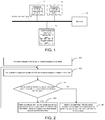
- FIG. 1 is a schematic diagram showing components of a tumor positioning apparatus, in accordance with embodiments of the present disclosure.
- the tumor positioning apparatus may include: at least one processor 11, a memory 12, a communication interface 13, and a communication bus 14.
- the components of the tumor positioning apparatus will be specifically described below with reference to FIG. 1 .
- the processor 11 is a control center of the tumor positioning apparatus, which may be a processor or a collective name for a plurality of processing elements.
- the processor 11 may be a central processing unit (CPU), or an application specific integrated circuit (ASIC), or is configured to implement one or more integrated circuits of the embodiments of the present disclosure, such as one or more digital signal processors (DSP), or one or more field programmable gate arrays (FPGA).
- DSP digital signal processors
- FPGA field programmable gate arrays
- the processor 11 may include one or more CPUs, such as a CPU0 and a CPU1 shown in FIG. 1 .
- the tumor positioning apparatus may include a plurality of processors, such as the processor 11 and a processor 15 shown in FIG. 1 .
- Each of these processors may be a single-CPU or a multi-CPU.
- the processor here may refer to one or more devices, circuits, and/or processing cores for processing data (such as computer program instructions).
- the memory 12 may be a read-only memory (ROM) or other types of static storage devices that may store static information and instructions, a random access memory (RAM) or other types of dynamic storage devices that may store information and instructions, or electrically erasable programmable read-only memory (EEPROM), compact disc read-only memory (CD-ROM), or other disc storage, optical disc storage (including compact discs, laser discs, optical discs, digital versatile discs, blue-ray discs, etc.), disk storage media or other magnetic storage devices, or any other medium that can carry or store expected program codes in a form of instructions or data structures and can be accessed by a computer, which is not limited thereto.
- the memory may exist independently and be connected to the processor 11 through the communication bus 14.
- the memory 12 may also be integrated with the processor 11.
- the memory 12 is used for storing data in the present disclosure and executing software programs in the present disclosure.
- the processor 11 may perform various functions of the tumor positioning apparatus by running or executing the software programs stored in the memory 12 and invoking the data stored in the memory 12.
- the communication interface 13 may use a device such as a transceiver to communicate with other devices or communication networks, such us a control system, a radio access network (RAN), or wireless local area networks (WLAN).
- the communication interface 13 may include a receiving unit to realize a receiving function, and a transmission unit to realize a transmission function.
- the communication bus 14 may be an industry standard architecture (ISA) bus, a peripheral component interconnect (PCI) bus, an extended industry standard architecture (EISA) bus, or the like.
- ISA industry standard architecture
- PCI peripheral component interconnect
- EISA extended industry standard architecture
- the bus may be classified into an address bus, a data bus, and a control bus, etc. For convenience of representation, only one thick line is used to represent the bus in FIG. 1 , but it does not mean that there is only one bus or one type of buses.
- the embodiments of the present disclosure provide a tumor positioning method. As shown in FIG. 2 , the method may include the following steps.
- the tumor positioning apparatus may obtain a reference image, and collect the projection images of the tumor at different angles according to pre-stored geometric information.
- the geometric information includes angle information for collecting the projection images.
- the projection images may be two-dimensional images, such as KV-level X-ray projection images, or may be three-dimensional images, such as three-dimensional images generated after reconstruction of the two-dimensional images of the tumor at the different angles.
- KV-level X-ray projection images of the tumor at two angles collected by the tumor positioning apparatus are used as an example for description.
- the tumor positioning apparatus may use the following two manners to collect the KV-level X-ray projection images at two angles.
- the tumor positioning apparatus may control the flat panel detector to collect a KV-level X-ray projection image at a first angle according to first angle information included in the geometric information, and control the flat panel detector to rotate to a second angle following a gantry and to collect a KV-level X-ray projection image at the second angle according to second angle information.
- the tumor positioning apparatus may control a first flat panel detector to collect the KV-level X-ray projection image at the first angle according to the first angle information included in the geometric information, and control a second flat panel detector to collect the KV-level X-ray projection image at the second angle according to the second angle information (forming a preset angle with the first angle, such as 90 degrees), so as to obtain the KV-level X-ray projection images at the two angles.
- the projection images are registered with the reference image to obtain a first offset.
- the tumor positioning apparatus may register the projection images with the reference image to obtain the first offset.
- the reference image may include a computed tomography (CT) image in a treatment plan, two-dimensional digitally reconstructed radiograph (DRR) images that are generated from the CT image and correspond to the projection images at the different angles one by one,, a magnetic resonance imaging (MRI) image, a positron emission computed tomography (PET) image, or the like.
- CT computed tomography
- DRR digitally reconstructed radiograph
- the tumor positioning apparatus registering the reference image with the projection images to obtain the first offset specifically is that: the tumor positioning apparatus reconstructs the projection images of the tumor collected at the different angles to obtain a target image.
- a dimension of the target image is the same as a dimension of the CT image, and the CT image is registered with the target image to obtain the first offset.
- the tumor positioning apparatus registering the reference images with the projection images to obtain the first offset specifically is that: the tumor positioning apparatus registers the projection images at the different angles with corresponding DRR images to obtain two-dimensional offsets at the different angles, and calculates the first offset according to the two-dimensional offsets at the different angles.
- the first offset may be any one of a three-dimensional offset, a four-dimensional offset, a five-dimensional offset, and a six-dimensional offset.
- the tumor positioning apparatus After the tumor positioning apparatus obtains the first offset, whether the virtual reacquisition operation needs to be performed may be determined according to the first offset.
- the tumor positioning apparatus may uses an automatic manner and a manual manner to determine whether the virtual reacquisition operation needs to be performed.
- the tumor positioning apparatus may perform the following step 205 when receiving an instruction that the operator determines that the virtual reacquisition operation needs to be performed according to the first offset; and perform the following step 204 when receiving an instruction that the operator determines that the the virtual reacquisition operation does not need to be performed according to the first offset.
- the tumor positioning apparatus may determine whether the first offset is within a preset offset range. If the first offset is within the preset offset range, it is determined that the virtual reacquisition operation does not need to be performed, and in this case, the following step 204 may be performed. If the first offset is not within the preset offset range, it is determined that the virtual reacquisition operation needs to be performed, and in this case, the following step 205 may be performed.
- the tumor positioning apparatus may output the first offset, and correct the position of the tumor according to the first offset, such as controlling the treatment couch to move.
- the tumor positioning apparatus may generate the reference image through a preset algorithm according to the first offset and the pre-stored geometric information. For example, when the reference images are the DRR images, the tumor positioning apparatus may generate DRR images that are in one-to-one correspondence with the projection images at the different angles through a DRR algorithm, according to the first offset, the CT image and the pre-stored geometric information.
- the tumor positioning apparatus may register the projection images collected in step 201 with the generated reference image according to step 202 to obtain a second offset, and determine a first accumulated offset.
- the first accumulated offset is a sum of the first offset and the second offset.
- the tumor positioning apparatus may re-generate a reference image according to the first accumulated offset, and register the projection images with the re-generated reference image to obtain a third offset, and determine a second accumulated offset.
- the second accumulated offset is a sum of the first offset, the second offset, and the third offset.
- the tumor positioning apparatus When it is determined that the virtual reacquisition operation needs to be performed according to the third offset, the tumor positioning apparatus re-generates a reference image according to the second accumulated offset, and registers the projection images with the re-generated reference image to obtain a fourth offset, and so on and so forth.
- the tumor positioning apparatus may generate two DRR images according to the CT image in the treatment plan and the preset geometric information, and collect two KV-level X-ray projection images that are approximately orthogonal. Moreover, the tumor positioning apparatus may also register the KV-level X-ray projection images collected at the two angles with corresponding DRR images to obtain the first offset, cumulatively display the first offset (an accumulated offset obtained by accumulating the first offset and a zero offset initially), and determine whether the virtual reacquisition operation needs to be performed according to the first offset.
- the tumor positioning apparatus outputs the first offset to the control system, and controls the treatment couch to move. If the virtual reacquisition operation needs to be performed, the tumor positioning apparatus may re-generate two DRR images according to the first offset, register the KV-level X-ray projection images collected at the two angles with the corresponding and re-generated DRR images to obtain the second offset, and determine and display the first accumulated offset, that is, the sum of the first offset and the second offset. Then, the tumor positioning apparatus may continue to determine whether the virtual reacquisition operation needs to be performed according to the second offset. If the virtual reacquisition operation does not need to be performed, the first accumulated offset is output in the display manner, and if the virtual reacquisition needs to be performed, the DRR images are re-generated according to the first accumulated offset, and so on and so forth.
- the tumor positioning apparatus may re-generate the reference image according to the first offset when it is determined that the virtual reacquisition operation needs to be performed, register the projection images with the re-generated reference image to obtain the second offset, and output a sum of the first offset and the second offset when it is determined that the virtual reacquisition operation does not need to be performed according to the second offset, that is, output the first accumulated offset, so that the treatment couch is moved according to the first accumulated offset.
- the tumor positioning method in the present disclosure may not only shorten the time for positioning the tumor, but also extends the service life of the treatment couch and the devices such as the detector, and the tube.
- the tumor positioning apparatus when it is determined that the virtual reacquisition operation needs to be performed according to the offset, the tumor positioning apparatus generates the reference image according to the accumulated offset, and registers the projection images with the generated reference image.
- the accumulated offset is considered when the tumor positioning apparatus generates the reference image, and the accumulated offset not only includes offset information in the X, Y, and Z directions, but also includes angle information.
- the angle information included in the offset cannot be accurately compensated. Therefore, in the embodiments of the present disclosure, the accumulated offset, which the three-dimensional couch is moved according to, is more accurate than the offset in the prior art, which improves an accuracy for positioning the tumor.
- the tumor positioning apparatus includes corresponding hardware structures and/or software modules to perform the functions.
- the present invention may implements the algorithm steps of the examples described in the embodiments disclosed herein in a form of hardware or a combination of hardware and computer software. Whether a function is performed through hardware or a manner of computer software driving the hardware depends on specific application and design constraints of the technical solution. Professional technicians may realize the described functions through different methods for each specific application, but such implementation should not be considered to go beyond the scope of the present invention.
- the embodiments of the present disclosure may divide the function modules of the tumor positioning apparatus according to the above methods and examples. For example, different function modules may be divided corresponding to different functions, or two or more functions may be integrated into one processing module.
- the above integrated modules may be implemented in the form of hardware, or software functional modules. It will be noted that, the division of the modules in the embodiments of the present disclosure is illustrative, and is only a logical function division, and there may be other division manners in actual implementation.
- FIG. 3 shows a schematic diagram showing components of another tumor positioning apparatus involved in the above embodiments.
- the tumor positioning apparatus may include: a collection unit 31, a registration unit 32, a determination unit 33, a generation unit 34, and an output unit 35.
- the collection unit 31 is used for supporting the tumor positioning apparatus to perform the step 201 in the tumor positioning method shown in FIG. 2 .
- the registration unit 32 is used for supporting the tumor positioning apparatus to perform the step 202 in the tumor positioning method shown in FIG. 2 .
- the determination unit 33 is used for supporting the tumor positioning apparatus to perform the step 204 and the step 205 of the tumor positioning method shown in FIG. 2 .
- step 204 it is determined that the virtual reacquisition operation does not need to be performed according to the first offset
- step 205 it is determined that the virtual reacquisition operation needs to be performed according to the first offset.
- the generation unit 34 is used for supporting the tumor positioning apparatus to perform the step 205 of the tumor positioning method shown in FIG. 2 .
- the reference image is re-generated according to the first offset.
- the output unit 35 is used for supporting the tumor positioning apparatus to perform the output of the first accumulated offset or the output of the second cumulative offset in the tumor positioning method shown in FIG. 2 .
- the tumor positioning apparatus may further include a display unit 36 and an acquisition unit 37.
- the display unit 36 is used for supporting the tumor positioning apparatus to perform the display of the accumulated offset described in step 204 in the tumor positioning method shown in FIG. 2 .
- the acquisition unit 37 is used for supporting the tumor positioning apparatus to perform the acquisition of the reference image in the tumor positioning method shown in FIG. 2 .
- the tumor positioning apparatus provided in the embodiments of the present disclosure may be used for performing the above tumor positioning methods. Therefore, it may achieve a same effect as the above tumor positioning method.
- FIG. 5 shows a schematic diagram showing components of yet another tumor positioning apparatus involved in the above embodiments.
- the tumor positioning apparatus includes: a processing module 41, a communication module 42 and a storage module 43.
- the processing module 41 is used for controlling and managing actions of the tumor positioning apparatus.
- the processing module 41 is used for supporting the tumor positioning apparatus to perform the step 201, the step 202, the step 203, the step 204, and the step 205 in FIG. 2 , and/or used for other processes of technologies described herein.
- the communication module 42 is used for supporting the tumor positioning apparatus to communicate with and other network entities.
- the storage module 43 is used for storing program codes and data of the tumor positioning apparatus.
- the processing module 41 may be the processor in FIG. 1 .
- the processing module 41 may implement or execute various exemplary logical blocks, modules and circuits described in the present disclosure.
- the processor may also be a combination of realizing computing functions.
- the processor may be a combination of one or more microprocessors, or a combination of DSP and microprocessor.
- the communication module 42 may be the communication interface in FIG. 1 .
- the storage module 43 may be the memory in FIG. 1 .
- the disclosed apparatus and method may be implemented through other manners.
- the embodiments about the apparatus described above are only illustrative.
- the division of the module or unit is only a logical function division, and there may be other division manners in actual implementation.
- multiple units or components may be combined or integrated into another device, or some features may be omitted or not performed.
- the displayed or discussed mutual coupling or direct coupling or communication connection may be indirect coupling or communication connection through some interfaces, devices or units,or may be in electrical, mechanical or other forms.
- the units described as separate components may or may not be physically separated, and the component displayed as a unit may be one physical unit or multiple physical units. That is, the component(s) may be positioned in one place, or may be distributed at many different places. Some or all of the units may be selected to achieve the purpose of the solutions of the embodiments according to actual needs.
- the functional units in the embodiments of the present disclosure may be integrated into one processing unit, or each unit may exist alone physically, or two or more units may be integrated into one unit.
- the integrated unit may be implemented in a form of hardware or software functional unit.
- the integrated unit When the integrated unit is realized in the form of the software functional unit and sold or used as an independent product, it may be stored in the readable storage medium.
- the software product is stored in a storage medium and includes many instructions to enable a device (such as a single-chip microcomputer or a chip) or a processor to execute all or part of the steps of the method in the embodiments of the present disclosure.
- the storage media include: a U disk, a mobile hard disk, a ROM, a RAM, a magnetic disk or an optical disk and other media that may store program codes.
Landscapes
- Health & Medical Sciences (AREA)
- Engineering & Computer Science (AREA)
- Biomedical Technology (AREA)
- Pathology (AREA)
- Nuclear Medicine, Radiotherapy & Molecular Imaging (AREA)
- Radiology & Medical Imaging (AREA)
- Life Sciences & Earth Sciences (AREA)
- Animal Behavior & Ethology (AREA)
- General Health & Medical Sciences (AREA)
- Public Health (AREA)
- Veterinary Medicine (AREA)
- Apparatus For Radiation Diagnosis (AREA)
Applications Claiming Priority (1)
| Application Number | Priority Date | Filing Date | Title |
|---|---|---|---|
| PCT/CN2018/100103 WO2020029304A1 (zh) | 2018-08-10 | 2018-08-10 | 一种肿瘤定位方法及装置 |
Publications (2)
| Publication Number | Publication Date |
|---|---|
| EP3834883A1 true EP3834883A1 (de) | 2021-06-16 |
| EP3834883A4 EP3834883A4 (de) | 2022-03-23 |
Family
ID=69413346
Family Applications (1)
| Application Number | Title | Priority Date | Filing Date |
|---|---|---|---|
| EP18929819.3A Pending EP3834883A4 (de) | 2018-08-10 | 2018-08-10 | Verfahren und vorrichtung zur tumorpositionierung |
Country Status (4)
| Country | Link |
|---|---|
| US (1) | US11628311B2 (de) |
| EP (1) | EP3834883A4 (de) |
| CN (1) | CN112384278B (de) |
| WO (1) | WO2020029304A1 (de) |
Families Citing this family (5)
| Publication number | Priority date | Publication date | Assignee | Title |
|---|---|---|---|---|
| WO2021159519A1 (zh) * | 2020-02-14 | 2021-08-19 | 西安大医集团股份有限公司 | 图像引导方法、装置、放疗设备和计算机存储介质 |
| WO2022116114A1 (zh) * | 2020-12-03 | 2022-06-09 | 西安大医集团股份有限公司 | 监测方法、装置及计算机存储介质 |
| WO2022120716A1 (zh) * | 2020-12-10 | 2022-06-16 | 西安大医集团股份有限公司 | 实时图像引导方法、装置及系统、放射治疗系统 |
| WO2024250210A1 (zh) * | 2023-06-08 | 2024-12-12 | 中国科学院深圳先进技术研究院 | 三维肿瘤实时跟踪方法、装置、计算机设备及存储介质 |
| CN119868824B (zh) * | 2024-12-18 | 2026-01-13 | 西安大医集团股份有限公司 | 肿瘤定位方法、设备及存储介质 |
Family Cites Families (20)
| Publication number | Priority date | Publication date | Assignee | Title |
|---|---|---|---|---|
| EP1674286B1 (de) * | 2004-12-23 | 2011-03-02 | Arjowiggins Security | Sicherheitselement mit einem digitalen Merkmal und Sicherheitsgegenstand oder Dokument mit demselben. |
| US7835500B2 (en) * | 2005-11-16 | 2010-11-16 | Accuray Incorporated | Multi-phase registration of 2-D X-ray images to 3-D volume studies |
| US20080037843A1 (en) * | 2006-08-11 | 2008-02-14 | Accuray Incorporated | Image segmentation for DRR generation and image registration |
| US7720196B2 (en) * | 2008-01-07 | 2010-05-18 | Accuray Incorporated | Target tracking using surface scanner and four-dimensional diagnostic imaging data |
| JP4956458B2 (ja) * | 2008-02-13 | 2012-06-20 | 三菱電機株式会社 | 患者位置決め装置及びその方法 |
| US7949089B2 (en) * | 2008-04-10 | 2011-05-24 | Arineta Ltd. | Apparatus and method for tracking feature's position in human body |
| JP2010246883A (ja) | 2009-03-27 | 2010-11-04 | Mitsubishi Electric Corp | 患者位置決めシステム |
| US8559596B2 (en) * | 2010-06-08 | 2013-10-15 | Accuray Incorporated | Target Tracking for image-guided radiation treatment |
| US9227087B2 (en) * | 2010-08-04 | 2016-01-05 | P-Cure Ltd. | Teletherapy control system and method |
| WO2012019162A1 (en) * | 2010-08-06 | 2012-02-09 | Accuray, Inc. | Systems and methods for real-time tumor tracking during radiation treatment using ultrasound imaging |
| CN102222331B (zh) * | 2011-05-16 | 2013-09-25 | 付东山 | 一种基于双平板的二维-三维医学图像配准方法及系统 |
| CN102440789B (zh) * | 2011-09-08 | 2014-07-09 | 付东山 | 一种基于双能x射线图像的软组织病灶定位系统 |
| JP2013099431A (ja) * | 2011-11-08 | 2013-05-23 | Natl Inst Of Radiological Sciences | 放射線治療における患者自動位置決め装置及び方法並びに患者自動位置決め用プログラム |
| US9672640B2 (en) * | 2013-01-24 | 2017-06-06 | Varian Medical Systems International Ag | Method for interactive manual matching and real-time projection calculation in imaging |
| US9949701B2 (en) * | 2013-02-14 | 2018-04-24 | Siemens Aktiengesellschaft | Registration for tracked medical tools and X-ray systems |
| US9844358B2 (en) * | 2014-06-04 | 2017-12-19 | Varian Medical Systems, Inc. | Imaging-based self-adjusting radiation therapy systems, devices, and methods |
| CN104587609B (zh) * | 2015-02-03 | 2017-05-31 | 瑞地玛医学科技有限公司 | 放射治疗摆位定位装置及静态、动态靶区摆位方法 |
| DE102015208929B3 (de) | 2015-05-13 | 2016-06-09 | Friedrich-Alexander-Universität Erlangen-Nürnberg | Verfahren zur 2D-3D-Registrierung, Recheneinrichtung und Computerprogramm |
| JP6281849B2 (ja) * | 2016-04-22 | 2018-02-21 | 国立研究開発法人量子科学技術研究開発機構 | 放射線治療における患者自動位置決め装置及び方法並びに患者自動位置決め用プログラム |
| US10713801B2 (en) * | 2017-01-06 | 2020-07-14 | Accuray Incorporated | Image registration of treatment planning image, intrafraction 3D image, and intrafraction 2D x-ray image |
-
2018
- 2018-08-10 EP EP18929819.3A patent/EP3834883A4/de active Pending
- 2018-08-10 CN CN201880095527.3A patent/CN112384278B/zh active Active
- 2018-08-10 WO PCT/CN2018/100103 patent/WO2020029304A1/zh not_active Ceased
-
2021
- 2021-02-09 US US17/171,460 patent/US11628311B2/en active Active
Also Published As
| Publication number | Publication date |
|---|---|
| US11628311B2 (en) | 2023-04-18 |
| CN112384278A (zh) | 2021-02-19 |
| CN112384278B (zh) | 2023-06-16 |
| US20210162235A1 (en) | 2021-06-03 |
| EP3834883A4 (de) | 2022-03-23 |
| WO2020029304A1 (zh) | 2020-02-13 |
Similar Documents
| Publication | Publication Date | Title |
|---|---|---|
| US11628311B2 (en) | Tumor positioning method and apparatus | |
| EP2399238B1 (de) | Funktionelle bildgebung | |
| US20200058389A1 (en) | Selecting acquisition parameter for imaging system | |
| US20120170820A1 (en) | Methods and apparatus for comparing 3d and 2d image data | |
| US20170091929A1 (en) | Method and system for determining a respiratory phase | |
| US9492124B2 (en) | System and method for treatment planning of organ disease at the functional and anatomical levels | |
| Hu et al. | Multimodal volume-aware detection and segmentation for brain metastases radiosurgery | |
| CN113262048A (zh) | 空间配准方法、装置、终端设备及术中导航系统 | |
| CN118570264A (zh) | 图像配准方法、装置、计算机设备及存储介质 | |
| CN117530707A (zh) | 图像标定方法、装置和图像标定设备 | |
| US20230023669A1 (en) | Methods and arrangements to describe deformity of a bone | |
| Liu et al. | 2D–3D radiograph to cone-beam computed tomography (CBCT) registration for C-arm image-guided robotic surgery | |
| US20210137454A1 (en) | Method and apparatus of locating tumor | |
| CN106600619A (zh) | 一种数据处理方法和装置 | |
| Yi et al. | DS-Net: an efficient dual-stage strategy for high-precision segmentation of gross tumor volumes in lung cancer CT images | |
| US11786203B2 (en) | Determining an image dataset | |
| CN112770810B (zh) | 肿瘤定位方法、装置及放射治疗系统 | |
| Mukhtar et al. | Imaging-Based Artificial Intelligence in Vascular and Interventional Radiology: A Narrative Review | |
| CN114299096B (zh) | 一种轮廓勾画方法、装置、设备及存储介质 | |
| Mori et al. | Development of digital reconstructed radiography software at new treatment facility for carbon-ion beam scanning of National Institute of Radiological Sciences | |
| US11406336B2 (en) | Tomosynthesis method with combined slice image datasets | |
| Wei et al. | A model that predicts a real-time tumour surface using intra-treatment skin surface and end-of-expiration and end-of-inhalation planning CT images | |
| GB2618334A (en) | A method for quantifying patient set up errors in radiotherapy | |
| US20210097735A1 (en) | Tomosynthesis method | |
| CN119868824B (zh) | 肿瘤定位方法、设备及存储介质 |
Legal Events
| Date | Code | Title | Description |
|---|---|---|---|
| STAA | Information on the status of an ep patent application or granted ep patent |
Free format text: STATUS: THE INTERNATIONAL PUBLICATION HAS BEEN MADE |
|
| PUAI | Public reference made under article 153(3) epc to a published international application that has entered the european phase |
Free format text: ORIGINAL CODE: 0009012 |
|
| STAA | Information on the status of an ep patent application or granted ep patent |
Free format text: STATUS: REQUEST FOR EXAMINATION WAS MADE |
|
| 17P | Request for examination filed |
Effective date: 20210303 |
|
| AK | Designated contracting states |
Kind code of ref document: A1 Designated state(s): AL AT BE BG CH CY CZ DE DK EE ES FI FR GB GR HR HU IE IS IT LI LT LU LV MC MK MT NL NO PL PT RO RS SE SI SK SM TR |
|
| DAV | Request for validation of the european patent (deleted) | ||
| DAX | Request for extension of the european patent (deleted) | ||
| REG | Reference to a national code |
Ref country code: DE Ref legal event code: R079 Free format text: PREVIOUS MAIN CLASS: A61N0005000000 Ipc: A61N0005100000 |
|
| A4 | Supplementary search report drawn up and despatched |
Effective date: 20220223 |
|
| RIC1 | Information provided on ipc code assigned before grant |
Ipc: A61N 5/10 20060101AFI20220217BHEP |
|
| STAA | Information on the status of an ep patent application or granted ep patent |
Free format text: STATUS: EXAMINATION IS IN PROGRESS |
|
| 17Q | First examination report despatched |
Effective date: 20250319 |