EP3851165B1 - Vorrichtung zur behandlung eines gewebes - Google Patents
Vorrichtung zur behandlung eines gewebes Download PDFInfo
- Publication number
- EP3851165B1 EP3851165B1 EP21157293.8A EP21157293A EP3851165B1 EP 3851165 B1 EP3851165 B1 EP 3851165B1 EP 21157293 A EP21157293 A EP 21157293A EP 3851165 B1 EP3851165 B1 EP 3851165B1
- Authority
- EP
- European Patent Office
- Prior art keywords
- treatment
- treatment head
- tissue
- transducer
- control unit
- Prior art date
- Legal status (The legal status is an assumption and is not a legal conclusion. Google has not performed a legal analysis and makes no representation as to the accuracy of the status listed.)
- Active
Links
Images

Classifications
-
- A—HUMAN NECESSITIES
- A61—MEDICAL OR VETERINARY SCIENCE; HYGIENE
- A61N—ELECTROTHERAPY; MAGNETOTHERAPY; RADIATION THERAPY; ULTRASOUND THERAPY
- A61N7/00—Ultrasound therapy
-
- A—HUMAN NECESSITIES
- A61—MEDICAL OR VETERINARY SCIENCE; HYGIENE
- A61B—DIAGNOSIS; SURGERY; IDENTIFICATION
- A61B8/00—Diagnosis using ultrasonic, sonic or infrasonic waves
- A61B8/08—Clinical applications
- A61B8/0833—Clinical applications involving detecting or locating foreign bodies or organic structures
- A61B8/085—Clinical applications involving detecting or locating foreign bodies or organic structures for locating body or organic structures, e.g. tumours, calculi, blood vessels, nodules
-
- A—HUMAN NECESSITIES
- A61—MEDICAL OR VETERINARY SCIENCE; HYGIENE
- A61B—DIAGNOSIS; SURGERY; IDENTIFICATION
- A61B8/00—Diagnosis using ultrasonic, sonic or infrasonic waves
- A61B8/42—Details of probe positioning or probe attachment to the patient
- A61B8/4272—Details of probe positioning or probe attachment to the patient involving the acoustic interface between the transducer and the tissue
- A61B8/4281—Details of probe positioning or probe attachment to the patient involving the acoustic interface between the transducer and the tissue characterised by sound-transmitting media or devices for coupling the transducer to the tissue
-
- A—HUMAN NECESSITIES
- A61—MEDICAL OR VETERINARY SCIENCE; HYGIENE
- A61B—DIAGNOSIS; SURGERY; IDENTIFICATION
- A61B8/00—Diagnosis using ultrasonic, sonic or infrasonic waves
- A61B8/44—Constructional features of the ultrasonic, sonic or infrasonic diagnostic device
- A61B8/4444—Constructional features of the ultrasonic, sonic or infrasonic diagnostic device related to the probe
- A61B8/4461—Features of the scanning mechanism, e.g. for moving the transducer within the housing of the probe
-
- A—HUMAN NECESSITIES
- A61—MEDICAL OR VETERINARY SCIENCE; HYGIENE
- A61B—DIAGNOSIS; SURGERY; IDENTIFICATION
- A61B8/00—Diagnosis using ultrasonic, sonic or infrasonic waves
- A61B8/54—Control of the diagnostic device
-
- A—HUMAN NECESSITIES
- A61—MEDICAL OR VETERINARY SCIENCE; HYGIENE
- A61N—ELECTROTHERAPY; MAGNETOTHERAPY; RADIATION THERAPY; ULTRASOUND THERAPY
- A61N7/00—Ultrasound therapy
- A61N7/02—Localised ultrasound hyperthermia
-
- A—HUMAN NECESSITIES
- A61—MEDICAL OR VETERINARY SCIENCE; HYGIENE
- A61B—DIAGNOSIS; SURGERY; IDENTIFICATION
- A61B34/00—Computer-aided surgery; Manipulators or robots specially adapted for use in surgery
- A61B34/10—Computer-aided planning, simulation or modelling of surgical operations
- A61B2034/101—Computer-aided simulation of surgical operations
- A61B2034/105—Modelling of the patient, e.g. for ligaments or bones
-
- A—HUMAN NECESSITIES
- A61—MEDICAL OR VETERINARY SCIENCE; HYGIENE
- A61B—DIAGNOSIS; SURGERY; IDENTIFICATION
- A61B90/00—Instruments, implements or accessories specially adapted for surgery or diagnosis and not covered by any of the groups A61B1/00 - A61B50/00, e.g. for luxation treatment or for protecting wound edges
- A61B90/36—Image-producing devices or illumination devices not otherwise provided for
- A61B90/37—Surgical systems with images on a monitor during operation
- A61B2090/378—Surgical systems with images on a monitor during operation using ultrasound
-
- A—HUMAN NECESSITIES
- A61—MEDICAL OR VETERINARY SCIENCE; HYGIENE
- A61N—ELECTROTHERAPY; MAGNETOTHERAPY; RADIATION THERAPY; ULTRASOUND THERAPY
- A61N7/00—Ultrasound therapy
- A61N2007/0052—Ultrasound therapy using the same transducer for therapy and imaging
-
- A—HUMAN NECESSITIES
- A61—MEDICAL OR VETERINARY SCIENCE; HYGIENE
- A61N—ELECTROTHERAPY; MAGNETOTHERAPY; RADIATION THERAPY; ULTRASOUND THERAPY
- A61N7/00—Ultrasound therapy
- A61N2007/0082—Scanning transducers
-
- A—HUMAN NECESSITIES
- A61—MEDICAL OR VETERINARY SCIENCE; HYGIENE
- A61N—ELECTROTHERAPY; MAGNETOTHERAPY; RADIATION THERAPY; ULTRASOUND THERAPY
- A61N7/00—Ultrasound therapy
- A61N2007/0086—Beam steering
Definitions
- the device includes a transducer for emitting a beam of ultrasound waves, preferably high intensity focused ultrasound waves for irradiating the tissue.
- the transducer is mounted on a movable treatment head.
- the beam is focused on or focusable onto a focal point.
- An ultrasonic imaging device is mounted on the treatment head and has an imaging plane intersecting the focal point.
- An inflatable balloon surrounding at least partially the treatment head, containing a coupling fluid and defining a contact surface of the treatment head is provided.
- a control unit is used for controlling the movement of the treatment head and the operation of the transducer and the imaging device.
- Ultrasound waves in particular high intensity focused ultrasound (HIFU) are mainly used for treatment of tumours of breast, thyroid, prostate and uterus.
- High intensity ultrasound waves are focused onto a focal point located within the tumour to be treated.
- the temperature can rise up to 85 °C, whereby the tissue is destroyed by coagulation necrosis.
- One big advantage of a treatment with HIFU is that it is non-invasive, thereby considerably reducing risks for the patient.
- the focal point is moved along the tissue (scanning). Scanning can occur by moving the treatment head, known as mechanical scanning, or by keeping the treatment head fixed and move the focal point using phase array technique, known as electronic scanning. Due to its simplicity and low cost, mechanical scanning is preferred.
- a treatment with HIFU also provides some energy in the tissue located adjacent to the focal point, therefore possibly damaging also those tissues, it is common to operate a device for treatment in a way known as "pulse and pause method" wherein an irradiation period is followed by a pause period without irradiation, in order allow the tissue region to cool down.
- a motion of the treatment head might displace the tissue of a patient, e.g. because of friction between the tissue and the balloon surface.
- motion of the treatment head may be impaired by mechanical interferences between the treatment head and the tissue of a patient, e.g. by friction or lack of clearance between the tissue and the balloon surface.
- tissue displacement certain areas of the tissue to be treated may be over- or undertreated.
- This movement of the treatment head is particularly advantageous in order to reduce or displace appearance of skin artefacts, also known as "skin ghost".
- skin ghost appears when the tissue boundary layer, depending from the tissue e.g. a skin or a mucosa, reflects the imaging beam back to the imaging device, which is successively reflected back to the skin or mucosa and then back to the imaging device again.
- the skin ghost appears in the image as a bright line located within the tissue at approximately twice the distance between the skin or mucosa and the beginning of the image, which may correspond to the surface of the imaging device or not.
- the skin ghost is particularly unwanted when it is located within the target area, that means within the tissue to be treated.
- control unit is adapted to move the treatment head to the travel position for further positioning of the treatment head once achievement of a desired level of treatment has been recognized.
- the treatment head is moved along a scanning path.
- the treatment head is preferably displaced only when in the travel position.
- the control unit is adapted to control the pressure in the balloon. Therefore, the device is equipped with a fluid system coupled to the balloon for handling the coupling fluid, preferably including at least one pump for filling and/or emptying the balloon. Further, a pressure sensor may be present in the balloon in order to control the pressure in the balloon, the pressure sensor and the at least one pump preferably being connected to the control unit or other adequate means for regulating the pressure in the balloon.
- the volume of the coupling fluid may also be controlled and regulated by known means.
- control unit is adapted to bring the pressure in the balloon to a first value when the treatment head is in the travel position and to a second value higher than the first value when the treatment head is in the treatment position and/or in the monitoring position. Accordingly, by reducing the pressure when the treatment head is in the travel position, friction between tissue and balloon can be reduced, with the known advantages. Further, by increasing the pressure when the treatment head is in the treatment position and/or in the monitoring position, the compression reduces blood flow in the tissue, thereby reducing also heat dissipation. In other words, an higher temperature in the focal point may be achieved with the same beam power.
- An example not presently claimed also relates to a method for operating a device for treatment of a tissue of a living being.
- the device is a device as described above. Reference to the advantages and alternatives cited above is therefore made.
- the distance between the skin surface line and the beginning of the image corresponding to the ultrasonic imaging device (y n ) for every width point (x n ) within the chosen width sector (x min , x max ) is then determined.
- the beginning of the image is meant to be the end of the image which corresponds to the location of the imaging device, thus being located in the ultrasound wave propagation direction upstream with respect to the skin surface.
- the offset value (A) is a characteristic of the imaging device and must therefore not be determined every time when performing the present invention.
- the treatment head 1 In the travel position T, the treatment head 1 is raised along an axis A with respect to the skin 6 compared to the treatment position, whereby a contact between the membrane 5 and the skin 6 is kept in order to ensure coupling of the echography device 2 and cooling of the skin 6 and tissue 7.
- the HIFU beam is also not focused on the tumour 8.
- the pressure in the membrane may be adjusted in order to reduce friction, while keeping the treatment head 1 in a constant position.
- the membrane 5 or other equivalent means may be displaced with respect to the treatment head 1, which is kept in the same position. Combination of those alternatives may also be possible.
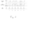
- the HIFU transducer 4 is located at a first treatment point I and is triggered to emit an HIFU beam and is successively moved along the scanning pattern in a plane substantially perpendicular to the axis A, referred to in figure 2 as the "scanning plane", to reach the next treatment point II.
- the scanning plane substantially perpendicular to the axis A
- no displacement of the treatment head 1 along the axis A takes place.
- the treatment head 1 is kept in the treatment position T, as known from the prior art.
- the treatment head 1 before the treatment head 1 is displaced from the treatment point II to the treatment point III, the treatment head 1 is moved from the treatment position T to the travel position S along the axis A, then displaced along the scanning plane and subsequently moved back to the treatment position T, where a HIFU beam is emitted.
- the treatment head 1 is further moved to the travel position S along the axis A and displaced to the next treatment point V, where it is moved back along the axis A to the treatment position T and a HIFU beam is delivered.
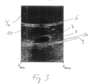
- the beginning of the image corresponding to the echography device 2 is the upper edge of the image.
- the width of the analyzed sector ranges from x min to x max .
- a second bright curved line 9 running parallel to the skin 6 is displayed.
- This curved line is referred to as a skin ghost and covers a sector of the tumour 8, thus impeding correct imaging of the tissue 7.
- a distance y n between the beginning of the image and the line 10 is determined over the whole width of sector (from x min to x max ). Then, a second line y s representing the skin ghost 9 is drawn at twice the distance y n from the beginning of the image. In figure 3 , an offset A is set to zero.
Landscapes
- Health & Medical Sciences (AREA)
- Life Sciences & Earth Sciences (AREA)
- Animal Behavior & Ethology (AREA)
- Veterinary Medicine (AREA)
- Nuclear Medicine, Radiotherapy & Molecular Imaging (AREA)
- Radiology & Medical Imaging (AREA)
- Engineering & Computer Science (AREA)
- Biomedical Technology (AREA)
- Public Health (AREA)
- General Health & Medical Sciences (AREA)
- Physics & Mathematics (AREA)
- Pathology (AREA)
- Surgery (AREA)
- Molecular Biology (AREA)
- Medical Informatics (AREA)
- Heart & Thoracic Surgery (AREA)
- Biophysics (AREA)
- Acoustics & Sound (AREA)
- Vascular Medicine (AREA)
- Surgical Instruments (AREA)
- Ultra Sonic Daignosis Equipment (AREA)
Claims (13)
- Eine Vorrichtung zur Behandlung eines Gewebes eines lebenden Wesens, wobei die Vorrichtung folgendes umfasst:- einen Wandler (4) zum Aussenden eines Bündels von Ultraschallwellen, vorzugsweise hochintensiver fokussierter Ultraschallwellen, zum Bestrahlen des Gewebes (8), wobei das Bündel auf einen Fokalpunkt (F) fokussiert oder fokussierbar ist, wobei der Wandler (4) auf einem beweglichen Behandlungskopf (1) montiert ist;- eine Ultraschall-Bildgebungseinrichtung (2, 3), die auf dem Behandlungskopf (1) montiert ist und eine Abbildungsebene aufweist, die den Fokalpunkt (f) schneidet;- eine Steuereinheit zur Steuerung der Bewegung und Position des Behandlungskopfes (1) sowie des Betriebs des Wandlers (4) und der Bildgebungseinrichtung (2, 3);wobei der Behandlungskopf (1) auf eine Behandlungsposition (T) und eine Überwachungsposition (M) verstellbar ist,wobei in der Überwachungsposition (M) der Abstand zwischen dem Wandler (4) und dem Gewebe (8) kleiner ist als in der Behandlungsposition (T),wobeidie Steuereinheit geeignet ist, umden Wandler (4) auszulösen, um mindestens ein Bündel von Ultraschallwellen auszusenden, wenn sich der Behandlungskopf (1) in der Behandlungsposition (T) befindet,
undeinen Bereich (7), der das Gewebe (8) umgibt, zumindest dann abzubilden, wenn sich der Behandlungskopf (1) in der Überwachungsposition (M) befindet, und dadurch gekennzeichnet, dass die Vorrichtung einen aufblasbaren Ballon (5) umfasst, der den Behandlungskopf (1) zumindest teilweise umgibt, eine Kopplungsflüssigkeit enthalten kann und eine Kontaktfläche des Behandlungskopfes (1) definiert,wobei die Steuereinheit so konfiguriert ist, dass sie den Druck im Ballon erhöht, wenn sich der Behandlungskopf in der Behandlungsposition befindet, und den Druck im Ballon senkt, wenn sich der Behandlungskopf in der Überwachungsposition befindet. - Vorrichtung nach Anspruch 1, dadurch gekennzeichnet, dass der Behandlungskopf (1) in eine Reiseposition (S) verstellbar ist und in der Reiseposition (S) ein Abstand zwischen der Oberfläche und dem Gewebe (8) oder zwischen dem Wandler (4) und dem Gewebe (8) grösser ist als in der Behandlungsposition (T) oder die Kompressionskraft reduziert ist, wobei die Steuereinheit dazu eingerichtet ist, den Behandlungskopf (1) von der Reiseposition (S) in die Behandlungsposition (T) zu bewegen, bevor ein Ultraschallpuls vom Wandler (4) ausgesendet wird.
- Vorrichtung nach Anspruch 2, dadurch gekennzeichnet, dass die Steuereinheit dazu eingerichtet ist, den Behandlungskopf (1) aus der Reiseposition (S) in die Überwachungsposition (M) zur Überwachung des Gewebes (8) mittels des Bildgebungsgeräts (2, 3) und anschliessend in die Behandlungsposition (T) zu bewegen, bevor ein Ultraschallpuls vom Wandler (4) abgegeben wird.
- Vorrichtung nach einem der Ansprüche 2 oder 3, dadurch gekennzeichnet, dass die Steuereinheit dazu eingerichtet ist, den Behandlungskopf (1) nach Emission eines Ultraschallpulses vom Wandler (4) von der Behandlungsposition (T) in die Überwachungsposition (M) zu bewegen.
- Vorrichtung nach Anspruch 4, dadurch gekennzeichnet, dass die Steuereinheit dazu eingerichtet ist, den Behandlungskopf (1) in die Behandlungsposition (T) zur weiteren Emission eines Ultraschallpulses vom Wandler (4) zu bewegen, nachdem der Behandlungskopf (1) von der Behandlungsposition (T) in die Überwachungsposition (M) gebracht worden ist.
- Vorrichtung nach einem der Ansprüche 2 bis 5, dadurch gekennzeichnet, dass die Steuereinheit dazu eingerichtet ist, den Behandlungskopf (1) zur weiteren Positionierung des Behandlungskopfes (1) nach der vorangegangenen Bewegung in die Reiseposition (S) zu bewegen.
- Vorrichtung nach einem der vorhergehenden Ansprüche, dadurch gekennzeichnet, dass die Steuereinheit dazu eingerichtet ist, die Position und/oder die Einstellung des Behandlungskopfes (1) mit Hilfe der Ultraschall-Bildgebungseinrichtung (2, 3) zu steuern.
- Vorrichtung nach einem der vorhergehenden Ansprüche, dadurch gekennzeichnet, dass die Steuereinheit dazu eingerichtet ist, den Druck im Ballon (5) zu steuern.
- Vorrichtung nach Anspruch 2 und Anspruch 8, dadurch gekennzeichnet, dass die Steuereinheit dazu eingerichtet ist, den Druck im Ballon (5) auf einen ersten Wert zu bringen, wenn sich der Behandlungskopf (1) in der Reiseposition (S) befindet, und auf einen zweiten Wert, der höher als der erste Wert ist, wenn sich der Behandlungskopf (1) in der Behandlungsposition (T) befindet.
- Vorrichtung nach Anspruch 2 und Anspruch 8, dadurch gekennzeichnet, dass die Steuereinheit dazu eingerichtet ist, den Druck im Ballon (5) von einem ersten Wert auf einen zweiten Wert zu ändern, der niedriger als der erste Wert ist, wenn sich der Behandlungskopf (1) in der Reiseposition befindet und in einer Ebene positioniert ist, die im Wesentlichen senkrecht zu einer Achse (A) zwischen der Behandlungsposition (T) und der Überwachungsposition (M) verläuft.
- Vorrichtung nach einem der Ansprüche 2 - 6 und 9 - 10, dadurch gekennzeichnet, dass die Bildgebung des Gewebes (8) in der Behandlungsposition (T), der Bildgebungsposition (M) und der Reiseposition (S) sowie in Positionen, die zwischen der Behandlungsposition (T), der Bildgebungsposition (M) und der Reiseposition (S) liegen, erfolgt.
- Vorrichtung nach einem der Ansprüche 2 - 6 und 9 - 11, dadurch gekennzeichnet, dass ein erhöhter Druck des Behandlungskopfes (1) in der Behandlungsposition (T) und/oder Überwachungsposition (M) gegenüber der Reiseposition (S) den Blutfluss im Gewebe reduziert.
- Vorrichtung nach einem der Ansprüche 2 - 6 und 9 - 12, dadurch gekennzeichnet, dass in der Reiseposition (S) der Behandlungskopf (1) entlang der Achse (A) gegenüber dem Gewebe (8) gegenüber der Behandlungsposition (T) erhöht werden kann, wobei ein Kontakt zwischen dem Ballon (5) und dem Gewebe (8) aufrechterhalten wird.
Applications Claiming Priority (3)
| Application Number | Priority Date | Filing Date | Title |
|---|---|---|---|
| EP13199292.7A EP2886160A1 (de) | 2013-12-23 | 2013-12-23 | Vorrichtung zur Behandlung eines Gewebes und Verfahren zur Herstellung eines Bildes einer bildgeführten Vorrichtung zur Behandlung eines Gewebes |
| PCT/EP2014/077446 WO2015096994A1 (en) | 2013-12-23 | 2014-12-11 | Device for treatment of a tissue and method of preparation of an image of an image-guided device for treatment of a tissue |
| EP14811883.9A EP3086843B1 (de) | 2013-12-23 | 2014-12-11 | Vorrichtung zur behandlung eines gewebes |
Related Parent Applications (1)
| Application Number | Title | Priority Date | Filing Date |
|---|---|---|---|
| EP14811883.9A Division EP3086843B1 (de) | 2013-12-23 | 2014-12-11 | Vorrichtung zur behandlung eines gewebes |
Publications (3)
| Publication Number | Publication Date |
|---|---|
| EP3851165A2 EP3851165A2 (de) | 2021-07-21 |
| EP3851165A3 EP3851165A3 (de) | 2021-10-13 |
| EP3851165B1 true EP3851165B1 (de) | 2025-04-09 |
Family
ID=49949446
Family Applications (3)
| Application Number | Title | Priority Date | Filing Date |
|---|---|---|---|
| EP13199292.7A Withdrawn EP2886160A1 (de) | 2013-12-23 | 2013-12-23 | Vorrichtung zur Behandlung eines Gewebes und Verfahren zur Herstellung eines Bildes einer bildgeführten Vorrichtung zur Behandlung eines Gewebes |
| EP21157293.8A Active EP3851165B1 (de) | 2013-12-23 | 2014-12-11 | Vorrichtung zur behandlung eines gewebes |
| EP14811883.9A Active EP3086843B1 (de) | 2013-12-23 | 2014-12-11 | Vorrichtung zur behandlung eines gewebes |
Family Applications Before (1)
| Application Number | Title | Priority Date | Filing Date |
|---|---|---|---|
| EP13199292.7A Withdrawn EP2886160A1 (de) | 2013-12-23 | 2013-12-23 | Vorrichtung zur Behandlung eines Gewebes und Verfahren zur Herstellung eines Bildes einer bildgeführten Vorrichtung zur Behandlung eines Gewebes |
Family Applications After (1)
| Application Number | Title | Priority Date | Filing Date |
|---|---|---|---|
| EP14811883.9A Active EP3086843B1 (de) | 2013-12-23 | 2014-12-11 | Vorrichtung zur behandlung eines gewebes |
Country Status (5)
| Country | Link |
|---|---|
| US (2) | US11123576B2 (de) |
| EP (3) | EP2886160A1 (de) |
| KR (2) | KR102609342B1 (de) |
| CN (1) | CN105828877B (de) |
| WO (1) | WO2015096994A1 (de) |
Families Citing this family (5)
| Publication number | Priority date | Publication date | Assignee | Title |
|---|---|---|---|---|
| KR102695747B1 (ko) * | 2017-12-08 | 2024-08-14 | 테라끌리옹 에스에이 | 초음파 장치 및 초음파 장치를 작동시키는 방법 |
| EP3643242A1 (de) * | 2018-10-25 | 2020-04-29 | Koninklijke Philips N.V. | Stützeinheit für ein medizinisches bildgebungselement |
| EP3986548A1 (de) * | 2019-06-19 | 2022-04-27 | Theraclion SA | Vorrichtung und verfahren zur behandlung eines patienten mittels hochintensivem fokussiertem ultraschall (hifu) |
| US20240050738A1 (en) * | 2022-08-11 | 2024-02-15 | Pacesetter, Inc. | Active implantable medical device |
| NL2035948B1 (en) * | 2023-10-03 | 2025-04-11 | Stichting Radboud Univ | Transcranial ultrasound stimulation device |
Family Cites Families (15)
| Publication number | Priority date | Publication date | Assignee | Title |
|---|---|---|---|---|
| CN2344037Y (zh) * | 1998-11-16 | 1999-10-20 | 重庆市高强度聚焦超声肿瘤治疗研究中心 | 伸缩式组合探头 |
| US7160258B2 (en) * | 2001-06-26 | 2007-01-09 | Entrack, Inc. | Capsule and method for treating or diagnosing the intestinal tract |
| FR2827149B1 (fr) * | 2001-07-13 | 2003-10-10 | Technomed Medical Systems | Sonde de traitement par ultrasons focalises |
| CN100542635C (zh) * | 2005-01-10 | 2009-09-23 | 重庆海扶(Hifu)技术有限公司 | 高强度聚焦超声治疗装置和方法 |
| JP4923436B2 (ja) | 2005-05-10 | 2012-04-25 | マックス株式会社 | ガス燃焼式打込み工具 |
| FR2886551B1 (fr) * | 2005-06-03 | 2007-09-07 | Theraclion Soc Par Actions Sim | Procede de determination de distance et appareil de traitement mettant en oeuvre ub tel procede |
| KR102087909B1 (ko) * | 2008-06-06 | 2020-03-12 | 얼테라, 인크 | 코스메틱 치료 시스템 |
| EP2327450A1 (de) * | 2009-11-27 | 2011-06-01 | Theraclion SAS | Eine Abdeckhaube, ein therapeutisches Gerät und seine Verwendung |
| EP2332614A1 (de) | 2009-12-10 | 2011-06-15 | Theraclion SAS | Ultraschallbehandlungsgerät |
| US20120172710A1 (en) * | 2009-12-18 | 2012-07-05 | Anthony Brian W | Quantitative elastography |
| WO2013009785A2 (en) * | 2011-07-10 | 2013-01-17 | Guided Therapy Systems, Llc. | Systems and methods for improving an outside appearance of skin using ultrasound as an energy source |
| CN103123721B (zh) * | 2011-11-17 | 2016-04-27 | 重庆海扶医疗科技股份有限公司 | 一种实时减少图像中伪影的方法以及装置 |
| CN102551804B (zh) * | 2011-12-31 | 2013-12-04 | 重庆海扶医疗科技股份有限公司 | 减少图像伪影的超声治疗仪监控系统及其图像获取方法 |
| KR101226659B1 (ko) * | 2012-08-22 | 2013-02-05 | 주식회사 하이로닉 | 피하 지방층의 감소를 위한 고강도 집속 초음파 생성 장치 |
| KR20140095848A (ko) * | 2013-01-25 | 2014-08-04 | 삼성전자주식회사 | 초음파 치료 방법 및 초음파 치료 시스템 |
-
2013
- 2013-12-23 EP EP13199292.7A patent/EP2886160A1/de not_active Withdrawn
-
2014
- 2014-12-11 CN CN201480066612.9A patent/CN105828877B/zh active Active
- 2014-12-11 KR KR1020227012558A patent/KR102609342B1/ko active Active
- 2014-12-11 US US15/106,892 patent/US11123576B2/en active Active
- 2014-12-11 EP EP21157293.8A patent/EP3851165B1/de active Active
- 2014-12-11 EP EP14811883.9A patent/EP3086843B1/de active Active
- 2014-12-11 KR KR1020167016108A patent/KR102388415B1/ko active Active
- 2014-12-11 WO PCT/EP2014/077446 patent/WO2015096994A1/en not_active Ceased
-
2021
- 2021-09-15 US US17/476,050 patent/US12090345B2/en active Active
Also Published As
| Publication number | Publication date |
|---|---|
| EP3086843A1 (de) | 2016-11-02 |
| EP3086843B1 (de) | 2021-02-17 |
| EP3851165A2 (de) | 2021-07-21 |
| KR102609342B1 (ko) | 2023-12-01 |
| EP2886160A1 (de) | 2015-06-24 |
| US12090345B2 (en) | 2024-09-17 |
| KR102388415B1 (ko) | 2022-04-20 |
| US11123576B2 (en) | 2021-09-21 |
| KR20220053041A (ko) | 2022-04-28 |
| WO2015096994A1 (en) | 2015-07-02 |
| EP3851165A3 (de) | 2021-10-13 |
| US20220001213A1 (en) | 2022-01-06 |
| CN105828877A (zh) | 2016-08-03 |
| KR20160101922A (ko) | 2016-08-26 |
| CN105828877B (zh) | 2019-04-23 |
| US20170001043A1 (en) | 2017-01-05 |
Similar Documents
| Publication | Publication Date | Title |
|---|---|---|
| US12090345B2 (en) | Device for treatment of a tissue and method of preparation of an image of an image-guided device for treatment of a tissue | |
| JP6444302B2 (ja) | 超音波誘導治療のためのヒューマンインターフェースおよびデバイス | |
| CN109689160B (zh) | 具有来自微泡的减小的干扰的治疗性超声波 | |
| US8715187B2 (en) | Systems and methods for automatically identifying and segmenting different tissue types in ultrasound images | |
| US10881378B2 (en) | Methods and systems for a display interface for diagnostic medical imaging | |
| KR102396008B1 (ko) | 정반사체를 트래킹하기 위한 초음파 이미징 시스템 및 방법 | |
| US20100241036A1 (en) | Controlled, non-linear focused ultrasound treatment | |
| JP2008526326A (ja) | 高強度集束超音波療法のための方法および装置 | |
| US20100049098A1 (en) | Automatic acoustic treatment device | |
| US20150305821A1 (en) | Image-guided therapeutic apparatus and method of preparation of an image-guided therapeutic appratus for treatment of tissue | |
| KR20140095848A (ko) | 초음파 치료 방법 및 초음파 치료 시스템 | |
| KR101725189B1 (ko) | 초음파를 이용한 의료장치 및 트랜스듀서의 이동 제어 방법 | |
| CN113332620B (zh) | 一种超声医疗设备 | |
| EP2481447A1 (de) | Medizinisches Instrument mit hochintensivem, gebündelten Ultraschall mit Doppelwandlern | |
| US11872413B2 (en) | Control method for the treatment of brain tissue using an ultrasonic probe and an implanted acoustic window on the cranium | |
| EP3366350A1 (de) | Vorrichtung zur therapeutischen behandlung, verfahren zur definition einer einstellung, verfahren zur behandlung von gewebe und computerprogrammprodukt | |
| JP2018083000A (ja) | 情報取得装置 | |
| CN114007688B (zh) | 用于通过高强度聚焦超声凝固静脉曲张的方法、装置和该装置的使用方法 | |
| KR102244287B1 (ko) | 신경을 감지하여 에너지를 조사하는 시술장치 | |
| WO2025163599A1 (en) | Systems, devices, and methods for non-invasive treatment of tissue using boiling histotripsy |
Legal Events
| Date | Code | Title | Description |
|---|---|---|---|
| PUAI | Public reference made under article 153(3) epc to a published international application that has entered the european phase |
Free format text: ORIGINAL CODE: 0009012 |
|
| STAA | Information on the status of an ep patent application or granted ep patent |
Free format text: STATUS: THE APPLICATION HAS BEEN PUBLISHED |
|
| AC | Divisional application: reference to earlier application |
Ref document number: 3086843 Country of ref document: EP Kind code of ref document: P |
|
| AK | Designated contracting states |
Kind code of ref document: A2 Designated state(s): AL AT BE BG CH CY CZ DE DK EE ES FI FR GB GR HR HU IE IS IT LI LT LU LV MC MK MT NL NO PL PT RO RS SE SI SK SM TR |
|
| PUAL | Search report despatched |
Free format text: ORIGINAL CODE: 0009013 |
|
| AK | Designated contracting states |
Kind code of ref document: A3 Designated state(s): AL AT BE BG CH CY CZ DE DK EE ES FI FR GB GR HR HU IE IS IT LI LT LU LV MC MK MT NL NO PL PT RO RS SE SI SK SM TR |
|
| RIC1 | Information provided on ipc code assigned before grant |
Ipc: A61N 7/00 20060101ALN20210908BHEP Ipc: A61B 8/08 20060101ALI20210908BHEP Ipc: A61N 7/02 20060101AFI20210908BHEP |
|
| STAA | Information on the status of an ep patent application or granted ep patent |
Free format text: STATUS: REQUEST FOR EXAMINATION WAS MADE |
|
| 17P | Request for examination filed |
Effective date: 20220412 |
|
| RBV | Designated contracting states (corrected) |
Designated state(s): AL AT BE BG CH CY CZ DE DK EE ES FI FR GB GR HR HU IE IS IT LI LT LU LV MC MK MT NL NO PL PT RO RS SE SI SK SM TR |
|
| RAP3 | Party data changed (applicant data changed or rights of an application transferred) |
Owner name: THERACLION SA |
|
| GRAP | Despatch of communication of intention to grant a patent |
Free format text: ORIGINAL CODE: EPIDOSNIGR1 |
|
| STAA | Information on the status of an ep patent application or granted ep patent |
Free format text: STATUS: GRANT OF PATENT IS INTENDED |
|
| RIC1 | Information provided on ipc code assigned before grant |
Ipc: A61N 7/00 20060101ALN20241104BHEP Ipc: A61B 8/08 20060101ALI20241104BHEP Ipc: A61N 7/02 20060101AFI20241104BHEP |
|
| INTG | Intention to grant announced |
Effective date: 20241128 |
|
| RIC1 | Information provided on ipc code assigned before grant |
Ipc: A61N 7/00 20060101ALN20241122BHEP Ipc: A61B 8/08 20060101ALI20241122BHEP Ipc: A61N 7/02 20060101AFI20241122BHEP |
|
| GRAS | Grant fee paid |
Free format text: ORIGINAL CODE: EPIDOSNIGR3 |
|
| GRAA | (expected) grant |
Free format text: ORIGINAL CODE: 0009210 |
|
| STAA | Information on the status of an ep patent application or granted ep patent |
Free format text: STATUS: THE PATENT HAS BEEN GRANTED |
|
| AC | Divisional application: reference to earlier application |
Ref document number: 3086843 Country of ref document: EP Kind code of ref document: P |
|
| AK | Designated contracting states |
Kind code of ref document: B1 Designated state(s): AL AT BE BG CH CY CZ DE DK EE ES FI FR GB GR HR HU IE IS IT LI LT LU LV MC MK MT NL NO PL PT RO RS SE SI SK SM TR |
|
| REG | Reference to a national code |
Ref country code: GB Ref legal event code: FG4D |
|
| REG | Reference to a national code |
Ref country code: CH Ref legal event code: EP |
|
| REG | Reference to a national code |
Ref country code: DE Ref legal event code: R096 Ref document number: 602014091794 Country of ref document: DE |
|
| REG | Reference to a national code |
Ref country code: IE Ref legal event code: FG4D |
|
| REG | Reference to a national code |
Ref country code: NL Ref legal event code: MP Effective date: 20250409 |
|
| PG25 | Lapsed in a contracting state [announced via postgrant information from national office to epo] |
Ref country code: NL Free format text: LAPSE BECAUSE OF FAILURE TO SUBMIT A TRANSLATION OF THE DESCRIPTION OR TO PAY THE FEE WITHIN THE PRESCRIBED TIME-LIMIT Effective date: 20250409 |
|
| REG | Reference to a national code |
Ref country code: AT Ref legal event code: MK05 Ref document number: 1783038 Country of ref document: AT Kind code of ref document: T Effective date: 20250409 |
|
| PG25 | Lapsed in a contracting state [announced via postgrant information from national office to epo] |
Ref country code: PT Free format text: LAPSE BECAUSE OF FAILURE TO SUBMIT A TRANSLATION OF THE DESCRIPTION OR TO PAY THE FEE WITHIN THE PRESCRIBED TIME-LIMIT Effective date: 20250811 Ref country code: ES Free format text: LAPSE BECAUSE OF FAILURE TO SUBMIT A TRANSLATION OF THE DESCRIPTION OR TO PAY THE FEE WITHIN THE PRESCRIBED TIME-LIMIT Effective date: 20250409 Ref country code: FI Free format text: LAPSE BECAUSE OF FAILURE TO SUBMIT A TRANSLATION OF THE DESCRIPTION OR TO PAY THE FEE WITHIN THE PRESCRIBED TIME-LIMIT Effective date: 20250409 |
|
| REG | Reference to a national code |
Ref country code: LT Ref legal event code: MG9D |
|
| PG25 | Lapsed in a contracting state [announced via postgrant information from national office to epo] |
Ref country code: GR Free format text: LAPSE BECAUSE OF FAILURE TO SUBMIT A TRANSLATION OF THE DESCRIPTION OR TO PAY THE FEE WITHIN THE PRESCRIBED TIME-LIMIT Effective date: 20250710 Ref country code: NO Free format text: LAPSE BECAUSE OF FAILURE TO SUBMIT A TRANSLATION OF THE DESCRIPTION OR TO PAY THE FEE WITHIN THE PRESCRIBED TIME-LIMIT Effective date: 20250709 |
|
| PG25 | Lapsed in a contracting state [announced via postgrant information from national office to epo] |
Ref country code: PL Free format text: LAPSE BECAUSE OF FAILURE TO SUBMIT A TRANSLATION OF THE DESCRIPTION OR TO PAY THE FEE WITHIN THE PRESCRIBED TIME-LIMIT Effective date: 20250409 |
|
| PG25 | Lapsed in a contracting state [announced via postgrant information from national office to epo] |
Ref country code: BG Free format text: LAPSE BECAUSE OF FAILURE TO SUBMIT A TRANSLATION OF THE DESCRIPTION OR TO PAY THE FEE WITHIN THE PRESCRIBED TIME-LIMIT Effective date: 20250409 |
|
| PG25 | Lapsed in a contracting state [announced via postgrant information from national office to epo] |
Ref country code: HR Free format text: LAPSE BECAUSE OF FAILURE TO SUBMIT A TRANSLATION OF THE DESCRIPTION OR TO PAY THE FEE WITHIN THE PRESCRIBED TIME-LIMIT Effective date: 20250409 |
|
| PG25 | Lapsed in a contracting state [announced via postgrant information from national office to epo] |
Ref country code: AT Free format text: LAPSE BECAUSE OF FAILURE TO SUBMIT A TRANSLATION OF THE DESCRIPTION OR TO PAY THE FEE WITHIN THE PRESCRIBED TIME-LIMIT Effective date: 20250409 |
|
| PG25 | Lapsed in a contracting state [announced via postgrant information from national office to epo] |
Ref country code: RS Free format text: LAPSE BECAUSE OF FAILURE TO SUBMIT A TRANSLATION OF THE DESCRIPTION OR TO PAY THE FEE WITHIN THE PRESCRIBED TIME-LIMIT Effective date: 20250709 |
|
| PG25 | Lapsed in a contracting state [announced via postgrant information from national office to epo] |
Ref country code: IS Free format text: LAPSE BECAUSE OF FAILURE TO SUBMIT A TRANSLATION OF THE DESCRIPTION OR TO PAY THE FEE WITHIN THE PRESCRIBED TIME-LIMIT Effective date: 20250809 |
|
| PG25 | Lapsed in a contracting state [announced via postgrant information from national office to epo] |
Ref country code: LV Free format text: LAPSE BECAUSE OF FAILURE TO SUBMIT A TRANSLATION OF THE DESCRIPTION OR TO PAY THE FEE WITHIN THE PRESCRIBED TIME-LIMIT Effective date: 20250409 |
|
| REG | Reference to a national code |
Ref country code: CH Ref legal event code: U11 Free format text: ST27 STATUS EVENT CODE: U-0-0-U10-U11 (AS PROVIDED BY THE NATIONAL OFFICE) Effective date: 20260101 |
|
| PGFP | Annual fee paid to national office [announced via postgrant information from national office to epo] |
Ref country code: DE Payment date: 20251126 Year of fee payment: 12 |
|
| PGFP | Annual fee paid to national office [announced via postgrant information from national office to epo] |
Ref country code: GB Payment date: 20251119 Year of fee payment: 12 |
|
| REG | Reference to a national code |
Ref country code: DE Ref legal event code: R097 Ref document number: 602014091794 Country of ref document: DE |
|
| PG25 | Lapsed in a contracting state [announced via postgrant information from national office to epo] |
Ref country code: SM Free format text: LAPSE BECAUSE OF FAILURE TO SUBMIT A TRANSLATION OF THE DESCRIPTION OR TO PAY THE FEE WITHIN THE PRESCRIBED TIME-LIMIT Effective date: 20250409 Ref country code: DK Free format text: LAPSE BECAUSE OF FAILURE TO SUBMIT A TRANSLATION OF THE DESCRIPTION OR TO PAY THE FEE WITHIN THE PRESCRIBED TIME-LIMIT Effective date: 20250409 |
|
| PGFP | Annual fee paid to national office [announced via postgrant information from national office to epo] |
Ref country code: FR Payment date: 20251120 Year of fee payment: 12 |
|
| PG25 | Lapsed in a contracting state [announced via postgrant information from national office to epo] |
Ref country code: CZ Free format text: LAPSE BECAUSE OF FAILURE TO SUBMIT A TRANSLATION OF THE DESCRIPTION OR TO PAY THE FEE WITHIN THE PRESCRIBED TIME-LIMIT Effective date: 20250409 |
|
| PG25 | Lapsed in a contracting state [announced via postgrant information from national office to epo] |
Ref country code: EE Free format text: LAPSE BECAUSE OF FAILURE TO SUBMIT A TRANSLATION OF THE DESCRIPTION OR TO PAY THE FEE WITHIN THE PRESCRIBED TIME-LIMIT Effective date: 20250409 |
|
| PG25 | Lapsed in a contracting state [announced via postgrant information from national office to epo] |
Ref country code: RO Free format text: LAPSE BECAUSE OF FAILURE TO SUBMIT A TRANSLATION OF THE DESCRIPTION OR TO PAY THE FEE WITHIN THE PRESCRIBED TIME-LIMIT Effective date: 20250409 Ref country code: SK Free format text: LAPSE BECAUSE OF FAILURE TO SUBMIT A TRANSLATION OF THE DESCRIPTION OR TO PAY THE FEE WITHIN THE PRESCRIBED TIME-LIMIT Effective date: 20250409 |
|
| PG25 | Lapsed in a contracting state [announced via postgrant information from national office to epo] |
Ref country code: IT Free format text: LAPSE BECAUSE OF FAILURE TO SUBMIT A TRANSLATION OF THE DESCRIPTION OR TO PAY THE FEE WITHIN THE PRESCRIBED TIME-LIMIT Effective date: 20250409 |
|
| PLBE | No opposition filed within time limit |
Free format text: ORIGINAL CODE: 0009261 |
|
| STAA | Information on the status of an ep patent application or granted ep patent |
Free format text: STATUS: NO OPPOSITION FILED WITHIN TIME LIMIT |
|
| REG | Reference to a national code |
Ref country code: CH Ref legal event code: L10 Free format text: ST27 STATUS EVENT CODE: U-0-0-L10-L00 (AS PROVIDED BY THE NATIONAL OFFICE) Effective date: 20260218 |
|
| 26N | No opposition filed |
Effective date: 20260112 |
|
| PGFP | Annual fee paid to national office [announced via postgrant information from national office to epo] |
Ref country code: CH Payment date: 20260101 Year of fee payment: 12 |