EP3855199B1 - Procédé et système de mappage d'une fraction de concentrations de tissus en irm - Google Patents
Procédé et système de mappage d'une fraction de concentrations de tissus en irm Download PDFInfo
- Publication number
- EP3855199B1 EP3855199B1 EP20152927.8A EP20152927A EP3855199B1 EP 3855199 B1 EP3855199 B1 EP 3855199B1 EP 20152927 A EP20152927 A EP 20152927A EP 3855199 B1 EP3855199 B1 EP 3855199B1
- Authority
- EP
- European Patent Office
- Prior art keywords
- voxel
- dictionary
- fat
- fraction
- signal profile
- Prior art date
- Legal status (The legal status is an assumption and is not a legal conclusion. Google has not performed a legal analysis and makes no representation as to the accuracy of the status listed.)
- Active
Links
Images


Classifications
-
- G—PHYSICS
- G06—COMPUTING OR CALCULATING; COUNTING
- G06T—IMAGE DATA PROCESSING OR GENERATION, IN GENERAL
- G06T7/00—Image analysis
- G06T7/0002—Inspection of images, e.g. flaw detection
- G06T7/0012—Biomedical image inspection
-
- G—PHYSICS
- G01—MEASURING; TESTING
- G01R—MEASURING ELECTRIC VARIABLES; MEASURING MAGNETIC VARIABLES
- G01R33/00—Arrangements or instruments for measuring magnetic variables
- G01R33/20—Arrangements or instruments for measuring magnetic variables involving magnetic resonance
- G01R33/44—Arrangements or instruments for measuring magnetic variables involving magnetic resonance using nuclear magnetic resonance [NMR]
- G01R33/48—NMR imaging systems
- G01R33/54—Signal processing systems, e.g. using pulse sequences ; Generation or control of pulse sequences; Operator console
- G01R33/543—Control of the operation of the MR system, e.g. setting of acquisition parameters prior to or during MR data acquisition, dynamic shimming, use of one or more scout images for scan plane prescription
-
- G—PHYSICS
- G01—MEASURING; TESTING
- G01R—MEASURING ELECTRIC VARIABLES; MEASURING MAGNETIC VARIABLES
- G01R33/00—Arrangements or instruments for measuring magnetic variables
- G01R33/20—Arrangements or instruments for measuring magnetic variables involving magnetic resonance
- G01R33/44—Arrangements or instruments for measuring magnetic variables involving magnetic resonance using nuclear magnetic resonance [NMR]
- G01R33/48—NMR imaging systems
- G01R33/50—NMR imaging systems based on the determination of relaxation times, e.g. T1 measurement by IR sequences; T2 measurement by multiple-echo sequences
-
- G—PHYSICS
- G01—MEASURING; TESTING
- G01R—MEASURING ELECTRIC VARIABLES; MEASURING MAGNETIC VARIABLES
- G01R33/00—Arrangements or instruments for measuring magnetic variables
- G01R33/20—Arrangements or instruments for measuring magnetic variables involving magnetic resonance
- G01R33/44—Arrangements or instruments for measuring magnetic variables involving magnetic resonance using nuclear magnetic resonance [NMR]
- G01R33/48—NMR imaging systems
- G01R33/54—Signal processing systems, e.g. using pulse sequences ; Generation or control of pulse sequences; Operator console
- G01R33/56—Image enhancement or correction, e.g. subtraction or averaging techniques, e.g. improvement of signal-to-noise ratio and resolution
- G01R33/5608—Data processing and visualization specially adapted for MR, e.g. for feature analysis and pattern recognition on the basis of measured MR data, segmentation of measured MR data, edge contour detection on the basis of measured MR data, for enhancing measured MR data in terms of signal-to-noise ratio by means of noise filtering or apodization, for enhancing measured MR data in terms of resolution by means for deblurring, windowing, zero filling, or generation of gray-scaled images, colour-coded images or images displaying vectors instead of pixels
-
- G—PHYSICS
- G01—MEASURING; TESTING
- G01R—MEASURING ELECTRIC VARIABLES; MEASURING MAGNETIC VARIABLES
- G01R33/00—Arrangements or instruments for measuring magnetic variables
- G01R33/20—Arrangements or instruments for measuring magnetic variables involving magnetic resonance
- G01R33/44—Arrangements or instruments for measuring magnetic variables involving magnetic resonance using nuclear magnetic resonance [NMR]
- G01R33/48—NMR imaging systems
- G01R33/4828—Resolving the MR signals of different chemical species, e.g. water-fat imaging
-
- G—PHYSICS
- G01—MEASURING; TESTING
- G01R—MEASURING ELECTRIC VARIABLES; MEASURING MAGNETIC VARIABLES
- G01R33/00—Arrangements or instruments for measuring magnetic variables
- G01R33/20—Arrangements or instruments for measuring magnetic variables involving magnetic resonance
- G01R33/44—Arrangements or instruments for measuring magnetic variables involving magnetic resonance using nuclear magnetic resonance [NMR]
- G01R33/48—NMR imaging systems
- G01R33/54—Signal processing systems, e.g. using pulse sequences ; Generation or control of pulse sequences; Operator console
- G01R33/56—Image enhancement or correction, e.g. subtraction or averaging techniques, e.g. improvement of signal-to-noise ratio and resolution
- G01R33/5602—Image enhancement or correction, e.g. subtraction or averaging techniques, e.g. improvement of signal-to-noise ratio and resolution by filtering or weighting based on different relaxation times within the sample, e.g. T1 weighting using an inversion pulse
-
- G—PHYSICS
- G01—MEASURING; TESTING
- G01R—MEASURING ELECTRIC VARIABLES; MEASURING MAGNETIC VARIABLES
- G01R33/00—Arrangements or instruments for measuring magnetic variables
- G01R33/20—Arrangements or instruments for measuring magnetic variables involving magnetic resonance
- G01R33/44—Arrangements or instruments for measuring magnetic variables involving magnetic resonance using nuclear magnetic resonance [NMR]
- G01R33/48—NMR imaging systems
- G01R33/54—Signal processing systems, e.g. using pulse sequences ; Generation or control of pulse sequences; Operator console
- G01R33/56—Image enhancement or correction, e.g. subtraction or averaging techniques, e.g. improvement of signal-to-noise ratio and resolution
- G01R33/561—Image enhancement or correction, e.g. subtraction or averaging techniques, e.g. improvement of signal-to-noise ratio and resolution by reduction of the scanning time, i.e. fast acquiring systems, e.g. using echo-planar pulse sequences
- G01R33/5613—Generating steady state signals, e.g. low flip angle sequences [FLASH]
-
- G—PHYSICS
- G01—MEASURING; TESTING
- G01R—MEASURING ELECTRIC VARIABLES; MEASURING MAGNETIC VARIABLES
- G01R33/00—Arrangements or instruments for measuring magnetic variables
- G01R33/20—Arrangements or instruments for measuring magnetic variables involving magnetic resonance
- G01R33/44—Arrangements or instruments for measuring magnetic variables involving magnetic resonance using nuclear magnetic resonance [NMR]
- G01R33/48—NMR imaging systems
- G01R33/54—Signal processing systems, e.g. using pulse sequences ; Generation or control of pulse sequences; Operator console
- G01R33/56—Image enhancement or correction, e.g. subtraction or averaging techniques, e.g. improvement of signal-to-noise ratio and resolution
- G01R33/561—Image enhancement or correction, e.g. subtraction or averaging techniques, e.g. improvement of signal-to-noise ratio and resolution by reduction of the scanning time, i.e. fast acquiring systems, e.g. using echo-planar pulse sequences
- G01R33/5613—Generating steady state signals, e.g. low flip angle sequences [FLASH]
- G01R33/5614—Generating steady state signals, e.g. low flip angle sequences [FLASH] using a fully balanced steady-state free precession [bSSFP] pulse sequence, e.g. trueFISP
-
- G—PHYSICS
- G06—COMPUTING OR CALCULATING; COUNTING
- G06T—IMAGE DATA PROCESSING OR GENERATION, IN GENERAL
- G06T2207/00—Indexing scheme for image analysis or image enhancement
- G06T2207/10—Image acquisition modality
- G06T2207/10072—Tomographic images
- G06T2207/10088—Magnetic resonance imaging [MRI]
-
- G—PHYSICS
- G06—COMPUTING OR CALCULATING; COUNTING
- G06T—IMAGE DATA PROCESSING OR GENERATION, IN GENERAL
- G06T2207/00—Indexing scheme for image analysis or image enhancement
- G06T2207/30—Subject of image; Context of image processing
- G06T2207/30004—Biomedical image processing
Definitions
- the present disclosure is directed, in general, to imaging techniques for imaging biological objects, like tissues, and more specifically to tissue fractions in Magnetic Resonance Imaging (MRI). More specifically, the present invention is directed to methods and systems for water-fat separation and voxel-wise fat fraction quantification using MRI.
- MRI Magnetic Resonance Imaging
- Non-spoiled steady-state gradient echo (GRE) acquisitions also referred to as steady state free precession (SSFP) or balanced steady state free precession (bSSFP) if the gradients are balanced
- GRE steady state gradient echo
- SSFP steady state free precession
- bSSFP balanced steady state free precession
- common fat suppression strategies proved to be poorly compatible with those acquisitions.
- One of the major limitations is their cost in terms of acquisition time, which compromises the time efficiency of GRE acquisitions.
- a method allowing to obtain voxel-wise estimates of fat fractions might add value to a multitude of clinical applications.
- the diagnosis of certain liver pathologies would benefit from a non-invasive imaging technique for the quantitative assessment of liver fat [3].
- the clinical gold standard for assessment of liver fat is liver biopsy, which is a highly invasive procedure that is prone to sampling errors.
- a non-invasive MRI technique to measure the proton density fat-fraction may improve patient comfort and would probably be the preferred choice compared to an invasive liver biopsy or to less accurate ultrasounds (US) and computed tomography (CT) imaging methodologies.
- fat quantification in bone marrow is increasingly being used as a tool for evaluating the relationship between osteoporosis and bone marrow adiposity as well as for characterizing cellularity for radiation dosimetry in cancer patients [4].
- the fat quantification in localized regions has been traditionally achieved by means of single-voxel MR spectroscopy (MRS).
- MRS single-voxel MR spectroscopy
- Recent studies [4] suggest that chemical shift-based water-fat imaging might be a valid alternative leading to comparable results.
- myocardial fat fraction quantification via MRI would provide additional prognostic information for cardiovascular diseases [5]. In fact, fatty infiltration in myocardium has been shown to be associated with heart failure [6].
- MFG magnetic resonance fingerprinting
- US 2018/0292492 A1 proposes a method wherein multiple MRF datasets are compared with a MRF dictionary to determine at least one tissue property.
- Cencini et al. proposed a novel dictionary-based multicomponent approach for water-fat separation.
- the method is based on a dictionary of simulated signals which is a combination of a 3D water-only dictionary (T1 water, off-resonance, B1+) and a 3D fat-only dictionary (T1 water, off-resonance, B1+) shifted by 3.5ppm.
- Dictionary entries are linear combinations of those two dictionaries with varying water and fat fractions, giving rise to a 5D dictionary (T1 water, T1 fat, off-resonance, B1+, fat fraction).
- the acquired MRF signals are then voxel-wise matched to the dictionary.
- tissue type fraction e.g. water and fat
- Dependent claims present further advantages of the invention.
- the present invention proposes a method for tissue separation and tissue fraction estimation based on a phase-cycled steady-state non-spoiled GRE acquisition (e.g. bSSFP) combined with a dictionary-based reconstruction.
- tissue it is meant the chemical species composing a biological object.
- the dictionary-based reconstruction involves a dictionary, for instance a two-dimensional (2D) dictionary as presented hereafter, wherein said dictionary contains, with respect to a biological object to be imaged, MRI simulated signal evolutions resulting from all combinations of the biological object tissue types and MRI apparatus imaging parameters that are going to be used for imaging said biological object.
- 2D two-dimensional
- the method according to the invention comprises the following successive steps:
- the present invention proposes to create parametric maps of f F , f W and M 0 by applying the above described method over the whole volume of the biological object.
- M 0 -weighted water-fat separated images are obtained by the system by multiplying the fraction maps by the M 0 map.
- the present invention concerns also a system, e.g. a MRI apparatus, configured for mapping a fat fraction in an image of a biological object by carrying out the steps of the previously described method.
- Said system comprises for instance:
- the present invention enables to determine fat and water fractions in each voxel of an image of a biological object.
- the concept of the invention may not only apply to the determination of fat and water fractions, but may apply to the determination of the fractions of other types of tissue.
- a phase-cycled acquisition is performed or controlled by the system according to the invention in order to acquire for each voxel within a biological object a complex signal profile.
- a phase-cycled bSSFP acquisition is used in order to obtain a bSSFP complex signal profile for each voxel.
- the bSSFP complex signal profile can be obtained by considering the evolution of the steady-state transverse magnetization of the given voxel across the N phase-cycled complex images.
- a dictionary of simulated signal profiles is generated by the system according to the invention.
- a dictionary of simulated bSSFP signal profiles is created through Bloch simulations (physical MR signal model).
- sets of parameters are defined for each dictionary entry.
- TR, TE and the RF excitation angle ⁇ are fixed to match the MRI sequence imaging parameters
- relaxation time ratios ⁇ and off-resonance frequencies are the variable parameters that can vary across dictionary entries.
- relaxation time ratio values ⁇ are chosen to cover ranges of values that are relevant for water and fat (e.g.
- each dictionary entry i.e. each element of the dictionary when the latter is represented as a matrix D with elements d i,j
- each dictionary entry is built by simulating a phase-cycled acquisition for a given set of parameters comprising the imaging parameters and said variable parameters. To do so, steady-state complex signals are simulated for each of the N different RF pulse phases. Then, the dictionary entry is obtained by concatenating the complex steady-state transverse magnetizations for the different RF pulse phases in order to obtain said simulated signal profile for the considered entry.
- the control unit of the system uses a weight optimization algorithm for expressing the complex signal profile acquired for each voxel as a weighted sum of all simulated signal profiles stored in the dictionary, wherein weights are optimized in order to minimize the distance between the complex signal and the weighted sum of the simulated signals.
- the weight optimization algorithm is therefore a dictionary-based fitting algorithm. The latter is for instance used in order to express the acquired bSSFP complex signal profile as a weighted sum of all the bSSFP signal profiles that have been simulated and stored in the dictionary.
- w are the weights associated to the dictionary entries.
- the squared norm (L 2 norm) is chosen as a distance metric and the constraint w ⁇ 0 is added to avoid the occurrence of negative weights that would hinder finding a biologically relevant solution.
- the Tikhonov regularization term ⁇ ⁇ w ⁇ 2 2 might be added in order to give preference to solutions with smaller weights.
- An example of implementation of the weight optimization algorithm consists in using a three-step algorithm.
- a single compartment fitting is performed in order to determine the one dictionary entry with the smallest distance to the acquired data.
- the distance is defined as the L2-norm of the difference between the two signals, i.e. the acquired signal and the signal obtained by fitting.
- a compartment refers to a dictionary entry.
- the single compartment fitting is configured for determining which single entry in the dictionary fits best the acquired signal.
- the use of a multicompartment fitting for the present invention would determine which combination of several dictionary entries fits best the acquired signal.
- a phase correction step is used to correct potential hardware-related phase offsets in the acquired signal profiles.
- a median distance between the phase profile (i.e. the phase of the signal as a function of the phase cycle) of the acquired signal profile and the phase profile of the best fit signal (i.e. the simulated signal profile which best fits the acquired signal profile) is computed.
- the obtained value represents a phase shift between the dictionary entry (i.e. the simulated signal profile) and the acquired signal profile and can be used to correct the phase of the acquired signal profile.
- the corrected acquired signal profile is fitted a second time to the dictionary (repeating therefore step 103 with the phase corrected acquired signal) in order to obtain a final array of optimized weights, notably according to Eq. 4.
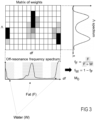
- a spectrum is extracted for each voxel and for each dimension of the dictionary from the obtained matrix of optimized weights. For instance, in the case of the present illustration based on a 2D dictionary, spectra of the voxel composition in terms of relaxation time ratios and off-resonance frequency are generated by the control unit from the obtained optimized weights. An illustration of such spectra is provided in Fig. 3 .
- the system determines one or several types of tissue composing a voxel (i.e. it determines at least one tissue type composing the voxel), and then, optionally a fraction of the type(s) of tissue composing the voxel by extracting information about tissue type(s) composing the voxel from the obtained spectra.
- the control unit is configured for estimating the fraction of tissue types (or more precisely of chemical species) that compose the voxel like the fraction of Fat f F , the fraction of Water f W , as well as other tissue type property, like its thermal equilibrium magnetization M 0 , from said the obtained spectra.
- the present invention proposes to obtain an estimation of the fat fraction by defining a range of frequencies for fat and respectively water based on the expected spectral resolution for fat and respectively water in MRI experiments.
- the control unit according to the invention is configured for estimating the fat fraction by calculating the integral of the off-resonance frequency spectrum over this range of frequencies defined for fat divided by the integral of the off-resonance frequency spectrum over both ranges defined for fat and water.
- the water fraction is then computed by the control unit as being one minus the estimated fat fraction (see Fig. 3 ).
- the control unit is further configured for estimating M 0 as being equal to the sum of all the weights.
- phase-cycled bSSFP acquisition has been performed for acquiring complex signal profiles.
- phase-cycled bSSFP acquisitions might be replaced by any steady-state non-spoiled GRE acquisition, as for example a phase-cycled FISS.
- the simulated signal profiles in the dictionary can be obtained by mean of any suitable numerical simulations, like Bloch simulations. Alternatively, an analytical description of the signal [18] or Extended Phase Graph (EPG) modelling can be used. Modelling of additional effects (e.g. MT, chemical exchange, B 1 ) could be included in the simulations. If additional variable parameters were to be considered, the dimensionality of the dictionary could be increased as needed while applying the present concept.
- EPG Extended Phase Graph
- alternative solutions can be implemented in order to estimate biological object tissue properties starting from the obtained voxel-wise spectra.
- the same procedure could be used to extract information from the relaxation time ratio spectra or by considering both dimensions at the same time.
- a watershed algorithm could be used in order to detect the peaks of water and fat.
- FIG. 2 schematically illustrates an embodiment of a system 200 according to the invention.
- Said system 200 is notably configured for mapping a fraction of a type of biological tissue present within a biological object, e.g. by mapping the fraction of fat within a biological tissue.
- the system is typically a computer-implemented system.
- the system 200 comprises:
- the present invention proposes a system and method enabling a determination of the fraction of a type of biological tissue comprised within a biological object and allowing a mapping of the fraction of tissue concentration in MRI images of said object, wherein tissue type separated images, e.g. fat-water separated images, are generated based on the extraction of multicomponent information from a phase-cycled signal profile by fitting of a multi-dimensional dictionary.
- tissue type separated images e.g. fat-water separated images
Landscapes
- Physics & Mathematics (AREA)
- Engineering & Computer Science (AREA)
- General Physics & Mathematics (AREA)
- High Energy & Nuclear Physics (AREA)
- Condensed Matter Physics & Semiconductors (AREA)
- Computer Vision & Pattern Recognition (AREA)
- Health & Medical Sciences (AREA)
- General Health & Medical Sciences (AREA)
- Nuclear Medicine, Radiotherapy & Molecular Imaging (AREA)
- Radiology & Medical Imaging (AREA)
- Signal Processing (AREA)
- Artificial Intelligence (AREA)
- Medical Informatics (AREA)
- Quality & Reliability (AREA)
- Theoretical Computer Science (AREA)
- Magnetic Resonance Imaging Apparatus (AREA)
Claims (10)
- Procédé (100) pour déterminer, par imagerie par résonance magnétique (ci-après IRM), un ou plusieurs types de tissu au sein d'un voxel dans un objet, le procédé comprenant :- la réalisation (101) d'une acquisition à cycle de phase consistant en l'acquisition, pour ledit objet, de plusieurs images d'écho de gradient, GRE, non altérées en régime permanent avec différents incréments de phase radiofréquence (RF) sur les acquisitions desdites plusieurs images de GRE non altérées en régime permanent, et la déduction d'un profil de signal complexe pour chaque voxel des images acquises, dans lequel pour chaque voxel, le profil de signal complexe est l'aimantation transversale en régime permanent pour chaque incrément de phase RF ;- la génération (102) d'un dictionnaire de profils de signaux simulés, dans lequel chaque profil de signal simulé est configuré pour simuler le profil de signal complexe précédemment déduit, avec un temps d'écho, TE, un temps de répétition, TR, et un angle d'excitation, RF, concordant avec les paramètres d'imagerie utilisés pour l'acquisition à cycle de phase, et dans lequel ledit dictionnaire est caractérisé par plusieurs dimensions, dans lequel chaque dimension est associée à un paramètre variable dont les valeurs varient au sein d'une plage de valeurs prédéfinie pertinente pour la détermination desdits un ou plusieurs types de tissu, et de sorte que chaque profil de signal simulé soit obtenu pour une valeur différente dudit paramètre ;- l'utilisation (103) d'un algorithme d'optimisation de pondération configuré pour exprimer le profil de signal complexe comme une somme pondérée des profils de signaux simulés, dans lequel l'algorithme d'optimisation de pondération reçoit en entrée le profil de signal complexe pour chaque voxel et les profils de signaux simulés du dictionnaire, et fournit en sortie, pour chaque voxel, une matrice M de pondérations optimisées ;- pour chaque voxel et chaque dimension de la matrice M obtenue, l'extraction (104), à partir de la matrice M, d'une distribution des pondérations optimisées obtenues ;- la détermination (105) desdits un ou plusieurs types de tissu composant chaque voxel à partir des distributions obtenues.
- Procédé selon la revendication 1, comprenant le calcul, à partir des distributions obtenues, de la fraction d'un type de tissu donné au sein d'un voxel.
- Procédé selon la revendication 1, dans lequel, pour chaque dimension de la matrice M, la distribution est calculée par la projection de la matrice M de pondérations optimisées sur un axe représentant les valeurs du paramètre variable associé à ladite dimension.
- Procédé selon la revendication 1 ou 3, comprenant le calcul de la fraction d'un type de tissu donné au sein d'un voxel comme le rapport entre la somme des pondérations à l'intérieur d'une plage desdits paramètres variables et la somme des pondérations dans la totalité de la plage simulée desdits paramètres variables.
- Procédé selon l'une des revendications 1 à 4, dans lequel le dictionnaire est un dictionnaire en 2D, dans lequel les deux dimensions sont le rapport de temps de relaxation T1/T2 et la fréquence hors résonance.
- Procédé selon l'une des revendications 1 à 5, dans lequel les pondérations sont optimisées afin de minimiser la distance entre le signal complexe et la somme pondérée des signaux simulés.
- Procédé selon l'une des revendications 1 à 6, comprenant le calcul d'une fraction graisseuse et d'une fraction hydrique pour chaque voxel.
- Procédé selon l'une des revendications 1 à 7, comprenant la création d'une carte paramétrique des fractions de tissu calculées sur l'ensemble du volume de l'objet.
- Procédé selon l'une des revendications 1 à 8, comprenant l'estimation d'une propriété de tissu objet à partir de la distribution extraite.
- Système (200) pour déterminer une fraction de type de tissu pour un objet imagé au moyen d'une IRM, le système (200) comprenant :- une unité de commande (203) comprenant un processeur et configurée pour être connectée à un appareil d'IRM (201) ;le système (200) étant caractérisé en ce que son unité de commande (203) est configurée pour mettre en œuvre le procédé selon l'une des revendications 1 à 9.
Priority Applications (2)
| Application Number | Priority Date | Filing Date | Title |
|---|---|---|---|
| EP20152927.8A EP3855199B1 (fr) | 2020-01-21 | 2020-01-21 | Procédé et système de mappage d'une fraction de concentrations de tissus en irm |
| US17/154,318 US11341642B2 (en) | 2020-01-21 | 2021-01-21 | Method and system for mapping a fraction of tissue concentrations in MRI |
Applications Claiming Priority (1)
| Application Number | Priority Date | Filing Date | Title |
|---|---|---|---|
| EP20152927.8A EP3855199B1 (fr) | 2020-01-21 | 2020-01-21 | Procédé et système de mappage d'une fraction de concentrations de tissus en irm |
Publications (3)
| Publication Number | Publication Date |
|---|---|
| EP3855199A1 EP3855199A1 (fr) | 2021-07-28 |
| EP3855199B1 true EP3855199B1 (fr) | 2023-10-11 |
| EP3855199C0 EP3855199C0 (fr) | 2023-10-11 |
Family
ID=69185533
Family Applications (1)
| Application Number | Title | Priority Date | Filing Date |
|---|---|---|---|
| EP20152927.8A Active EP3855199B1 (fr) | 2020-01-21 | 2020-01-21 | Procédé et système de mappage d'une fraction de concentrations de tissus en irm |
Country Status (2)
| Country | Link |
|---|---|
| US (1) | US11341642B2 (fr) |
| EP (1) | EP3855199B1 (fr) |
Families Citing this family (1)
| Publication number | Priority date | Publication date | Assignee | Title |
|---|---|---|---|---|
| JP7680978B2 (ja) * | 2022-03-14 | 2025-05-21 | 富士フイルム株式会社 | 磁気共鳴イメージング装置、画像処理装置、及び、信号分離方法 |
Family Cites Families (4)
| Publication number | Priority date | Publication date | Assignee | Title |
|---|---|---|---|---|
| US10429475B2 (en) * | 2013-03-12 | 2019-10-01 | The General Hospital Corporation | Method for increasing signal-to-noise ratio in magnetic resonance imaging using per-voxel noise covariance regularization |
| US10353034B2 (en) * | 2016-05-05 | 2019-07-16 | Siemens Healthcare Gmbh | Multi-echo pseudo-golden angle stack of stars thermometry with high spatial and temporal resolution using k-space weighted image contrast |
| CN108693491B (zh) * | 2017-04-07 | 2022-03-25 | 康奈尔大学 | 稳健的定量磁化率成像系统和方法 |
| US10564243B2 (en) * | 2017-04-07 | 2020-02-18 | Case Western Reserve University | System and method for phase cycling magnetic resonance fingerprinting (phc-MRF) |
-
2020
- 2020-01-21 EP EP20152927.8A patent/EP3855199B1/fr active Active
-
2021
- 2021-01-21 US US17/154,318 patent/US11341642B2/en active Active
Also Published As
| Publication number | Publication date |
|---|---|
| EP3855199A1 (fr) | 2021-07-28 |
| US11341642B2 (en) | 2022-05-24 |
| US20210224982A1 (en) | 2021-07-22 |
| EP3855199C0 (fr) | 2023-10-11 |
Similar Documents
| Publication | Publication Date | Title |
|---|---|---|
| Buonincontri et al. | Multi-site repeatability and reproducibility of MR fingerprinting of the healthy brain at 1.5 and 3.0 T | |
| Lam et al. | High‐resolution 1H‐MRSI of the brain using SPICE: data acquisition and image reconstruction | |
| Ostenson et al. | MR fingerprinting with simultaneous T1, T2, and fat signal fraction estimation with integrated B0 correction reduces bias in water T1 and T2 estimates | |
| US10345414B2 (en) | Rapid quantitative abdominal imaging with magnetic resonance fingerprinting (MRF) | |
| US10145917B2 (en) | Multi-component voxel separation using magnetic resonance fingerprinting with compartment exchange | |
| Weidlich et al. | T2 mapping with magnetization‐prepared 3D TSE based on a modified BIR‐4 T2 preparation | |
| Hermann et al. | Magnetic resonance fingerprinting for simultaneous renal T1 and T2* mapping in a single breath‐hold | |
| US8611624B2 (en) | Method for adipose tissue quantification with magnetic resonance imaging | |
| US8498461B2 (en) | Method and magnetic resonance system to determine system-dependent phase information | |
| Stern et al. | Mapping of magnetic resonance imaging’s transverse relaxation time at low signal‐to‐noise ratio using Bloch simulations and principal component analysis image denoising | |
| Omer et al. | Data‐driven algorithm for myelin water imaging: probing subvoxel compartmentation based on identification of spatially global tissue features | |
| Licht et al. | Multidimensional compressed sensing to advance 23Na multi‐quantum coherences MRI | |
| US11047944B2 (en) | Multi-point magnetic resonance imaging | |
| EP3855199B1 (fr) | Procédé et système de mappage d'une fraction de concentrations de tissus en irm | |
| KR102158268B1 (ko) | 뇌 대사물질 분석 및 뇌 네트워크 구현 장치 및 방법 | |
| Kathiravan et al. | A review of magnetic resonance imaging techniques | |
| Hu et al. | An Efficient Reconstruction Algorithm Based on the Alternating Direction Method of Multipliers for Joint Estimation of ${R} _ {{2}}^{*} $ and Off-Resonance in fMRI | |
| EP4246168B1 (fr) | Appareil d'imagerie par résonance magnétique et procédé de séparation de signal | |
| US20200033432A1 (en) | Method and apparatus for identifying at least one material comprised in a voxel | |
| Hansen et al. | Fast Padé transform accelerated CSI for hyperpolarized MRS | |
| US11360173B2 (en) | Method and system for mapping transverse relaxation in magnetic resonance scan | |
| US20190254596A1 (en) | Method for generating a magnetic resonance image dataset, computer program product, data medium, and magnetic resonance system | |
| US11841410B2 (en) | Method and system for analyzing multi-component magnetic resonance signals | |
| US12601804B2 (en) | Method and system of multi-parametric magnetic resonance imaging | |
| Garrido et al. | 3D Radial Knee Imaging at 0.55 T: Simultaneous T 1–T 2 Mapping and Synthetic Contrast Generation in Under 4 min |
Legal Events
| Date | Code | Title | Description |
|---|---|---|---|
| PUAI | Public reference made under article 153(3) epc to a published international application that has entered the european phase |
Free format text: ORIGINAL CODE: 0009012 |
|
| STAA | Information on the status of an ep patent application or granted ep patent |
Free format text: STATUS: THE APPLICATION HAS BEEN PUBLISHED |
|
| AK | Designated contracting states |
Kind code of ref document: A1 Designated state(s): AL AT BE BG CH CY CZ DE DK EE ES FI FR GB GR HR HU IE IS IT LI LT LU LV MC MK MT NL NO PL PT RO RS SE SI SK SM TR |
|
| STAA | Information on the status of an ep patent application or granted ep patent |
Free format text: STATUS: REQUEST FOR EXAMINATION WAS MADE |
|
| 17P | Request for examination filed |
Effective date: 20220119 |
|
| RBV | Designated contracting states (corrected) |
Designated state(s): AL AT BE BG CH CY CZ DE DK EE ES FI FR GB GR HR HU IE IS IT LI LT LU LV MC MK MT NL NO PL PT RO RS SE SI SK SM TR |
|
| GRAP | Despatch of communication of intention to grant a patent |
Free format text: ORIGINAL CODE: EPIDOSNIGR1 |
|
| STAA | Information on the status of an ep patent application or granted ep patent |
Free format text: STATUS: GRANT OF PATENT IS INTENDED |
|
| RIC1 | Information provided on ipc code assigned before grant |
Ipc: G01R 33/561 20060101ALN20230317BHEP Ipc: G01R 33/48 20060101ALN20230317BHEP Ipc: G01R 33/56 20060101ALI20230317BHEP Ipc: G01R 33/54 20060101AFI20230317BHEP |
|
| GRAJ | Information related to disapproval of communication of intention to grant by the applicant or resumption of examination proceedings by the epo deleted |
Free format text: ORIGINAL CODE: EPIDOSDIGR1 |
|
| STAA | Information on the status of an ep patent application or granted ep patent |
Free format text: STATUS: REQUEST FOR EXAMINATION WAS MADE |
|
| INTG | Intention to grant announced |
Effective date: 20230417 |
|
| INTC | Intention to grant announced (deleted) | ||
| RIC1 | Information provided on ipc code assigned before grant |
Ipc: G01R 33/561 20060101ALN20230605BHEP Ipc: G01R 33/48 20060101ALN20230605BHEP Ipc: G01R 33/56 20060101ALI20230605BHEP Ipc: G01R 33/54 20060101AFI20230605BHEP |
|
| GRAP | Despatch of communication of intention to grant a patent |
Free format text: ORIGINAL CODE: EPIDOSNIGR1 |
|
| STAA | Information on the status of an ep patent application or granted ep patent |
Free format text: STATUS: GRANT OF PATENT IS INTENDED |
|
| INTG | Intention to grant announced |
Effective date: 20230721 |
|
| GRAS | Grant fee paid |
Free format text: ORIGINAL CODE: EPIDOSNIGR3 |
|
| GRAA | (expected) grant |
Free format text: ORIGINAL CODE: 0009210 |
|
| STAA | Information on the status of an ep patent application or granted ep patent |
Free format text: STATUS: THE PATENT HAS BEEN GRANTED |
|
| AK | Designated contracting states |
Kind code of ref document: B1 Designated state(s): AL AT BE BG CH CY CZ DE DK EE ES FI FR GB GR HR HU IE IS IT LI LT LU LV MC MK MT NL NO PL PT RO RS SE SI SK SM TR |
|
| REG | Reference to a national code |
Ref country code: GB Ref legal event code: FG4D |
|
| REG | Reference to a national code |
Ref country code: CH Ref legal event code: EP |
|
| REG | Reference to a national code |
Ref country code: DE Ref legal event code: R096 Ref document number: 602020018866 Country of ref document: DE |
|
| REG | Reference to a national code |
Ref country code: IE Ref legal event code: FG4D |
|
| U01 | Request for unitary effect filed |
Effective date: 20231026 |
|
| U07 | Unitary effect registered |
Designated state(s): AT BE BG DE DK EE FI FR IT LT LU LV MT NL PT SE SI Effective date: 20231102 |
|
| U1N | Appointed representative for the unitary patent procedure changed after the registration of the unitary effect |
Representative=s name: SIEMENS HEALTHINEERS PATENT ATTORNEYS; DE |
|
| RAP2 | Party data changed (patent owner data changed or rights of a patent transferred) |
Owner name: CENTRE HOSPITALIER UNIVERSITAIRE VAUDOIS Owner name: SIEMENS HEALTHINEERS AG |
|
| U1K | Transfer of rights of the unitary patent after the registration of the unitary effect |
Owner name: SIEMENS HEALTHINEERS AG; DE Owner name: CENTRE HOSPITALIER UNIVERSITAIRE VAUDOIS; CH |
|
| U20 | Renewal fee for the european patent with unitary effect paid |
Year of fee payment: 5 Effective date: 20240119 |
|
| U1N | Appointed representative for the unitary patent procedure changed after the registration of the unitary effect |
Representative=s name: SIEMENS HEALTHINEERS PATENT ATTORNEYS; DE |
|
| PG25 | Lapsed in a contracting state [announced via postgrant information from national office to epo] |
Ref country code: GR Free format text: LAPSE BECAUSE OF FAILURE TO SUBMIT A TRANSLATION OF THE DESCRIPTION OR TO PAY THE FEE WITHIN THE PRESCRIBED TIME-LIMIT Effective date: 20240112 |
|
| PG25 | Lapsed in a contracting state [announced via postgrant information from national office to epo] |
Ref country code: IS Free format text: LAPSE BECAUSE OF FAILURE TO SUBMIT A TRANSLATION OF THE DESCRIPTION OR TO PAY THE FEE WITHIN THE PRESCRIBED TIME-LIMIT Effective date: 20240211 |
|
| PG25 | Lapsed in a contracting state [announced via postgrant information from national office to epo] |
Ref country code: ES Free format text: LAPSE BECAUSE OF FAILURE TO SUBMIT A TRANSLATION OF THE DESCRIPTION OR TO PAY THE FEE WITHIN THE PRESCRIBED TIME-LIMIT Effective date: 20231011 |
|
| PG25 | Lapsed in a contracting state [announced via postgrant information from national office to epo] |
Ref country code: IS Free format text: LAPSE BECAUSE OF FAILURE TO SUBMIT A TRANSLATION OF THE DESCRIPTION OR TO PAY THE FEE WITHIN THE PRESCRIBED TIME-LIMIT Effective date: 20240211 Ref country code: GR Free format text: LAPSE BECAUSE OF FAILURE TO SUBMIT A TRANSLATION OF THE DESCRIPTION OR TO PAY THE FEE WITHIN THE PRESCRIBED TIME-LIMIT Effective date: 20240112 Ref country code: ES Free format text: LAPSE BECAUSE OF FAILURE TO SUBMIT A TRANSLATION OF THE DESCRIPTION OR TO PAY THE FEE WITHIN THE PRESCRIBED TIME-LIMIT Effective date: 20231011 |
|
| PG25 | Lapsed in a contracting state [announced via postgrant information from national office to epo] |
Ref country code: RS Free format text: LAPSE BECAUSE OF FAILURE TO SUBMIT A TRANSLATION OF THE DESCRIPTION OR TO PAY THE FEE WITHIN THE PRESCRIBED TIME-LIMIT Effective date: 20231011 Ref country code: PL Free format text: LAPSE BECAUSE OF FAILURE TO SUBMIT A TRANSLATION OF THE DESCRIPTION OR TO PAY THE FEE WITHIN THE PRESCRIBED TIME-LIMIT Effective date: 20231011 Ref country code: NO Free format text: LAPSE BECAUSE OF FAILURE TO SUBMIT A TRANSLATION OF THE DESCRIPTION OR TO PAY THE FEE WITHIN THE PRESCRIBED TIME-LIMIT Effective date: 20240111 Ref country code: HR Free format text: LAPSE BECAUSE OF FAILURE TO SUBMIT A TRANSLATION OF THE DESCRIPTION OR TO PAY THE FEE WITHIN THE PRESCRIBED TIME-LIMIT Effective date: 20231011 |
|
| U1N | Appointed representative for the unitary patent procedure changed after the registration of the unitary effect |
Representative=s name: SIEMENS HEALTHINEERS PATENT ATTORNEYS; DE |
|
| REG | Reference to a national code |
Ref country code: DE Ref legal event code: R097 Ref document number: 602020018866 Country of ref document: DE |
|
| PG25 | Lapsed in a contracting state [announced via postgrant information from national office to epo] |
Ref country code: CZ Free format text: LAPSE BECAUSE OF FAILURE TO SUBMIT A TRANSLATION OF THE DESCRIPTION OR TO PAY THE FEE WITHIN THE PRESCRIBED TIME-LIMIT Effective date: 20231011 |
|
| PG25 | Lapsed in a contracting state [announced via postgrant information from national office to epo] |
Ref country code: SK Free format text: LAPSE BECAUSE OF FAILURE TO SUBMIT A TRANSLATION OF THE DESCRIPTION OR TO PAY THE FEE WITHIN THE PRESCRIBED TIME-LIMIT Effective date: 20231011 |
|
| PG25 | Lapsed in a contracting state [announced via postgrant information from national office to epo] |
Ref country code: SM Free format text: LAPSE BECAUSE OF FAILURE TO SUBMIT A TRANSLATION OF THE DESCRIPTION OR TO PAY THE FEE WITHIN THE PRESCRIBED TIME-LIMIT Effective date: 20231011 Ref country code: SK Free format text: LAPSE BECAUSE OF FAILURE TO SUBMIT A TRANSLATION OF THE DESCRIPTION OR TO PAY THE FEE WITHIN THE PRESCRIBED TIME-LIMIT Effective date: 20231011 Ref country code: RO Free format text: LAPSE BECAUSE OF FAILURE TO SUBMIT A TRANSLATION OF THE DESCRIPTION OR TO PAY THE FEE WITHIN THE PRESCRIBED TIME-LIMIT Effective date: 20231011 Ref country code: CZ Free format text: LAPSE BECAUSE OF FAILURE TO SUBMIT A TRANSLATION OF THE DESCRIPTION OR TO PAY THE FEE WITHIN THE PRESCRIBED TIME-LIMIT Effective date: 20231011 |
|
| PLBE | No opposition filed within time limit |
Free format text: ORIGINAL CODE: 0009261 |
|
| STAA | Information on the status of an ep patent application or granted ep patent |
Free format text: STATUS: NO OPPOSITION FILED WITHIN TIME LIMIT |
|
| PG25 | Lapsed in a contracting state [announced via postgrant information from national office to epo] |
Ref country code: MC Free format text: LAPSE BECAUSE OF FAILURE TO SUBMIT A TRANSLATION OF THE DESCRIPTION OR TO PAY THE FEE WITHIN THE PRESCRIBED TIME-LIMIT Effective date: 20231011 |
|
| PG25 | Lapsed in a contracting state [announced via postgrant information from national office to epo] |
Ref country code: MC Free format text: LAPSE BECAUSE OF FAILURE TO SUBMIT A TRANSLATION OF THE DESCRIPTION OR TO PAY THE FEE WITHIN THE PRESCRIBED TIME-LIMIT Effective date: 20231011 |
|
| REG | Reference to a national code |
Ref country code: CH Ref legal event code: PL |
|
| 26N | No opposition filed |
Effective date: 20240712 |
|
| PG25 | Lapsed in a contracting state [announced via postgrant information from national office to epo] |
Ref country code: CH Free format text: LAPSE BECAUSE OF NON-PAYMENT OF DUE FEES Effective date: 20240131 |
|
| PG25 | Lapsed in a contracting state [announced via postgrant information from national office to epo] |
Ref country code: CH Free format text: LAPSE BECAUSE OF NON-PAYMENT OF DUE FEES Effective date: 20240131 |
|
| PG25 | Lapsed in a contracting state [announced via postgrant information from national office to epo] |
Ref country code: IE Free format text: LAPSE BECAUSE OF NON-PAYMENT OF DUE FEES Effective date: 20240121 |
|
| PG25 | Lapsed in a contracting state [announced via postgrant information from national office to epo] |
Ref country code: IE Free format text: LAPSE BECAUSE OF NON-PAYMENT OF DUE FEES Effective date: 20240121 |
|
| U20 | Renewal fee for the european patent with unitary effect paid |
Year of fee payment: 6 Effective date: 20250120 |
|
| U1N | Appointed representative for the unitary patent procedure changed after the registration of the unitary effect |
Representative=s name: FISCHER, MICHAEL; DE |
|
| PG25 | Lapsed in a contracting state [announced via postgrant information from national office to epo] |
Ref country code: CY Free format text: LAPSE BECAUSE OF FAILURE TO SUBMIT A TRANSLATION OF THE DESCRIPTION OR TO PAY THE FEE WITHIN THE PRESCRIBED TIME-LIMIT; INVALID AB INITIO Effective date: 20200121 |
|
| PG25 | Lapsed in a contracting state [announced via postgrant information from national office to epo] |
Ref country code: HU Free format text: LAPSE BECAUSE OF FAILURE TO SUBMIT A TRANSLATION OF THE DESCRIPTION OR TO PAY THE FEE WITHIN THE PRESCRIBED TIME-LIMIT; INVALID AB INITIO Effective date: 20200121 |
|
| PG25 | Lapsed in a contracting state [announced via postgrant information from national office to epo] |
Ref country code: TR Free format text: LAPSE BECAUSE OF FAILURE TO SUBMIT A TRANSLATION OF THE DESCRIPTION OR TO PAY THE FEE WITHIN THE PRESCRIBED TIME-LIMIT Effective date: 20231011 |
|
| U20 | Renewal fee for the european patent with unitary effect paid |
Year of fee payment: 7 Effective date: 20260119 |
|
| PGFP | Annual fee paid to national office [announced via postgrant information from national office to epo] |
Ref country code: GB Payment date: 20260210 Year of fee payment: 7 |