EP3859015A1 - Procédés d'identification de l'état de fertilité masculine et de la qualité d'un embryon - Google Patents
Procédés d'identification de l'état de fertilité masculine et de la qualité d'un embryon Download PDFInfo
- Publication number
- EP3859015A1 EP3859015A1 EP20213740.2A EP20213740A EP3859015A1 EP 3859015 A1 EP3859015 A1 EP 3859015A1 EP 20213740 A EP20213740 A EP 20213740A EP 3859015 A1 EP3859015 A1 EP 3859015A1
- Authority
- EP
- European Patent Office
- Prior art keywords
- methylation
- pattern
- dna
- semen
- cytosines
- Prior art date
- Legal status (The legal status is an assumption and is not a legal conclusion. Google has not performed a legal analysis and makes no representation as to the accuracy of the status listed.)
- Ceased
Links
Images






Classifications
-
- C—CHEMISTRY; METALLURGY
- C12—BIOCHEMISTRY; BEER; SPIRITS; WINE; VINEGAR; MICROBIOLOGY; ENZYMOLOGY; MUTATION OR GENETIC ENGINEERING
- C12Q—MEASURING OR TESTING PROCESSES INVOLVING ENZYMES, NUCLEIC ACIDS OR MICROORGANISMS; COMPOSITIONS OR TEST PAPERS THEREFOR; PROCESSES OF PREPARING SUCH COMPOSITIONS; CONDITION-RESPONSIVE CONTROL IN MICROBIOLOGICAL OR ENZYMOLOGICAL PROCESSES
- C12Q1/00—Measuring or testing processes involving enzymes, nucleic acids or microorganisms; Compositions therefor; Processes of preparing such compositions
- C12Q1/68—Measuring or testing processes involving enzymes, nucleic acids or microorganisms; Compositions therefor; Processes of preparing such compositions involving nucleic acids
- C12Q1/6876—Nucleic acid products used in the analysis of nucleic acids, e.g. primers or probes
- C12Q1/6883—Nucleic acid products used in the analysis of nucleic acids, e.g. primers or probes for diseases caused by alterations of genetic material
-
- A—HUMAN NECESSITIES
- A61—MEDICAL OR VETERINARY SCIENCE; HYGIENE
- A61B—DIAGNOSIS; SURGERY; IDENTIFICATION
- A61B17/00—Surgical instruments, devices or methods
- A61B17/42—Gynaecological or obstetrical instruments or methods
- A61B17/425—Gynaecological or obstetrical instruments or methods for reproduction or fertilisation
- A61B17/43—Gynaecological or obstetrical instruments or methods for reproduction or fertilisation for artificial insemination
-
- C—CHEMISTRY; METALLURGY
- C12—BIOCHEMISTRY; BEER; SPIRITS; WINE; VINEGAR; MICROBIOLOGY; ENZYMOLOGY; MUTATION OR GENETIC ENGINEERING
- C12Q—MEASURING OR TESTING PROCESSES INVOLVING ENZYMES, NUCLEIC ACIDS OR MICROORGANISMS; COMPOSITIONS OR TEST PAPERS THEREFOR; PROCESSES OF PREPARING SUCH COMPOSITIONS; CONDITION-RESPONSIVE CONTROL IN MICROBIOLOGICAL OR ENZYMOLOGICAL PROCESSES
- C12Q1/00—Measuring or testing processes involving enzymes, nucleic acids or microorganisms; Compositions therefor; Processes of preparing such compositions
- C12Q1/68—Measuring or testing processes involving enzymes, nucleic acids or microorganisms; Compositions therefor; Processes of preparing such compositions involving nucleic acids
-
- G—PHYSICS
- G16—INFORMATION AND COMMUNICATION TECHNOLOGY [ICT] SPECIALLY ADAPTED FOR SPECIFIC APPLICATION FIELDS
- G16B—BIOINFORMATICS, i.e. INFORMATION AND COMMUNICATION TECHNOLOGY [ICT] SPECIALLY ADAPTED FOR GENETIC OR PROTEIN-RELATED DATA PROCESSING IN COMPUTATIONAL MOLECULAR BIOLOGY
- G16B20/00—ICT specially adapted for functional genomics or proteomics, e.g. genotype-phenotype associations
-
- C—CHEMISTRY; METALLURGY
- C12—BIOCHEMISTRY; BEER; SPIRITS; WINE; VINEGAR; MICROBIOLOGY; ENZYMOLOGY; MUTATION OR GENETIC ENGINEERING
- C12Q—MEASURING OR TESTING PROCESSES INVOLVING ENZYMES, NUCLEIC ACIDS OR MICROORGANISMS; COMPOSITIONS OR TEST PAPERS THEREFOR; PROCESSES OF PREPARING SUCH COMPOSITIONS; CONDITION-RESPONSIVE CONTROL IN MICROBIOLOGICAL OR ENZYMOLOGICAL PROCESSES
- C12Q2600/00—Oligonucleotides characterized by their use
- C12Q2600/112—Disease subtyping, staging or classification
-
- C—CHEMISTRY; METALLURGY
- C12—BIOCHEMISTRY; BEER; SPIRITS; WINE; VINEGAR; MICROBIOLOGY; ENZYMOLOGY; MUTATION OR GENETIC ENGINEERING
- C12Q—MEASURING OR TESTING PROCESSES INVOLVING ENZYMES, NUCLEIC ACIDS OR MICROORGANISMS; COMPOSITIONS OR TEST PAPERS THEREFOR; PROCESSES OF PREPARING SUCH COMPOSITIONS; CONDITION-RESPONSIVE CONTROL IN MICROBIOLOGICAL OR ENZYMOLOGICAL PROCESSES
- C12Q2600/00—Oligonucleotides characterized by their use
- C12Q2600/118—Prognosis of disease development
-
- C—CHEMISTRY; METALLURGY
- C12—BIOCHEMISTRY; BEER; SPIRITS; WINE; VINEGAR; MICROBIOLOGY; ENZYMOLOGY; MUTATION OR GENETIC ENGINEERING
- C12Q—MEASURING OR TESTING PROCESSES INVOLVING ENZYMES, NUCLEIC ACIDS OR MICROORGANISMS; COMPOSITIONS OR TEST PAPERS THEREFOR; PROCESSES OF PREPARING SUCH COMPOSITIONS; CONDITION-RESPONSIVE CONTROL IN MICROBIOLOGICAL OR ENZYMOLOGICAL PROCESSES
- C12Q2600/00—Oligonucleotides characterized by their use
- C12Q2600/154—Methylation markers
-
- G—PHYSICS
- G01—MEASURING; TESTING
- G01N—INVESTIGATING OR ANALYSING MATERIALS BY DETERMINING THEIR CHEMICAL OR PHYSICAL PROPERTIES
- G01N2800/00—Detection or diagnosis of diseases
- G01N2800/36—Gynecology or obstetrics
- G01N2800/367—Infertility, e.g. sperm disorder, ovulatory dysfunction
Definitions
- the present disclosure relates to methods of identifying a male fertility condition in a subject.
- the disclosure also relates to methods of identifying embryo quality. More particularly, the disclosure relates to methods of performing a fertility treatment.
- the main parameters evaluated in the semen analysis namely sperm count, motility, viability, and morphology, can be somewhat subjective, and consequently they can be subject to technical error. While continual training and assessment, quality control measures, and proficiency testing may all serve to minimize technical error, studies have demonstrated that coefficients of variation (CVs) between labs and technicians can fall in the 20-30% range, with higher CVs reported in some studies (see Gandini L et al. International Journal of Andrology. 2000;23(1):1-3 ; Neuwinger J et al. Fertility and Sterility. 1990;54(2):308-14 ; and Brazil C et al. Journal of Andrology. 2004;25(4):645-56 , each of which is incorporated by reference herein in its entirety).
- CVs coefficients of variation
- Adjunct tests have been developed, such as sperm DNA damage assessment, capacitation and acrosome reaction tests, egg and zona penetration assays, anti-sperm antibody testing, and aneuploidy screening (see Zini A et al. Human Reproduction. 2008;23(12):2663-8 ; Sanchez R et al. Andrologia. 1991;23(3): 197-203 ; Lee MA et al. Fertility and Sterility. 1987;48(4):649-58 ; Haas GG, Jr. et al. The New England Journal of Medicine. 1980;303(13):722-7 ; Burkman LJ et al. Fertility and Sterility.
- Y chromosome microdeletions see Tiepolo L et al. Human Genetics. 1976;34(2): 119-24 , which is incorporated by reference herein in its entirety
- Klinefelter syndrome see Lanfranco F et al. Lancet. 2004;364(9430):273-83 , which is incorporated by reference herein in its entirety
- DPY19L and SPATA16 mutations see Dam AH et al. American Journal of Human Genetics. 2007;81 (4):813-20 ; Elinati E et al. Human Molecular Genetics. 2012;21 (16):3695-702 ; Harbuz R et al. American Journal of Human Genetics.
- Epigenetics can refer to modifiable but generally stable, heritable modifications to the DNA or chromatin packaging.
- Direct DNA modifications can consist of methylation alteration to the 5-carbon position of cytosine bases, generally in the context of cytosine-guanine dinucleotides (CpGs).
- the present disclosure provides methods of identifying a male fertility condition in a subject and/or predicting a level of male fertility in a subject. This disclosure also provides methods of identifying and/or predicting embryo quality. Furthermore, the disclosure provides methods of performing fertility treatments.
- a "subject” is defined herein as a mammal that may experience a male fertility condition, a male fertility abnormality, decreased fertility, and/or a level of infertility. Examples of subjects include, but are not limited to, humans, horses, pigs, cattle, dogs, cats, rabbits, and aquatic mammals.
- a "methylation pattern" is defined herein as the methylation of a region of genetic material (e.g., a region of a DNA molecule); methylation of a region may include methylation of a plurality of regions. Such methylation can include the absence and/or presence of methylation in a region, the extent of methylation of a region, and/or the actual methylated sequence of the region.
- a “genome-wide methylation pattern" or a “genome-wide DNA methylation pattern” is defined herein as the methylation of a representative portion, a significant portion, and/or a substantial portion of a genome.
- a genome-wide methylation pattern may comprise a majority of CpGs from CpG islands, a portion of CpG sites outside of CpG islands, and/or a portion of non-CpG sites in the genome of a subject.
- a genome-wide methylation pattern may comprise a pattern of methylation at loci across the genome rather than at specific genes and/or genomic features.
- control pattern of methylation or a “control pattern of DNA methylation” is defined herein as a pattern of methylation from a population of males, or a representative sample thereof, that is indicative of a fertile male.
- a control pattern of methylation can be generated using techniques known in the art.
- the control pattern of methylation can be an average pattern of methylation generated from a population of males, or a representative sample thereof, that are determined to be fertile (e.g., normozoospermic males with proven fertility).
- an "aberrant pattern of methylation” or an “aberrant pattern of DNA methylation” is defined herein as a methylation pattern that deviates from a control pattern of methylation to an extent that is indicative of a male fertility condition.
- an aberrant pattern of methylation may be significantly different from a control pattern of methylation, or aberrant patterns of methylation may be significantly different from control patterns of methylation.
- a control pattern of methylation may be significantly different from an aberrant pattern of methylation, or control patterns of methylation may be significantly different from aberrant patterns of methylation.
- Methylation pattern differences may be observed at a plurality of consistently, or substantially consistently, altered loci, or methylation pattern differences may be manifest as increased variability in subjects having aberrant methylation patterns compared with control methylation patterns.
- differences between aberrant patterns of methylation and control patterns of methylation may be determined at a single CpG level.
- one or more CpGs may exhibit statistically significant differences in mean methylation between control groups and aberrant groups.
- differences between aberrant patterns of methylation and control patterns of methylation may be determined at an aggregate region level.
- one or more CpGs within a given region may exhibit statistically significant differences in mean methylation between control groups and aberrant groups, and/or the region as a whole may exhibit a statistically significant difference in mean methylation level.
- differences between aberrant patterns of methylation and control patterns of methylation may be determined from genome-wide comparisons.
- the differences between aberrant patterns of methylation and control patterns of methylation may be determined by simultaneously, or substantially simultaneously, comparing methylation patterns based on all profilable CpGs, substantially all profilable CpGs, or a large subset of CpGs.
- an analysis of the differences between aberrant patterns of methylation and control patterns of methylation may be determined in terms of "distance.” For example, each sample may represent a point in n-dimensional space, wherein "n" is the number of profilable or profiled CpGs.
- the "n" dimensions can be reduced to a single dimension, and it can be determined if the "distance” of an aberrant profile from a reference point shows a statistically significant difference from those one or more "distances" observed for control profiles.
- a maximally discriminative model may be utilized. For example, some machine learning methods may aim to learn the maximally discriminative model, regardless of whether any difference between groups or features reaches statistical significance.
- male fertility condition is defined herein as a condition that renders in a male subject some level of infertility, a fertility abnormality, and/or decreased fertility. Those of ordinary skill in the art are capable of distinguishing various levels of infertility, all of which would be considered to be within the present scope.
- a method for identifying an aberrant pattern of methylation associated with a male fertility condition may include assaying a pattern of methylation (e.g ., a test pattern of methylation) of a representative portion, a significant portion, and/or a substantial portion of a genome of a subject, and comparing the assayed pattern of methylation from the subject with a control pattern of DNA methylation to identify an aberrant pattern of methylation associated with a male fertility condition.
- a pattern of methylation e.g ., a test pattern of methylation
- a method for identifying a male fertility condition may include assaying the degree of methylation or a methylation pattern of a representative portion, a significant portion, and/or a substantial portion of a genome of sperm from a subject (e.g ., test sperm), and comparing the degree of methylation or the methylation pattern of the sperm from the subject with a control degree of methylation or a methylation pattern from control sperm to determine a male fertility condition.
- a subject e.g ., test sperm
- a useful method for assaying DNA methylation can include bisulfite treatment. Treating DNA with a bisulfite such as sodium bisulfite can convert unmethylated cytosines to uracils; methylated cytosines can resist deamination and thus generally remain unchanged.
- the cytosine to uracil transition following bisulfite treatment can provide a useful assay of the methylation pattern or profile of a region of genetic material. Following such treatment, the genetic material can be sequenced to determine the specific methylation pattern.
- Bisulfite treatment can be utilized in analyzing a few candidate loci, or it can be used to perform a broader genome-wide analysis. If a genome-wide or partially genome-wide analysis is desired, genetic material can be captured on an array of genome loci.
- methods according to the instant disclosure may be useful in predicting success with various treatments of reproductive disorders such as IVF or other artificial reproductive techniques.
- a male infertility condition may be a condition in a male subject that is associated with some level of infertility, a fertility abnormality, and/or decreased fertility.
- a method of identifying a male infertility condition may comprise extracting genetic material such as DNA from a semen sample from a subject.
- the semen sample may be a whole ejaculate sample.
- the semen sample may be purified, for example, by somatic cell lysis. Somatic cell lysis may reduce or eliminate white blood cell contamination in the semen sample.
- the semen sample may be purified by a density gradient purification.
- the semen sample, or the purified semen sample may be pelleted ( i.e ., via centrifugation) and/or visually inspected to determine whether substantially all contaminating cells are absent from or have been removed from the semen sample.
- the method may also comprise identifying a methylation pattern of the extracted DNA.
- the methylation pattern may be a genome-wide methylation pattern.
- the methylation pattern may include an assessment of the methylation status of cytosines and/or CpG dinucleotides from positions across the genome.
- the method may comprise comparing the methylation pattern of the extracted DNA to a control pattern of DNA methylation.
- the method may comprise determining if the methylation pattern of the extracted DNA is significantly different from the control pattern of DNA methylation, wherein a significantly different methylation pattern of the extracted DNA may indicate that the subject has a male fertility condition.
- the methylation pattern may be based on a methylation status of at least 50,000 cytosines from the extracted DNA.
- the methylation pattern may comprise the methylation status of at least 50,000 cytosines ( i . e ., whether or not cytosines disposed in at least 50,000 positions in the genome are methylated).
- the methylation pattern may be based on a methylation status of at least 100,000 cytosines from the extracted DNA, of at least 200,000 cytosines from the extracted DNA, of at least 300,000 cytosines from the extracted DNA, of at least 485,000 cytosines from the extracted DNA, or of another suitable number of cytosines from the extracted DNA.
- the methylation pattern may be based on a methylation status of at least 50,000 CpG dinucleotides from the extracted DNA.
- the methylation pattern may comprise the methylation status of at least 50,000 CpG dinucleotides ( i . e ., whether or not cytosines disposed in at least 50,000 CpG dinucleotide positions in the genome are methylated).
- the methylation pattern may be based on a methylation status of at least 100,000 CpG dinucleotides from the extracted DNA, of at least 200,000 CpG dinucleotides from the extracted DNA, of at least 300,000 CpG dinucleotides from the extracted DNA, of at least 485,000 CpG dinucleotides from the extracted DNA, or of another suitable number of CpG dinucleotides from the extracted DNA.
- the methylation pattern of the extracted DNA may be identified by a method selected from, but not limited to, analyzing the methylation pattern via a DNA methylation array, pyrosequencing, reduced representation bisulfite sequencing, targeted bisulfite sequencing, genome-wide bisulfite sequencing, bisulfite patch PCR, or a combination thereof.
- the methylation pattern of the extracted DNA may be identified by a method comprising treating at least a portion of the extracted DNA with bisulfite, amplifying the bisulfite-treated DNA, hybridizing the amplified DNA to a DNA methylation array, and/or processing the hybridized DNA methylation array to determine the methylation pattern of the extracted DNA.
- the method may comprise identifying a subject with a male fertility condition.
- the method may comprise identifying a DNA methylation pattern from a semen sample of the subject.
- the method may also comprise comparing the identified DNA methylation pattern to a control pattern of DNA methylation.
- the control pattern of DNA methylation may be a methylation pattern from DNA extracted from semen samples from a control population of fertile males.
- the method may further comprise classifying whether the identified DNA methylation pattern is significantly different from the control pattern of DNA methylation or whether the identified methylation pattern is not significantly different from the control pattern of DNA methylation.
- the method may comprise collecting a sample from the subject for use in an IVF procedure. In various embodiments, if the identified methylation pattern is not significantly different from the control pattern of DNA methylation, the method may comprise collecting a semen sample from the subject for use in an artificial insemination procedure.
- the methylation pattern may be based on a methylation status of at least 50,000 cytosines from the extracted DNA.
- the methylation pattern may comprise the methylation status of at least 50,000 cytosines ( i . e ., whether or not cytosines disposed in at least 50,000 positions in the genome are methylated).
- the methylation pattern may be based on a methylation status of at least 100,000 cytosines from the extracted DNA, of at least 200,000 cytosines from the extracted DNA, of at least 300,000 cytosines from the extracted DNA, of at least 485,000 cytosines from the extracted DNA, or of another suitable number of cytosines from the extracted DNA.
- the methylation pattern may be based on a methylation status of at least 50,000 CpG dinucleotides from the extracted DNA.
- the methylation pattern may comprise the methylation status of at least 50,000 CpG dinucleotides ( i . e ., whether or not cytosines disposed in at least 50,000 CpG dinucleotide positions in the genome are methylated).
- the methylation pattern may be based on a methylation status of at least 100,000 CpG dinucleotides from the extracted DNA, of at least 200,000 CpG dinucleotides from the extracted DNA, of at least 300,000 CpG dinucleotides from the extracted DNA, of at least 485,000 CpG dinucleotides from the extracted DNA, or of another suitable number of CpG dinucleotides from the extracted DNA.
- the method of fertility treatment may comprise purifying the semen sample.
- the method may comprise identifying a subject with a male fertility condition.
- the method may comprise identifying a DNA methylation pattern from a semen sample of the subject.
- the method may also comprise comparing the identified DNA methylation pattern to a control pattern of DNA methylation.
- the control pattern of DNA methylation may be a methylation pattern from DNA extracted from semen samples from a control population of fertile subjects.
- the method may further comprise classifying whether the identified DNA methylation pattern is significantly different from the control pattern of DNA methylation or whether the identified methylation pattern is not significantly different from the control pattern of DNA methylation.
- the method may comprise collecting a sample from the subject for use in an intra-cytoplasmic sperm injection (ICSI) procedure.
- ICSI intra-cytoplasmic sperm injection
- the method may comprise collecting a semen sample from the subject for use in a microdrop IVF procedure.
- sperm DNA methylation patterns may be a predictor of embryo quality following IVF.
- a method of predicting embryo quality may comprise collecting a semen sample from a subject, extracting DNA from a portion of the semen sample, identifying a methylation pattern of the extracted DNA, comparing the methylation pattern of the extracted DNA to a control pattern of DNA methylation, and/or determining if the methylation pattern of the extracted DNA is significantly different from the control pattern of DNA methylation, wherein a significantly different methylation pattern of the extracted DNA may indicate that an embryo generated via fertilization with the semen sample is at risk of being a poor-quality IVF embryo.
- the accuracy, the sensitivity, and/or the positive predictive value of the method for detecting a male infertility condition may be greater than 60%. In some other embodiments, the accuracy, the sensitivity, and/or the positive predictive value of the method for detecting a male infertility condition may be greater than 65%, greater than 70%, greater than 75%, greater than 80%, greater than 85%, greater than 90%, or greater than 99%. In certain embodiments, the accuracy, the sensitivity, and/or the positive predictive value of the method for detecting IVF embryo quality may be greater than 60%.
- the accuracy, the sensitivity, and/or the positive predictive value of the method for detecting IVF embryo quality may be greater than 65%, greater than 70%, greater than 75%, greater than 80%, greater than 85%, greater than 90%, or greater than 99%.
- kits for testing for a male fertility condition in a subject can include a kit housing containing assay components operable to detect a test pattern of methylation of genetic material from a semen sample from the subject, instructions describing how to perform an assay using the assay components, and a control pattern of DNA methylation to compare with the test pattern of methylation or to test for a male fertility condition or embryo quality.
- kit housings are contemplated, and are sufficient to contain the assay components, the instructions, and the control pattern of DNA methylation.
- a kit can comprise an assembly of the required elements and any helpful instructions to perform such methods and tests, for assaying genome-wide methylation to determine a likelihood of a fertility condition or to determine embryo quality.
- the kit housing can contain a receptacle for containing and/or transporting sperm.
- the kit can contain a genome array, PCR materials, or the like.
- the assay components included in the kit can be used to perform any assay capable of determining methylation patterns of genetic material.
- Non-limiting examples can include a DNA methylation array assay; capture or PCR reagents for pyrosequencing, reduced representation bisulfite sequencing, targeted bisulfite sequencing, genome-wide bisulfite sequencing, bisulfite patch PCR, or a combination thereof; and tools ( e.g ., computational tools) for data analysis and/or data interpretation.
- an assay component can be any component or material used to perform an assay.
- the control pattern of DNA methylation can be provided in a number of formats.
- the control pattern of DNA methylation can be provided as printed media, electronic media, biological material, and the like.
- the control pattern of DNA methylation can be derived from a variety of regions of genetic material.
- an assay can use a custom-designed DNA methylation array that covers a representative portion, a significant portion, and/or a substantial portion of a genome.
- Such an assay can specifically use semen samples processed by a density gradient preparation or other sperm preparation techniques used for assisted reproductive technology (ART).
- the selected sperm population used for testing can be a representative pool of sperm population used for ART ( e.g ., IVF, ICSI, etc.).
- DNA methylation is the attachment or coupling of a methyl group to DNA at a given locus.
- the human genome comprises about 27 million loci that can be methylated ( i . e ., CpG dinucleotides). At each of these loci, in an individual cell, the DNA is either methylated or it is not methylated ( i . e ., there are only two possible values).
- the measurement of DNA methylation in a sperm sample may be the fraction of sperm cells in the sperm sample that have methylation at a given locus.
- a value of 0 (zero) for a given locus may indicate that all sperm cells in the sperm sample are unmethylated.
- a value of 1 (one) for a given locus may indicate that all sperm cells in the sperm sample are methylated.
- a value of 0.5 for a given locus may indicate that half of the sperm cells in the sperm sample are methylated. Accordingly, a full human methylome can be about 27 million numbers, each ranging from 0 to 1.
- an expected range of normal methylation at the particular locus may first be defined. Accordingly, a subsequent methylation level seen outside of the expected range of normal methylation at the particular locus (e.g ., either higher than the maximum or lower than the minimum) may then be considered an aberrant or abnormal methylation level.
- b 0.2, however, other values are also within the scope of this disclosure.
- the value of 0.2 is specific to Illumina's 450K Human Methylation array (the "450K HM chip"), and is derived from Bibikova et al. (2011, Genomics. 2011 ;98:288-295 , which is incorporated by reference herein in its entirety) where the claim is made that the 450K HM chips "can detect a delta beta of 0.2 with 99% confidence" ('beta' refers to the chips' estimate of the methylation level at a given locus, 'delta' can be read as 'change in').
- the parameters of a beta distribution can be estimated from the n methylation levels at a given locus (standard methods can be used to estimate these, e.g ., maximum likelihood estimate, method of moments, etc.).
- Two critical values (x and x') in the distribution can be located such that Pr( X ⁇ x
- p can be corrected for multiple hypothesis testing using, for example, the Bonferoni method.
- an additional constraint can be added where a subsequent methylation measure at a given locus is only considered aberrant if it exceeds the critical values and the absolute value of the difference between the new methylation measure and either the mean of the beta distribution exceeds some threshold (e.g., a value of 0.2 for the 450K HM chip).
- some threshold e.g., a value of 0.2 for the 450K HM chip.
- a variation on this approach can involve not immediately computing the critical value, but using the beta distribution's cumulative distribution function to compute p -values for all loci, then correcting these for multiple hypothesis testing using, for example, the method of Benjamini and Hochberg ( see Benjamini Y and Hochberg Y. Journal of the Royal Statistical Society, Series B. 1995;57(1):289-300 , which is incorporated by reference herein in its entirety).
- Loci that exhibit a relationship between aberrant methylation and fertility outcome or embryo quality can be identified.
- a reference fertile methylome can be determined using one of the methods described above.
- those loci that have methylation levels outside of the normal range can be identified.
- a contingency table can be computed ( see Table 1).
- A is the count of the number of samples from known-fertile donors where aberrant methylation was observed at this locus
- B is the count of the number of samples from infertile men where aberrant methylation was observed at this locus
- C is the count of samples from known-fertile men where aberrant methylation was not observed at this locus
- D is the count of samples from infertile men where aberrant methylation was not observed at this locus.
- a standard statistical test called Fisher's exact test can be applied to the contingency table (e.g., Table 1) to determine whether there is a statistically significant association between fertility status and aberrant methylation at this locus.
- the method or process for determining embryo quality can be similar to the process described above for determining fertility status, but with the labels “Fertile” and “Infertile” in Table 1 replaced with “Good embryo quality” and “Poor embryo quality,” respectively.
- a subject may be shown a histogram depicting a distribution of the number of differentially methylated loci that are usually detected in fertile donors and infertile patients (or good/poor embryo patients), and where the subject falls on the distribution.
- fertile-donors/good-embryo-quality patients may have few aberrations, while infertile patients or poor-embryo-quality patients may have more aberrations.
- Genes exhibiting a relationship between aberrant methylation and fertility outcome and/or embryo quality can be determined.
- the number of loci with significant association between aberrant methylation and fertility outcome (or embryo quality) either overlapping the gene body, or 5 kilobases upstream of one of the gene's transcription start sites can be counted (referred to herein as "Method 1"). If the number of loci exceeds a predetermined threshold, for example, 2 loci, then it can be considered that the gene is differentially methylated. This method does not require p -values to be computed for individual loci and can be easily computationally tractable.
- a sliding window of fixed length can be moved across at least a portion of the whole genome. Determination of aberrant methylation within the window can be conducted as discussed above (referred to herein as "Method 2").
- Methods 1 and 2 can be conducted where p -values are available for individual loci. Stouffer's method or Fisher's method can be used for p -value combination to derive a region-wide p -value.
- a hidden Markov model can be trained to segment the genome into regions of aberrant methylation and no aberrant methylation.
- Genes can be ranked by prevalence of aberrant methylation. To determine which genes are likely to be most explanatory for fertility outcome or embryo-quality, genes can be ranked based on the number of infertile (or poor embryo-quality) samples present in a database where aberrant methylation is observed at these genes.
- Table 2 is a ranked list of genes that are aberrantly methylated in infertile patients. The number to the left of the gene name is the number of patients.
- Table 3 is a ranked list of genes that are aberrantly methylated in poor embryo-quality patients. As in Table 2, the number to the left of the gene name in Table 3 is the number of patients. Only genes which exhibit an association with fertility and/or embryo quality are included in each of the lists.
- Table 2 Infertility Genes, Ranked 64 SORCS2 26 ZNF137P 21 B4GALNT2 19 ABHD8 19 STON1-GTF2A1L 18 MAGEA10-MAGEA5 60 STARD13 25 C7orf50 21 BCYRN1 19 ANO1 52 STARD13-AS2 25 CSMD1 21 C1orf186 19 ASTN2 19 TBC1D16 18 MAGEC2 50 PTPRN2 25 EDNRB 21 C3orf24 19 ATF6B 19 TBCB 18 MDC1 48 INPP5A 25 FAM107B 21 CARD14 19 ATRAID 19 TMEM88 18 MEG3 46 HLA-DPB2 25 GABARAPL3 21 CEND1 19 C5orf63 19 TRIM39 18 MEST 45 MDGA2 25 GABRA2 21 CSGALNACT1 19 C6orf89 19 TRIM39-RPP21 18 MESTIT1 42 CNTNAP5 25 HDAC4 21 DFNA5 19 CALD1 18 MIR4456 42 Z
- Table 4 General composition of the study groups including semen parameters and IVF embryo quality. Good embryos, pregnant Poor embryos, not pregnant Poor embryos, pregnant Mean Std. Dev. Mean Std. Dev. Mean Std. Dev. ANOVA P Male age 33.08 5.97 32.98 4.29 33.15 5.03 0.9913 Sperm concentration (M/ml) 58.58 62.78 69.31 77.13 76.26 71.60 0.4527 Progressively motile sperm (%) 45.00 20.15 42.64 18.29 43.44 24.86 0.8867 Female age 30.93 4.52 32.60 5.12 30.98 4.51 0.242 # eggs retrieved 14.07 5.91 10.47 4.13 12.98 4.72 0.0103 # Mll eggs 12.09 5.15 8.87 3.89 11.29 3.83 0.0073 # eggs fertilized normally 10.74 4.82 7.23 3.33 9.26 4.00 0.0019 # embryos cryopreserved 4.21 3.10 0.26 1.16 0.81 1.55 ⁇ 0.0001 % fertilized eggs ⁇ level 2-
- Couples presenting with moderate to severe female factor infertility, including advanced maternal age and severe endometriosis or polycystic ovarian syndrome, were excluded from the study. A total of 55 patients were selected based on a high blastulation rate and confirmed pregnancy. Seventy-two patients were selected that displayed generally poor embryogenesis. Of these, 42 achieved a pregnancy, and 30 did not. A separate experiment was performed on a subset of the samples from IVF patients. For samples that contained > 5 x 10 6 progressively motile sperm (n 44), DNA methylation was assessed on the whole ejaculate, and separately a portion of the sample was purified using a 45%/90% discontinuous Isolate gradient, and the 90% fraction was also subjected to methylation analysis.
- Table 5 illustrates the frequency of male factor infertility (defined as being below the WHO threshold in at least one of the semen parameters), female factor infertility, the presence of both male and female factors, and the designation of idiopathic infertility among both partners. Also displayed are the natures of the various mild female factors within each group of IVF patients. The frequencies of all factors are statistically similar between groups (p > 0.05 by chi-squared analysis). Table 5: Frequency of male and mild female factors in 127 IVF patient-couples tested (no significant differences were observed between groups).
- Extracted sperm DNA was bisulfite converted with EZ-96 DNA Methylation-Gold kit (Zymo Research, Irvine, CA) according to the manufacturer's recommendations specifically for use with array platforms. Converted DNA was then delivered to the University of Utah Genomics Core Facility and hybridized to Infinium HumanMethylation450 BeadChip microarrays (Illumina) and analyzed according to Illumina protocols.
- genes were ranked by the number of differentially methylated CpGs ( p ⁇ 0.05, Wilcoxon rank sum test).
- individual p-values were combined for the CpGs within the promoter region (+/- 5 kb) of each gene using Stouffer's method (see Stouffer SA et al. Adjustment during Army Life. Princeton: Princeton University Press; 1949 , which is incorporated by reference herein in its entirety). Unless otherwise stated, all p-values are corrected for multiple hypothesis testing using Benjamini and Hochberg's method (see Benjamini Y and Hochberg Y. Journal of the Royal Statistical Society, Series B. 1995;57(1):289-300 , which is incorporated by reference herein in its entirety).
- ZeroR always predicts the majority class when classifying samples (the class which appears most often in the training data).
- Random assigns a class to each sample based on a Bernoulli trial with success probability of 0.5.
- a decision stump classifier For classifying samples based on Euclidean distance to other samples, a decision stump classifier was used. Briefly, this is a rule of the form 'if distance is greater than X, classify the sample as class A else classify it as class B,' where A and B depend on the comparison (for example, good and poor embryo quality).
- the value of X is learned by exhaustive enumeration of discriminative thresholds, and the one with the lowest relative cost is selected. The trade-off between sensitivity and specificity was modulated in this learning process by adjusting the relative cost of false positives to false negatives.
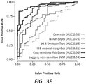
- the decision stump classifier For classifying samples based on the ⁇ -values from a subset of selected CpGs, the decision stump classifier, as described above, was used. More complex models and learning algorithms are also explored: a support vector machine (see Platt JC. Sequential Minimal Optimization: A Fast Algorithm for Training Support Vector Machines. Advances in kernel methods - support vector learning. 1998 , which is incorporated by reference herein in its entirety), a nearest neighbor classifier, a decision tree, and naive Bayes; WEKA (see Hall M et al. The WEKA Data Mining Software: An Update. SIGKDD Explorations. 2009; 11(1 ), which is incorporated by reference herein in its entirety) was used for its implementation of these, run with default parameters unless specified. Further, a number of these were augmented with adaptive boosting (AdaBoost), with bootstrap aggregating (bagging), and by weighting training data to effect a cost-sensitive training scheme (always favoring high specificity).
- FIG. 1A A hierarchical clustering of all 163 neat samples was performed, based on the methylation level of all 485,000+ CpGs interrogated by the array for each sample.
- This clustering revealed an out-group composed exclusively of IVF-patient samples (labeled in FIG. 1A as the patient-only cluster), which contained within it a subgroup almost exclusively composed of samples which lead to poor embryo quality (labeled in FIG. 1A as the poor embryo-quality cluster).
- the majority of IVF-patient samples and poor embryo-quality samples were distributed evenly amongst fertile donor samples and good embryo-quality samples outside of this group.
- the false positive cost of the training algorithm was adjusted (as described above) from 1 (i.e., a false positive was considered as costly as a false negative when training) to 10 (i.e., a false positive was considered 10 times more costly than a false negative when training). It was observed that with high false-positive costs, the classifier could achieve close to perfect specificity while identifying between a quarter and a third of the IVF-patient or poor embryo-quality samples (see FIGS. 1B and 1C ).
- differentially methylated positions were called using a Wilcoxon rank-sum test. As a control, this was repeated with randomly shuffled sample labels.
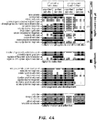
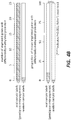
- the number of differentially methylated CpGs identified between IVF-patient samples and donor samples (here neat samples were concentrated on, as these generally showed the greater signal), both before and after correction for multiple hypothesis testing is shown in FIG. 3A .
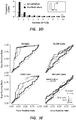
- the histogram of p-values from all CpGs is displayed in FIG. 3B , and shows a sharp spike in significant p-values for actual labels, with a relatively flat distribution for the shuffled control.
- the significant CpGs identified when using real labels showed no discernible bias towards particular genomic regions when compared to those from the randomly shuffled control (see FIG. 3C ).
- the samples were broken into 10 groups of approximately equal size. For each group, the selection of DMPs was repeated with those samples held out. It was observed that the number of groups for which a CpG was selected in the top 100 most differentially methylated CpGs was generally low when using randomly shuffled labels. In contrast, when using real labels, this followed a U-shaped distribution, with a substantially larger proportion of CpGs always, or generally always, appearing in the top 100 (Table 6 gives the genes which are intersected by these consistently differentially methylated CpGs).
- genes were ranked by the number of CpGs that were identified as differentially methylated within their promoter regions (+/- 5 kb around TSS) and GO analysis was performed on the top 1,000 genes.
- several gene families associated with embryogenesis and development are differentially methylated when comparing the good and poor embryogenesis groups.
- Table 7 Embryo quality (good vs. poor) Purified Embryo quality (good vs.
- Neat Source IVF patient vs. donor
- Purified Source IVF patient vs. donor
- Neat axon guidance NA 0.003784515 NA NA axonogenesis 0.008367064 1.10E-05 NA NA biological adhesion 1.36E-10 8.28E-07 6.41 E-12 2.02E-11 cell adhesion 1.74E-10 1.20E-06 9.03E-12 3.86E-11 cell morphogenesis 0.008091845 1.14E-06 0.015385704 0.013289352 cell morphogenesis involved in differentiation 0.002429427 1.37E-06 0.015840977 0.013687946 cell morphogenesis involved in neuron differentiation 0.008267382 1.05E-05 NA 0.034601396 cell motion NA 0.024863785 NA NA cell part morphogenesis 0.037927527 6.36E-06 NA NA cell projection morphogenesis 0.02677415 3.52E-06 NA NA cell projection organization NA 1.02E-06 NA NA cell-cell adhesion
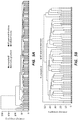
- Hierarchical clustering of samples showed that, while there were differences between purified and unpurified samples, purified and unpurified methylomes derived from the same sample always, or generally always, clustered with each other; between sample differences were greater than between purification method differences (see FIGS. 5A and 5B ).
- a fertile reference methylome may be used to identify infertile samples.
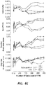
- all CpGs can be ranked by the number of times they are differentially methylated in patients, and the top ten (10) CpGs can be selected as predictive of patient status. It need not be ten, but ten is the number used in the plot shown in FIG. 7 (described below).
- each sample can be given a score between 0 and 10, representing the number of CpGs from the above list which it has aberrantly methylated.
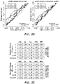
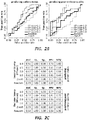
- all of the samples can then be ranked by their score, and an ROC curve can be plotted (see FIG. 7 ; a "positive” here is considered to be a diagnosis of infertility).
Landscapes
- Chemical & Material Sciences (AREA)
- Life Sciences & Earth Sciences (AREA)
- Health & Medical Sciences (AREA)
- Organic Chemistry (AREA)
- Engineering & Computer Science (AREA)
- Proteomics, Peptides & Aminoacids (AREA)
- Zoology (AREA)
- Wood Science & Technology (AREA)
- Analytical Chemistry (AREA)
- Genetics & Genomics (AREA)
- Molecular Biology (AREA)
- General Health & Medical Sciences (AREA)
- Physics & Mathematics (AREA)
- Bioinformatics & Cheminformatics (AREA)
- Biophysics (AREA)
- Biotechnology (AREA)
- Immunology (AREA)
- Biochemistry (AREA)
- Microbiology (AREA)
- General Engineering & Computer Science (AREA)
- Surgery (AREA)
- Medical Informatics (AREA)
- Pathology (AREA)
- Heart & Thoracic Surgery (AREA)
- Animal Behavior & Ethology (AREA)
- Veterinary Medicine (AREA)
- Gynecology & Obstetrics (AREA)
- Pregnancy & Childbirth (AREA)
- Reproductive Health (AREA)
- Nuclear Medicine, Radiotherapy & Molecular Imaging (AREA)
- Public Health (AREA)
- Biomedical Technology (AREA)
- Bioinformatics & Computational Biology (AREA)
- Spectroscopy & Molecular Physics (AREA)
- Evolutionary Biology (AREA)
- Theoretical Computer Science (AREA)
- Measuring Or Testing Involving Enzymes Or Micro-Organisms (AREA)
- Investigating Or Analysing Biological Materials (AREA)
Applications Claiming Priority (3)
| Application Number | Priority Date | Filing Date | Title |
|---|---|---|---|
| US201562201907P | 2015-08-06 | 2015-08-06 | |
| PCT/US2016/046060 WO2017024311A1 (fr) | 2015-08-06 | 2016-08-08 | Procédés d'identification de l'état de fertilité masculine et de la qualité des échantillons |
| EP16834003.2A EP3332035A4 (fr) | 2015-08-06 | 2016-08-08 | Procédés d'identification de l'état de fertilité masculine et de la qualité des échantillons |
Related Parent Applications (1)
| Application Number | Title | Priority Date | Filing Date |
|---|---|---|---|
| EP16834003.2A Division EP3332035A4 (fr) | 2015-08-06 | 2016-08-08 | Procédés d'identification de l'état de fertilité masculine et de la qualité des échantillons |
Publications (1)
| Publication Number | Publication Date |
|---|---|
| EP3859015A1 true EP3859015A1 (fr) | 2021-08-04 |
Family
ID=57943758
Family Applications (2)
| Application Number | Title | Priority Date | Filing Date |
|---|---|---|---|
| EP20213740.2A Ceased EP3859015A1 (fr) | 2015-08-06 | 2016-08-08 | Procédés d'identification de l'état de fertilité masculine et de la qualité d'un embryon |
| EP16834003.2A Withdrawn EP3332035A4 (fr) | 2015-08-06 | 2016-08-08 | Procédés d'identification de l'état de fertilité masculine et de la qualité des échantillons |
Family Applications After (1)
| Application Number | Title | Priority Date | Filing Date |
|---|---|---|---|
| EP16834003.2A Withdrawn EP3332035A4 (fr) | 2015-08-06 | 2016-08-08 | Procédés d'identification de l'état de fertilité masculine et de la qualité des échantillons |
Country Status (5)
| Country | Link |
|---|---|
| US (1) | US11401552B2 (fr) |
| EP (2) | EP3859015A1 (fr) |
| CN (1) | CN108699602B (fr) |
| AU (1) | AU2016301189B2 (fr) |
| WO (1) | WO2017024311A1 (fr) |
Families Citing this family (33)
| Publication number | Priority date | Publication date | Assignee | Title |
|---|---|---|---|---|
| CA2902916C (fr) | 2013-03-14 | 2018-08-28 | Mayo Foundation For Medical Education And Research | Detection de neoplasme |
| US10435755B2 (en) | 2015-03-27 | 2019-10-08 | Exact Sciences Development Company, Llc | Detecting esophageal disorders |
| CN114574585A (zh) | 2015-08-31 | 2022-06-03 | 梅约医药教育及研究基金会 | 检测胃肿瘤 |
| US10913986B2 (en) * | 2016-02-01 | 2021-02-09 | The Board Of Regents Of The University Of Nebraska | Method of identifying important methylome features and use thereof |
| US10370726B2 (en) * | 2016-04-14 | 2019-08-06 | Mayo Foundation For Medical Education And Research | Detecting colorectal neoplasia |
| CN109762826A (zh) * | 2017-12-21 | 2019-05-17 | 首都医科大学附属北京妇产医院 | Dnah6基因突变体及其应用 |
| CN108982867B (zh) * | 2018-05-31 | 2019-10-11 | 西北妇女儿童医院 | 一种利用蛋白芯片对囊胚质量判断的方法 |
| US10586164B1 (en) | 2018-10-15 | 2020-03-10 | AIble Inc. | Interface for visualizing and improving model performance |
| US10936768B2 (en) * | 2018-10-15 | 2021-03-02 | Aible, Inc. | Interface for visualizing and improving model performance |
| US11409549B2 (en) | 2018-10-15 | 2022-08-09 | AIble Inc. | Interface for generating models with customizable interface configurations |
| CN110042123B (zh) * | 2019-01-07 | 2023-01-17 | 西北农林科技大学 | 一种通过诱导表达zfp57提高牛体细胞克隆效率的方法 |
| KR102915151B1 (ko) * | 2019-04-15 | 2026-01-19 | 세르비조 갈레고 데 사우데 | 대장암 또는 이의 전암 단계의 진단 또는 예후예측을 위한 시험관내 방법 |
| KR102327509B1 (ko) * | 2019-05-16 | 2021-11-17 | 한국식품연구원 | 남성 불임 또는 생식 능력 저하 진단용 바이오 마커 |
| CN110205327B (zh) * | 2019-06-11 | 2020-09-01 | 华中农业大学 | 一种水稻温敏核不育基因tms3突变体及其分子标记与应用 |
| JP2022544368A (ja) * | 2019-08-15 | 2022-10-18 | ワシントン ステイト ユニバーシティー | 不妊症診断のための方法およびキット |
| US10704089B1 (en) * | 2019-12-12 | 2020-07-07 | The Florida International University Board Of Trustees | DNA methylation assays for body fluid identification |
| JP2023508853A (ja) * | 2019-12-16 | 2023-03-06 | エピゲノミクス アーゲー | 結腸直腸癌の検出方法 |
| JP7744928B2 (ja) * | 2020-04-29 | 2025-09-26 | イージー バイオメッド カンパニー リミテッド | 大腸癌の早期検出、治療応答性および予後の予測のための方法 |
| CN113517029A (zh) * | 2020-08-07 | 2021-10-19 | 广东省计划生育科学技术研究所(广东省计划生育专科医院) | 一种调控dpep3提高精液质量的方法 |
| TWI791347B (zh) * | 2020-12-11 | 2023-02-01 | 酷氏基因生物科技股份有限公司 | 一種用於評估胚胎植入成功率的預測方法 |
| AU2021442220B2 (en) * | 2021-04-23 | 2025-10-16 | Bgi Genomics Co., Ltd. | Composition, kit, and application for detection of colorectal cancer |
| CN113151453A (zh) * | 2021-05-31 | 2021-07-23 | 苏州市立医院 | 一种卵巢早衰诊断生物标志物及其应用 |
| WO2022256617A1 (fr) | 2021-06-03 | 2022-12-08 | Inherent Biosciences, Inc. | Analyse de méthylation d'adn pour identifier un type de cellule |
| CN115490758A (zh) * | 2021-06-17 | 2022-12-20 | 温州医科大学 | 一种用于治疗或辅助治疗结直肠癌的抗肿瘤多肽ccr313及其应用 |
| CN113584152A (zh) * | 2021-07-20 | 2021-11-02 | 上海市儿童医院 | Odnah基因突变在重度尿道下裂检测中的应用 |
| CA3233857A1 (fr) * | 2021-10-06 | 2023-04-13 | Ryan Miller | Procedes et systemes pour predire la qualite du sperme |
| EP4265738A4 (fr) * | 2022-03-03 | 2025-05-07 | Beijing OriginPoly Bio-Tec Co., Ltd. | Combinaison de marqueurs de méthylation de l'adn, utilisation, sonde d'amorce pour la détection précoce du cancer ovarien et kit |
| CN114875117A (zh) * | 2022-05-27 | 2022-08-09 | 中信湘雅生殖与遗传专科医院有限公司 | 检测女性不孕不育的基因文库的构建方法和试剂盒 |
| CN115807037B (zh) * | 2022-09-26 | 2024-08-02 | 湖南师范大学 | 一种遗传可控的四倍体鱼的选育方法及三倍体鱼的制备方法 |
| CN115807073B (zh) * | 2022-10-24 | 2024-03-12 | 北京大学第三医院(北京大学第三临床医学院) | 实现高着床潜能囊胚筛选的标志物及其应用 |
| CN118755837A (zh) * | 2024-08-12 | 2024-10-11 | 中国医学科学院肿瘤医院 | 一种检测肺癌脑转移瘤的甲基化生物标志物及其应用 |
| CN120400035B (zh) * | 2025-05-08 | 2026-02-10 | 华中农业大学 | Gpld1蛋白在制备抑制猪卵母细胞多精受精的试剂中的应用 |
| CN120138177B (zh) * | 2025-05-15 | 2025-08-29 | 山东省农业科学院畜牧兽医研究所 | 一种基于hspb9基因的公牛精子活力相关snp分子标记、检测产品及应用 |
Family Cites Families (14)
| Publication number | Priority date | Publication date | Assignee | Title |
|---|---|---|---|---|
| US6998235B2 (en) | 2001-06-15 | 2006-02-14 | Centre For Addiction And Mental Health | Method of determining susceptibility to bipolar disorders |
| US20050227230A1 (en) | 2001-09-14 | 2005-10-13 | Davies Collison Cave | Detection of dna methylation |
| EP2298932A1 (fr) * | 2005-09-29 | 2011-03-23 | Epigenomics AG | Procédé et l'ADN pour l'analyse d'expression du gène, particulièrement la méthylation de KAAG1, employant à classifier des cellules et tissus |
| GB0524110D0 (en) | 2005-11-28 | 2006-01-04 | Univ Cambridge Tech | Biomarkers and methods for identification of agents useful in the treatment of affective disorders |
| US7702468B2 (en) | 2006-05-03 | 2010-04-20 | Population Diagnostics, Inc. | Evaluating genetic disorders |
| US20100234242A1 (en) | 2007-01-23 | 2010-09-16 | Arturas Petronis | DNA Methylation Changes Associated with Major Psychosis |
| US20090246771A1 (en) * | 2007-11-02 | 2009-10-01 | University Of Southern California | Compositions and methods comprising biomarkers of sperm quality, semen quality and fertility |
| US20090307180A1 (en) | 2008-03-19 | 2009-12-10 | Brandon Colby | Genetic analysis |
| CA2729856A1 (fr) | 2008-07-04 | 2010-01-07 | Decode Genetics Ehf. | Variations du nombre de copies predictives d'un risque de schizophrenie |
| US20130338032A1 (en) * | 2009-02-17 | 2013-12-19 | Murdoch Childrens Research Institute | Assay for determining epigenetic profiles of markers of fragile x alleles |
| US9034161B2 (en) | 2012-08-23 | 2015-05-19 | Douglas T. Carrell | Sperm separation devices and associated methods |
| CN103103256B (zh) | 2012-12-20 | 2015-10-14 | 宁波大学 | 可用于检测与精神分裂症相关的drd4基因启动子区甲基化程度的试剂盒及其应用 |
| GB2533729A (en) | 2013-08-21 | 2016-06-29 | T Carrell Douglas | Systems and methods for determining impact of age related changes in sperm epigenome on offspring phenotype |
| CA3074808A1 (fr) | 2017-09-05 | 2019-03-14 | Igenomix S.L. | Procedes et dispositifs de detection de biomarqueurs associes a la preeclampsie |
-
2016
- 2016-08-08 EP EP20213740.2A patent/EP3859015A1/fr not_active Ceased
- 2016-08-08 WO PCT/US2016/046060 patent/WO2017024311A1/fr not_active Ceased
- 2016-08-08 US US15/750,715 patent/US11401552B2/en active Active
- 2016-08-08 CN CN201680057796.1A patent/CN108699602B/zh active Active
- 2016-08-08 EP EP16834003.2A patent/EP3332035A4/fr not_active Withdrawn
- 2016-08-08 AU AU2016301189A patent/AU2016301189B2/en active Active
Non-Patent Citations (28)
| Title |
|---|
| ARYEE MJ ET AL., BIOINFORMATICS, vol. 30, no. 10, 2014, pages 1363 - 9 |
| BARRATT CL ET AL., ASIAN JOURNAL OF ANDROLOGY, vol. 13, no. 1, 2011, pages 53 - 8 |
| BENCHAIB M. ET AL: "Influence of global sperm DNA methylation on IVF results", HUMAN REPRODUCTION, vol. 20, no. 3, 1 March 2005 (2005-03-01), GB, pages 768 - 773, XP055817308, ISSN: 0268-1161, DOI: 10.1093/humrep/deh684 * |
| BENJAMINI YHOCHBERG Y, JOURNAL OF THE ROYAL STATISTICAL SOCIETY, vol. 57, no. 1, 1995, pages 289 - 300 |
| BIBIKOVA ET AL., GENOMICS, vol. 98, 2011, pages 288 - 295 |
| BONDE JP ET AL., LANCET, vol. 352, no. 9135, 1998, pages 1172 - 7 |
| BRAZIL C ET AL., JOURNAL OF ANDROLOGY, vol. 25, no. 4, 2004, pages 645 - 56 |
| BURKMAN LJ ET AL., FERTILITY AND STERILITY, vol. 49, no. 4, 1988, pages 688 - 97 |
| COOPER TG ET AL., HUMAN REPRODUCTION UPDATE, vol. 16, no. 3, 2010, pages 231 - 45 |
| DAM AH ET AL., AMERICAN JOURNAL OF HUMAN GENETICS, vol. 81, no. 4, 2007, pages 813 - 20 |
| ELINATI E ET AL., HUMAN MOLECULAR GENETICS, vol. 21, no. 16, 2012, pages 3695 - 702 |
| ESTEVES SC ET AL., UROLOGY, vol. 79, no. 1, 2012, pages 16 - 22 |
| GANDINI L ET AL., INTERNATIONAL JOURNAL OF ANDROLOGY, vol. 23, no. 1, 2000, pages 1 - 3 |
| GUZICK DS ET AL., THE NEW ENGLAND JOURNAL OF MEDICINE, vol. 345, no. 19, 2001, pages 1388 - 93 |
| HAAS GG, JR. ET AL., THE NEW ENGLAND JOURNAL OF MEDICINE, vol. 303, no. 13, 1980, pages 722 - 7 |
| HALL M ET AL.: "The WEKA Data Mining Software: An Update", SIGKDD EXPLORATIONS, vol. 11, no. 1, 2009, XP055489105, DOI: 10.1145/1656274.1656278 |
| KOSCINSKI I ET AL., AMERICAN JOURNAL OF HUMAN GENETICS, vol. 88, no. 3, 2011, pages 344 - 50 |
| LANFRANCO F ET AL., LANCET, vol. 364, no. 9430, 2004, pages 273 - 83 |
| LEE MA ET AL., FERTILITY AND STERILITY, vol. 48, no. 4, 1987, pages 649 - 58 |
| LEUSHUIS E ET AL., FERTILITY AND STERILITY, vol. 94, no. 7, 2010, pages 2631 - 5 |
| MAKSIMOVIC J ET AL., GENOME BIOLOGY, vol. 13, no. 6, 2012, pages R44 |
| NEUWINGER J ET AL., FERTILITY AND STERILITY, vol. 54, no. 2, 1990, pages 308 - 14 |
| SANCHEZ R ET AL., ANDROLOGIA, vol. 23, no. 3, 1991, pages 197 - 203 |
| SCHAGDARSURENGIN UNDRAGA ET AL: "Analysing the sperm epigenome: roles in early embryogenesis and assisted reproduction", NATURE REVIEWS. UROLOGY, vol. 9, no. 11, 1 November 2012 (2012-11-01), US, pages 609 - 619, XP055817312, ISSN: 1759-4812, DOI: 10.1038/nrurol.2012.183 * |
| STOUFFER SA ET AL.: "Adjustment during Army Life", 1949, PRINCETON UNIVERSITY PRESS |
| TIEPOLO L ET AL., HUMAN GENETICS, vol. 34, no. 2, 1976, pages 119 - 24 |
| YANAGIMACHI R ET AL., BIOLOGY OF REPRODUCTION, vol. 15, no. 4, 1976, pages 471 - 6 |
| ZINI A ET AL., HUMAN REPRODUCTION, vol. 23, no. 12, 2008, pages 2663 - 8 |
Also Published As
| Publication number | Publication date |
|---|---|
| AU2016301189A1 (en) | 2018-03-08 |
| US11401552B2 (en) | 2022-08-02 |
| AU2016301189B2 (en) | 2022-07-28 |
| EP3332035A1 (fr) | 2018-06-13 |
| CN108699602B (zh) | 2022-01-11 |
| CN108699602A (zh) | 2018-10-23 |
| CA2994968A1 (fr) | 2017-02-09 |
| US20190112659A1 (en) | 2019-04-18 |
| EP3332035A4 (fr) | 2019-06-19 |
| WO2017024311A1 (fr) | 2017-02-09 |
Similar Documents
| Publication | Publication Date | Title |
|---|---|---|
| US11401552B2 (en) | Methods of identifying male fertility status and embryo quality | |
| AU2020274091B2 (en) | Systems and methods for multi-label cancer classification | |
| US20200399714A1 (en) | Cancer-related biological materials in microvesicles | |
| US20220401460A1 (en) | Modulating resistance to bcl-2 inhibitors | |
| CN110499364A (zh) | 一种用于检测扩展型遗传病全外显子的探针组及其试剂盒和应用 | |
| US20160340740A1 (en) | Methylation haplotyping for non-invasive diagnosis (monod) | |
| CN117730164A (zh) | 细胞的品质管理方法及制造细胞的方法 | |
| US12160984B2 (en) | Radio wave absorber | |
| JP7794719B2 (ja) | 細胞の品質管理方法及び細胞を製造する方法 | |
| WO2023286305A1 (fr) | Procédé de contrôle de qualité de cellules, et procédé de fabrication de cellules | |
| CN117677707A (zh) | 特定细胞的品质管理方法及制造特定细胞的方法 | |
| US20240233867A1 (en) | Quality management method for specific cell and method of producing specific cell | |
| US12054756B2 (en) | Engineered nucleases, compositions, and methods of use thereof | |
| US20250388634A1 (en) | Engineered gene effectors, compositions, and methods of use thereof | |
| US20250305061A1 (en) | Methods and systems for inferring gene expression using cell-free dna fragments | |
| CA2994968C (fr) | Procedes d'identification de l'etat de fertilite masculine et de la qualite des echantillons | |
| WO2026062505A1 (fr) | Procédés et systèmes de détection d'un organe ou d'un tissu atteint par un cancer ou une maladie, un trouble ou une affection | |
| Schwarzer et al. | Supplementary Data from Targeting Aggressive B-cell Lymphomas through Pharmacological Activation of the Mitochondrial Protease OMA1 [Dataset] | |
| HK40109321A (en) | Method for managing quality of specific cells, and method for manufacturing specific cells | |
| HK40109609A (en) | Cell quality management method and cell production method |
Legal Events
| Date | Code | Title | Description |
|---|---|---|---|
| PUAI | Public reference made under article 153(3) epc to a published international application that has entered the european phase |
Free format text: ORIGINAL CODE: 0009012 |
|
| STAA | Information on the status of an ep patent application or granted ep patent |
Free format text: STATUS: THE APPLICATION HAS BEEN PUBLISHED |
|
| AC | Divisional application: reference to earlier application |
Ref document number: 3332035 Country of ref document: EP Kind code of ref document: P |
|
| AK | Designated contracting states |
Kind code of ref document: A1 Designated state(s): AL AT BE BG CH CY CZ DE DK EE ES FI FR GB GR HR HU IE IS IT LI LT LU LV MC MK MT NL NO PL PT RO RS SE SI SK SM TR |
|
| STAA | Information on the status of an ep patent application or granted ep patent |
Free format text: STATUS: REQUEST FOR EXAMINATION WAS MADE |
|
| 17P | Request for examination filed |
Effective date: 20220203 |
|
| RBV | Designated contracting states (corrected) |
Designated state(s): AL AT BE BG CH CY CZ DE DK EE ES FI FR GB GR HR HU IE IS IT LI LT LU LV MC MK MT NL NO PL PT RO RS SE SI SK SM TR |
|
| P01 | Opt-out of the competence of the unified patent court (upc) registered |
Effective date: 20230527 |
|
| STAA | Information on the status of an ep patent application or granted ep patent |
Free format text: STATUS: EXAMINATION IS IN PROGRESS |
|
| 17Q | First examination report despatched |
Effective date: 20230803 |
|
| STAA | Information on the status of an ep patent application or granted ep patent |
Free format text: STATUS: THE APPLICATION HAS BEEN REFUSED |
|
| 18R | Application refused |
Effective date: 20250820 |