EP3876190B1 - Verfahren und system zur endoskopischen bildverarbeitung und computervorrichtung - Google Patents
Verfahren und system zur endoskopischen bildverarbeitung und computervorrichtung Download PDFInfo
- Publication number
- EP3876190B1 EP3876190B1 EP19879131.1A EP19879131A EP3876190B1 EP 3876190 B1 EP3876190 B1 EP 3876190B1 EP 19879131 A EP19879131 A EP 19879131A EP 3876190 B1 EP3876190 B1 EP 3876190B1
- Authority
- EP
- European Patent Office
- Prior art keywords
- endoscopic image
- layer
- convolutional network
- deep convolutional
- endoscopic
- Prior art date
- Legal status (The legal status is an assumption and is not a legal conclusion. Google has not performed a legal analysis and makes no representation as to the accuracy of the status listed.)
- Active
Links
Images


Classifications
-
- A—HUMAN NECESSITIES
- A61—MEDICAL OR VETERINARY SCIENCE; HYGIENE
- A61B—DIAGNOSIS; SURGERY; IDENTIFICATION
- A61B1/00—Instruments for performing medical examinations of the interior of cavities or tubes of the body by visual or photographical inspection, e.g. endoscopes; Illuminating arrangements therefor
- A61B1/00002—Operational features of endoscopes
- A61B1/00043—Operational features of endoscopes provided with output arrangements
- A61B1/00045—Display arrangement
- A61B1/0005—Display arrangement combining images e.g. side-by-side, superimposed or tiled
-
- G—PHYSICS
- G06—COMPUTING OR CALCULATING; COUNTING
- G06T—IMAGE DATA PROCESSING OR GENERATION, IN GENERAL
- G06T7/00—Image analysis
- G06T7/0002—Inspection of images, e.g. flaw detection
- G06T7/0012—Biomedical image inspection
-
- A—HUMAN NECESSITIES
- A61—MEDICAL OR VETERINARY SCIENCE; HYGIENE
- A61B—DIAGNOSIS; SURGERY; IDENTIFICATION
- A61B1/00—Instruments for performing medical examinations of the interior of cavities or tubes of the body by visual or photographical inspection, e.g. endoscopes; Illuminating arrangements therefor
- A61B1/00002—Operational features of endoscopes
- A61B1/00004—Operational features of endoscopes characterised by electronic signal processing
- A61B1/00009—Operational features of endoscopes characterised by electronic signal processing of image signals during a use of endoscope
- A61B1/000094—Operational features of endoscopes characterised by electronic signal processing of image signals during a use of endoscope extracting biological structures
-
- A—HUMAN NECESSITIES
- A61—MEDICAL OR VETERINARY SCIENCE; HYGIENE
- A61B—DIAGNOSIS; SURGERY; IDENTIFICATION
- A61B1/00—Instruments for performing medical examinations of the interior of cavities or tubes of the body by visual or photographical inspection, e.g. endoscopes; Illuminating arrangements therefor
- A61B1/00002—Operational features of endoscopes
- A61B1/00004—Operational features of endoscopes characterised by electronic signal processing
- A61B1/00009—Operational features of endoscopes characterised by electronic signal processing of image signals during a use of endoscope
- A61B1/000096—Operational features of endoscopes characterised by electronic signal processing of image signals during a use of endoscope using artificial intelligence
-
- G—PHYSICS
- G06—COMPUTING OR CALCULATING; COUNTING
- G06N—COMPUTING ARRANGEMENTS BASED ON SPECIFIC COMPUTATIONAL MODELS
- G06N3/00—Computing arrangements based on biological models
- G06N3/02—Neural networks
- G06N3/04—Architecture, e.g. interconnection topology
- G06N3/045—Combinations of networks
-
- G—PHYSICS
- G06—COMPUTING OR CALCULATING; COUNTING
- G06N—COMPUTING ARRANGEMENTS BASED ON SPECIFIC COMPUTATIONAL MODELS
- G06N3/00—Computing arrangements based on biological models
- G06N3/02—Neural networks
- G06N3/04—Architecture, e.g. interconnection topology
- G06N3/0464—Convolutional networks [CNN, ConvNet]
-
- G—PHYSICS
- G06—COMPUTING OR CALCULATING; COUNTING
- G06N—COMPUTING ARRANGEMENTS BASED ON SPECIFIC COMPUTATIONAL MODELS
- G06N3/00—Computing arrangements based on biological models
- G06N3/02—Neural networks
- G06N3/08—Learning methods
- G06N3/084—Backpropagation, e.g. using gradient descent
-
- G—PHYSICS
- G06—COMPUTING OR CALCULATING; COUNTING
- G06N—COMPUTING ARRANGEMENTS BASED ON SPECIFIC COMPUTATIONAL MODELS
- G06N3/00—Computing arrangements based on biological models
- G06N3/02—Neural networks
- G06N3/08—Learning methods
- G06N3/0895—Weakly supervised learning, e.g. semi-supervised or self-supervised learning
-
- G—PHYSICS
- G06—COMPUTING OR CALCULATING; COUNTING
- G06N—COMPUTING ARRANGEMENTS BASED ON SPECIFIC COMPUTATIONAL MODELS
- G06N3/00—Computing arrangements based on biological models
- G06N3/02—Neural networks
- G06N3/08—Learning methods
- G06N3/09—Supervised learning
-
- G—PHYSICS
- G06—COMPUTING OR CALCULATING; COUNTING
- G06T—IMAGE DATA PROCESSING OR GENERATION, IN GENERAL
- G06T2207/00—Indexing scheme for image analysis or image enhancement
- G06T2207/10—Image acquisition modality
- G06T2207/10068—Endoscopic image
-
- G—PHYSICS
- G06—COMPUTING OR CALCULATING; COUNTING
- G06T—IMAGE DATA PROCESSING OR GENERATION, IN GENERAL
- G06T2207/00—Indexing scheme for image analysis or image enhancement
- G06T2207/20—Special algorithmic details
- G06T2207/20081—Training; Learning
-
- G—PHYSICS
- G06—COMPUTING OR CALCULATING; COUNTING
- G06T—IMAGE DATA PROCESSING OR GENERATION, IN GENERAL
- G06T2207/00—Indexing scheme for image analysis or image enhancement
- G06T2207/30—Subject of image; Context of image processing
- G06T2207/30004—Biomedical image processing
Definitions
- the present disclosure relates to the field of image processing technologies, and in particular, to an endoscopic image processing method and system, and a computer device.
- stomach cancer and esophagus cancer are among the top five malignant tumor types that occur frequently in China and around the world. Both stomach cancer and esophagus cancer are malignant tumors occurring in an upper gastrointestinal tract.
- a doctor conducts an electronic examination by using an endoscope.
- the endoscope enters the upper gastrointestinal tract from the mouth of a patient. Strong light is emitted from a light source and an optical fiber allows the light to make a turn, so that the doctor can observe the health status of various organs in the upper gastrointestinal tract.
- a computer vision technology is usually used for extracting features such as a color, a texture, a gradient, and local binary patterns (LBP), and then organs are classified and identified by using a support vector machine (SVM) classification method.
- SVM support vector machine
- Such a technology requires researchers to have a deep understanding of medical images, in order to make a feasible feature extraction solution according to inherent characteristics of the images, which results in higher technical doorsill.
- the extracted features tend to be general features, rather than specific organ features extracted purposefully for a to-be-diagnosed body part. As a result, the coverage is incomplete and the solution is not robust enough.
- US 2018/247107 A1 discloses a method and system for classification of endoscopic images, where an initial trained deep network classifier is used to classify endoscopic images and determine confidence scores for the endoscopic images, the confidence score for each endoscopic image classified by the initial trained deep network classifier is compared to a learned confidence threshold, for endoscopic images with confidence scores higher than the learned threshold value, the classification result from the initial trained deep network classifier is output, and endoscopic images with confidence scores lower than the learned confidence threshold are classified using a first specialized network classifier built on a feature space of the initial trained deep network classifier;
- WO 2017/175282 A1 discloses a learning method performed by an image recognition device, which includes performing first learning by the convolutional neural network by using a first image group captured at a first frame rate, the first image group being input from outside, and performing, after performing the first learning, second learning by using a second image group that is captured at a second frame rate that is lower than the first frame rate, the second image group being input from outside, and the number of images in the second
- the present invention provides an endoscopic image processing method according to claim 1, an endoscopic image processing system according to claim 7, a computer device according to claim 10 and a storage medium according to claim 11.
- Preferred embodiments of the invention are defined in the dependent claims.
- Embodiments of the present disclosure provide an endoscopic image processing method and system, and a computer device, to make a prediction process more intelligent and more robust, thereby improving resource utilization of a processing apparatus.
- aspects of the present disclosure provide an endoscopic image processing method.
- the method includes:
- the present disclosure further provides an endoscopic image processing system that includes a human body detection device and an endoscopic image processing apparatus.
- the human body detection device is configured to detect a human body part, and transmit at least one detected first endoscopic image to the endoscopic image processing apparatus.
- the endoscopic image processing apparatus is configured to: acquire the at least one first endoscopic image from the human body detection device; create a deep convolutional network for endoscopic image prediction and determine a training parameter of the deep convolutional network according to the at least one first endoscopic image and at least one second endoscopic image transformed from the at least one first endoscopic image; and acquire a current endoscopic image of a to-be-examined user, perform prediction with the current endoscopic image by using the deep convolutional network based on the training parameter, to determine an organ category corresponding to the current endoscopic image.
- the present disclosure further provides a non-transitory computer readable storage medium that stores computer readable instructions that, when executed at by least one processor, cause the at least one processor to load and perform the operations including:
- the present disclosure further provides a computer device, including at least one processor and at least one memory that stores at least one piece of program code that, when executed by the at least one processor, can implement operations including:
- a feature extraction process is obtained completely by a model of a deep convolutional network through self-learning, while a researcher does not need to deeply understand a medical image, thus reducing the dependence on a professional level of a doctor, so that the whole prediction process is more intelligent.
- the amount of annotated data used in a training process can be reduced, thus improving a training convergence speed, providing clean available data for next disease diagnosis, providing available integration modules for disease diagnosis of different organs, and improving resource utilization of a processing apparatus.
- FIG. 1 is a schematic structural diagram of an endoscopic image processing system related to an embodiment of the present disclosure.
- an endoscopic image processing system 100 includes a to-be-examined user 101, a human body detection device 102 including an endoscope 1021, an endoscopic image processing apparatus 103, and a doctor 104.
- the endoscopic image processing apparatus 103 may include a real-time prediction sub-apparatus 1031, an offline training sub-apparatus 1032, and an endoscopic image database 1033.
- the human body detection device 102 detects a human body part of the to-be-examined user 101 by using the endoscope 1021.
- the human body detection device 102 transmits a collected endoscopic image to the endoscopic image processing apparatus 103.
- the collected endoscopic image may be transmitted to the real-time prediction sub-apparatus 1031 as a current endoscopic image for prediction, or may be transmitted to the endoscopic image database 1033 for storage. Images stored in the endoscopic image database 1033 are used for performing offline training.
- the real-time prediction sub-apparatus 1031 first needs to obtain a training parameter from the offline training sub-apparatus 1032, and then performs prediction with the current endoscopic image based on the training parameter and a created deep convolutional network, to determine an organ category corresponding to the current endoscopic image.
- the organ category may be a duodenum in an upper gastrointestinal tract.
- the offline training sub-apparatus 1032 uses the deep convolutional network the same as that of the real-time prediction sub-apparatus 1031 to acquire an image collected through an endoscope and an annotated label image from the endoscopic image database 1033, and performs off-line training according to the image collected by the endoscope and annotated label images, to output the training parameter of the deep convolutional network.
- the foregoing human body detection device 102 refers to a medical terminal device equipped with the endoscope 1021 and having an image collection function.
- the endoscope 1021 may include an image sensor, an optical lens, a light source for illumination, a mechanical device and so on.
- the endoscopic image processing apparatus 103 may be a server or a cloud server with image storage and processing functions. All the terminal devices are installed with an operating system, including but not limited to: an Android operating system, a Symbian operating system, a Windows mobile operating system, and an iPhone OS from Apple.
- the human body detection device 102 and the endoscopic image processing apparatus 103 may communicate with each other through a wired or a wireless network.
- FIG. 2 is a schematic flowchart of an endoscopic image processing method according to an embodiment of the present disclosure. The method is applied to a computer device.
- the computer device being a server is used as an example for description.
- This exemplary embodiment can include the following steps 201 to 204.
- step 201 the server acquires at least one first endoscopic image specific to a human body part.
- the at least one first endoscopic image corresponds to the human body part.
- step 202 the server creates a deep convolutional network for endoscopic image prediction.
- step 203 the server determines a training parameter of the deep convolutional network according to the at least one first endoscopic image and at least one second endoscopic image transformed from the at least one first endoscopic image.
- step 204 the server acquires a current endoscopic image of a to-be-examined user and performs prediction with the current endoscopic image by using the deep convolutional network and based on the training parameter, to determine an organ category corresponding to the current endoscopic image.
- the foregoing training parameter is determined according to the at least one first endoscopic image and the at least one second endoscopic image transformed from the at least one first endoscopic image.
- the human body part may be detected by using a detection device including an endoscope, to obtain the at least one first endoscopic image.
- the human body part includes one or more organs.
- the human body part is an upper gastrointestinal tract.
- the upper gastrointestinal tract includes five organs: a pharynx, an esophagus, a stomach, a cardia and a duodenum.
- the detection device may shoot a picture or a video, and the acquired first endoscopic image may be a white light RGB image.
- the deep convolutional network for classifying the endoscopic image is a convolutional neural network based on deep learning.
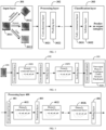
- the deep convolutional network includes an input layer, a processing layer, and a classification layer.
- FIG. 3 is a schematic structural diagram of a deep convolutional network according to an embodiment of the present disclosure.
- an input layer 301 determines at least one endoscopic image as an input.
- a processing layer 302 performs feature extraction on the inputted endoscopic images.
- a classification layer 303 outputs organ categories obtained by prediction with the inputted endoscopic images.
- the foregoing at least one endoscopic image may include first endoscopic images shot by a detection device. Certainly, the at least one endoscopic image may further include second endoscopic images obtained by transforming the first endoscopic images, to increase a sample size.
- a convolutional layer 3021 performs feature extraction on the endoscopic image by using a convolution matrix as a filter, to obtain a feature image.
- a pooling layer 3022 is configured to simplify information outputted by the convolutional layer, to reduce data dimensions and computational overheads, and control over-fitting.
- a fully connected layer 3031 is configured to determine an organ category closest to the acquired feature image.
- a softmax layer 3032 outputs a 1 ⁇ M-dimensional classification vector, the softmax layer being used for index normalization.
- M is the quantity of candidate organ categories. For example, there are six candidate organ categories: a non-organ map, a pharynx, an esophagus, a stomach, a cardia, and a duodenum.
- a value of an element in the classification vector is [0, 1], and an i th element represents a probability of the endoscopic image as an input belonging to an i th candidate organ category.
- the server may add at least one densely connected layer into the processing layer, the densely connected layer including a plurality of connected sub-layers. For each connected sub-layer, a feature outputted by other connected sub-layers before the connected sub-layer is used as an input of the connected sub-layer.
- FIG. 4 is a schematic structural diagram of a deep convolutional network according to another embodiment of the present disclosure.
- a processing layer 312 includes Y densely connected layer 3121 to 312Y.
- Each densely connected layer includes a plurality of connected sub-layers, as shown by solid circles in boxes 3121 to 312Y in FIG. 4 .
- Probabilities of six categories as shown in a box 3131 are outputted in an output layer 313.
- FIG. 5 is a schematic structural diagram of a processing layer according to still another embodiment of the present disclosure.
- a structure of a processing layer 400 there are K densely connected layers 4021 to 402K between a convolutional layer 401 and a pooling layer 404.
- a feature outputted by each connected sub-layer is inputted into subsequent other connected sub-layers.
- H j may be an operation such as batch normalization (BN), ReLU activation and 3 ⁇ 3 convolution.
- BN batch normalization
- ReLU activation ReLU activation
- 3 ⁇ 3 convolution 3 ⁇ 3 convolution
- a transition layer when at least one densely connected layer is added into the processing layer, in order to further compress parameters, a transition layer may further be added between two adjacent densely connected layers. As shown in FIG. 5 , a transition layer 403 is added between a densely connected layer 4021 and a densely connected layer 4022. If there are K densely connected layers, the quantity of the transition layers is K-1.
- a feature compression ratio of the transition layer may be set according to a preset prediction precision. Since a compression ratio affects the quantity of parameters and the prediction precision, a value of the feature compression ratio is set according to the preset prediction precision specific to the endoscopic image. For example, the value is set to 0.5.
- the server determines specific parameters of the processing layer and the classification layer in the deep convolutional network according to the quantity of endoscopic images for prediction, the prediction precision and the adjustment of a hyper parameter in a training process.
- Table 1 is an example of a structure and parameters of a deep convolutional network including 4 densely connected layers and 3 transition layers. The growth rate of each densely connected layer may be set to 24; a Error! Digit expected. convolution operation may further be performed before the Error! Digit expected, convolution operation, thereby reducing the quantity of the input feature images and fusing features of the channels. The Error! Digit expected, convolution operation in the transition layer may reduce the quantity of the input channels by a half.
- the server may determine a training parameter of the deep convolutional network according to at least one first endoscopic image and at least one second endoscopic image transformed from the at least one first endoscopic image.
- the server transforms the at least one first endoscopic image, to obtain at least one transformed second endoscopic image, and then inputs the at least one first endoscopic image and the at least one second endoscopic image into the deep convolutional network to perform training, to obtain the training parameter of the deep convolutional network.
- the transformation performed by the server on the at least one first endoscopic image includes at least one of cropping, rotation, brightness jitter, color jitter, or contrast jitter.
- Such a transformation operation achieves a data enhancement effect.
- the quantity of transformations may be determined according to the quantity of candidate organ categories and/or the preset prediction precision.
- 3011 represents the first endoscopic image acquired from the detection device.
- Two types of transformations are performed on 3011, including: a rotation transformation, to obtain a transformed second endoscopic image 3012; and a color transformation, to obtain a transformed second endoscopic image 3013.
- 3011, 3012 and 3013 are used as input images of the processing layer 302 to perform the feature extraction.
- the training parameter obtained in step 203 and the deep convolutional network created in step 202 are used for a subsequent real-time prediction.
- the server acquires a current endoscopic image of a to-be-examined user and performs prediction with the current endoscopic image by using the deep convolutional network and based on the training parameter, to determine an organ category corresponding to the current endoscopic image.
- prediction is performed with the current endoscopic image inputted in the input layer 301.
- the current endoscopic image is classified as an "esophagus" category; or, the image is not a valid medical image and does not correspond to any organ, belonging to a "non-organ-image” category, in which case a doctor does not need to refer to the image when diagnosing a disease.
- a training parameter of a deep convolutional network is determined according to at least one first endoscopic image and at least one second endoscopic image transformed from the at least one first endoscopic image.
- the transformed second endoscopic images may be used as auxiliary data for classification training of the first endoscopic images.
- the deep convolutional network when the deep convolutional network is created, at least one densely connected layer is added, which can maximize an information flow between all layers in the network, and mitigate a problem of gradient dissipation in the training process to a certain extent.
- a large quantity of features is reused, a large quantity of features can be generated by using a small quantity of convolution kernels.
- a size of the final model is also relatively small, which reduces the quantity of parameters.
- FIG. 6 is a schematic flowchart of an endoscopic image processing method according to another embodiment of the present disclosure. As shown in FIG. 6 , the method can include the following steps 501 to 507.
- a server acquires at least one first endoscopic image specific to a human body part.
- step 502 the server transforms the at least one first endoscopic image, to obtain at least one second endoscopic image after transformation.
- step 503 the server creates a deep convolutional network for endoscopic image prediction.
- step 504 the server determines at least one candidate organ category according to a structure of the human body part and a preset diagnosis target.
- the human body part may include a plurality of organs.
- a plurality of candidate organ categories in advance.
- a plurality of regions may be defined according to the preset diagnosis target, and then the plurality of candidate organ categories may be determined.
- stomach cancer and esophagus cancer are most common among malignant tumor types that occur frequently at present.
- the candidate organ categories may be three categories: stomach, esophagus, and others.
- step 505 the server acquires a label image corresponding to each candidate organ category.
- the label images may be acquired from a medical image database and annotated manually; alternatively, images with typical features of candidate organs may be filtered out from the collected first endoscopic images.
- FIG. 7 is a schematic diagram of label images according to an embodiment of the present disclosure. As shown in FIG. 7 , a plurality of label images of a duodenum, an esophagus, a stomach and an eye are provided respectively.
- the server performs training by using the at least one first endoscopic image and the at least one second endoscopic image as input samples and using the label images as ideal output samples (namely, target output samples), to obtain a training parameter of the deep convolutional network.
- weight values are gradually adjusted iteratively according to the inputted image samples and the ideal output samples until convergence.
- the server acquires a current endoscopic image of a to-be-examined user and performs prediction with the current endoscopic image by using the deep convolutional network based on the training parameter, to determine an organ category corresponding to the current endoscopic image.
- step 204 is the same as step 204, which is not described herein again.
- candidate organ categories are properly designed, and distorted second endoscopic images obtained by transforming first endoscopic images are included in input samples, so that the quantity of label images can be greatly reduced, thereby resolving the problem of the limited quantity of label images as annotated data during training of a deep convolutional network.
- FIG. 8 is a schematic flowchart of training a deep convolutional network according to an embodiment of the present disclosure, which as shown in FIG. 8 can include the following steps 701 to 713.
- a server acquires at least one first endoscopic image specific to a human body part.
- step 702 the server transforms the at least one first endoscopic image, to obtain at least one transformed second endoscopic image.
- the server creates a deep convolutional network for endoscopic image prediction, the deep convolutional network including an input layer, a processing layer, and a classification layer.
- a parameter may be adjusted by using a back propagation algorithm iteratively until convergence.
- the back propagation algorithm may be divided into four different parts: forward transmission, loss function calculation, reverse transmission, and parameter update.
- forward transmission initial sample data, including the at least one first endoscopic image and the at least one transformed second endoscopic image, is inputted and transmitted in the processing layer.
- the loss function is constructed, to help the deep convolutional network update a training parameter until convergence.
- step 704 the server pre-constructs a loss function used for training the deep convolutional network.
- the loss function is constructed according to a preset convergence strategy.
- the convergence strategy is specifically a consistency constraint strategy, that is, features extracted by a model from transformations of the same endoscopic image are close to each other.
- the convergence strategy is specifically a center aggregation strategy, that is, a distance between first endoscopic images belonging to the same organ category is reduced, that is, an intra-class distance is reduced, and a distance between endoscopic images belonging to different organ categories is increased, that is, an inter-class distance is increased.
- step 705 the server inputs the at least one first endoscopic image and the at least one second endoscopic image, and initializes the deep convolutional network.
- the initialization of the deep convolutional network can include the following two initialization processes.
- the initialized training parameter and the center feature lead to a high value of the loss function.
- the objective of training the deep neural network is to make a predicted value the same as a real value.
- the value of the loss function needs to be reduced to the greatest extent. A smaller loss value indicates that a prediction result is closer to the real value.
- the training parameter and center feature are adjusted iteratively, and the value of the loss function is calculated in each iteration, thereby minimizing a loss of the whole network.
- the following steps 706 and 707 correspond to the foregoing consistency constraint strategy, and the following step 708 corresponds to the foregoing center aggregation strategy.
- step 706 the server acquires at least one processed feature obtained by the processing layer processing the at least one first endoscopic image.
- step 707 the server calculates a value of the loss function in a current iteration according to the at least one processed feature and a feature of the at least one second endoscopic image.
- the foregoing consistency constraint strategy is as follows: in the loss function, a plurality of first distances between the processed features of the first endoscopic images and features of the second endoscopic images are calculated respectively, and consistency between the first endoscopic images and the second endoscopic images is constrained by the plurality of first distances.
- y i log f ( x i ; w) represents a cross-entropy loss of classification.
- ⁇ 2 ⁇ w ⁇ 2 2 represents an L2 regularization parameter.
- the variable h 0 is an eigenvector outputted by the processing layer according to the first endoscopic image, that is, a processed eigenvector.
- h k is an eigenvector of a k th second endoscopic image.
- r and ⁇ are hyper parameters, both of which are values greater than 0.
- i is an integer greater than or equal to 1 and less than or equal to n
- k is an integer greater than or equal to 1 and less than or equal to m.
- step 708 the server calculates a plurality of second distances between features of the first endoscopic images and center features corresponding to the first endoscopic images.
- the second distance is x i - c y i , reflecting a center loss L C .
- step 709 the server calculates the value of the loss function according to the plurality of first distances and the plurality of second distances.
- the value of loss function may be obtained through calculation according to the foregoing formula (2).
- step 710 the server determines whether a training process is finished or not according to the value of the loss function. If yes, step 713 is performed; otherwise, steps 711 and 712 are performed.
- the loss function is minimized, that is, obtaining minL (w). Whether to stop the iteration can be determined by determining whether the value of the loss function reaches an acceptable threshold. After the iteration stops, the whole training process is ended.
- step 711 the server updates the training parameter. Then, step 706 is further performed to perform the next iteration process.
- step 712 the server updates the center feature. Then, step 708 is further performed to perform the next iteration process.
- step 713 the server obtains the training parameter of the deep convolutional network after the training ends.
- a loss function used for training a deep convolutional network is pre-constructed.
- a value of the loss function in each iteration is calculated according to processed features of first endoscopic images and a feature of each second endoscopic image.
- a consistency constraint is introduced, so that a more stable feature can be preferentially found and a convergence speed of a training process can be accelerated, until an optimal solution is obtained.
- a center aggregation strategy is considered in the loss function, to ensure that features learned by a model for each category is more stable and cohesive, thereby further improving a generalization ability of the model in a real environment.
- FIG. 9 is a schematic structural diagram of an endoscopic image processing apparatus according to an embodiment of the present disclosure.
- an apparatus 800 can include: an acquisition module 810 that is configured to acquire a first endoscopic image specific to a human body part through a detection device including an endoscope, and acquire a current endoscopic image of a to-be-examined user.
- the apparatus 800 can further include a creation module 820 that is configured to create a deep convolutional network for endoscopic image prediction, and determine a training parameter of the deep convolutional network according to the at least one first endoscopic image acquired by the acquisition module 810 and at least one second endoscopic image transformed from the first endoscopic image.
- a creation module 820 that is configured to create a deep convolutional network for endoscopic image prediction, and determine a training parameter of the deep convolutional network according to the at least one first endoscopic image acquired by the acquisition module 810 and at least one second endoscopic image transformed from the first endoscopic image.
- the apparatus can include a prediction module 830 that is configured to perform prediction with the current endoscopic image by using the deep convolutional network created by the creation module 820 and based on the training parameter, to determine an organ category corresponding to the current endoscopic image.
- a prediction module 830 that is configured to perform prediction with the current endoscopic image by using the deep convolutional network created by the creation module 820 and based on the training parameter, to determine an organ category corresponding to the current endoscopic image.
- the apparatus 800 can further include: a determining module 840 that is configured to determine at least one candidate organ category according to a structure of the human body part and a preset diagnosis target, and acquire a label image corresponding to each candidate organ category.
- a determining module 840 that is configured to determine at least one candidate organ category according to a structure of the human body part and a preset diagnosis target, and acquire a label image corresponding to each candidate organ category.
- the creation module 820 can be configured to obtain the training parameter by using the first endoscopic image and the at least one second endoscopic image as input samples and using label images determined by the determining module 840 as ideal output samples.
- the deep convolutional network includes an input layer, a processing layer, and a classification layer.
- the creation module 820 is configured to add at least one densely connected layer into the processing layer, the densely connected layer including a plurality of connected sub-layers. For each connected sub-layer, a feature outputted by other connected sub-layers before the connected sub-layer is used as an input of the connected sub-layer.
- the creation module 820 is configured to add a transition layer between two adjacent densely connected layers, and set a value of a feature compression ratio of the transition layer according to a preset prediction precision.
- the deep convolutional network includes an input layer, a processing layer, and a classification layer.
- the apparatus 800 can further include: a construction module 850 that is configured to pre-construct a loss function used for training the deep convolutional network.
- the creation module 820 can be configured to perform the following processing iteratively during training of the deep convolutional network, including: acquiring at least one processed feature obtained by the processing layer processing the at least one first endoscopic image; calculating a value of the loss function in a current iteration according to the processed feature and a feature of each second endoscopic image; and determining whether a training process is finished or not according to the value of the loss function, where the training parameter is obtained in response to determining that the training process is finished.
- the creation module 820 is further configured to initialize a center feature of an organ category to which the at least one first endoscopic image belongs; calculate first distances between the processed features and features of the second endoscopic images; calculate second distances between features of the first endoscopic images and center features corresponding to the first endoscopic images; and calculate the value according to the first distances and the second distances.
- the transformation performed on the first endoscopic image includes at least one of cropping, rotation, brightness jitter, color jitter, or contrast jitter.
- FIG. 10 is a schematic structural diagram of an endoscopic image processing apparatus according to another embodiment of the present disclosure.
- an apparatus 900 can include: a processor 910, a memory 920, a port 930, and a bus 940.
- the processor 910 and the memory 920 are interconnected by using the bus 940.
- the processor 910 may receive and send data by using the port 930.
- the processor 910 is configured to execute non-transitory machine readable instruction modules stored in the memory 920.
- the memory 920 stores the non-transitory machine readable instruction modules executable by the processor 910.
- the instruction modules executable by the processor 910 can include an acquisition module 921, a creation module 922, and a prediction module 923, where when executed by the processor 910, the acquisition module 921 may be configured to acquire a first endoscopic image specific to a human body part through a detection device including an endoscope, and acquire a current endoscopic image of a to-be-examined user.
- the creation module 922 may be configured to create a deep convolutional network for endoscopic image prediction, and determine a training parameter of the deep convolutional network according to the at least one first endoscopic image acquired by the acquisition module 921 and at least one second endoscopic image transformed from the first endoscopic image; and Further, when the instruction modules are executed by the processor 910, the prediction module 923 may be configured to perform prediction with the current endoscopic image by using the deep convolutional network created by the creation module 922 and based on the training parameter, to determine an organ category corresponding to the current endoscopic image.
- the instruction modules executable by the processor 910 can further include a determining module 924.
- the determining module 924 may be configured to determine at least one candidate organ category according to a structure of the human body part and a preset diagnosis target, and acquire a label image corresponding to each candidate organ category before training the deep convolutional network.
- the creation module 922 may be configured to obtain the training parameter by using the at least one first endoscopic image and the at least one second endoscopic image as input samples and using label images determined by the determining module 924 as ideal output samples.
- the instruction modules executable by the processor 910 further include a construction module 925.
- the construction module 925 may be configured to pre-construct a loss function used for training the deep convolutional network.
- the creation module 922 may be configured to perform the following processing iteratively during training of the deep convolutional network: acquiring at least one processed feature obtained by a processing layer processing the at least one first endoscopic image; calculating a value of the loss function in a current iteration according to the processed feature and a feature of each second endoscopic image; and determining whether a training process is finished or not according to the value of the loss function, where the training parameter is obtained in response to determining that the training process is finished.
- the functional modules in the embodiments of the present disclosure may be integrated into one processing unit, or each of the modules may exist alone physically, or two or more modules are integrated into one unit.
- the integrated unit may be implemented in the form of hardware, or may be implemented in the form of a software functional unit.
- an endoscopic image processing system including: a human body detection device and an endoscopic image processing apparatus, the human body detection device being configured to detect a human body part, and transmit at least one detected first endoscopic image to the endoscopic image processing apparatus.
- the endoscopic image processing apparatus can be configured to: acquire the at least one first endoscopic image from the human body detection device; create a deep convolutional network for endoscopic image prediction and determine a training parameter of the deep convolutional network according to the at least one first endoscopic image and at least one second endoscopic image transformed from the at least one first endoscopic image; and acquire a current endoscopic image of a to-be-examined user, and perform prediction with the current endoscopic image by using the deep convolutional network based on the training parameter, to determine an organ category corresponding to the current endoscopic image.
- the endoscopic image processing apparatus is further configured to: determine at least one candidate organ category according to a structure of the human body part and a preset diagnosis target; acquire a label image corresponding to each candidate organ category; perform training by using the at least one first endoscopic image and the at least one second endoscopic image as input samples and using label images as target output samples, to obtain the training parameter.
- the deep convolutional network includes an input layer, a processing layer and a classification layer
- the endoscopic image processing apparatus is further configured to pre-construct a loss function used for training the deep convolutional network and perform the following processing iteratively during training of the deep convolutional network: acquiring at least one processed feature obtained by the processing layer processing the at least one first endoscopic image, calculating a value of the loss function in a current iteration according to the at least one processed feature and a feature of the at least one second endoscopic image, and determining whether a training process is finished or not according to the value of the loss function, where the training parameter is obtained in response to determining that the training process is finished.
- the endoscopic image processing apparatus is further configured to: initialize a center feature of an organ category to which the at least one first endoscopic image belongs; calculate a plurality of first distances between processed features and features of the second endoscopic images; calculate a plurality of second distances between features of the first endoscopic images and center features corresponding to the first endoscopic images; and calculate the value of the loss function according to the plurality of first distances and the plurality of second distances.
- a computer device including at least one processor and at least one memory, the at least one memory storing at least one piece of program code, and the at least one piece of program code being loaded and executed by the at least one processor to implement the steps of:
- the at least one processor can be configured to perform the steps of:
- the at least one processor is configured to perform the steps of:
- the at least one processor can be configured to perform the step of performing training by using the at least one first endoscopic image and the at least one second endoscopic image as input samples and using label images as target output samples, to obtain the training parameter.
- the deep convolutional network includes an input layer, a processing layer and a classification layer, and the at least one processor is configured to perform the steps of:
- the at least one processor is configured to perform the step of: adding a transition layer between two adjacent densely connected layers, and setting a feature compression ratio of the transition layer according to a preset prediction precision.
- the deep convolutional network includes an input layer, a processing layer and a classification layer, and the at least one processor is configured to perform the steps of:
- the at least one processor is configured to perform the steps of:
- the transformation performed on the at least one first endoscopic image includes at least one of cropping, rotation, brightness jitter, color jitter, or contrast jitter.
- each embodiment of the present disclosure may be implemented by a data processing program that is executed by a data processing device such as a computer.
- a data processing program constitutes the present disclosure.
- a data processing program stored in a storage medium is directly read from the storage medium for execution or the program is installed on or replicated to a storage device (such as a hard disk or memory) of a data processing device for execution. Therefore, such a storage medium also constitutes the present disclosure.
- the storage medium may use any type of recording manner, such as a paper storage medium (such as a paper tape), a magnetic storage medium (such as a floppy disk, a hard disk, or a flash memory), an optical storage medium (such as a CD-ROM), or a magneto-optical storage medium (such as an MO).
- a paper storage medium such as a paper tape
- a magnetic storage medium such as a floppy disk, a hard disk, or a flash memory
- an optical storage medium such as a CD-ROM
- a magneto-optical storage medium such as an MO
- the present disclosure further provides a storage medium, storing a data processing program, the data processing program being used for implementing any one of the embodiments of the foregoing methods in the present disclosure.
- the storage medium may be a computer readable storage medium, storing computer readable instructions, the computer readable instructions, when executed by at least one processor, causing the at least one processor to load and perform the steps of:
- the at least one processor is configured to perform the steps of:
- the at least one processor is configured to perform the steps of:
- the deep convolutional network includes an input layer, a processing layer and a classification layer, and the at least one processor is configured to perform the steps of:
- the at least one processor is configured to perform the step of: adding a transition layer between two adjacent densely connected layers, and setting a feature compression ratio of the transition layer according to a preset prediction precision.
- the deep convolutional network includes an input layer, a processing layer and a classification layer, and the at least one processor is configured to perform the steps of:
- the at least one processor is configured to perform the steps of:
- the transformation performed on the at least one first endoscopic image includes at least one of cropping, rotation, brightness jitter, color jitter, or contrast jitter.
Landscapes
- Engineering & Computer Science (AREA)
- Health & Medical Sciences (AREA)
- Life Sciences & Earth Sciences (AREA)
- Physics & Mathematics (AREA)
- Theoretical Computer Science (AREA)
- General Health & Medical Sciences (AREA)
- Biomedical Technology (AREA)
- Biophysics (AREA)
- Molecular Biology (AREA)
- Surgery (AREA)
- Medical Informatics (AREA)
- Evolutionary Computation (AREA)
- Artificial Intelligence (AREA)
- General Physics & Mathematics (AREA)
- Radiology & Medical Imaging (AREA)
- Nuclear Medicine, Radiotherapy & Molecular Imaging (AREA)
- Data Mining & Analysis (AREA)
- Computing Systems (AREA)
- Mathematical Physics (AREA)
- Software Systems (AREA)
- Computational Linguistics (AREA)
- General Engineering & Computer Science (AREA)
- Animal Behavior & Ethology (AREA)
- Heart & Thoracic Surgery (AREA)
- Pathology (AREA)
- Optics & Photonics (AREA)
- Public Health (AREA)
- Veterinary Medicine (AREA)
- Signal Processing (AREA)
- Quality & Reliability (AREA)
- Computer Vision & Pattern Recognition (AREA)
- Image Analysis (AREA)
- Image Processing (AREA)
- Endoscopes (AREA)
Claims (11)
- Endoskopisches Bildverarbeitungsverfahren, umfassend:Beziehen eines aktuellen endoskopischen Bildes eines zu untersuchenden Benutzers;Durchführen einer Vorhersage mit dem aktuellen endoskopischen Bild unter Verwendung eines tiefen Faltungsnetzes, basierend auf einem Trainingsparameter, der gemäß wenigstens einem ersten endoskopischen Bild und wenigstens einem zweiten endoskopischen Bild, das aus dem wenigstens einen ersten endoskopischen Bild umgewandelt wird, bestimmt wird, wobei das wenigstens eine erste endoskopische Bild einem menschlichen Körperteil entspricht; undBestimmen einer Organkategorie, die dem aktuellen endoskopischen Bild entspricht,wobei, vor dem Durchführen einer Vorhersage mit dem aktuellen endoskopischen Bild, das Verfahren ferner umfasst:Beziehen (201) des wenigstens einen ersten endoskopischen Bildes, das spezifisch für das menschliche Körperteil ist;Erstellen (202) des tiefen Faltungsnetzes zur endoskopischen Bildvorhersage; undBestimmen (203) des Trainingsparameters des tiefen Faltungsnetzes gemäß dem wenigstens einen ersten endoskopischen Bild und dem wenigstens einen zweiten endoskopischen Bild, das aus dem wenigstens einen ersten endoskopischen Bild umgewandelt wird,wobei das tiefe Faltungsnetz eine Eingabeschicht (301), eine Verarbeitungsschicht (302, 312, 400) und eine Klassifizierungsschicht (303) aufweist und das Verfahren ferner das Vorkonstruieren einer Verlustfunktion umfasst, die zum Trainieren des tiefen Faltungsnetzes verwendet wird,wobei das Bestimmen (203) des Trainingsparameters des tiefen Faltungsnetzes gemäß dem wenigstens einen ersten endoskopischen Bild und dem wenigstens einen zweiten endoskopischen Bild, das aus dem wenigstens einen ersten endoskopischen Bild umgewandelt wird, ferner das iterative Durchführen eines Prozesses während des Trainings des tiefen Faltungsnetzes umfasst, der beinhaltet:Beziehen (705) wenigstens eines verarbeiteten Merkmals, das durch die Verarbeitungsschicht (302, 312, 400) erhalten wird, die das wenigstens eine erste endoskopische Bild verarbeitet,Berechnen (706) eines Wertes der Verlustfunktion in einer aktuellen Iteration gemäß dem wenigstens einen verarbeiteten Merkmal und einem Merkmal des wenigstens einen zweiten endoskopischen Bildes undBestimmen (707), ob ein Trainingsprozess beendet ist, basierend auf dem Wert der Verlustfunktion, wobei der Trainingsparameter in Reaktion auf das Bestimmen, dass der Trainingsprozess beendet ist, erhalten wird.
- Verfahren gemäß Anspruch 1, ferner umfassend:Bestimmen (504) wenigstens einer potenziellen Organkategorie gemäß einer Struktur des menschlichen Körperteils und einem voreingestellten Diagnoseziel; undBeziehen (505) eines Beschriftungsbildes, das jeder potenziellen Organkategorie entspricht;wobei das Bestimmen (203) des Trainingsparameters des tiefen Faltungsnetzes gemäß dem wenigstens einen ersten endoskopischen Bild und dem wenigstens einen zweiten endoskopischen Bild, das aus dem wenigstens einen ersten endoskopischen Bild umgewandelt wird, ferner umfasst:
Durchführen (506) des Trainings unter Verwendung des wenigstens einen ersten endoskopischen Bildes und des wenigstens einen zweiten endoskopischen Bildes als Eingabeproben und unter Verwendung von Beschriftungsbildern als Zielausgabeproben, um den Trainingsparameter zu erhalten. - Verfahren gemäß Anspruch 1, wobei das tiefe Faltungsnetz eine Eingabeschicht (301), eine Verarbeitungsschicht (302, 312, 400) und eine Klassifizierungsschicht (303) aufweist und das Erstellen (202) des tiefen Faltungsnetzes zur endoskopischen Bildvorhersage ferner umfasst:Hinzufügen wenigstens einer dicht verbundenen Schicht (3121, 312Y, 4021, 4022, 402K) in die Verarbeitungsschicht (302, 312, 400), wobei die dicht verbundene Schicht (3121, 312Y) mehrere verbundene Teilschichten aufweist; undVerwenden eines Merkmals, das von anderen verbundenen Teilschichten vor der verbundenen Teilschicht ausgegeben wird, als Eingabe der verbundenen Teilschicht für jede verbundene Teilschicht.
- Verfahren gemäß Anspruch 3, wobei das Hinzufügen wenigstens einer dicht verbundenen Schicht (3121, 312Y, 4021, 4022, 402K) in die Verarbeitungsschicht (302, 312, 400) ferner umfasst:
Hinzufügen einer Übergangsschicht (403) zwischen zwei benachbarten dicht verbundenen Schichten (4021, 4022) und Einstellen eines Merkmalskomprimierverhältnisses der Übergangsschicht (403) gemäß einer voreingestellten Vorhersagegenauigkeit. - Verfahren gemäß Anspruch 1, wobei während des Trainings des tiefen Faltungsnetzes das Verfahren ferner das Initialisieren eines Mittenmerkmals einer Organkategorie umfasst, zu der das wenigstens eine erste endoskopische Bild gehört; und
wobei das Berechnen (706) des Wertes der Verlustfunktion in der aktuellen Iteration gemäß dem wenigstens einen verarbeiteten Merkmal und dem Merkmal des wenigstens einen zweiten endoskopischen Bildes ferner umfasst:Berechnen mehrerer erster Abstände zwischen verarbeiteten Merkmalen und Merkmalen der zweiten endoskopischen Bilder;Berechnen mehrerer zweiter Abstände zwischen Merkmalen der ersten endoskopischen Bilder und Mittenmerkmalen, die den ersten endoskopischen Bildern entsprechen; undBerechnen des Wertes der Verlustfunktion gemäß den mehreren ersten Abständen und den mehreren zweiten Abständen. - Verfahren gemäß Anspruch 1, wobei die Umwandlung, die für das wenigstens eine erste endoskopische Bild durchgeführt wird, wenigstens eines von Zuschneiden, Drehung, Helligkeitsjitter, Farbjitter oder Kontrastjitter umfasst.
- Endoskopisches Bildverarbeitungssystem (100), umfassend eine Detektionsvorrichtung für menschliche Körper (102) und eine endoskopische Bildverarbeitungseinrichtung (103),wobei die Detektionsvorrichtung für menschliche Körper (102) dafür ausgelegt ist, ein menschliches Köperteil zu detektieren und wenigstens ein detektiertes erstes endoskopisches Bild an die endoskopische Bildverarbeitungseinrichtung (103) zu übertragen, unddie endoskopische Bildverarbeitungseinrichtung (103) ausgelegt ist zum:Beziehen (201) des wenigstens einen ersten endoskopischen Bildes von der Detektionsvorrichtung für menschliche Körper (102);Erstellen (202) eines tiefen Faltungsnetzes zur endoskopischen Bildvorhersage und Bestimmen eines Trainingsparameters des tiefen Faltungsnetzes gemäß dem wenigstens einen ersten endoskopischen Bild und dem wenigstens einem zweiten endoskopischen Bild, das aus dem wenigstens einen ersten endoskopischen Bild umgewandelt wird; undBeziehen (203) eines aktuellen endoskopischen Bildes eines zu untersuchenden Benutzers und Durchführen einer Vorhersage mit dem aktuellen endoskopischen Bild unter Verwendung des tiefen Faltungsnetzes basierend auf dem Trainingsparameter, um eine Organkategorie zu bestimmen, die dem aktuellen endoskopischen Bild entspricht,wobei das tiefe Faltungsnetz ferner eine Eingabeschicht (301), eine Verarbeitungsschicht (302, 312, 400) und eine Klassifizierungsschicht (303) aufweist und die endoskopische Bildverarbeitungseinrichtung (103) ferner ausgelegt ist zum:
Vorkonstruieren einer Verlustfunktion, die zum Trainieren des tiefen Faltungsnetzes verwendet wird; und iteratives Durchführen eines Prozesses während des Trainings des tiefen Faltungsnetzes, beinhaltend:Beziehen (705) wenigstens eines verarbeiteten Merkmals, das durch die Verarbeitungsschicht (302, 312, 400) erhalten wird, die das wenigstens eine erste endoskopische Bild verarbeitet,Berechnen (706) eines Wertes der Verlustfunktion in einer aktuellen Iteration gemäß dem wenigstens einen verarbeiteten Merkmal und einem Merkmal des wenigstens einen zweiten endoskopischen Bildes undBestimmen (707), ob ein Trainingsprozess beendet ist, basierend auf dem Wert der Verlustfunktion, wobei der Trainingsparameter in Reaktion auf das Bestimmen, dass der Trainingsprozess beendet ist, erhalten wird. - System (100) gemäß Anspruch 7, wobei die endoskopische Bildverarbeitungseinrichtung (103) ferner ausgelegt ist zum:Bestimmen (504) wenigstens einer potenziellen Organkategorie gemäß einer Struktur des menschlichen Körperteils und einem voreingestellten Diagnoseziel; undBeziehen (505) eines Beschriftungsbildes, das jeder potenziellen Organkategorie entspricht; undDurchführen (506) des Trainings unter Verwendung des wenigstens einen ersten endoskopischen Bildes und des wenigstens einen zweiten endoskopischen Bildes als Eingabeproben und unter Verwendung von Beschriftungsbildern als Zielausgabeproben, um den Trainingsparameter zu erhalten.
- System (100) gemäß Anspruch 7, wobei während des Trainings des tiefen Faltungsnetzes die endoskopische Bildverarbeitungseinrichtung (103) ferner ausgelegt ist zum: Initialisieren eines Mittenmerkmals einer Organkategorie, zu der das wenigstens eine erste endoskopische Bild gehört; Berechnen mehrerer erster Abstände zwischen verarbeiteten Merkmalen und Merkmalen der zweiten endoskopischen Bilder; Berechnen mehrerer zweiter Abstände zwischen Merkmalen der ersten endoskopischen Bilder und Mittenmerkmalen, die den ersten endoskopischen Bildern entsprechen; und Berechnen des Wertes der Verlustfunktion gemäß den mehreren ersten Abständen und den mehreren zweiten Abständen.
- Computervorrichtung, umfassend wenigstens einen Prozessor und wenigstens einen Speicher, der wenigstens ein Stück von Programmcode speichert, der, wenn er von dem wenigstens einen Prozessor ausgeführt wird, das endoskopische Bildverarbeitungsverfahren gemäß einem der Ansprüche 1 bis 6 implementiert.
- Datenspeichermedium, das computerlesbare Anweisungen speichert, wobei die computerlesbaren Anweisungen, wenn sie von wenigstens einem Prozessor ausgeführt werden, den wenigstens einen Prozessor veranlassen, das endoskopische Bildverarbeitungsverfahren gemäß einem der Ansprüche 1 bis 6 durchzuführen.
Applications Claiming Priority (2)
| Application Number | Priority Date | Filing Date | Title |
|---|---|---|---|
| CN201811276885.2A CN109523522B (zh) | 2018-10-30 | 2018-10-30 | 内窥镜图像的处理方法、装置、系统及存储介质 |
| PCT/CN2019/112202 WO2020088288A1 (zh) | 2018-10-30 | 2019-10-21 | 内窥镜图像的处理方法、系统及计算机设备 |
Publications (3)
| Publication Number | Publication Date |
|---|---|
| EP3876190A1 EP3876190A1 (de) | 2021-09-08 |
| EP3876190A4 EP3876190A4 (de) | 2021-12-29 |
| EP3876190B1 true EP3876190B1 (de) | 2024-05-01 |
Family
ID=65774370
Family Applications (1)
| Application Number | Title | Priority Date | Filing Date |
|---|---|---|---|
| EP19879131.1A Active EP3876190B1 (de) | 2018-10-30 | 2019-10-21 | Verfahren und system zur endoskopischen bildverarbeitung und computervorrichtung |
Country Status (5)
| Country | Link |
|---|---|
| US (2) | US11849914B2 (de) |
| EP (1) | EP3876190B1 (de) |
| JP (1) | JP7214291B2 (de) |
| CN (1) | CN109523522B (de) |
| WO (1) | WO2020088288A1 (de) |
Cited By (1)
| Publication number | Priority date | Publication date | Assignee | Title |
|---|---|---|---|---|
| TWI868021B (zh) * | 2024-05-15 | 2024-12-21 | 國立成功大學 | 胃內視鏡影像分析方法與電腦系統 |
Families Citing this family (26)
| Publication number | Priority date | Publication date | Assignee | Title |
|---|---|---|---|---|
| CN109523522B (zh) | 2018-10-30 | 2023-05-09 | 腾讯医疗健康(深圳)有限公司 | 内窥镜图像的处理方法、装置、系统及存储介质 |
| CN110097083A (zh) * | 2019-03-29 | 2019-08-06 | 广州思德医疗科技有限公司 | 一种确定分类标签的方法及装置 |
| CN110084279A (zh) * | 2019-03-29 | 2019-08-02 | 广州思德医疗科技有限公司 | 一种确定分类标签的方法及装置 |
| CN110084280B (zh) * | 2019-03-29 | 2021-08-31 | 广州思德医疗科技有限公司 | 一种确定分类标签的方法及装置 |
| CN110490856B (zh) * | 2019-05-06 | 2021-01-15 | 腾讯医疗健康(深圳)有限公司 | 医疗内窥镜图像的处理方法、系统、机器设备和介质 |
| CN110495847B (zh) * | 2019-08-23 | 2021-10-08 | 重庆天如生物科技有限公司 | 基于深度学习的消化道早癌辅助诊断系统和检查装置 |
| EP3786765A1 (de) * | 2019-08-29 | 2021-03-03 | Leica Instruments (Singapore) Pte. Ltd. | Mikroskop, steuerschaltung, verfahren und computerprogramm zum erzeugen von informationen über mindestens einen inspizierten bereich eines bildes |
| CN113288007B (zh) * | 2019-12-06 | 2022-08-09 | 腾讯科技(深圳)有限公司 | 内窥镜移动时间确定方法、装置和计算机设备 |
| CN110859624A (zh) * | 2019-12-11 | 2020-03-06 | 北京航空航天大学 | 一种基于结构磁共振影像的大脑年龄深度学习预测系统 |
| CN110974142B (zh) * | 2019-12-20 | 2020-08-18 | 山东大学齐鲁医院 | 共聚焦激光显微内镜实时同步内镜病变定位系统 |
| CN113143168B (zh) * | 2020-01-07 | 2025-08-01 | 日本电气株式会社 | 医疗辅助操作方法、装置、设备和计算机存储介质 |
| EP4143786B1 (de) * | 2020-05-01 | 2025-11-12 | Given Imaging Ltd. | Systeme und verfahren zur analyse eines stroms von bildern |
| CN111814655B (zh) * | 2020-07-03 | 2023-09-01 | 浙江大华技术股份有限公司 | 目标重识别方法及其网络训练方法、相关装置 |
| WO2022001034A1 (en) | 2020-06-29 | 2022-01-06 | Zhejiang Dahua Technology Co., Ltd. | Target re-identification method, network training method thereof, and related device |
| CN111860542B (zh) * | 2020-07-22 | 2024-06-28 | 海尔优家智能科技(北京)有限公司 | 用于识别物品类别的方法及装置、电子设备 |
| CN112907726B (zh) * | 2021-01-25 | 2022-09-20 | 重庆金山医疗技术研究院有限公司 | 一种图像处理方法、装置、设备及计算机可读存储介质 |
| CN112906682A (zh) * | 2021-02-07 | 2021-06-04 | 杭州海康慧影科技有限公司 | 控制光源亮度的方法、装置及计算机存储介质 |
| CN113469959B (zh) * | 2021-06-16 | 2024-07-23 | 北京理工大学 | 基于质量缺陷成像模型的对抗训练优化方法及装置 |
| CN113486990B (zh) * | 2021-09-06 | 2021-12-21 | 北京字节跳动网络技术有限公司 | 内窥镜图像分类模型的训练方法、图像分类方法和装置 |
| CN113706526B (zh) * | 2021-10-26 | 2022-02-08 | 北京字节跳动网络技术有限公司 | 内窥镜图像特征学习模型、分类模型的训练方法和装置 |
| CN113822894B (zh) * | 2021-11-25 | 2022-02-08 | 武汉大学 | 十二指肠胰头图像识别方法和十二指肠胰头图像识别装置 |
| CN114464316B (zh) * | 2022-04-11 | 2022-07-19 | 武汉大学 | 胃部异常风险等级预测方法、装置、终端及可读存储介质 |
| CN114511749B (zh) * | 2022-04-19 | 2022-06-28 | 武汉大学 | 图像处理方法、装置、计算机设备及存储介质 |
| CN117974668B (zh) * | 2024-04-02 | 2024-08-13 | 青岛美迪康数字工程有限公司 | 基于ai的新型胃黏膜可视度评分量化方法、装置和设备 |
| CN118781118B (zh) * | 2024-09-11 | 2024-11-12 | 吉林大学 | 一种基于ai技术的消化内镜检查辅助系统及方法 |
| CN119169033B (zh) * | 2024-11-25 | 2025-05-23 | 新光维医疗科技(苏州)股份有限公司 | 内窥镜高分辨率图像自适应分块方法、系统、装置和介质 |
Family Cites Families (22)
| Publication number | Priority date | Publication date | Assignee | Title |
|---|---|---|---|---|
| US6292791B1 (en) * | 1998-02-27 | 2001-09-18 | Industrial Technology Research Institute | Method and apparatus of synthesizing plucked string instruments using recurrent neural networks |
| US10861151B2 (en) * | 2015-08-07 | 2020-12-08 | The Arizona Board Of Regents On Behalf Of Arizona State University | Methods, systems, and media for simultaneously monitoring colonoscopic video quality and detecting polyps in colonoscopy |
| JP6528608B2 (ja) * | 2015-08-28 | 2019-06-12 | カシオ計算機株式会社 | 診断装置、及び診断装置における学習処理方法、並びにプログラム |
| US10482313B2 (en) * | 2015-09-30 | 2019-11-19 | Siemens Healthcare Gmbh | Method and system for classification of endoscopic images using deep decision networks |
| JP6656357B2 (ja) * | 2016-04-04 | 2020-03-04 | オリンパス株式会社 | 学習方法、画像認識装置およびプログラム |
| US10007866B2 (en) * | 2016-04-28 | 2018-06-26 | Microsoft Technology Licensing, Llc | Neural network image classifier |
| CN106022221B (zh) | 2016-05-09 | 2021-11-30 | 腾讯科技(深圳)有限公司 | 一种图像处理方法及处理系统 |
| CN106097340A (zh) * | 2016-06-12 | 2016-11-09 | 山东大学 | 一种基于卷积分类器的自动检测并勾画肺结节所在位置的方法 |
| JP6927211B2 (ja) * | 2016-07-04 | 2021-08-25 | 日本電気株式会社 | 画像診断学習装置、画像診断装置、方法およびプログラム |
| CN106920227B (zh) * | 2016-12-27 | 2019-06-07 | 北京工业大学 | 基于深度学习与传统方法相结合的视网膜血管分割方法 |
| TW201902411A (zh) * | 2017-06-09 | 2019-01-16 | 多田智裕 | 藉由消化器官之內視鏡影像之疾病的診斷支援方法、診斷支援系統、診斷支援程式及記憶此診斷支援程式之電腦可讀取之記錄媒體 |
| US20190005377A1 (en) * | 2017-06-30 | 2019-01-03 | Advanced Micro Devices, Inc. | Artificial neural network reduction to reduce inference computation time |
| CN108304936B (zh) * | 2017-07-12 | 2021-11-16 | 腾讯科技(深圳)有限公司 | 机器学习模型训练方法和装置、表情图像分类方法和装置 |
| CN107730489A (zh) * | 2017-10-09 | 2018-02-23 | 杭州电子科技大学 | 无线胶囊内窥镜小肠病变计算机辅助检测系统及检测方法 |
| WO2019088121A1 (ja) * | 2017-10-30 | 2019-05-09 | 公益財団法人がん研究会 | 画像診断支援装置、資料収集方法、画像診断支援方法および画像診断支援プログラム |
| CN107977969B (zh) * | 2017-12-11 | 2020-07-21 | 北京数字精准医疗科技有限公司 | 一种内窥镜荧光图像的分割方法、装置及存储介质 |
| CN108108807B (zh) * | 2017-12-29 | 2020-06-02 | 北京达佳互联信息技术有限公司 | 学习型图像处理方法、系统及服务器 |
| CN108256450A (zh) * | 2018-01-04 | 2018-07-06 | 天津大学 | 一种基于深度学习的人脸识别和人脸验证的监督学习方法 |
| CN108596090B (zh) * | 2018-04-24 | 2019-08-27 | 北京达佳互联信息技术有限公司 | 人脸图像关键点检测方法、装置、计算机设备及存储介质 |
| CN108615037A (zh) * | 2018-05-31 | 2018-10-02 | 武汉大学人民医院(湖北省人民医院) | 基于深度学习的可控胶囊内镜操作实时辅助系统及操作方法 |
| US11455807B2 (en) * | 2018-09-20 | 2022-09-27 | Nvidia Corporation | Training neural networks for vehicle re-identification |
| CN109523522B (zh) * | 2018-10-30 | 2023-05-09 | 腾讯医疗健康(深圳)有限公司 | 内窥镜图像的处理方法、装置、系统及存储介质 |
-
2018
- 2018-10-30 CN CN201811276885.2A patent/CN109523522B/zh active Active
-
2019
- 2019-10-21 EP EP19879131.1A patent/EP3876190B1/de active Active
- 2019-10-21 JP JP2020560333A patent/JP7214291B2/ja active Active
- 2019-10-21 WO PCT/CN2019/112202 patent/WO2020088288A1/zh not_active Ceased
-
2020
- 2020-10-23 US US17/078,826 patent/US11849914B2/en active Active
-
2023
- 2023-11-10 US US18/506,545 patent/US12220102B2/en active Active
Cited By (1)
| Publication number | Priority date | Publication date | Assignee | Title |
|---|---|---|---|---|
| TWI868021B (zh) * | 2024-05-15 | 2024-12-21 | 國立成功大學 | 胃內視鏡影像分析方法與電腦系統 |
Also Published As
| Publication number | Publication date |
|---|---|
| US12220102B2 (en) | 2025-02-11 |
| EP3876190A4 (de) | 2021-12-29 |
| US20210052135A1 (en) | 2021-02-25 |
| WO2020088288A1 (zh) | 2020-05-07 |
| EP3876190A1 (de) | 2021-09-08 |
| US11849914B2 (en) | 2023-12-26 |
| CN109523522B (zh) | 2023-05-09 |
| JP2021519663A (ja) | 2021-08-12 |
| CN109523522A (zh) | 2019-03-26 |
| JP7214291B2 (ja) | 2023-01-30 |
| US20240081618A1 (en) | 2024-03-14 |
Similar Documents
| Publication | Publication Date | Title |
|---|---|---|
| EP3876190B1 (de) | Verfahren und system zur endoskopischen bildverarbeitung und computervorrichtung | |
| CN110889853B (zh) | 基于残差-注意力深度神经网络的肿瘤分割方法 | |
| CN110889852B (zh) | 基于残差-注意力深度神经网络的肝脏分割方法 | |
| CN110517256B (zh) | 一种基于人工智能的早期癌辅助诊断系统 | |
| CN113496489A (zh) | 内窥镜图像分类模型的训练方法、图像分类方法和装置 | |
| JP2020518915A (ja) | 自動眼底画像分析用のシステムおよび方法 | |
| Hasan et al. | Recent advancement of deep learning techniques for pneumonia prediction from chest X-ray image | |
| CN113781489A (zh) | 一种息肉影像语义分割方法及装置 | |
| CN113781488A (zh) | 舌象图像的分割方法、装置及介质 | |
| Lan et al. | Run: residual U-Net for computer-aided detection of pulmonary nodules without candidate selection | |
| Xie et al. | Optic disc and cup image segmentation utilizing contour-based transformation and sequence labeling networks | |
| Liu et al. | Diagnosis and detection of diabetic retinopathy based on transfer learning | |
| CN119444700A (zh) | 基于kan网络和可变注意力机制的结直肠病变分型方法 | |
| CN114155234A (zh) | 病灶肺段位置的识别方法、装置、存储介质及电子设备 | |
| CN117636449A (zh) | 一种眼底图像分类方法 | |
| CN115239695A (zh) | 一种基于时序图像的肺结节识别系统及方法 | |
| CN113570592B (zh) | 肠胃病检测和模型训练方法、装置、设备及介质 | |
| CN112001877B (zh) | 一种基于深度学习的甲状腺恶性结节检测方法 | |
| CN117831757B (zh) | 基于病理ct多模态先验知识引导的肺癌诊断方法及系统 | |
| CN117058467A (zh) | 一种胃肠道病变类型识别方法及系统 | |
| CN116703865A (zh) | 一种肺部影像识别方法、系统、存储介质及设备 | |
| WO2024098379A1 (zh) | 一种基于扩张残差网络的全自动心脏磁共振成像分割方法 | |
| CA3205896A1 (en) | Machine learning enabled system for skin abnormality interventions | |
| Narlı | Impact of local histogram equalization on deep learning architectures for diagnosis of COVID-19 on chest X-rays | |
| Paul et al. | Computer-Aided Diagnosis Using Hybrid Technique for Fastened and Accurate Analysis of Tuberculosis Detection with Adaboost and Learning Vector Quantization |
Legal Events
| Date | Code | Title | Description |
|---|---|---|---|
| STAA | Information on the status of an ep patent application or granted ep patent |
Free format text: STATUS: THE INTERNATIONAL PUBLICATION HAS BEEN MADE |
|
| PUAI | Public reference made under article 153(3) epc to a published international application that has entered the european phase |
Free format text: ORIGINAL CODE: 0009012 |
|
| STAA | Information on the status of an ep patent application or granted ep patent |
Free format text: STATUS: REQUEST FOR EXAMINATION WAS MADE |
|
| 17P | Request for examination filed |
Effective date: 20210531 |
|
| AK | Designated contracting states |
Kind code of ref document: A1 Designated state(s): AL AT BE BG CH CY CZ DE DK EE ES FI FR GB GR HR HU IE IS IT LI LT LU LV MC MK MT NL NO PL PT RO RS SE SI SK SM TR |
|
| A4 | Supplementary search report drawn up and despatched |
Effective date: 20211130 |
|
| RIC1 | Information provided on ipc code assigned before grant |
Ipc: G06N 3/08 20060101ALI20211124BHEP Ipc: G06N 3/04 20060101ALI20211124BHEP Ipc: A61B 1/00 20060101AFI20211124BHEP |
|
| DAV | Request for validation of the european patent (deleted) | ||
| DAX | Request for extension of the european patent (deleted) | ||
| RAP3 | Party data changed (applicant data changed or rights of an application transferred) |
Owner name: TENCENT TECHNOLOGY (SHENZHEN) COMPANY LIMITED |
|
| REG | Reference to a national code |
Ref country code: DE Ref legal event code: R079 Free format text: PREVIOUS MAIN CLASS: G06T0007000000 Ipc: A61B0001000000 Ref country code: DE Ref legal event code: R079 Ref document number: 602019051675 Country of ref document: DE Free format text: PREVIOUS MAIN CLASS: G06T0007000000 Ipc: A61B0001000000 |
|
| GRAP | Despatch of communication of intention to grant a patent |
Free format text: ORIGINAL CODE: EPIDOSNIGR1 |
|
| STAA | Information on the status of an ep patent application or granted ep patent |
Free format text: STATUS: GRANT OF PATENT IS INTENDED |
|
| RIC1 | Information provided on ipc code assigned before grant |
Ipc: G06N 3/084 20230101ALI20240119BHEP Ipc: G06N 3/045 20230101ALI20240119BHEP Ipc: A61B 1/00 20060101AFI20240119BHEP |
|
| GRAS | Grant fee paid |
Free format text: ORIGINAL CODE: EPIDOSNIGR3 |
|
| INTG | Intention to grant announced |
Effective date: 20240226 |
|
| GRAA | (expected) grant |
Free format text: ORIGINAL CODE: 0009210 |
|
| STAA | Information on the status of an ep patent application or granted ep patent |
Free format text: STATUS: THE PATENT HAS BEEN GRANTED |
|
| AK | Designated contracting states |
Kind code of ref document: B1 Designated state(s): AL AT BE BG CH CY CZ DE DK EE ES FI FR GB GR HR HU IE IS IT LI LT LU LV MC MK MT NL NO PL PT RO RS SE SI SK SM TR |
|
| REG | Reference to a national code |
Ref country code: GB Ref legal event code: FG4D |
|
| REG | Reference to a national code |
Ref country code: CH Ref legal event code: EP |
|
| REG | Reference to a national code |
Ref country code: IE Ref legal event code: FG4D |
|
| REG | Reference to a national code |
Ref country code: DE Ref legal event code: R096 Ref document number: 602019051675 Country of ref document: DE |
|
| REG | Reference to a national code |
Ref country code: LT Ref legal event code: MG9D |
|
| REG | Reference to a national code |
Ref country code: NL Ref legal event code: MP Effective date: 20240501 |
|
| PG25 | Lapsed in a contracting state [announced via postgrant information from national office to epo] |
Ref country code: IS Free format text: LAPSE BECAUSE OF FAILURE TO SUBMIT A TRANSLATION OF THE DESCRIPTION OR TO PAY THE FEE WITHIN THE PRESCRIBED TIME-LIMIT Effective date: 20240901 |
|
| PG25 | Lapsed in a contracting state [announced via postgrant information from national office to epo] |
Ref country code: BG Free format text: LAPSE BECAUSE OF FAILURE TO SUBMIT A TRANSLATION OF THE DESCRIPTION OR TO PAY THE FEE WITHIN THE PRESCRIBED TIME-LIMIT Effective date: 20240501 |
|
| PG25 | Lapsed in a contracting state [announced via postgrant information from national office to epo] |
Ref country code: HR Free format text: LAPSE BECAUSE OF FAILURE TO SUBMIT A TRANSLATION OF THE DESCRIPTION OR TO PAY THE FEE WITHIN THE PRESCRIBED TIME-LIMIT Effective date: 20240501 Ref country code: FI Free format text: LAPSE BECAUSE OF FAILURE TO SUBMIT A TRANSLATION OF THE DESCRIPTION OR TO PAY THE FEE WITHIN THE PRESCRIBED TIME-LIMIT Effective date: 20240501 |
|
| PG25 | Lapsed in a contracting state [announced via postgrant information from national office to epo] |
Ref country code: GR Free format text: LAPSE BECAUSE OF FAILURE TO SUBMIT A TRANSLATION OF THE DESCRIPTION OR TO PAY THE FEE WITHIN THE PRESCRIBED TIME-LIMIT Effective date: 20240802 |
|
| PG25 | Lapsed in a contracting state [announced via postgrant information from national office to epo] |
Ref country code: PT Free format text: LAPSE BECAUSE OF FAILURE TO SUBMIT A TRANSLATION OF THE DESCRIPTION OR TO PAY THE FEE WITHIN THE PRESCRIBED TIME-LIMIT Effective date: 20240902 |
|
| REG | Reference to a national code |
Ref country code: AT Ref legal event code: MK05 Ref document number: 1681200 Country of ref document: AT Kind code of ref document: T Effective date: 20240501 |
|
| PG25 | Lapsed in a contracting state [announced via postgrant information from national office to epo] |
Ref country code: NL Free format text: LAPSE BECAUSE OF FAILURE TO SUBMIT A TRANSLATION OF THE DESCRIPTION OR TO PAY THE FEE WITHIN THE PRESCRIBED TIME-LIMIT Effective date: 20240501 |
|
| PG25 | Lapsed in a contracting state [announced via postgrant information from national office to epo] |
Ref country code: ES Free format text: LAPSE BECAUSE OF FAILURE TO SUBMIT A TRANSLATION OF THE DESCRIPTION OR TO PAY THE FEE WITHIN THE PRESCRIBED TIME-LIMIT Effective date: 20240501 |
|
| PG25 | Lapsed in a contracting state [announced via postgrant information from national office to epo] |
Ref country code: AT Free format text: LAPSE BECAUSE OF FAILURE TO SUBMIT A TRANSLATION OF THE DESCRIPTION OR TO PAY THE FEE WITHIN THE PRESCRIBED TIME-LIMIT Effective date: 20240501 |
|
| PG25 | Lapsed in a contracting state [announced via postgrant information from national office to epo] |
Ref country code: PL Free format text: LAPSE BECAUSE OF FAILURE TO SUBMIT A TRANSLATION OF THE DESCRIPTION OR TO PAY THE FEE WITHIN THE PRESCRIBED TIME-LIMIT Effective date: 20240501 |
|
| PG25 | Lapsed in a contracting state [announced via postgrant information from national office to epo] |
Ref country code: LV Free format text: LAPSE BECAUSE OF FAILURE TO SUBMIT A TRANSLATION OF THE DESCRIPTION OR TO PAY THE FEE WITHIN THE PRESCRIBED TIME-LIMIT Effective date: 20240501 |
|
| PG25 | Lapsed in a contracting state [announced via postgrant information from national office to epo] |
Ref country code: PT Free format text: LAPSE BECAUSE OF FAILURE TO SUBMIT A TRANSLATION OF THE DESCRIPTION OR TO PAY THE FEE WITHIN THE PRESCRIBED TIME-LIMIT Effective date: 20240902 Ref country code: PL Free format text: LAPSE BECAUSE OF FAILURE TO SUBMIT A TRANSLATION OF THE DESCRIPTION OR TO PAY THE FEE WITHIN THE PRESCRIBED TIME-LIMIT Effective date: 20240501 Ref country code: NO Free format text: LAPSE BECAUSE OF FAILURE TO SUBMIT A TRANSLATION OF THE DESCRIPTION OR TO PAY THE FEE WITHIN THE PRESCRIBED TIME-LIMIT Effective date: 20240801 Ref country code: NL Free format text: LAPSE BECAUSE OF FAILURE TO SUBMIT A TRANSLATION OF THE DESCRIPTION OR TO PAY THE FEE WITHIN THE PRESCRIBED TIME-LIMIT Effective date: 20240501 Ref country code: LV Free format text: LAPSE BECAUSE OF FAILURE TO SUBMIT A TRANSLATION OF THE DESCRIPTION OR TO PAY THE FEE WITHIN THE PRESCRIBED TIME-LIMIT Effective date: 20240501 Ref country code: IS Free format text: LAPSE BECAUSE OF FAILURE TO SUBMIT A TRANSLATION OF THE DESCRIPTION OR TO PAY THE FEE WITHIN THE PRESCRIBED TIME-LIMIT Effective date: 20240901 Ref country code: HR Free format text: LAPSE BECAUSE OF FAILURE TO SUBMIT A TRANSLATION OF THE DESCRIPTION OR TO PAY THE FEE WITHIN THE PRESCRIBED TIME-LIMIT Effective date: 20240501 Ref country code: GR Free format text: LAPSE BECAUSE OF FAILURE TO SUBMIT A TRANSLATION OF THE DESCRIPTION OR TO PAY THE FEE WITHIN THE PRESCRIBED TIME-LIMIT Effective date: 20240802 Ref country code: FI Free format text: LAPSE BECAUSE OF FAILURE TO SUBMIT A TRANSLATION OF THE DESCRIPTION OR TO PAY THE FEE WITHIN THE PRESCRIBED TIME-LIMIT Effective date: 20240501 Ref country code: ES Free format text: LAPSE BECAUSE OF FAILURE TO SUBMIT A TRANSLATION OF THE DESCRIPTION OR TO PAY THE FEE WITHIN THE PRESCRIBED TIME-LIMIT Effective date: 20240501 Ref country code: BG Free format text: LAPSE BECAUSE OF FAILURE TO SUBMIT A TRANSLATION OF THE DESCRIPTION OR TO PAY THE FEE WITHIN THE PRESCRIBED TIME-LIMIT Effective date: 20240501 Ref country code: AT Free format text: LAPSE BECAUSE OF FAILURE TO SUBMIT A TRANSLATION OF THE DESCRIPTION OR TO PAY THE FEE WITHIN THE PRESCRIBED TIME-LIMIT Effective date: 20240501 Ref country code: RS Free format text: LAPSE BECAUSE OF FAILURE TO SUBMIT A TRANSLATION OF THE DESCRIPTION OR TO PAY THE FEE WITHIN THE PRESCRIBED TIME-LIMIT Effective date: 20240801 |
|
| PG25 | Lapsed in a contracting state [announced via postgrant information from national office to epo] |
Ref country code: DK Free format text: LAPSE BECAUSE OF FAILURE TO SUBMIT A TRANSLATION OF THE DESCRIPTION OR TO PAY THE FEE WITHIN THE PRESCRIBED TIME-LIMIT Effective date: 20240501 |
|
| PG25 | Lapsed in a contracting state [announced via postgrant information from national office to epo] |
Ref country code: EE Free format text: LAPSE BECAUSE OF FAILURE TO SUBMIT A TRANSLATION OF THE DESCRIPTION OR TO PAY THE FEE WITHIN THE PRESCRIBED TIME-LIMIT Effective date: 20240501 |
|
| PG25 | Lapsed in a contracting state [announced via postgrant information from national office to epo] |
Ref country code: CZ Free format text: LAPSE BECAUSE OF FAILURE TO SUBMIT A TRANSLATION OF THE DESCRIPTION OR TO PAY THE FEE WITHIN THE PRESCRIBED TIME-LIMIT Effective date: 20240501 |
|
| PG25 | Lapsed in a contracting state [announced via postgrant information from national office to epo] |
Ref country code: SK Free format text: LAPSE BECAUSE OF FAILURE TO SUBMIT A TRANSLATION OF THE DESCRIPTION OR TO PAY THE FEE WITHIN THE PRESCRIBED TIME-LIMIT Effective date: 20240501 Ref country code: RO Free format text: LAPSE BECAUSE OF FAILURE TO SUBMIT A TRANSLATION OF THE DESCRIPTION OR TO PAY THE FEE WITHIN THE PRESCRIBED TIME-LIMIT Effective date: 20240501 |
|
| PG25 | Lapsed in a contracting state [announced via postgrant information from national office to epo] |
Ref country code: SM Free format text: LAPSE BECAUSE OF FAILURE TO SUBMIT A TRANSLATION OF THE DESCRIPTION OR TO PAY THE FEE WITHIN THE PRESCRIBED TIME-LIMIT Effective date: 20240501 |
|
| PG25 | Lapsed in a contracting state [announced via postgrant information from national office to epo] |
Ref country code: SM Free format text: LAPSE BECAUSE OF FAILURE TO SUBMIT A TRANSLATION OF THE DESCRIPTION OR TO PAY THE FEE WITHIN THE PRESCRIBED TIME-LIMIT Effective date: 20240501 Ref country code: SK Free format text: LAPSE BECAUSE OF FAILURE TO SUBMIT A TRANSLATION OF THE DESCRIPTION OR TO PAY THE FEE WITHIN THE PRESCRIBED TIME-LIMIT Effective date: 20240501 Ref country code: RO Free format text: LAPSE BECAUSE OF FAILURE TO SUBMIT A TRANSLATION OF THE DESCRIPTION OR TO PAY THE FEE WITHIN THE PRESCRIBED TIME-LIMIT Effective date: 20240501 Ref country code: EE Free format text: LAPSE BECAUSE OF FAILURE TO SUBMIT A TRANSLATION OF THE DESCRIPTION OR TO PAY THE FEE WITHIN THE PRESCRIBED TIME-LIMIT Effective date: 20240501 Ref country code: DK Free format text: LAPSE BECAUSE OF FAILURE TO SUBMIT A TRANSLATION OF THE DESCRIPTION OR TO PAY THE FEE WITHIN THE PRESCRIBED TIME-LIMIT Effective date: 20240501 Ref country code: CZ Free format text: LAPSE BECAUSE OF FAILURE TO SUBMIT A TRANSLATION OF THE DESCRIPTION OR TO PAY THE FEE WITHIN THE PRESCRIBED TIME-LIMIT Effective date: 20240501 |
|
| REG | Reference to a national code |
Ref country code: DE Ref legal event code: R097 Ref document number: 602019051675 Country of ref document: DE |
|
| PG25 | Lapsed in a contracting state [announced via postgrant information from national office to epo] |
Ref country code: IT Free format text: LAPSE BECAUSE OF FAILURE TO SUBMIT A TRANSLATION OF THE DESCRIPTION OR TO PAY THE FEE WITHIN THE PRESCRIBED TIME-LIMIT Effective date: 20240501 |
|
| PLBE | No opposition filed within time limit |
Free format text: ORIGINAL CODE: 0009261 |
|
| STAA | Information on the status of an ep patent application or granted ep patent |
Free format text: STATUS: NO OPPOSITION FILED WITHIN TIME LIMIT |
|
| 26N | No opposition filed |
Effective date: 20250204 |
|
| PG25 | Lapsed in a contracting state [announced via postgrant information from national office to epo] |
Ref country code: SI Free format text: LAPSE BECAUSE OF FAILURE TO SUBMIT A TRANSLATION OF THE DESCRIPTION OR TO PAY THE FEE WITHIN THE PRESCRIBED TIME-LIMIT Effective date: 20240501 |
|
| REG | Reference to a national code |
Ref country code: CH Ref legal event code: PL |
|
| PG25 | Lapsed in a contracting state [announced via postgrant information from national office to epo] |
Ref country code: MC Free format text: LAPSE BECAUSE OF FAILURE TO SUBMIT A TRANSLATION OF THE DESCRIPTION OR TO PAY THE FEE WITHIN THE PRESCRIBED TIME-LIMIT Effective date: 20240501 |
|
| PG25 | Lapsed in a contracting state [announced via postgrant information from national office to epo] |
Ref country code: LU Free format text: LAPSE BECAUSE OF NON-PAYMENT OF DUE FEES Effective date: 20241021 Ref country code: BE Free format text: LAPSE BECAUSE OF NON-PAYMENT OF DUE FEES Effective date: 20241031 |
|
| PG25 | Lapsed in a contracting state [announced via postgrant information from national office to epo] |
Ref country code: CH Free format text: LAPSE BECAUSE OF NON-PAYMENT OF DUE FEES Effective date: 20241031 |
|
| REG | Reference to a national code |
Ref country code: BE Ref legal event code: MM Effective date: 20241031 |
|
| PG25 | Lapsed in a contracting state [announced via postgrant information from national office to epo] |
Ref country code: SE Free format text: LAPSE BECAUSE OF FAILURE TO SUBMIT A TRANSLATION OF THE DESCRIPTION OR TO PAY THE FEE WITHIN THE PRESCRIBED TIME-LIMIT Effective date: 20240501 |
|
| PG25 | Lapsed in a contracting state [announced via postgrant information from national office to epo] |
Ref country code: IE Free format text: LAPSE BECAUSE OF NON-PAYMENT OF DUE FEES Effective date: 20241021 |
|
| PGFP | Annual fee paid to national office [announced via postgrant information from national office to epo] |
Ref country code: DE Payment date: 20251020 Year of fee payment: 7 |
|
| PGFP | Annual fee paid to national office [announced via postgrant information from national office to epo] |
Ref country code: GB Payment date: 20251024 Year of fee payment: 7 |
|
| PGFP | Annual fee paid to national office [announced via postgrant information from national office to epo] |
Ref country code: FR Payment date: 20251017 Year of fee payment: 7 |
|
| PG25 | Lapsed in a contracting state [announced via postgrant information from national office to epo] |
Ref country code: CY Free format text: LAPSE BECAUSE OF FAILURE TO SUBMIT A TRANSLATION OF THE DESCRIPTION OR TO PAY THE FEE WITHIN THE PRESCRIBED TIME-LIMIT; INVALID AB INITIO Effective date: 20191021 |
|
| PG25 | Lapsed in a contracting state [announced via postgrant information from national office to epo] |
Ref country code: HU Free format text: LAPSE BECAUSE OF FAILURE TO SUBMIT A TRANSLATION OF THE DESCRIPTION OR TO PAY THE FEE WITHIN THE PRESCRIBED TIME-LIMIT; INVALID AB INITIO Effective date: 20191021 |