EP3889247A1 - Pigmentationshautmodell und verfahren zu dessen herstellung sowie verfahren zur bewertung des faktors zur behandlung oder vorbeugung der pigmentierung der haut - Google Patents
Pigmentationshautmodell und verfahren zu dessen herstellung sowie verfahren zur bewertung des faktors zur behandlung oder vorbeugung der pigmentierung der haut Download PDFInfo
- Publication number
- EP3889247A1 EP3889247A1 EP19888944.6A EP19888944A EP3889247A1 EP 3889247 A1 EP3889247 A1 EP 3889247A1 EP 19888944 A EP19888944 A EP 19888944A EP 3889247 A1 EP3889247 A1 EP 3889247A1
- Authority
- EP
- European Patent Office
- Prior art keywords
- pigmentation
- cell group
- cell
- skin model
- culturing
- Prior art date
- Legal status (The legal status is an assumption and is not a legal conclusion. Google has not performed a legal analysis and makes no representation as to the accuracy of the status listed.)
- Withdrawn
Links
- 208000012641 Pigmentation disease Diseases 0.000 title claims abstract description 154
- 238000000034 method Methods 0.000 title claims abstract description 55
- 238000004519 manufacturing process Methods 0.000 title claims abstract description 33
- 230000019612 pigmentation Effects 0.000 title claims abstract description 33
- 210000004027 cell Anatomy 0.000 claims abstract description 182
- 210000002950 fibroblast Anatomy 0.000 claims abstract description 90
- 210000002752 melanocyte Anatomy 0.000 claims abstract description 38
- 210000002510 keratinocyte Anatomy 0.000 claims abstract description 30
- 238000012258 culturing Methods 0.000 claims description 87
- 239000000758 substrate Substances 0.000 claims description 48
- -1 fibrinogen/thrombin Proteins 0.000 claims description 31
- 239000012528 membrane Substances 0.000 claims description 29
- 230000002265 prevention Effects 0.000 claims description 23
- CIWBSHSKHKDKBQ-JLAZNSOCSA-N Ascorbic acid Chemical compound OC[C@H](O)[C@H]1OC(=O)C(O)=C1O CIWBSHSKHKDKBQ-JLAZNSOCSA-N 0.000 claims description 17
- 239000000049 pigment Substances 0.000 claims description 17
- 230000000694 effects Effects 0.000 claims description 15
- ZCCUUQDIBDJBTK-UHFFFAOYSA-N psoralen Chemical group C1=C2OC(=O)C=CC2=CC2=C1OC=C2 ZCCUUQDIBDJBTK-UHFFFAOYSA-N 0.000 claims description 14
- 239000003795 chemical substances by application Substances 0.000 claims description 13
- 239000003504 photosensitizing agent Substances 0.000 claims description 11
- AUNGANRZJHBGPY-SCRDCRAPSA-N Riboflavin Chemical group OC[C@@H](O)[C@@H](O)[C@@H](O)CN1C=2C=C(C)C(C)=CC=2N=C2C1=NC(=O)NC2=O AUNGANRZJHBGPY-SCRDCRAPSA-N 0.000 claims description 10
- 239000001913 cellulose Substances 0.000 claims description 10
- 229920002678 cellulose Polymers 0.000 claims description 10
- 235000010980 cellulose Nutrition 0.000 claims description 10
- OVBPIULPVIDEAO-LBPRGKRZSA-N folic acid Chemical group C=1N=C2NC(N)=NC(=O)C2=NC=1CNC1=CC=C(C(=O)N[C@@H](CCC(O)=O)C(O)=O)C=C1 OVBPIULPVIDEAO-LBPRGKRZSA-N 0.000 claims description 10
- 108010008217 nidogen Proteins 0.000 claims description 10
- 239000011668 ascorbic acid Substances 0.000 claims description 9
- 235000010323 ascorbic acid Nutrition 0.000 claims description 9
- 229960005070 ascorbic acid Drugs 0.000 claims description 9
- VXGRJERITKFWPL-UHFFFAOYSA-N 4',5'-Dihydropsoralen Natural products C1=C2OC(=O)C=CC2=CC2=C1OCC2 VXGRJERITKFWPL-UHFFFAOYSA-N 0.000 claims description 7
- 108010035532 Collagen Proteins 0.000 claims description 6
- 102000008186 Collagen Human genes 0.000 claims description 6
- 229920001436 collagen Polymers 0.000 claims description 6
- WCDDVEOXEIYWFB-VXORFPGASA-N (2s,3s,4r,5r,6r)-3-[(2s,3r,5s,6r)-3-acetamido-5-hydroxy-6-(hydroxymethyl)oxan-2-yl]oxy-4,5,6-trihydroxyoxane-2-carboxylic acid Chemical compound CC(=O)N[C@@H]1C[C@H](O)[C@@H](CO)O[C@H]1O[C@@H]1[C@@H](C(O)=O)O[C@@H](O)[C@H](O)[C@H]1O WCDDVEOXEIYWFB-VXORFPGASA-N 0.000 claims description 5
- FHVDTGUDJYJELY-UHFFFAOYSA-N 6-{[2-carboxy-4,5-dihydroxy-6-(phosphanyloxy)oxan-3-yl]oxy}-4,5-dihydroxy-3-phosphanyloxane-2-carboxylic acid Chemical compound O1C(C(O)=O)C(P)C(O)C(O)C1OC1C(C(O)=O)OC(OP)C(O)C1O FHVDTGUDJYJELY-UHFFFAOYSA-N 0.000 claims description 5
- 229920001817 Agar Polymers 0.000 claims description 5
- 229920000936 Agarose Polymers 0.000 claims description 5
- 108010088751 Albumins Proteins 0.000 claims description 5
- 102000009027 Albumins Human genes 0.000 claims description 5
- 102000011632 Caseins Human genes 0.000 claims description 5
- 108010076119 Caseins Proteins 0.000 claims description 5
- 229920002101 Chitin Polymers 0.000 claims description 5
- 229920001661 Chitosan Polymers 0.000 claims description 5
- AUNGANRZJHBGPY-UHFFFAOYSA-N D-Lyxoflavin Chemical group OCC(O)C(O)C(O)CN1C=2C=C(C)C(C)=CC=2N=C2C1=NC(=O)NC2=O AUNGANRZJHBGPY-UHFFFAOYSA-N 0.000 claims description 5
- 102000016942 Elastin Human genes 0.000 claims description 5
- 108010014258 Elastin Proteins 0.000 claims description 5
- 108010073385 Fibrin Proteins 0.000 claims description 5
- 102000009123 Fibrin Human genes 0.000 claims description 5
- BWGVNKXGVNDBDI-UHFFFAOYSA-N Fibrin monomer Chemical compound CNC(=O)CNC(=O)CN BWGVNKXGVNDBDI-UHFFFAOYSA-N 0.000 claims description 5
- 108010010803 Gelatin Proteins 0.000 claims description 5
- 108010068370 Glutens Proteins 0.000 claims description 5
- 108090000288 Glycoproteins Proteins 0.000 claims description 5
- 102000003886 Glycoproteins Human genes 0.000 claims description 5
- 229920002683 Glycosaminoglycan Polymers 0.000 claims description 5
- QIVBCDIJIAJPQS-VIFPVBQESA-N L-tryptophane Chemical group C1=CC=C2C(C[C@H](N)C(O)=O)=CNC2=C1 QIVBCDIJIAJPQS-VIFPVBQESA-N 0.000 claims description 5
- 108010085895 Laminin Proteins 0.000 claims description 5
- 101100462438 Mus musculus Otulin gene Proteins 0.000 claims description 5
- OVBPIULPVIDEAO-UHFFFAOYSA-N N-Pteroyl-L-glutaminsaeure Chemical group C=1N=C2NC(N)=NC(=O)C2=NC=1CNC1=CC=C(C(=O)NC(CCC(O)=O)C(O)=O)C=C1 OVBPIULPVIDEAO-UHFFFAOYSA-N 0.000 claims description 5
- BAWFJGJZGIEFAR-NNYOXOHSSA-N NAD zwitterion Chemical group NC(=O)C1=CC=C[N+]([C@H]2[C@@H]([C@H](O)[C@@H](COP([O-])(=O)OP(O)(=O)OC[C@@H]3[C@H]([C@@H](O)[C@@H](O3)N3C4=NC=NC(N)=C4N=C3)O)O2)O)=C1 BAWFJGJZGIEFAR-NNYOXOHSSA-N 0.000 claims description 5
- 102100037369 Nidogen-1 Human genes 0.000 claims description 5
- 229920003171 Poly (ethylene oxide) Polymers 0.000 claims description 5
- 229920002125 Sokalan® Polymers 0.000 claims description 5
- 229920002472 Starch Polymers 0.000 claims description 5
- 102000007000 Tenascin Human genes 0.000 claims description 5
- 108010008125 Tenascin Proteins 0.000 claims description 5
- QIVBCDIJIAJPQS-UHFFFAOYSA-N Tryptophan Chemical group C1=CC=C2C(CC(N)C(O)=O)=CNC2=C1 QIVBCDIJIAJPQS-UHFFFAOYSA-N 0.000 claims description 5
- 108010031318 Vitronectin Proteins 0.000 claims description 5
- 102100035140 Vitronectin Human genes 0.000 claims description 5
- 239000008272 agar Substances 0.000 claims description 5
- 235000010419 agar Nutrition 0.000 claims description 5
- 229940072056 alginate Drugs 0.000 claims description 5
- 229920000615 alginic acid Polymers 0.000 claims description 5
- 235000010443 alginic acid Nutrition 0.000 claims description 5
- 239000005018 casein Substances 0.000 claims description 5
- BECPQYXYKAMYBN-UHFFFAOYSA-N casein, tech. Chemical compound NCCCCC(C(O)=O)N=C(O)C(CC(O)=O)N=C(O)C(CCC(O)=N)N=C(O)C(CC(C)C)N=C(O)C(CCC(O)=O)N=C(O)C(CC(O)=O)N=C(O)C(CCC(O)=O)N=C(O)C(C(C)O)N=C(O)C(CCC(O)=N)N=C(O)C(CCC(O)=N)N=C(O)C(CCC(O)=N)N=C(O)C(CCC(O)=O)N=C(O)C(CCC(O)=O)N=C(O)C(COP(O)(O)=O)N=C(O)C(CCC(O)=N)N=C(O)C(N)CC1=CC=CC=C1 BECPQYXYKAMYBN-UHFFFAOYSA-N 0.000 claims description 5
- 235000021240 caseins Nutrition 0.000 claims description 5
- 229960005188 collagen Drugs 0.000 claims description 5
- 229920001577 copolymer Polymers 0.000 claims description 5
- 229920002549 elastin Polymers 0.000 claims description 5
- 108060002895 fibrillin Proteins 0.000 claims description 5
- 102000013370 fibrillin Human genes 0.000 claims description 5
- 229950003499 fibrin Drugs 0.000 claims description 5
- 229940081104 fibrinogen / thrombin Drugs 0.000 claims description 5
- 239000011724 folic acid Chemical group 0.000 claims description 5
- 229960000304 folic acid Drugs 0.000 claims description 5
- 235000019152 folic acid Nutrition 0.000 claims description 5
- 239000008273 gelatin Substances 0.000 claims description 5
- 229920000159 gelatin Polymers 0.000 claims description 5
- 229940014259 gelatin Drugs 0.000 claims description 5
- 235000019322 gelatine Nutrition 0.000 claims description 5
- 235000011852 gelatine desserts Nutrition 0.000 claims description 5
- 235000021312 gluten Nutrition 0.000 claims description 5
- PCHJSUWPFVWCPO-UHFFFAOYSA-N gold Chemical compound [Au] PCHJSUWPFVWCPO-UHFFFAOYSA-N 0.000 claims description 5
- 239000010931 gold Substances 0.000 claims description 5
- 229910052737 gold Inorganic materials 0.000 claims description 5
- 229920000591 gum Polymers 0.000 claims description 5
- 229920002674 hyaluronan Polymers 0.000 claims description 5
- KIUKXJAPPMFGSW-MNSSHETKSA-N hyaluronan Chemical compound CC(=O)N[C@H]1[C@H](O)O[C@H](CO)[C@@H](O)C1O[C@H]1[C@H](O)[C@@H](O)[C@H](O[C@H]2[C@@H](C(O[C@H]3[C@@H]([C@@H](O)[C@H](O)[C@H](O3)C(O)=O)O)[C@H](O)[C@@H](CO)O2)NC(C)=O)[C@@H](C(O)=O)O1 KIUKXJAPPMFGSW-MNSSHETKSA-N 0.000 claims description 5
- 229940099552 hyaluronan Drugs 0.000 claims description 5
- 229940014041 hyaluronate Drugs 0.000 claims description 5
- 108010082117 matrigel Proteins 0.000 claims description 5
- CXKWCBBOMKCUKX-UHFFFAOYSA-M methylene blue Chemical group [Cl-].C1=CC(N(C)C)=CC2=[S+]C3=CC(N(C)C)=CC=C3N=C21 CXKWCBBOMKCUKX-UHFFFAOYSA-M 0.000 claims description 5
- 229960000907 methylthioninium chloride Drugs 0.000 claims description 5
- 229950006238 nadide Drugs 0.000 claims description 5
- 239000001814 pectin Substances 0.000 claims description 5
- 229920001277 pectin Polymers 0.000 claims description 5
- 235000010987 pectin Nutrition 0.000 claims description 5
- 229920002627 poly(phosphazenes) Polymers 0.000 claims description 5
- 229920002451 polyvinyl alcohol Polymers 0.000 claims description 5
- 150000004032 porphyrins Chemical group 0.000 claims description 5
- 235000019192 riboflavin Nutrition 0.000 claims description 5
- 239000002151 riboflavin Chemical group 0.000 claims description 5
- 229960002477 riboflavin Drugs 0.000 claims description 5
- 150000003839 salts Chemical class 0.000 claims description 5
- 239000008107 starch Substances 0.000 claims description 5
- 235000019698 starch Nutrition 0.000 claims description 5
- 229960004799 tryptophan Drugs 0.000 claims description 5
- 150000000996 L-ascorbic acids Chemical class 0.000 claims description 4
- 125000003396 thiol group Chemical group [H]S* 0.000 claims 2
- 210000003491 skin Anatomy 0.000 abstract description 12
- 238000004113 cell culture Methods 0.000 abstract description 9
- XUMBMVFBXHLACL-UHFFFAOYSA-N Melanin Chemical compound O=C1C(=O)C(C2=CNC3=C(C(C(=O)C4=C32)=O)C)=C2C4=CNC2=C1C XUMBMVFBXHLACL-UHFFFAOYSA-N 0.000 description 52
- 210000004207 dermis Anatomy 0.000 description 46
- 210000002615 epidermis Anatomy 0.000 description 46
- 210000004379 membrane Anatomy 0.000 description 25
- 238000010186 staining Methods 0.000 description 14
- 239000002609 medium Substances 0.000 description 12
- 102000003745 Hepatocyte Growth Factor Human genes 0.000 description 11
- 108090000100 Hepatocyte Growth Factor Proteins 0.000 description 11
- 239000001963 growth medium Substances 0.000 description 10
- 210000001519 tissue Anatomy 0.000 description 10
- 230000009758 senescence Effects 0.000 description 9
- 150000001875 compounds Chemical class 0.000 description 8
- LOKCTEFSRHRXRJ-UHFFFAOYSA-I dipotassium trisodium dihydrogen phosphate hydrogen phosphate dichloride Chemical compound P(=O)(O)(O)[O-].[K+].P(=O)(O)([O-])[O-].[Na+].[Na+].[Cl-].[K+].[Cl-].[Na+] LOKCTEFSRHRXRJ-UHFFFAOYSA-I 0.000 description 8
- 239000000284 extract Substances 0.000 description 8
- 239000010410 layer Substances 0.000 description 8
- 239000002953 phosphate buffered saline Substances 0.000 description 8
- 239000000126 substance Substances 0.000 description 8
- 239000008187 granular material Substances 0.000 description 6
- 238000012758 nuclear staining Methods 0.000 description 6
- 239000006144 Dulbecco’s modified Eagle's medium Substances 0.000 description 5
- 238000010586 diagram Methods 0.000 description 5
- 239000000203 mixture Substances 0.000 description 5
- 102000039446 nucleic acids Human genes 0.000 description 5
- 108020004707 nucleic acids Proteins 0.000 description 5
- 150000007523 nucleic acids Chemical class 0.000 description 5
- 239000000725 suspension Substances 0.000 description 5
- FWBHETKCLVMNFS-UHFFFAOYSA-N 4',6-Diamino-2-phenylindol Chemical compound C1=CC(C(=N)N)=CC=C1C1=CC2=CC=C(C(N)=N)C=C2N1 FWBHETKCLVMNFS-UHFFFAOYSA-N 0.000 description 4
- WZUVPPKBWHMQCE-UHFFFAOYSA-N Haematoxylin Chemical compound C12=CC(O)=C(O)C=C2CC2(O)C1C1=CC=C(O)C(O)=C1OC2 WZUVPPKBWHMQCE-UHFFFAOYSA-N 0.000 description 4
- 241000282414 Homo sapiens Species 0.000 description 4
- MLSJBGYKDYSOAE-DCWMUDTNSA-N L-Ascorbic acid-2-glucoside Chemical compound OC[C@H](O)[C@H]1OC(=O)C(O[C@@H]2[C@@H]([C@@H](O)[C@H](O)[C@@H](CO)O2)O)=C1O MLSJBGYKDYSOAE-DCWMUDTNSA-N 0.000 description 4
- 210000002469 basement membrane Anatomy 0.000 description 4
- 230000015572 biosynthetic process Effects 0.000 description 4
- 239000000499 gel Substances 0.000 description 4
- 238000012423 maintenance Methods 0.000 description 4
- 239000011148 porous material Substances 0.000 description 4
- 238000002360 preparation method Methods 0.000 description 4
- 230000035755 proliferation Effects 0.000 description 4
- 108090000790 Enzymes Proteins 0.000 description 3
- 102000004190 Enzymes Human genes 0.000 description 3
- 101000796203 Homo sapiens L-dopachrome tautomerase Proteins 0.000 description 3
- 102100031413 L-dopachrome tautomerase Human genes 0.000 description 3
- 241001465754 Metazoa Species 0.000 description 3
- 239000012228 culture supernatant Substances 0.000 description 3
- 230000000481 effect on pigmentation Effects 0.000 description 3
- 229940088598 enzyme Drugs 0.000 description 3
- 230000008099 melanin synthesis Effects 0.000 description 3
- 238000010899 nucleation Methods 0.000 description 3
- 238000011002 quantification Methods 0.000 description 3
- 239000000243 solution Substances 0.000 description 3
- 210000000434 stratum corneum Anatomy 0.000 description 3
- 150000003573 thiols Chemical group 0.000 description 3
- 108091032973 (ribonucleotides)n+m Proteins 0.000 description 2
- 241000283690 Bos taurus Species 0.000 description 2
- 108091003079 Bovine Serum Albumin Proteins 0.000 description 2
- 241000282693 Cercopithecidae Species 0.000 description 2
- 238000002965 ELISA Methods 0.000 description 2
- 241001494479 Pecora Species 0.000 description 2
- 241000251539 Vertebrata <Metazoa> Species 0.000 description 2
- 230000006907 apoptotic process Effects 0.000 description 2
- QVGXLLKOCUKJST-UHFFFAOYSA-N atomic oxygen Chemical compound [O] QVGXLLKOCUKJST-UHFFFAOYSA-N 0.000 description 2
- 239000000470 constituent Substances 0.000 description 2
- 230000034994 death Effects 0.000 description 2
- 230000002500 effect on skin Effects 0.000 description 2
- 210000001339 epidermal cell Anatomy 0.000 description 2
- 230000003834 intracellular effect Effects 0.000 description 2
- 239000007788 liquid Substances 0.000 description 2
- 230000000813 microbial effect Effects 0.000 description 2
- 238000004264 monolayer culture Methods 0.000 description 2
- 235000015097 nutrients Nutrition 0.000 description 2
- 210000000056 organ Anatomy 0.000 description 2
- 229910052760 oxygen Inorganic materials 0.000 description 2
- 239000001301 oxygen Substances 0.000 description 2
- 230000037361 pathway Effects 0.000 description 2
- 108090000765 processed proteins & peptides Proteins 0.000 description 2
- 239000000047 product Substances 0.000 description 2
- 230000001737 promoting effect Effects 0.000 description 2
- 235000018102 proteins Nutrition 0.000 description 2
- 102000004169 proteins and genes Human genes 0.000 description 2
- 239000002356 single layer Substances 0.000 description 2
- 210000000130 stem cell Anatomy 0.000 description 2
- 210000000437 stratum spinosum Anatomy 0.000 description 2
- 101000903927 Aspergillus oryzae (strain ATCC 42149 / RIB 40) Beta-galactosidase A Proteins 0.000 description 1
- 102100032528 C-type lectin domain family 11 member A Human genes 0.000 description 1
- 101710167766 C-type lectin domain family 11 member A Proteins 0.000 description 1
- 102100025064 Cellular tumor antigen p53 Human genes 0.000 description 1
- 206010014970 Ephelides Diseases 0.000 description 1
- 102400001301 Gasdermin-B, C-terminal Human genes 0.000 description 1
- 206010061218 Inflammation Diseases 0.000 description 1
- 108090001005 Interleukin-6 Proteins 0.000 description 1
- 241000124008 Mammalia Species 0.000 description 1
- 208000003351 Melanosis Diseases 0.000 description 1
- 108091005461 Nucleic proteins Proteins 0.000 description 1
- 238000011529 RT qPCR Methods 0.000 description 1
- 206010070834 Sensitisation Diseases 0.000 description 1
- 238000002835 absorbance Methods 0.000 description 1
- 230000004075 alteration Effects 0.000 description 1
- 238000010171 animal model Methods 0.000 description 1
- 230000003466 anti-cipated effect Effects 0.000 description 1
- 239000012911 assay medium Substances 0.000 description 1
- 230000004888 barrier function Effects 0.000 description 1
- 230000022131 cell cycle Effects 0.000 description 1
- 239000013592 cell lysate Substances 0.000 description 1
- 239000006285 cell suspension Substances 0.000 description 1
- 230000001413 cellular effect Effects 0.000 description 1
- 238000005119 centrifugation Methods 0.000 description 1
- 239000003153 chemical reaction reagent Substances 0.000 description 1
- 230000037319 collagen production Effects 0.000 description 1
- 210000002808 connective tissue Anatomy 0.000 description 1
- 230000006552 constitutive activation Effects 0.000 description 1
- 210000004748 cultured cell Anatomy 0.000 description 1
- 230000008021 deposition Effects 0.000 description 1
- 210000003595 dermal dendritic cell Anatomy 0.000 description 1
- 239000001177 diphosphate Substances 0.000 description 1
- XPPKVPWEQAFLFU-UHFFFAOYSA-J diphosphate(4-) Chemical compound [O-]P([O-])(=O)OP([O-])([O-])=O XPPKVPWEQAFLFU-UHFFFAOYSA-J 0.000 description 1
- 235000011180 diphosphates Nutrition 0.000 description 1
- 239000003814 drug Substances 0.000 description 1
- 239000012091 fetal bovine serum Substances 0.000 description 1
- 239000012894 fetal calf serum Substances 0.000 description 1
- 238000000684 flow cytometry Methods 0.000 description 1
- 235000013305 food Nutrition 0.000 description 1
- 230000006870 function Effects 0.000 description 1
- 230000012010 growth Effects 0.000 description 1
- 239000000383 hazardous chemical Substances 0.000 description 1
- 210000003630 histaminocyte Anatomy 0.000 description 1
- 210000003701 histiocyte Anatomy 0.000 description 1
- 229940088597 hormone Drugs 0.000 description 1
- 239000005556 hormone Substances 0.000 description 1
- 239000000017 hydrogel Substances 0.000 description 1
- 238000012151 immunohistochemical method Methods 0.000 description 1
- 230000001939 inductive effect Effects 0.000 description 1
- 230000008595 infiltration Effects 0.000 description 1
- 238000001764 infiltration Methods 0.000 description 1
- 230000004054 inflammatory process Effects 0.000 description 1
- 238000003780 insertion Methods 0.000 description 1
- 230000037431 insertion Effects 0.000 description 1
- 210000001821 langerhans cell Anatomy 0.000 description 1
- 239000006166 lysate Substances 0.000 description 1
- 159000000003 magnesium salts Chemical class 0.000 description 1
- 239000000463 material Substances 0.000 description 1
- 238000005259 measurement Methods 0.000 description 1
- 230000007246 mechanism Effects 0.000 description 1
- 210000002780 melanosome Anatomy 0.000 description 1
- 210000000716 merkel cell Anatomy 0.000 description 1
- 210000002901 mesenchymal stem cell Anatomy 0.000 description 1
- 108020004999 messenger RNA Proteins 0.000 description 1
- 229940028444 muse Drugs 0.000 description 1
- 238000012261 overproduction Methods 0.000 description 1
- 206010033675 panniculitis Diseases 0.000 description 1
- 239000008188 pellet Substances 0.000 description 1
- 210000004180 plasmocyte Anatomy 0.000 description 1
- 210000001778 pluripotent stem cell Anatomy 0.000 description 1
- 102000004196 processed proteins & peptides Human genes 0.000 description 1
- 230000002062 proliferating effect Effects 0.000 description 1
- 108090000623 proteins and genes Proteins 0.000 description 1
- 238000011160 research Methods 0.000 description 1
- 230000003248 secreting effect Effects 0.000 description 1
- 230000028327 secretion Effects 0.000 description 1
- 230000008313 sensitization Effects 0.000 description 1
- 238000000926 separation method Methods 0.000 description 1
- 230000004215 skin function Effects 0.000 description 1
- 159000000000 sodium salts Chemical class 0.000 description 1
- PPASLZSBLFJQEF-RXSVEWSESA-M sodium-L-ascorbate Chemical compound [Na+].OC[C@H](O)[C@H]1OC(=O)C(O)=C1[O-] PPASLZSBLFJQEF-RXSVEWSESA-M 0.000 description 1
- 239000011755 sodium-L-ascorbate Substances 0.000 description 1
- 235000019187 sodium-L-ascorbate Nutrition 0.000 description 1
- 210000004304 subcutaneous tissue Anatomy 0.000 description 1
- 239000006228 supernatant Substances 0.000 description 1
- 229940124597 therapeutic agent Drugs 0.000 description 1
- 238000012546 transfer Methods 0.000 description 1
- 230000007306 turnover Effects 0.000 description 1
- 238000001262 western blot Methods 0.000 description 1
Images


Classifications
-
- C—CHEMISTRY; METALLURGY
- C12—BIOCHEMISTRY; BEER; SPIRITS; WINE; VINEGAR; MICROBIOLOGY; ENZYMOLOGY; MUTATION OR GENETIC ENGINEERING
- C12M—APPARATUS FOR ENZYMOLOGY OR MICROBIOLOGY; APPARATUS FOR CULTURING MICROORGANISMS FOR PRODUCING BIOMASS, FOR GROWING CELLS OR FOR OBTAINING FERMENTATION OR METABOLIC PRODUCTS, i.e. BIOREACTORS OR FERMENTERS
- C12M35/00—Means for application of stress for stimulating the growth of microorganisms or the generation of fermentation or metabolic products; Means for electroporation or cell fusion
- C12M35/08—Chemical, biochemical or biological means, e.g. plasma jet, co-culture
-
- C—CHEMISTRY; METALLURGY
- C12—BIOCHEMISTRY; BEER; SPIRITS; WINE; VINEGAR; MICROBIOLOGY; ENZYMOLOGY; MUTATION OR GENETIC ENGINEERING
- C12M—APPARATUS FOR ENZYMOLOGY OR MICROBIOLOGY; APPARATUS FOR CULTURING MICROORGANISMS FOR PRODUCING BIOMASS, FOR GROWING CELLS OR FOR OBTAINING FERMENTATION OR METABOLIC PRODUCTS, i.e. BIOREACTORS OR FERMENTERS
- C12M21/00—Bioreactors or fermenters specially adapted for specific uses
- C12M21/08—Bioreactors or fermenters specially adapted for specific uses for producing artificial tissue or for ex-vivo cultivation of tissue
-
- C—CHEMISTRY; METALLURGY
- C12—BIOCHEMISTRY; BEER; SPIRITS; WINE; VINEGAR; MICROBIOLOGY; ENZYMOLOGY; MUTATION OR GENETIC ENGINEERING
- C12M—APPARATUS FOR ENZYMOLOGY OR MICROBIOLOGY; APPARATUS FOR CULTURING MICROORGANISMS FOR PRODUCING BIOMASS, FOR GROWING CELLS OR FOR OBTAINING FERMENTATION OR METABOLIC PRODUCTS, i.e. BIOREACTORS OR FERMENTERS
- C12M25/00—Means for supporting, enclosing or fixing the microorganisms, e.g. immunocoatings
- C12M25/02—Membranes; Filters
- C12M25/04—Membranes; Filters in combination with well or multiwell plates, i.e. culture inserts
-
- C—CHEMISTRY; METALLURGY
- C12—BIOCHEMISTRY; BEER; SPIRITS; WINE; VINEGAR; MICROBIOLOGY; ENZYMOLOGY; MUTATION OR GENETIC ENGINEERING
- C12M—APPARATUS FOR ENZYMOLOGY OR MICROBIOLOGY; APPARATUS FOR CULTURING MICROORGANISMS FOR PRODUCING BIOMASS, FOR GROWING CELLS OR FOR OBTAINING FERMENTATION OR METABOLIC PRODUCTS, i.e. BIOREACTORS OR FERMENTERS
- C12M25/00—Means for supporting, enclosing or fixing the microorganisms, e.g. immunocoatings
- C12M25/14—Scaffolds; Matrices
-
- C—CHEMISTRY; METALLURGY
- C12—BIOCHEMISTRY; BEER; SPIRITS; WINE; VINEGAR; MICROBIOLOGY; ENZYMOLOGY; MUTATION OR GENETIC ENGINEERING
- C12M—APPARATUS FOR ENZYMOLOGY OR MICROBIOLOGY; APPARATUS FOR CULTURING MICROORGANISMS FOR PRODUCING BIOMASS, FOR GROWING CELLS OR FOR OBTAINING FERMENTATION OR METABOLIC PRODUCTS, i.e. BIOREACTORS OR FERMENTERS
- C12M35/00—Means for application of stress for stimulating the growth of microorganisms or the generation of fermentation or metabolic products; Means for electroporation or cell fusion
- C12M35/02—Electrical or electromagnetic means, e.g. for electroporation or for cell fusion
-
- C—CHEMISTRY; METALLURGY
- C12—BIOCHEMISTRY; BEER; SPIRITS; WINE; VINEGAR; MICROBIOLOGY; ENZYMOLOGY; MUTATION OR GENETIC ENGINEERING
- C12N—MICROORGANISMS OR ENZYMES; COMPOSITIONS THEREOF; PROPAGATING, PRESERVING, OR MAINTAINING MICROORGANISMS; MUTATION OR GENETIC ENGINEERING; CULTURE MEDIA
- C12N5/00—Undifferentiated human, animal or plant cells, e.g. cell lines; Tissues; Cultivation or maintenance thereof; Culture media therefor
- C12N5/06—Animal cells or tissues; Human cells or tissues
- C12N5/0602—Vertebrate cells
- C12N5/0625—Epidermal cells, skin cells; Cells of the oral mucosa
- C12N5/0626—Melanocytes
-
- C—CHEMISTRY; METALLURGY
- C12—BIOCHEMISTRY; BEER; SPIRITS; WINE; VINEGAR; MICROBIOLOGY; ENZYMOLOGY; MUTATION OR GENETIC ENGINEERING
- C12N—MICROORGANISMS OR ENZYMES; COMPOSITIONS THEREOF; PROPAGATING, PRESERVING, OR MAINTAINING MICROORGANISMS; MUTATION OR GENETIC ENGINEERING; CULTURE MEDIA
- C12N5/00—Undifferentiated human, animal or plant cells, e.g. cell lines; Tissues; Cultivation or maintenance thereof; Culture media therefor
- C12N5/06—Animal cells or tissues; Human cells or tissues
- C12N5/0602—Vertebrate cells
- C12N5/0625—Epidermal cells, skin cells; Cells of the oral mucosa
- C12N5/0629—Keratinocytes; Whole skin
-
- C—CHEMISTRY; METALLURGY
- C12—BIOCHEMISTRY; BEER; SPIRITS; WINE; VINEGAR; MICROBIOLOGY; ENZYMOLOGY; MUTATION OR GENETIC ENGINEERING
- C12N—MICROORGANISMS OR ENZYMES; COMPOSITIONS THEREOF; PROPAGATING, PRESERVING, OR MAINTAINING MICROORGANISMS; MUTATION OR GENETIC ENGINEERING; CULTURE MEDIA
- C12N5/00—Undifferentiated human, animal or plant cells, e.g. cell lines; Tissues; Cultivation or maintenance thereof; Culture media therefor
- C12N5/06—Animal cells or tissues; Human cells or tissues
- C12N5/0602—Vertebrate cells
- C12N5/0652—Cells of skeletal and connective tissues; Mesenchyme
- C12N5/0656—Adult fibroblasts
-
- C—CHEMISTRY; METALLURGY
- C12—BIOCHEMISTRY; BEER; SPIRITS; WINE; VINEGAR; MICROBIOLOGY; ENZYMOLOGY; MUTATION OR GENETIC ENGINEERING
- C12N—MICROORGANISMS OR ENZYMES; COMPOSITIONS THEREOF; PROPAGATING, PRESERVING, OR MAINTAINING MICROORGANISMS; MUTATION OR GENETIC ENGINEERING; CULTURE MEDIA
- C12N5/00—Undifferentiated human, animal or plant cells, e.g. cell lines; Tissues; Cultivation or maintenance thereof; Culture media therefor
- C12N5/06—Animal cells or tissues; Human cells or tissues
- C12N5/0697—Artificial constructs associating cells of different lineages, e.g. tissue equivalents
- C12N5/0698—Skin equivalents
-
- G—PHYSICS
- G01—MEASURING; TESTING
- G01N—INVESTIGATING OR ANALYSING MATERIALS BY DETERMINING THEIR CHEMICAL OR PHYSICAL PROPERTIES
- G01N33/00—Investigating or analysing materials by specific methods not covered by groups G01N1/00 - G01N31/00
- G01N33/48—Biological material, e.g. blood, urine; Haemocytometers
- G01N33/50—Chemical analysis of biological material, e.g. blood, urine; Testing involving biospecific ligand binding methods; Immunological testing
- G01N33/5005—Chemical analysis of biological material, e.g. blood, urine; Testing involving biospecific ligand binding methods; Immunological testing involving human or animal cells
- G01N33/5008—Chemical analysis of biological material, e.g. blood, urine; Testing involving biospecific ligand binding methods; Immunological testing involving human or animal cells for testing or evaluating the effect of chemical or biological compounds, e.g. drugs, cosmetics
- G01N33/502—Chemical analysis of biological material, e.g. blood, urine; Testing involving biospecific ligand binding methods; Immunological testing involving human or animal cells for testing or evaluating the effect of chemical or biological compounds, e.g. drugs, cosmetics for testing non-proliferative effects
-
- G—PHYSICS
- G01—MEASURING; TESTING
- G01N—INVESTIGATING OR ANALYSING MATERIALS BY DETERMINING THEIR CHEMICAL OR PHYSICAL PROPERTIES
- G01N33/00—Investigating or analysing materials by specific methods not covered by groups G01N1/00 - G01N31/00
- G01N33/48—Biological material, e.g. blood, urine; Haemocytometers
- G01N33/50—Chemical analysis of biological material, e.g. blood, urine; Testing involving biospecific ligand binding methods; Immunological testing
- G01N33/5005—Chemical analysis of biological material, e.g. blood, urine; Testing involving biospecific ligand binding methods; Immunological testing involving human or animal cells
- G01N33/5008—Chemical analysis of biological material, e.g. blood, urine; Testing involving biospecific ligand binding methods; Immunological testing involving human or animal cells for testing or evaluating the effect of chemical or biological compounds, e.g. drugs, cosmetics
- G01N33/5082—Supracellular entities, e.g. tissue, organisms
- G01N33/5088—Supracellular entities, e.g. tissue, organisms of vertebrates
-
- C—CHEMISTRY; METALLURGY
- C12—BIOCHEMISTRY; BEER; SPIRITS; WINE; VINEGAR; MICROBIOLOGY; ENZYMOLOGY; MUTATION OR GENETIC ENGINEERING
- C12N—MICROORGANISMS OR ENZYMES; COMPOSITIONS THEREOF; PROPAGATING, PRESERVING, OR MAINTAINING MICROORGANISMS; MUTATION OR GENETIC ENGINEERING; CULTURE MEDIA
- C12N2500/00—Specific components of cell culture medium
- C12N2500/30—Organic components
- C12N2500/38—Vitamins
-
- C—CHEMISTRY; METALLURGY
- C12—BIOCHEMISTRY; BEER; SPIRITS; WINE; VINEGAR; MICROBIOLOGY; ENZYMOLOGY; MUTATION OR GENETIC ENGINEERING
- C12N—MICROORGANISMS OR ENZYMES; COMPOSITIONS THEREOF; PROPAGATING, PRESERVING, OR MAINTAINING MICROORGANISMS; MUTATION OR GENETIC ENGINEERING; CULTURE MEDIA
- C12N2500/00—Specific components of cell culture medium
- C12N2500/70—Undefined extracts
- C12N2500/76—Undefined extracts from plants
-
- C—CHEMISTRY; METALLURGY
- C12—BIOCHEMISTRY; BEER; SPIRITS; WINE; VINEGAR; MICROBIOLOGY; ENZYMOLOGY; MUTATION OR GENETIC ENGINEERING
- C12N—MICROORGANISMS OR ENZYMES; COMPOSITIONS THEREOF; PROPAGATING, PRESERVING, OR MAINTAINING MICROORGANISMS; MUTATION OR GENETIC ENGINEERING; CULTURE MEDIA
- C12N2502/00—Coculture with; Conditioned medium produced by
- C12N2502/09—Coculture with; Conditioned medium produced by epidermal cells, skin cells, oral mucosa cells
- C12N2502/091—Coculture with; Conditioned medium produced by epidermal cells, skin cells, oral mucosa cells melanocytes
-
- C—CHEMISTRY; METALLURGY
- C12—BIOCHEMISTRY; BEER; SPIRITS; WINE; VINEGAR; MICROBIOLOGY; ENZYMOLOGY; MUTATION OR GENETIC ENGINEERING
- C12N—MICROORGANISMS OR ENZYMES; COMPOSITIONS THEREOF; PROPAGATING, PRESERVING, OR MAINTAINING MICROORGANISMS; MUTATION OR GENETIC ENGINEERING; CULTURE MEDIA
- C12N2502/00—Coculture with; Conditioned medium produced by
- C12N2502/09—Coculture with; Conditioned medium produced by epidermal cells, skin cells, oral mucosa cells
- C12N2502/094—Coculture with; Conditioned medium produced by epidermal cells, skin cells, oral mucosa cells keratinocytes
-
- C—CHEMISTRY; METALLURGY
- C12—BIOCHEMISTRY; BEER; SPIRITS; WINE; VINEGAR; MICROBIOLOGY; ENZYMOLOGY; MUTATION OR GENETIC ENGINEERING
- C12N—MICROORGANISMS OR ENZYMES; COMPOSITIONS THEREOF; PROPAGATING, PRESERVING, OR MAINTAINING MICROORGANISMS; MUTATION OR GENETIC ENGINEERING; CULTURE MEDIA
- C12N2503/00—Use of cells in diagnostics
- C12N2503/02—Drug screening
-
- C—CHEMISTRY; METALLURGY
- C12—BIOCHEMISTRY; BEER; SPIRITS; WINE; VINEGAR; MICROBIOLOGY; ENZYMOLOGY; MUTATION OR GENETIC ENGINEERING
- C12N—MICROORGANISMS OR ENZYMES; COMPOSITIONS THEREOF; PROPAGATING, PRESERVING, OR MAINTAINING MICROORGANISMS; MUTATION OR GENETIC ENGINEERING; CULTURE MEDIA
- C12N2503/00—Use of cells in diagnostics
- C12N2503/04—Screening or testing on artificial tissues
- C12N2503/06—Screening or testing on artificial skin
-
- C—CHEMISTRY; METALLURGY
- C12—BIOCHEMISTRY; BEER; SPIRITS; WINE; VINEGAR; MICROBIOLOGY; ENZYMOLOGY; MUTATION OR GENETIC ENGINEERING
- C12N—MICROORGANISMS OR ENZYMES; COMPOSITIONS THEREOF; PROPAGATING, PRESERVING, OR MAINTAINING MICROORGANISMS; MUTATION OR GENETIC ENGINEERING; CULTURE MEDIA
- C12N2529/00—Culture process characterised by the use of electromagnetic stimulation
- C12N2529/10—Stimulation by light
Definitions
- the present invention relates to a pigmentation skin model and to a method for producing it.
- the invention also relates to a method for evaluating factors for treatment or prevention of skin pigmentation using the pigmentation skin model.
- the skin is an organ that covers the surface of the body, separating the interior of the body from its exterior. Skin functions as a physical barrier and protects the interior of the body from dryness and infiltration of hazardous substances, thus performing an indispensable role for life maintenance.
- the skin of higher vertebrates primarily consists of the epidermis, dermis and subcutaneous tissue layers, in that order from the outermost layer.
- the epidermis is composed mainly of cells known as keratinocytes, the keratinocytes dividing at the deepest part (basal lamina) of the epidermis while migrating to the surface and differentiating toward the upper layer into the stratum spinosum, granular layer and stratum corneum, eventually being shed as old skin.
- Melanocytes are present in the basal lamina of the epidermis, with melanin being produced by melanosomes inside the melanocytes.
- the melanin that is produced is taken up into the surrounding keratinocytes. After being taken up, the melanin migrates to the stratum corneum with turnover of keratinocytes, being discharged out of the body over a period of about 40 days.
- pigmentation of skin is caused by overproduction of melanin by melanocytes due to hormone imbalance, ultraviolet ray exposure or local inflammation, and by deposition of melanin granules inside keratinocytes in the basal epidermal layer.
- Methods and agents (skin whiteners) for treatment or prevention of pigmentation have been developed, but these have not provided the anticipated effects or have only exhibited temporary effects, resulting in relapse without continual use, and therefore an ongoing demand exists to develop new methods of treatment and prevention, and new therapeutic agents.
- the present inventors have found that it is possible to develop a pigmentation skin model that can be stably cultured for long periods by applying, on a first cell group that includes fibroblasts that have suffered damage by photoirradiation, a second cell group that includes melanocytes and keratinocytes.
- the present invention encompasses the following inventions.
- a pigmentation skin model that can be stably cultured for long periods, and which can be used to search for and evaluate factors for treatment or prevention of skin pigmentation.
- first, second and third throughout the present specification are used to distinguish one element from another, and a first element may be referred to as “second element”, or similarly a second element may be referred to as “first element”, without deviating from the gist of the invention.
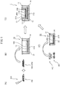
- Fig. 1 is a schematic diagram illustrating a pigmentation skin model 1 and a method for its production, according to an embodiment of the invention.
- the pigmentation skin model 1 comprises:
- the pigmentation skin model 1 of this embodiment can be provided by a method that comprises the following steps:
- the structure of the pigmentation skin model 1 comprising the first cell group 100 and third cell group 100a may be referred to as "dermis model” (first dermis model or second dermis model) since it models the structure of the skin dermis.
- the structure comprising the pigmentation skin model 1 containing the second cell group 200 may be referred to as "epidermis model” since it models the structure of the skin epidermis.
- the pigmentation skin model 1 may have a structure with the second cell group 200 directly seeded or layered on the first cell group 100, or it may have a structure as shown in Fig. 1 , having a space formed between the second cell group 200 and first cell group 100, in which medium is present.
- the second cell group 200 is preferably seeded on a porous membrane (corresponding to the second porous membrane 210 of Fig. 1 ) in such a manner that liquid components secreted by the first cell group 100 and second cell group 200 can mutually pass through.
- step (3) of the method of producing the pigmentation skin model 1 the step of applying the second cell group 200 that includes melanocytes and keratinocytes onto the first cell group 100 obtained in step (2) may be a step of directly seeding or layering the second cell group 200 on the first cell group 100, or it may be a step in which the second cell group 200 that has been seeded on a second cell culturing substrate 21 having a porous membrane is applied onto the first cell group 100.
- the first cell culturing substrate 11 on which the first cell group 100 is seeded may employ a known culture substrate that allows culturing of fibroblasts, but it is preferably a cell culturing substrate having a porous membrane, and more preferably a cell culture insert. If the first cell culturing substrate 11 has a first porous membrane 110 it will be possible to supply nutrients and oxygen both above and below the cells that have been seeded on the first porous membrane 110.
- the second cell culturing substrate 21 on which the second cell group 200 is seeded may employ a known culture substrate that allows culturing of melanocytes and keratinocytes, but it is preferably a cell culturing substrate having a porous membrane, and more preferably a cell culture insert. If the second cell culturing substrate 21 has a second porous membrane 210 it will be possible not only to supply nutrients and oxygen both above and below the cells that have been seeded on the second porous membrane 210, but also to allow mutual passage of liquid components secreted by the first cell group 100 and second cell group 200.
- the second cell culturing substrate 21 has a second porous membrane 210 it will also be possible to separate the dermis model and epidermis model during the course of maintenance culturing of the pigmentation skin model 1, allowing culturing of each under different conditions and allowing the epidermis model to be transferred onto a different dermis model and cultured. Since formation of the epidermis layer by the epidermal cell group is affected by the quality of the dermis model, the quality of the epidermis model in the pigmentation skin model 1 can be controlled by replacement with a dermis model having the desired quality.
- the mean pore size of the first porous membrane 110 and second porous membrane 210 may be selected as appropriate, and may be a mean pore size of about 0.01 ⁇ m to about 100 ⁇ m, for example (such as 0.01 ⁇ m to 100 ⁇ m, 0.01 ⁇ m to 50 ⁇ m, 0.01 ⁇ m to 10 ⁇ m, 0.1 ⁇ m to 50 ⁇ m or 0.1 ⁇ m to 10 ⁇ m).
- the density of the pores in the first porous membrane 110 and second porous membrane 210 may also be selected as appropriate, but it is preferably a pore density of 1 ⁇ 10 4 /cm 2 or greater, 1 ⁇ 10 5 /cm 2 or greater, 5 ⁇ 10 5 /cm 2 or greater, 10 ⁇ 10 5 /cm 2 or greater, 50 ⁇ 10 5 /cm 2 or greater or 100 ⁇ 10 5 /cm 2 or greater, and the range may be 1 ⁇ 10 4 /cm 2 to 100 ⁇ 10 8 /cm 2 or 1 ⁇ 10 5 /cm 2 to 100 ⁇ 10 8 /cm 2 .
- the second cell culturing substrate 21 is a cell culture insert
- the second cell culturing substrate 21 (cell culture insert) used has a smaller inner diameter than the first cell culturing substrate 11. This will allow the second cell culturing substrate 21 to be used by insertion into the culturing portion of the first cell culturing substrate 11.
- the cells used for the invention may be derived from any animal, but they are preferably derived from a vertebrate, more preferably from a mammal, and most preferably from a human.
- Fibroblasts are one of the types of cells constituting connective tissue, and they are present in a large number of organs and tissues. Fibroblasts are included in skin, and primarily the dermal tissue.
- the fibroblasts to be used for the pigmentation skin model 1 of the invention are preferably fibroblasts derived from the dermis.
- Keratinocytes are a type of cells composing the epidermis, and in biological epidermal tissue they divide at the deepest part (basal lamina) while migrating to the surface and differentiating toward the upper layer into the stratum spinosum, granular layer and stratum corneum, eventually being shed as old skin.
- Melanocytes are a type of cells composing epidermal tissue, which are present in the epidermal basal lamina of biological tissue and produce melanin.
- the fibroblasts, keratinocytes and melanocytes used for the invention may each be primary cultured cells harvested from biological tissue, or cells marketed or distributed after first having been isolated and/or grown, or cells of an established cell line, or cells differentiated from pluripotent stem cells such as ES cells, iPS cells or Muse cells.
- the first cell group 100 may also contain cells other than fibroblasts, such as mast cells, histiocytes, plasmocytes or dermal dendritic cells that are present within dermal tissue.
- the number of fibroblasts in the first cell group 100 may be 1 ⁇ 10 4 to 10 8 /cm 2 , and preferably 0.1 to 10 ⁇ 10 5 /cm 2 .
- the second cell group 200 may also include cells other than the keratinocytes and melanocytes, such as Langerhans cells in the epidermal tissue, or Merkel cells.
- the second cell group 200 may include the cells in a proportion of keratinocytes:melanocytes of 1:1 to 1000:1 and preferably 3:1 to 30:1.
- the second cell group 200 includes the keratinocytes at 1 ⁇ 10 2 to 10 6 /cm 2 , preferably 1.0 to 10 ⁇ 10 4 /cm 2 and more preferably about 4 to 8 ⁇ 10 4 /cm 2 , and the melanocytes at 1 to 10 ⁇ 10 3 /cm 2 and preferably 4 to 8 ⁇ 10 3 /cm 2 .
- the fibroblasts used in the first cell group 100 are fibroblasts that have been damaged by photoirradiation.
- the fibroblasts damaged by photoirradiation are preferably ones that have been damaged by photoirradiation in the presence of a photosensitizer.
- photosensitizers to be used include psoralen, NAD, riboflavin, tryptophan, folic acid, porphyrin, methylene blue, and thiol group-protected gold nanoclusters (AUxSRy).
- the light used to induce damage in the fibroblasts may have a wavelength which causes damage in intracellular nucleic acid such as DNA or RNA but does not result in death of all of the cells, and it is preferably ultraviolet light (about 200 nm to about 400 nm), and more preferably UVA (about 320 nm to about 400 nm).
- the intensity of the irradiated light may be such as to produce damage in the intracellular nucleic acid such as DNA or RNA while not inducing apoptosis and death in all of the cells, and it can be appropriately adjusted by the wavelength or irradiation time, and the cell density.
- Irradiation of UVA may be at 0.01 J/cm 2 to 100 J/cm 2 , preferably 0.1 J/cm 2 to 20 J/cm 2 and more preferably 0.5 J/cm 2 to 10 J/cm 2 .
- fibroblasts are cultured for a fixed time period it is possible to cause proliferation of only the surviving fibroblasts that have not undergone apoptosis ( Fig. 1(B) ). Fibroblasts that have suffered damage by photoirradiation have elongated cellular forms and reduced proliferation potency, as well as increased production of melanin-producing factors (such as stem cell growth factor (SCF), and thus an increased level of cell senescence (see Fig. 3 ).
- SCF stem cell growth factor
- the level of senescence of cells can be examined by measuring expression levels of commonly known cell senescence markers such as senescent acidic ⁇ -galactosidase (SA- ⁇ gal), constitutive activation of cell cycle check mechanisms such as the p21/p53 pathway or p16 pathway, or expression levels of cell senescence-associated secretory phenotype (SASP) factors such as IL-6. Adjustment of the photoirradiation dose allows fibroblasts with the desired level of senescence to be obtained.
- SA- ⁇ gal senescent acidic ⁇ -galactosidase
- SASP cell senescence-associated secretory phenotype
- the fibroblasts in the first cell group 100 of the pigmentation skin model 1 are fibroblasts that have been damaged by photoirradiation but have survived without dying.
- the pigmentation skin model 1 of the invention therefore contains largely homogeneous fibroblasts, and can thus provide a stable system with low variability of activity.
- the epidermis model 20 used to form the pigmentation skin model 1 may be TESTSKIN R LSE-melano (TOYOBO) or MelanoDerm R (MatTek), for example, which are commercially available.
- the pigmentation skin model 1 may be cultured using a culture solution commonly used as a culture solution for culturing of keratinocytes, such as KG medium, EpilifeKG2 (Kurabo Industries, Ltd.), Humedia-KG2 (Kurabo Industries, Ltd.) or assay medium (TOYOBO), at about 37°C for a period of 0 to 14 days.
- the culture medium used may alternatively be DMEM culture medium (Gibco), or a culture medium comprising a 1:1 mixture of ascorbic acid-containing KGM and DMEM.
- the first cell group 100 preferably is seeded together with a hydrogelling agent.
- hydrogelling agent is a substance added in order to form a hydrogel.
- Hydrogelling agents to be used for the invention may be selected from the group consisting of include collagen, gelatin, hyaluronate, hyaluronan, fibrin, alginate, agarose, chitosan, chitin, cellulose, pectin, starch, laminin, fibrinogen/thrombin, fibrillin, elastin, gum, cellulose, agar, gluten, casein, albumin, vitronectin, tenascin, entactin/nidogen, glycoproteins, glycosaminoglycan, poly(acrylic acid) and its derivatives, poly(ethylene oxide) and its copolymers, poly(vinyl alcohol), polyphosphazene, Matrigel, and combinations of the foregoing.
- step (2) may be carried out in the presence of ascorbic acid, an ascorbic acid derivative, or salt thereof.
- Ascorbic acid, an ascorbic acid derivative or salt thereof is preferably present in order to promote multilayer formation similar to the structure of the dermis, by accelerated proliferation of the fibroblasts and collagen production.
- “ascorbic acid derivative” refers to ascorbic acid diphosphate, ascorbic acid monophosphate, sodium L-ascorbate or L-ascorbic acid 2-glucoside, or a salt thereof (such as a sodium salt or magnesium salt).
- the pigmentation skin model 1 provided for the invention has fibroblasts damaged by photoirradiation that has acted directly or indirectly on melanocytes, accelerating melanin production. The extent of pigment production and/or pigmentation in the pigmentation skin model 1 can then be measured to evaluate factors that affect pigmentation.
- the invention can provide a method for evaluating factors for treatment or prevention of skin pigmentation using the pigmentation skin model.
- the method of the invention comprises:
- candidate factors include low molecular compounds, peptides, nucleic acids, proteins, mammalian animal (such as mouse, rat, porcine, bovine, sheep, monkey or human) cells, tissue extracts or cell culture supernatants, plant-derived compounds or extracts (such as galenical extracts or galenical-derived compounds), and microbial compounds or extracts, or culture products.
- mammalian animal such as mouse, rat, porcine, bovine, sheep, monkey or human
- tissue extracts or cell culture supernatants such as galenical extracts or galenical-derived compounds
- plant-derived compounds or extracts such as galenical extracts or galenical-derived compounds
- microbial compounds or extracts or culture products.
- a candidate substance may be added to the pigmentation skin model 1 and culturing carried out for a predetermined time period, and then the extent of pigment production and/or pigmentation in the pigmentation skin model 1 may be measured to evaluate the treatment or prevention effect that the candidate substance has on pigmentation. For example, it is possible to compare the extent of pigment production and/or pigmentation in a pigmentation skin model 1 without addition of a candidate substance, and with addition of a substance that does not have a treatment or prevention effect on pigmentation, and when the extent of pigment production and/or pigmentation in the pigmentation skin model 1 with the added candidate substance is higher, the candidate substance may be evaluated as having a treatment or prevention effect on pigmentation.
- pigment production refers to production of a pigment such as melanin pigment, which is produced by the pigmentation skin model.
- Melanin pigment is produced primarily by melanocytes.
- the amount of pigment production can be determined as the amount of melanin, by extracting the melanin pigment from the pigmentation skin model, and especially the second cell group (keratinocytes and melanocytes), and measuring the absorbance at 405 nm.
- the amount of pigment production can be measured as the level of melanin or nucleic acid (such as mRNA) coding therefor, present in the pigmentation skin model and particularly the second cell group (keratinocytes and melanocytes), using a method such as ELISA, flow cytometry, Western blotting, an immunohistochemical method or qPCR, with no limitation to these.
- melanin or nucleic acid such as mRNA
- a method such as ELISA, flow cytometry, Western blotting, an immunohistochemical method or qPCR, with no limitation to these.
- extentent of pigmentation means the color brightness of the pigmentation skin model, and particularly the second cell group (keratinocytes and melanocytes), under visible light.
- the pigmentation skin model of the invention has varying brightness depending on the amount of melanin produced by the melanocytes. A greater amount of melanin results in reduced brightness, so that the pigmentation skin model exhibits a darker color. A lower amount of melanin, conversely, increases the brightness and causes the pigmentation skin model to exhibit a lighter color. That is, the treatment or prevention effect on pigmentation by an added candidate substance can be evaluated by comparing the color brightness of the pigmentation skin model.
- the brightness can be quantified by recording the pigmentation skin model as an image and using publicly known image measuring means.
- the method of the invention comprises:
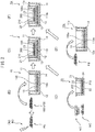
- Steps (1) to (3) are the same as the steps described for the method for producing a pigmentation skin model and will not be explained again here ( Fig. 2(A) to (D) ).
- step (5) is the same as step (2) described above for "Method for evaluating factors for treatment or prevention of skin pigmentation" (first mode) and will not be explained again here.
- the invention comprises applying the second cell group 200 obtained in step (3) onto a substrate containing a candidate factor, and culturing the obtained pigmentation skin model 1a (step (4), Fig. 2(F) ).
- the candidate factor on the substrate used in step (3) (such as the third cell culturing substrate 11a in Fig.
- 2(E) and (F) may be, for example, a low molecular compound, peptide, nucleic acid or protein, a mammalian animal (such as mouse, rat, porcine, bovine, sheep, monkey or human) cells, a tissue extract or cell culture supernatant, plant-derived compound or extract (such as a galenical extract or galenical-derived compound), a microbial compound or extract, or a culture product.
- a mammalian animal such as mouse, rat, porcine, bovine, sheep, monkey or human
- plant-derived compound or extract such as a galenical extract or galenical-derived compound
- a microbial compound or extract such as shown in Fig.
- the candidate factor may be any type of cells, such as fibroblast progenitor cells including mesenchymal stem cells, or fibroblasts (such as fibroblasts that have not been damaged by photoirradiation, or fibroblasts with low damage by photoirradiation).
- fibroblasts that have not been damaged by photoirradiation or "fibroblasts with low damage by photoirradiation” are fibroblasts that have not been subjected to a photoirradiation step, and that have a lower level of senescence than the "fibroblasts that have been damaged by photoirradiation” mentioned above.
- the third cell group 100a used as the candidate factor may be one that has been seeded on the third cell culturing substrate 11a having a porous membrane.
- step (4) may be directly seeding or layering the second cell group 200 obtained in step (3) onto the third cell group 100a used as the candidate factor, or it may be applying the second cell group 200 that has been seeded on the second cell culturing substrate 21 having a porous membrane, onto the third cell group 100a used as the candidate factor. In the latter case it is possible to easily transfer the second cell culturing substrate 21 onto another dermis model.
- fibroblasts (1 ⁇ 10 5 cells) were cultured with a growth medium (DMEM + 10% fetal calf serum). Before reaching 100% confluence, the medium was exchanged with growth medium containing psoralen (final concentration: 25 ng/mL) (Sigma-Aldrich), and culturing was continued.
- DMEM + 10% fetal calf serum 10% fetal calf serum
- a dermis model was prepared using fibroblasts subjected to the PUVA treatment of 1-1. above, or without PUVA treatment (control). For cursory examination, the procedure of 1. above was followed by removal of the growth medium and imbibing with 1 mL PBS. The PBS was then removed, a cell release agent (TrypLE SELECT, Thermo Fisher Scientific) was added at 300 ⁇ L per well of a 6-well plate, and the mixture was allowed to stand for 5 minutes in a 5% CO 2 incubator at 37°C as cell releasing treatment. Growth medium was added to suspend the reaction, and the cell suspension was recovered in a 15 ml centrifuge tube.
- a gel suspension comprising the composition listed in Table 1 was prepared on ice to prepare a dermis model for application to each well of a 6-well plate.
- Table 1 Composition of gel suspension for dermis model containing fibroblasts Reagent Amount Pig skin collagen (final concentration: 0.5% (w/v)) (NH Foods Ltd., 309-31595) 1.5 mL 5 ⁇ DMEM 0.6 mL Fetal bovine serum (final concentration: 10% (v/v)) 0.3 mL L-ascorbic acid 2-glucoside(AA2G) (final concentration: 250 ⁇ M) (Sigma-Aldrich, SMB00390) 3 ⁇ L Fibroblast suspension 0.6 mL Total volume 3 mL
- a cell culture insert for dermis model preparation was set in the 6-well plate and 3 mL of gel suspension was added into the cell culture insert. After solidifying the gel suspension with a 5% CO 2 incubator at 37°C, 2 mL of growth medium containing ascorbic acid (AA2G) was added to the 6-well plate and culturing was carried out for 24 hours in the 5% CO 2 incubator at 37°C.
- A2G ascorbic acid
- a commercially available epidermis model comprising melanocytes and keratinocytes was also used (see Fig. 1(C) ).
- the prepared epidermis model was laid over and combined with the dermis model of 1-2. above (see Fig. 1(D) ) and set in a specialized vessel (Corning BioCoat deep well plate, 6 wells) (Corning #355467), and then 9.5 mL of three-dimensional skin model medium (a 1:1 mixture of epidermis model special medium (MatTek #EPI-100-NMM-113) and DMEM) was added and medium exchange was carried out with the same medium at a frequency of once every 3 to 4 days.
- a specialized vessel Corning BioCoat deep well plate, 6 wells
- DMEM medium exchange was carried out with the same medium at a frequency of once every 3 to 4 days.
- An epidermis model cultured for 10 days in combination with a dermis model prepared using PUVA-treated fibroblasts was replaced with a dermis model prepared using PUVA-untreated normal fibroblasts, and culturing was continued for 10 days (see Fig. 2(E) and (F) ).
- the replacement dermis model was prepared on the day prior to replacement.
- Fig. 3 shows PUVA-treated and untreated fibroblasts.
- the PUVA-treated fibroblasts had become elongated ( Fig. 3(A) ).
- Fig. 3(B) Upon examining variation in the number of proliferated PUVA-treated and untreated fibroblasts, it was found that the PUVA-treated fibroblasts had a notably lower growth rate ( Fig. 3(B) ).
- Fig. 7 is a diagram showing pigmentation in an epidermis model, for a pigmentation skin model either with PUVA treatment (A) or without treatment (B).
- the epidermis model for the PUVA-treated pigmentation skin model showed accelerated pigmentation.
- An epidermis model cultured for 10 days in combination with a dermis model prepared using PUVA-treated fibroblasts was replaced with a dermis model prepared using PUVA-untreated normal fibroblasts, and after culturing for another 10 days, pigmentation was observed to be inhibited ( Fig. 8 ) compared to pigmentation in the epidermis model for the PUVA-treated pigmentation skin model ( Fig. 7(A) ).
- the ratio of the thickest pigmented region in the PUVA-treated pigmentation skin model was high, while the region of thick pigmentation in the pigmentation skin model under the Replace conditions was reduced ( Fig. 9(B) ).
- TRP2 antibody was used for staining of the activated melanocytes in the epidermis model slices, and the nuclei were stained by hematoxylin staining ( Fig. 11(A) ).
Landscapes
- Health & Medical Sciences (AREA)
- Engineering & Computer Science (AREA)
- Life Sciences & Earth Sciences (AREA)
- Biomedical Technology (AREA)
- Chemical & Material Sciences (AREA)
- Bioinformatics & Cheminformatics (AREA)
- Wood Science & Technology (AREA)
- Organic Chemistry (AREA)
- Zoology (AREA)
- Biotechnology (AREA)
- Genetics & Genomics (AREA)
- Microbiology (AREA)
- Biochemistry (AREA)
- General Health & Medical Sciences (AREA)
- Cell Biology (AREA)
- General Engineering & Computer Science (AREA)
- Immunology (AREA)
- Molecular Biology (AREA)
- Urology & Nephrology (AREA)
- Hematology (AREA)
- Sustainable Development (AREA)
- Dermatology (AREA)
- Physics & Mathematics (AREA)
- Toxicology (AREA)
- Food Science & Technology (AREA)
- Medicinal Chemistry (AREA)
- Tropical Medicine & Parasitology (AREA)
- Analytical Chemistry (AREA)
- General Physics & Mathematics (AREA)
- Pathology (AREA)
- Rheumatology (AREA)
- General Chemical & Material Sciences (AREA)
- Physiology (AREA)
- Electromagnetism (AREA)
- Micro-Organisms Or Cultivation Processes Thereof (AREA)
- Measuring Or Testing Involving Enzymes Or Micro-Organisms (AREA)
- Apparatus Associated With Microorganisms And Enzymes (AREA)
- Medicines Containing Material From Animals Or Micro-Organisms (AREA)
Applications Claiming Priority (2)
| Application Number | Priority Date | Filing Date | Title |
|---|---|---|---|
| JP2018226024 | 2018-11-30 | ||
| PCT/JP2019/046886 WO2020111265A1 (ja) | 2018-11-30 | 2019-11-29 | 色素沈着皮膚モデルおよびその製造方法、ならびに皮膚の色素沈着を治療または予防するための因子の評価方法 |
Publications (2)
| Publication Number | Publication Date |
|---|---|
| EP3889247A1 true EP3889247A1 (de) | 2021-10-06 |
| EP3889247A4 EP3889247A4 (de) | 2022-10-19 |
Family
ID=70853043
Family Applications (1)
| Application Number | Title | Priority Date | Filing Date |
|---|---|---|---|
| EP19888944.6A Withdrawn EP3889247A4 (de) | 2018-11-30 | 2019-11-29 | Pigmentationshautmodell und verfahren zu dessen herstellung sowie verfahren zur bewertung des faktors zur behandlung oder vorbeugung der pigmentierung der haut |
Country Status (7)
| Country | Link |
|---|---|
| US (1) | US20220025337A1 (de) |
| EP (1) | EP3889247A4 (de) |
| JP (1) | JP7522042B2 (de) |
| KR (1) | KR20210097116A (de) |
| CN (1) | CN113166694A (de) |
| SG (1) | SG11202105106VA (de) |
| WO (1) | WO2020111265A1 (de) |
Cited By (1)
| Publication number | Priority date | Publication date | Assignee | Title |
|---|---|---|---|---|
| EP4464766A4 (de) * | 2022-01-13 | 2026-02-18 | Riken | Behälter zur herstellung einer gespannten dreidimensionalen künstlichen haut und herstellungsverfahren dafür |
Families Citing this family (5)
| Publication number | Priority date | Publication date | Assignee | Title |
|---|---|---|---|---|
| JP7083866B2 (ja) * | 2020-06-11 | 2022-06-13 | 株式会社ナリス化粧品 | 皮膚モデル |
| JP7661133B2 (ja) * | 2021-05-31 | 2025-04-14 | 株式会社 資生堂 | 皮膚モデルおよびその製造方法、ならびに皮膚の色素沈着を治療または予防するための因子の評価方法 |
| JP7733479B2 (ja) * | 2021-05-31 | 2025-09-03 | 株式会社 資生堂 | 皮膚モデルおよびその製造方法、ならびに皮膚の色素沈着を治療または予防するための因子の評価方法 |
| CN113533649A (zh) * | 2021-07-14 | 2021-10-22 | 南开大学 | 一种适用于人体皮肤污染物暴露评估的模拟方法 |
| CN118995578B (zh) * | 2024-10-25 | 2025-03-21 | 济南磐升生物技术有限公司 | 一种组织工程皮肤及其构建方法和在评估注射用水光针产品保湿效果中的应用 |
Family Cites Families (13)
| Publication number | Priority date | Publication date | Assignee | Title |
|---|---|---|---|---|
| JPS5459875A (en) | 1977-10-20 | 1979-05-14 | Nec Corp | Semiconductor device |
| EP1482030A1 (de) * | 2003-05-28 | 2004-12-01 | Deutsches Krebsforschungszentrum Stiftung des öffentlichen Rechts | Methode zur Erhöhung des Ertrags an proliferierenden primären Keratinozyten |
| KR100806695B1 (ko) * | 2005-11-25 | 2008-02-27 | 주식회사 엠씨티티 | 색소화 질환 치료용 세포치료 약제학적 조성물 |
| FR2895748B1 (fr) * | 2006-01-05 | 2008-04-04 | Oreal | Modele de culture cellulaire et ses applications |
| FR2905380B1 (fr) * | 2006-09-04 | 2008-12-05 | Engelhard Lyon Sa | Procede de production de cellules de langerhans et/ou de cellules de dentritiques interstitielles/dermiques a partir de monocytes cd14+ |
| FR2930644B1 (fr) * | 2008-04-29 | 2012-08-03 | Oreal | Procede d'evaluation de la pigmentation |
| CA2658074C (fr) * | 2008-03-17 | 2017-11-07 | L'oreal | Equivalent de peau pigmentee fonctionnelle |
| FR2928654B1 (fr) * | 2008-03-17 | 2018-04-27 | L'oreal | Equivalent de peau pigmentee fonctionnelle. |
| CN101347640A (zh) * | 2008-09-05 | 2009-01-21 | 西安交通大学 | 用于筛选退色剂的色素化皮肤类似物模型及其构建方法 |
| JP2010172240A (ja) * | 2009-01-28 | 2010-08-12 | Shiseido Co Ltd | エラスチン発現低下を回復する薬剤のスクリーニング方法 |
| JP2011092179A (ja) * | 2009-09-30 | 2011-05-12 | Toyobo Co Ltd | メラノサイトを含む3次元培養皮膚モデルおよびその使用方法 |
| JP2012235921A (ja) * | 2011-05-12 | 2012-12-06 | Shiseido Co Ltd | 三次元皮膚モデルの製造方法 |
| GB201416006D0 (en) * | 2014-09-10 | 2014-10-22 | Univ Singapore | Organotypic skin model |
-
2019
- 2019-11-29 US US17/298,446 patent/US20220025337A1/en not_active Abandoned
- 2019-11-29 KR KR1020217015801A patent/KR20210097116A/ko not_active Ceased
- 2019-11-29 WO PCT/JP2019/046886 patent/WO2020111265A1/ja not_active Ceased
- 2019-11-29 EP EP19888944.6A patent/EP3889247A4/de not_active Withdrawn
- 2019-11-29 JP JP2020557877A patent/JP7522042B2/ja active Active
- 2019-11-29 SG SG11202105106VA patent/SG11202105106VA/en unknown
- 2019-11-29 CN CN201980078315.9A patent/CN113166694A/zh active Pending
Cited By (1)
| Publication number | Priority date | Publication date | Assignee | Title |
|---|---|---|---|---|
| EP4464766A4 (de) * | 2022-01-13 | 2026-02-18 | Riken | Behälter zur herstellung einer gespannten dreidimensionalen künstlichen haut und herstellungsverfahren dafür |
Also Published As
| Publication number | Publication date |
|---|---|
| WO2020111265A1 (ja) | 2020-06-04 |
| US20220025337A1 (en) | 2022-01-27 |
| JP7522042B2 (ja) | 2024-07-24 |
| KR20210097116A (ko) | 2021-08-06 |
| CN113166694A (zh) | 2021-07-23 |
| JPWO2020111265A1 (ja) | 2021-10-14 |
| EP3889247A4 (de) | 2022-10-19 |
| SG11202105106VA (en) | 2021-06-29 |
Similar Documents
| Publication | Publication Date | Title |
|---|---|---|
| US20220025337A1 (en) | Pigmentation skin model and method for producing same, and method for evaluating factor for treating or preventing pigmentation of skin | |
| EP3862009A1 (de) | Zusammensetzung mit induzierten exosomen zur regeneration der haut und wundheilung | |
| US20210038652A1 (en) | Extracellular vesicles derived from cells cultured under hypoxic conditions and uses thereof | |
| van de Kamp et al. | Hepatocyte growth factor‐loaded biomaterials for mesenchymal stem cell recruitment | |
| Papageorgiou et al. | Expanded skeletal stem and progenitor cells promote and participate in induced bone regeneration at subcritical BMP-2 dose | |
| KR20140115296A (ko) | 모낭 외모낭초로부터 멜라닌세포를 유도하기 위한 방법 및 이식을 위한 제조 | |
| US9592257B2 (en) | Complete human skin organ generated from culture-expanded cells | |
| KR20120120971A (ko) | 인간 다능성 줄기 세포로부터 인간 멜라닌세포를 제조하는 방법 | |
| US12357560B2 (en) | Composition for treating or preventing skin pigmentation | |
| CN104968298A (zh) | 使用自体成纤维细胞改变皮肤身份的方法 | |
| CN116410918B (zh) | 一种皮肤类器官外泌体及其制备方法和应用 | |
| JP7383619B2 (ja) | 皮膚細胞の細胞活性又は肌状態の決定方法、並びに皮膚細胞賦活剤のスクリーニング方法 | |
| JP7733479B2 (ja) | 皮膚モデルおよびその製造方法、ならびに皮膚の色素沈着を治療または予防するための因子の評価方法 | |
| de Andrés et al. | A bioengineered tumor matrix-based scaffold for the evaluation of melatonin efficacy on head and neck squamous cancer stem cells | |
| Riva et al. | Generation of human epidermal constructs on a collagen layer alone | |
| US12077780B2 (en) | Conditioned medium from cells cultured under hypoxic conditions and uses thereof | |
| US12442815B1 (en) | Viscoelastic hydrogel regulation of organoid patterning and vascularization | |
| US20140128272A1 (en) | Method for Inducing Dormancy of Cancer Tissue-Derived Cell Mass and Method for Evaluating Treating Means with the Use of Cancer-Tissue-Derived Cell Mass | |
| EP4397751A1 (de) | Verfahren zur herstellung menschlicher melanozyten aus pluripotenten stammzellen | |
| WO2025051858A1 (en) | Processes for producing a reconstructed skin model, and applications thereof | |
| MEAD | Incorporation of pigment producing cells into novel 3D skin equivalents to study the role of the dermal compartment upon pigmentation in skin equivalents | |
| CN117616114A (zh) | 生物工程真皮乳头和毛囊以及相关产品、方法和应用 | |
| López de Andrés | Biofabrication of complex tumor microenvironments with application in precision oncology | |
| Jiang et al. | 3D‐Cultured Mesenchymal Stem Cell‐Derived Exosomes Embedded in a Hybrid Scaffold Enhance Diabetic Wound Healing by Promoting Angiogenesis | |
| van de Kamp et al. | Research Article Hepatocyte Growth Factor-Loaded Biomaterials for Mesenchymal Stem Cell Recruitment |
Legal Events
| Date | Code | Title | Description |
|---|---|---|---|
| STAA | Information on the status of an ep patent application or granted ep patent |
Free format text: STATUS: THE INTERNATIONAL PUBLICATION HAS BEEN MADE |
|
| PUAI | Public reference made under article 153(3) epc to a published international application that has entered the european phase |
Free format text: ORIGINAL CODE: 0009012 |
|
| STAA | Information on the status of an ep patent application or granted ep patent |
Free format text: STATUS: REQUEST FOR EXAMINATION WAS MADE |
|
| 17P | Request for examination filed |
Effective date: 20210517 |
|
| AK | Designated contracting states |
Kind code of ref document: A1 Designated state(s): AL AT BE BG CH CY CZ DE DK EE ES FI FR GB GR HR HU IE IS IT LI LT LU LV MC MK MT NL NO PL PT RO RS SE SI SK SM TR |
|
| DAV | Request for validation of the european patent (deleted) | ||
| DAX | Request for extension of the european patent (deleted) | ||
| A4 | Supplementary search report drawn up and despatched |
Effective date: 20220916 |
|
| RIC1 | Information provided on ipc code assigned before grant |
Ipc: C12Q 1/02 20060101ALI20220912BHEP Ipc: C12N 5/071 20100101ALI20220912BHEP Ipc: C12M 1/34 20060101ALI20220912BHEP Ipc: C12M 1/00 20060101AFI20220912BHEP |
|
| STAA | Information on the status of an ep patent application or granted ep patent |
Free format text: STATUS: THE APPLICATION IS DEEMED TO BE WITHDRAWN |
|
| 18D | Application deemed to be withdrawn |
Effective date: 20240601 |