EP3896426B1 - Analyseur de sang - Google Patents
Analyseur de sang Download PDFInfo
- Publication number
- EP3896426B1 EP3896426B1 EP21163161.9A EP21163161A EP3896426B1 EP 3896426 B1 EP3896426 B1 EP 3896426B1 EP 21163161 A EP21163161 A EP 21163161A EP 3896426 B1 EP3896426 B1 EP 3896426B1
- Authority
- EP
- European Patent Office
- Prior art keywords
- red blood
- malaria
- blood cells
- information
- iron
- Prior art date
- Legal status (The legal status is an assumption and is not a legal conclusion. Google has not performed a legal analysis and makes no representation as to the accuracy of the status listed.)
- Active
Links
Images








Classifications
-
- G—PHYSICS
- G01—MEASURING; TESTING
- G01N—INVESTIGATING OR ANALYSING MATERIALS BY DETERMINING THEIR CHEMICAL OR PHYSICAL PROPERTIES
- G01N21/00—Investigating or analysing materials by the use of optical means, i.e. using sub-millimetre waves, infrared, visible or ultraviolet light
- G01N21/62—Systems in which the material investigated is excited whereby it emits light or causes a change in wavelength of the incident light
- G01N21/63—Systems in which the material investigated is excited whereby it emits light or causes a change in wavelength of the incident light optically excited
- G01N21/64—Fluorescence; Phosphorescence
- G01N21/6402—Atomic fluorescence; Laser induced fluorescence
-
- G—PHYSICS
- G01—MEASURING; TESTING
- G01N—INVESTIGATING OR ANALYSING MATERIALS BY DETERMINING THEIR CHEMICAL OR PHYSICAL PROPERTIES
- G01N21/00—Investigating or analysing materials by the use of optical means, i.e. using sub-millimetre waves, infrared, visible or ultraviolet light
- G01N21/62—Systems in which the material investigated is excited whereby it emits light or causes a change in wavelength of the incident light
- G01N21/63—Systems in which the material investigated is excited whereby it emits light or causes a change in wavelength of the incident light optically excited
- G01N21/64—Fluorescence; Phosphorescence
- G01N21/6428—Measuring fluorescence of fluorescent products of reactions or of fluorochrome labelled reactive substances, e.g. measuring quenching effects, using measuring "optrodes"
-
- G—PHYSICS
- G01—MEASURING; TESTING
- G01N—INVESTIGATING OR ANALYSING MATERIALS BY DETERMINING THEIR CHEMICAL OR PHYSICAL PROPERTIES
- G01N15/00—Investigating characteristics of particles; Investigating permeability, pore-volume or surface-area of porous materials
- G01N15/10—Investigating individual particles
- G01N15/14—Optical investigation techniques, e.g. flow cytometry
- G01N15/1456—Optical investigation techniques, e.g. flow cytometry without spatial resolution of the texture or inner structure of the particle, e.g. processing of pulse signals
- G01N15/1459—Optical investigation techniques, e.g. flow cytometry without spatial resolution of the texture or inner structure of the particle, e.g. processing of pulse signals the analysis being performed on a sample stream
-
- G—PHYSICS
- G01—MEASURING; TESTING
- G01N—INVESTIGATING OR ANALYSING MATERIALS BY DETERMINING THEIR CHEMICAL OR PHYSICAL PROPERTIES
- G01N1/00—Sampling; Preparing specimens for investigation
- G01N1/28—Preparing specimens for investigation including physical details of (bio-)chemical methods covered elsewhere, e.g. G01N33/50, C12Q
- G01N1/30—Staining; Impregnating ; Fixation; Dehydration; Multistep processes for preparing samples of tissue, cell or nucleic acid material and the like for analysis
- G01N1/31—Apparatus therefor
-
- G—PHYSICS
- G01—MEASURING; TESTING
- G01N—INVESTIGATING OR ANALYSING MATERIALS BY DETERMINING THEIR CHEMICAL OR PHYSICAL PROPERTIES
- G01N15/00—Investigating characteristics of particles; Investigating permeability, pore-volume or surface-area of porous materials
- G01N15/10—Investigating individual particles
- G01N15/1031—Investigating individual particles by measuring electrical or magnetic effects
- G01N15/12—Investigating individual particles by measuring electrical or magnetic effects by observing changes in resistance or impedance across apertures when traversed by individual particles, e.g. by using the Coulter principle
-
- G—PHYSICS
- G01—MEASURING; TESTING
- G01N—INVESTIGATING OR ANALYSING MATERIALS BY DETERMINING THEIR CHEMICAL OR PHYSICAL PROPERTIES
- G01N21/00—Investigating or analysing materials by the use of optical means, i.e. using sub-millimetre waves, infrared, visible or ultraviolet light
- G01N21/17—Systems in which incident light is modified in accordance with the properties of the material investigated
- G01N21/47—Scattering, i.e. diffuse reflection
- G01N21/49—Scattering, i.e. diffuse reflection within a body or fluid
-
- G—PHYSICS
- G01—MEASURING; TESTING
- G01N—INVESTIGATING OR ANALYSING MATERIALS BY DETERMINING THEIR CHEMICAL OR PHYSICAL PROPERTIES
- G01N21/00—Investigating or analysing materials by the use of optical means, i.e. using sub-millimetre waves, infrared, visible or ultraviolet light
- G01N21/62—Systems in which the material investigated is excited whereby it emits light or causes a change in wavelength of the incident light
- G01N21/63—Systems in which the material investigated is excited whereby it emits light or causes a change in wavelength of the incident light optically excited
- G01N21/64—Fluorescence; Phosphorescence
- G01N21/6486—Measuring fluorescence of biological material, e.g. DNA, RNA, cells
-
- G—PHYSICS
- G01—MEASURING; TESTING
- G01N—INVESTIGATING OR ANALYSING MATERIALS BY DETERMINING THEIR CHEMICAL OR PHYSICAL PROPERTIES
- G01N33/00—Investigating or analysing materials by specific methods not covered by groups G01N1/00 - G01N31/00
- G01N33/48—Biological material, e.g. blood, urine; Haemocytometers
- G01N33/483—Physical analysis of biological material
- G01N33/487—Physical analysis of biological material of liquid biological material
- G01N33/49—Blood
-
- G—PHYSICS
- G16—INFORMATION AND COMMUNICATION TECHNOLOGY [ICT] SPECIALLY ADAPTED FOR SPECIFIC APPLICATION FIELDS
- G16H—HEALTHCARE INFORMATICS, i.e. INFORMATION AND COMMUNICATION TECHNOLOGY [ICT] SPECIALLY ADAPTED FOR THE HANDLING OR PROCESSING OF MEDICAL OR HEALTHCARE DATA
- G16H10/00—ICT specially adapted for the handling or processing of patient-related medical or healthcare data
- G16H10/40—ICT specially adapted for the handling or processing of patient-related medical or healthcare data for data related to laboratory analysis, e.g. patient specimen analysis
-
- G—PHYSICS
- G16—INFORMATION AND COMMUNICATION TECHNOLOGY [ICT] SPECIALLY ADAPTED FOR SPECIFIC APPLICATION FIELDS
- G16H—HEALTHCARE INFORMATICS, i.e. INFORMATION AND COMMUNICATION TECHNOLOGY [ICT] SPECIALLY ADAPTED FOR THE HANDLING OR PROCESSING OF MEDICAL OR HEALTHCARE DATA
- G16H40/00—ICT specially adapted for the management or administration of healthcare resources or facilities; ICT specially adapted for the management or operation of medical equipment or devices
- G16H40/60—ICT specially adapted for the management or administration of healthcare resources or facilities; ICT specially adapted for the management or operation of medical equipment or devices for the operation of medical equipment or devices
- G16H40/63—ICT specially adapted for the management or administration of healthcare resources or facilities; ICT specially adapted for the management or operation of medical equipment or devices for the operation of medical equipment or devices for local operation
-
- G—PHYSICS
- G16—INFORMATION AND COMMUNICATION TECHNOLOGY [ICT] SPECIALLY ADAPTED FOR SPECIFIC APPLICATION FIELDS
- G16H—HEALTHCARE INFORMATICS, i.e. INFORMATION AND COMMUNICATION TECHNOLOGY [ICT] SPECIALLY ADAPTED FOR THE HANDLING OR PROCESSING OF MEDICAL OR HEALTHCARE DATA
- G16H50/00—ICT specially adapted for medical diagnosis, medical simulation or medical data mining; ICT specially adapted for detecting, monitoring or modelling epidemics or pandemics
- G16H50/30—ICT specially adapted for medical diagnosis, medical simulation or medical data mining; ICT specially adapted for detecting, monitoring or modelling epidemics or pandemics for calculating health indices; for individual health risk assessment
-
- G—PHYSICS
- G01—MEASURING; TESTING
- G01N—INVESTIGATING OR ANALYSING MATERIALS BY DETERMINING THEIR CHEMICAL OR PHYSICAL PROPERTIES
- G01N15/00—Investigating characteristics of particles; Investigating permeability, pore-volume or surface-area of porous materials
- G01N15/01—Investigating characteristics of particles; Investigating permeability, pore-volume or surface-area of porous materials specially adapted for biological cells, e.g. blood cells
- G01N2015/012—Red blood cells
-
- G—PHYSICS
- G01—MEASURING; TESTING
- G01N—INVESTIGATING OR ANALYSING MATERIALS BY DETERMINING THEIR CHEMICAL OR PHYSICAL PROPERTIES
- G01N15/00—Investigating characteristics of particles; Investigating permeability, pore-volume or surface-area of porous materials
- G01N15/10—Investigating individual particles
- G01N15/14—Optical investigation techniques, e.g. flow cytometry
- G01N2015/1486—Counting the particles
-
- G—PHYSICS
- G01—MEASURING; TESTING
- G01N—INVESTIGATING OR ANALYSING MATERIALS BY DETERMINING THEIR CHEMICAL OR PHYSICAL PROPERTIES
- G01N21/00—Investigating or analysing materials by the use of optical means, i.e. using sub-millimetre waves, infrared, visible or ultraviolet light
- G01N21/62—Systems in which the material investigated is excited whereby it emits light or causes a change in wavelength of the incident light
- G01N21/63—Systems in which the material investigated is excited whereby it emits light or causes a change in wavelength of the incident light optically excited
- G01N21/64—Fluorescence; Phosphorescence
- G01N21/6428—Measuring fluorescence of fluorescent products of reactions or of fluorochrome labelled reactive substances, e.g. measuring quenching effects, using measuring "optrodes"
- G01N2021/6439—Measuring fluorescence of fluorescent products of reactions or of fluorochrome labelled reactive substances, e.g. measuring quenching effects, using measuring "optrodes" with indicators, stains, dyes, tags, labels, marks
-
- G—PHYSICS
- G01—MEASURING; TESTING
- G01N—INVESTIGATING OR ANALYSING MATERIALS BY DETERMINING THEIR CHEMICAL OR PHYSICAL PROPERTIES
- G01N21/00—Investigating or analysing materials by the use of optical means, i.e. using sub-millimetre waves, infrared, visible or ultraviolet light
- G01N21/17—Systems in which incident light is modified in accordance with the properties of the material investigated
- G01N21/47—Scattering, i.e. diffuse reflection
- G01N21/49—Scattering, i.e. diffuse reflection within a body or fluid
- G01N21/53—Scattering, i.e. diffuse reflection within a body or fluid within a flowing fluid, e.g. smoke
- G01N21/534—Scattering, i.e. diffuse reflection within a body or fluid within a flowing fluid, e.g. smoke by measuring transmission alone, i.e. determining opacity
-
- G—PHYSICS
- G01—MEASURING; TESTING
- G01N—INVESTIGATING OR ANALYSING MATERIALS BY DETERMINING THEIR CHEMICAL OR PHYSICAL PROPERTIES
- G01N2333/00—Assays involving biological materials from specific organisms or of a specific nature
- G01N2333/435—Assays involving biological materials from specific organisms or of a specific nature from animals; from humans
- G01N2333/44—Assays involving biological materials from specific organisms or of a specific nature from animals; from humans from protozoa
- G01N2333/445—Plasmodium
-
- Y—GENERAL TAGGING OF NEW TECHNOLOGICAL DEVELOPMENTS; GENERAL TAGGING OF CROSS-SECTIONAL TECHNOLOGIES SPANNING OVER SEVERAL SECTIONS OF THE IPC; TECHNICAL SUBJECTS COVERED BY FORMER USPC CROSS-REFERENCE ART COLLECTIONS [XRACs] AND DIGESTS
- Y02—TECHNOLOGIES OR APPLICATIONS FOR MITIGATION OR ADAPTATION AGAINST CLIMATE CHANGE
- Y02A—TECHNOLOGIES FOR ADAPTATION TO CLIMATE CHANGE
- Y02A50/00—TECHNOLOGIES FOR ADAPTATION TO CLIMATE CHANGE in human health protection, e.g. against extreme weather
- Y02A50/30—Against vector-borne diseases, e.g. mosquito-borne, fly-borne, tick-borne or waterborne diseases whose impact is exacerbated by climate change
-
- Y—GENERAL TAGGING OF NEW TECHNOLOGICAL DEVELOPMENTS; GENERAL TAGGING OF CROSS-SECTIONAL TECHNOLOGIES SPANNING OVER SEVERAL SECTIONS OF THE IPC; TECHNICAL SUBJECTS COVERED BY FORMER USPC CROSS-REFERENCE ART COLLECTIONS [XRACs] AND DIGESTS
- Y02—TECHNOLOGIES OR APPLICATIONS FOR MITIGATION OR ADAPTATION AGAINST CLIMATE CHANGE
- Y02A—TECHNOLOGIES FOR ADAPTATION TO CLIMATE CHANGE
- Y02A90/00—Technologies having an indirect contribution to adaptation to climate change
- Y02A90/10—Information and communication technologies [ICT] supporting adaptation to climate change, e.g. for weather forecasting or climate simulation
Definitions
- the diagnosis of malaria is performed by detecting, for example, a DNA aptamer that specifically binds to pLDH (plasmodium lactate dehydrogenase) (for example, see Japanese Laid-Open Patent Publication No. 2012-254074 ).
- pLDH plasma lactate dehydrogenase
- a DNA aptamer amplified by polymerase chain reaction (PCR) is fluorescently labeled, and fluorescence caused by excitation is detected, whereby the DNA aptamer is detected.
- diagnosis of iron-deficiency anemia is performed by detecting, for example, an antibody bound to a repulsive guidance molecule (repulsive guidance molecule c) (for example, see PCT International Application Publication No. 2015-502753 ).
- Document EP 2 990 802 A1 relates to a blood analyzer that comprises a light source unit configured to irradiate light on a measurement sample prepared from blood, a fluorescent light detecting unit configured to detect auto-fluorescence produced by red blood cells in the measurement sample which is irradiated by light, an information processing unit configured to obtain auto-fluorescence information related to red blood cells which produce auto-fluorescence detected by the fluorescent light detecting unit.
- the information processing unit is configured to make a determination regarding anemia based on the auto-fluorescence information.
- a blood analyzer configured to include: a first sample preparation unit configured to prepare, from a blood sample, a first measurement sample for measuring a red blood cell infected with malaria; a second sample preparation unit configured to prepare, from the blood sample, a second measurement sample for measuring intrinsic fluorescence of a red blood cell; a light source unit configured to apply light to the first measurement sample and the second measurement sample; a detection unit configured to detect fluorescence and scattered light generated from the first measurement sample to which light has been applied, and detect intrinsic fluorescence generated from the second measurement sample to which light has been applied; and an information processing unit configured to generate information about malaria infection based on fluorescence and scattered light detected from the first measurement sample, and generate information about iron-deficiency anemia based on intrinsic fluorescence detected from the second measurement sample.
- one blood analyzer generates information for determining whether or not iron preparation is to be supplied to an iron-deficiency anemia patient. Therefore, measurement for determining whether or not iron preparation is to be supplied to an iron-deficiency anemia patient can be facilitated.
- a blood analyzer that determines whether or not malaria infection has occurred, based on intensities of fluorescence and scattered light generated by applying light to a first measurement sample prepared from a blood sample and a reagent, and that determines whether or not iron-deficiency anemia occurs, based on an intensity of intrinsic fluorescence from a red blood cell included in a second measurement sample prepared from the blood sample, will be described.
- the blood analyzer includes a measurement unit 2 and an information processing unit 3.
- the measurement unit 2 takes in a blood sample, prepares first and second measurement samples from the blood sample, and performs optical measurement of the first and the second measurement samples.
- the information processing unit 3 processes first measurement data derived from the first measurement sample through measurement by the measurement unit 2 and second measurement data derived from the second measurement sample through the measurement, and outputs a result of analysis of the blood sample.
- the measurement unit 2 includes a first sample preparation unit 5A that prepares, from a blood sample, the first measurement sample for measuring red blood cells that are infected with malaria, and a second sample preparation unit 5B that prepares, from a blood sample, the second measurement sample for measuring intrinsic fluorescence of red blood cells.
- the measurement unit 2 further includes a suction unit 4.
- the suction unit 4 has a suction tube 20 and a metering unit 21, and suctions a blood sample stored in a test tube, through the suction tube 20.
- the first sample preparation unit 5A has a first reaction chamber 54A, and is connected to reagent containers 51, 52, 53.
- the reagent container 51 stores a hemolyzing agent for contracting red blood cells.
- the reagent container 52 stores a nucleic acid staining reagent that contains a staining dye for staining nucleic acid such as DNA.
- the reagent container 53 stores a diluent that does not contain a hemolyzing agent and a staining dye.
- the suction unit 4 moves the suction tube 20 to a portion above the first reaction chamber 54A and discharges the suctioned blood sample into the first reaction chamber 54A.
- the hemolyzing agent is sent from the reagent container 51 into the first reaction chamber 54A.
- the nucleic acid staining reagent is sent from the reagent container 52 into the first reaction chamber 54A.
- the blood sample, the hemolyzing agent, and the nucleic acid staining reagent are mixed in the first reaction chamber 54A.
- the diluent is sent from the reagent container 53 into the first reaction chamber 54A, and a reaction solution of the blood sample, the hemolyzing agent, and the nucleic acid staining reagent is diluted by the diluent, to prepare the first measurement sample.
- the first measurement sample is used for measuring malaria-infected red blood cells.
- the hemolyzing agent stored in the reagent container 51 contains, for example, two kinds of surfactants having different hemolyzing abilities.
- the hemolyzing agent includes 2.95 mmol/L of lauryl trimethyl ammonium chloride that is a cationic surfactant, and 1.11 mmol/L of stearyl trimethyl ammonium chloride that is a cationic surfactant.
- Stearyl trimethyl ammonium chloride has a higher hemolyzing ability than lauryl trimethyl ammonium chloride.
- the two kinds of the surfactants may be another combination of surfactants in a case where the surfactants have different hemolyzing abilities.
- the hemolyzing agent further includes 2.90 mmol/L of PBC-44 which is a nonionic surfactant, 20 mmol/L of ADA, an appropriate amount of NaCl, and 1 L of purified water.
- the pH of the ADA is 6.1.
- the pH of the hemolyzing agent is not less than 5.0 and not greater than 7.0.
- the osmotic pressure of the hemolyzing agent is, for example, not lower than 200 mOsm/kg ⁇ H 2 O and not higher than 300 mOsm/kg ⁇ H 2 O.
- the hemolyzing agent include Lysercell (SYSMEX CORPORATION).
- the nucleic acid staining reagent stored in the reagent container 52 contains, for example, a staining dye (Hoechst 34580).
- the concentration of the staining dye in the nucleic acid staining reagent is, for example, not lower than 0.3 ⁇ mol/L and not higher than 0.6 ⁇ mol/L, or is 0.45 ⁇ mol/L.
- the concentration of the staining dye in the measurement sample is, for example, not lower than 0.15 ⁇ mol/L and not higher than 1.0 ⁇ mol/L.
- the chemical formula of Hoechst 34580 is indicated below.
- One reagent that contains the staining dye and the hemolyzing agent for contracting a red blood cell may be mixed with a blood sample without separately using the hemolyzing agent and the nucleic acid staining reagent, to prepare the first measurement sample.
- Examples of the diluent that is stored in the reagent container 53 and that does not contain a hemolyzing agent and a staining dye include buffer solutions such as CELLPACK DFL (SYSMEX CORPORATION).
- the diluent that does not contain a hemolyzing agent is used also as a sheath liquid in measurement of blood cells in a flow cytometry.
- the second sample preparation unit 5B has a second reaction chamber 54B and is connected to the reagent container 53.
- the suction unit 4 moves the suction tube 20 to a portion above the second reaction chamber 54B and discharges the suctioned blood sample into the second reaction chamber 54B.
- a diluent is sent from the reagent container 53 into the second reaction chamber 54B.
- the blood sample and the diluent are mixed in the second reaction chamber 54B, to prepare the second measurement sample.
- the second measurement sample is used for measuring intrinsic fluorescence of a red blood cell.
- the suction unit 4 includes the suction tube 20 in which liquid passes, and the metering unit 21.
- a metering unit 22 and electromagnetic valves 23, 24 are disposed in a flow path between the reagent container 51 that stores the hemolyzing agent, and the first reaction chamber 54A.
- a syringe pump is used as the metering unit 22. Instead of the syringe pump, for example, a diaphragm pump can also be used.
- the electromagnetic valves 23, 24 open and close the flow path. The metering unit 22 and the electromagnetic valves 23, 24 quantitatively send the hemolyzing agent in the reagent container 51 to the first reaction chamber 54A.
- the reagent container 52 that stores the nucleic acid staining reagent is held by a reagent container holder 25.
- the reagent container holder 25 has a suction tube 26 for suctioning the nucleic acid staining reagent in the reagent container 52, and a suction tube elevating/lowering mechanism 27 for elevating and lowering the suction tube 26.
- the tip of the suction tube 26 can penetrate (puncture) a sealing member of the reagent container 52.
- a cover 28 is connected to the suction tube elevating/lowering mechanism 27.
- a metering unit 33 and electromagnetic valves 34, 35 are disposed in a flow path between the reagent container 53 that stores the diluent, and the first reaction chamber 54A.
- a syringe pump is used as the metering unit 33. Instead of the syringe pump, for example, a diaphragm pump can also be used.
- the electromagnetic valves 34, 35 open and close the flow path.
- the metering unit 33 and the electromagnetic valves 34, 35 quantitatively send the diluent in the reagent container 53 into the first reaction chamber 54A.
- a waste liquid chamber 36 for storing unnecessary solution is connected to the first reaction chamber 54A.
- An electromagnetic valve 37 for opening and closing a flow path is disposed between the first reaction chamber 54A and the waste liquid chamber 36.
- a heater 55A for heating liquid in the first reaction chamber 54A is disposed adjacent to the first reaction chamber 54A.
- the first reaction chamber 54A is connected to a pump 56A for supplying air into the first reaction chamber 54A in order to stir liquid in the first reaction chamber 54A.
- the first measurement sample prepared by the first sample preparation unit 5A, the second measurement sample prepared by the second sample preparation unit 5B, and the diluent stored in the reagent container 53 are supplied into the flow cell 61.
- each of the first measurement sample and the second measurement sample is covered by the diluent serving as a sheath liquid.
- the light source unit 62 applies, to the flow cell 61, light in a band of wavelength that is not less than 400 nm and not greater than 435 nm.
- the light source unit 62 is a semiconductor laser light source, and applies, to the flow cell 61, blue-violet laser light having a wavelength of 405 nm.
- a detector may be disposed at an appropriate position relative to the flow cell 61 and detect other scattered light such as side scattered light or backward scattered light instead of forward scattered light.
- Each of the detectors 63, 64 outputs an analog signal indicating an intensity of received light.
- the analog signal outputted from the detector 63 is referred to as "fluorescence signal”
- the analog signal outputted from the detector 64 is referred to as "forward scattered light signal”.
- the first measurement sample and the second measurement sample may be caused to flow in the flow cell 61 in any order.
- the first measurement sample may be caused to flow in the flow cell 61 and the second measurement sample may be caused to subsequently flow therein.
- the second measurement sample may be caused to flow in the flow cell 61 and the first measurement sample may be caused to subsequently flow therein.
- the detector 7d detects the change of the voltage to detect the electric resistance, thereby detecting the blood cell.
- the detector 7d outputs an analog signal indicating the voltage.
- a red blood cell can be detected also by detection of scattered light by the optical detector unit 6. Therefore, the second detector unit 7 may be omitted.
- the information processing unit 3 includes a main body 300, an input unit 309, and a display unit 310.
- the main body 300 has a CPU (central processing unit) 301, a ROM (read only memory) 302, a RAM (random access memory) 303, a hard disk 304, a reading unit 305, an input/output interface 306, an image output interface 307, and a communication interface 308.
- the display unit 310 displays characters and images.
- the CPU 301 executes a computer program stored in the ROM 302 and executes a computer program loaded into the RAM 303.
- the RAM 303 is used for reading the computer programs stored in the ROM 302 and the hard disk 304.
- the RAM 303 is used also as a work area for the CPU 301 when the computer program is executed.
- a computer program 320 for analyzing the first and the second measurement data provided by the measurement unit 2 and outputting an analysis result is installed in the hard disk 304.
- the reading unit 305 is implemented by a flexible disc drive, a CD-ROM drive, a DVD-ROM drive, or the like, and can read a computer program or data stored in a portable storage medium 321.
- the computer program 320 for causing a computer to function as the information processing unit 3 is stored in the portable storage medium 321.
- the computer program 320 read from the portable storage medium 321 is installed in the hard disk 304.
- the CPU 301 of the information processing unit 3 receives an instruction for performing measurement, from a user, via the input unit 309 (step S101). Upon receiving the instruction for performing the measurement, the CPU 301 operates to transmit, to the measurement unit 2, instruction data of an instruction for starting the measurement (step S102), and the measurement unit 2 receives the instruction data (step S103).
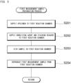
- the microcomputer 82 performs a first measurement sample preparation process (step S104), a first measurement process (step S105), a second measurement sample preparation process (step S106), and a second measurement process (step S107).
- the second measurement sample preparation process (step S106) and the second measurement process (step S107) may be performed before the first measurement sample preparation process (step S104) and first measurement process (step S105).
- the first reaction chamber 54A is heated to a predetermined temperature by the heater 55A. In the heated state, the mixture in the first reaction chamber 54A is stirred by using the pump 56A (step S203).
- the blood sample, the hemolyzing agent, and the nucleic acid staining reagent are caused to react with each other, and the microcomputer 82 thereafter performs control such that the first sample preparation unit 5A supplies a predetermined amount of the diluent from the reagent container 53 to the first reaction chamber 54A, to dilute the reaction solution of the blood sample, the hemolyzing agent, and the nucleic acid staining reagent, thereby preparing the first measurement sample.
- the microcomputer 82 performs control such that the first sample preparation unit 5A introduces the first measurement sample from the first reaction chamber 54A to the optical detector unit 6 (step S204).
- FIG. 4 is referred to again.
- the optical detector unit 6 measures the first measurement sample.
- the first sample preparation unit 5A supplies both the sheath liquid and the first measurement sample to the flow cell 61.
- the light source unit 62 applies light to flow of the first measurement sample in the flow cell 61.
- the first measurement sample flows in the flow cell 61, and blood cells including white blood cells, aggregate platelets, and red blood cells sequentially pass through the flow cell 61.
- the "aggregate platelets” refer to an aggregate of two or more platelets.
- Plasmodium is in the malaria-infected red blood cell.
- the red blood cell in the measurement sample is contracted by the action of the hemolyzing agent.
- the malaria-infected red blood cell is contracted by the hemolyzing agent while having Plasmodium therein.
- Plasmodium has a nucleus, and the nucleus is stained by the nucleic acid staining reagent.
- the white blood cell also has a nucleus, and is thus stained by the staining reagent.
- Neither red blood cells hereinafter, referred to as "normal red blood cell" which are not infected with Plasmodium nor aggregate platelets have nuclei, and are hardly stained by the nucleic acid staining reagent.
- the detector 63 detects the fluorescence generated from the blood cell.
- the detector 64 detects the scattered light (forward scattered light) generated from the blood cell.
- Each of the detectors 63, 64 outputs an electric signal based on the level of the received light, as the fluorescence signal and the forward scattered light signal.
- the signal processing circuit 81 extracts the fluorescence intensity from the fluorescence signal and extracts the forward scattered light intensity from the forward scattered light signal. After the first measurement sample has flowed through the flow cell 61, the washing unit 8 washes the flow cell 61.
- step S106 The second measurement sample preparation process (step S106) will be described.
- the microcomputer 82 performs control such that the suction unit 4 suctions a predetermined amount of blood sample from a test tube, and supplies 5 ⁇ L of the sample to the second reaction chamber 54B.
- the microcomputer 82 performs control such that the second sample preparation unit 5B supplies 1020 ⁇ L of the diluent from the reagent container 53 to the second reaction chamber 54B.
- the second reaction chamber 54B is heated to a predetermined temperature by the heater 55B. In the heated state, the mixture in the second reaction chamber 54B is stirred by using the pump 56B in step S303. In the operations from step S301 to S303, the second measurement sample is prepared in the second reaction chamber 54B. In the second measurement sample, the blood cells are not hemolyzed and stained. In step S304, the microcomputer 82 performs control such that the second sample preparation unit 5B introduces the second measurement sample from the second reaction chamber 54B to the optical detector unit 6.
- step S304 the microcomputer 82 returns the process to the main routine.
- the second measurement sample flows in the flow cell 61, and blood cells including red blood cells sequentially pass through the flow cell 61.
- An amount of protoporphyrin in red blood cells of a healthy person is small, whereas a large amount of protoporphyrin is contained in red blood cells of an iron-deficiency anemia patient.
- blue-violet laser light is applied to red blood cells containing a large amount of protoporphyrin, intrinsic fluorescence is generated.
- the intrinsic fluorescence is red light having a wavelength of not less than 600 nm and not greater than 700 nm, and the intrinsic fluorescence generated from each of the red blood cells is individually detected by the detector 63.
- the scattered light (forward scattered light) generated from the red blood cell has a wavelength of 405 nm and is detected by the detector 64.
- the detector 63 and the detector 64 each output an electric signal based on the level of the received light, as the fluorescence signal and the forward scattered light signal.
- the signal processing circuit 81 extracts the fluorescence intensity from the fluorescence signal, and extracts the forward scattered light intensity from the forward scattered light signal. After the second measurement sample has flowed through the flow cell 61, the washing unit 8 washes the flow cell 61.
- the second detector unit 7 measures the third measurement sample.
- the second detector unit 7 applies a voltage to the third measurement sample flowing in the sheath flow cell, and outputs an analog signal indicating the voltage, to the signal processing circuit 81.
- the signal processing circuit 81 detects the change of the electric resistance by signal processing and detects the red blood cell.
- the signal processing circuit 81 converts the output signal from the second detector unit 7, to detection data of the red blood cell.
- the microcomputer 82 operates to transmit first, second, and third measurement data including feature parameters to the information processing unit 3 (step S110), and ends the process.
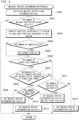
- the first measurement data analysis process will be described with reference to FIG. 7 .
- the CPU 301 firstly sorts the detected blood cells into white blood cells, aggregate platelets, normal red blood cells, and malaria-infected red blood cells based on the fluorescence intensity and the forward scattered light intensity included in the first measurement data, and counts each of the white blood cells, the aggregate platelets, the normal red blood cells, and the malaria-infected red blood cells (step S401).
- FIG. 8 An operation of sorting the detected blood cells into white blood cells, aggregate platelets, normal red blood cells, and malaria-infected red blood cells will be described with reference to FIG. 8 .
- the vertical axis represents forward scattered light intensities
- the horizontal axis represents fluorescence intensities.
- the white blood cells substantially appear in a region 401 (hereinafter, a particle group in the region 401 is referred to as a first particle group) in which the forward scattered light intensity is higher than that of an aggregation of normal red blood cells and malaria-infected red blood cells, and the fluorescence intensity is higher than that of an aggregation of the normal red blood cells and aggregate platelets.
- the aggregate platelets substantially appear in a region 402 in which the forward scattered light intensity is almost the same as that in the region 401, and the fluorescence intensity is lower than that in the region 401.
- the normal red blood cell is highly contracted by the hemolyzing agent.
- the malaria-infected red blood cells are contracted by the hemolyzing agent and each contains Plasmodium having a nucleus therein, and is thus stained by the nucleic acid staining reagent. Therefore, the malaria-infected red blood cells substantially appear in a region 404 (hereinafter, a particle group in the region 404 is referred to as a third particle group) in which the forward scattered light intensity is lower than that in the region 401 and the fluorescence intensity is higher than that in the region 403.
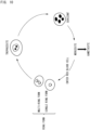
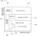
- Sorting the malaria-infected red blood cells into sub-aggregations will be described with reference to FIG. 10 and FIG. 11 .
- FIG. 10 if blood is sucked by an anopheline having Plasmodium, both saliva and a protozoan of the anopheline are injected into blood.
- the protozoan enters a hepatic cell and proliferates in the cell, and is released again into the blood.
- the morphology of the protozoan is called merozoite. If the merozoite is released into the blood, the merozoite immediately enters a red blood cell, and grows while changing the morphology.
- the trophozoite or schizont appears in a region 413 in which the fluorescence intensity and the forward scattered light intensity are higher than those of the ring form in a region 404 in which malaria-infected red blood cells appear.
- the gametocyte appears in a region 414 in which the fluorescence intensity is lower than that of the trophozoite or schizont, and the forward scattered light intensity is about the same as that of the trophozoite or schizont which has a higher forward scattered light intensity.
- the single ring form appears in a region 411 in which the fluorescence intensity is about the same as that of the gametocyte having a lower fluorescence intensity, and the forward scattered light intensity is lower than that of the gametocyte.
- the multi-ring form appears in a region 412 in which the fluorescence intensity is higher than that of the single ring form, and the forward scattered light intensity is about the same as that of the single ring form having a lower forward scattered light intensity.
- the CPU 301 performs a malaria species determination process in step S402.
- That the blood sample is suspected of being infected with Plasmodium falciparum means that the blood sample is likely to be infected with Plasmodium falciparum but is not highly likely to be infected therewith.
- Both of the malaria species/stage determination flags having been set as 2 and 3 mean that infection with Plasmodium falciparum is likely to occur.
- the malaria species/stage determination flag having been set as 3 indicates that the blood sample is suspected of being infected with the other species of Plasmodium.
- the malaria species/stage determination flag having been set as 4 indicates that the blood sample is highly likely to be infected with the other species of Plasmodium.
- That the blood sample is suspected of being infected with the other species of Plasmodium means that the blood sample is likely to be infected with the other species of Plasmodium but is not highly likely to be infected therewith.
- Both of the malaria species/stage determination flags having been set as 3 and 4 mean that infection with the other species of Plasmodium is likely to occur.
- step S502 the CPU 301 determines whether or not the number of malaria-infected red blood cells counted in step S401 is not less than a threshold value T1.
- the threshold value T1 is set such that, in a case where the number of malaria-infected red blood cells is less than T1, it can be determined that malaria-infected red blood cells do not appear.
- the blood sample can be determined as being not infected with Plasmodium.
- the CPU 301 ends the malaria species determination process, and the process proceeds to calculation of a malaria-infected red blood cell ratio (step S403).
- the blood sample can be determined as being likely to be infected with Plasmodium.
- the CPU 301 shifts the process to step S503.
- step S503 the CPU 301 counts particles belonging to the region 411 in the scattergram illustrated in FIG. 11 , as single ring forms, counts particles belonging to the region 412, as multi-ring forms, counts particles belonging to the region 413, as trophozoites or schizonts (hereinafter, referred to as "trophozoites/schizonts"), and counts particles belonging to the region 414, as gametocytes (step S503).
- step S506 the CPU 301 determines whether or not the number of single ring forms is not less than a threshold value T3 (step S506).
- the threshold value T3 is set such that, in a case where the number of single ring forms is less than T3, it can be determined that single ring forms do not appear. In a case where the number of the single ring forms is less than T3 (NO in step S506), the CPU 301 shifts the process to step S511.
- the CPU 301 determines whether or not a representative value of the forward scattered light intensities is not less than a threshold value T4 in the region 411 in which the single ring forms appear (step S507).
- the size of the single ring form of Plasmodium falciparum is smaller than the size of the single ring form of the other species of Plasmodium.
- the forward scattered light intensity represents the size of a cell.
- the representative value of the forward scattered light intensities in the region 411 in which the single ring forms appear is an average value of the forward scattered light intensities of a plurality of particles that appear in the region 411.
- the threshold value T4 is set such that, in a case where the representative value is less than T4, it can be determined that falciparum malaria infection is likely to occur.
- the CPU 301 determines whether or not the number of multi-ring forms is not less than a threshold value T5 (step S508).
- the threshold value T5 is set such that, in a case where the number of the multi-ring forms is less than T5, it can be determined that multi-ring forms do not appear.
- the CPU 301 sets the malaria species determination flag as 1 (step S509), ends the malaria species determination process, and returns the process to the first measurement data analysis process.
- multi-ring forms can be determined as being likely to appear in the blood sample.
- the blood sample can be determined as being highly likely to be infected with Plasmodium falciparum. Therefore, the CPU 301 sets the malaria species determination flag as 2 (step S510), ends the malaria species determination process, and returns the process to the first measurement data analysis process.
- the CPU 301 determines whether or not the number of trophozoites/schizonts is not less than a threshold value T6 (step S511).
- the threshold value T6 is set such that, in a case where the number of trophozoites/schizonts is less than T6, it can be determined that trophozoites/schizonts do not appear in the blood sample.
- the number of trophozoites/schizonts appearing in peripheral blood of a person infected with Plasmodium falciparum is small, and trophozoites/schizonts often appear in peripheral blood of a person infected with the other species of Plasmodium.
- the number of trophozoites/schizonts is less than T6 (NO in step S511), it can be determined that trophozoites/schizonts do not appear in the blood sample.
- the blood sample cannot be determined as being highly likely to be infected with the other species of Plasmodium.
- the CPU 301 sets the malaria species/stage determination flag as 3 (step S512), ends the malaria species determination process, and returns the process to the first measurement data analysis process.
- trophozoites/schizonts can be determined as appearing in the blood sample. Furthermore, in this case, the blood sample can be determined as being highly likely to be infected with the other species of Plasmodium. Therefore, the CPU 301 sets the malaria species/stage determination flag as 4 (step S513), ends the malaria species determination process, and returns the process to the first measurement data analysis process.
- FIG. 12 illustrates an example of a scattergram in which an aggregation of single ring forms and an aggregation of multi-ring forms appear.
- the forward scattered light intensity and the fluorescence intensity of the aggregation of single ring forms are lower than those of the aggregation of white blood cells.
- the fluorescence intensity of the aggregation of single ring forms is lower than that of the aggregation of multi-ring forms.
- FIG. 13 illustrates an example of a scattergram in which an aggregation of single ring forms and an aggregation of gametocytes appear. As illustrated in FIG.
- FIG. 14 illustrates an example of a scattergram in which an aggregation of single ring forms and an aggregation of trophozoites/schizonts appear. As illustrated in FIG. 14 , the fluorescence intensity of the aggregation of trophozoites/schizonts is higher than that of the aggregation of single ring forms.
- FIG. 7 is referred to again.
- the malaria species determination process as described above ends, and the CPU 301 then calculates a ratio (hereinafter, referred to as "malaria-infected red blood cell ratio") of the number of malaria-infected red blood cells (the number of blood cells in the region 404) to the total number of red blood cells (the number of red blood cells counted in step S112) (step S403).
- the CPU 301 then ends the first measurement data analysis process, and returns the process to the main routine shown in FIG. 4 .
- the CPU 301 performs a second measurement data analysis process for generating information about iron-deficiency anemia, generates a result of analysis of the blood sample, and stores the analysis result in the hard disk 304 (step S114).
- the CPU 301 may firstly perform the second measurement data analysis process (step S114) and subsequently perform the first measurement data analysis process (step S 113).
- the second measurement data analysis process will be described with reference to FIG. 15 .
- the second measurement data analysis process starts, and, then, the CPU 301 firstly sets, as 0, an initial value of an iron-deficiency anemia determination flag representing possibility of iron-deficiency anemia in step S601.
- the iron-deficiency anemia determination flag is stored in a specific region of the RAM 303.
- the iron-deficiency anemia determination flag having been set as 0 indicates that iron-deficiency anemia is less likely to occur, and the iron-deficiency anemia determination flag having been set as 1 indicates that iron-deficiency anemia is likely to occur.
- the CPU 301 extracts particles in which the fluorescence intensity is not less than a predetermined threshold value, as red blood cells (hereinafter, referred to as "intrinsically fluorescent red blood cell”) that generate intrinsic fluorescence, from an aggregation of particles determined as red blood cells, and counts the intrinsically fluorescent red blood cells.
- red blood cells hereinafter, referred to as "intrinsically fluorescent red blood cell”
- the number of the intrinsically fluorescent red blood cells is referred to as “intrinsically fluorescent red blood cell number”.
- red blood cell in which the fluorescence intensity having the threshold value or a greater value is not detected is referred to as "red blood cell that does not generate intrinsic fluorescence”.
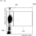
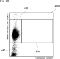
- the process step of step S602 will be described with reference to FIG. 16A to FIG. 16D .
- the CPU 301 generates a scattergram 4000, for a group of particles in the region 403 in the scattergram generated in step S401 shown in FIG. 7 , in which the vertical axis represents forward scattered light intensities, and the horizontal axis represents fluorescence intensities.
- the CPU 301 identifies, as red blood cells that do not generate intrinsic fluorescence, particles in a region 400 in which the forward scattered light intensity is higher than that in a noise region 490, and the fluorescence intensity is lower than that in a region 410 in which intrinsically fluorescent red blood cells appear.
- the CPU 301 identifies, as intrinsically fluorescent red blood cells, particles in the region 410 in which the forward scattered light intensity is about the same as that in the region 400, and the fluorescence intensity is higher than that in the region 400.
- the CPU 301 counts the particles in the region 410, and determines the intrinsically fluorescent red blood cell number.
- FIG. 16A shows a result of measurement of a normal sample, that is, a blood sample collected from a healthy person.
- the number of particles appearing in the region 410 is small, and particles regarded as the intrinsically fluorescent red blood cells are hardly detected.
- FIG. 16B shows a result of measurement of a blood sample (hereinafter, referred to as "iron-deficiency anemia sample”) collected from an iron-deficiency anemia patient, and a lot of particles appear in the region 410 and a lot of particles regarded as intrinsically fluorescent red blood cells are detected.
- iron-deficiency anemia sample a blood sample collected from an iron-deficiency anemia patient
- FIG. 16C and FIG. 16D each show a result of measurement of a blood sample (hereinafter, referred to as "thalassemia sample”) collected from a thalassemia patient, and the number of particles appearing in the region 410 is small, and particles regarded as intrinsically fluorescent red blood cells are hardly detected.
- thalassemia sample a blood sample collected from a thalassemia patient
- the number of particles appearing in the region 410 is small, and particles regarded as intrinsically fluorescent red blood cells are hardly detected.
- thalassemia sample a blood sample collected from a thalassemia patient
- thalassemia sample a blood sample collected from a thalassemia patient
- the number of particles appearing in the region 410 is small, and particles regarded as intrinsically fluorescent red blood cells are hardly detected.
- thalassemia sample is classified as microcytic anemia and resembles iron-deficiency anemia in symptoms, a test value of a complete blood count (CBC) item, and
- step S603 the CPU 301 calculates, as intrinsic fluorescence information, a ratio (hereinafter, referred to as "intrinsic fluorescence ratio") of the intrinsically fluorescent red blood cell number obtained in step S602 to the number of red blood cells counted in step S 112 ( FIG. 4 ).
- intrinsic fluorescence ratio a ratio (hereinafter, referred to as "intrinsic fluorescence ratio") of the intrinsically fluorescent red blood cell number obtained in step S602 to the number of red blood cells counted in step S 112 ( FIG. 4 ).
- the sum of the number of particles in the region 400 shown in FIG. 16A to FIG. 16D and the number of particles in the region 410 shown therein may be used as the number of red blood cells.
- the CPU 301 determines, in step S604, whether or not the intrinsic fluorescence ratio is not less than a threshold value T7.
- the threshold value T7 is set such that, in a case where the intrinsic fluorescence ratio is not less than T7, iron-deficiency anemia can be determined as being likely to occur.
- the CPU 301 sets the iron-deficiency anemia determination flag as 1 in step S605, ends the second measurement data analysis process, and returns the process to the main routine.
- the CPU 301 maintains the iron-deficiency anemia determination flag as 0, ends the second measurement data analysis process, and returns the process to the main routine.
- FIG. 4 is referred to again.
- the second measurement data analysis process as described above ends, and the CPU 301 then causes the display unit 310 to display the analysis result (step S115) and ends the process.
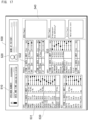
- An analysis result screen displayed on the display unit 310 will be described with reference to FIG. 17 .
- An analysis result screen 500 includes a sample information display region 510, a patient information display region 520, a measurement result display region 530, and a reference information display region 540.
- the measurement result display region 530 includes a CBC item display region 531, a malaria item display region 532, and an intrinsic fluorescence item display region 533.
- Information displayed in the sample information display region 510 represents a blood sample from which the analysis result displayed on the analysis result screen 500 has been obtained.
- Information displayed in the patient information display region 520 information of a subject from which the blood sample is collected is displayed.
- measured values of the respective items which are obtained by the measurement data analysis process are displayed.
- measured values of basic measurement items in blood cell analysis are displayed.
- the measured values displayed in the CBC item display region 531 include measured values of red blood cells (RBC) and white blood cells (WBC).
- information used for determining whether or not iron preparation is to be supplied to an iron-deficiency anemia patient is generated by a single blood analyzer. Therefore, measurement for determining whether or not iron preparation is to be supplied to an iron-deficiency anemia patient can be facilitated.
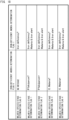
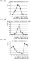
- an automatic multi-item blood cell analyzer XN-30 manufactured by SYSMEX CORPORATION was used to measure a ratio of the number of malaria-infected red blood cells to the total number of red blood cells, the intrinsically fluorescent red blood cell number, and the number of red blood cells.
- FIG. 19 shows the result.
- the automatic multi-item blood cell analyzer XN-30 has the same configuration as the optical detector unit 6.
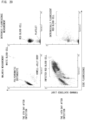
- FIG. 20 illustrates scattergrams on the 0th day after the infection and the 14th day after the infection. It is indicated that the infection increased red blood cells exhibiting staining fluorescence characteristics and scattered light characteristics specific to malaria infection, and increased red blood cells exhibiting intrinsic fluorescence characteristics specific to iron-deficiency anemia.
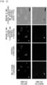
- both staining fluorescence derived from nucleic acid of Plasmodium and intrinsic fluorescence derived from protoporphyrin IX were not observed in the uninfected mouse.
- both fluorescence derived from a dye by which nucleic acid of Plasmodium was stained and intrinsic fluorescence derived from protoporphyrin IX were observed in the malaria-infected mouse.
- the optical detector unit 6 of the blood analyzer according to the present embodiment was able to detect that malaria-infected red blood cells and protoporphyrin IX-containing red blood cells (iron-deficient red blood cells) were in a malaria-infected mouse.
Landscapes
- Health & Medical Sciences (AREA)
- Life Sciences & Earth Sciences (AREA)
- Engineering & Computer Science (AREA)
- Chemical & Material Sciences (AREA)
- General Health & Medical Sciences (AREA)
- Physics & Mathematics (AREA)
- Pathology (AREA)
- Immunology (AREA)
- Biomedical Technology (AREA)
- Biochemistry (AREA)
- Analytical Chemistry (AREA)
- General Physics & Mathematics (AREA)
- Public Health (AREA)
- Medical Informatics (AREA)
- Primary Health Care (AREA)
- Epidemiology (AREA)
- Molecular Biology (AREA)
- Nuclear Medicine, Radiotherapy & Molecular Imaging (AREA)
- Optics & Photonics (AREA)
- Dispersion Chemistry (AREA)
- Hematology (AREA)
- General Business, Economics & Management (AREA)
- Chemical Kinetics & Catalysis (AREA)
- Databases & Information Systems (AREA)
- Business, Economics & Management (AREA)
- Data Mining & Analysis (AREA)
- Food Science & Technology (AREA)
- Biophysics (AREA)
- Urology & Nephrology (AREA)
- Medicinal Chemistry (AREA)
- Ecology (AREA)
- Investigating, Analyzing Materials By Fluorescence Or Luminescence (AREA)
- Investigating Or Analysing Biological Materials (AREA)
- Measuring Or Testing Involving Enzymes Or Micro-Organisms (AREA)
- Investigating Or Analysing Materials By Optical Means (AREA)
- Apparatus Associated With Microorganisms And Enzymes (AREA)
Claims (14)
- Analyseur de sang (1) comprenant :une première unité (5A) de préparation d'échantillon configurée de manière à préparer, à partir d'un échantillon de sang, un premier échantillon de mesure pour mesurer un globule rouge infecté par le paludisme ;une seconde unité (5B) de préparation d'échantillon configurée de manière à préparer, à partir de l'échantillon de sang, un second échantillon de mesure pour mesurer la fluorescence intrinsèque d'un globule rouge ;une unité (62) formant source de lumière configurée de manière à appliquer de la lumière au premier échantillon de mesure et au second échantillon de mesure ;une unité de détection (63, 64) configurée de manière à détecter la fluorescence et la lumière diffusée générées par le premier échantillon de mesure auquel de la lumière a été appliquée, et à détecter la fluorescence intrinsèque générée par le second échantillon de mesure auquel de la lumière a été appliquée ; etune unité (3) de traitement d'informations configurée de manière à générer des informations relative à une infection palustre sur la base de la fluorescence et de la lumière diffusée détectées à partir du premier échantillon de mesure, et à générer des informations relatives à une anémie ferriprive sur la base de la fluorescence intrinsèque détectée à partir du second échantillon de mesure,dans lequel l'unité (3) de traitement d'informations émet des informations d'affichage afin d'afficher simultanément, sur une unité d'affichage (310), les informations relatives à une infection palustre et les informations relatives à une anémie ferriprive,dans lequelles informations relatives à une infection palustre incluent le résultat d'une comparaison entre un nombre de globules rouges infectés par le paludisme ou un rapport entre le nombre de globules rouges infectés par le paludisme et un nombre de globules rouges, et une valeur de seuil pour déterminer la possibilité d'une infection palustre,les informations relatives à une anémie ferriprive incluent le résultat d'une comparaison entre un nombre de globules rouges qui génèrent une fluorescence intrinsèque ou le rapport entre le nombre de globules rouges qui génèrent une fluorescence intrinsèque et le nombre de globules rouges, et une valeur de seuil pour déterminer la possibilité d'une anémie ferriprive, etl'unité (3) de traitement d'informations génère un indicateur d'alerte dans un cas où le résultat de la comparaison avec la valeur de seuil pour déterminer la possibilité d'une infection palustre indique qu'une infection palustre est probable, et le résultat de la comparaison avec la valeur de seuil pour déterminer la possibilité d'une anémie ferriprive indique qu'une anémie ferriprive est probable.
- Analyseur de sang (1) selon la revendication 1, dans lequel la première unité (5A) de préparation d'échantillon mélange l'échantillon de sang, un agent hémolytique pour contracter un globule rouge, et un colorant de coloration d'acide nucléique, afin de préparer le premier échantillon de mesure.
- Analyseur de sang (1) selon la revendication 1 ou la revendication 2, dans lequel la seconde unité (5B) de préparation d'échantillon mélange l'échantillon de sang et un diluant qui ne contient pas d'agent hémolytique pour contracter un globule rouge et un colorant de coloration d'acide nucléique, afin de préparer le second échantillon de mesure.
- Analyseur de sang (1) selon l'une quelconque des revendications 1 à 3, comprenant en outre une unité d'aspiration (4) configurée de manière à aspirer l'échantillon de sang à partir d'un récipient d'échantillon qui stocke l'échantillon de sang, dans lequel
la première unité (5A) de préparation d'échantillon et la seconde unité (5B) de préparation d'échantillon préparent le premier échantillon de mesure et le second échantillon de mesure, respectivement, à partir d'une partie de l'échantillon de sang aspiré par l'unité d'aspiration (4). - Analyseur de sang (1) selon l'une quelconque des revendications 1 à 4, dans lequel l'unité formant source (62) de lumière émet de la lumière dans une bande de longueur d'onde qui n'est pas inférieure à 400 nm et pas supérieure à 435 nm.
- Analyseur de sang (1) selon l'une quelconque des revendications 1 à 5, comprenant en outre une cellule d'écoulement (61) dans laquelle le premier et le second échantillons de mesure s'écoulent, dans lequell'unité (62) formant source de lumière est configurée de manière à appliquer de la lumière au premier et au second échantillons de mesure qui s'écoulent dans la cellule d'écoulement, etle détecteur (63, 64) est configuré de manière à détecter la fluorescence et la lumière diffusée générées par le premier échantillon de mesure qui s'écoule dans la cellule d'écoulement, et à détecter la fluorescence intrinsèque générée par le second échantillon de mesure qui s'écoule dans la cellule d'écoulement.
- Analyseur de sang (1) selon la revendication 6, comprenant en outre une unité de lavage (8) configurée de manière à laver la cellule d'écoulement (61) après que l'un parmi le premier et le second échantillons de mesure soit passé à travers la cellule d'écoulement et avant que l'autre parmi le premier et le second échantillons de mesure passe à travers la cellule d'écoulement.
- Analyseur de sang (1) selon l'une quelconque des revendications 1 à 7, dans lequel les informations relatives à une infection palustre incluent un nombre de globules rouges infectés par la paludisme ou un rapport entre le nombre de globules rouges infectés par la paludisme et un nombre de globules rouges.
- Analyseur de sang (1) selon l'une quelconque des revendications 1 à 8, dans lequel les informations relatives à une infection palustre incluent des informations indiquant la possibilité d'une infection palustre sur la base du résultat d'une comparaison entre un nombre de globules rouges infectés par le paludisme ou un rapport entre le nombre de globules rouges infectés par le paludisme et un nombre de globules rouges, et une valeur de seuil pour déterminer la possibilité d'une infection palustre.
- Analyseur de sang (1) selon la revendication 9, dans lequel les informations indiquant la possibilité d'une infection palustre incluent des informations indiquant une espèce plasmodiale qui a provoqué l'infection.
- Analyseur de sang (1) selon l'une quelconque des revendications 1 à 10, dans lequel les informations relatives à une anémie ferriprive incluent un nombre de globules rouges qui génèrent une fluorescence intrinsèque, ou un rapport entre le nombre de globules rouges qui génèrent une fluorescence intrinsèque et un nombre de globules rouges.
- Analyseur de sang (2) selon l'une quelconque des revendications 1 à 11, dans lequel les informations relatives à une anémie ferriprive incluent des informations indiquant la possibilité d'une anémie ferriprive sur la base du résultat d'une comparaison entre un nombre de globules rouges qui génèrent une fluorescence intrinsèque ou un rapport entre le nombre de globules rouges qui génèrent une fluorescence intrinsèque et un nombre de globules rouges, et une valeur de seuil pour déterminer la possibilité d'une anémie ferriprive.
- Appareil de traitement d'informations (3) pour générer des informations relatives à une infection palustre et des informations relatives à une anémie ferriprive, l'appareil de traitement d'informations (2) comprenant un dispositif de commande (301), dans lequelle dispositif de commande (301)est configuré de manière à être connecté à une unité de détection (6A) dans le but de détecter la fluorescence et la lumière diffusée générées par une application de lumière à un premier échantillon de mesure afin de mesurer un globule rouge infecté par le paludisme, et de détecter la fluorescence intrinsèque générée par une application de lumière à un second échantillon de mesure afin de mesurer la fluorescence intrinsèque d'un globule rouge, etest programmé pour générer des informations relatives à une infection palustre sur la base de la fluorescence détectée et de la lumière diffusée, et pour générer des informations relatives à une anémie ferriprive sur la base de la fluorescence intrinsèque détectée à partir du second échantillon de mesure, l'appareil de traitement d'informations (3) comprenant en outre une unité d'affichage (310),dans lequel le dispositif de commande (301) génère des informations d'affichage afin d'afficher simultanément, sur l'unité d'affichage (310), les informations relatives à une infection palustre et les informations relatives à une anémie ferriprive,dans lequelles informations relatives à une infection palustre incluent le résultat d'une comparaison entre un nombre de globules rouges infectés par le paludisme ou un rapport entre le nombre de globules rouges infectés par le paludisme et un nombre de globules rouges, et une valeur de seuil pour déterminer la possibilité d'une infection palustre,les informations relatives à une anémie ferriprive incluent le résultat d'une comparaison entre le nombre de globules rouges qui génèrent une fluorescence intrinsèque ou le rapport entre le nombre de globules rouges qui génèrent une fluorescence intrinsèque et le nombre de globules rouges, et une valeur de seuil pour déterminer la possibilité d'une anémie ferriprive, etle dispositif de commande (301) génère un indicateur d'alerte dans un cas où le résultat de la comparaison avec la valeur de seuil pour déterminer la possibilité d'une infection palustre indique qu'une infection palustre est probable, et le résultat de la comparaison avec la valeur de seuil pour déterminer la possibilité d'une anémie ferriprive indique qu'une anémie ferriprive est probable.
- Programme de commande d'un appareil de traitement d'informations (3) qui génère des informations relatives à une infection palustre et des informations relatives à une anémie ferriprive, dans lequell'appareil de traitement d'informations (3) est connecté à une unité de détection (6A) pour détecter la fluorescence et la lumière diffusée générées par une application de lumière à un premier échantillon de mesure afin de mesurer un globule rouge infecté par le paludisme, et pour détecter la fluorescence intrinsèque générée par une application de lumière à un second échantillon de mesure afin de mesurer la fluorescence intrinsèque d'un globule rouge, etle programme amène l'appareil de traitement d'informations (3) à exécuter les étapes suivantes :générer des informations relatives à une infection palustre sur la base de la fluorescence détectée et de la lumière diffusée ;générer des informations relatives à une anémie ferriprive sur la base de la fluorescence intrinsèque détectée à partir du second échantillon de mesure,générer des informations d'affichage afin d'afficher simultanément, sur une unité d'affichage de l'appareil de traitement d'informations, les informations relatives à une infection palustre et les informations relatives à une anémie ferriprive,dans lequelles informations relatives à une infection palustre incluent le résultat d'une comparaison entre un nombre de globules rouges infectés par le paludisme ou un rapport entre le nombre de globules rouges infectés par le paludisme et un nombre de globules rouges, et une valeur de seuil pour déterminer la possibilité d'une infection palustre,les informations relatives à une anémie ferriprive incluent le résultat d'une comparaison entre le nombre de globules rouges qui génèrent une fluorescence intrinsèque ou le rapport entre le nombre de globules rouges qui génèrent une fluorescence intrinsèque et le nombre de globules rouges, et une valeur de seuil pour déterminer la possibilité d'une anémie ferriprive, etgénérer un indicateur d'alerte dans un cas où le résultat de la comparaison avec la valeur de seuil pour déterminer la possibilité d'une infection palustre indique qu'une infection palustre est probable, et le résultat de la comparaison avec la valeur de seuil pour déterminer la possibilité d'une anémie ferriprive indique qu'une anémie ferriprive est probable.
Applications Claiming Priority (1)
| Application Number | Priority Date | Filing Date | Title |
|---|---|---|---|
| JP2020071704A JP7583401B2 (ja) | 2020-04-13 | 2020-04-13 | 血液分析装置 |
Publications (2)
| Publication Number | Publication Date |
|---|---|
| EP3896426A1 EP3896426A1 (fr) | 2021-10-20 |
| EP3896426B1 true EP3896426B1 (fr) | 2023-12-20 |
Family
ID=75396660
Family Applications (1)
| Application Number | Title | Priority Date | Filing Date |
|---|---|---|---|
| EP21163161.9A Active EP3896426B1 (fr) | 2020-04-13 | 2021-03-17 | Analyseur de sang |
Country Status (4)
| Country | Link |
|---|---|
| US (1) | US12174118B2 (fr) |
| EP (1) | EP3896426B1 (fr) |
| JP (1) | JP7583401B2 (fr) |
| CN (1) | CN113533265B (fr) |
Families Citing this family (2)
| Publication number | Priority date | Publication date | Assignee | Title |
|---|---|---|---|---|
| JP2023137082A (ja) * | 2022-03-17 | 2023-09-29 | シスメックス株式会社 | 測定装置および分析方法 |
| JP2024066938A (ja) * | 2022-11-02 | 2024-05-16 | シスメックス株式会社 | 光学システムおよび検体分析装置 |
Family Cites Families (16)
| Publication number | Priority date | Publication date | Assignee | Title |
|---|---|---|---|---|
| US6100038A (en) * | 1996-09-06 | 2000-08-08 | Litron Laboratories Limited | Method for the enumeration of micronucleated erythrocyte populations with a single laser flow cytometer |
| KR100381725B1 (ko) * | 2000-12-29 | 2003-04-26 | (학)가톨릭학원가톨릭중앙의료원 | 용혈성 빈혈 검사 방법 |
| US20060257883A1 (en) * | 2005-05-10 | 2006-11-16 | Bjoraker David G | Detection and measurement of hematological parameters characterizing cellular blood components |
| JP4861874B2 (ja) * | 2007-03-29 | 2012-01-25 | シスメックス株式会社 | 分析装置 |
| WO2009136573A1 (fr) * | 2008-05-09 | 2009-11-12 | シスメックス株式会社 | Dispositif d'analyse de sang, procédé d'analyse de sang, agent hémolytique et agent colorant |
| KR101319202B1 (ko) | 2011-06-07 | 2013-10-23 | 포항공과대학교 산학협력단 | pLDH에 특이적으로 결합하는 DNA 압타머 |
| CN104144947B (zh) | 2011-12-14 | 2017-10-27 | 艾伯维德国有限责任两合公司 | 用于诊断和治疗铁相关病症的组合物和方法 |
| JP6027742B2 (ja) * | 2011-12-28 | 2016-11-16 | シスメックス株式会社 | 血球分析装置、血球分析方法、及びコンピュータプログラム |
| CA2796794C (fr) * | 2012-11-23 | 2015-06-16 | Westport Power Inc. | Procede et systeme de distribution d'un carburant gazeux dans le systeme d'admission d'air d'un moteur a combustion interne |
| JP6612050B2 (ja) * | 2014-08-28 | 2019-11-27 | シスメックス株式会社 | 血液分析装置、診断支援方法、およびコンピュータプログラム |
| WO2016094615A1 (fr) * | 2014-12-11 | 2016-06-16 | The Penn State Research Foundation | Aliment médical pour le traitement de la malaria et/ou la déficience en fer |
| JP6542557B2 (ja) * | 2015-03-27 | 2019-07-10 | シスメックス株式会社 | 血液測定装置および血液測定装置の制御方法 |
| JP6609429B2 (ja) * | 2015-06-30 | 2019-11-20 | 日本光電工業株式会社 | 血液分析方法および血液分析装置 |
| JP6635720B2 (ja) * | 2015-08-31 | 2020-01-29 | シスメックス株式会社 | 血液分析装置及び血液分析方法 |
| JP6710512B2 (ja) * | 2015-09-14 | 2020-06-17 | シスメックス株式会社 | 血液分析装置、血液分析方法及びコンピュータプログラム |
| JP6871729B2 (ja) * | 2016-12-06 | 2021-05-12 | シスメックス株式会社 | マラリア原虫に感染した赤血球の検出方法及び血液分析装置 |
-
2020
- 2020-04-13 JP JP2020071704A patent/JP7583401B2/ja active Active
-
2021
- 2021-03-17 EP EP21163161.9A patent/EP3896426B1/fr active Active
- 2021-04-12 CN CN202110387267.0A patent/CN113533265B/zh active Active
- 2021-04-12 US US17/228,220 patent/US12174118B2/en active Active
Also Published As
| Publication number | Publication date |
|---|---|
| EP3896426A1 (fr) | 2021-10-20 |
| CN113533265A (zh) | 2021-10-22 |
| JP7583401B2 (ja) | 2024-11-14 |
| CN113533265B (zh) | 2025-03-28 |
| US20210318240A1 (en) | 2021-10-14 |
| US12174118B2 (en) | 2024-12-24 |
| JP2021167795A (ja) | 2021-10-21 |
Similar Documents
| Publication | Publication Date | Title |
|---|---|---|
| US10107754B2 (en) | Blood analyzer and blood analyzing method | |
| US20070020721A1 (en) | Blood analyzing method and blood analyzer | |
| JP5670052B2 (ja) | 血液分析装置、血液分析方法及びコンピュータプログラム | |
| JP5721874B2 (ja) | 血液分析装置 | |
| CN102016573B (zh) | 血液分析装置,血液分析方法,溶血剂及染色剂 | |
| JP7340002B2 (ja) | 血液分析装置、血液分析方法およびプログラム | |
| US20250237644A1 (en) | Bone marrow fluid analysis method, sample analyzer, and non-transitory storage medium | |
| JP7209771B2 (ja) | 血液分析装置および血液分析方法 | |
| US11002746B2 (en) | Blood analyzer and blood analyzing method for determining if plasmodium infection is present | |
| US7354775B2 (en) | Reagent for partially lysing a cell membrane of a red blood cell, a reagent for detecting malaria infected red blood cells, and a sample analyzing method for detecting malaria infected red blood cells | |
| US12174118B2 (en) | Blood analyzer | |
| CN106483278B (zh) | 血液分析方法以及在其中使用的染色液及血液分析装置 | |
| CN119290715A (zh) | 血小板凝集的检测方法 | |
| JP2026059656A (ja) | 測定装置 | |
| JP2026059654A (ja) | 測定装置、試薬及び分析方法 | |
| JP2026074826A (ja) | 検体測定装置 | |
| JP2005333868A (ja) | マラリア原虫測定方法及び測定装置 | |
| JP2021103109A (ja) | 血液分析方法 |
Legal Events
| Date | Code | Title | Description |
|---|---|---|---|
| PUAI | Public reference made under article 153(3) epc to a published international application that has entered the european phase |
Free format text: ORIGINAL CODE: 0009012 |
|
| STAA | Information on the status of an ep patent application or granted ep patent |
Free format text: STATUS: THE APPLICATION HAS BEEN PUBLISHED |
|
| AK | Designated contracting states |
Kind code of ref document: A1 Designated state(s): AL AT BE BG CH CY CZ DE DK EE ES FI FR GB GR HR HU IE IS IT LI LT LU LV MC MK MT NL NO PL PT RO RS SE SI SK SM TR |
|
| B565 | Issuance of search results under rule 164(2) epc |
Effective date: 20210906 |
|
| STAA | Information on the status of an ep patent application or granted ep patent |
Free format text: STATUS: REQUEST FOR EXAMINATION WAS MADE |
|
| 17P | Request for examination filed |
Effective date: 20220331 |
|
| RBV | Designated contracting states (corrected) |
Designated state(s): AL AT BE BG CH CY CZ DE DK EE ES FI FR GB GR HR HU IE IS IT LI LT LU LV MC MK MT NL NO PL PT RO RS SE SI SK SM TR |
|
| GRAP | Despatch of communication of intention to grant a patent |
Free format text: ORIGINAL CODE: EPIDOSNIGR1 |
|
| STAA | Information on the status of an ep patent application or granted ep patent |
Free format text: STATUS: GRANT OF PATENT IS INTENDED |
|
| RIC1 | Information provided on ipc code assigned before grant |
Ipc: G01N 33/569 20060101ALI20230626BHEP Ipc: G01N 33/49 20060101ALI20230626BHEP Ipc: G01N 15/14 20060101ALI20230626BHEP Ipc: G01N 15/12 20060101AFI20230626BHEP |
|
| INTG | Intention to grant announced |
Effective date: 20230718 |
|
| GRAS | Grant fee paid |
Free format text: ORIGINAL CODE: EPIDOSNIGR3 |
|
| GRAA | (expected) grant |
Free format text: ORIGINAL CODE: 0009210 |
|
| STAA | Information on the status of an ep patent application or granted ep patent |
Free format text: STATUS: THE PATENT HAS BEEN GRANTED |
|
| AK | Designated contracting states |
Kind code of ref document: B1 Designated state(s): AL AT BE BG CH CY CZ DE DK EE ES FI FR GB GR HR HU IE IS IT LI LT LU LV MC MK MT NL NO PL PT RO RS SE SI SK SM TR |
|
| REG | Reference to a national code |
Ref country code: GB Ref legal event code: FG4D |
|
| REG | Reference to a national code |
Ref country code: CH Ref legal event code: EP |
|
| REG | Reference to a national code |
Ref country code: DE Ref legal event code: R096 Ref document number: 602021007779 Country of ref document: DE |
|
| REG | Reference to a national code |
Ref country code: IE Ref legal event code: FG4D |
|
| PG25 | Lapsed in a contracting state [announced via postgrant information from national office to epo] |
Ref country code: GR Free format text: LAPSE BECAUSE OF FAILURE TO SUBMIT A TRANSLATION OF THE DESCRIPTION OR TO PAY THE FEE WITHIN THE PRESCRIBED TIME-LIMIT Effective date: 20240321 |
|
| REG | Reference to a national code |
Ref country code: LT Ref legal event code: MG9D |
|
| PG25 | Lapsed in a contracting state [announced via postgrant information from national office to epo] |
Ref country code: LT Free format text: LAPSE BECAUSE OF FAILURE TO SUBMIT A TRANSLATION OF THE DESCRIPTION OR TO PAY THE FEE WITHIN THE PRESCRIBED TIME-LIMIT Effective date: 20231220 |
|
| REG | Reference to a national code |
Ref country code: NL Ref legal event code: MP Effective date: 20231220 |
|
| PG25 | Lapsed in a contracting state [announced via postgrant information from national office to epo] |
Ref country code: ES Free format text: LAPSE BECAUSE OF FAILURE TO SUBMIT A TRANSLATION OF THE DESCRIPTION OR TO PAY THE FEE WITHIN THE PRESCRIBED TIME-LIMIT Effective date: 20231220 |
|
| PG25 | Lapsed in a contracting state [announced via postgrant information from national office to epo] |
Ref country code: LT Free format text: LAPSE BECAUSE OF FAILURE TO SUBMIT A TRANSLATION OF THE DESCRIPTION OR TO PAY THE FEE WITHIN THE PRESCRIBED TIME-LIMIT Effective date: 20231220 Ref country code: GR Free format text: LAPSE BECAUSE OF FAILURE TO SUBMIT A TRANSLATION OF THE DESCRIPTION OR TO PAY THE FEE WITHIN THE PRESCRIBED TIME-LIMIT Effective date: 20240321 Ref country code: FI Free format text: LAPSE BECAUSE OF FAILURE TO SUBMIT A TRANSLATION OF THE DESCRIPTION OR TO PAY THE FEE WITHIN THE PRESCRIBED TIME-LIMIT Effective date: 20231220 Ref country code: ES Free format text: LAPSE BECAUSE OF FAILURE TO SUBMIT A TRANSLATION OF THE DESCRIPTION OR TO PAY THE FEE WITHIN THE PRESCRIBED TIME-LIMIT Effective date: 20231220 Ref country code: BG Free format text: LAPSE BECAUSE OF FAILURE TO SUBMIT A TRANSLATION OF THE DESCRIPTION OR TO PAY THE FEE WITHIN THE PRESCRIBED TIME-LIMIT Effective date: 20240320 |
|
| REG | Reference to a national code |
Ref country code: AT Ref legal event code: MK05 Ref document number: 1642855 Country of ref document: AT Kind code of ref document: T Effective date: 20231220 |
|
| PG25 | Lapsed in a contracting state [announced via postgrant information from national office to epo] |
Ref country code: NL Free format text: LAPSE BECAUSE OF FAILURE TO SUBMIT A TRANSLATION OF THE DESCRIPTION OR TO PAY THE FEE WITHIN THE PRESCRIBED TIME-LIMIT Effective date: 20231220 |
|
| PG25 | Lapsed in a contracting state [announced via postgrant information from national office to epo] |
Ref country code: SE Free format text: LAPSE BECAUSE OF FAILURE TO SUBMIT A TRANSLATION OF THE DESCRIPTION OR TO PAY THE FEE WITHIN THE PRESCRIBED TIME-LIMIT Effective date: 20231220 Ref country code: RS Free format text: LAPSE BECAUSE OF FAILURE TO SUBMIT A TRANSLATION OF THE DESCRIPTION OR TO PAY THE FEE WITHIN THE PRESCRIBED TIME-LIMIT Effective date: 20231220 Ref country code: NO Free format text: LAPSE BECAUSE OF FAILURE TO SUBMIT A TRANSLATION OF THE DESCRIPTION OR TO PAY THE FEE WITHIN THE PRESCRIBED TIME-LIMIT Effective date: 20240320 Ref country code: NL Free format text: LAPSE BECAUSE OF FAILURE TO SUBMIT A TRANSLATION OF THE DESCRIPTION OR TO PAY THE FEE WITHIN THE PRESCRIBED TIME-LIMIT Effective date: 20231220 Ref country code: LV Free format text: LAPSE BECAUSE OF FAILURE TO SUBMIT A TRANSLATION OF THE DESCRIPTION OR TO PAY THE FEE WITHIN THE PRESCRIBED TIME-LIMIT Effective date: 20231220 Ref country code: HR Free format text: LAPSE BECAUSE OF FAILURE TO SUBMIT A TRANSLATION OF THE DESCRIPTION OR TO PAY THE FEE WITHIN THE PRESCRIBED TIME-LIMIT Effective date: 20231220 |
|
| PG25 | Lapsed in a contracting state [announced via postgrant information from national office to epo] |
Ref country code: IS Free format text: LAPSE BECAUSE OF FAILURE TO SUBMIT A TRANSLATION OF THE DESCRIPTION OR TO PAY THE FEE WITHIN THE PRESCRIBED TIME-LIMIT Effective date: 20240420 |
|
| PG25 | Lapsed in a contracting state [announced via postgrant information from national office to epo] |
Ref country code: CZ Free format text: LAPSE BECAUSE OF FAILURE TO SUBMIT A TRANSLATION OF THE DESCRIPTION OR TO PAY THE FEE WITHIN THE PRESCRIBED TIME-LIMIT Effective date: 20231220 Ref country code: AT Free format text: LAPSE BECAUSE OF FAILURE TO SUBMIT A TRANSLATION OF THE DESCRIPTION OR TO PAY THE FEE WITHIN THE PRESCRIBED TIME-LIMIT Effective date: 20231220 |
|
| PG25 | Lapsed in a contracting state [announced via postgrant information from national office to epo] |
Ref country code: SK Free format text: LAPSE BECAUSE OF FAILURE TO SUBMIT A TRANSLATION OF THE DESCRIPTION OR TO PAY THE FEE WITHIN THE PRESCRIBED TIME-LIMIT Effective date: 20231220 |
|
| PG25 | Lapsed in a contracting state [announced via postgrant information from national office to epo] |
Ref country code: SM Free format text: LAPSE BECAUSE OF FAILURE TO SUBMIT A TRANSLATION OF THE DESCRIPTION OR TO PAY THE FEE WITHIN THE PRESCRIBED TIME-LIMIT Effective date: 20231220 Ref country code: SK Free format text: LAPSE BECAUSE OF FAILURE TO SUBMIT A TRANSLATION OF THE DESCRIPTION OR TO PAY THE FEE WITHIN THE PRESCRIBED TIME-LIMIT Effective date: 20231220 Ref country code: RO Free format text: LAPSE BECAUSE OF FAILURE TO SUBMIT A TRANSLATION OF THE DESCRIPTION OR TO PAY THE FEE WITHIN THE PRESCRIBED TIME-LIMIT Effective date: 20231220 Ref country code: IT Free format text: LAPSE BECAUSE OF FAILURE TO SUBMIT A TRANSLATION OF THE DESCRIPTION OR TO PAY THE FEE WITHIN THE PRESCRIBED TIME-LIMIT Effective date: 20231220 Ref country code: IS Free format text: LAPSE BECAUSE OF FAILURE TO SUBMIT A TRANSLATION OF THE DESCRIPTION OR TO PAY THE FEE WITHIN THE PRESCRIBED TIME-LIMIT Effective date: 20240420 Ref country code: EE Free format text: LAPSE BECAUSE OF FAILURE TO SUBMIT A TRANSLATION OF THE DESCRIPTION OR TO PAY THE FEE WITHIN THE PRESCRIBED TIME-LIMIT Effective date: 20231220 Ref country code: CZ Free format text: LAPSE BECAUSE OF FAILURE TO SUBMIT A TRANSLATION OF THE DESCRIPTION OR TO PAY THE FEE WITHIN THE PRESCRIBED TIME-LIMIT Effective date: 20231220 Ref country code: AT Free format text: LAPSE BECAUSE OF FAILURE TO SUBMIT A TRANSLATION OF THE DESCRIPTION OR TO PAY THE FEE WITHIN THE PRESCRIBED TIME-LIMIT Effective date: 20231220 |
|
| PG25 | Lapsed in a contracting state [announced via postgrant information from national office to epo] |
Ref country code: PT Free format text: LAPSE BECAUSE OF FAILURE TO SUBMIT A TRANSLATION OF THE DESCRIPTION OR TO PAY THE FEE WITHIN THE PRESCRIBED TIME-LIMIT Effective date: 20240422 Ref country code: PL Free format text: LAPSE BECAUSE OF FAILURE TO SUBMIT A TRANSLATION OF THE DESCRIPTION OR TO PAY THE FEE WITHIN THE PRESCRIBED TIME-LIMIT Effective date: 20231220 |
|
| PG25 | Lapsed in a contracting state [announced via postgrant information from national office to epo] |
Ref country code: PT Free format text: LAPSE BECAUSE OF FAILURE TO SUBMIT A TRANSLATION OF THE DESCRIPTION OR TO PAY THE FEE WITHIN THE PRESCRIBED TIME-LIMIT Effective date: 20240422 Ref country code: PL Free format text: LAPSE BECAUSE OF FAILURE TO SUBMIT A TRANSLATION OF THE DESCRIPTION OR TO PAY THE FEE WITHIN THE PRESCRIBED TIME-LIMIT Effective date: 20231220 |
|
| REG | Reference to a national code |
Ref country code: DE Ref legal event code: R097 Ref document number: 602021007779 Country of ref document: DE |
|
| PG25 | Lapsed in a contracting state [announced via postgrant information from national office to epo] |
Ref country code: DK Free format text: LAPSE BECAUSE OF FAILURE TO SUBMIT A TRANSLATION OF THE DESCRIPTION OR TO PAY THE FEE WITHIN THE PRESCRIBED TIME-LIMIT Effective date: 20231220 |
|
| PLBE | No opposition filed within time limit |
Free format text: ORIGINAL CODE: 0009261 |
|
| STAA | Information on the status of an ep patent application or granted ep patent |
Free format text: STATUS: NO OPPOSITION FILED WITHIN TIME LIMIT |
|
| PG25 | Lapsed in a contracting state [announced via postgrant information from national office to epo] |
Ref country code: SI Free format text: LAPSE BECAUSE OF FAILURE TO SUBMIT A TRANSLATION OF THE DESCRIPTION OR TO PAY THE FEE WITHIN THE PRESCRIBED TIME-LIMIT Effective date: 20231220 |
|
| PG25 | Lapsed in a contracting state [announced via postgrant information from national office to epo] |
Ref country code: SI Free format text: LAPSE BECAUSE OF FAILURE TO SUBMIT A TRANSLATION OF THE DESCRIPTION OR TO PAY THE FEE WITHIN THE PRESCRIBED TIME-LIMIT Effective date: 20231220 Ref country code: DK Free format text: LAPSE BECAUSE OF FAILURE TO SUBMIT A TRANSLATION OF THE DESCRIPTION OR TO PAY THE FEE WITHIN THE PRESCRIBED TIME-LIMIT Effective date: 20231220 |
|
| REG | Reference to a national code |
Ref country code: CH Ref legal event code: PL |
|
| PG25 | Lapsed in a contracting state [announced via postgrant information from national office to epo] |
Ref country code: LU Free format text: LAPSE BECAUSE OF NON-PAYMENT OF DUE FEES Effective date: 20240317 |
|
| PG25 | Lapsed in a contracting state [announced via postgrant information from national office to epo] |
Ref country code: MC Free format text: LAPSE BECAUSE OF FAILURE TO SUBMIT A TRANSLATION OF THE DESCRIPTION OR TO PAY THE FEE WITHIN THE PRESCRIBED TIME-LIMIT Effective date: 20231220 |
|
| 26N | No opposition filed |
Effective date: 20240923 |
|
| PG25 | Lapsed in a contracting state [announced via postgrant information from national office to epo] |
Ref country code: MC Free format text: LAPSE BECAUSE OF FAILURE TO SUBMIT A TRANSLATION OF THE DESCRIPTION OR TO PAY THE FEE WITHIN THE PRESCRIBED TIME-LIMIT Effective date: 20231220 Ref country code: LU Free format text: LAPSE BECAUSE OF NON-PAYMENT OF DUE FEES Effective date: 20240317 |
|
| REG | Reference to a national code |
Ref country code: BE Ref legal event code: MM Effective date: 20240331 |
|
| PG25 | Lapsed in a contracting state [announced via postgrant information from national office to epo] |
Ref country code: BE Free format text: LAPSE BECAUSE OF NON-PAYMENT OF DUE FEES Effective date: 20240331 |
|
| PG25 | Lapsed in a contracting state [announced via postgrant information from national office to epo] |
Ref country code: IE Free format text: LAPSE BECAUSE OF NON-PAYMENT OF DUE FEES Effective date: 20240317 |
|
| PG25 | Lapsed in a contracting state [announced via postgrant information from national office to epo] |
Ref country code: IE Free format text: LAPSE BECAUSE OF NON-PAYMENT OF DUE FEES Effective date: 20240317 Ref country code: BE Free format text: LAPSE BECAUSE OF NON-PAYMENT OF DUE FEES Effective date: 20240331 Ref country code: CH Free format text: LAPSE BECAUSE OF NON-PAYMENT OF DUE FEES Effective date: 20240331 |
|
| PG25 | Lapsed in a contracting state [announced via postgrant information from national office to epo] |
Ref country code: CY Free format text: LAPSE BECAUSE OF FAILURE TO SUBMIT A TRANSLATION OF THE DESCRIPTION OR TO PAY THE FEE WITHIN THE PRESCRIBED TIME-LIMIT; INVALID AB INITIO Effective date: 20210317 |
|
| PG25 | Lapsed in a contracting state [announced via postgrant information from national office to epo] |
Ref country code: HU Free format text: LAPSE BECAUSE OF FAILURE TO SUBMIT A TRANSLATION OF THE DESCRIPTION OR TO PAY THE FEE WITHIN THE PRESCRIBED TIME-LIMIT; INVALID AB INITIO Effective date: 20210317 |
|
| PG25 | Lapsed in a contracting state [announced via postgrant information from national office to epo] |
Ref country code: TR Free format text: LAPSE BECAUSE OF FAILURE TO SUBMIT A TRANSLATION OF THE DESCRIPTION OR TO PAY THE FEE WITHIN THE PRESCRIBED TIME-LIMIT Effective date: 20231220 |
|
| PGFP | Annual fee paid to national office [announced via postgrant information from national office to epo] |
Ref country code: GB Payment date: 20260323 Year of fee payment: 6 |
|
| PGFP | Annual fee paid to national office [announced via postgrant information from national office to epo] |
Ref country code: DE Payment date: 20260323 Year of fee payment: 6 |
|
| PGFP | Annual fee paid to national office [announced via postgrant information from national office to epo] |
Ref country code: FR Payment date: 20260323 Year of fee payment: 6 |