EP3901903B1 - Classification d'une lésion sur la base d'études longitudinales - Google Patents
Classification d'une lésion sur la base d'études longitudinales Download PDFInfo
- Publication number
- EP3901903B1 EP3901903B1 EP20171081.1A EP20171081A EP3901903B1 EP 3901903 B1 EP3901903 B1 EP 3901903B1 EP 20171081 A EP20171081 A EP 20171081A EP 3901903 B1 EP3901903 B1 EP 3901903B1
- Authority
- EP
- European Patent Office
- Prior art keywords
- img
- input data
- medical image
- training
- lesion
- Prior art date
- Legal status (The legal status is an assumption and is not a legal conclusion. Google has not performed a legal analysis and makes no representation as to the accuracy of the status listed.)
- Active
Links
Images


Classifications
-
- G—PHYSICS
- G06—COMPUTING OR CALCULATING; COUNTING
- G06T—IMAGE DATA PROCESSING OR GENERATION, IN GENERAL
- G06T7/00—Image analysis
- G06T7/0002—Inspection of images, e.g. flaw detection
- G06T7/0012—Biomedical image inspection
- G06T7/0014—Biomedical image inspection using an image reference approach
- G06T7/0016—Biomedical image inspection using an image reference approach involving temporal comparison
-
- G—PHYSICS
- G16—INFORMATION AND COMMUNICATION TECHNOLOGY [ICT] SPECIALLY ADAPTED FOR SPECIFIC APPLICATION FIELDS
- G16H—HEALTHCARE INFORMATICS, i.e. INFORMATION AND COMMUNICATION TECHNOLOGY [ICT] SPECIALLY ADAPTED FOR THE HANDLING OR PROCESSING OF MEDICAL OR HEALTHCARE DATA
- G16H50/00—ICT specially adapted for medical diagnosis, medical simulation or medical data mining; ICT specially adapted for detecting, monitoring or modelling epidemics or pandemics
- G16H50/70—ICT specially adapted for medical diagnosis, medical simulation or medical data mining; ICT specially adapted for detecting, monitoring or modelling epidemics or pandemics for mining of medical data, e.g. analysing previous cases of other patients
-
- A—HUMAN NECESSITIES
- A61—MEDICAL OR VETERINARY SCIENCE; HYGIENE
- A61B—DIAGNOSIS; SURGERY; IDENTIFICATION
- A61B6/00—Apparatus or devices for radiation diagnosis; Apparatus or devices for radiation diagnosis combined with radiation therapy equipment
- A61B6/52—Devices using data or image processing specially adapted for radiation diagnosis
- A61B6/5211—Devices using data or image processing specially adapted for radiation diagnosis involving processing of medical diagnostic data
- A61B6/5217—Devices using data or image processing specially adapted for radiation diagnosis involving processing of medical diagnostic data extracting a diagnostic or physiological parameter from medical diagnostic data
-
- G—PHYSICS
- G06—COMPUTING OR CALCULATING; COUNTING
- G06F—ELECTRIC DIGITAL DATA PROCESSING
- G06F18/00—Pattern recognition
- G06F18/20—Analysing
- G06F18/21—Design or setup of recognition systems or techniques; Extraction of features in feature space; Blind source separation
- G06F18/214—Generating training patterns; Bootstrap methods, e.g. bagging or boosting
-
- G—PHYSICS
- G06—COMPUTING OR CALCULATING; COUNTING
- G06F—ELECTRIC DIGITAL DATA PROCESSING
- G06F18/00—Pattern recognition
- G06F18/20—Analysing
- G06F18/24—Classification techniques
- G06F18/241—Classification techniques relating to the classification model, e.g. parametric or non-parametric approaches
-
- G—PHYSICS
- G06—COMPUTING OR CALCULATING; COUNTING
- G06T—IMAGE DATA PROCESSING OR GENERATION, IN GENERAL
- G06T7/00—Image analysis
- G06T7/0002—Inspection of images, e.g. flaw detection
- G06T7/0012—Biomedical image inspection
- G06T7/0014—Biomedical image inspection using an image reference approach
-
- G—PHYSICS
- G06—COMPUTING OR CALCULATING; COUNTING
- G06T—IMAGE DATA PROCESSING OR GENERATION, IN GENERAL
- G06T7/00—Image analysis
- G06T7/60—Analysis of geometric attributes
- G06T7/62—Analysis of geometric attributes of area, perimeter, diameter or volume
-
- G—PHYSICS
- G06—COMPUTING OR CALCULATING; COUNTING
- G06V—IMAGE OR VIDEO RECOGNITION OR UNDERSTANDING
- G06V10/00—Arrangements for image or video recognition or understanding
- G06V10/20—Image preprocessing
- G06V10/25—Determination of region of interest [ROI] or a volume of interest [VOI]
-
- G—PHYSICS
- G06—COMPUTING OR CALCULATING; COUNTING
- G06V—IMAGE OR VIDEO RECOGNITION OR UNDERSTANDING
- G06V10/00—Arrangements for image or video recognition or understanding
- G06V10/40—Extraction of image or video features
- G06V10/44—Local feature extraction by analysis of parts of the pattern, e.g. by detecting edges, contours, loops, corners, strokes or intersections; Connectivity analysis, e.g. of connected components
- G06V10/443—Local feature extraction by analysis of parts of the pattern, e.g. by detecting edges, contours, loops, corners, strokes or intersections; Connectivity analysis, e.g. of connected components by matching or filtering
- G06V10/449—Biologically inspired filters, e.g. difference of Gaussians [DoG] or Gabor filters
- G06V10/451—Biologically inspired filters, e.g. difference of Gaussians [DoG] or Gabor filters with interaction between the filter responses, e.g. cortical complex cells
- G06V10/454—Integrating the filters into a hierarchical structure, e.g. convolutional neural networks [CNN]
-
- G—PHYSICS
- G06—COMPUTING OR CALCULATING; COUNTING
- G06V—IMAGE OR VIDEO RECOGNITION OR UNDERSTANDING
- G06V10/00—Arrangements for image or video recognition or understanding
- G06V10/70—Arrangements for image or video recognition or understanding using pattern recognition or machine learning
- G06V10/82—Arrangements for image or video recognition or understanding using pattern recognition or machine learning using neural networks
-
- G—PHYSICS
- G16—INFORMATION AND COMMUNICATION TECHNOLOGY [ICT] SPECIALLY ADAPTED FOR SPECIFIC APPLICATION FIELDS
- G16H—HEALTHCARE INFORMATICS, i.e. INFORMATION AND COMMUNICATION TECHNOLOGY [ICT] SPECIALLY ADAPTED FOR THE HANDLING OR PROCESSING OF MEDICAL OR HEALTHCARE DATA
- G16H10/00—ICT specially adapted for the handling or processing of patient-related medical or healthcare data
- G16H10/60—ICT specially adapted for the handling or processing of patient-related medical or healthcare data for patient-specific data, e.g. for electronic patient records
-
- G—PHYSICS
- G16—INFORMATION AND COMMUNICATION TECHNOLOGY [ICT] SPECIALLY ADAPTED FOR SPECIFIC APPLICATION FIELDS
- G16H—HEALTHCARE INFORMATICS, i.e. INFORMATION AND COMMUNICATION TECHNOLOGY [ICT] SPECIALLY ADAPTED FOR THE HANDLING OR PROCESSING OF MEDICAL OR HEALTHCARE DATA
- G16H30/00—ICT specially adapted for the handling or processing of medical images
-
- G—PHYSICS
- G06—COMPUTING OR CALCULATING; COUNTING
- G06T—IMAGE DATA PROCESSING OR GENERATION, IN GENERAL
- G06T2207/00—Indexing scheme for image analysis or image enhancement
- G06T2207/10—Image acquisition modality
- G06T2207/10072—Tomographic images
- G06T2207/10081—Computed x-ray tomography [CT]
-
- G—PHYSICS
- G06—COMPUTING OR CALCULATING; COUNTING
- G06T—IMAGE DATA PROCESSING OR GENERATION, IN GENERAL
- G06T2207/00—Indexing scheme for image analysis or image enhancement
- G06T2207/10—Image acquisition modality
- G06T2207/10116—X-ray image
-
- G—PHYSICS
- G06—COMPUTING OR CALCULATING; COUNTING
- G06T—IMAGE DATA PROCESSING OR GENERATION, IN GENERAL
- G06T2207/00—Indexing scheme for image analysis or image enhancement
- G06T2207/20—Special algorithmic details
- G06T2207/20081—Training; Learning
-
- G—PHYSICS
- G06—COMPUTING OR CALCULATING; COUNTING
- G06T—IMAGE DATA PROCESSING OR GENERATION, IN GENERAL
- G06T2207/00—Indexing scheme for image analysis or image enhancement
- G06T2207/20—Special algorithmic details
- G06T2207/20084—Artificial neural networks [ANN]
-
- G—PHYSICS
- G06—COMPUTING OR CALCULATING; COUNTING
- G06T—IMAGE DATA PROCESSING OR GENERATION, IN GENERAL
- G06T2207/00—Indexing scheme for image analysis or image enhancement
- G06T2207/30—Subject of image; Context of image processing
- G06T2207/30004—Biomedical image processing
- G06T2207/30096—Tumor; Lesion
-
- G—PHYSICS
- G06—COMPUTING OR CALCULATING; COUNTING
- G06V—IMAGE OR VIDEO RECOGNITION OR UNDERSTANDING
- G06V2201/00—Indexing scheme relating to image or video recognition or understanding
- G06V2201/03—Recognition of patterns in medical or anatomical images
Definitions
- CT computed tomography
- lung nodules between 6mm to 30mm are detected in each screening, follow-up screening tests (also denoted as “longitudinal studies") are scheduled to confirm the malignancy predictions before patients are being sent to pathologic evaluation.
- follow-up screening tests also denoted as “longitudinal studies”
- detecting lung nodules manually by radiologists is a time-consuming task.
- radiologists Besides the difficulty of detecting nodules in a single CT scan, it is also challenging for radiologists to track and compare multiple nodules between longitudinal CT scans to grade the malignancy of lung nodules.
- there are different guidelines for guiding radiologists to make decisions for scheduling future follow-ups as well as calling patients for pathology test such decisions remain highly subjective.
- the problem is solved by a method for classifying a lesion, a method for providing a trained classification function, a classification system, a medical imaging, a computer-program product and a computer-readable medium according to the independent claims.
- Advantageous embodiments are described within the dependent claims and within the following description.
- the solution according to the invention is described with respect to methods and systems for providing a medical data record, with respect to methods and systems for classifying a lesion, and with respect to methods and systems for providing a trained classifying function.
- Features, advantages or alternative embodiments herein can be assigned to the other corresponding claimed objects and vice versa.
- features of the methods and systems for classifying a lesion can be improved with features described or claimed in the context of methods and systems for providing a trained classifying function.
- the trained classifying function provided by a method or a system for providing the trained classifying function can be used within a method or a system for classifying a lesion.
- the invention relates to a computer-implemented method as disclosed in claim 1.
- the steps of receiving the first medical image and receiving the second medical image are executed by an interface, in particular, by an interface of a classification system.
- the step of determining the first lesion area, of determining the registration function, of determining the second lesion area and of classifying the lesion are executed by a computation unit, in particular, by a computation unit of the classification system.
- a lesion corresponds to a damage or a change in a tissue of an organism (in particular, a mammal, in particular, a human), usually caused by disease or trauma.
- a lesion can be caused by a tumor, and classified according to its benignancy and/or its malignancy.
- a medical image corresponds to the result of a medical imaging examination.
- a medical image can comprise additional data (in particular, meta-data).
- a medical image can be an X-ray image, a computed-tomography image, an ultrasound image, a magnetic resonance image, a positron emission tomography image, a single-photon emission computed tomography image, and/or a digital pathology image.
- the first medical image and the second medical image are of the same type.
- a medical image is two-dimensional medical image, a three-dimensional medical image and/or a four-dimensional medical image.
- a medical image can comprise a plurality of pixels or voxels (the terms "pixels” and “voxels” are used as synonyms within this specification and correspond to the elementary building blocks of an image).
- each pixel or voxel comprises an assigned intensity value.
- the intensity value of a pixel can correspond to an X-ray attenuation value of tissue mapped by the respective pixel or voxel.
- each medical image corresponds to an examination time, being the point in time the medical image was created.
- the first medical examination time can be earlier in time than the second medical examination time, or vice versa.
- a registration function is a function which maps an input medical image to an output medical image.
- the registration function assigns for a subset of pixels or voxels of the input medical image corresponding pixels or voxels in the output medical image.
- the input medical image and the output medical image have the same dimensionality.
- the first medical image is the input medical image
- the second medical image is the output medical image.
- the registration function can be an intensity-based registration function and/or a feature-based registration function.
- the registration function can be based on a linear (or affine) transformation or based on a non-linear transformation.
- a non-linear transformation can be based on radial basis functions, physical continuum models and/or large deformation models (e.g. diffeomorphisms).
- a registration function can be based on a frequency-domain representation of the first and/or the second medical image, e.g. a Fourier or a Laplace transformation of the first and/or the second medical image.
- a registration function can be determined manually, interactively, semi-automatically or automatically.
- a registration function can be determined by applying a trained registration function (e.g. a convolutional or a non-convolutional neural network) based on known training registrations of pairs of training images.
- a trained registration function e.g. a convolutional or a non-convolutional neural network
- a lesion area is an area or a region in a medical image corresponding to a lesion.
- the lesion area can comprise all pixels or voxels of the medical image corresponding to the lesion.
- the first lesion area is a lesion area within the first medical image.
- the second lesion area is a lesion area within the second medical image.
- the first lesion area and the second lesion area correspond to the same lesion.
- a lesion area can be a segmentation or a mask of a lesion within the corresponding medical image.
- the step of classifying a lesion can comprise determining a classification value corresponding to a lesion (or the first and the second lesion area) and/or providing the classification value.
- Providing the classification value can comprise storing, displaying or transmitting the classification value.
- the inventors recognized that by the classification being based on the second lesion area and the second medical image, longitudinal information of the disease pathway can be considered.
- longitudinal information of the disease pathway can be considered.
- determining the second lesion area is based on applying the registration function to the first lesion area.
- the pixels or voxels corresponding to the first lesion area are mapped to pixels or voxels within the second medical image, and the set of these pixels or voxels within the second medical image defines the second lesion area within the second medical image.
- the second lesion area defined by a set of pixels and voxels can be the convex hull of said set of pixels or voxels.
- the pixels or voxels corresponding to the boundary of the first lesion area are mapped to pixels or voxels within the second medical image, and the convex hull of the set of these pixels or voxels within the second medical image defines the second lesion are within the second medical image.
- the inventors recognized that by applying the registration function to the first lesion area the second lesion area can be determined in a fast and effective manner. By relying on the convex hull, an efficient error correction algorithm can be used for cases where some pixels or voxels of the second medical image are not a mapping target due to numerical or rounding errors.
- the step of classifying the lesion is furthermore based on the first lesion area.
- the inventors recognized that by classifying the lesion being based on the first lesion area and the second lesion area longitudinal information can be incorporated very efficiently into the classification process.
- the step of classifying the lesion comprises the sub-step of applying a trained classifying function to first input data and second input data, thereby generating output data, wherein the first input data is based on the first lesion area, wherein the second input data is based on the second lesion area, and wherein the step of classifying the lesion comprises the sub-step of determining a lesion classification based on the output data.
- the lesion classification can be identical with the output data.
- a trained classifying function is a trained function.
- a trained function mimics cognitive functions that humans associate with other human minds.
- the trained function is able to adapt to new circumstances and to detect and extrapolate patterns.
- parameters of a trained function can be adapted by means of training.
- supervised training semi-supervised training, unsupervised training, reinforcement learning and/or active learning can be used.
- representation learning an alternative term is "feature learning”
- the parameters of the trained functions can be adapted iteratively by several steps of training.
- a trained function can comprise a neural network, a support vector machine, a decision tree and/or a Bayesian network, and/or the trained function can be based on k-means clustering, Q-learning, genetic algorithms and/or association rules.
- a neural network can be a deep neural network, a convolutional neural network or a convolutional deep neural network.
- a neural network can be an adversarial network, a deep adversarial network and/or a generative adversarial network.
- the inventors recognized that using a trained classification function all features corresponding to the first and the second lesion area can be considered for creating the output data and the classification value. In particular, also correlations not recognized by a human expert can be utilized for inferring the output data and the classification value.
- the lesion qualification can be better and more exact than based only separately on medical images of different points in time.
- the trained classifying function is a recurrent neural network, wherein the first input data and the second input data are independently used as input data for the recurrent neural network.
- a recurrent neural network is an artificial neural network where connections between nodes form a directed graph along a temporal sequence.
- a recurrent neural network can be interpreted as directed acyclic graph.
- the recurrent neural network can be a finite impulse recurrent neural network or an infinite impulse recurrent neural network (wherein a finite impulse network can be unrolled and replaced with a strictly feedforward neural network, and an infinite impulse network cannot be unrolled and replaced with a strictly feedforward neural network).
- the recurrent neural network can comprise additional storage states or additional network structures that incorporate time delays or comprise feedback loops.
- a recurrent neural network could also be defined as a neural network whose output does not only depend on the input value and the edge weights, but also on a hidden state vector, wherein the hidden state vector is based on previous inputs used on the recurrent neural network.
- information from applying the recurrent neural network to the second input data is stored within the hidden state, and is used as additional information when applying the recurrent network to the first input data.
- This behavior leads to the output data and the classification value being based on the sequence of lesion areas, so the classification of the lesion can be done with a high precision and taking into account the time development of the lesion.
- the recurrent neural network comprises at least one LSTM (acronym for "long short-term memory”) block.
- a LSTM block comprises a cell, an input gate, an output gate and a forget gate, wherein the cell corresponds to the hidden vector, and the input gate, the output gate and the forget gate regulate the flow of information into and out of the cell.
- LSTM blocks can prevent exploding and vanishing gradient problems that can be encountered when training other types of recurrent neural networks.
- LSTM blocks are suited for input data that is separated by diverse or even unknown time intervals, and so are suited for a use in longitudinal medical imaging studies, because the time differences between different images in the longitudinal studies are in general not constant, and may even be unknown.
- the step of classifying furthermore comprises the sub-step of determining the first input data by applying a trained preprocessing function to at least a part of the first medical image containing the first lesion area. Furthermore, the step of classifying comprises the sub-step of determining the second input data by applying the trained preprocessing function to at least a part of the second medical image containing the second lesion area.
- the trained preprocessing function is a trained function.
- the at least part of the first medical image containing the first lesion area is a subset of the first medical image, wherein the subset comprises the first lesion area.
- the at least part of the second medical image containing the second lesion area is a subset of the second medical image, wherein the subset comprises the second lesion area.
- the trained preprocessing function is applied to the at least part of the first medical image by applying the trained preprocessing function to a pixel-wise or voxel-wise product of a mask defined by the first lesion area and the first medical image.
- the trained preprocessing function is applied to at least part of the second medical image by applying the trained preprocessing function to a product of a mask defined by the second lesion area and the second medical image.
- the trained preprocessing function can be used for normalization of the input data (e.g. to get input data with the same brightness and contrast) .
- the trained preprocessing function is configured by training to classify lesions within single medical images.
- the trained preprocessing function is trained to classify lesions within single medical images by at least one of their parameters being adapted based on a comparison of an training output and a real classification value of a lesion, wherein the training output is the result of applying the trained preprocessing function to a training medical image.
- classifying based on single medical images means that only medical images acquired at a certain time are used as input, and not longitudinal studies.
- the trained preprocessing function is a convolutional neural network.
- the trained preprocessing function is a deep convolutional network.
- the inventors recognized that a convolutional network and a deep convolutional network are well-suited for image processing and extracting features from images.
- the registration function is a non-rigid registration function.
- a synonym for "non-rigid registration function” is “elastic registration function”.
- a rigid registration function preserves value of the Euclidean distance of two points, so that d(TF(x), TF(y)) d(x, y), where d(x, y) is the distance of two points, and TF(x) is the result of applying the registration function to a point.
- a non-rigid registration function does not preserve the value of the Euclidean distance of two points.
- the non-rigid registration function can be a radial basis function (in particular, selected from the group of thin-plate transformations or surface spline transformations, multiquadric transformations, and compactly-supported transformations), physical continuum models (e.g., viscous fluid models), and large deformation models (diffeomorphism transformations).
- the inventors recognized that by using a non-rigid registration function local geometric differences between the first medical image and the second medical image can be considered.
- those local geometric differences can occur due to physical changes in the examination volume in-between the first and the second medical imaging examination, or due to different poses of the patient when performing the first and the second medical imaging examination.
- non-rigid transformations can be helpful if a growing lesion effects changes in the neighboring examination volume.
- determining the registration function is based on a vector momentumparameterized stationary velocity field (an acronym is "vSVF").
- vSVF vector momentumparameterized stationary velocity field
- Methods for vSVF are known e.g. from Z. Shen et al., “Networks for Joint Affine and Non-Parametric Image Registration” (20019) 4219-4228. 10.1109/CVPR.2019.00435 .
- vSVF is a fluid dynamic method that deforms the image according to a smooth velocity field, where the deformation map can be accumulated along the time.
- the first medical image and the second medical image are two-dimensional or three-dimensional X-ray based medical images.
- an X-ray based medical image is a medical image that was recorded by means of X-ray radiation.
- a three-dimensional X-ray based medical image can be a computed tomography image.
- the invention relates to a computer-implemented method for providing a trained classifying as disclosed in claim 8.
- the steps of receiving the first medical training image, of receiving the second medical training image, of receiving the training classification and of providing the trained classifying function are executed by an interface, in particular, by an interface of a providing system.
- the steps of determining the registration function, of determining the second lesion area, of applying the trained classifying function, and of adjusting at least one parameter of the trained classifying function are executed by a computation unit, in particular, by a computation unit of the providing system.
- the inventors recognized that based on this method a trained classification function can be provided in an efficient way, wherein at the same time the trained classification function can produce high quality results in classifying lesions in longitudinal studies.
- the invention relates to a classification system as disclosed in claim 9.
- the classification system is configured for executing the method for classifying a lesion according to the invention and its aspects.
- the classification system is configured for executing the method for classifying a lesion by the interface and the computation unit being configured for executing the single steps of the method for classifying a lesion.
- the realization of the invention by a computer program product and/or a computer-readable medium has the advantage that already existing providing systems can be easily adopted by software updates in order to work as proposed by the invention.
- the computer program product can be, for example, a computer program or comprise another element apart from the computer program.
- This other element can be hardware, for example a memory device, on which the computer program is stored, a hardware key for using the computer program and the like, and/or software, for example a documentation or a software key for using the computer program.
- Fig. 1 displays the relation between medical images IMG.1, IMG.2, IMG.3 and registration functions TF(t.2, t.1), TF(t.3, t.1) .
- the first medical image IMG.1 corresponds to a first examination time t.1
- the second medical image IMG.2 corresponds to a second examination time t.2
- the third medical image IMG.3 corresponds to a third examination time t.3.
- the first medical examination time t.1 is later than the second medical examination time t.2, and the second medical examination time t.2 is later than the third medical examination time t.3.
- Each registration function TF(t.2, t.1), TF(t.3, t.1) is based on a pair of medical images IMG.1, IMG.2, IMG.3.
- each registration function TF(t.2, t.1), TF(t.3, t.1) is based on a pair of medical images IMG.1, IMG.2, IMG.3 comprising the first medical image IMG.1 and another medical image IMG.2, IMG.3.
- the registration function maps coordinates of the first medical image IMG.1 to coordinates of the another medical image IMG.2, IMG.3.
- the coordinates can be represented by real numbers, and/or by integer numbers corresponding to pixels.
- the registration function maps pixels or voxels of the first medical image to pixels or voxels of the second medical image.
- the registration function TF(t.2, t.1) can also be used for mapping a first area within the first medical image IMG.1 to a second area within the second medical image IMG.2, wherein an area is e.g. by a set of pixels or voxels of the respective image.
- the first area comprising pixels of first medical image IMG.1 is mapped to a set of pixels in the second medial image IMG.2 forming the second area in the second medical image IMG.2, wherein the second area in the second medical image comprises the resulting pixels of applying the registration function TF(t.2, t.1) pixel-wise to the pixels of the first area.
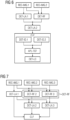
- Fig. 2 displays a data flow diagram for some embodiments of the method for classifying a lesion.
- three medical images IMG.1, IMG.2, IMG.3 are used as input, wherein a registration function TF(t.2, t.1) for the first medical image IMG.1 and the second medical image IMG.2 and a registration function TF(t.3, t.1) for the first medical image IMG.1 and the third medical image IMG.3 is calculated as displayed in Fig. X.
- the first medical image IMG.1 comprises a first lesion area LA.1, comprising several pixels or voxels of the first medical image IMG.1.
- the trained preprocessing function TPF is trained convolutional neural network.
- the lesion areas LA.1, LA.2, LA.3 can be used as inputs for the trained preprocessing function TPF also in combination with the respective medical image IMG.1, IMG.2, IMG.3, e.g.
- the input data ID.1, ID.2, ID.3 is equivalent with the lesion area LA.1, LA.2, LA.3 (or, e.g., with the pixel-wise or voxel-wise product of the lesion area LA.1, LA.2, LA.3 as mask and the respective medical image IMG.1, IMG.2, IMG . 3) .
- the input data ID.1, ID.2, ID.3 is then used as input for a trained classification function TCF.
- the trained classification function TCF is a recurrent neural network comprising recurrent neural network blocks RNB.1, RNB.2, RNB.3.
- each input data ID.1, ID.2, ID.3 is used as input for a different one of the recurrent neural network blocks RNB.1, RNB.2, RNB.3.
- Every recurrent neural network block RNB.1, RNB.2, RNB.3 determines output data OD.1, OD.2, OD.3 and intermediate data BD.1, BD.2, BD.3, wherein the intermediate data BD.1, BD.2, BD.3 is used as additional input for the next of the successive recurrent neural network blocks RNB.1, RNB.2, RNB.3.
- the classification value of the lesion can then be based on all of the output data OD.1, OD.2, OD.3, or only on some of the output data OD.1, OD.2, OD.3.
- the classification value of the lesion is based on the output data OD.1 related to the first medical image IMG.1, and potentially on further output data OD.2, OD.3.
- the most recent one of the medical images IMG.1, IMG.2, IMG.3 can be used with a high weight in the classification of the lesion, implying a better and more exact classification.
- Fig. 3 displays a detailed view of an LSTM network comprising several recurrent neural network blocks RNB.i, RNB.j.
- Each recurrent neural network block RNB.i, RNB.j uses input data ID.i, ID.j to generate or calculate output data OD.i, OD.j.
- each recurrent neural network block RNB.i, RNB.j takes as additional input intermediate data IBD.i, IBD.j and produces as additional output intermediate data OBD.i, OBD.j, wherein output intermediate data OBD.i, OBD.j can be used as input intermediate data IBD.i, IBD.j within the next step.
- Fig. 3 displays an iterative process, which was unfolded for two inputs.
- the iteration can be extended to cover an arbitrary number of input data ID.i, ID.j.
- the recurrent neural network blocks RNB.i, RNB.j are the same up to a number of internal states IG.i, IG.j, OG.i, OG.j, FG.i, FG.j.
- the neural network is an LSTM network
- the recurrent neural network block RNB.i, RNB.j has internal states denoted as input gate IG.i, IG.j, output gate OG.i, OG.j and forget gate FG.i, FG.j.
- the operation ".” is a point-wise multiplication
- "*" is a convolution operation
- " ⁇ " denotes the Sigmoid function.
- the values i j , o j and f j correspond to the values of the input gate IG.j, the output gate OG.j and the forget gate FG.j.
- the values x j and y j correspond to the input data ID.j and the output data OD.j of the respective block.
- the values c i and c j correspond to the intermediate input intermediate data IBD.i and the output intermediate data OBD.i, OBD.j, and are often denoted as "cell state”.
- the values W and b correspond to weights of the network, which are fixed by training the recurrent neural network.
- Fig. 4 displays a first embodiment of the computer-implemented method for classifying a lesion.
- the first two steps of the displayed embodiment are receiving REC-IMG.1 a first medical image IMG.1 of an examination volume and receiving REC-IMG.2 a second medical image IMG.2 of the examination volume.
- the first medical image IMG.1 corresponds to a first examination time t.1 (which is the time the first medical image IMG.1 was determined based on a medical imaging examination)
- the second medical image IMG.2 corresponds to a second examination time t.2 (which is the time the second medical image IMG.2 was determined based on another medical imaging examination).
- the second examination time t.2 is earlier than the first examination time t.1, and the second examination time and the first examination time are different.
- the first and the second medical image IMG.1, IMG.2 can be part of a longitudinal study relating to a certain patient.
- both the first and the second medical image IMG.1, IMG.2 are three-dimensional images generated by means of a computed tomography imaging examination.
- the first and/or the second medical image IMG.1, IMG.2 can be based on other known method of medical imaging, e.g. magnetic resonance imaging, positron emission tomography or single photon emission computed tomography.
- both the first and the second medical image IMG.1, IMG.2 are DICOM images (acronym for "Digital Imaging and Communications in Medicine").
- both the first and the second medical image IMG.1, IMG.2 have the same size (measured in terms of number of voxels) with respect to each of the dimensions of the respective medical images IMG.1, IMG.2.
- a next step of the displayed first embodiment is determining DET-LA.1 a first lesion area LA.1 corresponding to a lesion within the first medical image IMG.1.
- the lesion area corresponds to a segmentation of the lesion within the first medical image IMG.1.
- all voxels that are part of a segment corresponding to the lesion are considered as being part of the first lesion area LA.1, and all other voxels are considered as being not part of the first lesion area LA.1.
- a further step of the displayed embodiment is determining DET-RF a registration function RF(t.2, t.1) based on a comparison of the first medical image IMG.1 and the second medical image IMG.2. This further step can be executed before, after or in parallel with the step of determining DET-LA.1 the first lesion area LA.1.
- the registration function RF(t.2, t.1) is a non-rigid registration function based on a vector momentum-parameterized stationary velocity field.
- Methods for determining such a registration function RF(t.2, t.1) are known e.g. from the paper Z. Shen et al. "Networks for Joint Affine and Non-Parametric Image Registration” (20019) 4219-4228, 10.1109/CVPR.2019.00435 .
- ⁇ -1 denotes the registration function RF(t.2, t.1) (also denoted as registration map)
- I 1 denotes the first medical image IMG.1
- I 2 denotes the second medical image IMG.2
- Picking a specific L implies picking an expected model of deformation.
- the differential operator is spatially invariant and is predefined to encode a desired level of smoothness.
- the optimization takes places on m, where the velocity is smoothed from it. The resulting transformation is guaranteed to be diffeomorphic.
- the next step of the displayed first embodiment is determining DET-LA.2 a second lesion area LA.2 within the second medical image IMG.2 based on the registration function RF(t.2, t.1) and the first lesion area LA.1.
- the registration function RF(t.2, t.1) is used to map every voxel of the first medical image IMG.1 corresponding to the first lesion area LA.1 within the first medical image IMG.1 to at least one voxel of the second medical image IMG.2. All the voxels of the second medical image IMG.2 which are the result of such mapping are at least a part of the second lesion area LA.2 within the second medical image IMG.2. Further normalization procedures can be applied, e.g. the second lesion area LA.2 can be defined as the convex hull of all the voxels of the second medical image IMG.2 which are the result of such mapping.
- the last step of the displayed first embodiment is classifying CLF the lesion within the first medical image IMG.1 based on the second lesion area LA.2.
- the step of classifying CLF the lesion is furthermore based on the first lesion area LA.1.
- the first lesion area LA.1 and the second lesion area LA.2 (or, equivalently, the first and the second medical image IMG.1, IMG.2 with the first and the second lesion area LA.1, LA.2 being used as masks) are used as an input of a trained classification function TCF, which creates as an output a classification value of the lesion (e.g. a binary value whether the lesion is benign or malign).
- Fig. 5 displays a second embodiment of the computer-implemented method for classifying a lesion.
- the steps of receiving REC-IMG.1, REC-IMG.2 the first and the second medical image IMG.1, IMG.2, the step of determining DET-RF the registration function RF(t.2, t.1), and the steps of determining DET-LA.1, DET-LA.2 the first and the second lesion area LA.1, LA.2 are equivalent to the corresponding steps of the first embodiment and can comprise all advantageous embodiments of the respective steps.
- the step of classifying CLF the lesion comprises the substep of applying APL-TCF a trained classifying function TCF to first input data ID.1 and second input data ID.2, thereby generating output data OD.1, OD.2.
- the first input data ID.1 is based on the first lesion area LA.1
- the second input data ID.2 is based on the second lesion area LA.2.
- the first input data ID.1 is based on a mask defined by the first lesion area LA.1 and the first medical image IMG.1
- the second input data ID.2 is based on a mask defined by the second lesion areas LA.2 and the second medical image IMG.2.
- the trained classifying function TCF is a recurrent neural network.
- the recurrent network can be applied to a sequence of input data ID.1, ID.2 by iteratively applying a block (comprising a or defined by a neural network) to an each input data ID.1, ID.2, keeping an internal state.
- the initial value h of the hidden state vector can be initialized randomly or by a certain sequence (e.g., all entries can be set to 0 or 1).
- the step of classifying the lesion furthermore comprises the substep of determining DET-LC a lesion classification based on the output data OD.1, OD.2.
- the lesion classification is a real number between 0 and 1, corresponding to the probability of the classified lesion being malign.
- the lesion classification can be a class label indicating a type of a lesion.
- the lesion classification can be equivalent to the output data OD.1 corresponding to the first medical image IMG.1.
- Fig. 6 displays a third embodiment of the computer-implemented method for classifying a lesion.
- the steps of receiving REC-IMG.1, REC-IMG.2 the first and the second medical image IMG.1, IMG.2, the step of determining DET-RF the registration function RF(t.2, t.1), the steps of determining DET-LA.1, DET-LA.2, the first and the second lesion area LA.1, LA.2, as well as the substeps of applying APL-TCF a trained classifying function TCF to first input data ID.1 and second input data ID.2 and of determining DET-LC a lesion classification are equivalent to the corresponding steps of the first embodiment and/or of the second embodiment and can comprise all advantageous embodiments of the respective steps.
- the step of classifying CLF the lesion comprises the substep of determining DET-ID.1 the first input data ID.1 by applying a trained preprocessing function TPF to at least a part of the first medical image IMG.1 containing the first lesion area LA.1, and the substep of determining DET-ID.2 the second input data ID.2 by applying the trained preprocessing function TPF to at least a part of the second medical image IMG.2 containing the second lesion area LA.2.
- the trained preprocessing function TPF is a convolutional deep neural network.
- the trained preprocessing function TPF is applied to a pixel-wise or voxel-wise multiplication of the respective lesion area LA.1, LA.2 being interpreted as mask and the respective medical image IMG.1, IMG.2.
- Fig. 7 displays a fourth embodiment of the computer-implemented method for classifying a lesion.
- the steps of receiving REC-IMG.1, REC-IMG.2 the first and the second medical image IMG.1, IMG.2, the step of determining DET-LA.1 the first lesion area LA.1, the step of determining DET-LA.2 the second lesion area LA.2 and the step of classifying the lesion can comprise all alternatives and advantageous embodiments of the first, the second and the third embodiment of the method for classifying a lesion above.
- the fourth embodiment displays the extension of the method for three medical images IMG.1, IMG.2, IMG.3 within a longitudinal study.
- the third medical image IMG.3 is of the same type as the first and the second medical image IMG.1, IMG.2.
- the third medical image IMG.3 corresponds to a third time t.3, wherein the third time t.3 is earlier than the second time.
- the fourth embodiment also comprises the step of receiving REC-IMG.3 the third medical image IMG.3. If there would be additional medical images, there would be an additional step of receiving for each of the additional medical images.
- the step of determining DET-RF a registration function RF(t.2, t.1), RF(t.3, t.1) comprises for each of the medical images IMG.2, IMG.3 except the first medical image IMG.1 a substep of determining DET-RF.2, DET-RF.3 a registration function RF(t.2, t.1), RF(t.3, t.1) based on the first medical image IMG.1 and the respective other medical image IMG.2, IMG.3.
- the step of determining DET-RF a registration function RF(t.2, t.1), RF(t.3, t.1) comprises a substep of determining a first registration function RF(t.2, t.1) based on the first medical image IMG.1 and the second medical image IMG.2 and a substep of determining a second registration function RF(t.3, t.1) based on the first medical image IMG.1 and the third medical image IMG.3. If there would be additional medical images, there would be an additional substep of determining a registration function based on the first medical image and the respective additional medical image.
- the fourth embodiment also comprises an additional step of determining DET-LA.3 a third lesion area LA.3 within the third medical image IMG.3.
- the step of determining DET-LA.3 the third lesion area LA.3 is equivalent to the step of determining DET-LA.2 the second lesion area LA.2, wherein the second medical image IMG.2 is replaced with the third medical image IMG.3 (and the corresponding registration function RF(t.3, t.1) is used). If there would be additional medical images, for each of the additional medical images there would be an additional step of determining a lesion area.
- the step of classifying CLF is based on all the lesion areas LA.2, LA.3.
- Fig. 8 displays a flow-chart of an embodiment of a computer-implemented method for providing a trained classifying function TCF.
- the first steps of the displayed embodiments are receiving TREC-TIMG.1 a first medical training image of a training examination volume, wherein the first medical training image corresponds to a first examination time, and receiving a second medical training image of the training examination volume, wherein the second medical training image corresponds to a second examination time being different from the first examination time.
- the first and the second medical training image can comprise all advantageous features described in context of the first and the second medical training image with respect to Fig. 4 .
- both the first medical training image and the second medical training image are two-dimensional or three-dimensional X-ray based medical images.
- Further steps of the displayed embodiment are determining TDET-LA.1 a first lesion area corresponding to a lesion within the first medical training image, determining TDET-RF a registration function based on a comparison of the first medical training image and the second medical training image, and determining a second lesion area within the second medical training image based on the registration function and the first medical training image.
- the registration function is a non-rigid registration function.
- determining TDET-RF the registration function is based on a vector momentum-parameterized stationary velocity field.
- the registration function can be determined as described with respect to Fig. 4 and the method for classifying a lesion.
- a further step of the displayed embodiment is receiving TREC-TCL a training classification corresponding to the first lesion area within the first medical training image.
- the training classification can be a binary value corresponding to whether the lesion corresponding to the first lesion area is a malign lesion or a benign lesion.
- the training classification can be a class label corresponding to the class of the lesion corresponding to the first lesion area, e.g. a BI-RADS (acronym for "Breast Imaging Reporting and Data System”) classification value.
- a further step of the displayed embodiment is applying TAPL-TCF a trained classifying function TCF to first training input data and second training input data, thereby generating training output data, wherein the first training input data is based on the first lesion area, and wherein the second training input data is based on the second lesion area.
- the relation between the first training input data, the lesion area and the first medical training image is equivalent to the relation between the first input data, the first lesion area and the first medical image described with respect to the method for classifying a lesion.
- the relation between the second training input data, the second lesion area and the second medical training image is equivalent to the relation between the second input data, the second lesion area and the second medical image described with respect to the method for classifying a lesion.
- the trained classifying function can comprise all advantageous features and embodiments as the trained classifying function described with respect to the method of classifying a lesion.
- the trained classifying function TCF is a recurrent neural network, and the first training input data and the second training input data are independently used as training input data for the recurrent neural network.
- the recurrent neural network comprises at least one LSTM block.
- the step of applying TAPL-TCF the trained classifying function TCF furthermore comprises the substeps of determining the first training input data by applying a trained preprocessing function TPF to at least a part of the first medical training image containing the first lesion area, and determining the second training input data by applying the trained preprocessing function TPF to at least a part of the second medical training image containing the second lesion area.
- the trained preprocessing function is configured by training to classify lesions within single medical images, and/or wherein the trained preprocessing function TPF is a convolutional neural network.
- the trained preprocessing function TPF can be pretrained based on a comparison between training classifications of lesions within a single medical image, and the result of its application to these single medical images.
- a further step of the displayed embodiment of the method for providing a trained classifying function is adjusting TADJ-TCF at least one parameter of the trained classifying function TCF based on a comparison of the training classification and the training output data.
- the parameter is adapted by minimizing a cost function, wherein the training classification and the trained output parameter are used as input values for the cost function.
- the cost function has a (local or global) extremum if the training classification equals the training output data.
- adjusting TADJ-TCF at least one parameter of the trained classifying function TCF can comprise a gradient descent algorithm and/or a backpropagation algorithm.
- the last step of the displayed embodiment is providing TPROV-TCF the trained classifying function TCF.
- providing TPROV-TCF the trained classifying function TCF can comprise displaying, storing and/or transmitting the trained classifying function TCF.
- Fig. 9 displays a classification system CSYS
- Fig. 10 displays a training system TSYS.
- the classification system CSYS comprises an interface CSYS.IF, a computation unit CSYS.CU, and a memory unit CSYS.MU.
- the training system TSYS comprises an interface TSYS.IF, a computation unit TSYS.CU, and a memory unit TSYS.MU.
- the classification system CSYS and/or the training system TSYS can be a (personal) computer, a workstation, a virtual machine running on host hardware, a microcontroller, or an integrated circuit.
- the classification system CSYS and/or the training system TSYS can be mobile devices, e.g. a smartphone or a tablet.
- the classification system CSYS and/or the training system TSYS can be a real or a virtual group of computers (the technical term for a real group of computers is "cluster", the technical term for a virtual group of computers is "cloud").
- An interface CSYS.IF, TSYS.IF can be embodied as a hard-ware interface or as a software interface (e.g. PCIBus, USB or Firewire).
- a computation unit CU.1, CU.2, CU.3 can comprise hardware elements and software el-ements, for example a microprocessor, a CPU (acronym for "central processing unit"), a GPU (acronym for "graphical processing unit”), a field programmable gate array (an ac-ronym is "FPGA”) or an ASIC.(acronym for "application-specific integrated circuit”).
- the computation unit CSYS.CU, TSYS.CU can be configured for multithreading, i.e.
- a memory unit CSYS.MU, TSYS.MU can be e.g. non-permanent main memory (e.g. random access memory) or permanent mass storage (e.g. hard disk, USB stick, SD card, solid state disk).
- An interface CSYS.IF, TSYS.IF can comprise several (potentially spatially) separate sub-interfaces, each having the characteristics of an interface described above.
- a computation unit CSYS.CU, TSYS.CU can comprise several (potentially spatially) separate computation sub-units, each having the characteristics of a computation unit described above.
- a memory unit CSYS.MU, TSYS.MU can comprise several (potentially spatially) separate memory sub-units, each having the characteristics of a memory unit described above.
- the classification system CSYS is configured to executed one of the embodiments of the method for classifying a lesion.
- the classification CSYS is configured to execute this method by the interface CSYS.IF and the computation unit CSYS.CU being configured to execute the respective steps of the method.
- the training system TSYS is configured to execute one of the embodiments of the method for providing a trained classifying function TCF.
- the training system TSYS is configured to execute this method by the interface TSYS.IF and the computation unit TSYS.CU being configured to execute the respective steps of the method.
Landscapes
- Engineering & Computer Science (AREA)
- Theoretical Computer Science (AREA)
- Health & Medical Sciences (AREA)
- Physics & Mathematics (AREA)
- General Physics & Mathematics (AREA)
- Computer Vision & Pattern Recognition (AREA)
- General Health & Medical Sciences (AREA)
- Medical Informatics (AREA)
- Evolutionary Computation (AREA)
- Life Sciences & Earth Sciences (AREA)
- Multimedia (AREA)
- Artificial Intelligence (AREA)
- Data Mining & Analysis (AREA)
- Radiology & Medical Imaging (AREA)
- Nuclear Medicine, Radiotherapy & Molecular Imaging (AREA)
- Public Health (AREA)
- Biomedical Technology (AREA)
- Quality & Reliability (AREA)
- Molecular Biology (AREA)
- Databases & Information Systems (AREA)
- Primary Health Care (AREA)
- Epidemiology (AREA)
- Computing Systems (AREA)
- Software Systems (AREA)
- Biodiversity & Conservation Biology (AREA)
- Evolutionary Biology (AREA)
- Pathology (AREA)
- General Engineering & Computer Science (AREA)
- Bioinformatics & Computational Biology (AREA)
- Bioinformatics & Cheminformatics (AREA)
- Geometry (AREA)
- High Energy & Nuclear Physics (AREA)
- Biophysics (AREA)
- Optics & Photonics (AREA)
- Heart & Thoracic Surgery (AREA)
- Surgery (AREA)
- Animal Behavior & Ethology (AREA)
- Veterinary Medicine (AREA)
- Physiology (AREA)
- Image Analysis (AREA)
Claims (13)
- Procédé mis en oeuvre par ordinateur de classement d'une lésion, comprenant- la réception (REC-IMG.1) d'une première image (IMG.1) médicale d'un volume à examiner, dans lequel la première image (IMG.1) médicale correspond à un premier temps (t.1) d'examen,- la réception (REC-IMG.2) d'une deuxième image (IMG.2) médicale du volume à examiner, dans lequel la deuxième image (IMG.2) médicale correspond à un deuxième temps (t.2) d'examen, qui est différent du premier temps (t.1) d'examen,- la détermination (DET-LA.1) d'une première zone (LA.1) de lésion correspondant à la lésion dans la première image (IMG.1) médicale,- la détermination (DET-RF) d'une fonction (RF (t.2, t.1)) d'enregistrement sur la base d'une comparaison entre la première images (IMG.1) médicale et la deuxième image (IMG.2) médicale,- la détermination (DET-LA.2) d'une seconde zone (LA.2) de lésion dans la deuxième image (IMG.2) médicale sur la base de la fonction (RF (t.2, t.1)) d'enregistrement et de la première zone (LA.1) de lésion,- l'application (APL-TCF) d'une fonction (TCF) de classement ayant subi un apprentissage à une première donnée (ID.1) d'entrée et à une deuxième donnée (ID.2) d'entrée, en créant ainsi une donnée (OD.1, OD.2) de sortie,dans lequel la première donnée (ID.1) d'entrée repose sur la première zone (LA.1) de lésion,dans lequel la deuxième donnée (ID.2) d'entrée repose sur la deuxième zone (LA.2) de lésion,dans lequel la fonction (TCF) de classement ayant subi un apprentissage est un réseau neuronal récurrent,dans lequel la première donnée (ID. 1) d'entrée et la deuxième donnée (ID.2) d'entrée sont utilisées indépendamment comme données d'entrée pour le réseau neuronal récurrent,dans lequel une information provenant de l'application du réseau neuronal récurrent à la deuxième (ID.2) d'entrée est mise en mémoire dans un état caché et est utilisée comme information supplémentaire, lors de l'application du réseau récurrent à la première donnée d'entrée,et dans lequel la donnée (OD.1, OD.2) de sortie du réseau neuronal récurrent dépend de l'état caché - la détermination (DET-LC) d'un classement de la lésion reposant sur la donnée (OD.1, OD.2) de sortie.
- Procédé suivant la revendication 1, dans lequel le réseau neuronal récurrent comprend au moins un bloc LSTM.
- Procédé suivant la revendication 1 ou la revendication 2, dans lequel le stade de classement (CLF) comprend en outre les sous-stades suivants :- la détermination (DET-ID.1) de la première donnée (ID.1) d'entrée en appliquant une fonction (TPF) de prétraitement ayant subi un apprentissage à au moins une partie de la première image (IMG.1) médicale contenant la première zone (LA.1) de lésion,- la détermination (DET-ID.2) de la deuxième donnée (ID.2) d'entrée en appliquant la fonction (TPF) de prétraitement ayant subi un apprentissage à au moins une partie de la deuxième image (IMG.2) médicale contenant la deuxième zone (LA.2) de lésion.
- Procédé suivant la revendication 3, dans lequel la fonction (TPF) de prétraitement ayant subi un apprentissage est configurée en faisant subir un apprentissage à des lésions de classement dans des images médicales uniques.
- Procédé suivant l'une des revendications précédentes, dans lequel la fonction (RF (t.2, t.1)) d'enregistrement est une fonction d'enregistrement non rigide.
- Procédé suivant la revendication 5, dans lequel la détermination de la fonction (RF (t.2, t.1)) d'enregistrement repose sur un champ de vélocité stationnaire de vecteurs paramétrés par moment.
- Procédé suivant l'une des revendications précédentes, dans lequel la première image (IMG.1) médicale et la deuxième image (IMG.2) médicale sont des images médicales en deux dimensions ou en trois dimensions reposant sur les rayons X.
- Procédé mis en oeuvre par ordinateur pour fournir une fonction (TCF) de classement ayant subi un apprentissage comprenant :- la réception (TREC-TIMG.1) d'une première image (TIMG.1) médicale d'apprentissage d'un volume à examiner en apprentissage, dans lequel la première (TIMG.1) médicale d'apprentissage correspond à un premier temps (t.1) d'examen,- la réception (TREC-TIMG.2) d'une deuxième image (TIMG.2) médicale d'apprentissage du volume à examiner en apprentissage, dans lequel la deuxième image (TIMG.2) médicale d'apprentissage correspond à un deuxième (t.2) d'examen différent du premier temps (t.1) d'examen,- la détermination (TDET-LA.1) d'une première zone (LA.1) de lésion correspondant à une lésion dans la première image (IMG.1) d'apprentissage,- la détermination (TDET-REF) d'une fonction (RF (t.2, t.1) d'enregistrement sur la base d'une comparaison entre la première image (TIMG.1) médicale d'apprentissage et la deuxième image (TIMG.2) médicale d'apprentissage,- la réception (TREC-TCL) d'un classement d'apprentissage correspondant à une première zone (LA.1) de lésion dans la première image (TIMG.1) médicale d'apprentissage,- la détermination (TDET-LA.2) d'une deuxième zone (LA.2) de lésion dans la deuxième image (TIMG.2) médicale d'apprentissage sur la base de la fonction (RF (t.2, t.1)) d'enregistrement et de la première image (TIMG.1) médicale d'apprentissage,- l'application (TAPL-TCF) d'une fonction (TCF) de classement ayant subi un apprentissage à une première donnée (TID.1) d'entrée d'apprentissage et à une deuxième donnée (TID.2) de donnée d'apprentissage, en créant ainsi une donnée (TOD.1) de sortie d'apprentissage,dans lequel la première donnée (TID.1) d'entrée d'apprentissage repose sur la première zone (LA.1) de lésion,dans lequel la deuxième donnée (TID.2) d'entrée d'apprentissage repose sur la deuxième zone (LA.2) de lésion,dans lequel la fonction (TCF) de classement ayant subi un apprentissage est un réseau neuronal récurrent,dans lequel la première donnée (TID.1) d'entrée d'apprentissage et la deuxième donnée (TID.2) d'entrée d'apprentissage sont utilisées indépendamment comme données d'entrée pour le réseau neuronal récurrent,dans lequel une information provenant de l'application du réseau neuronal récurrent à la deuxième donnée (TID.2) d'entrée d'apprentissage est mise en mémoire dans un état caché et est utilisée comme information supplémentaire, lors de l'application du réseau neuronal à la première donnée d'entrée,et dans lequel la donnée (TOD.1) de sortie d'apprentissage du réseau neuronal récurrent dépend de l'état caché,- l'ajustement (TADJ-TCF) d'au moins un paramètre de la fonction (TCF) de classement ayant subi un apprentissage sur la base d'une comparaison entre le classement d'apprentissage et la donnée (TOD.1) de sortie d'apprentissage,- la mise à disposition (TPROV-TCF) de la fonction (TCF) de classement ayant subi un apprentissage.
- Système (CSYS) de classement pour classer une lésion comprenant- une interface (CSYS.IF) configurée pour recevoir (REC-IMG.1) une première image (IMG.1) médicale d'un volume à examiner,dans lequel la première image (IMG.1) médicale correspond à un premier temps (t.1) d'examen,configurée en outre pour recevoir (REC-IMG.2) une deuxième image (IMG.2) médicale du volume à examiner, dans lequel la deuxième image (IMG.2) médicale correspond à un deuxième temps (t.2) d'examen différent du premier temps (t.2) d'examen, et- une unité (CSYS.CU) informatique configurée pour déterminer (DET-LA.1) une première zone (LA.1) de lésion correspondant à la lésion dans la première image (IMG.1) médicale,configurée en outre pour déterminer (DET-RF) une fonction (RF (t.2, t.1)) d'enregistrement reposant sur une comparaison entre la première image (IMG.1) médicale et la deuxième image (IMG.2) médicale,configurée en outre pour déterminer (DET-LA.2) une deuxième zone (LA.2) de lésion dans la deuxième image (IMG.2) médicale sur la base de la fonction (RF (t.2, t.1)) d'enregistrement et de la première zone (LA.1) de lésion,configurée en outre pour appliquer (APL-TCF) une fonction (TCF) de classement ayant subi un apprentissage à une première donnée (ID.1) d'entrée et à une deuxième donnée (ID.2) d'entrée, en créant ainsi une ainsi une donnée (OD.1, OD.2) de sortie, dans lequel la première donnée (ID.1) d'entrée repose sur la première zone (LA.1) de lésion, dans lequel la deuxième donnée (ID.2) d'entrée repose sur la deuxième zone (LA.2) de liaison, dans lequel la fonction (TCF) de classement ayant subi un apprentissage est un réseau neuronal récurrent, dans lequel la première entrée (ID.1) d'entrée et la deuxième donnée (ID.2) d'entrée sont utilisées indépendamment comme données d'entrée pour le réseau neuronal récurrent, dans lequel une information provenant de l'application du réseau neuronal récurrent à la deuxième zone (ID.2) d'entrée est mise en mémoire dans un état caché, et est utilisée comme information supplémentaire, lors de l'application du réseau récurrent à la première donnée d'entrée, dans lequel la donnée (OD.1, OD.2) de sortie du réseau neuronal récurrent dépend de l'état caché,configurée en outre pour outre pour déterminer (DET-LC) un classement de la lésion sur la base de la donnée (OD.1, OD.2) de sortie.
- Système d'imagerie médicale comprenant un système (CSYS) de classement suivant la revendication 9.
- Programme d'ordinateur comprenant des instructions qui, lorsque le programme est exécuté par un système (CSYS) de classement, font que le système (CSYS) de classement effectue le procédé suivant l'une des revendications 1 à 7.
- Support, déchiffrable par ordinateur, comprenant des instructions qui, lorsqu'elles sont exécutées par un système (CSYS) de classement, font que le système (CSYS) de classement effectue le procédé suivant l'une des revendications 1 à 7.
- Support de mémoire, déchiffrable par ordinateur, comprenant une fonction (TCF) de classement ayant subi un apprentissage fournie par un procédé suivant la revendication 8, à utiliser dans un procédé de classement d'une lésion suivant l'une des revendications 1 à 7.
Priority Applications (4)
| Application Number | Priority Date | Filing Date | Title |
|---|---|---|---|
| EP20171081.1A EP3901903B1 (fr) | 2020-04-23 | 2020-04-23 | Classification d'une lésion sur la base d'études longitudinales |
| US17/228,813 US11748886B2 (en) | 2020-04-23 | 2021-04-13 | Classifying a lesion based on longitudinal studies |
| CN202110437627.3A CN113555125B (zh) | 2020-04-23 | 2021-04-22 | 病变分类和分类函数提供方法、分类和成像系统以及介质 |
| US18/349,576 US20230351601A1 (en) | 2020-04-23 | 2023-07-10 | Classifying a lesion based on longitudinal studies |
Applications Claiming Priority (1)
| Application Number | Priority Date | Filing Date | Title |
|---|---|---|---|
| EP20171081.1A EP3901903B1 (fr) | 2020-04-23 | 2020-04-23 | Classification d'une lésion sur la base d'études longitudinales |
Publications (3)
| Publication Number | Publication Date |
|---|---|
| EP3901903A1 EP3901903A1 (fr) | 2021-10-27 |
| EP3901903B1 true EP3901903B1 (fr) | 2023-06-14 |
| EP3901903C0 EP3901903C0 (fr) | 2023-06-14 |
Family
ID=70417449
Family Applications (1)
| Application Number | Title | Priority Date | Filing Date |
|---|---|---|---|
| EP20171081.1A Active EP3901903B1 (fr) | 2020-04-23 | 2020-04-23 | Classification d'une lésion sur la base d'études longitudinales |
Country Status (3)
| Country | Link |
|---|---|
| US (2) | US11748886B2 (fr) |
| EP (1) | EP3901903B1 (fr) |
| CN (1) | CN113555125B (fr) |
Families Citing this family (5)
| Publication number | Priority date | Publication date | Assignee | Title |
|---|---|---|---|---|
| CN113196340B (zh) * | 2018-11-13 | 2025-02-18 | 皇家飞利浦有限公司 | 用于成像的方法、非瞬态计算机可读介质和系统 |
| EP4006832A1 (fr) * | 2020-11-30 | 2022-06-01 | Koninklijke Philips N.V. | Prédiction de la probabilité qu'un individu présente une ou plusieurs lésions |
| US12340527B2 (en) * | 2021-10-05 | 2025-06-24 | Koninklijke Philips N.V. | Inter- and extrapolation of chest image and mechanical ventilation settings into a time lapse series for progression monitoring and outcome prediction during long term mechanical ventilation |
| CN119124629B (zh) * | 2024-07-29 | 2025-09-23 | 清华大学 | 一种航空发动机状态监测方法、装置及电子设备 |
| US20260087615A1 (en) | 2024-09-23 | 2026-03-26 | Siemens Healthineers Ag | Advanced pulmonary nodule analysis system with malignancy-weighted detection and customized visualization |
Family Cites Families (15)
| Publication number | Priority date | Publication date | Assignee | Title |
|---|---|---|---|---|
| WO2012107057A1 (fr) * | 2011-02-10 | 2012-08-16 | Mevis Medical Solutions Ag | Dispositif de traitement d'images |
| US10417788B2 (en) * | 2016-09-21 | 2019-09-17 | Realize, Inc. | Anomaly detection in volumetric medical images using sequential convolutional and recurrent neural networks |
| US10561373B2 (en) * | 2017-01-31 | 2020-02-18 | International Business Machines Corporation | Topological evolution of tumor imagery |
| US11589924B2 (en) * | 2017-08-01 | 2023-02-28 | Siemens Healthcare Gmbh | Non-invasive assessment and therapy guidance for coronary artery disease in diffuse and tandem lesions |
| US10762637B2 (en) * | 2017-10-27 | 2020-09-01 | Siemens Healthcare Gmbh | Vascular segmentation using fully convolutional and recurrent neural networks |
| US10628659B2 (en) * | 2017-11-27 | 2020-04-21 | International Business Machines Corporation | Intelligent tumor tracking system |
| US20190295709A1 (en) | 2018-03-20 | 2019-09-26 | Siemens Healthcare Gmbh | Automatic analysis of a large patient population using medical image data |
| CN112292691B (zh) * | 2018-06-18 | 2024-06-04 | 谷歌有限责任公司 | 用于使用深度学习提高癌症检测的方法与系统 |
| US10643092B2 (en) * | 2018-06-21 | 2020-05-05 | International Business Machines Corporation | Segmenting irregular shapes in images using deep region growing with an image pyramid |
| CN112292086B (zh) * | 2018-06-22 | 2024-05-07 | 皇家飞利浦有限公司 | 超声病变评估及相关联的设备、系统和方法 |
| CN109308495B (zh) * | 2018-07-05 | 2021-07-02 | 科亚医疗科技股份有限公司 | 从患者的医学图像自动预测生理状况的装置和系统 |
| EP3611733B1 (fr) * | 2018-08-15 | 2024-09-25 | Siemens Healthineers AG | Recherche d'une image de référence médicale |
| US11937767B2 (en) * | 2019-07-18 | 2024-03-26 | Hoya Corporation | Endoscope |
| US11024027B2 (en) | 2019-09-13 | 2021-06-01 | Siemens Healthcare Gmbh | Manipulable object synthesis in 3D medical images with structured image decomposition |
| IL274016A (en) * | 2020-04-18 | 2021-10-31 | Highrad Ltd | Methods for automated lesion analysis in longitudinal volumetric medical image studies |
-
2020
- 2020-04-23 EP EP20171081.1A patent/EP3901903B1/fr active Active
-
2021
- 2021-04-13 US US17/228,813 patent/US11748886B2/en active Active
- 2021-04-22 CN CN202110437627.3A patent/CN113555125B/zh active Active
-
2023
- 2023-07-10 US US18/349,576 patent/US20230351601A1/en active Pending
Also Published As
| Publication number | Publication date |
|---|---|
| CN113555125A (zh) | 2021-10-26 |
| EP3901903A1 (fr) | 2021-10-27 |
| CN113555125B (zh) | 2025-02-28 |
| US11748886B2 (en) | 2023-09-05 |
| US20230351601A1 (en) | 2023-11-02 |
| US20210334970A1 (en) | 2021-10-28 |
| EP3901903C0 (fr) | 2023-06-14 |
Similar Documents
| Publication | Publication Date | Title |
|---|---|---|
| EP3901903B1 (fr) | Classification d'une lésion sur la base d'études longitudinales | |
| CN113313234B (zh) | 用于图像分割的神经网络系统和方法 | |
| US11810291B2 (en) | Medical image synthesis of abnormality patterns associated with COVID-19 | |
| US11263744B2 (en) | Saliency mapping by feature reduction and perturbation modeling in medical imaging | |
| Huang et al. | Medical image segmentation | |
| US20200074634A1 (en) | Recist assessment of tumour progression | |
| CN110050281A (zh) | 学习图像中的对象的注释 | |
| Maity et al. | Automatic lung parenchyma segmentation using a deep convolutional neural network from chest X-rays | |
| US12106549B2 (en) | Self-supervised learning for artificial intelligence-based systems for medical imaging analysis | |
| CN115205192B (zh) | 根据头部ct图像的自动出血扩张检测 | |
| Deeparani et al. | Efficient image segmentation and implementation of K-means clustering | |
| US11908047B2 (en) | Generating synthetic x-ray images and object annotations from CT scans for augmenting x-ray abnormality assessment systems | |
| Hassan et al. | Image classification based deep learning: A Review | |
| JP7395668B2 (ja) | 高速マンモグラフィ・データ・ハンドリングのためのシステム及び方法 | |
| US20240005650A1 (en) | Representation learning | |
| US11861828B2 (en) | Automated estimation of midline shift in brain ct images | |
| Gilani et al. | Skin lesion analysis using generative adversarial networks: a review | |
| Wajid et al. | Lung cancer detection using Local Energy-based Shape Histogram (LESH) feature extraction and cognitive machine learning techniques | |
| Sudha et al. | Automatic lung cancer detection using hybrid particle snake swarm optimization with optimized mask RCNN | |
| EP4375927A1 (fr) | Suivi de lésion dans des études d'imagerie longitudinale 4d | |
| US12211204B2 (en) | AI driven longitudinal liver focal lesion analysis | |
| Tamilmani et al. | Medical image segmentation using grey wolf-based u-net with bi-directional convolutional LSTM | |
| US20240177343A1 (en) | Lesion tracking in 4d longitudinal imaging studies | |
| Sihare et al. | MRI-based tumour prediction based on U-Net and VGG-Net | |
| Deepak et al. | Liver Tumor Segmentation using Hybrid Residual Network and Conditional Random Fields |
Legal Events
| Date | Code | Title | Description |
|---|---|---|---|
| PUAI | Public reference made under article 153(3) epc to a published international application that has entered the european phase |
Free format text: ORIGINAL CODE: 0009012 |
|
| STAA | Information on the status of an ep patent application or granted ep patent |
Free format text: STATUS: REQUEST FOR EXAMINATION WAS MADE |
|
| 17P | Request for examination filed |
Effective date: 20210319 |
|
| AK | Designated contracting states |
Kind code of ref document: A1 Designated state(s): AL AT BE BG CH CY CZ DE DK EE ES FI FR GB GR HR HU IE IS IT LI LT LU LV MC MK MT NL NO PL PT RO RS SE SI SK SM TR |
|
| B565 | Issuance of search results under rule 164(2) epc |
Effective date: 20200929 |
|
| STAA | Information on the status of an ep patent application or granted ep patent |
Free format text: STATUS: EXAMINATION IS IN PROGRESS |
|
| 17Q | First examination report despatched |
Effective date: 20220223 |
|
| GRAP | Despatch of communication of intention to grant a patent |
Free format text: ORIGINAL CODE: EPIDOSNIGR1 |
|
| STAA | Information on the status of an ep patent application or granted ep patent |
Free format text: STATUS: GRANT OF PATENT IS INTENDED |
|
| INTG | Intention to grant announced |
Effective date: 20230209 |
|
| RIC1 | Information provided on ipc code assigned before grant |
Ipc: G06V 10/44 20220101ALI20230127BHEP Ipc: G06V 10/82 20220101ALI20230127BHEP Ipc: G06V 10/25 20220101ALI20230127BHEP Ipc: G06T 7/00 20060101AFI20230127BHEP |
|
| GRAS | Grant fee paid |
Free format text: ORIGINAL CODE: EPIDOSNIGR3 |
|
| GRAA | (expected) grant |
Free format text: ORIGINAL CODE: 0009210 |
|
| STAA | Information on the status of an ep patent application or granted ep patent |
Free format text: STATUS: THE PATENT HAS BEEN GRANTED |
|
| AK | Designated contracting states |
Kind code of ref document: B1 Designated state(s): AL AT BE BG CH CY CZ DE DK EE ES FI FR GB GR HR HU IE IS IT LI LT LU LV MC MK MT NL NO PL PT RO RS SE SI SK SM TR |
|
| REG | Reference to a national code |
Ref country code: CH Ref legal event code: EP |
|
| REG | Reference to a national code |
Ref country code: DE Ref legal event code: R096 Ref document number: 602020012421 Country of ref document: DE |
|
| REG | Reference to a national code |
Ref country code: AT Ref legal event code: REF Ref document number: 1579769 Country of ref document: AT Kind code of ref document: T Effective date: 20230715 |
|
| U01 | Request for unitary effect filed |
Effective date: 20230627 |
|
| U07 | Unitary effect registered |
Designated state(s): AT BE BG DE DK EE FI FR IT LT LU LV MT NL PT SE SI Effective date: 20230726 |
|
| REG | Reference to a national code |
Ref country code: LT Ref legal event code: MG9D |
|
| PG25 | Lapsed in a contracting state [announced via postgrant information from national office to epo] |
Ref country code: NO Free format text: LAPSE BECAUSE OF FAILURE TO SUBMIT A TRANSLATION OF THE DESCRIPTION OR TO PAY THE FEE WITHIN THE PRESCRIBED TIME-LIMIT Effective date: 20230914 Ref country code: ES Free format text: LAPSE BECAUSE OF FAILURE TO SUBMIT A TRANSLATION OF THE DESCRIPTION OR TO PAY THE FEE WITHIN THE PRESCRIBED TIME-LIMIT Effective date: 20230614 |
|
| PG25 | Lapsed in a contracting state [announced via postgrant information from national office to epo] |
Ref country code: RS Free format text: LAPSE BECAUSE OF FAILURE TO SUBMIT A TRANSLATION OF THE DESCRIPTION OR TO PAY THE FEE WITHIN THE PRESCRIBED TIME-LIMIT Effective date: 20230614 Ref country code: HR Free format text: LAPSE BECAUSE OF FAILURE TO SUBMIT A TRANSLATION OF THE DESCRIPTION OR TO PAY THE FEE WITHIN THE PRESCRIBED TIME-LIMIT Effective date: 20230614 Ref country code: GR Free format text: LAPSE BECAUSE OF FAILURE TO SUBMIT A TRANSLATION OF THE DESCRIPTION OR TO PAY THE FEE WITHIN THE PRESCRIBED TIME-LIMIT Effective date: 20230915 |
|
| U1N | Appointed representative for the unitary patent procedure changed after the registration of the unitary effect |
Representative=s name: SIEMENS HEALTHINEERS PATENT ATTORNEYS; DE |
|
| PG25 | Lapsed in a contracting state [announced via postgrant information from national office to epo] |
Ref country code: SK Free format text: LAPSE BECAUSE OF FAILURE TO SUBMIT A TRANSLATION OF THE DESCRIPTION OR TO PAY THE FEE WITHIN THE PRESCRIBED TIME-LIMIT Effective date: 20230614 |
|
| PG25 | Lapsed in a contracting state [announced via postgrant information from national office to epo] |
Ref country code: IS Free format text: LAPSE BECAUSE OF FAILURE TO SUBMIT A TRANSLATION OF THE DESCRIPTION OR TO PAY THE FEE WITHIN THE PRESCRIBED TIME-LIMIT Effective date: 20231014 |
|
| PG25 | Lapsed in a contracting state [announced via postgrant information from national office to epo] |
Ref country code: SM Free format text: LAPSE BECAUSE OF FAILURE TO SUBMIT A TRANSLATION OF THE DESCRIPTION OR TO PAY THE FEE WITHIN THE PRESCRIBED TIME-LIMIT Effective date: 20230614 Ref country code: SK Free format text: LAPSE BECAUSE OF FAILURE TO SUBMIT A TRANSLATION OF THE DESCRIPTION OR TO PAY THE FEE WITHIN THE PRESCRIBED TIME-LIMIT Effective date: 20230614 Ref country code: RO Free format text: LAPSE BECAUSE OF FAILURE TO SUBMIT A TRANSLATION OF THE DESCRIPTION OR TO PAY THE FEE WITHIN THE PRESCRIBED TIME-LIMIT Effective date: 20230614 Ref country code: IS Free format text: LAPSE BECAUSE OF FAILURE TO SUBMIT A TRANSLATION OF THE DESCRIPTION OR TO PAY THE FEE WITHIN THE PRESCRIBED TIME-LIMIT Effective date: 20231014 Ref country code: CZ Free format text: LAPSE BECAUSE OF FAILURE TO SUBMIT A TRANSLATION OF THE DESCRIPTION OR TO PAY THE FEE WITHIN THE PRESCRIBED TIME-LIMIT Effective date: 20230614 |
|
| RAP2 | Party data changed (patent owner data changed or rights of a patent transferred) |
Owner name: SIEMENS HEALTHINEERS AG |
|
| U1K | Transfer of rights of the unitary patent after the registration of the unitary effect |
Owner name: SIEMENS HEALTHINEERS AG; DE |
|
| PG25 | Lapsed in a contracting state [announced via postgrant information from national office to epo] |
Ref country code: PL Free format text: LAPSE BECAUSE OF FAILURE TO SUBMIT A TRANSLATION OF THE DESCRIPTION OR TO PAY THE FEE WITHIN THE PRESCRIBED TIME-LIMIT Effective date: 20230614 |
|
| REG | Reference to a national code |
Ref country code: DE Ref legal event code: R097 Ref document number: 602020012421 Country of ref document: DE |
|
| PLBE | No opposition filed within time limit |
Free format text: ORIGINAL CODE: 0009261 |
|
| STAA | Information on the status of an ep patent application or granted ep patent |
Free format text: STATUS: NO OPPOSITION FILED WITHIN TIME LIMIT |
|
| 26N | No opposition filed |
Effective date: 20240315 |
|
| U20 | Renewal fee for the european patent with unitary effect paid |
Year of fee payment: 5 Effective date: 20240418 |
|
| U1N | Appointed representative for the unitary patent procedure changed after the registration of the unitary effect |
Representative=s name: SIEMENS HEALTHINEERS PATENT ATTORNEYS; DE |
|
| PG25 | Lapsed in a contracting state [announced via postgrant information from national office to epo] |
Ref country code: MC Free format text: LAPSE BECAUSE OF FAILURE TO SUBMIT A TRANSLATION OF THE DESCRIPTION OR TO PAY THE FEE WITHIN THE PRESCRIBED TIME-LIMIT Effective date: 20230614 |
|
| PG25 | Lapsed in a contracting state [announced via postgrant information from national office to epo] |
Ref country code: MC Free format text: LAPSE BECAUSE OF FAILURE TO SUBMIT A TRANSLATION OF THE DESCRIPTION OR TO PAY THE FEE WITHIN THE PRESCRIBED TIME-LIMIT Effective date: 20230614 |
|
| REG | Reference to a national code |
Ref country code: CH Ref legal event code: PL |
|
| PG25 | Lapsed in a contracting state [announced via postgrant information from national office to epo] |
Ref country code: CH Free format text: LAPSE BECAUSE OF NON-PAYMENT OF DUE FEES Effective date: 20240430 |
|
| PG25 | Lapsed in a contracting state [announced via postgrant information from national office to epo] |
Ref country code: IE Free format text: LAPSE BECAUSE OF NON-PAYMENT OF DUE FEES Effective date: 20240423 |
|
| U20 | Renewal fee for the european patent with unitary effect paid |
Year of fee payment: 6 Effective date: 20250417 |
|
| PGFP | Annual fee paid to national office [announced via postgrant information from national office to epo] |
Ref country code: GB Payment date: 20250507 Year of fee payment: 6 |
|
| PG25 | Lapsed in a contracting state [announced via postgrant information from national office to epo] |
Ref country code: CY Free format text: LAPSE BECAUSE OF FAILURE TO SUBMIT A TRANSLATION OF THE DESCRIPTION OR TO PAY THE FEE WITHIN THE PRESCRIBED TIME-LIMIT; INVALID AB INITIO Effective date: 20200423 |
|
| PG25 | Lapsed in a contracting state [announced via postgrant information from national office to epo] |
Ref country code: HU Free format text: LAPSE BECAUSE OF FAILURE TO SUBMIT A TRANSLATION OF THE DESCRIPTION OR TO PAY THE FEE WITHIN THE PRESCRIBED TIME-LIMIT; INVALID AB INITIO Effective date: 20200423 |