EP3916014A1 - Gegen avss3-integrin gerichtete einzeldomänen-antikörper - Google Patents
Gegen avss3-integrin gerichtete einzeldomänen-antikörper Download PDFInfo
- Publication number
- EP3916014A1 EP3916014A1 EP19938916.4A EP19938916A EP3916014A1 EP 3916014 A1 EP3916014 A1 EP 3916014A1 EP 19938916 A EP19938916 A EP 19938916A EP 3916014 A1 EP3916014 A1 EP 3916014A1
- Authority
- EP
- European Patent Office
- Prior art keywords
- seq
- domain antibody
- integrin
- vhh
- present disclosure
- Prior art date
- Legal status (The legal status is an assumption and is not a legal conclusion. Google has not performed a legal analysis and makes no representation as to the accuracy of the status listed.)
- Pending
Links
Images








Classifications
-
- A—HUMAN NECESSITIES
- A61—MEDICAL OR VETERINARY SCIENCE; HYGIENE
- A61K—PREPARATIONS FOR MEDICAL, DENTAL OR TOILETRY PURPOSES
- A61K49/00—Preparations for testing in vivo
- A61K49/001—Preparation for luminescence or biological staining
- A61K49/0013—Luminescence
- A61K49/0017—Fluorescence in vivo
- A61K49/0019—Fluorescence in vivo characterised by the fluorescent group, e.g. oligomeric, polymeric or dendritic molecules
- A61K49/0021—Fluorescence in vivo characterised by the fluorescent group, e.g. oligomeric, polymeric or dendritic molecules the fluorescent group being a small organic molecule
- A61K49/0032—Methine dyes, e.g. cyanine dyes
-
- A—HUMAN NECESSITIES
- A61—MEDICAL OR VETERINARY SCIENCE; HYGIENE
- A61K—PREPARATIONS FOR MEDICAL, DENTAL OR TOILETRY PURPOSES
- A61K49/00—Preparations for testing in vivo
- A61K49/001—Preparation for luminescence or biological staining
- A61K49/0013—Luminescence
- A61K49/0017—Fluorescence in vivo
- A61K49/005—Fluorescence in vivo characterised by the carrier molecule carrying the fluorescent agent
- A61K49/0058—Antibodies
-
- C—CHEMISTRY; METALLURGY
- C07—ORGANIC CHEMISTRY
- C07K—PEPTIDES
- C07K16/00—Immunoglobulins [IG], e.g. monoclonal or polyclonal antibodies
- C07K16/18—Immunoglobulins [IG], e.g. monoclonal or polyclonal antibodies against material from animals or humans
- C07K16/28—Immunoglobulins [IG], e.g. monoclonal or polyclonal antibodies against material from animals or humans against receptors, cell surface antigens or cell surface determinants
-
- C—CHEMISTRY; METALLURGY
- C07—ORGANIC CHEMISTRY
- C07K—PEPTIDES
- C07K16/00—Immunoglobulins [IG], e.g. monoclonal or polyclonal antibodies
- C07K16/18—Immunoglobulins [IG], e.g. monoclonal or polyclonal antibodies against material from animals or humans
- C07K16/28—Immunoglobulins [IG], e.g. monoclonal or polyclonal antibodies against material from animals or humans against receptors, cell surface antigens or cell surface determinants
- C07K16/2839—Immunoglobulins [IG], e.g. monoclonal or polyclonal antibodies against material from animals or humans against receptors, cell surface antigens or cell surface determinants against the integrin superfamily
- C07K16/2848—Immunoglobulins [IG], e.g. monoclonal or polyclonal antibodies against material from animals or humans against receptors, cell surface antigens or cell surface determinants against the integrin superfamily against integrin beta3-subunit-containing molecules, e.g. CD41, CD51, CD61
-
- G—PHYSICS
- G01—MEASURING; TESTING
- G01N—INVESTIGATING OR ANALYSING MATERIALS BY DETERMINING THEIR CHEMICAL OR PHYSICAL PROPERTIES
- G01N33/00—Investigating or analysing materials by specific methods not covered by groups G01N1/00 - G01N31/00
- G01N33/48—Biological material, e.g. blood, urine; Haemocytometers
- G01N33/50—Chemical analysis of biological material, e.g. blood, urine; Testing involving biospecific ligand binding methods; Immunological testing
- G01N33/53—Immunoassay; Biospecific binding assay; Materials therefor
- G01N33/575—Immunoassay; Biospecific binding assay; Materials therefor for cancer
- G01N33/5758—Immunoassay; Biospecific binding assay; Materials therefor for cancer involving compounds serving as markers for tumours, cancers or neoplasias, e.g. cellular determinants, receptors, heat shock/stress proteins, A-protein, oligosaccharides or metabolites
- G01N33/5759—Immunoassay; Biospecific binding assay; Materials therefor for cancer involving compounds serving as markers for tumours, cancers or neoplasias, e.g. cellular determinants, receptors, heat shock/stress proteins, A-protein, oligosaccharides or metabolites involving compounds localised on the membrane of tumour or cancer cells
-
- A—HUMAN NECESSITIES
- A61—MEDICAL OR VETERINARY SCIENCE; HYGIENE
- A61K—PREPARATIONS FOR MEDICAL, DENTAL OR TOILETRY PURPOSES
- A61K39/00—Medicinal preparations containing antigens or antibodies
- A61K2039/505—Medicinal preparations containing antigens or antibodies comprising antibodies
-
- C—CHEMISTRY; METALLURGY
- C07—ORGANIC CHEMISTRY
- C07K—PEPTIDES
- C07K2317/00—Immunoglobulins specific features
- C07K2317/20—Immunoglobulins specific features characterized by taxonomic origin
- C07K2317/22—Immunoglobulins specific features characterized by taxonomic origin from camelids, e.g. camel, llama or dromedary
-
- C—CHEMISTRY; METALLURGY
- C07—ORGANIC CHEMISTRY
- C07K—PEPTIDES
- C07K2317/00—Immunoglobulins specific features
- C07K2317/50—Immunoglobulins specific features characterized by immunoglobulin fragments
- C07K2317/56—Immunoglobulins specific features characterized by immunoglobulin fragments variable (Fv) region, i.e. VH and/or VL
- C07K2317/569—Single domain, e.g. dAb, sdAb, VHH, VNAR or nanobody®
-
- C—CHEMISTRY; METALLURGY
- C07—ORGANIC CHEMISTRY
- C07K—PEPTIDES
- C07K2317/00—Immunoglobulins specific features
- C07K2317/90—Immunoglobulins specific features characterized by (pharmaco)kinetic aspects or by stability of the immunoglobulin
- C07K2317/92—Affinity (KD), association rate (Ka), dissociation rate (Kd) or EC50 value
-
- G—PHYSICS
- G01—MEASURING; TESTING
- G01N—INVESTIGATING OR ANALYSING MATERIALS BY DETERMINING THEIR CHEMICAL OR PHYSICAL PROPERTIES
- G01N2333/00—Assays involving biological materials from specific organisms or of a specific nature
- G01N2333/435—Assays involving biological materials from specific organisms or of a specific nature from animals; from humans
- G01N2333/705—Assays involving receptors, cell surface antigens or cell surface determinants
- G01N2333/70546—Integrin superfamily, e.g. VLAs, leuCAM, GPIIb/GPIIIa, LPAM
- G01N2333/70557—Integrin beta3-subunit-containing molecules, e.g. CD41, CD51, CD61
-
- G—PHYSICS
- G01—MEASURING; TESTING
- G01N—INVESTIGATING OR ANALYSING MATERIALS BY DETERMINING THEIR CHEMICAL OR PHYSICAL PROPERTIES
- G01N2800/00—Detection or diagnosis of diseases
- G01N2800/70—Mechanisms involved in disease identification
- G01N2800/7014—(Neo)vascularisation - Angiogenesis
Definitions
- the present disclosure relates to an ⁇ v ⁇ 3 integrin targeting single-domain antibody and various applications thereof.
- Integrin is a cell surface receptor that regulates important physiological functions of cells such as cell adhesion and migration, differentiation, and proliferation.
- the integrin acts as a heterodimer in which ⁇ and ⁇ subunits have non-covalent bonds, and the ⁇ and ⁇ subunits are paired to form 22 integrin families.
- Integrin mainly binds to extracellular matrix proteins such as vitronectin, fibronectin, collagen, laminin, vWF, and fibrinogen.
- different types of integrins have different specificities to ligands.
- One type of integrin may bind to several ligands simultaneously.
- integrin ⁇ v ⁇ 3 is expressed in most of the aggressive tumor cells among various cancers including skin cancer, prostate cancer, breast cancer, cervical cancer, colon cancer, lung cancer, gallbladder cancer, pancreatic cancer, and stomach cancer.
- the integrin ⁇ v ⁇ 3 is known to regulate adhesion-dependent tumor cell growth, survival and invasion to improve the malignancy of various human tumors. Recently, it has been shown that ⁇ integrin regulates intracellular signaling to act as a mediator independent of adhesion to increase tumor growth and metastasis ( David A Cheresh et al., Nature Medicine 2009, 15 (10): 1163 ). Further, the ⁇ v ⁇ 3 integrin is highly expressed during neovascularization.
- Angiogenesis means creation of new capillary vessels from existing blood vessels.
- Angiogenesis is a strictly controlled phenomenon that rarely occurs under normal physiological conditions, or occurs when the embryo develops during the development of the fertilized egg, when the wound is healed in adults, and during changes in the reproductive system in the female reproductive cycle. In adults, capillary endothelial cells do not divide relatively well, and the rate of division is usually several months to several years.
- Angiogenesis occurs in a complex process via the interaction of various types of cells with water-soluble factors and extracellular matrix components, and its mechanism of action has not been fully identified. The angiogenesis is responsible for several diseases.
- the single-domain antibody is highly water-soluble and stable even under extreme pH conditions and temperature conditions up to 90°C ( Dumoulin, M. et al. 2002. Protein Sciii.11:500-515 , Dumoulin, M. et al. 2003. Nature 424:783-788 ).
- a purpose of the present disclosure may provide an ⁇ v ⁇ 3 integrin targeting single-domain antibody.
- a purpose of the present disclosure may provide a recombinant vector containing at least one selected from the group consisting of base sequences represented by SEQ ID NO: 1, SEQ ID NO: 2, SEQ ID NO: 3, SEQ ID NO: 4, SEQ ID NO: 5, SEQ ID NO: 6, SEQ ID NO: 7, SEQ ID NO: 8, SEQ ID NO: 9, and SEQ ID NO: 10.
- a purpose of the present disclosure may provide a recombinant microorganism transformed with the recombinant vector.
- a purpose of the present disclosure may provide a composition for detecting angiogenesis.
- a purpose of the present disclosure may provide a kit for detecting angiogenesis.
- a purpose of the present disclosure may provide a composition for diagnosing angiogenesis-related diseases, the composition includes the single-domain antibody.
- a purpose of the present disclosure may provide a preparation method of an ⁇ v ⁇ 3 integrin targeting single-domain antibody.
- a purpose of the present disclosure may provide a method for screening an angiogenesis inhibitor or promoter.
- a purpose of the present disclosure may provide a method for providing information for diagnosis of angiogenesis-related disease.
- the present disclosure provides an ⁇ v ⁇ 3 integrin targeting single-domain antibody encoded by at least one selected from the group consisting of base sequences represented by SEQ ID NO: 1, SEQ ID NO: 2, SEQ ID NO: 3, SEQ ID NO: 4, SEQ ID NO: 5, SEQ ID NO: 6, SEQ ID NO: 7, SEQ ID NO: 8, SEQ ID NO: 9, and SEQ ID NO: 10.
- the present disclosure provides a recombinant vector containing at least one selected from the group consisting of base sequences represented by SEQ ID NO: 1, SEQ ID NO: 2, SEQ ID NO: 3, SEQ ID NO: 4, SEQ ID NO: 5, SEQ ID NO: 6, SEQ ID NO: 7, SEQ ID NO: 8, SEQ ID NO: 9, and SEQ ID NO: 10.
- the present disclosure provides a recombinant microorganism transformed with the recombinant vector.
- the present disclosure provides a composition for detecting angiogenesis, the composition including the single-domain antibody.
- kits for detecting angiogenesis including the single-domain antibody.
- the present disclosure provides a composition for diagnosing angiogenesis-related diseases, the composition includes the single-domain antibody.
- the present disclosure provides a preparation method of an ⁇ v ⁇ 3 integrin targeting single-domain antibody, the method including: (1) culturing a recombinant microorganism transformed with a recombinant vector containing at least one selected from the group consisting of base sequences represented by SEQ ID NO: 1, SEQ ID NO: 2, SEQ ID NO: 3, SEQ ID NO: 4, SEQ ID NO: 5, SEQ ID NO: 6, SEQ ID NO: 7 SEQ ID NO: 8, SEQ ID NO: 9 and SEQ ID NO: 10; and (2) expressing a single-domain antibody for ⁇ v ⁇ 3 integrin in the microorganism.
- the present disclosure provides a method for screening an angiogenesis inhibitor or promoter, the method comprising (1) treating a biological sample isolated from a specimen or an animal model with candidate drugs to be analyzed; (2) binding the ⁇ v ⁇ 3 integrin targeting single-domain antibody to the sample and measuring a level of ⁇ v ⁇ 3 integrin protein; and (3) selecting a candidate drug by which the protein level of the (2) is suppressed or enhanced compared to a control.
- the present disclosure provides a method for providing information for diagnosis of angiogenesis-related diseases, the method including (a) binding the ⁇ v ⁇ 3 integrin targeting single-domain antibody to a biological sample isolated from a specimen and measuring a level of ⁇ v ⁇ 3 integrin protein; and (b) comparing the level of the ⁇ v ⁇ 3 integrin protein with a reference value obtained from a control sample.
- the ⁇ v ⁇ 3 integrin targeting single-domain antibody according to the present disclosure exhibits high binding ability to ⁇ v ⁇ 3 integrin related to angiogenesis, excellent tissue permeability, and biostability compared to conventional antibodies.
- the single-domain antibody according to the present disclosure may be combined with fluorescent particles and thus may be easily measured in vitro, in vivo or ex vivo, and may be effective in detecting angiogenesis and diagnosing angiogenesis related diseases, therefore it may be usefully used in related industries.
- the present disclosure provides an ⁇ v ⁇ 3 integrin targeting single-domain antibody encoded by at least one selected from the group consisting of base sequences represented by SEQ ID NO: 1, SEQ ID NO: 2, SEQ ID NO: 3, SEQ ID NO: 4, SEQ ID NO: 5, SEQ ID NO: 6, SEQ ID NO: 7, SEQ ID NO: 8, SEQ ID NO: 9, and SEQ ID NO: 10.
- ⁇ v ⁇ 3 integrin in the present disclosure refers to a receptor molecule that exists on the cell surface and acts when cells adhere to extracellular matrix such as fibronectin and collagen.
- the integrin refers to a transmembrane glycoprotein composed of heterodimers of two subunits, that is, ⁇ and ⁇ subunits.
- the existence of 21 types of integrins has been revealed so far. Among them, ⁇ v ⁇ 3 integrin has been reported to play a very important role in maintaining the structure of the cardiovascular system and bone tissue.
- single-domain antibody in the present disclosure refers to an antibody in which a CDR is a portion of a single domain polypeptide, and includes heavy chain antibodies, antibodies naturally devoid of light chains, single-domain antibodies derived from conventional 4-chain antibodies, engineered antibodies and single domain scaffolds other than those derived from antibodies.
- the single-domain antibody is referred to as a variable region of a heavy chain antibody (VHH), a nanobody, or an sdAb.
- the single-domain antibody according to the present disclosure refers to a naturally occurring single-domain antibody derived from a heavy chain naturally free of a light chain, and acts as a specific antibody to ⁇ v ⁇ 3 integrin, and has a molecular weight of about 14 KDa to 15 KDa.
- the single-domain antibody according to the present disclosure is an antibody that is a VHH derived from Camelidae, and may be derived from camels, dromedaries, llama, alpaca and wild llama.
- species other than Camellidae may naturally produce the single-domain antibody as heavy chain antibodies without light chains.
- the present disclosure is not limited thereto.
- the single-domain antibody according to the present disclosure is about 10 times smaller than an IgG molecule and is very stable as single polypeptides and are stable under extreme pH and temperature conditions. Further, the single-domain antibody is resistant to the action of proteases, unlike conventional antibodies. When expressed in vitro, mass production of the single-domain antibody may be realized at a high yield.
- the base sequence represented by SEQ ID NO: 1 encodes the amino acid sequence of the single-domain antibody represented by SEQ ID NO: 11.
- the base sequence represented by SEQ ID NO: 2 encodes the amino acid sequence thereof represented by SEQ ID NO: 12.
- the base sequence represented by SEQ ID NO: 3 may encode the amino acid sequence thereof represented by SEQ ID NO: 13. Further, the amino acid sequences containing the His6 tag in SEQ ID NOs: 1, 2 or 3 may be represented by amino acids of SEQ ID NOs: 14, 15 and 16, respectively.
- camels were immunized by injecting, thereto, recombinant ⁇ v ⁇ 3 integrin with cross reactivity in mouse and human, as a targeting protein. Subsequently, mRNA was separated and purified from peripheral lymphocytes isolated from camel blood to synthesize cDNA. After amplifying the variable domains (VHH) of the heavy chain-only antibody by PCR, the VHH was cloned into the M13 bacteriophage gene. Phage display using immobilized immunogen (phosphatidylserine) was performed to select phage clones that react specifically to immobilized antigens.
- VHH variable domains
- DNA sequences of 10 candidate groups were identified. Among them, 20 DNA sequences are shown in FIG. 6A to FIG. 6I . Among them, clones 14, 11, 29, 17, 15, 28, 12, 19, 13, 21, 27, 16, 18, 20, 30, 23, 24, 25, 22, and 26 showing high responses in 10 groups were selected, and their amino acid sequences are shown in FIG. 8A to FIG. 8C .
- Base sequence of the clones VHH-25, VHH-13, VHH-22, VHH-11, VHH-17, VHH-15, VHH-12, VHH-16, VHH-20, and VHH-23 exhibiting strong positives were respectively represented by SEQ ID NOs: 1 to 10.
- the amino acid sequences of VHH-25, VHH-13, and VHH-22 are represented by SEQ ID NOs: 11, 12, and 13, respectively.
- nucleic acid molecules that may be used as genes encoding proteins constituting a single-domain antibody according to the present disclosure includes functional equivalents of nucleic acid molecules constituting the same, for example, variants in which some base sequences of nucleic acid molecules have been modified by deletion, substitution or insertion, but may function functionally identically to nucleic acid molecules.
- the gene may include a base sequence having a sequence homology of at least 70%, more preferably at least 80%, even more preferably at least 90%, most preferably at least 95% to the nucleotide sequence according to the present disclosure.
- % of sequence homology for a polynucleotide is identified by comparing two optimally arranged sequences with a comparison region.
- a portion of the polynucleotide sequence in the comparison region may contain additions or deletions (that is, gaps) compared to the reference sequence (not containing additions or deletions) for the optimal arrangement of the two sequences.
- the single-domain antibody according to the present disclosure may contain a variant of the amino acid sequence as described in the attached sequence list as long as the variant may specifically recognize ⁇ v ⁇ 3 integrin.
- modifications may be made to the amino acid sequence of an antibody to alleviate its binding affinity and/or other biological properties. Such modifications include, for example, deletions, insertions and/or substitutions of amino acid sequence residues of the antibody.
- the amino acid variation is performed based on the relative similarity of amino acid side chain substituents, such as hydrophobicity, hydrophilicity, charge, size, etc. Based on analysis of the size, shape and type of amino acid side chain substituents, it may be identified that arginine, lysine and histidine are all positively charged residues; alanine, glycine and serine have similar sizes; and phenylalanine, tryptophan and tyrosine have similar shapes. Thus, based on these considerations, arginine, lysine and histidine; alanine, glycine and serine; and phenylalanine, tryptophan, and tyrosine are biologically functional equivalents.
- the hydropathic index of the amino acid may be considered.
- Each amino acid is assigned a hydropathic index according to its hydrophobicity and charge: isoleucine (+4.5); valine (+4.2); leucine (+3.8); phenylalanine (+2.8); cysteine (+2.5); methionine (+1.9); alanine (+1.8); glycine (-0.4); threonine (-0.7); serine (-0.8); tryptophan (-0.9); tyrosine (-1.3); proline (-1.6); histidine (-3.2); glutamate (-3.5); glutamine (-3.5); aspartate (-3.5); asparagine (-3.5); lysine (-3.9); and arginine (-4.5).
- the hydropathic amino acid index is very important in imparting an interactive biological function of a protein. It is a known fact that a similar biological activity may be achieved only by substitution with an amino acid having a similar hydropathic index. When introducing the variation with reference to the hydropathic index, substitutions are made between amino acids showing a difference in the hydropathic index, preferably within ⁇ 2, more preferably within ⁇ 1, and even more preferably within ⁇ 0.5.
- hydrophilicity values are assigned to the amino acid residues: arginine (+3.0); lysine (+3.0); aspartate (+3.0 ⁇ 1); glutamate (+3.0 ⁇ 1); serine (+0.3); asparagine (+0.2); glutamine (+0.2); glycine (0); threonine (-0.4); proline (-0.5 ⁇ 1); alanine (-0.5); histidine (-0.5); cysteine (-1.0); methionine (-1.3); valine (-1.5); leucine (-1.8); isoleucine (-1.8); tyrosine (-2.3); phenylalanine (-2.5); tryptophan (-3.4).
- substitution may be made between amino acids showing a difference in hydrophilicity values of preferably within ⁇ 2, more preferably within ⁇ 1, and even more preferably within ⁇ 0.5.
- Functional molecules or elements may be additionally coupled to the single-domain antibody according to the present disclosure.
- the functional molecule may be one or more selected from the group consisting of inorganic particles, chemicals, peptides, polypeptides, nucleic acids, carbohydrates, radioactive isotopes and lipids.
- the inorganic particles are fluorescent labels or dyes, and may be selected from the group consisting of GFP (Green Fluorescent Protein), YFP (Yellow Fluorescent Protein), BFP (Blue fluorescent protein), CFP (Cyan fluorescent protein), Acridine dyes, Cyanine dyes, fluorone dyes, oxazine dyes, phenanthridine dyes, and rhodamine dyes, but is not limited thereto.
- GFP Green Fluorescent Protein
- YFP Yellow Fluorescent Protein
- BFP Blue fluorescent protein
- CFP Cyan fluorescent protein
- Acridine dyes Cyanine dyes, fluorone dyes, oxazine dyes, phenanthridine dyes, and rhodamine dyes, but is not limited thereto.
- the radioactive isotopes may be at least one selected from the group consisting of 18 F (fluorine), 11 C (carbon), 15 O (oxygen), 13 N (nitrogen), 89 Zr (zirconium), C 15 O, 13 N (ammonia), H 2 15 O and 18 FDG (F-Deoxy Glucose), but is not limited thereto.
- the chemical substance may be a substance that inhibits diseases related to angiogenesis.
- the chemical substance that inhibits angiogenesis related diseases may be combined with the single-domain antibody according to the present disclosure without limitation.
- the chemical substance may be an angiogenesis inhibitor, an anticancer agent, or an atherosclerosis inhibitor.
- the angiogenesis inhibitor may be, for example, avastin, itraconazole, carboxyamidotriazole, suramin, SU5416, thrombospondin, sorafenib, sunitinib, pazopanib, everolimus, and mixtures thereof, but is not limited thereto.
- the anticancer agent may be, for example, acivicin, aclarubicin, acodazole, acronycin, adozelesin, alanosine, aldesleukin, allopurinol sodium, altretamine, aminoglutethimide, amonafide, ampligen, amsacrine, androgens, anguidine, apidicholine glycinate, asarey, asparaginase, 5-azacytidine, azathioprine, Bacillus calmette-guerin (BCG), Bakers antipol, beta-2-dioxythioguanosine, bisantrene HCl, Bleomycin Sulfate, Bulseophan, Butionine Sulfoximine, BWA 773U82, BW 502U83/HCl, BW 7U85 Mesylate, cerasemide, carbetimer, carboplatin, carmustine, chlorambucil, chloroquinoxa
- the peptide or polypeptide as a functional molecule is not particularly limited, and may include hormones, hormone analogs, enzymes, enzyme inhibitors, signaling proteins or portions thereof, antibodies or portions thereof, single chain antibodies, binding proteins or binding domains thereof, antigens, adhesion proteins, structural proteins, regulatory proteins, toxin proteins, cytokines, transcriptional regulatory factors, blood coagulation factors, and vaccines, but is not limited thereto.
- the peptide or polypeptide additionally bound to the single-domain antibody may include insulin, IGF-1 (insulin-like growth factor 1), growth hormone, erythropoietin, G-CSFs (granulocyte-colony stimulating factors), GM-CSFs (granulocyte/macrophage-colony stimulating factors), interferon alpha, interferon beta, interferon gamma, interleukin-1 alpha and beta, interleukin-3, interleukin-4, interleukin-6, interleukin-2, EGFs (epidermal growth factors), calcitonin, ACTH (adrenocorticotropic hormone), TNF (tumor necrosis factor), atobisban, buserelin, cetrorelix, deslorelin, desmopressin, dynorphin A (1-13), elcatonin, eleidosin, eptifibatide, GHRHII(growth hormone releasing hormone-I
- a sequence of a suitable peptide linker used when the functional molecule bound to the single-domain antibody is a peptide or a polypeptide may be selected in consideration of the following factors: (a) ability at which the liner is applied to a flexible elongated structure; (b) the ability not to create secondary structures that interact with the epitope; and (c) the absence of a hydrophobic moiety capable of reacting with the epitope or a moiety having a charge.
- the preferred peptide linker may include Gly, Asn and Ser moieties. Other neutral amino acids such as Thr and Ala may also be included in the linker sequence.
- the present disclosure provides a recombinant vector containing at least one selected from the group consisting of base sequences represented by SEQ ID NO: 1, SEQ ID NO: 2, SEQ ID NO: 3, SEQ ID NO: 4, SEQ ID NO: 5, SEQ ID NO: 6, SEQ ID NO: 7, SEQ ID NO: 8, SEQ ID NO: 9, and SEQ ID NO: 10.
- the term "vector” refers means for expressing a target gene in a host cell, and may include a plasmid vector; a phagemid vector; a cozmid vector; viral vectors such as bacteriophage vector, adenovirus vector, retrovirus vector, and adeno-associated virus vector.
- the vector is a phagemid vector or a plasmid vector.
- the recombinant vector may be represented as the vector map of FIG. 10A or FIG. 10B .
- FIG. 10A shows the expression vector map (pALT-Avb3 VHH25) of the single-domain antibody VHH-25 containing the His6 tag.
- FIG. 10B shows the expression vector map (pALT-Avb3 VHH25K1) of the single-domain antibody VHH-25 containing the Lysin and His6 tags.
- the vector is not limited thereto.
- a nucleic acid molecule encoding a single-domain antibody in a vector according to the present disclosure is operatively linked with a promoter.
- operably linked refers to a functional linkage between a nucleic acid expression regulatory sequence (e.g., a promoter, a signal sequence, or an array of transcriptional control factor binding sites) and another nucleic acid sequence.
- a nucleic acid expression regulatory sequence e.g., a promoter, a signal sequence, or an array of transcriptional control factor binding sites
- the regulatory sequence controls the transcription and/or translation of the other nucleic acid sequence.
- the vector system according to the present disclosure may be constructed through various methods known in the art. A specific example of the method is disclosed in Sambrook et al. (2001), Molecular Cloning, A Laboratory Manual, Cold Spring Harbor Laboratory Press , which is incorporated herein by reference.
- the vector according to the present disclosure may typically be constructed as vectors for cloning or as vectors for expression. Further, the vector according to the present disclosure may be constructed using prokaryotic or eukaryotic cells as a host.
- the vector according to the present disclosure is an expression vector and a prokaryotic cell is a host, strong promoter capable of proceeding transcription (e.g., tac promoter, lac promoter, lacUV5 promoter, lpp promoter, pL ⁇ promoter, pR ⁇ promoter, rac5 promoter, amp promoter, recA promoter, SP6 promoter, trp promoter, and T7 promoter, etc.), a ribosome binding site for initiation of translation and a transcription/translation termination sequence may be contained in the vector.
- strong promoter capable of proceeding transcription e.g., tac promoter, lac promoter, lacUV5 promoter, lpp promoter, pL ⁇ promoter, pR ⁇ promoter, rac5 promoter, amp promoter, recA
- E. coli e.g., HB101, BL21, DH5 ⁇ , etc.
- E. coli tryptophan biosynthetic pathway Yanofsky, C. (1984), J. Bacteriol., 158:1018-1024
- the left-handed promoter of phage ⁇ pL ⁇ promoter, Herskowitz, I. and Hagen, D. (1980), Ann. Rev. Genet., 14:399-445
- pL ⁇ promoter Herskowitz, I. and Hagen, D. (1980), Ann. Rev. Genet., 14:399-445
- a vector that may be used in the present disclosure may be produced by manipulating a plasmid often used in the art (e.g. pSC101, pGV1106, pACYC177, ColE1, pKT230, pME290, pBR322, pUC8/9, pUC6, pBD9, pHC79, pIJ61, pLAFR1, pHV14, pGEX series, pET series and pUC19, etc.), phagemid (e.g., pComb3X), phage (e.g., ⁇ gt4 ⁇ B, ⁇ -Charon and M13, etc.) or virus (e.g., SV40, etc.).
- a plasmid often used in the art
- a plasmid often used in the art
- the vector according to the present disclosure is an expression vector and a eukaryotic cell is a host
- the promoters derived from the genome of mammalian cells e.g. metallotionine promoter
- promoters derived from mammalian viruses e.g. adenovirus late promoter, vaccinia virus 7.5K promoter, SV40 promoter, cytomegalovirus promoter and HSV pseudo-promoter
- the vector generally has a polyadenylation sequence as a transcription termination sequence.
- the vector according to the present disclosure may be fused with other sequences as necessary to facilitate purification of the protein. Sequences to be fused therewith may include, for example, glutathione S-transferase (Pharmacia, USA), maltose binding protein (NEB, USA), FLAG (IBI, USA) and 6x His (hexahistidine; Quiagen, USA) and the like, but are not limited thereto. Further, the expression vector according to the present disclosure may contain an antibiotic resistance gene commonly used in the art as a selection label. Examples of the antibiotic resistance gene include resistance genes to ampicillin, gentamicin, carbenicillin, chloramphenicol, streptomycin, kanamycin, geneticin, neomycin, and tetracycline.
- the present disclosure provides a recombinant microorganism transformed with the recombinant vector.
- any host cell known in the art may be used as a host cell capable of stably and continuously cloning and expressing the vector according to the present disclosure.
- the host cell may include strains of the genus Bacillus such as Escherichia coli, Bacillus subtilis and Bacillus thuringiensis , or a prokaryotic host cell such as Streptomyces, Pseudomonas ( e.g. Pseudomonas putida ), Proteus mirabilis, Staphylococcus (e.g., Staphylocus carnosus ) , but is not limited thereto.
- the host cell is preferably E. coli, more preferably E. coli ER2537, E.
- the single-domain antibody may be overexpressed using E. coli and thus mass production thereof may be realized at low cost.
- a method of transporting the vector according to the present disclosure into the host cell may include the CaC12 method ( Cohen, SN et al. (1973), Proc. Natl. Acac. Sci. USA, 9:2110-2114 ), Hanahan method ( Cohen , SN et al. (1973), Proc. Natl. Acac. Sci. USA, 9:2110-2114 ; and Hanahan, D. (1983), J. Mol. Biol., 166:557-580 ) and electroporation method ( Dower, W.J. et al. (1988), Nucleic. Acids Res., 16:6127-6145 ).
- the vector injected into the host cell may be expressed in the host cell.
- a large amount of single-domain antibodies according to the present disclosure may be obtained.
- the present disclosure provides a composition for detecting angiogenesis, the composition includes the single-domain antibody.
- the term "detection" may mean detecting an antigen-antibody complex and may be performed using various labels.
- Specific examples of the label include enzymes, fluorescent substances, ligands, luminescent substances, microparticles, or radioactive isotope.
- Enzymes used as detection labels include acetylcholinesterase, alkaline phosphatase, ⁇ -D-galactosidase, horseradish peroxidase and ⁇ -latamase, and the like.
- the fluorescent material includes fluorescein, Eu 3+ , Eu 3+ chelate or cryptate, etc.
- the ligand includes a biotin derivative, etc.
- the luminescent material includes acridinium ester and isoluminol derivative.
- the microparticles includes colloidal gold and colored latex.
- the radioactive isotope includes 57 Co, 3 H, 125 I and 125 I-Bonton Hunter reagent.
- kits for detecting angiogenesis the kit includes the single-domain antibody.
- the kit according to the present disclosure may contain not only a single-domain antibody according to the present disclosure, but also a color development substrate solution to react with the label, a washing solution to be used in each reaction step, and an enzyme reaction stop solution.
- the disclosure is not limited thereto.
- the present disclosure provides a composition for diagnosing angiogenesis-related diseases, the composition includes the single-domain antibody.
- the angiogenesis-related disease may include at least one selected from the group consisting of arteriosclerosis, cancer, diabetic retinopathy, angiogenesis glaucoma, posterior lens fibrosis, proliferative vitreous retinopathy, immature retinopathy, ophthalmic inflammation, corneal ulcer, conical cornea, macular degeneration, Sjogren's syndrome, myopia eye tumor, corneal graft rejection, abnormal wound union, trachoma, bone disease, rheumatoid arthritis, osteoarthritis, septicemia arthritis, hemangiomas, angiofibroma, psoriasis, pyogenic granuloma, proteinuria, abdominal aortic aneurysm disease, degenerative cartilage loss due to traumatic joint damage, neuro demyelination disease, liver cirrhosis, glomerular disease, immature rupture of the embryonic membrane, inflammatory bowel disease, periodontal ligament disease, restenosis, inflammation
- the cancer may include bile duct cancer, bladder cancer, brain tumor, breast cancer, cervical cancer, chorionic cancer, colon cancer, endometrial cancer, esophageal cancer, gastric cancer, multiple myeloma, AIDS-related leukemia and adult T-cell lymphoma/leukemia, intraepithelial cancer, liver cancer, lung cancer, lymphoma, neuroblastoma, oral cancer, ovarian cancer, pancreatic cancer, prostate cancer, rectal cancer, sarcoma, skin cancer, testicular cancer, thyroid cancer or renal cell carcinoma, and cancers in which ⁇ v ⁇ 3 integrin are expressed.
- the disclosure is not limited thereto.
- the present disclosure provides a preparation method of an ⁇ v ⁇ 3 integrin targeting single-domain antibody, the method including: (1) culturing a recombinant microorganism transformed with a recombinant vector containing at least one selected from the group consisting of base sequences represented by SEQ ID NO: 1, SEQ ID NO: 2, SEQ ID NO: 3, SEQ ID NO: 4, SEQ ID NO: 5, SEQ ID NO: 6, SEQ ID NO: 7 SEQ ID NO: 8, SEQ ID NO: 9 and SEQ ID NO: 10; and (2) expressing a single-domain antibody for ⁇ v ⁇ 3 integrin in the microorganism.
- cultivation means growing microorganisms under appropriately artificially controlled environmental conditions.
- the microorganism may be grown in a conventional medium.
- the medium contains nutrients required by a culture target, that is, a microorganism in order to cultivate a specific microorganism, and may additionally contain a material for a special purpose.
- the medium may be referred to as an incubator or culture liquid and contains natural, synthetic, or selective medium.
- the medium used for cultivation must meet the requirements for a specific strain in an appropriate manner by controlling a temperature, pH, etc. in a conventional medium containing an appropriate carbon source, nitrogen source, amino acid, vitamin, etc.
- the carbon source may include a mixed sugar of glucose and xylose as the main carbon source.
- the carbon source may further include sugars and carbohydrates such as sucrose, lactose, fructose, maltose, starch, cellulose, oils and fats such as soybean oil, sunflower oil, castor oil, and coconut oil, fatty acids such as palmitic acid, stearic acid and linoleic acid, alcohols such as glycerol and ethanol, and organic acids such as acetic acid. These materials may be used individually or as a mixture thereof.
- the nitrogen source that may be used may include ammonia, ammonium sulfate, inorganic nitrogen sources such as ammonium chloride, ammonium acetate, ammonium phosphate, ammonium carbonate, and ammonium nitrate; organic nitrogen sources such as amino acids such as glutamic acid, methionine, glutamine, and peptone, NZ-amine, meat extract, yeast extract, malt extract, corn steep liquor, casein hydrolysate, fish or a decomposition product thereof, skim soybean cake or a decomposition product thereof. These nitrogen sources may be used alone or in combination thereof.
- the medium may contain first potassium phosphate, second potassium phosphate and a corresponding sodium-containing salt as a phosphate source.
- the phosphate source that may be used may include potassium dihydrogen phosphate or dipotassium hydrogen phosphate or the corresponding sodium-containing salt.
- the inorganic compound may include sodium chloride, calcium chloride, iron chloride, magnesium sulfate, iron sulfate, manganese sulfate and calcium carbonate.
- essential growth materials such as amino acids and vitamins may be used.
- precursors suitable for the culture medium may be used.
- the raw materials as described above may be added in a batch, fed-batch or continuous manner to the culture during the culture process, but are not particularly limited thereto.
- Basic compounds such as sodium hydroxide, potassium hydroxide, ammonia, or acid compounds such as phosphoric acid or sulfuric acid may be used in an appropriate manner to adjust the pH of the culture.
- the present disclosure provides a method for screening an angiogenesis inhibitor or promoter, the method comprising (1) treating a biological sample isolated from a specimen or an animal model with candidate drugs to be analyzed; (2) binding the ⁇ v ⁇ 3 integrin targeting single-domain antibody to the sample and measuring a level of ⁇ v ⁇ 3 integrin protein; and (3) selecting a candidate drug by which the protein level of the (2) is suppressed or enhanced compared to a control.
- the present disclosure provides a method for providing information for diagnosis of angiogenesis-related diseases, the method including (a) binding the ⁇ v ⁇ 3 integrin targeting single-domain antibody to a biological sample isolated from a specimen and measuring a level of ⁇ v ⁇ 3 integrin protein; and (b) comparing the level of the ⁇ v ⁇ 3 integrin protein with a reference value obtained from a control sample.
- the single-domain antibody according to the present disclosure may be provided as a pharmaceutical composition for the prevention and treatment of angiogenesis-related diseases when combined with a functional molecule.
- the pharmaceutical composition according to the present disclosure may additionally contain an adjuvant in addition to the single-domain antibody.
- the adjuvant may be used without limitation, as long as it is known in the art.
- the pharmaceutical composition may further contain a Freund's complete adjuvant or incomplete adjuvant, thereby increasing the effect.
- the pharmaceutical composition according to the present disclosure may be prepared in the form of incorporating an active ingredient into a pharmaceutically acceptable carrier.
- the pharmaceutically acceptable carrier includes carriers, excipients, and diluents commonly used in the pharmaceutical field.
- the pharmaceutically acceptable carriers that may be used for pharmaceutical composition according to the present disclosure may include lactose, dextrose, sucrose, sorbitol, mannitol, xylitol, erythritol, maltitol, starch, gum acacia, alginate, gelatin, calcium phosphate, calcium silicate, cellulose, methyl cellulose, polyvinyl pyrrolidone, water, methylhydroxybenzoate, propylhydroxybenzoate, talc, magnesium stearate, and mineral oils.
- the present disclosure is limited thereto.
- compositions according to the present disclosure may be formulated and used in the form of oral dosage forms such as powders, granules, tablets, capsules, suspensions, emulsions, syrups, and aerosols, external preparations, suppositories or sterile injectable solutions according to conventional methods.
- the formulation may be prepared using diluents or excipients such as commonly used fillers, extenders, binders, wetting agents, disintegrants, and surfactants.
- Solid preparations for oral administration include tablets, pills, powders, granules, capsules, etc.
- the solid preparation may be prepared by mixing at least one excipient such as starch, calcium carbonate, sucrose, lactose, and gelatin, with the active ingredient. Further, in addition to simple excipients, lubricants such as magnesium stearate and talc may be used.
- Liquid preparations for oral administration include suspensions, solvents, emulsions, syrups, etc.
- excipients such as wetting agents, sweeteners, fragrances, and preservatives, are included therein in addition to water and liquid paraffin, which are commonly used diluents.
- Preparations for parenteral administration include sterile aqueous solutions, non-aqueous solutions, suspensions, emulsions, lyophilized preparations, and suppositories.
- the non-aqueous solvent and suspension may include propylene glycol, polyethylene glycol, vegetable oil such as olive oil, injectable ester such as ethyloleate.
- a base for suppositories may include witepsol, tween 61, cacao butter, laurin paper, glycerogelatin, and the like.
- composition according to the present disclosure may be administered to an individual by various routes. All modes of administration may be considered, for example, including oral, intravenous, intramuscular, subcutaneous, intraperitoneal injection.
- the dosage of the pharmaceutical composition according to the present disclosure is selected in consideration of the individual's age, weight, sex, and physical condition. It is obvious that the concentration of the single-domain antibody contained in the pharmaceutical composition may be selected in various ways depending on the subject. Preferably, the single-domain antibody is contained in a concentration of 0.01 ⁇ g/ml to 5,000 ⁇ g/ml in the pharmaceutical composition. When the concentration is lower than 0.01 ⁇ g/ml, pharmaceutical activity may not occur. When the concentration exceeds 5,000 ⁇ g/ml, the single-domain antibody may be toxic to the human body.
- ⁇ v ⁇ 3 integrin ITGAVB3 2 mg/ml human ⁇ v ⁇ 3 integrin ITGAVB3 was used as an antigen, mixed in advance with 1 mg/ml PBS, and the mixture was divided into 800 ⁇ l per EP tube at 4°C.
- CFA Complete Freund's Adjuvant
- IFA incomplete Freund's Adjuvant
- the antigen and adjuvant were thoroughly mixed to prepare a stable emulsion.
- the emulsion was administered subcutaneously to a portion around the back of a camel ( Camelus bacterianus ) .
- the first immunization was induced by mixing 2 ml of the collected blood (collected from 1 ml of pre-immune serum) with complete Freud adjuvant (CFA) and 600 ⁇ g antigen mixture/camel.
- Boost immunity was induced by mixing the blood with incomplete Freud adjuvant (IFA) and 600 ⁇ g antigen mixture/camel. A total of 4 times were performed at intervals of 2 weeks, and 50 ml of blood was collected 15 days after the fourth boost immunization was performed.
- ELISA was performed as follows to identify the titer of the immune-induced camel serum of Example 1-1.
- the serum titer was measured the day after the fourth boost immunization was performed.
- 100 ⁇ L of targeting protein was coated with coating buffer containing 10 ⁇ L antigen and was incubated overnight at 4°C. After washing twice with a washing buffer (PBST, PBS containing 0.05% Tween-20), the well was blocked using 350 ⁇ L blocking buffer (PBSM, PBS containing 4% milk) and was placed at room temperature for 2 hours. Thereafter, the blocking buffer was removed. The plate was placed on a clean side of a paper towel while being washed for 5 times using the washing buffer. 100 ⁇ L diluted serum was added thereto, incubated at room temperature for 1 hour, and washed 5 times with washing buffer.
- Rabbit anti-camel IgG pAb-HRP was diluted with PBS, and 100 ⁇ L diluted antibody was added to each well, followed by incubation at room temperature for 1 hour.
- a HRP (horseradish peroxidase) substrate solution was prepared as follows: A stock solution of OPD (o-phenylenediamine) was prepared by dissolving 22 mg OPD (Sigma) in 100 mL of 50 mM sodium citrate at pH 4.0. After the filter was sterilized and stored at 4 degrees C, 36 ⁇ L 30% H 2 O 2 was added to 21 mL OPD stock solution before the detection step. 100 ⁇ L substrate solution was added to each well and incubated for 30 minutes at room temperature. Each plate was analyzed using a microplate reader set at 490 nm. The results are shown in Table 1 below.
- RNA isolation and cDNA synthesis of isolated peripheral blood leukocytes PBLs
- RNA isolation and cDNA synthesis of isolated peripheral blood leukocytes were performed as follows.
- peripheral blood leukocytes were isolated from blood of camel in which the immunity was induced in the Example 1-1 via density gradient centrifugation using the Ficoll Hypaque technique.
- Total RNA was isolated according to the manufacturer's protocol using the Trizol method. The separated RNA pellet was dried and dissolved in 100 ⁇ l DEPC-treated water, and then used for RT-PCR.
- RNAsin Promega
- Super RT HT Biotechnology Ltd, Cambridge, UK
- the cDNA synthesized in the 2-1 was quantified by UV spectroscopy.
- FC downstream primer, VH/VHH-FR1 upstream primer and cDNA synthesized in the 2-1 as diluted 200 times were used as templates to perform the first PCR amplification of the VH-CH1-FC/VHH-FC gene.
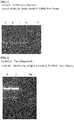
- the first PCR amplified DNA band of about 600 bp was collected, and a Gel purification process was performed using Qiagen Kit. Thereafter, in order to amplify the VHH gene, the second PCR amplification was performed using the VHH-FR4 downstream primer, the VHH-FR1 upstream primer, and the purified DNA. The results are shown in FIG. 1 and FIG. 2 .
- the DNA band of the single-domain antibody according to the present disclosure as amplified at about 600 bp could be identified.
- Example 2 The selection and search of an antibody that recognizes ⁇ v ⁇ 3 integrin from the single-domain antibody gene library as prepared in Example 2 was performed using a phage display method. Thus, a single-domain antibody clone library was prepared.
- the cut gene was inserted into the phagemid.
- a following process was carried out to transform the same in E. coli TG1.
- the second PCR product obtained in the 2-2 and pDisplay-3M TM were cleaved with BssHII and NheI, and purified simultaneously with Qiagen kit. Thereafter, the DNA fragment was ligated using NEB's T4 DNA ligase, and then the product was transformed via electro-transformation into E . coli TG1 cells. The results are shown in FIG. 3 and FIG. 4 .
- the E. coli TG1 transformed in the 3-1 was infected with the M13K07 helper phage.
- a process was performed as follows.
- log-phase TG1 cells and M13K07 phage were infected with different dilutions for 30 minutes at 37°C, and inoculated on agar of 2TY plates. After inoculating and culturing a small plaque in the 3 mL liquid 2TY medium, 30 ⁇ L of overnight cultured TG1 cells were added thereto and then culturing occurred at 37 degrees C for 2 hours. After diluting the culture medium of 1L 2TY medium and incubating for 1 hour, 50 ⁇ g/mL kanamycin was added thereto and culturing occurred under the condition of 37 degrees C for 16 hours.
- phage particles were removed by centrifugation (10 minutes, 5,000 g), and 0.25 volume of phage precipitate was added thereto to precipitate phage from the supernatant. After incubation on ice for 30 minutes, centrifugation (10 minutes, 5,000 g) was performed to obtain phage particles. After resuspending the pellet with 5 mL PBS, the pellet passed through a 0.22- ⁇ m filter. Helper phages were identified by counting the number of plaque-forming units (pfu) on a 2TY plate for the top-agar layer containing 100 ⁇ L TG1 (saturated culture). After diluting the phage stock solution to 1 ⁇ 10 13 pfu/mL, the phage stock solution was stored at -20 degrees C.
- the library glycerol stock and 500 mL 2TY-G were incubated and incubated at 37 degrees C with shaking at 250 rpm until the optical concentration reached 0.8 to 0.9 at 600 nm.
- M13KO7 helper phage was added thereto so that the final concentration was 5 ⁇ 10 9 pfu/mL, and incubated at 37°C for 30 minutes without shaking, followed by appropriate shaking (200 rpm) to perform phage infection for 30 minutes. Thereafter, the cells were collected by centrifugation at 2200 g for 15 minutes, and the pellet was resuspended in the same amount of 2TY-AK. Rapid shaking (300 rpm) was performed and incubation occurred overnight at 30 degrees C.
- 9 of the 10 clones were identified to have about 600 bp, and thus the single domain according to the present disclosure was correctly inserted thereto.
- ⁇ v ⁇ 3 integrin ITGAVB3 antigen to be used for specific antibody selection via panning was verified using SDS-PAGE.
- Example 4 After the panning process of Example 4, 40 single-domain antibody phage clones were extracted, and the binding ability thereof to ⁇ v ⁇ 3 integrin-specific single-domain antibody was analyzed using ELISA.
- an eluate of a single clone was inoculated into 5 mL 2YT-AG medium and culturing thereof occurred overnight at 37°C.
- a glycerol stock for each clone was prepared and cultured overnight, and 100 ⁇ L culture was put in 20 mL of 2YT-AG medium and cultured. The culture was carried out for several hours at 37 degrees C until the optical density of OD600 reached 0.4 to 0.5.
- M13KO7 helper phage was added in 20 multiple infections (that is, number of phage particles/host cells). The cells were incubated at 37°C for 30 minutes, and the cells were infected while shaking for 30 minutes.
- Infected cells were collected via centrifugation (10 minutes at 5,000 g), resuspended in 2YT-AK, and incubated at 30 degrees C for 16 hours. Thereafter, phage particles were precipitated in the supernatant, and the phage pellet was resuspended in 1 mL PBS and centrifuged (10 minutes at 5,000 g) to remove cell debris. Ab fragments irrelevant to phage particles were removed, phage pellets were removed in 250 ⁇ L PBS, and centrifugation was performed again.
- phage ELISA 2.5 ⁇ g/mL to 5 ⁇ g/mL of a target protein and a control protein were coated at 4 degrees C using a coating buffer, and the wells were washed 3 times with 200 ⁇ L washing buffer per well. The wells were blocked with 200 ⁇ L PBS-M for two hours at room temperature. The blocking buffer was removed and the plate was washed 6 times with the washing solution. Thereafter, 100 ⁇ L of phage solution was added to the washing buffer per well and incubation was executed at room temperature for 1 to 2 hours. Then, washing was performed 6 times with washing buffer. HRP-binding anti-M13 antibody (GE healthcare) was diluted with 1:5,000 blocking buffer.
- the HRP substrate solution was prepared as follows: A stock solution of OPD was prepared by dissolving 22 mg OPD (Sigma) in 100 mL of 50 mg sodium citrate (pH 4.0), and 36 ⁇ L 30% H 2 O 2 was added to 21 mL of OPL stock solution immediately before the detection step. 100 ⁇ L of substrate solution was added to each well and incubation was performed for 30 minutes at room temperature. Plates were analyzed using a micro plate reader set at 490 nm. Table 4 shows the results of analyzing the clones.
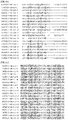
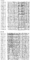
- the returned sequence was transformed using Vector NTI ® , Version 10, and the protein sequence was aligned. Thereafter, clones encoding the same protein sequence were grouped and analyzed. The results are shown in Table 5, FIG. 8A to FIG. 8C .
- the single-domain antibody clone according to the present disclosure is exemplified as VHH for convenience.
- clones 14, 11, 29, 17, 15, 28, 12, 19, 13, 21, 27, 16, 18, 20, 30, 23, 24, 25, 22, 26 showing high responses were selected from the 10 groups and their amino acid sequences were shown.
- the base sequence of clones VHH-25, VHH-13, VHH-22, VHH-11, VHH-17, VHH-15, VHH-12, VHH-16, VHH-20, VHH-23 exhibiting strong positives among them were represented by SEQ ID NOs: 1 to 10, respectively.
- the amino acid sequences of VHH-25, VHH-13, and VHH-22 among them were represented by SEQ ID NOs: 11, 12 and 13, respectively.
- the ELISA method of Example 5 was used to select single-domain antibody clones from among clones VHH-11, VHH-17, VHH-15, VHH-12, VHH-13, VHH-16, VHH-20, VHH-23, VHH-25, and VHH-22 exhibiting strong positives.
- the results are shown in Tables 6 and 7.
- VHH-12, VHH-13, VHH-16, VHH-22, and VHH-25 among the 10 candidate VHH clones exhibited relatively high binding ability.
- VHH-13, VHH-22 and VHH-25 were selected from among single-domain antibodies targeting the ⁇ v ⁇ 3 integrin in the Example 6, and were compared with conventional ⁇ v ⁇ 3 integrin antibodies, and thus SPR analysis was performed.
- CMDH chip Carboxymethyl Dextran Hydrogel Surface Sensor Chip, Reichert Technologies
- SPR equipment Reichert SR7500DC system
- VHH-13, VHH-22, and VHH-25 were the best among single-domain antibody clones according to the present disclosure.
- a vector to produce them was constructed and introduced into E. coli, thereby to express the single-domain antibody VHH-25.
- a vector containing a His6 tag was constructed for expression of VHH-13, VHH-22 or VHH-25, which are single-domain antibody clones selected according to the present disclosure.
- the vector map for VHH-25 (pALT-Avb3 VHH25) is shown in FIG. 10A .
- a vector containing Lysin and His6 tags was prepared, and a vector map (pALT-Avb3 VHH25K1) thereof is shown in FIG. 10B .
- the amino acid sequences containing His6 tag in clones VHH-25, VHH-13 and VHH-22 are represented by SEQ ID NOs: 14, 15 and 16, respectively.
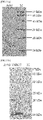
- VHH-13, VHH-22 or VHH-25 containing the His6 tag were identified by performing reduced SDS-PAGE. The results are shown in FIG. 11A to FIG. 11C .
- the expression results of VHH-25 containing Lysin and His6 tags are shown in FIG. 11D .
- VHH-13 was identified to have about 15.9 kDa ( FIG. 11A )
- VHH-22 was identified to have about 14.1 kDa ( FIG. 11B ).
- VHH-25 had about 14.5 kDa ( FIG. 11C )
- lysine-containing VHH-25 had about 14.2 kDa ( FIG. 11D ).
- an animal model was produced by inoculating U87-MG cells into Athymic nude mice (female, 6 to 8 weeks old). When the tumor was grown to a size of 0.8 cm to 1 cm, Cy5.5 was bound to VHH-25-K which in turn was injected into the tail vein (1 mg/kg). The contrast effect was analyzed using IVIS (PerkinElmer) over time. The results are shown in FIG. 13 .
- the contrast effect may be identified at the tumor site. It was identified that the contrast signal appeared strongly for 4 hours. Then, it was identified that a continuous contrast signal appeared at the tumor site for 24 hours.
- an animal model in which intravascular plaques are formed is produced by performing a high fat diet in ApoE-/- mice (Female, 10 weeks old) for 36 weeks. Thereafter, Cy5.5 was conjugated to VHH-25-K as prepared in the same manner as in Example 9, which in turn was injected into the tail vein (1 mg/kg), and 4 hours later, the mice were sacrificed and the aorta was separated therefrom. The contrast effect of the isolated aorta was analyzed using IVIS. The results are shown in FIG. 14 . As shown in FIG. 14 , it was identified that on 4 hours after the injection, a selective contrast effect appeared in the area where the plaque was formed.
Landscapes
- Health & Medical Sciences (AREA)
- Life Sciences & Earth Sciences (AREA)
- Immunology (AREA)
- Chemical & Material Sciences (AREA)
- Engineering & Computer Science (AREA)
- General Health & Medical Sciences (AREA)
- Biomedical Technology (AREA)
- Molecular Biology (AREA)
- Public Health (AREA)
- Animal Behavior & Ethology (AREA)
- Veterinary Medicine (AREA)
- Epidemiology (AREA)
- Organic Chemistry (AREA)
- Biochemistry (AREA)
- Medicinal Chemistry (AREA)
- Hematology (AREA)
- Urology & Nephrology (AREA)
- Proteomics, Peptides & Aminoacids (AREA)
- Genetics & Genomics (AREA)
- Biophysics (AREA)
- Cell Biology (AREA)
- Pathology (AREA)
- Microbiology (AREA)
- Food Science & Technology (AREA)
- Analytical Chemistry (AREA)
- General Physics & Mathematics (AREA)
- Physics & Mathematics (AREA)
- Biotechnology (AREA)
- Peptides Or Proteins (AREA)
- Micro-Organisms Or Cultivation Processes Thereof (AREA)
- Oncology (AREA)
Applications Claiming Priority (1)
| Application Number | Priority Date | Filing Date | Title |
|---|---|---|---|
| PCT/KR2019/009144 WO2021015336A1 (ko) | 2019-07-24 | 2019-07-24 | αVβ3 인테그린 표적 단일 도메인 항체 |
Publications (2)
| Publication Number | Publication Date |
|---|---|
| EP3916014A1 true EP3916014A1 (de) | 2021-12-01 |
| EP3916014A4 EP3916014A4 (de) | 2022-10-26 |
Family
ID=74193762
Family Applications (1)
| Application Number | Title | Priority Date | Filing Date |
|---|---|---|---|
| EP19938916.4A Pending EP3916014A4 (de) | 2019-07-24 | 2019-07-24 | Gegen avss3-integrin gerichtete einzeldomänen-antikörper |
Country Status (4)
| Country | Link |
|---|---|
| US (1) | US12383634B2 (de) |
| EP (1) | EP3916014A4 (de) |
| CN (1) | CN113557246B (de) |
| WO (1) | WO2021015336A1 (de) |
Family Cites Families (12)
| Publication number | Priority date | Publication date | Assignee | Title |
|---|---|---|---|---|
| US4554101A (en) | 1981-01-09 | 1985-11-19 | New York Blood Center, Inc. | Identification and preparation of epitopes on antigens and allergens on the basis of hydrophilicity |
| DE69322860T2 (de) * | 1992-04-03 | 1999-07-01 | Genentech, Inc., South San Francisco, Calif. | Antikörper gegen alpha v beta 3 integrin |
| US5753230A (en) * | 1994-03-18 | 1998-05-19 | The Scripps Research Institute | Methods and compositions useful for inhibition of angiogenesis |
| JP2001521520A (ja) * | 1997-04-11 | 2001-11-06 | ジー.ディー.サール アンド カンパニー | 抗α▲下v▼β▲下3▼インテグリン抗体アンタゴニスト |
| US20040208870A1 (en) * | 2003-01-30 | 2004-10-21 | Medimmune, Inc. | Stabilized high concentration anti-integrin alphanubeta3 antibody formulations |
| CN102875681A (zh) * | 2005-07-08 | 2013-01-16 | 拜奥根Idec马萨诸塞公司 | 抗-αvβ6抗体及其用途 |
| EP3023497A1 (de) * | 2005-11-18 | 2016-05-25 | Glenmark Pharmaceuticals S.A. | Anti-alpha2-integrin-antikörper und deren verwendungen |
| WO2007134876A2 (en) * | 2006-05-24 | 2007-11-29 | Bayer Schering Pharma Aktiengesellschaft | HIGH AFFINITY HUMAN AND HUMANIZED ANTI-α5β1 INTEGRIN FUNCTION BLOCKING ANTIBODIES WITH REDUCED IMMUNOGENICITY |
| TWI746473B (zh) * | 2015-11-02 | 2021-11-21 | 美商辛分子醫藥有限公司 | 針對細胞內抗原之單域抗體 |
| KR102630070B1 (ko) * | 2017-03-31 | 2024-01-26 | 더 리젠츠 오브 더 유니버시티 오브 캘리포니아 | 알파-v 베타-3-양성인 암 줄기 세포(csc)를 표적화하여 사멸시키고 약물 내성 암을 치료하기 위한 조성물 및 방법 |
| KR102098462B1 (ko) * | 2018-03-14 | 2020-04-08 | 가톨릭대학교 산학협력단 | αvβ3 인테그린 표적 단일 도메인 항체 |
| KR102101561B1 (ko) * | 2018-07-25 | 2020-04-20 | 가톨릭대학교 산학협력단 | 신생혈관 표적용 조영제 조성물 및 이의 제조방법 |
-
2019
- 2019-07-24 WO PCT/KR2019/009144 patent/WO2021015336A1/ko not_active Ceased
- 2019-07-24 US US17/431,357 patent/US12383634B2/en active Active
- 2019-07-24 CN CN201980093592.7A patent/CN113557246B/zh active Active
- 2019-07-24 EP EP19938916.4A patent/EP3916014A4/de active Pending
Also Published As
| Publication number | Publication date |
|---|---|
| WO2021015336A1 (ko) | 2021-01-28 |
| US20220133913A1 (en) | 2022-05-05 |
| CN113557246A (zh) | 2021-10-26 |
| EP3916014A4 (de) | 2022-10-26 |
| CN113557246B (zh) | 2023-09-01 |
| US12383634B2 (en) | 2025-08-12 |
Similar Documents
| Publication | Publication Date | Title |
|---|---|---|
| JP7700159B2 (ja) | Axlまたはror2に対するキメラ抗原受容体およびその使用方法 | |
| Dong et al. | A toolbox of nanobodies developed and validated for use as intrabodies and nanoscale immunolabels in mammalian brain neurons | |
| KR101151805B1 (ko) | 바이포달 펩타이드 바인더 | |
| EP1962961B1 (de) | Demikörper: dimerisations-aktivierte therapeutische mittel | |
| Hong et al. | Site-specific C-terminal dinitrophenylation to reconstitute the antibody Fc functions for nanobodies | |
| JP2016519933A (ja) | ソルタギング可能なタンパク質を有する赤血球のinvitro生成 | |
| Meléndez et al. | Novel lectin-based chimeric antigen receptors target Gb3-positive tumour cells | |
| CN114478800B (zh) | 基于血清白蛋白的融合蛋白、纳米组装体及其制备方法和应用 | |
| KR20060036476A (ko) | 뇌 이동 활성을 가지는 폴리펩티드 및 그 이용 | |
| JP2025113367A (ja) | 生理活性を有するペプチドの製造方法及び短鎖リンカーを含むペプチド | |
| CN117916271A (zh) | Her2单域抗体变体及其car | |
| CN118126201B (zh) | 一种靶向细胞膜grp94嵌合抗原受体、工程化细胞和应用 | |
| CN113088495A (zh) | 工程改造的t细胞、其制备及应用 | |
| CN117843794A (zh) | 一种抗ny-eso-1的纳米抗体及其应用 | |
| KR102098462B1 (ko) | αvβ3 인테그린 표적 단일 도메인 항체 | |
| US12383634B2 (en) | Single domain antibody targeting αVβ3 integrin | |
| CN119613550B (zh) | 非天然氨基酸修饰的抗pd-l1纳米抗体 | |
| Voloshchuk et al. | Sortase A mediated protein modifications and peptide conjugations | |
| CN120209142A (zh) | 一种靶向Ly6G6D的纳米抗体及其应用 | |
| CN110734494B (zh) | 抗tspan8单克隆抗体及其用途 | |
| UA81908C2 (uk) | Рекомбінантний химерний білок - антагоніст anth1 | |
| WO2015156644A1 (ko) | 환형의 β-헤어핀 기반 펩타이드 바인더 및 이의 제조방법 | |
| US20260071182A1 (en) | Monobody and NK Cells | |
| KR101443839B1 (ko) | 펩타이드의 타겟 친화도 개선방법 | |
| CN109943572B (zh) | 生产抗体片段的方法 |
Legal Events
| Date | Code | Title | Description |
|---|---|---|---|
| STAA | Information on the status of an ep patent application or granted ep patent |
Free format text: STATUS: THE INTERNATIONAL PUBLICATION HAS BEEN MADE |
|
| PUAI | Public reference made under article 153(3) epc to a published international application that has entered the european phase |
Free format text: ORIGINAL CODE: 0009012 |
|
| STAA | Information on the status of an ep patent application or granted ep patent |
Free format text: STATUS: REQUEST FOR EXAMINATION WAS MADE |
|
| 17P | Request for examination filed |
Effective date: 20210825 |
|
| AK | Designated contracting states |
Kind code of ref document: A1 Designated state(s): AL AT BE BG CH CY CZ DE DK EE ES FI FR GB GR HR HU IE IS IT LI LT LU LV MC MK MT NL NO PL PT RO RS SE SI SK SM TR |
|
| A4 | Supplementary search report drawn up and despatched |
Effective date: 20220923 |
|
| RIC1 | Information provided on ipc code assigned before grant |
Ipc: A61K 51/10 20060101ALI20220919BHEP Ipc: G01N 33/68 20060101ALI20220919BHEP Ipc: G01N 33/58 20060101ALI20220919BHEP Ipc: G01N 33/574 20060101ALI20220919BHEP Ipc: C07K 16/28 20060101AFI20220919BHEP |
|
| DAV | Request for validation of the european patent (deleted) | ||
| DAX | Request for extension of the european patent (deleted) | ||
| STAA | Information on the status of an ep patent application or granted ep patent |
Free format text: STATUS: EXAMINATION IS IN PROGRESS |
|
| 17Q | First examination report despatched |
Effective date: 20250901 |