EP3920134A1 - Procédé multicanal à profondeur de champ étendue et procédé de cytologie numérique automatisée - Google Patents
Procédé multicanal à profondeur de champ étendue et procédé de cytologie numérique automatisée Download PDFInfo
- Publication number
- EP3920134A1 EP3920134A1 EP20305602.3A EP20305602A EP3920134A1 EP 3920134 A1 EP3920134 A1 EP 3920134A1 EP 20305602 A EP20305602 A EP 20305602A EP 3920134 A1 EP3920134 A1 EP 3920134A1
- Authority
- EP
- European Patent Office
- Prior art keywords
- color
- image
- wavelet
- coefficient
- edf
- Prior art date
- Legal status (The legal status is an assumption and is not a legal conclusion. Google has not performed a legal analysis and makes no representation as to the accuracy of the status listed.)
- Granted
Links
Images
Classifications
-
- G—PHYSICS
- G06—COMPUTING OR CALCULATING; COUNTING
- G06T—IMAGE DATA PROCESSING OR GENERATION, IN GENERAL
- G06T5/00—Image enhancement or restoration
- G06T5/50—Image enhancement or restoration using two or more images, e.g. averaging or subtraction
-
- G—PHYSICS
- G06—COMPUTING OR CALCULATING; COUNTING
- G06T—IMAGE DATA PROCESSING OR GENERATION, IN GENERAL
- G06T7/00—Image analysis
- G06T7/0002—Inspection of images, e.g. flaw detection
- G06T7/0012—Biomedical image inspection
-
- G—PHYSICS
- G06—COMPUTING OR CALCULATING; COUNTING
- G06T—IMAGE DATA PROCESSING OR GENERATION, IN GENERAL
- G06T3/00—Geometric image transformations in the plane of the image
- G06T3/40—Scaling of whole images or parts thereof, e.g. expanding or contracting
-
- G—PHYSICS
- G06—COMPUTING OR CALCULATING; COUNTING
- G06T—IMAGE DATA PROCESSING OR GENERATION, IN GENERAL
- G06T5/00—Image enhancement or restoration
- G06T5/20—Image enhancement or restoration using local operators
-
- G—PHYSICS
- G06—COMPUTING OR CALCULATING; COUNTING
- G06T—IMAGE DATA PROCESSING OR GENERATION, IN GENERAL
- G06T7/00—Image analysis
- G06T7/10—Segmentation; Edge detection
-
- G—PHYSICS
- G06—COMPUTING OR CALCULATING; COUNTING
- G06V—IMAGE OR VIDEO RECOGNITION OR UNDERSTANDING
- G06V10/00—Arrangements for image or video recognition or understanding
- G06V10/40—Extraction of image or video features
- G06V10/56—Extraction of image or video features relating to colour
-
- H—ELECTRICITY
- H04—ELECTRIC COMMUNICATION TECHNIQUE
- H04N—PICTORIAL COMMUNICATION, e.g. TELEVISION
- H04N1/00—Scanning, transmission or reproduction of documents or the like, e.g. facsimile transmission; Details thereof
- H04N1/40—Picture signal circuits
- H04N1/40012—Conversion of colour to monochrome
-
- H—ELECTRICITY
- H04—ELECTRIC COMMUNICATION TECHNIQUE
- H04N—PICTORIAL COMMUNICATION, e.g. TELEVISION
- H04N23/00—Cameras or camera modules comprising electronic image sensors; Control thereof
- H04N23/60—Control of cameras or camera modules
- H04N23/67—Focus control based on electronic image sensor signals
- H04N23/676—Bracketing for image capture at varying focusing conditions
-
- G—PHYSICS
- G06—COMPUTING OR CALCULATING; COUNTING
- G06T—IMAGE DATA PROCESSING OR GENERATION, IN GENERAL
- G06T2207/00—Indexing scheme for image analysis or image enhancement
- G06T2207/10—Image acquisition modality
- G06T2207/10024—Color image
-
- G—PHYSICS
- G06—COMPUTING OR CALCULATING; COUNTING
- G06T—IMAGE DATA PROCESSING OR GENERATION, IN GENERAL
- G06T2207/00—Indexing scheme for image analysis or image enhancement
- G06T2207/10—Image acquisition modality
- G06T2207/10056—Microscopic image
-
- G—PHYSICS
- G06—COMPUTING OR CALCULATING; COUNTING
- G06T—IMAGE DATA PROCESSING OR GENERATION, IN GENERAL
- G06T2207/00—Indexing scheme for image analysis or image enhancement
- G06T2207/20—Special algorithmic details
- G06T2207/20024—Filtering details
- G06T2207/20032—Median filtering
-
- G—PHYSICS
- G06—COMPUTING OR CALCULATING; COUNTING
- G06T—IMAGE DATA PROCESSING OR GENERATION, IN GENERAL
- G06T2207/00—Indexing scheme for image analysis or image enhancement
- G06T2207/20—Special algorithmic details
- G06T2207/20048—Transform domain processing
- G06T2207/20064—Wavelet transform [DWT]
-
- G—PHYSICS
- G06—COMPUTING OR CALCULATING; COUNTING
- G06T—IMAGE DATA PROCESSING OR GENERATION, IN GENERAL
- G06T2207/00—Indexing scheme for image analysis or image enhancement
- G06T2207/20—Special algorithmic details
- G06T2207/20212—Image combination
- G06T2207/20221—Image fusion; Image merging
Definitions
- the present invention relates to the field of image processing of biological samples images.
- the present invention relates to the field of image analysis of urine samples in the context of automated digital cytology.
- one commonly used solution consists in fusing the sequence of acquired images in a single composite image. To do so, it is possible to select the image containing the most information among all the images of the volume, i.e. the stack of images. In this «best-focus» selection method almost no computation is required However, «best-focus» methods result in loss of information when the objects of interest are thicker than the depth of field of the acquisition module, for instance in presence of cell clumps.
- Extended Depth of Field (EDF) methods are used to fuse the images of the z-stack into a single 2D image (EDF image) in which all the parts of the object of interest are seemingly in focus, while avoiding the loss of information associated with the previously described "best-focus" method.
- EDF methods are usually classified in 3 categories: spatial domain approaches; transform-based methods, such as those based on wavelet transforms (WT), and deep-learning based methods.
- WT wavelet transforms
- One spatial domain approach which is relatively easy to implement, consists in selecting a focused area within each image of the z-stack; then fusing the selected areas to obtain one single 2D image in which all the different parts of the object of interest are in focus.
- SME smooth manifold extraction
- Common wavelet transform-based methods include Discrete Wavelet Transform (DWT) algorithms and Stationary Wavelet Transform (SWT) algorithms.
- DWT Discrete Wavelet Transform
- SWT Stationary Wavelet Transform
- A. P. Bradley and P. C. Bamford have shown, in "A One-pass Extended Depth of Field Algorithm Based on the Over-complete Discrete Wavelet Transform” (2004 ), that the latter is more performing.
- wavelet transform-based methods for computing an EDF image of a stack of images consist in applying a wavelet transform algorithm on every image in the stack, thus obtaining a stack of wavelet transforms; then applying a coefficient selection rule to select the most relevant wavelet coefficients in the stack of wavelet transforms.
- the coefficient selection rules are well defined for grayscale volumes and several techniques of the prior art compute an EDF image of a stack by applying wavelet transforms on grayscale images, i.e. images comprising only one channel. Such techniques can also be applied on color images, i.e. images comprising more than one color channel.
- One possible approach to manage multi-channel images is to apply on each channel, in a succession: a wavelet transform, a coefficient selection strategy and an inverse wavelet transform, thereby obtaining one EDF image for each channel.
- the EDF images thus obtained are then merged to obtain one color EDF image.
- the main drawback of this method is that it produces color saturation and/or artifacts.
- B. Forster et al. in "Extended Depth-of-Focus for multi-channel microscopy images: a complex wavelet approach" (2004 ), propose to perform a preprocessing step of color-to-grayscale conversion and a post-processing step of color reassignment.
- Urinary cytopathology is the method of choice for non-invasive detection of bladder cancer; it consists in analyzing microscopy slides containing urothelial cells, looking for abnormalities. This method gives satisfactory results for the detection of advanced cancers; however, it is time-consuming, expensive and ineffective for the detection of early-stage bladder cancers. Early detection is a key issue in order to increase patients' chances of survival while drastically reducing the cost of the treatments, which is known to be more elevated for advanced stage cancers. Automatic solutions may help the management of early-stage cancers by reducing the analysis time, thus accelerating the diagnostic process, and by improving the detection and quantification of abnormalities by means of performing segmentation algorithms.
- Fine detail recovery is fundamental to achieve precise segmentation and, in the context of automated digital cytology, precise cytoplasm and nucleus segmentation is crucial (i) to improve the detection of abnormal cells; (ii) to ease cell counting and (iii) to calculate, with high accuracy, biomarkers correlated with clinically relevant information, such as the Nuclear/Cytoplasmic Ratio.
- the present invention aims to overcome the problems associated with the generation of an EDF image from a color volume in the field of image analysis of biological samples.
- the present invention relates to a computer implemented method for generating a color-faithful extended depth-of-field (EDF) image from a color volume of a biological sample having dimension ( N , M , L ) and values I( n , m , l ), the voxels ( n , m , l ) for a fixed l going from 1 to L being images acquired at different focal depths in a z-direction using a microscope or a scanner, said method comprising the following steps:
- the method of the present invention allows accurate color reproduction and fine detail recovery in an EDF image obtained from a color volume.
- this method drastically reduces color inaccuracies associated with the color reassignment strategies of the prior art, without compromising the level of details.
- the set of wavelet coefficients comprises at least four wavelet coefficients and the subset of wavelet coefficients selected using a predefined coefficient selection rule comprises at least one wavelet coefficient.
- the number of wavelet coefficients in the 3D wavelet coefficient matrix depends on the decomposition level of the wavelet transform.
- the wavelet transform comprises multiple levels of decomposition applied in succession.
- the wavelet transform comprises only one level of decomposition, so that a color-faithful extended depth-of-field (EDF) image with fine detail recovery is obtained with less computational steps.
- the set of wavelet coefficients comprises four wavelet coefficients and the subset of selected wavelet coefficients comprises at least one and no more than four coefficients.
- a first subset of wavelet coefficients is used in a first predefined coefficient selection rule and a second subset of wavelet coefficients is used in a second predefined coefficient selection rule.
- the step of generating a 2D wavelet coefficient matrix WCM and a 2D coefficient map CM comprises:
- the value of the voxel of the 3D wavelet coefficient matrix having a l index equal to the value of the 2D coefficient map CM comprises the full set of wavelet coefficients.
- the value of each pixel having a ( n , m ) index is the l index of the voxel of the 3D wavelet coefficient matrix that comprises the subset of wavelet coefficients selected by the first coefficient selection rule.
- the value of each pixel having a ( n , m ) index is the l index of the voxel of the 3D wavelet coefficient matrix that comprises the subset of wavelet coefficients selected by the second coefficient selection rule.
- the set of wavelet coefficient in a pixel having a ( n , m ) index is the set of coefficients in the 3D wavelet coefficient matrix having the same ( n , m ) index and having as l index the value of the 2D coefficient maps.
- the 2D coefficient maps is obtained by combining a first and a second coefficient maps.
- a first 2D wavelet coefficient matrix WCM is generated as well.
- a second 2D wavelet coefficient matrix WCM is generated in the step of generating a second 2D coefficient map.
- said first and second 2D wavelet coefficient matrices do not need to be combined.
- the 2D wavelet coefficient matrix WCM is generated based on the first and second 2D coefficient maps.
- This embodiment allows to select detail information by means of the first coefficient selection rule and to obtain a denoising effect by means of the second coefficient selection rule.
- the step of combining the first and the second 2D coefficient maps allows to obtain one 2D coefficient map CM from which is then computed one wavelet coefficient matrix WCM.
- the combination step allows to obtain a wavelet coefficient matrix WCM and a 2D coefficient map CM that have both detail information, obtained by means of the first coefficient selection rule, and a denoising effect, obtained by means of the second coefficient selection rule.
- the step of combining the 2D coefficient maps comprises:
- This embodiment allows to avoid an interpolation step.
- the values obtained in the rounded 2D coefficient map CM are used to select wavelet coefficients from the 3D wavelet coefficient matrix having a l index equal to said obtained values.
- the wavelet coefficient matrix WCM is built based on the selected coefficients.
- the color-faithful extended depth-of-field (EDF) image obtained with the present invention is converted to a different color space, preferentially the color space of the color volume. This allows to ease further steps of image analysis, such as segmentation, by selecting the color space in which the algorithms used in the further steps of image analysis work best.
- the invertible color-to-grayscale transformation is a principal component analysis (PCA).
- PCA has the advantage of being a standard and easy-to-implement technique for dimensionality reduction having the further advantage of being invertible.
- a stationary wavelet transform in the step of applying a wavelet transform, is applied.
- the SWT is associated with better performance metrics than other methods, such as CWT-EDF and "best-focus" methods.
- SWT-EDF achieves the best color fidelity when compared to other wavelet-based methods followed by color reconstruction strategies.
- the color volume is a biomedical image and the method further comprises a step of segmentation of the color-faithful extended depth-of-field (EDF) image. Segmentation performances are improved when the segmentation is applied to an image with fine reproduction of details and elevated color fidelity.
- EDF extended depth-of-field
- the present invention also relates to a system for analysis of biological samples, said system having a processor and a computation module configured to:
- the system for analysis of biological samples is an automated digital cytology system for the analysis of urine sample.
- the system further comprises an acquisition module configured to acquire a color volume, the color volume comprising at least two color images.
- the system of the present invention is a bladder cancer detection system.
- the present invention also relates to a computer program product for analysis of biological samples, the computer program product comprising instructions which, when the program is executed by a computer, cause the computer to carry out the steps of the method according to the present invention.
- the present invention also relates to a computer-readable storage medium comprising instructions which, when the program is executed by a computer, cause the computer to carry out the steps of the method according to any one of the embodiments described hereabove.
- the present invention relates to a computer implemented method for generating an extended depth-of-field (EDF) image from a volume comprising a stack of 2D microscopy images acquired at different focal depths in a z-direction.
- EDF extended depth-of-field
- the EDF image obtained with the present method is characterized by an elevated color fidelity and fine detail recovery.
- the volume of the present method is a color volume.
- the method M used in the present invention comprises a receiving step to receive a volume 1.
- the volume 1 is a stack of 2D color images acquired by moving a microscopy slide comprising an object of interest along a z-direction parallel to the optical axis of a bright-field microscope.
- the volume 1 has dimension ( N , M , L ) and each voxel has a value I( n , m , I ).
- the set of voxels of the volume 1 having a fixed l , comprised between 1 and L are 2D images acquired at different focal depths in a z-direction using a microscope.
- the L dimension is equal to the number of acquired images.
- the l index of a voxel is the focal depth at which the initial image comprising said voxel has been acquired.
- the L dimension of the volume 1 is equal or superior to 2, so that said volume 1 is a stack of 2D images comprising at least two 2D-images acquired at different focal depths.
- the volume 1 according to the present invention may be represented in any color space.
- the initial color volume i.e. the stack of 2D images, is an RGB volume.
- the method M herein described comprises a step of generating a grayscale volume 2 from a color volume 1 by applying M10 an invertible color-to-grayscale transformation to said color volume 1.
- the color-to-grayscale transformation need to be an invertible transformation, in order to convert back the grayscale EDF image 4, which is obtained after applying M50 an inverse wavelet transformation, to a color image.
- the color-to-grayscale invertible transformation may be a principal component analysis (PCA) transformation; in this particular embodiment, its inverse transformation is an inverse PCA allowing to reconstruct the original color variables from the principal components.
- PCA principal component analysis
- the method M of the present invention comprises a step M20 of applying a wavelet transform to the grayscale volume 2.
- the application of the wavelet transform allows to obtain a 3D wavelet coefficient matrix 3 having ( N, M, L ) dimension, wherein the value of each voxel having a ( n , m, l ) index comprises a set of wavelet coefficients.
- Said set of coefficients comprises at least four wavelet coefficients.
- the term "3D" refers to a three-dimensional matrix.
- the wavelet coefficient matrix 3 is a 3D matrix since it has dimension ( N , M , L ).
- the color volume 1, the greyscale volume 2 and the 3D wavelet coefficient matrix 3 are three-dimensional matrices having dimension ( N , M , L ); whereas the wavelet coefficient matrix WCM, the coefficient map CM, the grayscale EDF image 6, the color composite image CC, the color EDF image 7 and the color-faithful extended depth-of-field (EDF) image are 2D, i.e. bi-dimensional matrices having dimension ( N , M ).
- the step M20 of applying a wavelet transform to the grayscale volume 2 is performed in parallel on each set of NxM voxels of the grayscale volume 2 having a same l index and ( n , m ) index going from (1,1) to ( N , M ). Therefore, for a grayscale volume 2 having ( N , M, L) dimension, L wavelet transforms are applied in said step M20.
- the wavelet transform applied in step M20 is a stationary wavelet transform (SWT) because among wavelet-based methods with reconstructed colors, SWT-EDF achieves the best color fidelity.
- SWT-EDF achieves the best color fidelity.
- This is particularly advantageous in case the image obtained with the present method M, after the concatenating step M90, is further segmented.
- SWT-EDF outperforms both CWT-EDF and "best-focus" methods according to the common evaluation metric Intersection over Union (IoU).
- a wavelet transform produces a decomposition of a signal as wavelet coefficients, said wavelet coefficients comprising approximation coefficients and detail coefficients.
- the wavelet transform is known as a decomposition of the original image into sub-images of wavelet coefficients. This decomposition is reversible, the original image can be reconstructed from the wavelet coefficients by applying the inverse wavelet transform.
- a wavelet transform of an image is computed by applying a first decomposition to each row of pixels of the image and then a second decomposition to each column of the result of the first decomposition.
- One level of wavelet decomposition of an image is the result of said first and second decompositions, i.e.
- One level of wavelet decomposition of an image produces four matrices of wavelet coefficients (sub-images).
- the values of the pixels of said sub-images comprise the wavelet coefficients.
- several levels of subsequent decomposition may be performed.
- four sub-images are obtained: three "detail" sub-images comprising the details of the original image and a fourth "approximation" sub-image comprising an approximation of the original image.
- the values of the pixels of said sub-images comprise the wavelet coefficients.
- the values of the pixels of the three "detail" sub-images comprise: in a first sub-image, the horizontal detail coefficients; in a second sub-image, the vertical detail coefficients; and, in a third sub-image, the diagonal detail coefficients.
- Said horizontal detail coefficients, vertical details coefficients and diagonal detail coefficients are a function of the horizontal details, vertical details and diagonal details of the original image, respectively.
- the values of the pixels of the fourth approximation sub-image comprise the approximation coefficients.
- Said approximation coefficients are a function of the approximation of the original image.
- the image is filtered with a high-pass filter to generate the detail coefficients and with a low-pass filter to generate the approximation coefficients.
- filtering is performed on the image obtained in the previous decomposition step.
- the original image can be reconstructed by applying an inverse wavelet transform on the decomposition.
- the wavelet transform applied in step M20 comprises one level of decomposition, hence it decomposes each image of the grayscale volume 2 in four sub-images comprising the wavelet coefficients.
- image of the grayscale volume 2 is meant the set of N x M voxels of the grayscale volume 2 having a same l index, so that the l th image of the grayscale volume 2 is the set of voxels having a fixed l and ( n , m ) index going from (1,1) to ( N , M ).
- the present method M also comprises a selection step M30 where for each group of L voxels of the 3D wavelet coefficient matrix 3 having a same ( n , m ) index and a l index going from 1 to L , the step is configured to select a set of wavelet coefficients using a predefined coefficient selection rule.
- Said set of wavelet coefficients comprise at least one detail coefficient or approximation coefficient from the sub-images defined hereabove.
- the whole set of wavelet coefficients is used in the further steps of the present invention.
- one subset of wavelet coefficients is selected from the whole set of wavelet coefficients, and said subset is further used to compute the 2D coefficient map CM.
- several 2D coefficient maps may be computed; in particular, each of the 2D coefficient maps is computed based on a different subset of wavelet coefficients; then one 2D coefficient map is obtained by combining the several 2D coefficient maps.
- the coefficient selection rule applied in step M30 may be any coefficient selection rule known by a person skilled in the art.
- the coefficient selection rule is adapted for grayscale images or volumes.
- applying the coefficient selection rule on every color channel and merging the results can produce false colors; it is therefore necessary to convert the initial color image to a grayscale image, then to compute a 2D greyscale EDF image, finally to perform a color reconstruction.
- one or more coefficient selection rules may be applied.
- the method M of the present invention further comprises a step of generating M40:
- a first subset of wavelet coefficients is selected using a first predefined coefficient selection rule and a second subset of wavelet coefficients is selected using a second predefined coefficient selection rule.
- the step of generating M40 a 2D wavelet coefficient matrix WCM and a 2D coefficient map CM comprises multiple steps M41, M42, M43 and M44.
- the step M41 consists in generating a first 2D wavelet coefficient matrix and a first 2D coefficient map from the first subset of wavelet coefficients.
- the step M41 is followed by a step M42 of generating a second 2D wavelet coefficient matrix and a second 2D coefficient map from the second subset of wavelet coefficients.
- the step M43 combines the first and the second 2D coefficient maps, so as to obtain one final 2D coefficient map CM.
- a 2D wavelet coefficient matrix WCM is generated in step M44.
- the value WCM( n , m ) of each pixel of the 2D wavelet coefficient matrix WCM is the set of coefficients of the 3D wavelet coefficient matrix having a ( n , m ) index and having a l index equal to the value CM( n , m ) of the final 2D coefficient map CM.
- the step M43 of combining the first and the second 2D coefficient maps, so as to obtain one 2D coefficient map CM may comprise multiple steps.
- it comprises:
- the median filter is a 3x3 median filter.
- two 2D wavelet coefficient matrices and two 2D coefficient maps are generated.
- the value of each pixel having a ( n , m ) index is the first subset of wavelet coefficients selected from the 3D wavelet coefficient matrix 3 by the first coefficient selection rule.
- the value of each pixel having a ( n , m ) index is the second subset of wavelet coefficients selected from the 3D wavelet coefficient matrix 3 by the second coefficient selection rule.
- the value of each pixel having a ( n , m ) index is the l index of the voxel of the 3D wavelet coefficient matrix 3 comprising the first subset of wavelet coefficients selected by the first coefficient selection rule.
- the value of each pixel having a ( n , m ) index is the l index of the voxel of the 3D wavelet coefficient matrix 3 comprising the second set of wavelet coefficients selected by the second coefficient selection rule.
- the first selection rule is applied to select the most relevant coefficients from the subset of coefficients comprising detail coefficients
- the second selection rule is applied to select the most relevant coefficients from a subset of coefficients comprising the approximation coefficients.
- the detail coefficients comprise the horizontal detail coefficients, the vertical detail coefficients and the diagonal detail coefficients.
- the first coefficient selection rule comprises:
- the second coefficient selection rule comprises:
- the first 2D wavelet coefficient matrix comprises information about the image details
- the second 2D wavelet coefficient matrix is a smoothened representation of the image due to the denoising effect resulting from the low-pass filter of the wavelet transform.
- the method comprises a step of applying M50 an inverse transformation of the wavelet transform to the 2D wavelet coefficient matrix WCM so as to obtain a 2D grayscale EDF image 4.
- the present method comprises a step of generating M60 a 2D color composite image CC, wherein the value CC ( n , m ) of each pixel having ( n , m ) index is the value I( n , m , l ) of the voxel of the color volume 1 having a l index equal to the value CM( n , m ) of the 2D coefficient map CM.
- the method also comprises a step M70 of applying an inverse transformation of the invertible color-to-grayscale transformation to the 2D grayscale EDF image 4 so as to obtain a 2D color EDF image 5.
- the method also comprises a step M80 of converting the 2D color composite image CC and the 2D color EDF image 5 into a color space comprising at least one chromaticity component and at least one intensity component.
- color space refers to the mathematical representation of a color in a coordinate axis system, in which each color is defined by a vector, the projections of the vector on the axis of the coordinate axis system being the different components of the color in said color space.
- Most color spaces use three components to represent a color.
- biomedical images are acquired by devices such as cameras and scanners whose sensors comprise three channels, namely, red (R), green (G), and blue (B), hence generating an image in the RGB color space.
- R, G and B components are highly correlated; moreover, luminance information is not available. Therefore, for some applications, a conversion, as in step M70, from the RGB color space to another color space with a different coordinate axis system is suitable.
- Each coordinate axis of the color space comprises a color information, said information may be luma values, the luminance, a function of luminance; or alternatively the chrominance (combination of a chroma value and a luma value) or a function of chrominance.
- each coordinate axis comprises either chromaticity information, i.e. chrominance or a function of chrominance, or intensity information, i.e. , luma values or luminance or a function of luminance; and none of the coordinate axis comprises a combination of said chromaticity information and intensity information.
- RGB in color spaces such as YUV, YES, YT 1 T 2 , CIE L*a*b*, YcbCr, CIE L*U*V*, HSV the coordinate axis comprising intensity information are separated from the coordinate axis comprising chromaticity information.
- these color spaces comprise one luminance component and two chrominance components.
- the conversion step M80 is configured to convert the 2D color composite image CC and the 2D color EDF image 5 into a YUV color space in which the luminance component Y is the intensity component while U and V are the chromaticity components.
- the color space of the conversion step M80 is one of the following color spaces: CIE L*a*b*, YcbCr, CIE L*U*V*, HSV.
- the method further comprises the step M90 of concatenating the at least one chromaticity component of the 2D color composite image CC and the at least one intensity component of the 2D color EDF image 5, to obtain a color-faithful extended depth-of-field (EDF) image.
- EDF extended depth-of-field
- the 2D color composite image CC Since the chromaticity information of the 2D color composite image CC is retrieved from the initial color volume 1 in the step of generating M60 the 2D color composite image CC, the 2D color composite image CC has elevated color fidelity compared to the volume 1. Since the detail information in the 2D color EDF image 5 is maximized by means of the coefficient selection rule, the 2D color EDF image 5 has elevated recovery of fine details from the color volume 1. Concatenating the intensity component of the 2D color EDF image 5 and the chromaticity components of the 2D color composite image CC advantageously allows to combine fine recovery of details from the color volume 1 and elevated color fidelity to the color volume 1. As a consequence, the method M allows to obtain a "color-faithful" extended depth-of-field (EDF) image with fine detail recovery from the initial volume 1.
- EDF extended depth-of-field
- the color space has one intensity component and two chromaticity components and in the concatenating step M90 the one intensity component of the color EDF image 7 and the two chromaticity components of the color composite image CC are concatenated. Therefore, in this particular embodiment, the color-faithful extended depth-of-field (EDF) image has one intensity component and two chromaticity components.
- EDF extended depth-of-field
- the color-faithful extended depth-of-field (EDF) image obtained in the concatenating step M90 may further be converted to any color space.
- it is converted to the color space of the initial color volume 1.
- This may be an RGB color space.
- the color-faithful extended depth-of-field (EDF) image obtained is converted to a color space different than the color space of the initial volume 1.
- the color-faithful extended depth-of-field (EDF) image obtained in the concatenating step M90 is further segmented.
- the segmentation step is performed using a U-net segmentation model.
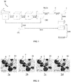
- FIG. 1 A non-limiting example of the steps M10 to M90 of the method according to the present inventions is represented in figure 1 .
- the selection step M30 is not illustrated.
- Various modifications of this steps may be implemented in the steps M10 to M70 without departing from the result of obtaining a 2D wavelet coefficient matrix WCM, a 2D coefficient map CM, a 2D color composite image CC and a 2D color EDF image 5.
- the method M of the present invention allows to obtain from a color volume 1, a 2D image with elevated color fidelity and fine detail recovery by concatenating, in a color space, the intensity component of the 2D color EDF image 5 and the chromaticity components of the color composite image 2D color composite image CC.
- FIG. 2 One advantage of the present invention will be better understood when read in conjunction with figure 2 .
- the objects of interest are cells 6, some of which form cell clumps 7.
- figures 2a , 2b and 2d represent non-limitative examples of images obtained before or after performing different steps of the method M of the present invention.
- Figure 2c and 2d represent, respectively: a color image obtained from the image in figure 2b after a color reassignment strategy according to the prior art; and a color image obtained from the image in figure 2b after a color reassignment strategy according to the present invention.
- figure 2a is an example of an image belonging to an initial color volume 1, comprising in-focus objects 8 and out-of-focus objects 9.
- a color volume 1 of a biological sample usually one color is predominant.
- the main color of all the objects is blue, with the exception of the element 10, whose main color is red.
- blue is the predominant color and red is a rare color.
- All objects in the images represented in figures 2b to 2d are in-focus.
- Figure 2b is an example of a 2D color EDF image 5 obtained as output of the step of applying M70 an inverse transformation of the invertible color-to-grayscale to the 2D grayscale EDF image 4.
- Figure 2b represents a 2D color EDF image 5 obtained from the image in figure 2b , in which a color assignment technique according to the prior art is applied. The red color of the element 10 following the color assignment strategy of the prior art is partially recovered.
- Figure 2d represents a 2D color EDF image 5 obtained with the method M of the present invention.
- the image in figure 2d is obtained after converting the 2D color EDF image 5 and the 2D color composite image CC in the YUV color space, then concatenating the luminance component Y of the 2D color EDF image 5 and the U, V chromaticity components of the 2D color composite image CC.
- the red color of the element 10 is fully recovered and the element 10 has the same color as in the image in figure 2a .
- Color fidelity obtained with the prior art and color fidelity obtained with the present method are compared by means of the chi-squared distance ( ⁇ ).
- the chi-squared distance ( ⁇ ) between the RGB normalized color histogram of each image represented in figure 2 and the color histogram of the image in figure 2a is calculated.
- the chi-squared distance ( ⁇ ) between the image in figure 2d and the image in figure 2a is smaller than the chi-squared distance ( ⁇ ) between the image in figure 2c and the image in figure 2a .
- the present invention ensures accurate reproduction of elements with rare colors, i.e ., elements whose chrominance component is not the predominant chrominance component of the original color volume 1.
- the main issues observed in a 2D color EDF image 5 are: the lack of a color accuracy and the presence of artifact.
- the present method achieves the best color fidelity when compared to other wavelet-based methods followed by color reconstruction strategies. For instance, the example in figure 2 shows that an element with rare colors is better reproduced with the present method M than with the color reconstruction methods of the prior art.
- the present invention also relates to a system for analysis of a color volume 1 acquired from a biological sample.
- Said system comprises a storage medium and processor having means for carrying out the steps of the method according to any one of the embodiments described above.
- the system further comprises a visualization module for displaying the volume 1, the grayscale volume 2, the 2D color composite image CC, the 2D grayscale EDF image 4, the 2D color EDF image 5 and the color-faithful extended depth-of-field (EDF) image.
- the visualization module may further display the 3D wavelet coefficient matrix 3, the 2D wavelet coefficient matrix WCM, the 2D coefficient map CM.
- the biological sample is a urine sample.
- the system comprises an automated digital cytology system configured to acquire a volume 1, the volume 1 comprising at least two 2D color images.
- the present invention further comprises of a computer program product for generating a color-faithful extended depth-of-field (EDF) image from a volume 1 obtained from a microscope, the computer program product comprising instructions which, when the program is executed by a computer, cause the computer to carry out the steps of the method according to any one of the embodiments described hereabove.
- EDF extended depth-of-field
- the computer program product to perform the method as described above may be written as computer programs, code segments, instructions or any combination thereof, for individually or collectively instructing or configuring the processor or computer to operate as a machine or special-purpose computer to perform the operations performed by hardware components.
- the computer program product includes machine code that is directly executed by a processor or a computer, such as machine code produced by a compiler.
- the computer program product includes higher-level code that is executed by a processor or a computer using an interpreter. Programmers of ordinary skill in the art can readily write the instructions or software based on the block diagrams and the flow charts illustrated in the drawings and the corresponding descriptions in the specification, which disclose algorithms for performing the operations of the method as described above.
- the present invention further comprises a computer-readable storage medium comprising instructions which, when the program is executed by a computer, cause the computer to carry out the steps of the method according to any one of the embodiments described hereabove.
- the computer-readable storage medium is a non-transitory computer-readable storage medium.
- Computer programs implementing the method of the present embodiments can commonly be distributed to users on a distribution computer-readable storage medium such as, but not limited to, an SD card, an external storage device, a microchip, a flash memory device, a portable hard drive and software websites. From the distribution medium, the computer programs can be copied to a hard disk or a similar intermediate storage medium. The computer programs can be run by loading the computer instructions either from their distribution medium or their intermediate storage medium into the execution memory of the computer, configuring the computer to act in accordance with the method of this invention. All these operations are well- known to those skilled in the art of computer systems.
- the instructions or software to control a processor or computer to implement the hardware components and perform the methods as described above, and any associated data, data files, and data structures, are recorded, stored, or fixed in or on one or more non-transitory computer-readable storage media.
- Examples of a non-transitory computer-readable storage medium include read-only memory (ROM), random-access memory (RAM), flash memory, CD- ROMs, CD- Rs, CD+Rs, CD- RWs, CD+RWs, DVD-ROMs, DVD- Rs, DVD+Rs, DVD- RWs, DVD+RWs, DVD- RAMs, BD- ROMs, BD- Rs, BD- R LTHs, BD- REs, magnetic tapes, floppy disks, magneto-optical data storage devices, optical data storage devices, hard disks, solid-state disks, and any device known to one of ordinary skill in the art that is capable of storing the instructions or software and any associated data, data files, and data structures in a non-transitory manner
- the instructions or software and any associated data, data files, and data structures are distributed over network-coupled computer systems so that the instructions and software and any associated data, data files, and data structures are stored, accessed, and executed in a distributed fashion by the processor or computer.
Landscapes
- Engineering & Computer Science (AREA)
- Physics & Mathematics (AREA)
- General Physics & Mathematics (AREA)
- Theoretical Computer Science (AREA)
- Multimedia (AREA)
- Signal Processing (AREA)
- Computer Vision & Pattern Recognition (AREA)
- General Health & Medical Sciences (AREA)
- Medical Informatics (AREA)
- Nuclear Medicine, Radiotherapy & Molecular Imaging (AREA)
- Radiology & Medical Imaging (AREA)
- Quality & Reliability (AREA)
- Health & Medical Sciences (AREA)
- Image Processing (AREA)
- Microscoopes, Condenser (AREA)
- Image Analysis (AREA)
Priority Applications (9)
| Application Number | Priority Date | Filing Date | Title |
|---|---|---|---|
| EP20305602.3A EP3920134B1 (fr) | 2020-06-05 | 2020-06-05 | Méthode de profondeur de champ étendue multi-canaux pour la cytologie numérique automatisée |
| ES20305602T ES2991834T3 (es) | 2020-06-05 | 2020-06-05 | Método multicanal de profundidad de campo extendida para citología digital automatizada |
| CA3179368A CA3179368A1 (fr) | 2020-06-05 | 2021-06-04 | Procede a profondeur de champ etendue a canaux multiples pour cytologie numerique automatisee |
| US18/007,862 US12283045B2 (en) | 2020-06-05 | 2021-06-04 | Multi-channel extended depth-of-field method for automated digital cytology |
| JP2022574393A JP7749171B2 (ja) | 2020-06-05 | 2021-06-04 | 自動デジタル細胞診のためのマルチチャンネル拡張被写界深度法 |
| EP21730226.4A EP4162441B1 (fr) | 2020-06-05 | 2021-06-04 | Procédé multicanal à profondeur de champ étendue et procédé de cytologie numérique automatisée |
| PCT/EP2021/065055 WO2021245262A1 (fr) | 2020-06-05 | 2021-06-04 | Procédé à profondeur de champ étendue à canaux multiples pour cytologie numérique automatisée |
| KR1020227045075A KR102951862B1 (ko) | 2020-06-05 | 2021-06-04 | 자동화된 디지털 세포학을 위한 다중 채널 확장 피사계 심도 방법 |
| CN202180040219.2A CN115943423A (zh) | 2020-06-05 | 2021-06-04 | 自动数字细胞学的多通道扩展景深方法 |
Applications Claiming Priority (1)
| Application Number | Priority Date | Filing Date | Title |
|---|---|---|---|
| EP20305602.3A EP3920134B1 (fr) | 2020-06-05 | 2020-06-05 | Méthode de profondeur de champ étendue multi-canaux pour la cytologie numérique automatisée |
Publications (3)
| Publication Number | Publication Date |
|---|---|
| EP3920134A1 true EP3920134A1 (fr) | 2021-12-08 |
| EP3920134C0 EP3920134C0 (fr) | 2024-09-25 |
| EP3920134B1 EP3920134B1 (fr) | 2024-09-25 |
Family
ID=71575262
Family Applications (2)
| Application Number | Title | Priority Date | Filing Date |
|---|---|---|---|
| EP20305602.3A Active EP3920134B1 (fr) | 2020-06-05 | 2020-06-05 | Méthode de profondeur de champ étendue multi-canaux pour la cytologie numérique automatisée |
| EP21730226.4A Active EP4162441B1 (fr) | 2020-06-05 | 2021-06-04 | Procédé multicanal à profondeur de champ étendue et procédé de cytologie numérique automatisée |
Family Applications After (1)
| Application Number | Title | Priority Date | Filing Date |
|---|---|---|---|
| EP21730226.4A Active EP4162441B1 (fr) | 2020-06-05 | 2021-06-04 | Procédé multicanal à profondeur de champ étendue et procédé de cytologie numérique automatisée |
Country Status (8)
| Country | Link |
|---|---|
| US (1) | US12283045B2 (fr) |
| EP (2) | EP3920134B1 (fr) |
| JP (1) | JP7749171B2 (fr) |
| KR (1) | KR102951862B1 (fr) |
| CN (1) | CN115943423A (fr) |
| CA (1) | CA3179368A1 (fr) |
| ES (1) | ES2991834T3 (fr) |
| WO (1) | WO2021245262A1 (fr) |
Families Citing this family (1)
| Publication number | Priority date | Publication date | Assignee | Title |
|---|---|---|---|---|
| CN118800320B (zh) * | 2024-09-14 | 2024-11-15 | 山东大学 | 一种基于共识聚类的空间多组学技术空间域识别方法及系统 |
Citations (1)
| Publication number | Priority date | Publication date | Assignee | Title |
|---|---|---|---|---|
| CN102609931A (zh) * | 2012-02-01 | 2012-07-25 | 广州市明美光电技术有限公司 | 一种显微图像的景深扩展方法及装置 |
Family Cites Families (35)
| Publication number | Priority date | Publication date | Assignee | Title |
|---|---|---|---|---|
| JPH11328394A (ja) * | 1998-05-18 | 1999-11-30 | Ricoh Co Ltd | 画像処理装置 |
| JP2008245033A (ja) | 2007-03-28 | 2008-10-09 | Institute Of National Colleges Of Technology Japan | 被写界深度制御方法、画像処理装置及びプログラムと記録媒体 |
| CN101399900A (zh) * | 2008-11-11 | 2009-04-01 | 南京大学 | 一种基于小波变换的彩色序列图像融合方法 |
| JP5336947B2 (ja) * | 2009-06-29 | 2013-11-06 | 株式会社東芝 | バッチ式製造プロセスの監視方法及び監視装置 |
| CN101930606A (zh) * | 2010-05-14 | 2010-12-29 | 深圳市海量精密仪器设备有限公司 | 一种图像边缘检测的景深扩展方法 |
| US8433132B2 (en) * | 2011-04-12 | 2013-04-30 | Sony Corporation | Method for efficient representation and processing of color pixel data in digital pathology images |
| JP5705096B2 (ja) * | 2011-12-02 | 2015-04-22 | キヤノン株式会社 | 画像処理装置及び画像処理方法 |
| CN102496135B (zh) * | 2011-12-06 | 2013-10-30 | 银江股份有限公司 | 一种基于dwt域的数字水印方法及系统 |
| JP6081139B2 (ja) * | 2011-12-26 | 2017-02-15 | 東芝メディカルシステムズ株式会社 | 超音波診断装置、医用画像処理装置、及び医用画像処理方法 |
| JP5909149B2 (ja) * | 2012-05-15 | 2016-04-26 | 日本放送協会 | 色変換装置、符号化器および復号器ならびにそれらのプログラム |
| US9105078B2 (en) * | 2012-05-31 | 2015-08-11 | Apple Inc. | Systems and methods for local tone mapping |
| WO2014083574A2 (fr) * | 2012-11-30 | 2014-06-05 | Larsen & Toubro Limited | Procédé et système de calcul de profondeur de champ étendue pour des images microscopiques |
| CN104077746B (zh) * | 2013-03-29 | 2017-03-01 | 富士通株式会社 | 灰度图像处理方法及其装置 |
| WO2015054666A1 (fr) * | 2013-10-10 | 2015-04-16 | Board Of Regents, The University Of Texas System | Systèmes et procédés d'analyse quantitative d'images histopathologiques au moyen de schémas d'ensemble de multi-classificateurs |
| JP6598473B2 (ja) * | 2015-02-27 | 2019-10-30 | キヤノン株式会社 | 撮像装置および画像処理装置 |
| US11004199B2 (en) * | 2016-11-11 | 2021-05-11 | University Of South Florida | Automated stereology for determining tissue characteristics |
| CN106803242A (zh) * | 2016-12-26 | 2017-06-06 | 江南大学 | 基于四元数小波变换的多聚焦图像融合方法 |
| CN106910179B (zh) * | 2017-01-22 | 2019-03-15 | 卡本(深圳)医疗科技有限公司 | 基于小波变换的多模态医学图像融合方法 |
| JP2019058074A (ja) * | 2017-09-25 | 2019-04-18 | オリンパス株式会社 | 画像処理装置、細胞集塊認識装置、細胞集塊認識方法および細胞集塊認識プログラム |
| DE102017123511A1 (de) * | 2017-10-10 | 2019-04-11 | Carl Zeiss Microscopy Gmbh | Mikroskop und Verfahren zum Erzeugen eines mikroskopischen Bildes mit einer erweiterten Schärfentiefe |
| EP3698193A1 (fr) * | 2017-10-19 | 2020-08-26 | Scopio Labs Ltd. | Détection adaptative basée sur la profondeur |
| CA3078762A1 (fr) * | 2017-10-20 | 2019-04-25 | Institut National D'optique | Systemes et procedes de camera a haute resolution et a grande profondeur de champ mettant en uvre un empilement de mises au point |
| CN107784638A (zh) * | 2017-10-27 | 2018-03-09 | 北京信息科技大学 | 一种优化的东巴古籍图像增强方法 |
| EP3721373B1 (fr) * | 2017-12-07 | 2025-03-05 | Ventana Medical Systems, Inc. | Systèmes d'apprentissage profond et procédés permettant de classifier conjointement des cellules et des régions d'images biologiques |
| TWI699816B (zh) * | 2017-12-26 | 2020-07-21 | 雲象科技股份有限公司 | 自動化顯微鏡系統之控制方法、顯微鏡系統及電腦可讀取記錄媒體 |
| US11222415B2 (en) * | 2018-04-26 | 2022-01-11 | The Regents Of The University Of California | Systems and methods for deep learning microscopy |
| US20200095299A1 (en) * | 2018-07-14 | 2020-03-26 | City Of Hope | STEM CELL MODEL OF APOE GENOTYPE AND Abeta42-DEPENDENT NEURODEGENERATION AND METHODS OF USING THE SAME |
| JP7166456B2 (ja) * | 2018-08-01 | 2022-11-07 | シーディーエックス・メディカル・アイピー・インコーポレイテッド | 生体試料への拡張強化された合焦深度 |
| CN109509164B (zh) * | 2018-09-28 | 2023-03-28 | 洛阳师范学院 | 一种基于gdgf的多传感器图像融合方法及系统 |
| US11501446B2 (en) * | 2018-10-30 | 2022-11-15 | Allen Institute | Segmenting 3D intracellular structures in microscopy images using an iterative deep learning workflow that incorporates human contributions |
| EP3903092A4 (fr) * | 2018-12-26 | 2022-02-16 | The Regents of the University of California | Systèmes et procédés de propagation bidimensionnelle d'ondes de fluorescence sur des surfaces à l'aide d'un apprentissage profond |
| US11803963B2 (en) * | 2019-02-01 | 2023-10-31 | Sartorius Bioanalytical Instruments, Inc. | Computational model for analyzing images of a biological specimen |
| CN110633651B (zh) * | 2019-08-26 | 2022-05-13 | 武汉大学 | 一种基于图像拼接的异常细胞自动识别方法 |
| CN111063029B (zh) * | 2019-12-11 | 2023-06-09 | 深圳市优必选科技股份有限公司 | 地图构建方法、装置、计算机可读存储介质及机器人 |
| US12300006B2 (en) * | 2019-12-23 | 2025-05-13 | The Regents Of The University Of California | Method and system for digital staining of microscopy images using deep learning |
-
2020
- 2020-06-05 ES ES20305602T patent/ES2991834T3/es active Active
- 2020-06-05 EP EP20305602.3A patent/EP3920134B1/fr active Active
-
2021
- 2021-06-04 US US18/007,862 patent/US12283045B2/en active Active
- 2021-06-04 CA CA3179368A patent/CA3179368A1/fr active Pending
- 2021-06-04 WO PCT/EP2021/065055 patent/WO2021245262A1/fr not_active Ceased
- 2021-06-04 JP JP2022574393A patent/JP7749171B2/ja active Active
- 2021-06-04 KR KR1020227045075A patent/KR102951862B1/ko active Active
- 2021-06-04 EP EP21730226.4A patent/EP4162441B1/fr active Active
- 2021-06-04 CN CN202180040219.2A patent/CN115943423A/zh active Pending
Patent Citations (1)
| Publication number | Priority date | Publication date | Assignee | Title |
|---|---|---|---|---|
| CN102609931A (zh) * | 2012-02-01 | 2012-07-25 | 广州市明美光电技术有限公司 | 一种显微图像的景深扩展方法及装置 |
Non-Patent Citations (4)
| Title |
|---|
| A. SHIHAVUDDIN ET AL., SMOOTH 2D MANIFOLD EXTRACTION FROM 3D IMAGE STACK, 2017 |
| B. FORSTER ET AL., EXTENDED DEPTH-OF-FOCUS FOR MULTI-CHANNEL MICROSCOPY IMAGES: A COMPLEX WAVELET APPROACH, 2004 |
| BRADLEY A P ET AL: "Virtual Microscopy with Extended Depth of Field", DIGITAL IMAGE COMPUTING: TECHNIQUES AND APPLICATIONS, 2005. DICTA '05. PROCEEDINGS 2005 QUEENSLAND, AUSTRALIA 06-08 DEC. 2005, PISCATAWAY, NJ, USA,IEEE, 6 December 2005 (2005-12-06), pages 35, XP032158974, ISBN: 978-0-7695-2467-2, DOI: 10.1109/DICTA.2005.87 * |
| FORSTER B ET AL: "COMPLEX WAVELETS FOR EXTENDED DEPTH-OF-FIELD: A NEW METHOD FOR THE FUSION OF MULTICHANNEL MICROSCOPY IMAGES", MICROSCOPY RESEARCH AND TECHNIQUE, WILEY-LISS, CHICHESTER, GB, vol. 65, 2004, pages 33 - 42, XP008060702, ISSN: 1059-910X, DOI: 10.1002/JEMT.20092 * |
Also Published As
| Publication number | Publication date |
|---|---|
| EP3920134C0 (fr) | 2024-09-25 |
| JP7749171B2 (ja) | 2025-10-06 |
| EP3920134B1 (fr) | 2024-09-25 |
| EP4162441B1 (fr) | 2026-03-11 |
| EP4162441A1 (fr) | 2023-04-12 |
| ES2991834T3 (es) | 2024-12-05 |
| JP2023528882A (ja) | 2023-07-06 |
| CN115943423A (zh) | 2023-04-07 |
| US12283045B2 (en) | 2025-04-22 |
| KR20230049586A (ko) | 2023-04-13 |
| CA3179368A1 (fr) | 2021-12-09 |
| US20230237651A1 (en) | 2023-07-27 |
| WO2021245262A1 (fr) | 2021-12-09 |
| KR102951862B1 (ko) | 2026-04-13 |
Similar Documents
| Publication | Publication Date | Title |
|---|---|---|
| CN107452010B (zh) | 一种自动抠图算法和装置 | |
| TWI430184B (zh) | 結合全色像素之邊緣映射 | |
| BRPI0719548A2 (pt) | Método para aumentar, aparelho para expandir e método para melhorar a gama dinâmica dos dados de imagem originais. | |
| DE112009005382T5 (de) | Verfahren zum Erzeugen von Tiefenkarten aus monokularen Bildern und Systeme, die dieses verwenden | |
| CN113538295B (zh) | 内窥镜弱纹理图像增强方法及装置 | |
| RU2400815C2 (ru) | Способ повышения качества цифрового фотоизображения | |
| EP3143549B1 (fr) | Transformation d'image basée sur une segmentation | |
| Piccinini et al. | Extended depth of focus in optical microscopy: Assessment of existing methods and a new proposal | |
| JP2006271971A (ja) | ボリュメトリック画像強調システム及び方法 | |
| CN110852953A (zh) | 图像插值方法及装置、存储介质、图像信号处理器、终端 | |
| CN118781170A (zh) | 一种用于光场相机的高精度多视角图像渲染方法 | |
| EP3920134B1 (fr) | Méthode de profondeur de champ étendue multi-canaux pour la cytologie numérique automatisée | |
| CN114170097B (zh) | 基于大气散射模型的低照度图像和视频增强方法、装置 | |
| US8265413B2 (en) | Digital image degraining filter and method | |
| CN101751664B (zh) | 立体深度资讯的产生系统及产生方法 | |
| CN117058049B (zh) | 新视角图像合成方法、合成模型训练方法及存储介质 | |
| JP7417166B2 (ja) | デプスマップの精度向上装置、方法、およびプログラム | |
| CN121353218A (zh) | 一种基于灰度阈值分层的细胞病理涂片数字切片有形成分分离与轻量化处理系统及方法 | |
| CN120655552A (zh) | 图像处理方法、系统、车辆及计算机可读存储介质 | |
| Indhumathi | Volume processing of fluorescent laser confocal microscopic cellular images | |
| CN121169679A (zh) | 基于重复度定位的大尺寸病理切片全景图生成方法 |
Legal Events
| Date | Code | Title | Description |
|---|---|---|---|
| PUAI | Public reference made under article 153(3) epc to a published international application that has entered the european phase |
Free format text: ORIGINAL CODE: 0009012 |
|
| STAA | Information on the status of an ep patent application or granted ep patent |
Free format text: STATUS: THE APPLICATION HAS BEEN PUBLISHED |
|
| AK | Designated contracting states |
Kind code of ref document: A1 Designated state(s): AL AT BE BG CH CY CZ DE DK EE ES FI FR GB GR HR HU IE IS IT LI LT LU LV MC MK MT NL NO PL PT RO RS SE SI SK SM TR |
|
| B565 | Issuance of search results under rule 164(2) epc |
Effective date: 20201113 |
|
| STAA | Information on the status of an ep patent application or granted ep patent |
Free format text: STATUS: REQUEST FOR EXAMINATION WAS MADE |
|
| 17P | Request for examination filed |
Effective date: 20220608 |
|
| RBV | Designated contracting states (corrected) |
Designated state(s): AL AT BE BG CH CY CZ DE DK EE ES FI FR GB GR HR HU IE IS IT LI LT LU LV MC MK MT NL NO PL PT RO RS SE SI SK SM TR |
|
| GRAP | Despatch of communication of intention to grant a patent |
Free format text: ORIGINAL CODE: EPIDOSNIGR1 |
|
| STAA | Information on the status of an ep patent application or granted ep patent |
Free format text: STATUS: GRANT OF PATENT IS INTENDED |
|
| INTG | Intention to grant announced |
Effective date: 20240516 |
|
| GRAS | Grant fee paid |
Free format text: ORIGINAL CODE: EPIDOSNIGR3 |
|
| GRAA | (expected) grant |
Free format text: ORIGINAL CODE: 0009210 |
|
| STAA | Information on the status of an ep patent application or granted ep patent |
Free format text: STATUS: THE PATENT HAS BEEN GRANTED |
|
| RIN1 | Information on inventor provided before grant (corrected) |
Inventor name: OLIVO-MARIN, JEAN-CHRISTOPHE Inventor name: FEZZANI, RIADH Inventor name: BOUYSSOUX, ALEXANDRE |
|
| AK | Designated contracting states |
Kind code of ref document: B1 Designated state(s): AL AT BE BG CH CY CZ DE DK EE ES FI FR GB GR HR HU IE IS IT LI LT LU LV MC MK MT NL NO PL PT RO RS SE SI SK SM TR |
|
| REG | Reference to a national code |
Ref country code: GB Ref legal event code: FG4D |
|
| RIN1 | Information on inventor provided before grant (corrected) |
Inventor name: OLIVO-MARIN, JEAN-CHRISTOPHE Inventor name: FEZZANI, RIADH Inventor name: BOUYSSOUX, ALEXANDRE |
|
| REG | Reference to a national code |
Ref country code: CH Ref legal event code: EP |
|
| REG | Reference to a national code |
Ref country code: DE Ref legal event code: R096 Ref document number: 602020038241 Country of ref document: DE |
|
| REG | Reference to a national code |
Ref country code: IE Ref legal event code: FG4D |
|
| U01 | Request for unitary effect filed |
Effective date: 20241016 |
|
| U07 | Unitary effect registered |
Designated state(s): AT BE BG DE DK EE FI FR IT LT LU LV MT NL PT RO SE SI Effective date: 20241104 |
|
| REG | Reference to a national code |
Ref country code: ES Ref legal event code: FG2A Ref document number: 2991834 Country of ref document: ES Kind code of ref document: T3 Effective date: 20241205 |
|
| PG25 | Lapsed in a contracting state [announced via postgrant information from national office to epo] |
Ref country code: NO Free format text: LAPSE BECAUSE OF FAILURE TO SUBMIT A TRANSLATION OF THE DESCRIPTION OR TO PAY THE FEE WITHIN THE PRESCRIBED TIME-LIMIT Effective date: 20241225 |
|
| PG25 | Lapsed in a contracting state [announced via postgrant information from national office to epo] |
Ref country code: GR Free format text: LAPSE BECAUSE OF FAILURE TO SUBMIT A TRANSLATION OF THE DESCRIPTION OR TO PAY THE FEE WITHIN THE PRESCRIBED TIME-LIMIT Effective date: 20241226 |
|
| PG25 | Lapsed in a contracting state [announced via postgrant information from national office to epo] |
Ref country code: RS Free format text: LAPSE BECAUSE OF FAILURE TO SUBMIT A TRANSLATION OF THE DESCRIPTION OR TO PAY THE FEE WITHIN THE PRESCRIBED TIME-LIMIT Effective date: 20241225 |
|
| PG25 | Lapsed in a contracting state [announced via postgrant information from national office to epo] |
Ref country code: RS Free format text: LAPSE BECAUSE OF FAILURE TO SUBMIT A TRANSLATION OF THE DESCRIPTION OR TO PAY THE FEE WITHIN THE PRESCRIBED TIME-LIMIT Effective date: 20241225 Ref country code: NO Free format text: LAPSE BECAUSE OF FAILURE TO SUBMIT A TRANSLATION OF THE DESCRIPTION OR TO PAY THE FEE WITHIN THE PRESCRIBED TIME-LIMIT Effective date: 20241225 Ref country code: GR Free format text: LAPSE BECAUSE OF FAILURE TO SUBMIT A TRANSLATION OF THE DESCRIPTION OR TO PAY THE FEE WITHIN THE PRESCRIBED TIME-LIMIT Effective date: 20241226 |
|
| PG25 | Lapsed in a contracting state [announced via postgrant information from national office to epo] |
Ref country code: IS Free format text: LAPSE BECAUSE OF FAILURE TO SUBMIT A TRANSLATION OF THE DESCRIPTION OR TO PAY THE FEE WITHIN THE PRESCRIBED TIME-LIMIT Effective date: 20250125 |
|
| PG25 | Lapsed in a contracting state [announced via postgrant information from national office to epo] |
Ref country code: SM Free format text: LAPSE BECAUSE OF FAILURE TO SUBMIT A TRANSLATION OF THE DESCRIPTION OR TO PAY THE FEE WITHIN THE PRESCRIBED TIME-LIMIT Effective date: 20240925 |
|
| PG25 | Lapsed in a contracting state [announced via postgrant information from national office to epo] |
Ref country code: CZ Free format text: LAPSE BECAUSE OF FAILURE TO SUBMIT A TRANSLATION OF THE DESCRIPTION OR TO PAY THE FEE WITHIN THE PRESCRIBED TIME-LIMIT Effective date: 20240925 Ref country code: PL Free format text: LAPSE BECAUSE OF FAILURE TO SUBMIT A TRANSLATION OF THE DESCRIPTION OR TO PAY THE FEE WITHIN THE PRESCRIBED TIME-LIMIT Effective date: 20240925 |
|
| PG25 | Lapsed in a contracting state [announced via postgrant information from national office to epo] |
Ref country code: SK Free format text: LAPSE BECAUSE OF FAILURE TO SUBMIT A TRANSLATION OF THE DESCRIPTION OR TO PAY THE FEE WITHIN THE PRESCRIBED TIME-LIMIT Effective date: 20240925 |
|
| PGFP | Annual fee paid to national office [announced via postgrant information from national office to epo] |
Ref country code: GB Payment date: 20250618 Year of fee payment: 6 |
|
| PLBE | No opposition filed within time limit |
Free format text: ORIGINAL CODE: 0009261 |
|
| STAA | Information on the status of an ep patent application or granted ep patent |
Free format text: STATUS: NO OPPOSITION FILED WITHIN TIME LIMIT |
|
| U20 | Renewal fee for the european patent with unitary effect paid |
Year of fee payment: 6 Effective date: 20250627 |
|
| 26N | No opposition filed |
Effective date: 20250626 |
|
| PGFP | Annual fee paid to national office [announced via postgrant information from national office to epo] |
Ref country code: ES Payment date: 20250731 Year of fee payment: 6 |
|
| PGFP | Annual fee paid to national office [announced via postgrant information from national office to epo] |
Ref country code: CH Payment date: 20250701 Year of fee payment: 6 |
|
| PG25 | Lapsed in a contracting state [announced via postgrant information from national office to epo] |
Ref country code: HR Free format text: LAPSE BECAUSE OF FAILURE TO SUBMIT A TRANSLATION OF THE DESCRIPTION OR TO PAY THE FEE WITHIN THE PRESCRIBED TIME-LIMIT Effective date: 20240925 |
|
| PG25 | Lapsed in a contracting state [announced via postgrant information from national office to epo] |
Ref country code: MC Free format text: LAPSE BECAUSE OF FAILURE TO SUBMIT A TRANSLATION OF THE DESCRIPTION OR TO PAY THE FEE WITHIN THE PRESCRIBED TIME-LIMIT Effective date: 20240925 |
|
| PG25 | Lapsed in a contracting state [announced via postgrant information from national office to epo] |
Ref country code: IE Free format text: LAPSE BECAUSE OF NON-PAYMENT OF DUE FEES Effective date: 20250605 |