EP3925703A2 - Procédé d'extraction de microvésicules d'un échantillon biologique - Google Patents
Procédé d'extraction de microvésicules d'un échantillon biologique Download PDFInfo
- Publication number
- EP3925703A2 EP3925703A2 EP20754903.1A EP20754903A EP3925703A2 EP 3925703 A2 EP3925703 A2 EP 3925703A2 EP 20754903 A EP20754903 A EP 20754903A EP 3925703 A2 EP3925703 A2 EP 3925703A2
- Authority
- EP
- European Patent Office
- Prior art keywords
- microvesicles
- aggregates
- capture filter
- biological sample
- aggregate
- Prior art date
- Legal status (The legal status is an assumption and is not a legal conclusion. Google has not performed a legal analysis and makes no representation as to the accuracy of the status listed.)
- Granted
Links
Images






Classifications
-
- B—PERFORMING OPERATIONS; TRANSPORTING
- B01—PHYSICAL OR CHEMICAL PROCESSES OR APPARATUS IN GENERAL
- B01D—SEPARATION
- B01D37/00—Processes of filtration
- B01D37/03—Processes of filtration using flocculating agents
-
- B—PERFORMING OPERATIONS; TRANSPORTING
- B01—PHYSICAL OR CHEMICAL PROCESSES OR APPARATUS IN GENERAL
- B01L—CHEMICAL OR PHYSICAL LABORATORY APPARATUS FOR GENERAL USE
- B01L3/00—Containers or dishes for laboratory use, e.g. laboratory glassware; Droppers
- B01L3/50—Containers for the purpose of retaining a material to be analysed, e.g. test tubes
- B01L3/502—Containers for the purpose of retaining a material to be analysed, e.g. test tubes with fluid transport, e.g. in multi-compartment structures
- B01L3/5023—Containers for the purpose of retaining a material to be analysed, e.g. test tubes with fluid transport, e.g. in multi-compartment structures with a sample being transported to, and subsequently stored in an absorbent for analysis
-
- B—PERFORMING OPERATIONS; TRANSPORTING
- B01—PHYSICAL OR CHEMICAL PROCESSES OR APPARATUS IN GENERAL
- B01D—SEPARATION
- B01D15/00—Separating processes involving the treatment of liquids with solid sorbents; Apparatus therefor
- B01D15/08—Selective adsorption, e.g. chromatography
- B01D15/26—Selective adsorption, e.g. chromatography characterised by the separation mechanism
- B01D15/36—Selective adsorption, e.g. chromatography characterised by the separation mechanism involving ionic interaction, e.g. ion-exchange, ion-pair, ion-suppression or ion-exclusion
- B01D15/361—Ion-exchange
- B01D15/362—Cation-exchange
-
- B—PERFORMING OPERATIONS; TRANSPORTING
- B01—PHYSICAL OR CHEMICAL PROCESSES OR APPARATUS IN GENERAL
- B01D—SEPARATION
- B01D21/00—Separation of suspended solid particles from liquids by sedimentation
- B01D21/01—Separation of suspended solid particles from liquids by sedimentation using flocculating agents
-
- B—PERFORMING OPERATIONS; TRANSPORTING
- B01—PHYSICAL OR CHEMICAL PROCESSES OR APPARATUS IN GENERAL
- B01D—SEPARATION
- B01D61/00—Processes of separation using semi-permeable membranes, e.g. dialysis, osmosis or ultrafiltration; Apparatus, accessories or auxiliary operations specially adapted therefor
- B01D61/14—Ultrafiltration; Microfiltration
- B01D61/16—Feed pretreatment
-
- B—PERFORMING OPERATIONS; TRANSPORTING
- B01—PHYSICAL OR CHEMICAL PROCESSES OR APPARATUS IN GENERAL
- B01L—CHEMICAL OR PHYSICAL LABORATORY APPARATUS FOR GENERAL USE
- B01L3/00—Containers or dishes for laboratory use, e.g. laboratory glassware; Droppers
-
- B—PERFORMING OPERATIONS; TRANSPORTING
- B01—PHYSICAL OR CHEMICAL PROCESSES OR APPARATUS IN GENERAL
- B01D—SEPARATION
- B01D2311/00—Details relating to membrane separation process operations and control
- B01D2311/04—Specific process operations in the feed stream; Feed pretreatment
-
- B—PERFORMING OPERATIONS; TRANSPORTING
- B01—PHYSICAL OR CHEMICAL PROCESSES OR APPARATUS IN GENERAL
- B01D—SEPARATION
- B01D2311/00—Details relating to membrane separation process operations and control
- B01D2311/12—Addition of chemical agents
-
- B—PERFORMING OPERATIONS; TRANSPORTING
- B01—PHYSICAL OR CHEMICAL PROCESSES OR APPARATUS IN GENERAL
- B01D—SEPARATION
- B01D2311/00—Details relating to membrane separation process operations and control
- B01D2311/26—Further operations combined with membrane separation processes
- B01D2311/2642—Aggregation, sedimentation, flocculation, precipitation or coagulation
-
- B—PERFORMING OPERATIONS; TRANSPORTING
- B01—PHYSICAL OR CHEMICAL PROCESSES OR APPARATUS IN GENERAL
- B01D—SEPARATION
- B01D29/00—Filters with filtering elements stationary during filtration, e.g. pressure or suction filters, not covered by groups B01D24/00 - B01D27/00; Filtering elements therefor
- B01D29/01—Filters with filtering elements stationary during filtration, e.g. pressure or suction filters, not covered by groups B01D24/00 - B01D27/00; Filtering elements therefor with flat filtering elements
- B01D29/05—Filters with filtering elements stationary during filtration, e.g. pressure or suction filters, not covered by groups B01D24/00 - B01D27/00; Filtering elements therefor with flat filtering elements supported
-
- B—PERFORMING OPERATIONS; TRANSPORTING
- B01—PHYSICAL OR CHEMICAL PROCESSES OR APPARATUS IN GENERAL
- B01L—CHEMICAL OR PHYSICAL LABORATORY APPARATUS FOR GENERAL USE
- B01L2200/00—Solutions for specific problems relating to chemical or physical laboratory apparatus
- B01L2200/06—Fluid handling related problems
- B01L2200/0647—Handling flowable solids, e.g. microscopic beads, cells, particles
- B01L2200/0652—Sorting or classification of particles or molecules
-
- B—PERFORMING OPERATIONS; TRANSPORTING
- B01—PHYSICAL OR CHEMICAL PROCESSES OR APPARATUS IN GENERAL
- B01L—CHEMICAL OR PHYSICAL LABORATORY APPARATUS FOR GENERAL USE
- B01L2300/00—Additional constructional details
- B01L2300/06—Auxiliary integrated devices, integrated components
- B01L2300/0681—Filter
-
- B—PERFORMING OPERATIONS; TRANSPORTING
- B01—PHYSICAL OR CHEMICAL PROCESSES OR APPARATUS IN GENERAL
- B01L—CHEMICAL OR PHYSICAL LABORATORY APPARATUS FOR GENERAL USE
- B01L2300/00—Additional constructional details
- B01L2300/08—Geometry, shape and general structure
- B01L2300/0832—Geometry, shape and general structure cylindrical, tube shaped
-
- B—PERFORMING OPERATIONS; TRANSPORTING
- B01—PHYSICAL OR CHEMICAL PROCESSES OR APPARATUS IN GENERAL
- B01L—CHEMICAL OR PHYSICAL LABORATORY APPARATUS FOR GENERAL USE
- B01L2300/00—Additional constructional details
- B01L2300/12—Specific details about materials
Definitions
- the present disclosure relates to a method for extracting microvesicles from a biological sample. More specifically, the present disclosure relates to a method for extracting microvesicles from a biological sample in which the method may extract microvesicles using a polyvalent cationic substance without a centrifugation process.
- In vivo microvesicle refers to a small vesicle having a membrane structure which exists in various types of cells or is secreted from the cell.
- Microvesicles secreted out of the cell include exosomes, shedding microvesicles (SMVs), and apoptotic blebs.
- exosomes are membranous vesicles with a diameter of 30 to 100 nm and of phagocytic origin
- shedding microvesicles are large membranous vesicles with a diameter of 50 to 1000 nm which are shed directly from a plasma membrane.
- the apoptotic bleb refers to a vesicle with a diameter of 50 to 5000 nm which are shed from dying cells.
- Such in vivo microvesicles are tens of nanometer-sized vesicles secreted from cells and have a structure in which proteins and RNA generated in the cytoplasm or cells are contained in a lipid bilayer or a lipid monolayer.
- the exosomes act as means for communication between cells via exchange of proteins and RNA, and is responsible for discharge of unnecessary substances in cells.
- the exosome contains microRNA (miRNA) and thus is expected to be used as a useful marker for molecular diagnosis such as early diagnosis of diseases such as cancer.
- the ultracentrifugation isolation method is the most widely used method for isolating the microvesicles, and is recognized as the most reliable method as well as having the simple principle itself.
- a centrifuge that may rotate at 100,000 to 200,000 RPMs should be used.
- an expensive centrifuge must be used in the ultracentrifugation isolation method.
- the size exclusion method is mainly used together with the ultracentrifugation isolation method and has the advantage of increasing the purity of microvesicles. Due to the phenomenon in which microvesicles stick to a filter, the yield is lowered in the size exclusion method.
- the size exclusion method should be used together with the ultracentrifugation isolation method.
- the immunoaffinity isolation method is intended for isolating microvesicles by attaching antibodies to microvesicles.
- the immunoaffinity isolation method isolates specific microvesicles at very high selectivity.
- extraction efficiency is low because there is no antibody universally applicable to general microvesicles.
- the immunoaffinity isolation method is not suitable for practical diagnosis.
- the microvesicles settle down by adding PEG (polyethylene glycol) to a sample to lower the solubility thereof.
- PEG polyethylene glycol
- the sedimentation efficiency may be improved using a centrifugal separator.
- an inspection cost is high because the centrifugal separator is used.
- the purity of the sediment is lowered because impurities such as EV settle down together. This method is not suitable for use for diagnosis.
- a purpose of the present disclosure is to provide a method for extracting microvesicles from a biological sample in which the method may extract microvesicles using a polyvalent cationic substance without a centrifugation process.
- a method for extracting microvesicles from a biological sample comprising: adding a polyvalent cationic substance to the biological sample to form an aggregate in which the microvesicles and the polyvalent cationic substance are aggregated with other via an electrical force; passing the biological sample containing the aggregate through a capture filter while the aggregate is captured by the capture filter; and passing an elution solution through the capture filter where the aggregate is captured such that the microvesicles are separated from the aggregate, and then extracting the separated microvesicles.
- a method for extracting microvesicles from a biological sample comprising: adding a polyvalent cationic substance to the biological sample to form an aggregate in which the microvesicles and the polyvalent cationic substance are aggregated with other via an electrical force; passing the biological sample containing the aggregate through a capture filter while the aggregate is captured by the capture filter; passing a buffer solution having a negative charge through the capture filter in which the aggregate is captured such that the aggregate is released from the capture filter; and injecting an elution solution into the aggregate released from the capture filter such that the microvesicles are separated from the aggregate, and then extracting the separated microvesicles.
- the polyvalent cationic substance may include poly lysine, polyarginine, poly histidine, protamine, cationic dextran, cationic dendrimer, cationic polysaccharide, poly amidoamine, polyethyleneimine, polyquaternium, or a combination thereof.
- the elution solution may contain a chaotropic agent or protease.
- the biological sample may include blood, plasma, serum, urine, saliva, cerebrospinal fluid, tear, sweat, feces, ascites, amniotic fluid, semen, milk, cell medium, tissue extract or cancer tissue.
- the chaotropic agent may include a salt including guanidinium ion, n-butanol, ethanol, lithium perchlorate, lithium acetate, magnesium chloride, phenol, 2-propanol, sodium dodecyl sulfate, thiourea, urea, or a combination thereof.
- protease may include proteinase K, pepsin, trypsin or chymotrypsin.
- the capture filter may have pores of a size capable of capturing the aggregate.
- the capture filter may include at least one of: a membrane having pores defined therein; or a bead packing filter in which a plurality of micro-beads are closely packed in a 3 dimensional manner so that pores are formed therein.
- the capture filter may be made of a hydrophilic material or a cation exchange resin.
- the capture filter may be made of a hydrophilic material or a cation exchange resin so that the aggregate is captured in the capture filter via an electric force, wherein the buffer solution contains at least one of Ca 2 + , Mg 2 + , Na + , K + , NH 4+ ions, or a combination thereof.
- a method for extracting microvesicles from a biological sample may be realized in which the method may extract microvesicles using a polyvalent cationic substance without a centrifugation process.
- the term "microvesicle” is used synonymously with an extracellular vesicle released from a cell membrane of Archaea, Prokarya, or Eukarya.
- the microvesicle may include exosomes, argosomes, dexosomes, ectosomes, exovesicle, oncosome, prominosome, prostasome, tolerosome, microparticle, nanovesicle, blebbing vesicle, budding vesicle, exosome-like vesicle, matrix vesicle, membrane vesicle, shedding vesicle, membrane particle, shedding microvesicle, membrane bleb, epididimosome, promininosome, texosome or archeosome, but may not be limited thereto.
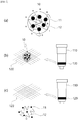
- FIG. 1 is a diagram to illustrate a method for extracting microvesicles from a biological sample according to the present disclosure.
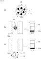
- FIG. 2 is a diagram showing an implementation of a method for extracting microvesicles from a biological sample according to the present disclosure.
- the method for extracting microvesicles in accordance with the present disclosure may include add a polyvalent cationic substance 12 to a biological sample containing microvesicles 11 S10.
- the biological sample may include a cell culture solution, a tissue sample, etc. containing microvesicles.
- the biological sample may include animal, plant or microorganism-derived cell medium, tissue extract, cancer tissue, blood, plasma, serum, urine, saliva, cerebrospinal fluid, tears, sweat, feces, ascite, amniotic fluid, semen, or milk, but may not be limited thereto.
- the microvesicles 11 have electrically negative (-) properties.
- the polyvalent cationic substance 12 when added to the biological sample, the polyvalent cationic substance 12 and the microvesicles 11 are aggregated with each other via an electrical force, thereby forming an aggregate 10 as shown in (a) of FIG. 2 S11.
- the polyvalent cationic substance includes a cationic polymer.
- the polyvalent cationic substance may include polylysine (which may be available in a form of a salt such as poly-L-lysine hydrobromide or poly-L-lysine hydrochloride), polyarginine, poly histidine, protamine (which may be available in a form of a salt such as protamine chloride or protamine sulfate), cationic dextran, cationic dendrimer, cationic polysaccharide, polyamidoamine, polyethyleneimine, polyquaternium, or a combination thereof.
- the polyvalent cationic substance may be a polylysine salt.
- the polyvalent cationic substance employed poly-L-lysine (PLL) having a molecular weight of 150 to 300 kD at a concentration of 0.02 to 2.00 mg/mL (present example 1).
- a size of each of the microvesicles 11 is in a range of nanometers. Thus, it is difficult to extract the microvesicles 11.
- the microvesicles 11 having the electrical negative (-) property are aggregated with the polyvalent cationic substance 12 via the electrical force to form the aggregate having an increased size, which may be easily captured.
- an incubation process for aggregation between the polyvalent cationic substance 12 and the microvesicles 11 is carried out for a certain period of time and under a certain temperature condition.
- the incubation process may be performed under a temperature condition of 0.1 to 40 °C, preferably, lower than 25 °C.
- the incubation process may be performed at a temperature lower than 20 °C, or lower than 15 °C, or lower than 12 °C, or lower than 10 °C.
- the process may be performed under a temperature condition of 1 to 8 °C.
- the incubation process may be performed for 0.5 to 100 minutes, preferably for a time duration smaller than 30 minutes.
- the incubation process may be performed for a time duration smaller than 25 minutes, or smaller than 20 minutes, or smaller than 15 minutes.
- the process may be performed for 5 to 15 minutes.
- microvesicle aggregates were effectively formed when the incubation process was performed at 4 °C for 10 minutes ( FIG. 10 ).
- the incubation time duration for the aggregation may be determined experimentally.
- a time required for the aggregation using the electrical force may be significantly reduced, compared to a time required in the conventional immunoaffinity isolation method.
- the polyvalent cationic substance 12 employs poly lysine salt, poly arginine, poly histidine salt, protamine chloride, protamine sulfate, cationic dextran, cationic dendrimer, cationic polysaccharide, poly amidoamine, polyethylenimine or polyquaternium, or combinations thereof.
- the polyvalent cationic substance 12 and the microvesicles 11 in the biological sample are aggregated with each other to form the aggregate 10.
- the aggregate 10 may be captured by the capture filter 120 S12.
- the biological sample passes through the capture filter 120 installed inside an extraction tube 110 by gravity or a negative pressure by way of example.
- the capture filter 120 has pores of a size capable of capturing the aggregate 10.
- the capture filter 120 may be embodied as a membrane in which the pores are formed, or a bead packing filter in which a plurality of micro-beads are closely packed in a 3D manner so as to form the pores.
- the capture filter may have the pores of a size capable of capturing the microvesicle aggregates therein.
- the size of the pores formed in the capture filter may be set to a size in which the microvesicle aggregate cannot escape from the capture filter.
- the pore may be sized such that when the aggregate is disaggregated by the elution solution in a subsequent elution step, the separated microvesicle may flow through the pore.
- the size of the pore in the capture filter may be in a range of 50 nm to 1,000 nm, specifically 100 nm to 500 nm, and more specifically 150 nm to 300 nm.
- a surface portion of the capture filter in contact with the microvesicle aggregate may be embodied as a membrane, a bead, a column, or a combination thereof.
- the capture filter may be provided in a form of a pore-formed membrane or a bead packing filter in which a plurality of micro-beads are closely packed to each other so as to form pores.
- the capture filter 120 may be made of a hydrophilic material or a cation exchange resin so that the aggregate 10 having a positive electrical characteristic is more easily captured therein.
- the capture filter When the capture filter is made of a hydrophilic material, the microvesicle aggregate may be easily caught in the pores and physically captured.
- the hydrophilic material may include sliver metal, polyethersulfone, glass fiber, polycarbonate track etch (PCTE), polyester, mixed cellulose esters (MCE), nylon, cellulose acetate, or combinations thereof.
- the cation exchange resin may include carboxymethyl, methyl sulfonate, a sulphonyl group, or combinations thereof.
- An embodiment shown in (b) of FIG. 3 shows an example of a capture filter 120a made of the cation exchange resin having a negative charge which may capture the aggregate 10 using an electrical force.
- a manner in which the aggregate 10 is captured by the capture filter 120 in which pores are formed and/or a manner in which the aggregate 10 is captured by the capture filter 120 made of the cation exchange resin having a negative charge via an electrical force may be employed.
- the nanometer-scale microvesicles 11 may be aggregated with the polyvalent cationic substance 12 to form the aggregate 10 which may be easily caught in the pores or easily captured using the electrical force.
- the microvesicle aggregate was filtered using a syringe filter having a pore size of about 220 nm (present example 1).
- an extraction process of the microvesicles 11 using the elution solution is performed S13 and S14.
- a washing solution may pass through the capture filter 120 to wash the capture filter 120. This may remove residues such as EVs collected on the microvesicles 11 in addition to the aggregate 10. Thus, a purity of eventually extracted microvesicles 11 may be improved.
- the washing solution may contain a chaotropic agent to be described later.
- the chaotropic agent contained in the washing solution is preferably contained at a lower concentration than that of the chaotropic agent contained in the elution solution.
- the washing solution may also contain a salt including guanidinium ions as a chaotropic agent.
- the concentration of the chaotropic agent contained in the washing solution is lower than the concentration of the chaotropic agent contained in the elution solution.
- the former may be in a range of 0.1 to 4.5 M, 0.5 to 4 M, and 1 to 3 M.
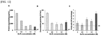
- the captured microvesicle aggregate was washed using the washing solution containing guanidium thiocyanate at 0 to 2 M concentration ( FIG. 12 ).
- the washing solution may contain an appropriate salt to effectively wash impurities.
- the salt may contain monovalent cations such as NH 4 + , Na + , and K + , divalent cations such as Ca 2+ , Mg 2+ , or combinations thereof.
- the salt in the washing solution may be (NH 4 ) 2 SO 4 , Na 2 SO 4 , NaCl, KCl, CH 3 COONH 4 , or a combination thereof.
- the salt may be contained in the washing solution at a concentration lower than a certain concentration, for example, lower than 10 mM.
- the elution solution may pass S13 through the capture filter 120 where the aggregate 10 is captured to isolate the microvesicles 11 from the aggregate 10 and may extract the microvesicles 11 S14.
- the elution solution may contain a chaotropic agent or protease.
- the chaotropic agent refers to a substance or an ion that destabilizes or destroys a cargo structure of a water molecule in a water solution. Due to the addition of the chaotropic agent into the water solution, an entropy of water is increased to affect a force between proteins, hydrogen bond, Van der Waals force, or hydrophobic bond. Thus, a molecular structure thereof is destabilized.
- the chaotropic agent may include a salt containing guanidinium ion, such as guanidinium isothiocyanate, guanidinium thiocyanate, guanidinium chloride, or guanidine hydrochloride; n-butanol, ethanol, lithium perchlorate, lithium acetate, magnesium chloride, phenol, 2-propanol, sodium dodecyl sulfate, thiourea, urea, or a combination thereof, but may be not limited thereto.
- guanidinium ion such as guanidinium isothiocyanate, guanidinium thiocyanate, guanidinium chloride, or guanidine hydrochloride
- n-butanol ethanol, lithium perchlorate, lithium acetate, magnesium chloride, phenol, 2-propanol, sodium dodecyl sulfate, thiourea, urea, or a combination thereof, but may
- the chaotropic agent according to the present disclosure includes guanidinium thiocyanate (GuTc) which may be contained in the elution solution at a concentration of 1 to 9 M, specifically 2 to 8 M, more specifically 3 to 7 M, or 4 to 6 M.
- GuTc guanidinium thiocyanate
- the protease may include proteinase K, pepsin, trypsin or chymotrypsin.
- the elution solution as described above may remove the electrical bond between the microvesicles 11 and the polyvalent cations constituting the aggregate 10 to isolate the microvesicles 11 from the aggregate 10, thereby enabling extraction of the microvesicles 11. That is, isolating the microvesicles 11 from the aggregate 10 using the elution solution may allow eliminating a centrifugal process for extracting the microvesicles 11. Thus, the microvesicles 11 may be extracted at a lower cost and for a smaller time duration, compared to the conventional method using the centrifugal separator.
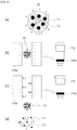
- FIG. 4 is a diagram showing another implementation of a method for extracting microvesicles from a biological sample according to the present disclosure.
- the capture filter 120a is made of a hydrophilic material or a cation exchange resin.
- the aggregate 10 is captured by the capture filter 120a using the electrical force.
- a buffer solution having a negative charge may pass through the capture filter 120a in which the aggregate 10 is captured, such that aggregate 10 is released from the capturer filter 120a, as shown in (c) of FIG. 4 .
- the elution solution may be injected to the aggregate 10 isolated from the capture filter 120a as in the above embodiment, such that the microvesicles 11 are isolated from the aggregate 10 to extract the microvesicles 11.
- the buffer solution may contain at least one of Ca 2 + , Mg 2 + , Na + , K + , NH 4+ ions, or a combination thereof by way of example.
- FIG. 5 to FIG. 13 is a diagram to illustrate an experimental example of a method for extracting microvesicles 11 from a biological sample according to the present disclosure.
- This experimental example uses plasma as the biological sample.
- poly-L-lysine solution is used as the polyvalent cationic substance 12.
- the microvesicles 11 and the polyvalent cationic substance 12 are aggregated with each other to form the aggregate 10. This experimental example will be described below.
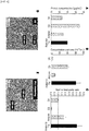
- FIG. 5 shows that a size of the aggregate 10 is significantly increased compared to a size of the microvesicle 11 according to an embodiment.
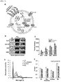
- FIG. 6 shows that an extraction yield and a purity of microvesicles 11 using the method according to an embodiment of the present disclosure are improved, compared to the comparative example method used in the prior art.
- FIG. 7 shows that biomarkers of extracellular vesicles (EVs) are well expressed in the microvesicles 11 extracted according to an embodiment of the present disclosure, and thus are usefully used for analysis such as subsequent molecular diagnosis.
- EVs extracellular vesicles
- FIG. 10 shows an optimum temperature and time required for the formation of the aggregate 10 of the microvesicles 11.
- FIG. 13 shows the results of an experiment on whether the aggregate 10 is decomposed and resuspended after adding a poly-L-lysine solution to plasma to form the aggregate 10, and then adding proteinase K as an elution solution thereto.
- 0.25 mg/mL of poly-L-lysine solution was added, and 100 ⁇ l, 200 ⁇ l and 300 ⁇ l of proteinase K was added by way of example.
- FIG. 13 it was identified that the aggregate 10 was decomposed and resuspended.
- FIG. 12 shows the measurement of protein impurities and microvesicles based on a concentration of the chaotropic agent GuTc in the washing solution. It was confirmed based on FIG. 12 that the washing process proceeded efficiently using GuTc at a concentration of 2M.
- a cationic polymer was added to the prepared plasma sample to induce aggregation between extracellular vesicles and the polymer via interaction between charges. Aggregates with a size of several hundred nanometers were formed. Poly-L-lysine (PLL, Sigma-Aldrich, USA) having a molecular weight of 150 to 300 kDa was used as the cationic polymer. After preparing a PLL stock solution at a concentration of 10 mg/mL, we additionally added the stock solution thereto, such that a final PLL concentration (0.02 to 2.00 mg/mL) in each plasma sample was adjusted. We carefully considered the yield and purity of the isolated extracellular vesicle, and then adjusted the final PLL concentration to 0.5 mg/mL.
- PLL Poly-L-lysine
- a mixture of plasma and PLL solution was incubated at 4°C for 10 minutes. After the incubation, the mixture was filtered using a syringe filter having a pore size of 220 nm. After the filtration, large aggregated particles were left in the filter.
- UC ultracentrifugation
- the supernatant was filtered using a syringe filter (Merck Millipore, USA) having a 200 ⁇ m pore size, followed by ultracentrifugation at 120,000 g and 4° C for 2 hours (CP100WX; Hitachi, Japan). After aspirating the supernatant, bottom pellets were carefully washed with PBS at 120,000 g and 4°C for 1 hour, and then were resuspended in 50 ⁇ L of PBS.
- a syringe filter Merck Millipore, USA having a 200 ⁇ m pore size
- ExoQuick TM exosome precipitation solution (EXOQ5A-1, System Biosciences, USA) as a representative product that was commercialized for extracellular vesicle isolation was used.
- a standard protocol may refer to the ExoQuick user manual.
- Plasma samples were mixed with ExoQuick solution as a PEG-based solution, and then the mixture was incubated at 4°C for 30 minutes. After the incubation, the mixture was centrifuged at 1,500 g for 30 min. The supernatant was carefully aspirated, leaving pellets on the bottom. Further centrifugation at 1,500 g for 5 minutes was performed, and then, the ExoQuick solution was completely removed. The remaining pellets were then resuspended in 200 ⁇ L of PBS.
- an exoEasy TM method using a commercialized spin column-type extracellular vesicle isolation kit that utilizes the electrical interaction between the membrane and the extracellular vesicle was carried out.
- the particle diameter according to Present present example 1 was measured using the dynamic light scattering method (DLS). As a result, it was identified that the extracellular vesicle aggregate (600 nm) has a significantly larger size than that of the extracellular vesicle (200 nm). The results indicate that the extracellular vesicles were aggregated with PLL. The isolated extracellular vesicles and the extracellular vesicles aggregated with PLL were also visualized using SEM. In the aggregated extracellular vesicle, the extracellular vesicle was completely surrounded with polymer. A size of the extracellular vesicle and a size of the extracellular vesicle aggregate were identified as 171 nm and 815 nm, respectively ( FIG. 5 ).
- Comparative example 2 was based on a polymer (PEG)-based precipitation and isolation method for extracellular vesicles
- the comparative example 3 was based on a method using a spin column-type extracellular vesicle isolation kit utilizing the electrical interaction between the extracellular vesicle and the membrane.
- the size distribution of extracellular vesicles was measured based on nanoparticle tracking analysis (NTA) (Malvern Panalytical, USA). The shape and size thereof were identified using cryo-TEM (TecnaiG2-F20,FEI,USA).
- the extracellular vesicle isolated using the comparative example 1 had the size distribution of about 20 to130 based on a result of SEM measurement, whereas the extracellular vesicle isolated using ExoCAS had the size distribution of 30 to 100 nm based on a result of SEM measurement ((d) and (e) in FIG. 6 ).
- the TEM image morphology of the extracellular vesicle isolated according to the present example 1 was compared with that of the extracellular vesicle isolated using the comparative example 1.
- a clear morphology was observed in vesicles with a diameter of 100 to 200 nm ((d) and (e) in FIG. 6 ).
- no significant difference was found between the TEM images of the comparative example 1 and the present example 1.
- protein contamination which determines the quality of subsequent analysis is also important. That is, the protein amount in the elution buffer should be minimized, while the extracellular vesicle therein should be concentrated at high concentration. According to BCA protein assay method (#23225; Thermo Scientific, USA), the protein contamination levels based on different extracellular vesicle isolation methods were measured. The tested sample was a final eluent as obtained in each method.
- the amounts of residual proteins in the comparative example 1, the comparative example 3 and the present example 1 were 232 ⁇ g/mL, 301 ⁇ g/mL, and 041 ⁇ g/mL, respectively, whereas that of the comparative example 2 was significantly higher (33,730 ⁇ g/mL) ((a) in FIG. 6 ).
- the purity ratio represents the number of isolated particles relative to the protein concentration.
- the comparative example 1 based on the UC method showed a purity ratio of 1.0, the comparative example 2 showed the purity ratio of 0.1, and the comparative example 3 showed a purity ratio of 1.8 whereas the present example 1 showered a significantly higher purity ratio of 3.5 ((c) in FIG. 6 ).
- exosomes contain protein markers such as Alix (ALG-2-interacting protein X), TSG101 (tumor susceptibility gene 101 protein), HSP70 (heat shock protein), and the tetraspanins CD63, CD81, and CD9.
- ALG-2-interacting protein X protein markers
- TSG101 tumor susceptibility gene 101 protein
- HSP70 heat shock protein
- CD63, CD81, and CD9 protein markers
- the present inventors analyzed the characteristics of the isolated extracellular vesicles using the four reference protein markers (ALIX, TSG101: inner protein markers; CD9, and CD81: surface protein markers) ((a) in FIG. 7 ).
- the total level of the protein marker may be measured from the intensity of the protein band ((b) and (c) in FIG. 7 ).
- the level of the exosome protein marker was as follows: present example 1 > comparative example 3 > comparative example 1.
- Re transcription of RNA eluates was performed using TaqMan MicroRNA RT Kit (4366596, Life Technologies, USA) and TaqMan Micro RNA Assay (4427975, Life Technologies, USA).
- TaqMan Universal Master Mix II, no UNG (4440040, Life Technologies, USA) and miRNA assays hsa-let-7a-5p, ID 000377, and hsa-miR-142-3p, ID 000464 were used. Additional experiments were performed on the RNA eluate using an Agilent Eukaryote Total RNA Pico chip and on an Agilent 2100 bioanalyzer (Agilent Technologies, USA).
- RNA amount was as follows: present example 1 > comparative example 3 > comparative example 1 > comparative example 2 ((d) in FIG. 7 ).
- the PLL aggregation method yields the microvesicle containing more abundant RNA, compared to other extracellular vesicle isolation methods. Further, based on a result of RT-qPCR experiments using hsa-let-7a-5p and hsa-miR-142-3p as known miRNA markers of extracellular vesicles, the PLL aggregation method showed a similar Ct value compared to that of the UC method ((e) in FIG. 7 ).
- the zeta potential analysis method of extracellular vesicles and polycationic polymers is as follows: the zeta potentials for extracellular vesicles isolated according to the comparative example 1, PLL solution (10 mg/mL), and PLL and extravesicular vesicle aggregates were measured using a zeta potential analyzer (Zetasizer Pro; Malvern Panalytical; UK). Since it is difficult to resuspend the aggregates in deionized water, the aggregated pellets (PLL 0.5 mg/mL) from 1 mL of plasma were first resuspended in 10 ⁇ L of GuTc solution (10 M), and then 990 ⁇ L of deionized water was added to the solution. The mixture was carefully dispersed for 5 minutes using a vortex mixer.
- PLL solution 10 mg/mL
- PLL and extravesicular vesicle aggregates were measured using a zeta potential analyzer (Zetasizer Pro; Malver
- the extracellular vesicle exhibits a negative charge of -19.7 mV, whereas the PLL solution (10 mg/mL) exhibits a strong positive charge zeta potential of 42.4 mV.
- the zeta potential of the extracellular vesicle-PLL aggregate was neutralized to -5.6 mV.
- the results indicate a strong electrostatic interaction between the negative charge of the extracellular vesicle and the positive charge of the PLL polymer.
- Albumin showed a weak negative charge
- ⁇ -globulin showed a pharmacologically positive charge.
- fibrinogen exhibits a negative charge of - 18.7 mV similar to that of the extracellular vesicles, and thus strongly interacts with cationic PLL ((a) and (b) in FIG. 8 and FIG. 9 ).
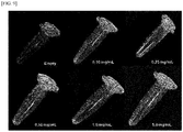
- the present inventors investigated the possibility of aggregate formation of extracellular vesicle-deficient plasma when adding the PLL solution thereto ((d) in FIG. 8 ). Despite the absence of extracellular vesicles, relatively weak aggregate formation was found, compared to control plasma. Therefore, the present inventors further investigated the aggregate formation of extracellular vesicle-deficient serum using serum (fibrinogen-free) and PLL ((e) and (f) in FIG. 8 ). Surprisingly, no apparent aggregate formation was observed. The results show that plasma proteins and PLL are aggregated with each other via electrical interaction, and fibrinogen is the main protein that is aggregated with PLL.
- Extracellular vesicle aggregates were successfully obtained by simply adding PLL solution to plasma. For isolation of extracellular vesicles for subsequent application, resuspension of extracellular vesicle aggregates in an appropriate buffer was required. However, strongly aggregated polymers and extracellular vesicles could not be suspended in deionized water or PBS solution ((a) in FIG. 11 ). Therefore, the chaotropic agent (GuTc) was used for dissolution of PLL and extracellular vesicle precipitates. The dissolution ability of the extracellular vesicle-PLL aggregate based on various concentrations of GuTc solution (0 to 4 M) was measured.
- the size and particle concentration of isolated extracellular vesicles based on GuTc concentrations were analyzed using NTA ((b) and (c) in FIG. 11 ). As the concentration of the GuTc solution increased, the size of the extracellular vesicles gradually decreased. At low concentrations of GuTc (1M and 2M), larger particles were observed (about 300 nm). At 1M GuTc, the extracellular vesicle concentration was very low (0.03 ⁇ 10 10 ). On the other hand, at high concentration of GuTc (4M), a peak value of the particle size significantly decreased to 102 nm. Thus, the extracellular vesicle was considered to be completely released from the aggregate. At a fixed PLL concentration (0.2 mg/mL) in plasma, 4 M GuTc solution among various concentration solutions showed the best aggregate-dissolving effect. The optimized GuTc buffer successfully dissolved the extracellular vesicle aggregate pellets within 1 minute.
- Reference numerals 10 Aggregate 11: Microvesicles 12: Polyvalent cationic substance 110: Extraction tube 120, and 120a : Capture filter
- the method according to the present disclosure may extract the microvesicles from the biological sample using the polyvalent cationic substance without the centrifugation process and thus may be useful in various industrial fields where the microvesicles are utilized.
Landscapes
- Chemical & Material Sciences (AREA)
- Chemical Kinetics & Catalysis (AREA)
- Health & Medical Sciences (AREA)
- Analytical Chemistry (AREA)
- Clinical Laboratory Science (AREA)
- Engineering & Computer Science (AREA)
- Water Supply & Treatment (AREA)
- Hematology (AREA)
- General Health & Medical Sciences (AREA)
- Peptides Or Proteins (AREA)
- Micro-Organisms Or Cultivation Processes Thereof (AREA)
- Investigating Or Analysing Biological Materials (AREA)
- Measuring Or Testing Involving Enzymes Or Micro-Organisms (AREA)
- Sampling And Sample Adjustment (AREA)
Applications Claiming Priority (2)
| Application Number | Priority Date | Filing Date | Title |
|---|---|---|---|
| KR20190017053 | 2019-02-14 | ||
| PCT/KR2020/001987 WO2020166978A2 (fr) | 2019-02-14 | 2020-02-12 | Procédé d'extraction de microvésicules d'un échantillon biologique |
Publications (3)
| Publication Number | Publication Date |
|---|---|
| EP3925703A2 true EP3925703A2 (fr) | 2021-12-22 |
| EP3925703A4 EP3925703A4 (fr) | 2022-11-30 |
| EP3925703B1 EP3925703B1 (fr) | 2025-04-16 |
Family
ID=72045018
Family Applications (1)
| Application Number | Title | Priority Date | Filing Date |
|---|---|---|---|
| EP20754903.1A Active EP3925703B1 (fr) | 2019-02-14 | 2020-02-12 | Procédé d'extraction de microvésicules d'échantillons biologiques |
Country Status (5)
| Country | Link |
|---|---|
| US (1) | US12551828B2 (fr) |
| EP (1) | EP3925703B1 (fr) |
| KR (1) | KR102403334B1 (fr) |
| CN (1) | CN113631240B (fr) |
| WO (1) | WO2020166978A2 (fr) |
Cited By (1)
| Publication number | Priority date | Publication date | Assignee | Title |
|---|---|---|---|---|
| JP2023149346A (ja) * | 2022-03-31 | 2023-10-13 | ヒューマン・メタボローム・テクノロジーズ株式会社 | 脂質二重膜に覆われた微粒子又は細胞外小胞の回収方法および脂質二重膜に覆われた微粒子又は細胞外小胞を回収するための回収キット |
Families Citing this family (9)
| Publication number | Priority date | Publication date | Assignee | Title |
|---|---|---|---|---|
| JP7625408B2 (ja) * | 2020-11-30 | 2025-02-03 | 社会医療法人大雄会 | 細胞外小胞および/またはセルフリーdnaを濃縮する方法 |
| US20240400995A1 (en) * | 2020-12-29 | 2024-12-05 | Union Korea Life Sciences Inc. | Method for isolating exosomes with high efficiency and high purity |
| JP2024521372A (ja) * | 2021-06-04 | 2024-05-31 | インスティチュート フォー ベーシック サイエンス | 塩分別沈殿を用いた細胞外小胞体分離方法 |
| CN113462520A (zh) * | 2021-07-26 | 2021-10-01 | 百奥芯(苏州)生物科技有限公司 | 一种循环肿瘤外泌体富集芯片及其应用 |
| CN115074301A (zh) * | 2022-08-22 | 2022-09-20 | 博奥生物集团有限公司 | 利用ε-聚赖氨酸富集细菌细胞外囊泡的方法 |
| WO2024205013A1 (fr) * | 2023-03-29 | 2024-10-03 | 고려대학교 산학협력단 | Procédé et appareil d'extraction et de purification de masse de microvésicules à partir d'échantillons biologiques |
| CN116773500B (zh) * | 2023-06-26 | 2024-02-23 | 四川大学 | 一种细胞外囊泡的分离和鉴定方法 |
| KR20250146978A (ko) * | 2024-04-03 | 2025-10-13 | 고려대학교 산학협력단 | 생물학적 샘플로부터 세포외 소포체를 대량으로 추출하고 고순도로 정제하는 방법 및 장치 |
| CN121022735A (zh) * | 2025-08-29 | 2025-11-28 | 广东龄值生物科技有限公司 | 一种间充质干细胞外泌体的纯化方法及制得的间充质干细胞外泌体颗粒和其应用 |
Family Cites Families (15)
| Publication number | Priority date | Publication date | Assignee | Title |
|---|---|---|---|---|
| US5000854A (en) * | 1989-06-14 | 1991-03-19 | The University Of Michigan | Protamine-based filter device for removal of heparin from blood samples |
| GB0212825D0 (en) * | 2002-05-31 | 2002-07-10 | Dna Res Innovations Ltd | Methods compositions and kits for cell separation |
| CA2645739A1 (fr) * | 2006-03-20 | 2007-09-27 | Medarex, Inc. | Purification de proteines |
| KR101233636B1 (ko) * | 2010-10-20 | 2013-02-15 | 아주대학교산학협력단 | 혈색소 측정용 혈액 성분 분리 장치 및 분리 방법 |
| US9745616B2 (en) * | 2011-05-17 | 2017-08-29 | Dxterity Diagnostics Incorporated | Methods and compositions for detecting target nucleic acids |
| KR101495631B1 (ko) | 2012-03-30 | 2015-02-26 | (주)어핀텍 | 원심분리 키트 및 이를 이용한 원심분리 방법 |
| CN108103057B (zh) * | 2012-08-28 | 2021-09-03 | 阿科尼生物系统公司 | 用于纯化核酸的方法和试剂盒 |
| US9920498B2 (en) * | 2013-11-05 | 2018-03-20 | Crystal Lagoons (Curacao) B.V. | Floating lake system and methods of treating water within a floating lake |
| US9568404B2 (en) * | 2014-05-16 | 2017-02-14 | Junyu Mai | Method and apparatus for biomolecule analysis |
| US10968443B2 (en) * | 2014-12-31 | 2021-04-06 | The Rockefeller University | Method of RNA isolation from clinical samples |
| KR101761680B1 (ko) * | 2015-03-31 | 2017-08-23 | 포항공과대학교 산학협력단 | 수용액 이상계를 이용한 세포 밖 소포체의 분리방법 |
| KR101726286B1 (ko) * | 2015-08-11 | 2017-04-12 | 한국과학기술연구원 | 세포외 소포체 포집 장치 및 그 사용 방법 |
| EP3443112B1 (fr) | 2016-04-12 | 2020-08-05 | Unicyte EV AG | Isolation de vesicules extracellulaires ( evs ) à partir d'échantillons de liquide biologique |
| KR101951549B1 (ko) * | 2017-04-21 | 2019-02-22 | 고려대학교 산학협력단 | 미세 소포체의 분리 장치 및 방법 |
| JP2020528766A (ja) | 2017-07-26 | 2020-10-01 | ロゼッタ エクソソーム | 陽イオンを利用した細胞外小胞体の分離方法 |
-
2020
- 2020-02-12 KR KR1020200017213A patent/KR102403334B1/ko active Active
- 2020-02-12 US US17/430,429 patent/US12551828B2/en active Active
- 2020-02-12 EP EP20754903.1A patent/EP3925703B1/fr active Active
- 2020-02-12 WO PCT/KR2020/001987 patent/WO2020166978A2/fr not_active Ceased
- 2020-02-12 CN CN202080024409.0A patent/CN113631240B/zh active Active
Cited By (1)
| Publication number | Priority date | Publication date | Assignee | Title |
|---|---|---|---|---|
| JP2023149346A (ja) * | 2022-03-31 | 2023-10-13 | ヒューマン・メタボローム・テクノロジーズ株式会社 | 脂質二重膜に覆われた微粒子又は細胞外小胞の回収方法および脂質二重膜に覆われた微粒子又は細胞外小胞を回収するための回収キット |
Also Published As
| Publication number | Publication date |
|---|---|
| WO2020166978A9 (fr) | 2021-05-27 |
| WO2020166978A2 (fr) | 2020-08-20 |
| KR102403334B1 (ko) | 2022-06-02 |
| US20220111318A1 (en) | 2022-04-14 |
| CN113631240B (zh) | 2023-08-08 |
| CN113631240A (zh) | 2021-11-09 |
| WO2020166978A3 (fr) | 2021-04-22 |
| KR20200099489A (ko) | 2020-08-24 |
| EP3925703A4 (fr) | 2022-11-30 |
| EP3925703B1 (fr) | 2025-04-16 |
| US12551828B2 (en) | 2026-02-17 |
Similar Documents
| Publication | Publication Date | Title |
|---|---|---|
| EP3925703B1 (fr) | Procédé d'extraction de microvésicules d'échantillons biologiques | |
| EP3443112B1 (fr) | Isolation de vesicules extracellulaires ( evs ) à partir d'échantillons de liquide biologique | |
| Konoshenko et al. | Isolation of extracellular vesicles: general methodologies and latest trends | |
| KR101761680B1 (ko) | 수용액 이상계를 이용한 세포 밖 소포체의 분리방법 | |
| US11479765B2 (en) | Method of isolating exosomes using encapsulation and aqueous micellar system | |
| KR102708451B1 (ko) | 미세소포체 분리방법 및 미세소포체 분리장치 | |
| EP4141107B1 (fr) | Procédé d'isolement de microvésicule et dispositif d'isolation de microvésicule | |
| US10669535B2 (en) | Methods for the isolation of extracellular vesicles and other bioparticles from urine and other biofluids | |
| Kluszczyńska et al. | Methods for the Determination of the Purity of Exosomes | |
| EP3658125A1 (fr) | Composites biomoléculaires comprenant des fantômes cellulaires modifiés | |
| US20190040093A1 (en) | Bioparticle isolation and therapeutic application thereof | |
| Kotmakçı et al. | Exosome isolation: is there an optimal method with regard to diagnosis or treatment | |
| CN111670067A (zh) | 用于从生物材料中分离细胞外囊泡的方法和固定相 | |
| Marzouk et al. | Polyethylene glycol (PEG)-based precipitation for exosome enrichment: A review on recent developments, current challenges, and future perspectives | |
| Notarangelo et al. | Rapid nickel-based isolation of extracellular vesicles from different biological fluids | |
| CN116948947A (zh) | 一种外泌体的分离纯化方法 | |
| US12441993B2 (en) | Isolation of extracellular vesicles by precipitation or immobilization using polyethylenimine and polyethylenimine-coated solid supports | |
| RU2678988C1 (ru) | Способ выделения внеклеточных везикул из биологических жидкостей | |
| US20240400995A1 (en) | Method for isolating exosomes with high efficiency and high purity | |
| WO2026075627A1 (fr) | Procédé d'isolement d'exosomes | |
| Ciftci-Dede et al. | Comparative analysis of magnetically activated cell sorting and ultracentrifugation for exosome isolation from human mesenchymal stem cell culture supernatant |
Legal Events
| Date | Code | Title | Description |
|---|---|---|---|
| STAA | Information on the status of an ep patent application or granted ep patent |
Free format text: STATUS: THE INTERNATIONAL PUBLICATION HAS BEEN MADE |
|
| PUAI | Public reference made under article 153(3) epc to a published international application that has entered the european phase |
Free format text: ORIGINAL CODE: 0009012 |
|
| STAA | Information on the status of an ep patent application or granted ep patent |
Free format text: STATUS: REQUEST FOR EXAMINATION WAS MADE |
|
| 17P | Request for examination filed |
Effective date: 20210812 |
|
| AK | Designated contracting states |
Kind code of ref document: A2 Designated state(s): AL AT BE BG CH CY CZ DE DK EE ES FI FR GB GR HR HU IE IS IT LI LT LU LV MC MK MT NL NO PL PT RO RS SE SI SK SM TR |
|
| DAV | Request for validation of the european patent (deleted) | ||
| DAX | Request for extension of the european patent (deleted) | ||
| A4 | Supplementary search report drawn up and despatched |
Effective date: 20221031 |
|
| RIC1 | Information provided on ipc code assigned before grant |
Ipc: B01D 29/05 20060101ALI20221025BHEP Ipc: B01D 37/03 20060101ALI20221025BHEP Ipc: B01D 21/01 20060101ALI20221025BHEP Ipc: B01L 3/00 20060101AFI20221025BHEP |
|
| GRAP | Despatch of communication of intention to grant a patent |
Free format text: ORIGINAL CODE: EPIDOSNIGR1 |
|
| STAA | Information on the status of an ep patent application or granted ep patent |
Free format text: STATUS: GRANT OF PATENT IS INTENDED |
|
| INTG | Intention to grant announced |
Effective date: 20241112 |
|
| GRAS | Grant fee paid |
Free format text: ORIGINAL CODE: EPIDOSNIGR3 |
|
| GRAA | (expected) grant |
Free format text: ORIGINAL CODE: 0009210 |
|
| STAA | Information on the status of an ep patent application or granted ep patent |
Free format text: STATUS: THE PATENT HAS BEEN GRANTED |
|
| AK | Designated contracting states |
Kind code of ref document: B1 Designated state(s): AL AT BE BG CH CY CZ DE DK EE ES FI FR GB GR HR HU IE IS IT LI LT LU LV MC MK MT NL NO PL PT RO RS SE SI SK SM TR |
|
| REG | Reference to a national code |
Ref country code: GB Ref legal event code: FG4D |
|
| REG | Reference to a national code |
Ref country code: CH Ref legal event code: EP Ref country code: DE Ref legal event code: R096 Ref document number: 602020049561 Country of ref document: DE |
|
| REG | Reference to a national code |
Ref country code: IE Ref legal event code: FG4D |
|
| REG | Reference to a national code |
Ref country code: NL Ref legal event code: MP Effective date: 20250416 |
|
| PG25 | Lapsed in a contracting state [announced via postgrant information from national office to epo] |
Ref country code: NL Free format text: LAPSE BECAUSE OF FAILURE TO SUBMIT A TRANSLATION OF THE DESCRIPTION OR TO PAY THE FEE WITHIN THE PRESCRIBED TIME-LIMIT Effective date: 20250416 |
|
| REG | Reference to a national code |
Ref country code: AT Ref legal event code: MK05 Ref document number: 1785226 Country of ref document: AT Kind code of ref document: T Effective date: 20250416 |
|
| PG25 | Lapsed in a contracting state [announced via postgrant information from national office to epo] |
Ref country code: FI Free format text: LAPSE BECAUSE OF FAILURE TO SUBMIT A TRANSLATION OF THE DESCRIPTION OR TO PAY THE FEE WITHIN THE PRESCRIBED TIME-LIMIT Effective date: 20250416 Ref country code: PT Free format text: LAPSE BECAUSE OF FAILURE TO SUBMIT A TRANSLATION OF THE DESCRIPTION OR TO PAY THE FEE WITHIN THE PRESCRIBED TIME-LIMIT Effective date: 20250818 Ref country code: ES Free format text: LAPSE BECAUSE OF FAILURE TO SUBMIT A TRANSLATION OF THE DESCRIPTION OR TO PAY THE FEE WITHIN THE PRESCRIBED TIME-LIMIT Effective date: 20250416 |
|
| REG | Reference to a national code |
Ref country code: LT Ref legal event code: MG9D |
|
| PG25 | Lapsed in a contracting state [announced via postgrant information from national office to epo] |
Ref country code: NO Free format text: LAPSE BECAUSE OF FAILURE TO SUBMIT A TRANSLATION OF THE DESCRIPTION OR TO PAY THE FEE WITHIN THE PRESCRIBED TIME-LIMIT Effective date: 20250716 Ref country code: GR Free format text: LAPSE BECAUSE OF FAILURE TO SUBMIT A TRANSLATION OF THE DESCRIPTION OR TO PAY THE FEE WITHIN THE PRESCRIBED TIME-LIMIT Effective date: 20250717 |
|
| PG25 | Lapsed in a contracting state [announced via postgrant information from national office to epo] |
Ref country code: PL Free format text: LAPSE BECAUSE OF FAILURE TO SUBMIT A TRANSLATION OF THE DESCRIPTION OR TO PAY THE FEE WITHIN THE PRESCRIBED TIME-LIMIT Effective date: 20250416 |
|
| PG25 | Lapsed in a contracting state [announced via postgrant information from national office to epo] |
Ref country code: BG Free format text: LAPSE BECAUSE OF FAILURE TO SUBMIT A TRANSLATION OF THE DESCRIPTION OR TO PAY THE FEE WITHIN THE PRESCRIBED TIME-LIMIT Effective date: 20250416 |
|
| PG25 | Lapsed in a contracting state [announced via postgrant information from national office to epo] |
Ref country code: HR Free format text: LAPSE BECAUSE OF FAILURE TO SUBMIT A TRANSLATION OF THE DESCRIPTION OR TO PAY THE FEE WITHIN THE PRESCRIBED TIME-LIMIT Effective date: 20250416 |
|
| PG25 | Lapsed in a contracting state [announced via postgrant information from national office to epo] |
Ref country code: AT Free format text: LAPSE BECAUSE OF FAILURE TO SUBMIT A TRANSLATION OF THE DESCRIPTION OR TO PAY THE FEE WITHIN THE PRESCRIBED TIME-LIMIT Effective date: 20250416 |
|
| PG25 | Lapsed in a contracting state [announced via postgrant information from national office to epo] |
Ref country code: RS Free format text: LAPSE BECAUSE OF FAILURE TO SUBMIT A TRANSLATION OF THE DESCRIPTION OR TO PAY THE FEE WITHIN THE PRESCRIBED TIME-LIMIT Effective date: 20250716 |
|
| PG25 | Lapsed in a contracting state [announced via postgrant information from national office to epo] |
Ref country code: IS Free format text: LAPSE BECAUSE OF FAILURE TO SUBMIT A TRANSLATION OF THE DESCRIPTION OR TO PAY THE FEE WITHIN THE PRESCRIBED TIME-LIMIT Effective date: 20250816 |
|
| PG25 | Lapsed in a contracting state [announced via postgrant information from national office to epo] |
Ref country code: LV Free format text: LAPSE BECAUSE OF FAILURE TO SUBMIT A TRANSLATION OF THE DESCRIPTION OR TO PAY THE FEE WITHIN THE PRESCRIBED TIME-LIMIT Effective date: 20250416 |
|
| PG25 | Lapsed in a contracting state [announced via postgrant information from national office to epo] |
Ref country code: DK Free format text: LAPSE BECAUSE OF FAILURE TO SUBMIT A TRANSLATION OF THE DESCRIPTION OR TO PAY THE FEE WITHIN THE PRESCRIBED TIME-LIMIT Effective date: 20250416 Ref country code: SM Free format text: LAPSE BECAUSE OF FAILURE TO SUBMIT A TRANSLATION OF THE DESCRIPTION OR TO PAY THE FEE WITHIN THE PRESCRIBED TIME-LIMIT Effective date: 20250416 |
|
| REG | Reference to a national code |
Ref country code: DE Ref legal event code: R097 Ref document number: 602020049561 Country of ref document: DE |
|
| PG25 | Lapsed in a contracting state [announced via postgrant information from national office to epo] |
Ref country code: CZ Free format text: LAPSE BECAUSE OF FAILURE TO SUBMIT A TRANSLATION OF THE DESCRIPTION OR TO PAY THE FEE WITHIN THE PRESCRIBED TIME-LIMIT Effective date: 20250416 |
|
| PG25 | Lapsed in a contracting state [announced via postgrant information from national office to epo] |
Ref country code: EE Free format text: LAPSE BECAUSE OF FAILURE TO SUBMIT A TRANSLATION OF THE DESCRIPTION OR TO PAY THE FEE WITHIN THE PRESCRIBED TIME-LIMIT Effective date: 20250416 |
|
| PG25 | Lapsed in a contracting state [announced via postgrant information from national office to epo] |
Ref country code: SK Free format text: LAPSE BECAUSE OF FAILURE TO SUBMIT A TRANSLATION OF THE DESCRIPTION OR TO PAY THE FEE WITHIN THE PRESCRIBED TIME-LIMIT Effective date: 20250416 Ref country code: RO Free format text: LAPSE BECAUSE OF FAILURE TO SUBMIT A TRANSLATION OF THE DESCRIPTION OR TO PAY THE FEE WITHIN THE PRESCRIBED TIME-LIMIT Effective date: 20250416 |
|
| PG25 | Lapsed in a contracting state [announced via postgrant information from national office to epo] |
Ref country code: IT Free format text: LAPSE BECAUSE OF FAILURE TO SUBMIT A TRANSLATION OF THE DESCRIPTION OR TO PAY THE FEE WITHIN THE PRESCRIBED TIME-LIMIT Effective date: 20250416 |
|
| PLBE | No opposition filed within time limit |
Free format text: ORIGINAL CODE: 0009261 |
|
| STAA | Information on the status of an ep patent application or granted ep patent |
Free format text: STATUS: NO OPPOSITION FILED WITHIN TIME LIMIT |
|
| REG | Reference to a national code |
Ref country code: CH Ref legal event code: L10 Free format text: ST27 STATUS EVENT CODE: U-0-0-L10-L00 (AS PROVIDED BY THE NATIONAL OFFICE) Effective date: 20260225 |
|
| 26N | No opposition filed |
Effective date: 20260119 |