EP3932437A1 - Drucksystem zur gewinnung von biologischen fasern - Google Patents
Drucksystem zur gewinnung von biologischen fasern Download PDFInfo
- Publication number
- EP3932437A1 EP3932437A1 EP20382597.1A EP20382597A EP3932437A1 EP 3932437 A1 EP3932437 A1 EP 3932437A1 EP 20382597 A EP20382597 A EP 20382597A EP 3932437 A1 EP3932437 A1 EP 3932437A1
- Authority
- EP
- European Patent Office
- Prior art keywords
- nozzle
- cross
- biocompatible
- fibers
- biocompatible hydrogel
- Prior art date
- Legal status (The legal status is an assumption and is not a legal conclusion. Google has not performed a legal analysis and makes no representation as to the accuracy of the status listed.)
- Withdrawn
Links
Images




Classifications
-
- A—HUMAN NECESSITIES
- A61—MEDICAL OR VETERINARY SCIENCE; HYGIENE
- A61L—METHODS OR APPARATUS FOR STERILISING MATERIALS OR OBJECTS IN GENERAL; DISINFECTION, STERILISATION OR DEODORISATION OF AIR; CHEMICAL ASPECTS OF BANDAGES, DRESSINGS, ABSORBENT PADS OR SURGICAL ARTICLES; MATERIALS FOR BANDAGES, DRESSINGS, ABSORBENT PADS OR SURGICAL ARTICLES
- A61L27/00—Materials for grafts or prostheses or for coating grafts or prostheses
- A61L27/50—Materials characterised by their function or physical properties, e.g. injectable or lubricating compositions, shape-memory materials, surface modified materials
- A61L27/52—Hydrogels or hydrocolloids
-
- A—HUMAN NECESSITIES
- A61—MEDICAL OR VETERINARY SCIENCE; HYGIENE
- A61L—METHODS OR APPARATUS FOR STERILISING MATERIALS OR OBJECTS IN GENERAL; DISINFECTION, STERILISATION OR DEODORISATION OF AIR; CHEMICAL ASPECTS OF BANDAGES, DRESSINGS, ABSORBENT PADS OR SURGICAL ARTICLES; MATERIALS FOR BANDAGES, DRESSINGS, ABSORBENT PADS OR SURGICAL ARTICLES
- A61L27/00—Materials for grafts or prostheses or for coating grafts or prostheses
- A61L27/14—Macromolecular materials
- A61L27/20—Polysaccharides
-
- A—HUMAN NECESSITIES
- A61—MEDICAL OR VETERINARY SCIENCE; HYGIENE
- A61L—METHODS OR APPARATUS FOR STERILISING MATERIALS OR OBJECTS IN GENERAL; DISINFECTION, STERILISATION OR DEODORISATION OF AIR; CHEMICAL ASPECTS OF BANDAGES, DRESSINGS, ABSORBENT PADS OR SURGICAL ARTICLES; MATERIALS FOR BANDAGES, DRESSINGS, ABSORBENT PADS OR SURGICAL ARTICLES
- A61L27/00—Materials for grafts or prostheses or for coating grafts or prostheses
- A61L27/14—Macromolecular materials
- A61L27/22—Polypeptides or derivatives thereof, e.g. degradation products
-
- A—HUMAN NECESSITIES
- A61—MEDICAL OR VETERINARY SCIENCE; HYGIENE
- A61L—METHODS OR APPARATUS FOR STERILISING MATERIALS OR OBJECTS IN GENERAL; DISINFECTION, STERILISATION OR DEODORISATION OF AIR; CHEMICAL ASPECTS OF BANDAGES, DRESSINGS, ABSORBENT PADS OR SURGICAL ARTICLES; MATERIALS FOR BANDAGES, DRESSINGS, ABSORBENT PADS OR SURGICAL ARTICLES
- A61L27/00—Materials for grafts or prostheses or for coating grafts or prostheses
- A61L27/14—Macromolecular materials
- A61L27/22—Polypeptides or derivatives thereof, e.g. degradation products
- A61L27/222—Gelatin
-
- A—HUMAN NECESSITIES
- A61—MEDICAL OR VETERINARY SCIENCE; HYGIENE
- A61L—METHODS OR APPARATUS FOR STERILISING MATERIALS OR OBJECTS IN GENERAL; DISINFECTION, STERILISATION OR DEODORISATION OF AIR; CHEMICAL ASPECTS OF BANDAGES, DRESSINGS, ABSORBENT PADS OR SURGICAL ARTICLES; MATERIALS FOR BANDAGES, DRESSINGS, ABSORBENT PADS OR SURGICAL ARTICLES
- A61L27/00—Materials for grafts or prostheses or for coating grafts or prostheses
- A61L27/14—Macromolecular materials
- A61L27/22—Polypeptides or derivatives thereof, e.g. degradation products
- A61L27/225—Fibrin; Fibrinogen
-
- A—HUMAN NECESSITIES
- A61—MEDICAL OR VETERINARY SCIENCE; HYGIENE
- A61L—METHODS OR APPARATUS FOR STERILISING MATERIALS OR OBJECTS IN GENERAL; DISINFECTION, STERILISATION OR DEODORISATION OF AIR; CHEMICAL ASPECTS OF BANDAGES, DRESSINGS, ABSORBENT PADS OR SURGICAL ARTICLES; MATERIALS FOR BANDAGES, DRESSINGS, ABSORBENT PADS OR SURGICAL ARTICLES
- A61L27/00—Materials for grafts or prostheses or for coating grafts or prostheses
- A61L27/14—Macromolecular materials
- A61L27/22—Polypeptides or derivatives thereof, e.g. degradation products
- A61L27/24—Collagen
-
- A—HUMAN NECESSITIES
- A61—MEDICAL OR VETERINARY SCIENCE; HYGIENE
- A61L—METHODS OR APPARATUS FOR STERILISING MATERIALS OR OBJECTS IN GENERAL; DISINFECTION, STERILISATION OR DEODORISATION OF AIR; CHEMICAL ASPECTS OF BANDAGES, DRESSINGS, ABSORBENT PADS OR SURGICAL ARTICLES; MATERIALS FOR BANDAGES, DRESSINGS, ABSORBENT PADS OR SURGICAL ARTICLES
- A61L27/00—Materials for grafts or prostheses or for coating grafts or prostheses
- A61L27/14—Macromolecular materials
- A61L27/26—Mixtures of macromolecular compounds
-
- A—HUMAN NECESSITIES
- A61—MEDICAL OR VETERINARY SCIENCE; HYGIENE
- A61L—METHODS OR APPARATUS FOR STERILISING MATERIALS OR OBJECTS IN GENERAL; DISINFECTION, STERILISATION OR DEODORISATION OF AIR; CHEMICAL ASPECTS OF BANDAGES, DRESSINGS, ABSORBENT PADS OR SURGICAL ARTICLES; MATERIALS FOR BANDAGES, DRESSINGS, ABSORBENT PADS OR SURGICAL ARTICLES
- A61L27/00—Materials for grafts or prostheses or for coating grafts or prostheses
- A61L27/28—Materials for coating prostheses
- A61L27/34—Macromolecular materials
-
- A—HUMAN NECESSITIES
- A61—MEDICAL OR VETERINARY SCIENCE; HYGIENE
- A61L—METHODS OR APPARATUS FOR STERILISING MATERIALS OR OBJECTS IN GENERAL; DISINFECTION, STERILISATION OR DEODORISATION OF AIR; CHEMICAL ASPECTS OF BANDAGES, DRESSINGS, ABSORBENT PADS OR SURGICAL ARTICLES; MATERIALS FOR BANDAGES, DRESSINGS, ABSORBENT PADS OR SURGICAL ARTICLES
- A61L27/00—Materials for grafts or prostheses or for coating grafts or prostheses
- A61L27/36—Materials for grafts or prostheses or for coating grafts or prostheses containing ingredients of undetermined constitution or reaction products thereof, e.g. transplant tissue, natural bone, extracellular matrix
- A61L27/3604—Materials for grafts or prostheses or for coating grafts or prostheses containing ingredients of undetermined constitution or reaction products thereof, e.g. transplant tissue, natural bone, extracellular matrix characterised by the human or animal origin of the biological material, e.g. hair, fascia, fish scales, silk, shellac, pericardium, pleura, renal tissue, amniotic membrane, parenchymal tissue, fetal tissue, muscle tissue, fat tissue, enamel
- A61L27/3633—Extracellular matrix [ECM]
-
- A—HUMAN NECESSITIES
- A61—MEDICAL OR VETERINARY SCIENCE; HYGIENE
- A61L—METHODS OR APPARATUS FOR STERILISING MATERIALS OR OBJECTS IN GENERAL; DISINFECTION, STERILISATION OR DEODORISATION OF AIR; CHEMICAL ASPECTS OF BANDAGES, DRESSINGS, ABSORBENT PADS OR SURGICAL ARTICLES; MATERIALS FOR BANDAGES, DRESSINGS, ABSORBENT PADS OR SURGICAL ARTICLES
- A61L27/00—Materials for grafts or prostheses or for coating grafts or prostheses
- A61L27/36—Materials for grafts or prostheses or for coating grafts or prostheses containing ingredients of undetermined constitution or reaction products thereof, e.g. transplant tissue, natural bone, extracellular matrix
- A61L27/38—Materials for grafts or prostheses or for coating grafts or prostheses containing ingredients of undetermined constitution or reaction products thereof, e.g. transplant tissue, natural bone, extracellular matrix containing added animal cells
- A61L27/3804—Materials for grafts or prostheses or for coating grafts or prostheses containing ingredients of undetermined constitution or reaction products thereof, e.g. transplant tissue, natural bone, extracellular matrix containing added animal cells characterised by specific cells or progenitors thereof, e.g. fibroblasts, connective tissue cells, kidney cells
- A61L27/3826—Muscle cells, e.g. smooth muscle cells
-
- A—HUMAN NECESSITIES
- A61—MEDICAL OR VETERINARY SCIENCE; HYGIENE
- A61L—METHODS OR APPARATUS FOR STERILISING MATERIALS OR OBJECTS IN GENERAL; DISINFECTION, STERILISATION OR DEODORISATION OF AIR; CHEMICAL ASPECTS OF BANDAGES, DRESSINGS, ABSORBENT PADS OR SURGICAL ARTICLES; MATERIALS FOR BANDAGES, DRESSINGS, ABSORBENT PADS OR SURGICAL ARTICLES
- A61L27/00—Materials for grafts or prostheses or for coating grafts or prostheses
- A61L27/36—Materials for grafts or prostheses or for coating grafts or prostheses containing ingredients of undetermined constitution or reaction products thereof, e.g. transplant tissue, natural bone, extracellular matrix
- A61L27/38—Materials for grafts or prostheses or for coating grafts or prostheses containing ingredients of undetermined constitution or reaction products thereof, e.g. transplant tissue, natural bone, extracellular matrix containing added animal cells
- A61L27/3839—Materials for grafts or prostheses or for coating grafts or prostheses containing ingredients of undetermined constitution or reaction products thereof, e.g. transplant tissue, natural bone, extracellular matrix containing added animal cells characterised by the site of application in the body
- A61L27/3873—Muscle tissue, e.g. sphincter
-
- B—PERFORMING OPERATIONS; TRANSPORTING
- B33—ADDITIVE MANUFACTURING TECHNOLOGY
- B33Y—ADDITIVE MANUFACTURING, i.e. MANUFACTURING OF THREE-DIMENSIONAL [3D] OBJECTS BY ADDITIVE DEPOSITION, ADDITIVE AGGLOMERATION OR ADDITIVE LAYERING, e.g. BY 3D PRINTING, STEREOLITHOGRAPHY OR SELECTIVE LASER SINTERING
- B33Y70/00—Materials specially adapted for additive manufacturing
-
- B—PERFORMING OPERATIONS; TRANSPORTING
- B33—ADDITIVE MANUFACTURING TECHNOLOGY
- B33Y—ADDITIVE MANUFACTURING, i.e. MANUFACTURING OF THREE-DIMENSIONAL [3D] OBJECTS BY ADDITIVE DEPOSITION, ADDITIVE AGGLOMERATION OR ADDITIVE LAYERING, e.g. BY 3D PRINTING, STEREOLITHOGRAPHY OR SELECTIVE LASER SINTERING
- B33Y80/00—Products made by additive manufacturing
-
- A—HUMAN NECESSITIES
- A61—MEDICAL OR VETERINARY SCIENCE; HYGIENE
- A61L—METHODS OR APPARATUS FOR STERILISING MATERIALS OR OBJECTS IN GENERAL; DISINFECTION, STERILISATION OR DEODORISATION OF AIR; CHEMICAL ASPECTS OF BANDAGES, DRESSINGS, ABSORBENT PADS OR SURGICAL ARTICLES; MATERIALS FOR BANDAGES, DRESSINGS, ABSORBENT PADS OR SURGICAL ARTICLES
- A61L2400/00—Materials characterised by their function or physical properties
- A61L2400/12—Nanosized materials, e.g. nanofibres, nanoparticles, nanowires, nanotubes; Nanostructured surfaces
-
- A—HUMAN NECESSITIES
- A61—MEDICAL OR VETERINARY SCIENCE; HYGIENE
- A61L—METHODS OR APPARATUS FOR STERILISING MATERIALS OR OBJECTS IN GENERAL; DISINFECTION, STERILISATION OR DEODORISATION OF AIR; CHEMICAL ASPECTS OF BANDAGES, DRESSINGS, ABSORBENT PADS OR SURGICAL ARTICLES; MATERIALS FOR BANDAGES, DRESSINGS, ABSORBENT PADS OR SURGICAL ARTICLES
- A61L2400/00—Materials characterised by their function or physical properties
- A61L2400/16—Materials with shape-memory or superelastic properties
-
- A—HUMAN NECESSITIES
- A61—MEDICAL OR VETERINARY SCIENCE; HYGIENE
- A61L—METHODS OR APPARATUS FOR STERILISING MATERIALS OR OBJECTS IN GENERAL; DISINFECTION, STERILISATION OR DEODORISATION OF AIR; CHEMICAL ASPECTS OF BANDAGES, DRESSINGS, ABSORBENT PADS OR SURGICAL ARTICLES; MATERIALS FOR BANDAGES, DRESSINGS, ABSORBENT PADS OR SURGICAL ARTICLES
- A61L2430/00—Materials or treatment for tissue regeneration
- A61L2430/30—Materials or treatment for tissue regeneration for muscle reconstruction
Definitions
- the present disclosure relates to a printing system for obtaining individual biological fibers and methods for obtaining one or more individual biological fibers.
- the present disclosure also relates to methods of manufacturing a hybrid biocompatible machine or a biomimetic structure comprising one or more individual fibers, and a biomimetic structure for use as a medicament or for use in muscle tissue regeneration.
- 3D bioprinting is a technique that allows for printing of biological materials such as cell-laden hydrogels according to different designs, in order to mimic the 3D environment of biology such as native tissue.
- Two-dimensional cultures have been widely used in biological research, but there is an increasing understanding that some of the properties of biological materials might be completely different in native three-dimensional environments.
- 3D bioprinting offers an advantage over two-dimensional cultures by more closely approximating the native 3D environment of a biological material.
- Pneumatic extrusion-based bioprinting is the most widely used technique, and it is based on the extrusion of a cell-laden hydrogel (or any other polymer) through a thin nozzle by the application of pressure.
- This technique uses materials that have enough pseudoplastic or shear-thinning behaviour that decreases their viscosity when shear rate is increased (through the nozzle). This reduction of viscosity allows printing at lower pressures but also helps protect the cells from high shear stresses by decreasing the viscosity.
- skeletal muscle tissue has an inherently three-dimensional architecture, composed of bundles of muscle fibers (myocytes), created after fusion of myoblasts during the differentiation process. Myocytes within the bundles are densely packed and highly aligned in order to achieve very efficient and longitudinal contractions. Inside each myocyte, groups of internal protein structures (myofibrils) are formed. These structures contain periodic units (sarcomeres), which are mainly composed of actin fibers and myosin, and have the ability of contracting the myocyte upon certain stimulus. Therefore, given the complex three-dimensionality of skeletal muscle tissue in its natural form, any tissue model that aims at recreating its complexity must take a three-dimensional form.
- each bundle of myocytes is further organized in fascia, or fascicles, surrounded by the perimysium, a layer of connective tissue (mainly collagen) that stabilizes and separates the bundles of muscle fibers, containing from 10 to 100 of them.
- This fascicle structure has not been completely mimicked yet with 3D bioprinting, as subsequent layers of 3D-bioprinted fibers fuse with each other to form an even wider construct.
- the closest analogs to these kinds of structures have relied on sacrificial molds and microfabrication techniques ( D. Neal, M. S. Sakar, L.-L. S. Ong, and H. Harry Asada, Formation of elongated fascicle-inspired 3D tissues consisting of high-density, aligned cells using sacrificial outer molding, Lab Chip 14, 1907 (2014 ).
- Mouse myoblasts have already been 3D-bioprinted embedded in bioinks and their differentiation induced in this environment to form multinucleated myotubes ( H.-W. Kang, S. J. Lee, I. K. Ko, C. Kengla, J. J. Yoo, and A. Atala, A 3D bioprinting system to produce human-scale tissue constructs with structural integrity, Nature Biotechnology 34, 312 (2016 ).
- Other approaches not based on 3D bioprinting, but mold casting techniques, have also been described for the obtaining of muscle tissue ( R. Raman, C. Cvetkovic, and R. Bashir, A modular approach to the design, fabrication, and characterization of muscle-powered biological machines, Nature Protocols 12, 519 (2017 )). Nonetheless, these techniques lack the versatility, automation and fast-prototyping capabilities of 3D bioprinting.
- the thickness of the biological fibers that can be achieved with 3D bioprinting is difficult to control.
- the oxygen diffusion limit within a tissue is around 200 ⁇ m, wider tissues can suffer from biocompatibility issues or low cell density in the inner parts of the 3D-printed hydrogel. This causes a great loss of efficiency, as the bundle of myotubes will not be densely packed, but actually depleted.
- the lower density of packing will diminish the total force per cross-sectional area that can be achieved.
- some printing systems manufacture fibers by extruding the liquid phase biomaterial from the bioprinter nozzle into a support bath filled with a cross-linking solution
- a cross-linking solution US 10156560 B1 ; Hinton T.J. et al., Three-dimensional printing of complex biological structures by freeform reversible embedding of suspended hydrogels; Sci. Adv.; 1:e1500758 (2015 )).
- fixation of the printed filament diameter is not simultaneous to printing but only occurs after deposition.
- the type of cross-linking system which can be used is limited to chemical cross-linking.
- a co-axial printing system wherein an alginate hydrogel is extruded from an internal nozzle whilst a cross-linking CaCl 2 solution is flowed out of the external needle
- Ahn et al. A novel cell-printing method and its application to hepatogenic differentiation of human adipose stem cell-embedded mesh structures, Scientific Reports volume 5, 13427 (2015 ).
- this system does not enable cross-linking by methods other than chemical cross-linking. Thus, it limits the nature of the printed hydrogel.
- the flow of the cross-linking solution is difficult to control.
- obstruction of the nozzle due to accumulation of cross-linked alginate at the exit is very common. Therefore, it is difficult to finely control the diameter of the printed fiber.
- the inventors have provided such universal method for the fabrication of thin, homogeneous and width-controlled fibres of virtually any hydrogel by separating the step of fixating the 3D-printed fiber structure from the cross-linking step.
- This effect is obtained by the physical confinement of the fibers upon extrusion, more specifically by coating the extruded hydrogel with a polymeric composition (e.g. Pluronic ® F-127) in a gel state.
- a polymeric composition e.g. Pluronic ® F-127
- the hydrogel can subsequently be cross-linked by any known method, e.g, by chemical, thermal or enzymatic cross-linking or by cross-linking induced by exposure to UV light, and then the coating polymer can be removed (e.g., Pluronic ® F-127 is easily removed with a cold aqueous solution).
- Pluronic ® F-127 is easily removed with a cold aqueous solution.
- the physical confinement and chemical cross-linking can be combined, for instance by incorporating a chemical cross-linking agent into the polymeric composition (e.g. CaCl 2 for alginate hydrogels).
- a method for obtaining one or more individual fibers of biocompatible hydrogels with a predefined diameter comprises the use of a printing system comprising at least a first nozzle and a second nozzle surrounding the first nozzle, wherein said method comprises the following steps: a) providing a printable biocompatible hydrogel in the first nozzle; b) providing a printable composition comprising a non-toxic polymer in the second nozzle; c) extruding the biocompatible hydrogel in a) and the composition in b) simultaneously through the nozzles, wherein the composition in b) coats the extruded biocompatible hydrogel in a solidified state; d) optionally, submitting the obtained one or more individual fibers to a cross-linking treatment; e) optionally, removing the composition in b) from the external surface of the deposited one or more fibers.
- a method of manufacturing a hybrid biocompatible machine or a biomimetic structure comprising one or more individual fibers of biocompatible hydrogels, wherein the one or more individual fibers are obtained by the method of the first aspect.
- a hybrid biocompatible machine comprising individual skeletal muscle myotubes obtained by the method of the first aspect.
- a biomimetic structure comprising individual skeletal muscle myotubes obtained by the method of the first aspect.
- a use of the hybrid biocompatible machine of the third aspect or the biomimetic structure of the fourth aspect in testing a medicament comprising applying the medicament to the individual skeletal myotubes.
- a printing system for obtaining one or more individual fibers of biocompatible hydrogels with a predefined diameter
- the printing system comprises: at least a first nozzle and a second nozzle surrounding the first nozzle; a source of a printable biocompatible hydrogel connected to the first nozzle; and a source of a non-toxic polymer composition connected to the second nozzle; wherein the printing system is configured to extrude the biocompatible hydrogel and the non-toxic polymer simultaneously through the nozzles, such that when extruded, the non-toxic polymer coats the extruded biocompatible composition in a solidified state.
- a "printable" substance is any substance which is extrudable from a nozzle.
- Preferred biopolymers for 3D bioprinting are those presenting pseudoplastic or shear-thinning behaviour that decreases their viscosity when shear rate is increased (through the nozzle). Due to the shear stress felt at the nozzle, the free chains of an uncrosslinked polymer are free to re-orient longitudinally in the same direction. At this point, the viscosity of the material decreases locally, allowing a smooth printing, and, after deposition, the chains take again random orientations, increasing its viscosity and retaining the printed shape.
- the viscosity of the hydrogels formed by suitable polymers during extrusion are within the range of 30 mPa.s to 6x10 7 mPa.s.
- Measurements of (dynamic) viscosity of the hydrogels can be done by using any controlled-stress rheometer and performing a flow ramp measurement of shear stress vs shear rate, preferably at 25 °C (room temperature) or by setting a temperature that defines the bioprinting environment. From this shear stress vs shear rate plot, the viscosity of the material at a frequency of 1 Hz is taken as representative of the dynamic viscosity of the material.
- the term "non-toxic" is to be understood as biocompatible.
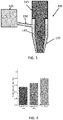
- FIG. 1 shows a cross-sectional side view of a printing system 100 according to one or more embodiments.
- the printing system 100 comprises a first nozzle 110 and a second nozzle 120 surrounding the first nozzle 110.
- a printable biological composition, and more specifically a biocompatible hydrogel 130 is provided in the first nozzle 110 and a printable composition 140 comprising one or more non-toxic polymers is provided in the second nozzle 120.
- the printing system 100 is configured so that the biocompatible hydrogel 130 and the printable composition 140 are extruded from the first nozzle 110 and second nozzle 120 simultaneously, such that the printable composition 140 coats the biocompatible hydrogel 130.
- the printable composition 140 is configured to coat the biocompatible hydrogel 130 in a solidified state. As a result, the printing system 100 prints individual fibers of biocompatible hydrogels which are coated in a solidified composition 140 comprising a non-toxic polymer.
- composition 140 Any printable composition which is non-toxic and configured to coat the extruded biocompatible hydrogel 130 in a solidified state may be used for the composition 140.
- the printable composition 140 preferably comprises one or more non-toxic thermoreversible gelation polymers.
- a polymer which is solid at a temperature between 10 °C - 40 °C is preferred, as this allows the extruded fibers to be incubated at a particular temperature (for example for temperature-assisted crosslinking) whilst the composition 140 is in a solid state, thereby maintaining the shape of the fiber whilst being incubated.
- the composition 140 is a poloxamer-based thermoreversible gel.
- Poloxamers or Pluronics ® are a class of water-soluble nonionic triblock copolymers formed by polar (poly ethylene oxide) and non-polar (poly propylene oxide) blocks which confer amphiphilic and surface active properties to the polymers. Their aqueous solutions undergo sol-to-gel transition with increasing the temperature above a lower critical gelation temperature (LCGT); moreover, the coexistence of hydrophilic and hydrophobic monomers into block copolymers allows the formation of ordered structures in solution, the most common of these being the micelles.
- LCGT critical gelation temperature
- poloxamers A variety of poloxamers is available on the market, differing on the molecular weight of the building blocks and on the hydrophobic-hydrophilic ratio, allowing the preparation of thermosensitive hydrogels with different properties, e.g., in terms of critical gelation concentration (CGC) and gelation time at physiological condition.
- CGC critical gelation concentration
- the most significant physical properties of most common poloxamers are described in Russo E. and Villa C. Poloxamer Hydrogels for Biomedical Applications, Pharmaceutics 2019, 11, 671 .
- said printable composition 140 is a poloxamer hydrogel in water-based media and comprises poloxamer 407 (e.g., Pluronic ® F-127).
- poloxamer 407 may be at a concentration of at least 25%, preferably 25-45 % (wt/v), more preferably of 30-40% (wt/v), more preferably of 33-37 % (wt/v). It is noted that an even higher concentration of poloxamer 407 may be possible, so long as the printing system is able to apply the required pressure for extrusion.
- the biocompatible hydrogel 130 may comprise one or more synthetic or natural biopolymers in an aqueous media. Natural biopolymers would include any polysaccharides and/or proteins which provide a printable composition.
- the biocompatible hydrogel 130 may comprise for instance one or more of a decellularized extracellular matrix (ECM), alginate, modified alginate comprising inserted cell attachment sites, gelatin, fibrinogen, laminin, collagen, or other ECM proteins, hyaluronic acid, as well as any of these modified with acrylamide groups, such as gelatin methacrylate (GelMA), alginate methacrylate (AlgMA) collagen methacrylate (ColMA), or hyaluronan methacrylate (HA-MA), chitosan, gellan gum, xanthan gum, agarose., poly(ethylene glycol) diacrylate (PEGDA), N-Isopropylacrylamide (NIPAM), nanocellulose.
- ECM extracellular matrix
- alginate alg
- biocompatible hydrogels are described in Table 1.
- Preferred embodiments comprise or consist of (i) fibrinogen and gelatin, (ii) fibrinogen, a decellularized ECM (e.g. Matrigel ® ) and gelatin, (iii) a decellularized ECM or other cell matrices (e.g. Matrigel ® ), alginate and gelatin, (iv) fibrinogen, gelatin and alginate, (v) fibrinogen, a decellularized ECM or other cell matrices (e.g.
- this biocompatible hydrogel 130 may be in nanostructured form further to the addition of nanocomposites.
- the biocompatible hydrogel 130 may also comprise any living cells. They type of cells included in the hydrogel can be selected, as appropriate, depending on the purpose. In preferred embodiments, these cells are animal cells, preferably these cells are mammalian cells. For example, these mammalian cells may be selected from a human, mouse, rat, guinea pig, dog, cat, cow, pig, sheep, horse, bear, and so on. In a preferred embodiment, said cells are mouse or human cells.
- the type of animal cells e.g., human-derived cells
- somatic stem cells examples include mesenchymal stem cells (MSC), hematopoietic stem cells, and neural stem cells.
- MSC mesenchymal stem cells
- progenitor cells and mature cells examples include cells derived from the skin, dermis, epidermis, muscle, cardiac muscle, nerve, bone, cartilage, endodermis, brain, epithelium, heart, kidney, liver, pancreas, spleen, oral cavity, cornea, or hair.
- human-derived cells examples include ES cells, iPS cells, MSC, chondrocytes, osteoblasts, osteoprogenitor cells, mesenchyme cells, myoblasts, cardiac muscle cells, nerve cells, hepatic cells, beta cells, fibroblasts, corneal endothelial cells, vascular endothelial cells, corneal epithelial cells, and hematopoietic stem cells.
- the origin of cells may be either autologous or allogeneic.
- these cells are myoblasts (e.g., C2C12 myoblasts).
- Skeletal muscle myoblasts can differentiate into myotubes, complex cellular structures composed of several fused myoblasts with contractile abilities.
- the construct can be incubated in differentiation medium, such as the DM medium described in Example 2, which promotes cell differentiation into myotubes.
- the myoblasts may be cultured in the differentiation medium for 5-7 days, although more days may be preferred to achieve full maturation, preferably the cells are cultured for a total period of 7 to 14 days.
- myotubes are aligned in the direction of the 3D bioprinted fibers, which is a necessary condition for maximum force generation.
- 3D bioprinting can further help in the alignment of fibers due to the shear stress at the nozzle. This stress aligns the hydrogel fibers which in turn helps in the alignment of the myotubes, as they tend to follow topographical cues.
- FIG. 2A shows a cross-sectional side view of an individual non-hollow fiber 200A obtained by the printing system 100 of FIG. 1 .
- the individual fiber 200A comprises a non-hollow inner fiber 210 of the biocompatible hydrogel, and a solidified outer coating 220 of the composition 140 containing the non-toxic polymer which coats the inner fiber 210.
- the inner fiber 210 is provided with physical support by the outer coating 220, preventing the inner fiber 210 from losing its shape over time.
- FIG. 2B shows another cross-sectional side view of an individual fiber 200B obtained by the printing system 100 of FIG. 1 .
- the individual fiber 200B comprises an inner fiber 210 of the biocompatible hydrogel, and a solidified outer coating 220 of the composition 140 containing the non-toxic polymer.
- the individual fiber 200B has been printed in a closed loop.
- the diameter of the biocompatible hydrogel obtained will depend on the diameter of the first nozzle 110, as well as the relative rates of extrusion of the biocompatible hydrogel 130 and the printable composition 140.
- the pressure applied to each nozzle may be independently controllable in order to control the diameter of the biocompatible hydrogel that is printed. For example, if the pressure applied to the first nozzle 110 is increased relative to the pressure applied to the second nozzle 120, the extrusion rate of the biocompatible hydrogel 130 will be greater and thus the coated fiber will have a greater diameter.
- FIG. 3 shows a cross-sectional side view of a printing system 300 according to one or more embodiments.
- the printing system 300 comprises a first nozzle 110 and a second nozzle 120 surrounding the first nozzle 110.
- a printable biocompatible hydrogel 130 is provided in the first nozzle 110 and a printable composition 140 comprising a non-toxic polymer is provided in the second nozzle 120.
- the biocompatible hydrogel 130 is provided to the first nozzle 110 by a first cartridge 310 which contains a source of the biocompatible hydrogel 130, and which is in fluidic communication with the first nozzle 110.
- the composition 140 is provided to the second nozzle 120 by a second cartridge 320, which contains a source of the composition 140 and is fluidically connected to the second nozzle 120 via a conduit 330 extending laterally into the second nozzle 120.
- the conduit 330 may extend into the second nozzle 120 at an acute angle to the nozzle (i.e. at an angle less than 90 degrees), which may help prevent the conduit 330 and second cartridge 320 from contacting a vessel into which the fibers are printed (for example a petri dish).
- the conduit 330 may comprise a flexible material such that the second cartridge 320 is moveable to avoid contact with a vessel into which the fibers are printed.
- FIG. 4 shows the cell viability of an extruded biocompatible hydrogel when a thin cylindrical nozzle (of approximately 700 ⁇ m diameter), wide cylindrical (of approximately 840 ⁇ m diameter) and a conical nozzle (of approximately 200 ⁇ m diameter) is used for the same biocompatible hydrogel.
- the cell viability of the conical nozzle was higher than the cylindrical nozzles, even with a smaller diameter, as conical nozzles provide better stress profiles for bioprinting, by reducing the amount of shear stress applied to the biocompatible hydrogel during extrusion, which results in the greater cell viability.
- the length of the cylindrical nozzles may be 25 mm or more.
- the first nozzle 110 may be made of any suitable material, such as a metal or plastic.
- the first nozzle 110 is preferably made of plastic as this further reduces the amount of shear stress applied to the biocompatible hydrogel during extrusion, which improves the viability of the extruded hydrogel.
- the diameter of the hole of the first nozzle 110 is preferably from 100 ⁇ m to 800 ⁇ m and the diameter of the hole of the second nozzle 120 is preferably from 1 mm to 5 mm.
- the diameter of the extruded biological fiber can be varied relative to the diameter of the first nozzle 110.
- the one or more fibers have a predetermined diameter with a standard deviation of 20% or less, such as 15% or less, 10% or less and 5% or less.
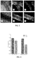
- FIG. 5 shows imaged fibers obtained with a printing system according to one or more embodiments (images A to C) as compared to fibers extruded from a printing system without an outer non-toxic polymer provided (images D to F).
- Images A and D show a simple image of the produced fibers.
- Images B and E show fluorescence images of the live assay showing the number of live cells in the fibers.
- Images C and F show fluorescence images of the dead assay showing the number of dead cells in the fubers.
- FIG. 6 shows a graph illustrating this effect. As can be seen, the fibers obtained with the polymer coating still had 80% viable cells after 24 hours, whereas the fibers without the polymer coating had less than 60% viable cells.
- a printable composition 140 comprising poloxamer 407 (e.g., Pluronic ® F-127) was used.
- Poloxamer 407 is particularly useful due to its thermoreversible properties. Using rheological characterisation methods, it has been observed that it behaves as a Newtonian liquid a low temperature (e.g. at 4 °C), and at room temperature (20 °C to 25 °C) shows a strong shear-thinning effect, meaning it has smooth printability whilst also retaining its shape once printed.
- the polymer may therefore be used as the printable composition 140 and may also be easily removed from the fibers by a wash with a cold aqueous composition such as water or phosphate buffered saline (i.e. at a temperature between 0 °C and 4 °C).
- a cold aqueous composition such as water or phosphate buffered saline (i.e. at a temperature between 0 °C and 4 °C).
- the printing systems and methods disclosed herein may be used to obtain one or more individual fibers of biocompatible hydrogels having a predefined diameter, by simultaneously extruding the biocompatible hydrogel 130 and the printable composition 140 such that the printable composition 140 coats the extruded biocompatible hydrogel 130 in a solidified state.
- the fibers may be submitted to a cross-linking treatment.
- the fibers may be submitted to one or more of UV cross-linking, temperature-assisted cross-linking, or cross-linking induced by a cross-linking agent (e.g., enzymatic, ionic or other chemical cross-linking agent), as described in further detail below.
- a cross-linking agent e.g., enzymatic, ionic or other chemical cross-linking agent
- the non-toxic polymer composition is a thermoreversible gelation non-toxic polymer which transitions from sol into a gel state. That is, the composition exhibits solid properties at a first temperature whilst being extrudable, such that the composition may be used as the printable composition 140 discussed above, and at a second temperature the composition may behave as a Newtonian fluid such that the composition may be removed by changing the temperatures of the extruded fibers.
- Poloxamer 407 behaves as a Newtonian liquid a low temperature (e.g.
- compositions are solid at a temperature between 20 °C to 25 °C.
- Such compositions may be removable by a wash with a cold aqueous solution, such as water or phosphate buffered saline applied to the extruded biological fibers.
- the steps of submitting the fibers to a cross-linking treatment may be performed at the same time as removing the composition coating the extruded fiber.
- a solution containing a cross-linking agent for example an ionic, enzymatic or other chemical cross-linking agent
- the solution may be applied to the fibers at a temperature which causes the polymer composition to dissolve.
- Poloxamer 407 is used as the composition 140
- the fibers may be washed with a solution containing cross-linking agent which has a temperature of about 4 °C, such that the solution both washes the composition 140 from the fibers, and also cross-links the fibers.
- the polymer composition 140 may comprise a cross-linking agent such that the cross-linking treatment is applied to the extruded biological fibers (with the polymer composition 140 further providing structural support to the biological fibers).
- the combination of physical and chemical confinement provides for a finer control of the fibers width.
- the fibers may be submitted to any cross-linking method, for instance one or more of UV cross-linking, temperature-assisted cross-linking, or cross-linking induced by a cross-linking agent (e.g., enzymatic, ionic or other chemical cross-linking agent),.
- a cross-linking agent e.g., enzymatic, ionic or other chemical cross-linking agent
- the biocompatible hydrogel 130 may comprise a substance which crosslinks upon exposure to a cross-linking agent, wherein the extruded biological fiber is cross-linked exposing the obtained fiber to the cross-linking agent.
- the polymer composition 140 may comprise the cross-linking agent such that cross-linking occurs simultaneously to extrusion; in other examples, the biological fibers may be extruded, and the cross-linking agent performed in a separate step (for example after the polymer composition 140 is removed).
- the biocompatible hydrogel 130 comprises one or more of alginate, modified alginate comprising inserted cell attachment sites (e.g., Arg-Gly-Asp (RGD) motifs), such as in Alginate-RGD bioink (Sigma Aldrich), gellan gum and chitosan.
- the cross-linking agent may comprise a divalent cation, like Ca 2+ , Ba 2+ , Mg 2+ and can be found at a concentration of 10 - 300 mM, preferably of 25 - 300 mM.
- the ionic cross-linking agent comprising a divalent cation is CaCl 2 .
- the cross-linking agent may comprise negatively charged ions, preferably wherein the cross-linking agent comprises negatively charged Molybdenum or Platinum ions.
- the biocompatible hydrogel 130 may comprise alginate having a concentration of 0.5% to 2% (wt/v).
- the polymer composition may comprise CaCl 2 at a concentration of 10 mM to 300 mM, preferably of 25 - 300 mM.
- the biocompatible hydrogel 130 may comprise one or more of the biocompatible polymers described herein above, preferably selected from collagen, gelatin, chitosan or combinations thereof and said cross-linking agent is another chemical cross-linking agent, including but not limited to genipin, proanthocyanidin and epigallocatechin gallate ( Pinheiro A, Cooley A, Liao J, Prabhu R, Elder S. Comparison of natural crosslinking agents for the stabilization of xenogenic articular cartilage. J Orthop Res. 2016;34(6):1037-1046. doi:10.1002/jor.23121 ).
- the amount of cross-linking agent which is extruded is highly controllable as opposed to the use of cross-linking liquid phase solution, which is hard to control and can cause blockage of the printing system.
- the biocompatible hydrogel 130 may comprise a substance which crosslinks upon exposure to UV light, and the crosslinking treatment may include exposing the extruded biological fiber to UV light.
- a substance include any hydrogel modified with acrylamide groups (i.e., a poly(acrylic) acid hydrogel), such as gelatin methacrylate (GelMA), poly(ethylene glycol) diacrylate (PEGDA), alginate methacrylate (AlgMA), collagen methacrylate (ColMA) or hyaluronan methacrylate (HA-MA) .
- a poly(acrylic) acid hydrogel such as gelatin methacrylate (GelMA), poly(ethylene glycol) diacrylate (PEGDA), alginate methacrylate (AlgMA), collagen methacrylate (ColMA) or hyaluronan methacrylate (HA-MA) .
- the biocompatible hydrogel 130 may comprise a substance which crosslinks upon heating, and the crosslinking treatment may comprise heating the biological fiber, generally to a temperature of more than 30 °C, preferably about 37 °C during at least 30 minutes, preferably 1 hour.
- the biocompatible hydrogel 130 may comprise one or more of collagen, a decellularized extracellular matrix (ECM) or other cell matrices, such as Matrigel ® .
- the biocompatible hydrogel may comprise collagen at a concentration of about 2 mg/mL to about 10 mg/mL.
- the biocompatible hydrogel may comprise a decellularized extracellular matrix (ECM) or other cell matrices, such as Matrigel ® at concentrations of between 25% and 75% (v/v).
- ECM extracellular matrix
- Matrigel ® at concentrations of between 25% and 75% (v/v).
- the biocompatible hydrogel 130 may comprise a substance which crosslinks enzymatically, and the crosslinking treatment may comprise applying an enzymatic cross-linking agent to the biological fiber.
- the agent may be comprised in the polymer composition 140 such that the steps of extruding the fiber and performing the cross-linking treatment occurs simultaneously.
- the cross-linking agent may be separately applied to the biological fiber.
- the biocompatible hydrogel 130 may comprise fibrinogen and the enzymatic cross-linking agent may be an enzyme solution comprising thrombin, for example at a concentration of 5 U/mL to 20 U/mL.
- the biocompatible hydrogel may comprise fibrinogen or gelatin and the enzymatic cross-linking agent may be an enzyme solution comprising transglutaminase. Enzymatic cross-linking is typically conducted at room temperature..
- the biocompatible hydrogel 130 may comprise at least a first cross-linkable substance and a second cross-linkable substance, wherein the cross-linking of the second substance is reversible.
- the second cross-linkable substance may be removed from the biocompatible hydrogel after the cross-linking treatment is applied for the first and second cross-linkable substances. This may be preferably when the second cross-linkable substance is useful in providing an initial structure to the obtained fibers, but may inhibit cell proliferation and differentiation.
- the second substance may be alginate and the alginate may be removed by application of a calcium chelator, such as ethylenediaminetetraacetic acid (EDTA), egtazic acid (EGTA), citric acid or etidronic acid.
- a calcium chelator such as ethylenediaminetetraacetic acid (EDTA), egtazic acid (EGTA), citric acid or etidronic acid.
- EDTA ethylenediaminetetraacetic acid
- EGTA egtazic acid
- citric acid or etidronic acid ethylenediaminetetraacetic acid
- said calcium chelator is EDTA which can be used for example at a concentration of 10mM-30mM, preferably about 20mM.
- biocompatible hydrogel 130 and the polymer composition 140.
- the physical confinement of the biocompatible hydrogel 130 by the polymer composition 140 relies on the gelation of the hydrogel 130 and polymer composition 140 such that diffusion of the biocompatible hydrogel 130 through the polymer composition 140 is negligible. It will be appreciated that for any given composition, the optimal concentrations for gelation can be determined by routine experimentation.
- the polymer composition additionally comprises a cross-linking agent such that cross-linking occurs immediately upon extrusion, this may immediately chemically confine the biocompatible hydrogel 130 inside the polymer composition such that the gelation of the biocompatible hydrogel 130 and the polymer composition 140 may take a wider range of values (as the chemical cross-linking inhibits diffusion of the biocompatible hydrogel).
- the fiber quality is influenced by both the homogeneity of the printing and the stability of the fibers.
- ECM extracellular matrix
- Matrigel ® cell matrices
- fibrinogen added to the mixture increases their long-term stability, as fibrin is more robust, creating high quality fibers.
- the polymer composition contains a cross-linking agent.
- the polymer composition may comprise CaCl 2 and the biocompatible hydrogel may comprise alginate. If the main biomaterial is too liquid, like Matrigel ® , it will diffuse through the polymer composition before the Ca-crosslinking of alginate can form homogeneous fibers, unless the concentration of alginate is increased to accelerate this process. Since alginate does not have cell attachment motives, it is preferable to keep its concentration as low as possible; in that case, however, the quality of the fibre will not be high.
- Collagen and Matrigel ® which are crosslinked slowly at 37 °C during approximately 30 min in an irreversible manner, deserve special consideration.
- Collagen is one of the main components of many tissue's ECM and, in particular, of skeletal muscle tissue, making it especially interesting for 3D bioengineering applications.
- Matrigel ® is one of the most widely used basement membrane matrices for 2D and 3D culture, since it is rich in collagen and many other ECM proteins, but shares the same difficulties.
- their irreversible and low temperature-dependent crosslinking makes their bioprinting difficult, as opposed to gelatin, which has reversible crosslinking.
- Table 1 shows a list of preferred examples. It will be appreciated that other combinations of cross-linking substances are possible, in which case other concentrations may be possible so long as the biocompatible hydrogel and the polymer compositions are extrudable, and the relative viscosity of the biocompatible hydrogel inhibits diffusion of the biocompatible hydrogel through the polymer composition.
- Table 1 presents a guide to understand the possible combinations of materials according to their confinement method. It also provides illustrative concentrations of the biocompatible hydrogel materials, as well as of the polymer composition and even CaCl 2 .
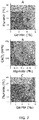
- FIG. 7 focuses on the description of these possibilities and shows a number of graphs illustrating possible combination of materials in the biocompatible hydrogel and the non-toxic polymer to achieve a homogeneous individual fiber.
- Graph A illustrates optimal concentrations of poloxamer 407 (Pluronic ® F-127) as the polymer composition, without a cross-linking agent, combined with gelatin in the biocompatible hydrogel.
- concentration of gelatin with respect to the concentration of poloxamer 407 needs to be adjusted to avoid diffusion of the biocompatible hydrogel through the pluronic.
- gelatin in concentrations below 3% (wt/v) diffuses, since its gelation is not high enough.
- pluronic at concentrations below 33% (wt/v) causes the same problem.
- the preferred range of concentrations for gelatin to get homogeneous fibers is between 3-5% (wt/v); for the poloxamer 407, it is between 33-37 % (wt/v).
- Higher concentrations of both pluronic and gelatin can still be used successfully; however, they might require pressures too high for the 3D bioprinter (this depends on the specific printing system) and cells might suffer too much shear stress during extrusion.
- the chemically assisted method based on alginate crosslinking on extrusion, provides the best homogeneity of fibers compared to physical confinement based on relative gelation properties. Moreover, it does not present clogging problems, since the flow of the polymer composition can be carefully controlled by the applied pressure. Whilst alginate also depends on the concentration of pluronic in the same way as gelatin, it is more dependent on the concentration of CaCl 2 dissolved in it with respect to its own concentration. Alginate concentrations ranging from as low as 0.25% (wt/v) can yield homogeneous fibers if the concentration of CaCl 2 is high enough and other materials that increase the viscosity are present in the mixture.
- alginate can, naturally, produce very homogeneous fibers, but its stiffness might be too high for the cells, taking into account that other biocompatible polymers with attachment sites, like fibrinogen or collagen, may be included in the biocompatible hydrogel. If the presence of alginate is a problem for cell proliferation or differentiation due to low biocompatibility, alginate can be removed after crosslinking of the other cross-linkable materials in the biocompatible hydrogel by the addition of ethylenediaminetetraacetic acid (EDTA) for 5 min.
- EDTA is a calcium chelator that will attract the Ca+2 ions that reversibly crosslink alginate, making it dissolve in the culture medium.
- Graph B illustrates optimal concentrations of CaCl 2 in a polymer composition, combined with alginate in a biocompatible hydrogel which additionally comprises gelatin at 3% (wt/v) as extra support.
- the preferred range of concentrations for obtaining homogeneous fibers is indicated by the region marked "X". If the concentration of alginate and CaCl 2 is too high, then the fibers are too stiff. Conversely, if the concentration is too low then the fibers do not form.
- graph C illustrates the optimal concentrations of poloxamer 407 (Pluronic) as the polymer composition, without a cross-linking agent, combined with GelMA rather than gelatin in the biocompatible hydrogel.
- the preferred range of concentrations for obtaining homogeneous fibers is indicated by the region marked "X”. It is noted that GelMA gelifies at higher concentrations than gelatin, and a minimum concentration of 5% (wt/v) should be used, compared to the minimum of 3% (wt/v) for gelatin. This creates a smaller range to obtain homogeneous fibers, between 5-7% (wt/v). For concentrations higher than this, the mixture is mostly unextrudable.
- homogeneous fibers can be obtained with this method by choosing the concentrations as shown in the region marked "X" in graph C.
- alginate alginate may additionally be used to provide extra support. If GelMA is later on crosslinked with UV light, the material will remain in the biocompatible hydrogel, providing cell attachment sites, instead of being washed away, as in the case of gelatin. Further optimization may be achieved by mixing both gelatin and GelMA, to regulate density of GelMA that will remain in the mixture after UV crosslinking.
- FIG. 8 shows a number of graphs illustrating possible combinations of materials that provide sufficient cell attachment sites.
- Graph A illustrates preferred concentrations of fibrinogen and Matrigel ® in the biocompatible hydrogel.
- fibrinogen should be decreased to the range of 10 mg/mL.
- fibrinogen should be reduced too much, the density of attachment sites of Matrigel ® , as well as its stiffness, is not high enough for proper cell differentiation.
- the preferred range of concentrations is indicated by the region marked "X".
- Graph B illustrates preferred concentrations of fibrinogen and Matrigel ® in the biocompatible hydrogel. If GelMA is used as a confinement material, instead of gelatin, which is dissolved during incubation, its proportionality with respect to fibrinogen is also important. As already mentioned, GelMA concentrations below 5% (wt/v) do not produce homogeneous fibers, since they dissolve through the pluronic. In this range of applicability, fibrinogen should be kept between 10-20 mg/mL to have enough attachment sites. The preferred range of concentrations is indicated by the region marked "X".
- fibrinogen is used alone as the main component for the biocompatible hydrogel (e.g. for a myoblast-laden hydrogel), it should be kept at a high concentration, with 20 mg/mL being a preferred value for cell differentiation.
- biocompatible hydrogel is confined due to the gelation properties of the biocompatible hydrogel and the polymer composition alone (i.e. without a cross-linking agent in the polymer composition)
- biological fibers having a diameter of between 300 ⁇ m and 900 ⁇ m are obtainable by varying the pressure applied to the inner nozzle between 50 kPa and 80 kPa.
- FIG. 12 shows a graph illustrating the width of the homogeneous fibers obtained by physical confinement of the fibers (i.e. due to the gelation properties alone) and by chemical confinement (using simultaneous cross-linking during extrusion), with an inner nozzle diameter of 200 ⁇ m. It is noted that fibers with lower and higher thickness than those shown in FIG. 12 can be obtained by varying the pressure further.
- FIGs. 13A to 13D illustrate images of homogeneous fibers obtained at points 1 to 4 on the graph shown in FIG. 12 . As can be seen, the fibers obtained have a high homogeneity.
- one or more obtained individual fibers may be comprised in or form a biomimetic structure (e.g. a tissue construct).
- Said biomimetic structure can further comprise a biomaterial.
- biomaterial may refer to any biocompatible material which serves as a substrate or guide for tissue repair or regeneration, for instance tissue scaffolds, tissue implants, stents or valves.
- the 3D-printed fibers of the invention can be used for the 3D bioengineering of tissue and obtaining of tissue constructs. These tissue constructs may be embedded with or without cells. These cells may be as described herein above.
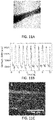
- said tissue is skeletal muscle tissue, preferably human or mouse skeletal muscle tissue. As shown in the Examples, the inventors have obtained a skeletal muscle construct with the 3D printing method and system of the invention with aligned myotubes and good contractibility features (see Fig. 10 ).
- the obtained biomimetic structure (e.g. tissue construct) can be used for research purposes.
- the obtained tissue can be used for drug testing purposes.
- the obtained biomimetic structure (e.g. tissue construct) can also be used for medical purposes, such as for tissue replacement or regeneration purposes. Accordingly, in an aspect of the invention said biomimetic structure (e.g. tissue construct) is for use as a medicament.
- the individual fibers obtained by the printing system and methods of the invention are individual skeletal muscle myotubes.
- myoblasts may be embedded in the hydrogel 130 and myoblasts may be induced to differentiate into multi-nucleated myotube structures.
- said biomimetic structure e.g. tissue construct
- said biomimetic structure comprises individual skeletal muscle myotubes obtained by a method of the invention and is for use in muscle tissue regeneration.
- multiple individual fibers (such as that shown in FIG.2A ) may be obtained which are aligned in the same uniaxial direction (either with or without the polymer coating, depending on if it is removed after cross-linking).
- Biological actuators based on 3D-printed skeletal muscle tissue can be used to study muscle development, maturation or healing, or even act as drug testing platforms for muscle; and more complex, untethered actuators can take the form of hybrid biocompatible machines comprising 3D-printed muscle fibers.
- hybrid biocompatible machines may comprise one or more individual fibers obtained by a bioprinting system as disclosed herein.
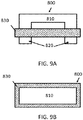
- FIGs. 8A and 8B show a hybrid biocompatible machine 800 according to one or more embodiments shown and described herein.
- the machine 800 comprises a main body 810 and two legs 820 extending from the main body 810.
- the main body is formed of a flexible material such as Polydimethylsiloxane (PDMS) or Poly(ethylene glycol) diacrylate (PEGDA).
- PDMS Polydimethylsiloxane
- PEGDA Poly(ethylene glycol) diacrylate
- One or more individual biological fibers 830 e.g. comprising skeletal muscle myotubes obtained by a printing system disclosed herein extend in a loop about the legs 820. When the biological fibers 830 are stimulated to contract, the contracting force is transmitted to the legs 820 of the machine 800 and the main body 810 is caused to flex.
- the machine 800 could be provided with an asymmetry such that contraction of the one of more biological fibers 830 causes the machine 800 to translate in a particular direction.
- the individual fibers printed using the printing systems and methods disclosed herein could be used in other biological machines, such as tethered biological actuators, for example as disclosed in Force Modulation and Adaptability of 3D-Bioprinted Biological Actuators Based on Skeletal Muscle Tissue (Rafael Mestre et al.; Adv. Mater. Technol. 2019, 4, 1800631 ).
- the hybrid biocompatible machines described above can be used for a variety of purposes, such as evaluation of tissue differentiation, functionality, drug testing platforms, drug screening platforms, force measurement platforms or as tissue models of young or old muscle (for example by assessing morphological and functional changes in the aging process of muscular tissue).
- the hybrid biocompatible machine may be studied for the force generation and contraction profiles of the printed fiber, allowing for a more detailed analysis of the tissue.
- a chemical confinement strategy was carried out with C2C12 myoblast concentration of 5 million/mL and an hydrogel composed of 0.5% (wt/v) of alginate, 3% (wt/v) gelatin to increase its viscosity and help with the homogeneity of the fibers, and 20 mg/mL fibrinogen for cell attachment sites.
- poloxamer 407 at 33% (wt/v) with 300 mM CaCl 2 was used as support material.
- a cold solution of thrombin (5 U/mL) was added to the construct for 5 min at 4°C. At this temperature, poloxamer 407 dissolved while fibrinogen was being crosslinked into fibrin. After this time, several washes with cold PBS removed completely the remaining poloxamer 407.
- alginate is a polysaccharide which does not possess cell attachment motifs. Therefore, individual fibers crosslinked with alginate, even at low a concentration of 0.5% (wt/v), did not show much compaction, something necessary for the proper differentiation of the cells.
- 3D-printed fibers showed compaction after incubation with EDTA (see Fig. 10A ), indicating the removal of alginate and the progression of the differentiation process.
- EPS could induced contractions in the fibers, as shown in Fig. 10B and immunostaining of Myosin Heavy Chain II and cell nuclei showed thin fibers with aligned myotubes ( Fig. 10C ).
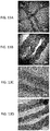
- the physical confinement method produces less homogeneous fibres, but they are still highly controlled in thickness and can yield thin skeletal muscle constructs (see Figure 11A ).
- This method as opposed to the chemically assisted one, only requires the presence of gelatin in the cell-laden hydrogel, although at a higher concentration to ensure physical confinement within the polymer coating.
- the hydrogel consisted gelatin at 5% (wt/v) with fibrinogen at 20 mg/mL and myoblasts at a density of 5 million cells/mL.
- the protocol after co-axial bioprinting was the same as for Example 1, except without the addition of EDTA: the fibre constructs were left in a solution of cold thrombin (5 U/mL) for 5 min at 4°C for fibrinogen to crosslink to fibrin and poloxamer 407 to dissolve. After this, additional washes with cold PBS (at 4°C) removed the remaining poloxamer 407 and the constructs were left to proliferate in GM in a cell incubator. As before, the fibers could compact during maturation of the tissue ( FIG. 11A ) and after several days of differentiation, they could respond to electrical stimulation with contractions, as shown in FIG. 11B . Immunostaining of Myosin Heavy Chain II and cell nuclei revealed thin fibres packed with aligned myotubes ( FIG. 11C ).
- the co-axial nozzles were manually fabricated using different types of commercially available nozzles and tips.
- the inner nozzle, where the cell-laden hydrogel passed through, was a 200-_m (G27) plastic conical nozzle (Optimum ® SmoothFlow TM tapered tips, Nordson ® , ref. 7018417).
- the luer lock of this nozzle was left free to be connected to the first bioprinting barrel, where the cell-laden hydrogel would be loaded.
- the outer nozzle that covered the inner one was a filtered P1000 pipette tip (Labclinics, ref. LAB1000ULFNL), cut approximately at 5 cm from its end. The tip was trimmed to increase its diameter at the final point. Both nozzles were assembled and glued together.
- This secondary nozzle was a flexible polypropylene 800 -_ m nozzle (G18) from Nordson ® (EFD ® 7018138).
- a silicone tubing of 0.8 mm of diameter (ibidi, ref. 10841) was attached to this external nozzle through a male elbow luer connector (ibidi, ref. 10802).
- a 1.1-mm (19G) nozzle (BBraun Sterican ® , ref. 4657799) was inserted through the other end of the tubing, so that its luer lock connector could be connected to the second bioprinting barrel, containing poloxamer 407 acid.
- gelatin from porcine skin type A (Sigma-Aldrich, G2500), fibrinogen from bovine plasma (Sigma-Aldrich, F8630) with thrombin from bovine plasma (Sigma-Aldrich, T4648) as crosslinker, Matrigel ® Basement TM membrane matrix (Corning ® , 354234), sodium alginate (Sigma-Aldrich, W201502), GelMA with lithium phenyl-2,4,6-trimethylbenzoylphosphinate (LAP) at 0.25% (wt/v) as photoinitiator (CELLINK ® , LIK-3050V-1), collagen type I high concentration (Corning ® , 354249), and Pluronic ® F-127 powder (Sigma-Aldrich, P2443).
- Poloxamer 407 was dissolved at concentrations ranging from 30-40% (wt/v) in ultrapure water with CaCl 2 (at molar concentrations ranging from 50 mM to 300 mM) under stirring in a refrigerator (4 °C) until fully dissolved.
- hydrogels containing gelatin, alginate and/or GelMA in the same composition these components were mixed together in PBS at the desired concentrations. If the hydrogel contained fibrinogen, this component was dissolved in PBS at a double concentration and then added to the hydrogel containing alginate and/or gelatin, also at a double concentration, in order to avoid pipetting the viscous mixture. If the hydrogel also contained Matrigel ® , the concentrations were also adjusted to achieve the desired concentrations reducing the need for pipetting (for instance, at 1:1:1 ratios, or 2:1:1 ratios, etc.).
- C2C12 myoblasts were harvested by a 0.25% (wt/v) Trypsin-0.53 mM EDTA solution, centrifuged at 300g for 5 min and the pellet re-suspended at a concentration of 5x10 6 cells/mL in the hydrogel mixture, at 37 °C.
- the cell-laden hydrogel was loaded into a 3-mL plastic syringe (Nordson ® , ref. 7012085) coupled to the inner nozzle of the co-axial nozzle.
- Poloxamer 407 was loaded while cold into a secondary barrel and left at RT to gellify beforehand.
- CELLINK ® Inkredible+ 3D bioprinter was used to bioprint the hydrogel fibers.
- the cell-laden hydrogel was inserted in the first cartridge and the poloxamer 407 barrel to the second barrel, and all the nozzles connected as previously explained.
- the pressure for the pluronic barrel was kept between 250-350 kPa and adjusted manually, although these values are highly dependent on the diameter and length of the silicone tubing and the concentration of pluronic.
- the pressures were kept between 40-80 kPa, also adjusted manually, depending on the type of hydrogel.
- the designs were directly written in GCode with the help of the open-source software Slic3r (v. 1.2.9) and the bioprinter was controlled with RepetierHost (v. 2.0.5).
- hydrogel contained fibrinogen
- a solution of 5 U/mL of thrombin was added to the Petri dish for 5 min in a refrigerator at 4 °C.
- poloxamer 407 would dissolve and fibrinogen would crosslink to form fibrin.
- several washes with cold PBS were done to completely remove poloxamer 407, GM supplemented with ACA added and the constructs left in a cell incubator at 37 °C and 5% CO 2 atmosphere.
- the hydrogel contained Matrigel ® , the as-printed constructs were left in an incubator for at least 30 min, without removing pluronic.
- Rheological characterization of pluronic was performed using a Discovery HR-2 controlled-stress rheometer (TA Instruments) equipped with a Peltier steel cone geometry of 40 mm of diameter, 26 ⁇ m of truncation, and an angle of 1.019°.
- the Peltier element was set to 4 °C and 25 °C to demonstrate the behaviour of the block co-polymer at low and room temperatures.
- the sample was left to acquire the desire temperature for 1 min.
- a flow ramp with shear rate from 100 1/s to 0.01 1/s was performed, in logarithmic mode with 600 s of duration per point, with a pre-conditioning to the temperature of 30 s and pre-shear of 3 rad/s for 10 s.
- Example 7 cell culture, differentiation and electrical stimulation
- C2C12 mouse myoblasts were purchased from ATCC and maintained in growth medium (GM) consisting of high glucose Dullbecco's Modified Eagle's Medium (DMEM; Gibco ® ) supplemented with 10% Fetal Bovine Serum (FBS), 200 nM L-Glutamine and 1% Penicillin/Streptomycin, in a 37 °C and 5% CO 2 atmosphere. Cells were passaged before reaching 80% confluency in Corning ® T-75 flasks.
- GM growth medium
- DMEM Dullbecco's Modified Eagle's Medium
- FBS Fetal Bovine Serum
- Penicillin/Streptomycin penicillin/Streptomycin
- GM was substituted by DM, consisting of high glucose DMEM containing 10% Horse Serum (Gibco ® ), 200 nM L-Glutamine (Gibco ® ), 1% Penicillin-Streptomycin (Gibco ® ), 50 ng/ml IGF-1 (Sigma-Aldrich) and 1 mg/ml 6-aminocaproic acid (ACA, Sigma-Aldrich).
- 3D-bioprinted fibres were stimulated with a set of carbon-made electrodes attached to the cover of a Petri dish under an inverted microscope (Leica's DMi8) with pulses of 2 ms and 1 V/mm. Analysis of the contractions was performed with a home-made Python algorithm based on computer vision techniques that computed the distance between frames of a selected ROI by applying an L2-norm to the image pixels.
Landscapes
- Health & Medical Sciences (AREA)
- Life Sciences & Earth Sciences (AREA)
- Chemical & Material Sciences (AREA)
- General Health & Medical Sciences (AREA)
- Veterinary Medicine (AREA)
- Dermatology (AREA)
- Medicinal Chemistry (AREA)
- Oral & Maxillofacial Surgery (AREA)
- Transplantation (AREA)
- Epidemiology (AREA)
- Public Health (AREA)
- Animal Behavior & Ethology (AREA)
- Engineering & Computer Science (AREA)
- Biomedical Technology (AREA)
- Zoology (AREA)
- Cell Biology (AREA)
- Botany (AREA)
- Chemical Kinetics & Catalysis (AREA)
- Dispersion Chemistry (AREA)
- Materials Engineering (AREA)
- Manufacturing & Machinery (AREA)
- Biophysics (AREA)
- Urology & Nephrology (AREA)
- Vascular Medicine (AREA)
- Molecular Biology (AREA)
- Materials For Medical Uses (AREA)
Priority Applications (4)
| Application Number | Priority Date | Filing Date | Title |
|---|---|---|---|
| EP20382597.1A EP3932437A1 (de) | 2020-07-03 | 2020-07-03 | Drucksystem zur gewinnung von biologischen fasern |
| EP21735334.1A EP4175688A1 (de) | 2020-07-03 | 2021-07-05 | Drucksystem zur gewinnung von formfreien breitengesteuerten einzelnen biologischen fasern |
| US18/013,859 US20230338619A1 (en) | 2020-07-03 | 2021-07-05 | Printing system for obtaining free-form width-controlled individual biological fibers |
| PCT/EP2021/068502 WO2022003203A1 (en) | 2020-07-03 | 2021-07-05 | Printing system for obtaining free-form width-controlled individual biological fibers |
Applications Claiming Priority (1)
| Application Number | Priority Date | Filing Date | Title |
|---|---|---|---|
| EP20382597.1A EP3932437A1 (de) | 2020-07-03 | 2020-07-03 | Drucksystem zur gewinnung von biologischen fasern |
Publications (1)
| Publication Number | Publication Date |
|---|---|
| EP3932437A1 true EP3932437A1 (de) | 2022-01-05 |
Family
ID=71607875
Family Applications (2)
| Application Number | Title | Priority Date | Filing Date |
|---|---|---|---|
| EP20382597.1A Withdrawn EP3932437A1 (de) | 2020-07-03 | 2020-07-03 | Drucksystem zur gewinnung von biologischen fasern |
| EP21735334.1A Pending EP4175688A1 (de) | 2020-07-03 | 2021-07-05 | Drucksystem zur gewinnung von formfreien breitengesteuerten einzelnen biologischen fasern |
Family Applications After (1)
| Application Number | Title | Priority Date | Filing Date |
|---|---|---|---|
| EP21735334.1A Pending EP4175688A1 (de) | 2020-07-03 | 2021-07-05 | Drucksystem zur gewinnung von formfreien breitengesteuerten einzelnen biologischen fasern |
Country Status (3)
| Country | Link |
|---|---|
| US (1) | US20230338619A1 (de) |
| EP (2) | EP3932437A1 (de) |
| WO (1) | WO2022003203A1 (de) |
Cited By (3)
| Publication number | Priority date | Publication date | Assignee | Title |
|---|---|---|---|---|
| CN115944837A (zh) * | 2022-07-07 | 2023-04-11 | 哈尔滨工业大学 | 一种组合层级螺旋式微纳米游泳机器人 |
| WO2023113354A1 (ko) * | 2021-12-13 | 2023-06-22 | 전남대학교산학협력단 | 관형 생체조직용 바이오잉크조성물키트, 그 제조방법, 이를 이용한 관형 생체조직 제조방법 및 그 방법으로 제조된 관형 생체조직 |
| WO2023139272A1 (en) * | 2022-01-24 | 2023-07-27 | Scipio Bioscience | Gelation device with piston |
Families Citing this family (9)
| Publication number | Priority date | Publication date | Assignee | Title |
|---|---|---|---|---|
| US20250241906A1 (en) * | 2022-02-15 | 2025-07-31 | University Of Central Florida Research Foundation, Inc. | Anabolic drugs stimulating type ii collagen production from chondrocytes or their progenitors |
| CN114532413A (zh) * | 2022-02-15 | 2022-05-27 | 江南大学 | 一种用于脂肪替代物的3d打印的乳液凝胶及其制备方法 |
| WO2023201047A1 (en) * | 2022-04-14 | 2023-10-19 | University Of Cincinnati | Recapitulating tissue-native architectures in bio-printable hydrogels |
| CN115990291B (zh) * | 2022-12-12 | 2024-06-04 | 中国科学院上海硅酸盐研究所 | 生物墨水、可免疫调节促毛囊再生的细胞支架及其制备方法和应用 |
| CN117752651B (zh) * | 2023-05-04 | 2026-08-07 | 南昌大学第一附属医院 | 含有表没食子儿茶素没食子酸酯的组合物及其应用 |
| CN116731970B (zh) * | 2023-06-26 | 2024-07-12 | 河南迈酷贝塔医药科技有限公司 | 基于复合水凝胶的食管癌仿生器官模型构建方法及其应用 |
| CN117298337B (zh) * | 2023-11-10 | 2024-08-16 | 广州贝奥吉因生物科技股份有限公司 | 一种骨修复水凝胶支架及其制备方法 |
| CN118563455B (zh) * | 2024-07-29 | 2024-10-22 | 四川大学华西医院 | 一种水凝胶纤维材料、制备方法 |
| CN120860300B (zh) * | 2025-09-25 | 2025-12-05 | 金士达医疗(咸宁)有限公司 | 一种杀菌止血的医用敷料的制备方法 |
Citations (6)
| Publication number | Priority date | Publication date | Assignee | Title |
|---|---|---|---|---|
| WO2015066705A1 (en) * | 2013-11-04 | 2015-05-07 | University Of Iowa Research Foundation | Bioprinter and methods of using same |
| WO2017187114A1 (en) * | 2016-04-29 | 2017-11-02 | The University Of Bristol | Improvements in 3d printing |
| WO2018055452A1 (en) * | 2016-09-21 | 2018-03-29 | Cellink Heal As | Implantable device and 3d bioprinting methods for preparing implantable device to deliver islets of langerhans |
| WO2018053565A1 (en) * | 2016-09-22 | 2018-03-29 | St Vincent's Hospital | Apparatus and method for handheld free-form biofabrication |
| US10156560B1 (en) | 2013-09-13 | 2018-12-18 | The Board Of Trustees Of The University Of Illinois | Locomotive biological machines |
| WO2019060518A1 (en) * | 2017-09-21 | 2019-03-28 | President And Fellows Of Harvard College | TISSUE CONSTRUCTION, METHODS OF PRODUCTION AND USE THEREOF |
Family Cites Families (1)
| Publication number | Priority date | Publication date | Assignee | Title |
|---|---|---|---|---|
| PT3670155T (pt) * | 2013-06-13 | 2025-07-08 | Aspect Biosystems Ltd | Cabeça de impressão, sistema de fabrico aditivo de estruturas tridimensionais e respetivo método |
-
2020
- 2020-07-03 EP EP20382597.1A patent/EP3932437A1/de not_active Withdrawn
-
2021
- 2021-07-05 WO PCT/EP2021/068502 patent/WO2022003203A1/en not_active Ceased
- 2021-07-05 US US18/013,859 patent/US20230338619A1/en active Pending
- 2021-07-05 EP EP21735334.1A patent/EP4175688A1/de active Pending
Patent Citations (6)
| Publication number | Priority date | Publication date | Assignee | Title |
|---|---|---|---|---|
| US10156560B1 (en) | 2013-09-13 | 2018-12-18 | The Board Of Trustees Of The University Of Illinois | Locomotive biological machines |
| WO2015066705A1 (en) * | 2013-11-04 | 2015-05-07 | University Of Iowa Research Foundation | Bioprinter and methods of using same |
| WO2017187114A1 (en) * | 2016-04-29 | 2017-11-02 | The University Of Bristol | Improvements in 3d printing |
| WO2018055452A1 (en) * | 2016-09-21 | 2018-03-29 | Cellink Heal As | Implantable device and 3d bioprinting methods for preparing implantable device to deliver islets of langerhans |
| WO2018053565A1 (en) * | 2016-09-22 | 2018-03-29 | St Vincent's Hospital | Apparatus and method for handheld free-form biofabrication |
| WO2019060518A1 (en) * | 2017-09-21 | 2019-03-28 | President And Fellows Of Harvard College | TISSUE CONSTRUCTION, METHODS OF PRODUCTION AND USE THEREOF |
Non-Patent Citations (13)
| Title |
|---|
| AHN ET AL.: "A novel cell-printing method and its application to hepatogenic differentiation of human adipose stem cell-embedded mesh structures", SCIENTIFIC REPORTS, vol. 5, 2015, pages 13427 |
| COSTANTINI MARCO ET AL: "Microfluidic-enhanced 3D bioprinting of aligned myoblast-laden hydrogels leads to functionally organized myofibersin vitroandin vivo", BIOMATERIALS, ELSEVIER, AMSTERDAM, NL, vol. 131, 23 March 2017 (2017-03-23), pages 98 - 110, XP029991706, ISSN: 0142-9612, DOI: 10.1016/J.BIOMATERIALS.2017.03.026 * |
| D. NEALM. S. SAKARL.-L. S. ONGH. HARRY ASADA: "Formation of elongated fascicle-inspired 3D tissues consisting of high-density, aligned cells using sacrificial outer molding", LAB CHIP, vol. 14, 2014, pages 1907, XP055351029, DOI: 10.1039/c4lc00023d |
| H.-W. KANGS. J. LEEI. K. KOC. KENGLAJ. J. YOOA. ATALA: "A 3D bioprinting system to produce human-scale tissue constructs with structural integrity", NATURE BIOTECHNOLOGY, vol. 34, 2016, pages 312 |
| HINTON T.J. ET AL.: "Three-dimensional printing of complex biological structures by freeform reversible embedding of suspended hydrogels", SCI. ADV., vol. 1, 2015, pages e1500758, XP055491252, DOI: 10.1126/sciadv.1500758 |
| HYUN-WOOK KANG ET AL: "A 3D bioprinting system to produce human-scale tissue constructs with structural integrity", NATURE BIOTECHNOLOGY, vol. 34, no. 3, 1 March 2016 (2016-03-01), us, pages 312 - 319, XP055556663, ISSN: 1087-0156, DOI: 10.1038/nbt.3413 * |
| KATJA HÖLZL ET AL: "Bioink properties before, during and after 3D bioprinting", BIOFABRICATION, vol. 8, no. 3, 23 September 2016 (2016-09-23), pages 032002, XP055599735, DOI: 10.1088/1758-5090/8/3/032002 * |
| MESTRE RAFAEL ET AL: "3D Bioprinted Muscle-Based Bio-Actuators: Force Adaptability Due to Training", 7 July 2018, BIG DATA ANALYTICS IN THE SOCIAL AND UBIQUITOUS CONTEXT : 5TH INTERNATIONAL WORKSHOP ON MODELING SOCIAL MEDIA, MSM 2014, 5TH INTERNATIONAL WORKSHOP ON MINING UBIQUITOUS AND SOCIAL ENVIRONMENTS, MUSE 2014 AND FIRST INTERNATIONAL WORKSHOP ON MACHINE LE, ISBN: 978-3-642-17318-9, XP047478021 * |
| PINHEIRO ACOOLEY ALIAO JPRABHU RELDER S.: "Comparison of natural crosslinking agents for the stabilization of xenogenic articular cartilage", J ORTHOP RES., vol. 34, no. 6, 2016, pages 1037 - 1046 |
| R. RAMANC. CVETKOVICR. BASHIR: "A modular approach to the design, fabrication, and characterization of muscle-powered biological machines", NATURE PROTOCOLS, vol. 12, 2017, pages 519 |
| RAFAEL MESTRE ET AL.: "Force Modulation and Adaptability of 3D-Bioprinted Biological Actuators Based on Skeletal Muscle Tissue", ADV. MATER. TECHNOL., vol. 4, 2019, pages 1800631 |
| RUSSO E.VILLA C.: "Poloxamer Hydrogels for Biomedical Applications", PHARMACEUTICS, vol. 11, 2019, pages 671 |
| T. J. HINTON ET AL: "Three-dimensional printing of complex biological structures by freeform reversible embedding of suspended hydrogels", SCIENCE ADVANCES, vol. 1, no. 9, 23 October 2015 (2015-10-23), pages e1500758 - e1500758, XP055491252, DOI: 10.1126/sciadv.1500758 * |
Cited By (3)
| Publication number | Priority date | Publication date | Assignee | Title |
|---|---|---|---|---|
| WO2023113354A1 (ko) * | 2021-12-13 | 2023-06-22 | 전남대학교산학협력단 | 관형 생체조직용 바이오잉크조성물키트, 그 제조방법, 이를 이용한 관형 생체조직 제조방법 및 그 방법으로 제조된 관형 생체조직 |
| WO2023139272A1 (en) * | 2022-01-24 | 2023-07-27 | Scipio Bioscience | Gelation device with piston |
| CN115944837A (zh) * | 2022-07-07 | 2023-04-11 | 哈尔滨工业大学 | 一种组合层级螺旋式微纳米游泳机器人 |
Also Published As
| Publication number | Publication date |
|---|---|
| EP4175688A1 (de) | 2023-05-10 |
| WO2022003203A1 (en) | 2022-01-06 |
| US20230338619A1 (en) | 2023-10-26 |
Similar Documents
| Publication | Publication Date | Title |
|---|---|---|
| US20230338619A1 (en) | Printing system for obtaining free-form width-controlled individual biological fibers | |
| Gungor-Ozkerim et al. | Bioinks for 3D bioprinting: an overview | |
| Ostrovidov et al. | 3D bioprinting in skeletal muscle tissue engineering | |
| US11850324B2 (en) | Three-dimensional (3-D) printing inks made from natural extracellular matrix molecules | |
| Agarwal et al. | Insights of 3D bioprinting and focusing the paradigm shift towards 4D printing for biomedical applications | |
| CN108273140B (zh) | 用于生物打印的生物砖及其用途 | |
| EP3065791B1 (de) | Verfahren zum drucken eines gewebekonstrukts mit eingebetteter vaskulatur | |
| US20220273844A1 (en) | Bio-ink formulations, bio-printed corneal lenticule, and applications thereof | |
| US20050129730A1 (en) | Tissue composites and uses thereof | |
| CN110205280A (zh) | 人工组织前体及制备其的方法 | |
| Tamay et al. | Bioinks—materials used in printing cells in designed 3D forms | |
| MXPA06006124A (es) | Composicion biomimetica reforzada por un complejo polielectrolitico de acido hialuronico y quitosan. | |
| Correia et al. | Design principles and multifunctionality in cell encapsulation systems for tissue regeneration | |
| Deshpande et al. | Silk based bio–inks for medical applications | |
| WO2017187114A1 (en) | Improvements in 3d printing | |
| EP4259223B1 (de) | Teilchenförmige materialien für gewebe-mimetika | |
| Banach-Kopeć et al. | Marine polymers in tissue bioprinting: Current achievements and challenges | |
| KR20200012723A (ko) | 광가교제 없이 3d 프린팅할 수 있는 세리신 기반 바이오잉크 조성물 | |
| WO2025099720A1 (en) | A shrinking and setting bioink | |
| US20240075189A1 (en) | Particulate materials for tissue mimics | |
| US20210236697A1 (en) | Injectable composite inks and methods for making and using thereof | |
| US20260115355A1 (en) | Bioengineering muscle constructs using 3d printed micro- and ultra-fine fibers | |
| Puistola | Novel Bioink Design for 3d Bioprinting of Human Pluripotent Stem Cell Derived Corneal Epithelial Cells | |
| da Luz Mano | Maria Clara Rosa da Silva Correia | |
| CA3037280C (en) | Three-dimensional (3-d) printing inks made from natural extracellular matrix molecules |
Legal Events
| Date | Code | Title | Description |
|---|---|---|---|
| PUAI | Public reference made under article 153(3) epc to a published international application that has entered the european phase |
Free format text: ORIGINAL CODE: 0009012 |
|
| STAA | Information on the status of an ep patent application or granted ep patent |
Free format text: STATUS: THE APPLICATION HAS BEEN PUBLISHED |
|
| AK | Designated contracting states |
Kind code of ref document: A1 Designated state(s): AL AT BE BG CH CY CZ DE DK EE ES FI FR GB GR HR HU IE IS IT LI LT LU LV MC MK MT NL NO PL PT RO RS SE SI SK SM TR |
|
| B565 | Issuance of search results under rule 164(2) epc |
Effective date: 20210112 |
|
| STAA | Information on the status of an ep patent application or granted ep patent |
Free format text: STATUS: THE APPLICATION IS DEEMED TO BE WITHDRAWN |
|
| 18D | Application deemed to be withdrawn |
Effective date: 20220706 |