EP3932866A1 - Nanoparticule dotée d'une couche tampon - Google Patents
Nanoparticule dotée d'une couche tampon Download PDFInfo
- Publication number
- EP3932866A1 EP3932866A1 EP20184045.1A EP20184045A EP3932866A1 EP 3932866 A1 EP3932866 A1 EP 3932866A1 EP 20184045 A EP20184045 A EP 20184045A EP 3932866 A1 EP3932866 A1 EP 3932866A1
- Authority
- EP
- European Patent Office
- Prior art keywords
- nanoparticle
- core
- outer layer
- buffer layer
- imaging
- Prior art date
- Legal status (The legal status is an assumption and is not a legal conclusion. Google has not performed a legal analysis and makes no representation as to the accuracy of the status listed.)
- Withdrawn
Links
Images


Classifications
-
- C—CHEMISTRY; METALLURGY
- C01—INORGANIC CHEMISTRY
- C01F—COMPOUNDS OF THE METALS BERYLLIUM, MAGNESIUM, ALUMINIUM, CALCIUM, STRONTIUM, BARIUM, RADIUM, THORIUM, OR OF THE RARE-EARTH METALS
- C01F17/00—Compounds of rare earth metals
- C01F17/30—Compounds containing rare earth metals and at least one element other than a rare earth metal, oxygen or hydrogen, e.g. La4S3Br6
- C01F17/36—Compounds containing rare earth metals and at least one element other than a rare earth metal, oxygen or hydrogen, e.g. La4S3Br6 halogen being the only anion, e.g. NaYF4
-
- B—PERFORMING OPERATIONS; TRANSPORTING
- B82—NANOTECHNOLOGY
- B82Y—SPECIFIC USES OR APPLICATIONS OF NANOSTRUCTURES; MEASUREMENT OR ANALYSIS OF NANOSTRUCTURES; MANUFACTURE OR TREATMENT OF NANOSTRUCTURES
- B82Y30/00—Nanotechnology for materials or surface science, e.g. nanocomposites
-
- B—PERFORMING OPERATIONS; TRANSPORTING
- B82—NANOTECHNOLOGY
- B82Y—SPECIFIC USES OR APPLICATIONS OF NANOSTRUCTURES; MEASUREMENT OR ANALYSIS OF NANOSTRUCTURES; MANUFACTURE OR TREATMENT OF NANOSTRUCTURES
- B82Y40/00—Manufacture or treatment of nanostructures
-
- B—PERFORMING OPERATIONS; TRANSPORTING
- B82—NANOTECHNOLOGY
- B82Y—SPECIFIC USES OR APPLICATIONS OF NANOSTRUCTURES; MEASUREMENT OR ANALYSIS OF NANOSTRUCTURES; MANUFACTURE OR TREATMENT OF NANOSTRUCTURES
- B82Y5/00—Nanobiotechnology or nanomedicine, e.g. protein engineering or drug delivery
-
- C—CHEMISTRY; METALLURGY
- C01—INORGANIC CHEMISTRY
- C01P—INDEXING SCHEME RELATING TO STRUCTURAL AND PHYSICAL ASPECTS OF SOLID INORGANIC COMPOUNDS
- C01P2002/00—Crystal-structural characteristics
- C01P2002/50—Solid solutions
- C01P2002/52—Solid solutions containing elements as dopants
-
- C—CHEMISTRY; METALLURGY
- C01—INORGANIC CHEMISTRY
- C01P—INDEXING SCHEME RELATING TO STRUCTURAL AND PHYSICAL ASPECTS OF SOLID INORGANIC COMPOUNDS
- C01P2002/00—Crystal-structural characteristics
- C01P2002/50—Solid solutions
- C01P2002/52—Solid solutions containing elements as dopants
- C01P2002/54—Solid solutions containing elements as dopants one element only
-
- C—CHEMISTRY; METALLURGY
- C01—INORGANIC CHEMISTRY
- C01P—INDEXING SCHEME RELATING TO STRUCTURAL AND PHYSICAL ASPECTS OF SOLID INORGANIC COMPOUNDS
- C01P2002/00—Crystal-structural characteristics
- C01P2002/70—Crystal-structural characteristics defined by measured X-ray, neutron or electron diffraction data
- C01P2002/72—Crystal-structural characteristics defined by measured X-ray, neutron or electron diffraction data by d-values or two theta-values, e.g. as X-ray diagram
-
- C—CHEMISTRY; METALLURGY
- C01—INORGANIC CHEMISTRY
- C01P—INDEXING SCHEME RELATING TO STRUCTURAL AND PHYSICAL ASPECTS OF SOLID INORGANIC COMPOUNDS
- C01P2002/00—Crystal-structural characteristics
- C01P2002/80—Crystal-structural characteristics defined by measured data other than those specified in group C01P2002/70
- C01P2002/82—Crystal-structural characteristics defined by measured data other than those specified in group C01P2002/70 by IR- or Raman-data
-
- C—CHEMISTRY; METALLURGY
- C01—INORGANIC CHEMISTRY
- C01P—INDEXING SCHEME RELATING TO STRUCTURAL AND PHYSICAL ASPECTS OF SOLID INORGANIC COMPOUNDS
- C01P2002/00—Crystal-structural characteristics
- C01P2002/80—Crystal-structural characteristics defined by measured data other than those specified in group C01P2002/70
- C01P2002/84—Crystal-structural characteristics defined by measured data other than those specified in group C01P2002/70 by UV- or VIS- data
-
- C—CHEMISTRY; METALLURGY
- C01—INORGANIC CHEMISTRY
- C01P—INDEXING SCHEME RELATING TO STRUCTURAL AND PHYSICAL ASPECTS OF SOLID INORGANIC COMPOUNDS
- C01P2002/00—Crystal-structural characteristics
- C01P2002/80—Crystal-structural characteristics defined by measured data other than those specified in group C01P2002/70
- C01P2002/85—Crystal-structural characteristics defined by measured data other than those specified in group C01P2002/70 by XPS, EDX or EDAX data
-
- C—CHEMISTRY; METALLURGY
- C01—INORGANIC CHEMISTRY
- C01P—INDEXING SCHEME RELATING TO STRUCTURAL AND PHYSICAL ASPECTS OF SOLID INORGANIC COMPOUNDS
- C01P2004/00—Particle morphology
- C01P2004/01—Particle morphology depicted by an image
- C01P2004/04—Particle morphology depicted by an image obtained by TEM, STEM, STM or AFM
-
- C—CHEMISTRY; METALLURGY
- C01—INORGANIC CHEMISTRY
- C01P—INDEXING SCHEME RELATING TO STRUCTURAL AND PHYSICAL ASPECTS OF SOLID INORGANIC COMPOUNDS
- C01P2004/00—Particle morphology
- C01P2004/80—Particles consisting of a mixture of two or more inorganic phases
- C01P2004/82—Particles consisting of a mixture of two or more inorganic phases two phases having the same anion, e.g. both oxidic phases
- C01P2004/84—Particles consisting of a mixture of two or more inorganic phases two phases having the same anion, e.g. both oxidic phases one phase coated with the other
Definitions
- the invention is in the field of radiation therapy.
- the present invention is directed to a nanoparticle, a nanoparticle for use in therapy and in vivo imaging, and a method for the preparation of a nanoparticle.
- Radiotherapy Radiation therapy, also known as radiotherapy, is often used for treatment of cancer, tumors, or other malignancies.
- ionizing radiation is used to kill malignant cells or to slow their growth by damaging the DNA of these cells.
- PDT photodynamic therapy
- a photosensitizer is activated by electromagnetic radiation to produce cytotoxic moieties, that damage or kill cells that are exposed to these moieties. If compounds comprising such photosensitizers are activated in close proximity to malignant cells, this cytotoxicity can be used to kill malignant cells, such as cancer cells.
- radiotherapy and photodynamic therapy can be used supplementary to each other.
- activation of the photosensitizer can be done using the same radiation that is used for radiotherapy or using radiation of a different type or wavelength.
- photosensitizers used in photodynamic therapy are activated by electromagnetic radiation with wavelengths in the ultraviolet or visible region. Such wavelengths are used for instance in EP 1100366 .
- a disadvantage of using ultraviolet (UV) or visible (vis) light is the limited penetration depth of these types of radiation, which generally restricts the use of PDT with ultraviolet or visible radiation to superficial lesions ( e.g. skin cancer).
- Another problem related to radiotherapy and photodynamic therapy is that healthy cells close to the malignant cells are also exposed to the harmful radiation and/or cytotoxicity.
- a slightly region that is slightly larger than the malignancy is irradiated. If this region is too large, this can result in a large amount of healthy cells getting damaged, which can have serious negative effects on a patient. Therefore, it is desirable order to minimize the region that needs to be irradiated, and to expose as little healthy cells as possible to damaging radiation.
- One of the ways in which this can be achieved is to combine radiotherapy with imaging techniques. If the location of malignant cells can be determined very accurately, it means that the safe margin around the malignancy that needs to be irradiated can be made smaller, resulting in less exposure of healthy cells to damaging radiation.
- particle that can act both as an agent for therapy (such as photodynamic therapy) as well as for imaging In US20160271251 , particles that are used for photodynamic therapy comprising a magnetic resonance imaging (MRI) contrast agent are described. In J. Am. Chem. Soc. 2013, 135, 6494-6503 , particles with an upconversion nanoparticle core and a porous silica shell are disclosed. The particle can deliver a chemotherapy drug to tumors, and the upconversion nanoparticle core may be used for imaging.
- MRI magnetic resonance imaging
- a problem associated with using the same particle for both imaging and therapy is that the two functions can negatively influence each other. For instance, if a particle is irradiated with electromagnetic radiation for the purpose of imaging, some of the energy that is absorbed in the imaging process that is supposed to converted into electromagnetic radiation of another wavelength may also be transferred to the parts of the particle that are responsible for the therapeutic functionality of the particle. Such energy transfer processes may lead to several problems. For example, imaging may become less accurate or less efficient, because this unintended energy transfer to other parts of the particle results in less of the absorbed energy being converted for the purpose of imaging. Another negative effect may be that when the particles are used for imaging in order to accurately determine which region in the tissue should be treated, energy transfer towards a region of the particle that is used for treatment may result in activation of therapy, such as damaging of cells by cytotoxicity, in unwanted locations.
- An object of the present invention is to address one or more of these problems associated with radiation therapy. Specifically, an object of the present invention is to provide a possibility to effectively combine radiation therapy, photodynamic therapy, and/or imaging while limiting unwanted interference between these applications.
- a nanoparticle comprising
- the buffer layer prevents energy transfer from the core to the outer layer, and/or from the outer layer to the core. By preventing these energy transfer processes, the core and/or outer layer of the particle can be used efficiently for applications such as medical imaging and treatment.
- nanoparticle as described herein for use in therapy a nanoparticle according as described herein for use in treatment of a tumor, and a nanoparticle as used herein for use in in vivo imaging.
- Another aspect of the invention is a method for in vivo imaging or treatment or both of a human or animal, comprising the steps of:
- a nanoparticle comprising
- the buffer layer prevents energy transfer between the core and the outer layer by preventing energy transfer from the core to the outer layer, and/or from the outer layer to the core. By preventing these energy transfer processes, the core and/or outer layer of the particle can be used efficiently for applications such as medical imaging and treatment.
- Energy transfer refers to electronic energy transfer, i.e ., the radiative or non-radiative transfer of energy from an electronically excited donor to an acceptor. As used herein, energy transfer does not refer to other types of processes in which energy is transferred from one system to another, such as mechanical energy transfer or vibrational energy transfer.
- Radiotherapy refers to treatment of cancer, tumor or other malignancies using ionizing radiation.
- Radiosensitizer refers to any compound that, when used in combination with ionizing radiation (i.e ., particle radiation such as proton radiation, or electromagnetic radiation with photon energy greater than 10 eV such as X-rays or y-rays), enhances the damage to cells caused by the ionizing radiation.
- ionizing radiation i.e ., particle radiation such as proton radiation, or electromagnetic radiation with photon energy greater than 10 eV such as X-rays or y-rays
- Photosensitizer refers to any compound that, when used in combination with non-ionizing radiation (i.e ., electromagnetic radiation with photon energy of 10 eV or lower, including ultraviolet, visible, near-infrared, and infrared radiation), enhances the damage to cells caused by the non-ionizing electromagnetic radiation.
- non-ionizing radiation i.e ., electromagnetic radiation with photon energy of 10 eV or lower, including ultraviolet, visible, near-infrared, and infrared radiation
- a certain compound can act both as a photosensitizer and as a radiosensitizer, depending on the conditions.
- An example of such a compound is rose bengal (RB).
- RB can act as a photosensitizer when irradiated with green light.
- RB can also act as a radiosensitizer under influence of X-ray irradiation, either activated directly by X-rays or indirectly through energy transfer from another compound that is activated by X-rays.
- photodynamic therapy A specific type of therapy in which photosensitizers and/or radiosensitizers are applied is photodynamic therapy (PDT).
- photo- and/or radiosensitizers are activated by radiation, which causes them to produce oxygen radicals and/or singlet oxygen species, which cause cytotoxicity in the direct vicinity of the photo-and/or radiosensitizers.
- photodynamic therapy refers to the therapy in which photosensitizers provide the cytotoxic effects when activated by non-ionizing electromagnetic radiation.
- photodynamic therapy can also encompass therapy in which radiosensitizers provide the cytotoxic effects performed when activated by ionizing radiation. In the latter case, the therapy is sometimes also referred to as X-ray induced or X-ray mediated photodynamic therapy, or X-PDT.
- the nanoparticle has a therapeutic function
- the therapeutic functionality is provided by the outer layer, since therapeutic effects such as cytotoxicity typically occur only very locally.
- the core, the outer layer, and the buffer layer may have a wide variety of compositions. These compositions can be chosen depending on the desired functionality of the core and the outer layer. These functionalities include imaging techniques such as magnetic resonance imaging (MRI), computerized tomography (CT), fluorescence imaging techniques, and upconversion luminescence (UCL) imaging, as well as therapeutic functionalities, such as functionality in photodynamic therapy, drug delivery, targeting of tumor cells. Each of these functionalities require different compositions of the core and/or outer layer. Therefore, the composition of the buffer layer can be chosen based on the functionality of the core and/or the outer layer and the composition that is connected with this functionality.
- imaging techniques such as magnetic resonance imaging (MRI), computerized tomography (CT), fluorescence imaging techniques, and upconversion luminescence (UCL) imaging
- therapeutic functionalities such as functionality in photodynamic therapy, drug delivery, targeting of tumor cells.
- Each of these functionalities require different compositions of the core and/or outer layer. Therefore, the composition of the buffer layer can be chosen based on the functionality of the
- a buffer layer composition may be chosen of which the energy levels do not match the conditions for any radiative or nonradiative energy transfer between the buffer layer and the core and/or between the buffer layer and the outer layer. In this way, energy transfer from the core to the outer layer and/or from the outer layer to the core cannot proceed via the buffer layer.
- the thickness of the buffer layer should be high enough, because energy transfer processes are heavily dependent on the distance between the materials between which the energy is transferred.
- the preferred thickness of the buffer layer may depend on the specific materials of the core, the buffer layer, and the outer layer.
- the thickness of the buffer layer is 1 nm or more, more preferably 2 nm or more, even more preferably 3 nm or more.
- the thickness of the buffer layer may be as high as 30 nm, but is preferably lower, such as 5-20 nm or 3-10 nm.
- Another way to determine whether a certain combination of compositions of core, buffer layer and outer layer can be used for a nanoparticle according to the invention is to synthesize a particle comprising a core, a buffer layer and an outer layer, and test whether excitation of the outer layer by radiation leads to interference with the function of the core, and/or vice versa.
- the buffer layer should prevent interference with the imaging functionality of the core in case the outer layer is excited by X-rays, and the X-PDT functionality should not be activated by excitation of the core with NIR radiation. This can for instance be assessed by measuring cytotoxicity of the nanoparticle upon irradiation with NIR radiation, or by testing and comparing imaging accuracy and efficiency with and without X-ray irradiation.
- the core, the outer layer, and the buffer layer are crystalline, because this generally results in higher efficiency of the functionality of the core and the outer layer, such as radiation-induced imaging and therapy.
- Another advantage is that the separation between the core and the different layers of the nanoparticles is more pronounced in case the core, the buffer layer and the outer layer are crystalline.
- the core, the buffer layer and the outer layer have substantially the same crystal structure, such as a hexagonal crystal structure. If the core, the buffer layer and the outer layer have substantially the same crystal structure, less defects will be present at the interface between the different layers. This results in the nanoparticles functioning more efficiently.
- Other advantages of the core, the buffer layer and the outer layer having substantially the same crystal structure are that this may result in the particles having larger mechanical strength. Also, it may have positive influence on the synthesis of the particles, especially in case the synthesis of the nanoparticles includes crystal growth.
- the lattice mismatch between the buffer layer and the core and between the buffer layer and the outer layer is sufficiently small to accommodate layer growth. If the lattice mismatch is too large, this can result in the increase of defects between the layers, and it can have a negative effect on the crystallinity of the layers.
- the lattice mismatch is 25% or less, more preferably 10% or less, such as 5% or less.
- the core of the nanoparticle as described herein comprises one or more rare-earth elements.
- Rare-earth containing nanoparticles for example upconversion nanoparticles, can advantageously be used for imaging in deeper layers of tissue by using near-infrared radiation to excite the material, thereby causing emission in the near-infrared and/or deep red spectral region, without the adverse effects of imaging techniques such as computerized tomography (CT) using X-rays.
- CT computerized tomography
- the core of the nanoparticle as described herein comprises an upconversion material.
- the core, the buffer layer, and/or the outer layer comprise an oxide or fluoride material comprising one or more rare-earth elements.
- oxide or fluoride material comprising one or more rare-earth elements.
- examples of such materials include CaO-X 2 O 3 wherein X is a rare-earth element.
- Other examples include NaXF 4 , wherein X is a rare-earth element, optionally doped with one or more other rare-earth elements. The presence of rare-earth elements in these materials may enable the use of these materials for imaging and/or as a radiosensitizer.
- the choice of rare-earth elements influences the wavelength at which luminescence occurs. For instance, when irradiated with NIR radiation with a wavelength of 980 nm, NaYF 4 doped with Er 3+ and Yb 3+ emits light mainly at 650nm and 1530 nm, whereas NaYF 4 doped with Yb 3+ and Tm 3+ emits mainly light of 808 nm. This results in a nanoparticle which can be used for imaging using NIR irradiation, for instance through upconversion luminescence (UCL).
- UCL upconversion luminescence
- lanthanide or rare-earth elements or combination thereof present in any amount in which they cause emission in the red, deep red, and/or NIR spectral range upon NIR irradiation, can also be used to provide the nanoparticle, preferably the core of the nanoparticle, with imaging functionality.
- the outer layer of the nanoparticle according to the invention can act as a radiosensitizer, and/or act as a substrate for X-ray induced photodynamic therapy.
- rare earth elements in the outer layer can be chosen such that the energy level scheme of the outer layer facilitates an efficient energy transfer to sensitizers that may be anchored on the particle surface.
- the outer layer may comprise NaGdF 4 doped with Tb 3+ or NaGdF 4 doped with Tb 3+ and Ce 3+ .
- rare earth elements which have emission in UV to visible spectral region under X-ray radiation can be used as radiosensitizing dopant in the outer layer.
- the amount of dopant atoms can vary to a large extent and can be chosen and optimized for optimal imaging and/or therapeutic effects.
- the one or more dopants may be present in an amount of 0-50 atom%, Preferably, the dopants are present in an amount of 0.1-30 atom%, such as 0.5-20 atom%.
- the nanoparticle as described herein further comprises one or more photosensitizers, radiosensitizers, and/or tumor targeting moieties attached to the outer layer.
- the photosensitizers and/or radiosensitizers may be activated directly by non-ionizing and/or ionizing radiation, or they can be activated indirectly via radiosensitizing ions in the outer layer that are excited by non-ionizing and/or ionizing radiation.
- This indirect activation of the photosensitizers and/or radiosensitizers can be caused by non-radiative and/or radiative energy transfer from the excited atoms in the outer layer.
- indirect activation of the photosensitizers and/or radiosensitizers takes places by non-radiative energy transfer, such as via Forster resonance energy transfer (FRET), because very high efficiencies can be achieved with FRET.
- FRET Forster resonance energy transfer
- the photosensitizers, radiosensitizers, and/or tumor targeting moieties are anchored to the outer layer via a functionalized groups, such as poly(allylamine) groups.
- Photo- and/or radiosensitizers that can be used include tirapazamine (TPZ); rose bengal (RB); porphyrin derivatives such as porfimer sodium, hematoporphyrin (HP), protoporphyrin IX (PpIX); phthalocyanine (Pc) derivatives, such as ZnPc, AlC 4 PC; chlorin derivatives such as chlorin e6 (Ce6), meta-tetra(hydroxyphenyl)chlorin (mTHPC); benzoporphyrin derivatives such as verteporfin; NIR-absorbing photosensitizers such as indocyanine green (ICG), cypate, naphthalocyanines; as well as other compounds that can be activated by ultraviolet or visible light.
- TPZ tirapazamine
- RB rose bengal
- porphyrin derivatives such as porfimer sodium, hematoporphyrin (HP), protoporphyrin IX (Pp
- nanoparticle as described herein for use in therapy.
- the core and/or outer layer of the nanoparticle as described herein may have provide the nanoparticle with therapeutic functionality, such as transportation and delivery of drugs, and cytotoxicity.
- nanoparticle as described herein for use in treatment of a tumor.
- the nanoparticle can be used for instance for killing tumor cells or slowing down the growth of tumors.
- the nanoparticles can transport drugs that are effective in the treatment of malignancies, and deliver these drugs specifically in the proximity of malignant cells.
- nanoparticles as described herein for use in in vivo imaging.
- the core and/or outer layer of the nanoparticle comprise an nanoparticle that can be used for imaging in deep red or NIR spectral region using e.g . NIR radiation
- the nanoparticles can be used for imaging in deep tissue, for instance imaging cancer, tumors, or other malignancies without exposing healthy cells to ionizing radiation.
- the imaging function can enable more targeted treatment of tumors using ionizing radiation, thereby limiting the amount of harmful radiation that healthy cells are exposed to.
- Another aspect of the invention is a method for the production of a nanoparticle as described herein, comprising the steps of
- the solution is heated to a temperature of 200 °C or higher, preferably 250 °C or higher, such as 300 °C or higher.
- the crystal growth preferably takes place in the absence of water and OH - .
- OH- which can be contained in the reactants (such as rare earth chloride hexahydrate and NaOH, which are typically used in the synthesis of nanoparticles comprising rare earth elements) or solvents (such as H 2 O) can readily be incorporated into the crystal lattice by substituting F- in the NaYF 4 sublattices, due to their similarities in terms electric charge, ion size and electronegativity. This is unwanted, because the incorporation of OH - in the lattice may result in serious quenching of the luminescence during imaging.
- the solution and dispersions contain less than 200 ppm of water by total weight of the solution or dispersion, preferably less than 100 ppm. More preferably, the solution and dispersions are substantially free of water.
- the method may further comprise the step of functionalization of the surface of the outer layer.
- Surface functionalization can be performed using ligand exchange.
- Preferred molecules for functionalization of the surface of the outer layer are poly(allylamine), as well as PEG derivatives such as poly(acrylamide) (PAAm) or poly(acrylic acid) (PAA).
- functional groups such as photosensitizers, radiosensitizers, tumor targeting moieties, and/or chemotherapy medicines can be bound to the to the functionalized nanoparticle.
- the functional groups may be bound to the nanoparticle using covalent bonds, and/or physical adsorption.
- these functional groups are covalently bound to the functionalized outer layer, because this results in a more stable bond between the functional groups and the nanoparticle.
- Another aspect of the invention is a method for in vivo imaging or treatment or both of a human or animal, comprising the steps of:
- oleic acid (90%), 1-octadecene (ODE, 90%), NaOH, NH 4 F, sodium oleate (NaOA), acetic acid, acetic anhydride, rose bengal (RB), poly(allylamine) 20 wt.% solution in water (PAAm), 6-bromohexanoic acid, N,N-dimethylformamide (DMF), N-(3-dimethylaminopropyl)-N'-ethylcarbodiimide hydrochloride (EDC-HCl), N-hydroxy-succinimide (NHS), 1,3-diphenylisobenzofuran (DPBF), folic acid
- 160 mL oleic acid and 160 mL ODE in 500 ml three necks flask were heated up to 100 °C and kept under vacuum for 60 min. 20 mmol Y(CH 3 COO) 3 ⁇ x H 2 O was then added in under nitrogen flow. Subsequently, 5 mL acetic anhydride was injected in the solution under nitrogen and heated up to 100 °C for 60 min. Acetic anhydride was removed under vacuum at 100 °C. Thereafter, 30 mmol NaOA was added under nitrogen flow and the atmosphere was switched to vacuum.
- ⁇ -NaGdF 4 :Tb The synthesis route for ⁇ -NaGdF 4 :Tb was the same as that of ⁇ -NaYF 4 , except that 17 mmol Gd(CH 3 COO) 3 ⁇ x H 2 O and 3 mmol Tb(CH 3 COO) 3 ⁇ x H 2 O were used.
- TEM images indicate that the core nanoparticles prepared in example 1 are spherical and monodispersed with the average diameter of ⁇ 9 nm ( figure 2a ).
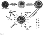
- the NaYF 4 buffer layer prevents energy transfer between the core and the outer layer.
- Figures 2b and 2c are TEM images of the nanoparticles, showing the uniform size and morphology of the nanoparticles with the average diameter of ca. 17 nm.
- HRTEM high-resolution TEM image
- HTEM high-resolution TEM image
- the X-ray powder diffraction (XRD) pattern in figure 7 indicates that both the core nanoparticles and the multilayer nanoparticles exhibit the hexagonal phase.
- High-angle annular dark field scanning transmission electron microscopy (HAADF-STEM) images are shown in figure 2e , the heavier lanthanide elements (Er, Gd, and Tb) which appear brighter are distributed in the core and outer layer of the nanoparticles, whereas the lighter metal (Y) is distributed in the middle layer of the nanoparticles.
- Figure 3a shows the upconversion luminescence spectra of the nanoparticles.
- the nanoparticles exhibit the characteristic emission of Tb 3+ ( figure 3b ) which overlaps reasonably well with the rose bengal (RB) absorption ( figure 8 ), indicating that excited energy of Tb 3+ could be effectively transferred to the surface bounded RB through nonradiative resonant energy transfer and induces PDT ( i.e ., X-PDT).
- the NaYF 4 buffer layer plays a key role not only in minimizing the interaction between Er 3+ ions in the core and the surface related quenching centers, e.g. defects, ligands, solvent molecules etc ., but also in blocking the energy transfer pathway between the core and the X-ray active outer layer of the nanoparticle, thus enhancing the local density of excitation energy ( cf. figure 3c ).
- the outer layer also plays as a layer that prevents interference by minimizing surface quenching.
- the relationship between the UCL intensity of Er 3+ and the distance between the core and surface of the nanoparticle, i.e. the buffer layer plus the outer layer was measured using NIR excitation (see figures 3d and 9 ).
- the UCL intensity increases with the thickness of the middle layer until core/buffer layer/outer layer ratio reaches 1:3:3, corresponding to a 2.6 nm thick buffer layer and a 1.5 nm thick outer layer.
- the as-synthesized oleic acid-capped nanoparticles were coated with poly(allylamine) (PAAm) to form NPs-NH2 via ligand exchange.
- PAAm poly(allylamine)
- NPs-NH 2 were washed with water and DMF. Finally, the NPs-NH 2 were obtained and dispersed in DMF.
- RB-HA rose bengal hexanoic acid
- 0.5 mg FA, 3 mg NHS and 3 mg EDC-HCl were mixed in 5 mL DMF at 25 °C for 2 h.
- 10 mg of NPs-NH2 was added and the reaction was run for 24 h.
- the obtained nanoparticles were then washed with DMF to remove the unreacted reagents. Finally, 10 mg PEG-NHS was reacted with the particles in 15 mL DMSO and 5 mL ethanol for 24h.
- upconversion luminescence-imaging-guided synergistic RT&X-PDT nanoplatforms were dispersed in phosphate buffered saline (PBS).
- the nanoparticle diameter as obtained from dynamic light scattering and the zeta potential increased from ca. 26 nm to ca. 52 nm and from +5.5 mV to +30 mV, respectively, after amino group modification.
- RB and cancer targeting moiety-folic acid (FA) were covalently conjugated with NPs-NH 2 to form NPs-RB/FA, followed by surface modification with PEG-NHS to form UIRPPs and to improve further the biocompatibility and specificity.
- the diameter of UIRPPs increased to 77 nm and the zeta potential decreased to +17 mV.
- Figure 10 is the ten days track of UCL spectrum of UIRPPs dispersed in water.
- the neglected spectral overlap between the UCL of NPs and the RB absorption indicates that UCL under NIR excitation can be used for imaging without inducing phototoxicity.
- the multilayer UIRPP structure enables effective nonradiative energy transfer from donor atoms inside the outer layer of the nanoparticles to the surface-anchored photosensitizers.
- the X-ray active Tb 3+ ions were doped solely in the outer layer and the thickness of the outer layer was controlled to be less than 2 nm, ensuring that the Tb 3+ is distributed like ring rather than a like a ball.
- This structure facilitates energy transfer by optimizing the effective amount of energy donors and by ensuring a short distance to the anchored surface-anchored photosensitizers.
- the loading efficiency was estimated high to be 12% (w/w) due to the covalent bonding.
- one RB molecule has four iodine atoms, which could also contribute in absorbing X-rays for computed tomography (CT) imaging and enhancing the effects of radiotherapy.
- CT computed tomography
- the efficacies of PDT activated with NIR laser or X-ray were estimated in solution by evaluating 1 O 2 generation efficiency using singlet oxygen detector diphenylisobenzofuran (DPBF).
- DPBF singlet oxygen detector diphenylisobenzofuran
- 2 mL of UIRPPs-water solution and 10 ⁇ L (1 mg/mL) of DPBF-ethanol solution were mixed and kept in the dark overnight.
- the absorption intensity of DPBF at 410 nm was recorded every 5 min (5, 10, 15, 20, 25 and 30 min) after NIR laser irradiation (808 nm, 980 nm or 1530 nm) of 0.7 W/cm 2 .
- UCL imaging and synergistic effect of RT&X-PDT of UIRPPs were tested in vitro.
- Breast carcinoma MCF-7 cells were maintained in the Dulbecco's modified Eagle medium (DMEM) under 5% CO 2 atmosphere at 37 °C.
- DMEM Dulbecco's modified Eagle medium
- the cells were incubated in a special confocal well plate (1 ⁇ 10 4 per well) for 24 h.
- the UIRPPs were added in the plate with 0 ⁇ g/mL, 50 ⁇ g/mL, 100 ⁇ g/mL, or 200 ⁇ g/mL, and incubation for 8 h.
- the nucleus was stained with 4',6-diamidino-2-phenylindole (DAPI), and the UCL imaging was measured by confocal microscopy with an irradiation wavelength of 980 nm and an emission wavelength of 650 nm or 540 nm.
- the UCL-RB was measured with 980 nm laser irradiation and 590 nm emission, the RB was measured with 540 nm irradiation and 590 nm emission.
- the MCF-7 cells were incubated in the 96-well plate (1 ⁇ 10 4 per well) for 24 h. Then UIRPPs of different concentration groups (0, 0, 0, 0, 50, 100, 200, 400 ⁇ g/mL) were added in the plate, each group was with five parallels. The group with 0 ⁇ g/mL of UIRPPs were irradiated with 808 nm, 980 nm or 1530 nm laser for 15 min. Further incubated for 24 h, the cell viability was assessed using a standard MTS assay.
- Figure 13 is the viability of a breast cancer line (MCF-7) subject to treatments.
- MCF-7 breast cancer line
- the cells emitted bright red UCL signals around the nuclei upon the laser irradiation at 980 or 808 nm ( figure 14 ). It is worth noting that there was no green UCL, guaranteeing the desired monochromic red UCL for imaging.
- the nanoparticles were modified with folic acid (FA) to enhance the cellular uptake efficiency via FA-receptor-mediated endocytosis.
- the positive targeting was successful as is shown in figure 5a where the UCL signal increased sharply with the concentration of UIRPPs.
- the UCL imaging maintained high resolution with the concentration of UIRPPs varying from 50 to 200 ⁇ g/mL.
- a comparison was made with the fluorescence imaging using RB emission under 540 nm laser irradiation ( cf. figure 5a ).
- RB aided imaging increased also sharply with RB concentration, image became more and more blurry mainly due to scattering of the irradiation light.
- Figure 15 is the confocal images of Z-scan to exhibit the endocytosis and the fine resolution of UCL imaging. Photoactivity of RB subject to NIR laser irradiation was assessed ( cf. figure 5b ), neither UCL green nor RB signal was observed, supporting that bio-functionalized UIRPPs are suitable for the specific monochromic red UCL imaging without severe photodamage.
- the cells were separated in 8 groups (G1 to G8) in the 96-well plate, G1 and G5 were incubated with saline, G2, G3, G4 and G8 were incubated with UIRPPs of different concentrations (0, 50, 100, 200, 400 ⁇ g/mL), G6 were incubated with 12% RB (0, 6, 12, 24, 48 ⁇ g/mL) and G7 were incubated with Er@Y@Gd/Tb NPs. After 8h incubation all groups were washed with PBS.
- the saline (G1 and G5) were taken as the control groups, the G2, G3 and G4 were irradiated with the NIR laser (800 nm, 980 nm or 1530 nm) for 15 min at 0.7 W/cm 2 .
- the G6, G7 and G8 were irradiated with X-rays for 45 min at 80 kV and 0.5 mA. After that the cells were cultured for another 48 h, the PDT efficacy of all groups was assessed by MTS assay.
- Flow cytometry analysis was used to characterize the apoptosis of MCF-7 cells experiencing synergistic RT&X-PDT.
- the cells were trypsinized and stained by Annexin V-FITC and propidium iodide (Annexin V-FITC Apoptosis Staining/Detection Kit) to measure the cell apoptosis.
- Annexin-V-fluorescein isothiocyanate (FITC) and propidium iodide (PI) staining were performed using an Annexin-V-FITC/PI kit (BD) according to the manufacturer's instructions. Briefly, MCF-7 cells were cultured in 6-well plates and then treated with different reagents (G1: saline, G2: saline, G3: 100 ⁇ g/mL UIRPPs, G4: 100 ⁇ g/mL G5: 100 ⁇ g/mL Er@Y@Gd/Tb NPs, G6: 50 ⁇ g/mL, UIRPPs, G7: 100 ⁇ g/mL UIRPPs, G8: 100 ⁇ g/mL UIRPPs) for 8 h, each with 3 parallels.
- G1 saline
- G2 saline
- G3 100 ⁇ g/mL UIRPPs
- G4 100 ⁇ g/mL
- G5 100
- the cells treated with saline (G1) were for the control.
- the cells were exposed to the 980 nm laser for 15 min at 0.7 W/cm 2 .
- the cells were irradiated with X-ray for 45 min at 80 kV and 0.5 mA. Then the cells were harvested using 0.05% trypsin after 24 h incubation and washed twice with cold PBS, and suspended in binding buffer. 1 ⁇ 10 5 cells in 100 pL binding buffer were added to a tube and incubated with 5 pL of Annexin-V-FITC and 5 pL of propidium iodide (PI). Cells were gently mixed and incubated for 15 min at room temperature.
- PI propidium iodide
- the nanoparticles were not coupled with photosensitizers, and the X-ray radiation effects caused about 30% of cell deaths.
- the “UIRPPs” group there were only 38.3%, 26.5% or 15.6% alive cells after irradiation with X-ray corresponding to different concentrations of UIRPPs (50 ⁇ g/mL, 100 ⁇ g/mL or 200 ⁇ g/mL). The death ratio of cells was as high as 84.4%, fully showing the synergistic RT&X-PDT.
Landscapes
- Chemical & Material Sciences (AREA)
- Engineering & Computer Science (AREA)
- Nanotechnology (AREA)
- Organic Chemistry (AREA)
- Crystallography & Structural Chemistry (AREA)
- Life Sciences & Earth Sciences (AREA)
- General Physics & Mathematics (AREA)
- Condensed Matter Physics & Semiconductors (AREA)
- Physics & Mathematics (AREA)
- Geology (AREA)
- Inorganic Chemistry (AREA)
- Bioinformatics & Cheminformatics (AREA)
- Biophysics (AREA)
- Medicinal Chemistry (AREA)
- Molecular Biology (AREA)
- Pharmacology & Pharmacy (AREA)
- General Health & Medical Sciences (AREA)
- General Engineering & Computer Science (AREA)
- Biotechnology (AREA)
- Composite Materials (AREA)
- Medical Informatics (AREA)
- Health & Medical Sciences (AREA)
- Materials Engineering (AREA)
- Manufacturing & Machinery (AREA)
- Medicines That Contain Protein Lipid Enzymes And Other Medicines (AREA)
- Medicinal Preparation (AREA)
- Radiation-Therapy Devices (AREA)
- Medicines Containing Antibodies Or Antigens For Use As Internal Diagnostic Agents (AREA)
Priority Applications (6)
| Application Number | Priority Date | Filing Date | Title |
|---|---|---|---|
| EP20184045.1A EP3932866A1 (fr) | 2020-07-03 | 2020-07-03 | Nanoparticule dotée d'une couche tampon |
| US18/003,507 US20230242411A1 (en) | 2020-07-03 | 2021-07-05 | Nanoparticle with a Buffer Layer |
| PCT/NL2021/050425 WO2022005293A1 (fr) | 2020-07-03 | 2021-07-05 | Nanoparticule avec une couche tampon |
| EP21737820.7A EP4175912A1 (fr) | 2020-07-03 | 2021-07-05 | Nanoparticule avec une couche tampon |
| CN202180054111.9A CN116075477A (zh) | 2020-07-03 | 2021-07-05 | 具有缓冲层的纳米粒子 |
| JP2023524266A JP2023531833A (ja) | 2020-07-03 | 2021-07-05 | バッファ層を有するナノ粒子 |
Applications Claiming Priority (1)
| Application Number | Priority Date | Filing Date | Title |
|---|---|---|---|
| EP20184045.1A EP3932866A1 (fr) | 2020-07-03 | 2020-07-03 | Nanoparticule dotée d'une couche tampon |
Publications (1)
| Publication Number | Publication Date |
|---|---|
| EP3932866A1 true EP3932866A1 (fr) | 2022-01-05 |
Family
ID=71514975
Family Applications (2)
| Application Number | Title | Priority Date | Filing Date |
|---|---|---|---|
| EP20184045.1A Withdrawn EP3932866A1 (fr) | 2020-07-03 | 2020-07-03 | Nanoparticule dotée d'une couche tampon |
| EP21737820.7A Withdrawn EP4175912A1 (fr) | 2020-07-03 | 2021-07-05 | Nanoparticule avec une couche tampon |
Family Applications After (1)
| Application Number | Title | Priority Date | Filing Date |
|---|---|---|---|
| EP21737820.7A Withdrawn EP4175912A1 (fr) | 2020-07-03 | 2021-07-05 | Nanoparticule avec une couche tampon |
Country Status (5)
| Country | Link |
|---|---|
| US (1) | US20230242411A1 (fr) |
| EP (2) | EP3932866A1 (fr) |
| JP (1) | JP2023531833A (fr) |
| CN (1) | CN116075477A (fr) |
| WO (1) | WO2022005293A1 (fr) |
Citations (7)
| Publication number | Priority date | Publication date | Assignee | Title |
|---|---|---|---|---|
| EP1100366A1 (fr) | 1998-07-09 | 2001-05-23 | Curelight Ltd. | Appareil et procede de therapie photodynamique haute energie efficace de l'acne simple et de la seborrhee |
| US20130158336A1 (en) | 2006-09-18 | 2013-06-20 | Board Of Reagents, The University Of Texas System | Use of nanoparticles in the photodynamic treatment of tumors and non-destructive testing |
| WO2013181076A1 (fr) * | 2012-05-30 | 2013-12-05 | University Of Massachusetts Medical School | Nanoparticules enrobées à conversion ascendante |
| US20160271251A1 (en) | 2013-10-23 | 2016-09-22 | Synchrotron Soleil | Radioluminescent compound for radiotherapy and deep photodynamic therapy and device for deep photodynamic therapy |
| CN109481680A (zh) * | 2019-01-09 | 2019-03-19 | 中国科学院长春光学精密机械与物理研究所 | 一种内外兼修的复合纳米光敏剂及其制备方法和应用 |
| US20190210886A1 (en) * | 2018-01-11 | 2019-07-11 | National Yang-Ming University | Lanthanide-doped fluoride nanocomposites, production method and applications |
| CN110478483A (zh) * | 2019-08-22 | 2019-11-22 | 青岛大学 | 一种多色上转换纳米探针及制备方法与应用 |
Family Cites Families (1)
| Publication number | Priority date | Publication date | Assignee | Title |
|---|---|---|---|---|
| JP5830243B2 (ja) * | 2007-09-28 | 2015-12-09 | ナノコ テクノロジーズ リミテッド | ナノ粒子 |
-
2020
- 2020-07-03 EP EP20184045.1A patent/EP3932866A1/fr not_active Withdrawn
-
2021
- 2021-07-05 EP EP21737820.7A patent/EP4175912A1/fr not_active Withdrawn
- 2021-07-05 US US18/003,507 patent/US20230242411A1/en not_active Abandoned
- 2021-07-05 WO PCT/NL2021/050425 patent/WO2022005293A1/fr not_active Ceased
- 2021-07-05 JP JP2023524266A patent/JP2023531833A/ja active Pending
- 2021-07-05 CN CN202180054111.9A patent/CN116075477A/zh active Pending
Patent Citations (7)
| Publication number | Priority date | Publication date | Assignee | Title |
|---|---|---|---|---|
| EP1100366A1 (fr) | 1998-07-09 | 2001-05-23 | Curelight Ltd. | Appareil et procede de therapie photodynamique haute energie efficace de l'acne simple et de la seborrhee |
| US20130158336A1 (en) | 2006-09-18 | 2013-06-20 | Board Of Reagents, The University Of Texas System | Use of nanoparticles in the photodynamic treatment of tumors and non-destructive testing |
| WO2013181076A1 (fr) * | 2012-05-30 | 2013-12-05 | University Of Massachusetts Medical School | Nanoparticules enrobées à conversion ascendante |
| US20160271251A1 (en) | 2013-10-23 | 2016-09-22 | Synchrotron Soleil | Radioluminescent compound for radiotherapy and deep photodynamic therapy and device for deep photodynamic therapy |
| US20190210886A1 (en) * | 2018-01-11 | 2019-07-11 | National Yang-Ming University | Lanthanide-doped fluoride nanocomposites, production method and applications |
| CN109481680A (zh) * | 2019-01-09 | 2019-03-19 | 中国科学院长春光学精密机械与物理研究所 | 一种内外兼修的复合纳米光敏剂及其制备方法和应用 |
| CN110478483A (zh) * | 2019-08-22 | 2019-11-22 | 青岛大学 | 一种多色上转换纳米探针及制备方法与应用 |
Non-Patent Citations (6)
| Title |
|---|
| ANGEW. CHEM. INT. ED., vol. 54, 2015, pages 1770 - 1774 |
| BING XU ET AL: "Facile-Controlled Epitaxial Growth Direction of Heterogeneous Core/Shell Structured NaLnF 4 Nanocrystals through Traditional Methods", JOURNAL OF NANOMATERIALS, vol. 2020, 28 August 2020 (2020-08-28), US, pages 1 - 7, XP055749726, ISSN: 1687-4110, DOI: 10.1155/2020/6196520 * |
| J. AM. CHEM. SOC., vol. 135, 2013, pages 6494 - 6503 |
| MINGYE DING ET AL: "Highly enhanced upconversion luminescence in lanthanide-doped active-core/luminescent-shell/active-shell nanoarchitectures", JOURNAL OF MATERIALS CHEMISTRY C, vol. 4, no. 13, 1 January 2016 (2016-01-01), GB, pages 2432 - 2437, XP055750207, ISSN: 2050-7526, DOI: 10.1039/C6TC00163G * |
| STEFAN FISCHER ET AL: "Small Alkaline-Earth-based Core/Shell Nanoparticles for Efficient Upconversion", NANO LETTERS, vol. 19, no. 6, 6 May 2019 (2019-05-06), US, pages 3878 - 3885, XP055749587, ISSN: 1530-6984, DOI: 10.1021/acs.nanolett.9b01057 * |
| XIAOHUI ZHU ET AL: "Recent Progress of Rare-Earth Doped Upconversion Nanoparticles: Synthesis, Optimization, and Applications", ADVANCED SCIENCE, vol. 6, no. 22, 30 September 2019 (2019-09-30), pages 1901358, XP055749584, ISSN: 2198-3844, DOI: 10.1002/advs.201901358 * |
Also Published As
| Publication number | Publication date |
|---|---|
| US20230242411A1 (en) | 2023-08-03 |
| EP4175912A1 (fr) | 2023-05-10 |
| JP2023531833A (ja) | 2023-07-25 |
| WO2022005293A1 (fr) | 2022-01-06 |
| CN116075477A (zh) | 2023-05-05 |
Similar Documents
| Publication | Publication Date | Title |
|---|---|---|
| Yang et al. | Mesoporous silica nanorods intrinsically doped with photosensitizers as a multifunctional drug carrier for combination therapy of cancer | |
| Larue et al. | Using X-rays in photodynamic therapy: an overview | |
| Liu et al. | “Three birds with one stone” nanoplatform: Efficient near‐infrared‐triggered type‑I AIE photosensitizer for mitochondria‐targeted photodynamic therapy against hypoxic tumors | |
| Kamkaew et al. | Scintillating nanoparticles as energy mediators for enhanced photodynamic therapy | |
| Guan et al. | Photodynamic therapy based on nanoscale metal–organic frameworks: from material design to cancer nanotherapeutics | |
| Zhao et al. | Multifunctional core–shell upconverting nanoparticles for imaging and photodynamic therapy of liver cancer cells | |
| US10646570B2 (en) | Induced photodynamic therapy using nanoparticle scintillators as transducers | |
| US8771741B2 (en) | In vivo photodynamic therapy of cancer via a near infrared agent encapsulated in calcium phosphate nanoparticles | |
| Zhang et al. | Low-dose x-ray excited photodynamic therapy based on naluf4: Tb 3+–rose bengal nanocomposite | |
| Zhu et al. | An efficient tumor-inducible nanotheranostics for magnetic resonance imaging and enhanced photodynamic therapy | |
| Kalluru et al. | Unprecedented “All‐in‐One” Lanthanide‐Doped Mesoporous Silica Frameworks for Fluorescence/MR Imaging and Combination of NIR Light Triggered Chemo‐Photodynamic Therapy of Tumors | |
| Zhang et al. | Synthesis of porphyrin-conjugated silica-coated Au nanorods for synergistic photothermal therapy and photodynamic therapy of tumor | |
| CN111194232A (zh) | 用于x射线诱导的光动力学疗法、放射疗法、放射动力学疗法、化学疗法、免疫疗法以及它们的任何组合的纳米级金属有机层和金属有机纳米片 | |
| Gonzalez-Bejar et al. | NIR excitation of upconversion nanohybrids containing a surface grafted Bodipy induces oxygen-mediated cancer cell death | |
| Chuang et al. | Annealing-modulated nanoscintillators for nonconventional X-ray activation of comprehensive photodynamic effects in deep cancer theranostics | |
| JP2017532340A (ja) | 光線力学療法、x線誘起光線力学療法、放射線療法、化学療法、免疫療法、及びこれらの任意の組み合わせのためのナノ粒子 | |
| Buchner et al. | Photosensitiser functionalised luminescent upconverting nanoparticles for efficient photodynamic therapy of breast cancer cells | |
| Sun et al. | Ce6-C6-TPZ co-loaded albumin nanoparticles for synergistic combined PDT-chemotherapy of cancer | |
| Macchi et al. | Enhanced photothermal heating and combination therapy of NIR dye via conversion to self-assembled ionic nanomaterials | |
| Bekah et al. | Synthesis and characterization of biologically stable, doped LaF3 nanoparticles co-conjugated to PEG and photosensitizers | |
| Gao et al. | A small-sized and stable 2D metal–organic framework: a functional nanoplatform for effective photodynamic therapy | |
| Akhtar et al. | PDT-active upconversion nanoheaters for targeted imaging guided combinatorial cancer phototherapies with low-power single NIR excitation | |
| Zhang et al. | Rare-earth scintillating nanoparticles for X-ray induced photodynamic therapy | |
| Feng et al. | A scintillating nanoplatform with upconversion function for the synergy of radiation and photodynamic therapies for deep tumors | |
| Alizadeh Nobari et al. | Emerging trends in quantum dot-based photosensitizers for enhanced photodynamic therapy in cancer treatment |
Legal Events
| Date | Code | Title | Description |
|---|---|---|---|
| PUAI | Public reference made under article 153(3) epc to a published international application that has entered the european phase |
Free format text: ORIGINAL CODE: 0009012 |
|
| STAA | Information on the status of an ep patent application or granted ep patent |
Free format text: STATUS: THE APPLICATION HAS BEEN PUBLISHED |
|
| AK | Designated contracting states |
Kind code of ref document: A1 Designated state(s): AL AT BE BG CH CY CZ DE DK EE ES FI FR GB GR HR HU IE IS IT LI LT LU LV MC MK MT NL NO PL PT RO RS SE SI SK SM TR |
|
| B565 | Issuance of search results under rule 164(2) epc |
Effective date: 20201123 |
|
| STAA | Information on the status of an ep patent application or granted ep patent |
Free format text: STATUS: THE APPLICATION IS DEEMED TO BE WITHDRAWN |
|
| 18D | Application deemed to be withdrawn |
Effective date: 20220706 |