EP3945132A1 - Mikrofluidisches system, verfahren zur hemmung, verzögerung oder umkehrung der zellulären seneszenz unter verwendung eines mikrofluidischen systems und so erhaltene zelle - Google Patents
Mikrofluidisches system, verfahren zur hemmung, verzögerung oder umkehrung der zellulären seneszenz unter verwendung eines mikrofluidischen systems und so erhaltene zelle Download PDFInfo
- Publication number
- EP3945132A1 EP3945132A1 EP21188286.5A EP21188286A EP3945132A1 EP 3945132 A1 EP3945132 A1 EP 3945132A1 EP 21188286 A EP21188286 A EP 21188286A EP 3945132 A1 EP3945132 A1 EP 3945132A1
- Authority
- EP
- European Patent Office
- Prior art keywords
- cell
- cells
- flow
- present
- senescence
- Prior art date
- Legal status (The legal status is an assumption and is not a legal conclusion. Google has not performed a legal analysis and makes no representation as to the accuracy of the status listed.)
- Pending
Links
Images










Classifications
-
- B—PERFORMING OPERATIONS; TRANSPORTING
- B01—PHYSICAL OR CHEMICAL PROCESSES OR APPARATUS IN GENERAL
- B01L—CHEMICAL OR PHYSICAL LABORATORY APPARATUS FOR GENERAL USE
- B01L3/00—Containers or dishes for laboratory use, e.g. laboratory glassware; Droppers
- B01L3/50—Containers for the purpose of retaining a material to be analysed, e.g. test tubes
- B01L3/502—Containers for the purpose of retaining a material to be analysed, e.g. test tubes with fluid transport, e.g. in multi-compartment structures
- B01L3/5027—Containers for the purpose of retaining a material to be analysed, e.g. test tubes with fluid transport, e.g. in multi-compartment structures by integrated microfluidic structures, i.e. dimensions of channels and chambers are such that surface tension forces are important, e.g. lab-on-a-chip
- B01L3/502746—Containers for the purpose of retaining a material to be analysed, e.g. test tubes with fluid transport, e.g. in multi-compartment structures by integrated microfluidic structures, i.e. dimensions of channels and chambers are such that surface tension forces are important, e.g. lab-on-a-chip characterised by the means for controlling flow resistance, e.g. flow controllers, baffles or throttle valves
-
- C—CHEMISTRY; METALLURGY
- C12—BIOCHEMISTRY; BEER; SPIRITS; WINE; VINEGAR; MICROBIOLOGY; ENZYMOLOGY; MUTATION OR GENETIC ENGINEERING
- C12N—MICROORGANISMS OR ENZYMES; COMPOSITIONS THEREOF; PROPAGATING, PRESERVING, OR MAINTAINING MICROORGANISMS; MUTATION OR GENETIC ENGINEERING; CULTURE MEDIA
- C12N5/00—Undifferentiated human, animal or plant cells, e.g. cell lines; Tissues; Cultivation or maintenance thereof; Culture media therefor
- C12N5/06—Animal cells or tissues; Human cells or tissues
- C12N5/0602—Vertebrate cells
- C12N5/0652—Cells of skeletal and connective tissues; Mesenchyme
- C12N5/0662—Stem cells
- C12N5/0665—Blood-borne mesenchymal stem cells, e.g. from umbilical cord blood
-
- C—CHEMISTRY; METALLURGY
- C12—BIOCHEMISTRY; BEER; SPIRITS; WINE; VINEGAR; MICROBIOLOGY; ENZYMOLOGY; MUTATION OR GENETIC ENGINEERING
- C12M—APPARATUS FOR ENZYMOLOGY OR MICROBIOLOGY; APPARATUS FOR CULTURING MICROORGANISMS FOR PRODUCING BIOMASS, FOR GROWING CELLS OR FOR OBTAINING FERMENTATION OR METABOLIC PRODUCTS, i.e. BIOREACTORS OR FERMENTERS
- C12M23/00—Constructional details, e.g. recesses, hinges
- C12M23/02—Form or structure of the vessel
- C12M23/16—Microfluidic devices; Capillary tubes
-
- C—CHEMISTRY; METALLURGY
- C12—BIOCHEMISTRY; BEER; SPIRITS; WINE; VINEGAR; MICROBIOLOGY; ENZYMOLOGY; MUTATION OR GENETIC ENGINEERING
- C12M—APPARATUS FOR ENZYMOLOGY OR MICROBIOLOGY; APPARATUS FOR CULTURING MICROORGANISMS FOR PRODUCING BIOMASS, FOR GROWING CELLS OR FOR OBTAINING FERMENTATION OR METABOLIC PRODUCTS, i.e. BIOREACTORS OR FERMENTERS
- C12M35/00—Means for application of stress for stimulating the growth of microorganisms or the generation of fermentation or metabolic products; Means for electroporation or cell fusion
- C12M35/04—Mechanical means, e.g. sonic waves, stretching forces, pressure or shear stimuli
-
- C—CHEMISTRY; METALLURGY
- C12—BIOCHEMISTRY; BEER; SPIRITS; WINE; VINEGAR; MICROBIOLOGY; ENZYMOLOGY; MUTATION OR GENETIC ENGINEERING
- C12N—MICROORGANISMS OR ENZYMES; COMPOSITIONS THEREOF; PROPAGATING, PRESERVING, OR MAINTAINING MICROORGANISMS; MUTATION OR GENETIC ENGINEERING; CULTURE MEDIA
- C12N5/00—Undifferentiated human, animal or plant cells, e.g. cell lines; Tissues; Cultivation or maintenance thereof; Culture media therefor
- C12N5/06—Animal cells or tissues; Human cells or tissues
- C12N5/0602—Vertebrate cells
- C12N5/0652—Cells of skeletal and connective tissues; Mesenchyme
- C12N5/0662—Stem cells
- C12N5/0668—Mesenchymal stem cells from other natural sources
-
- B—PERFORMING OPERATIONS; TRANSPORTING
- B01—PHYSICAL OR CHEMICAL PROCESSES OR APPARATUS IN GENERAL
- B01L—CHEMICAL OR PHYSICAL LABORATORY APPARATUS FOR GENERAL USE
- B01L2400/00—Moving or stopping fluids
- B01L2400/04—Moving fluids with specific forces or mechanical means
- B01L2400/0475—Moving fluids with specific forces or mechanical means specific mechanical means and fluid pressure
- B01L2400/0478—Moving fluids with specific forces or mechanical means specific mechanical means and fluid pressure pistons
-
- C—CHEMISTRY; METALLURGY
- C12—BIOCHEMISTRY; BEER; SPIRITS; WINE; VINEGAR; MICROBIOLOGY; ENZYMOLOGY; MUTATION OR GENETIC ENGINEERING
- C12N—MICROORGANISMS OR ENZYMES; COMPOSITIONS THEREOF; PROPAGATING, PRESERVING, OR MAINTAINING MICROORGANISMS; MUTATION OR GENETIC ENGINEERING; CULTURE MEDIA
- C12N2521/00—Culture process characterised by the use of hydrostatic pressure, flow or shear forces
-
- C—CHEMISTRY; METALLURGY
- C12—BIOCHEMISTRY; BEER; SPIRITS; WINE; VINEGAR; MICROBIOLOGY; ENZYMOLOGY; MUTATION OR GENETIC ENGINEERING
- C12N—MICROORGANISMS OR ENZYMES; COMPOSITIONS THEREOF; PROPAGATING, PRESERVING, OR MAINTAINING MICROORGANISMS; MUTATION OR GENETIC ENGINEERING; CULTURE MEDIA
- C12N2527/00—Culture process characterised by the use of mechanical forces, e.g. strain, vibration
Definitions
- the present invention relates to a method for inhibiting, delaying, or reversing cellular senescence by applying an impact to cells in flow using a microfluidic system, the microfluidic system, and a cell obtained from the method or the microfluidic system, in which senescence is inhibited, delayed, or reversed.
- Cytoskeleton induces various intracellular changes through polymerization/depolymerization of actin in response to extracellular stimuli. These changes regulate cell migration and invasion, and affect the activity of ion channels present in the cell, ultimately leading to cell survival, proliferation, death, fluidity, and secretion through various intracellular signaling pathways.
- Cellular senescence refers to an irreversible life phenomenon in which the cell cycle of cells exposed to various physical, chemical, and biological stresses is slowed and cell division is reduced or permanently stopped. With the progress of senescence, cell division not only decreases or stops, but also accumulates cell damage due to unstable transmission of genetic information. The causes of aging related to this genetic information include genome instability, loss of telomeres, and epigenetic changes.
- senescence at the cellular level is a complex process that cannot be efficiently controlled by single-cause regulation.
- a method for effectively inhibiting or delaying cellular senescence or reversibly returning senescent cells to normal cells to have a phenotype similar to that of young cells has not yet been developed.
- mesenchymal stem cells first identified in bone marrow are totipotent cells with great potential in regenerative medicine.
- the mesenchymal stem cells may differentiate into several types of mesenchymal lineages, such as osteocytes, chondrocytes, adipocytes, myocytes, and fibroblasts. Since mesenchymal stem cells have immunomodulatory activity, it can be used as a composition for treating various autoimmune and inflammatory diseases, such as a transplant promoter, a fetal graft, and a host disease.
- the demand in regenerative medicine is continuously increasing due to the excellent therapeutic utility of mesenchymal stem cells.
- Patent reference 0001 Korean application No. 2015-0167693
- the present invention provides a method for inhibiting, delaying, or reversing cellular senescence and a cell in which senescence is inhibited, delayed, or reversed using the same.
- the present invention also provides a method for maintaining or enhancing the multipotency of stem cells.
- the present invention also provides a device for inhibiting, delaying, or reversing senescence of an isolated cell.
- the present invention provides a method for inhibiting, delaying, or reversing cellular senescence including:
- the inventors of the present invention made intensive research efforts to develop a method for delaying senescence of various therapeutic and research cells, including stem cells, and maintaining high levels of biological activities such as survival rate, proliferation rate, and differentiation ability for a long period of time.
- the inventors found that in an in vitro environment including a flow channel and an impact surface, that is, in a microfluidic system, by allowing a target cell to flow with a certain parameter and crashing the cell into the impact surface to apply a physical impact to the cell, reorganization of the cytoskeleton may be induced, thereby significantly delaying, reversing, or inhibiting the cellular senescence.
- the inventors have compared various aging factors, such as the cell size, the level of intracellular reactive oxygen species, and the expression levels of senescence-related proteins or genes of the cells that have passed through the microfluidic system as described above, with those of the control cells to which the microfluidic system as described above is not applied, to confirm the senescence inhibitory effect of the cells according to the present invention, and completed the present invention.
- various aging factors such as the cell size, the level of intracellular reactive oxygen species, and the expression levels of senescence-related proteins or genes of the cells that have passed through the microfluidic system as described above, with those of the control cells to which the microfluidic system as described above is not applied, to confirm the senescence inhibitory effect of the cells according to the present invention, and completed the present invention.
- flow comprises a fulid's flowing including a cell.
- the term "impact surface” refers to a surface into which a cell in flow crashes by being installed on the flow path while forming a predetermined angle with the flow path of the cell.
- the angle between the axis of the flow path and the impact surface may be about less than 180 degress, for example, from about 10 degrees to about 170 degress, from about 20 degrees to about 170 degress, from about 20 degrees to about 160 degress, from about 30 degrees to about 160 degress, from about 30 degrees to about 150 degrees, from about 40 degrees to about 150 degrees, from about 40 degrees to about 140 degress, from about 50 degrees to about 140 degress, from about 50 degrees to about 135 degrees, from about 50 degrees to about 130 degrees, from about 60 degrees to about 130 degress, from about 60 degrees to about 130 degress, from about 60 degrees to about 120 degrees, from about 70 degrees to about 120 degress, from about 70 degrees to about 110 degrees, from about 80 degrees to about 110 degress, from about 80 degrees to about 100 degrees, from about 85 degrees to about 100 degress, from about 85 degrees to about
- the flow path may be, for example, a T-shape in which cells flow in a straight line and then split at 90 degrees left and right from the flow direction at a certain point.
- the impact surface becomes the surface of the junction at perpendicular angles to the axis of the flow path.
- the impact surface may have a flat shape or may include a concave-convex structure including depression portions, protruding portions, or a combination thereof for efficient cell impact and cytoskeletal reorganization.
- the flowing may be performed by injecting a fluid containing cells into a microchannel in the microfluidic system according to an embodiment of the present invention to flow it.
- microchannel refers to a migration passage of a fluid having a hydraulic diameter of less than about 1 mm or a flow volume per minute of the fluid at the micro level ( ⁇ l).
- the flow volume of the fluid may be from about several tens to several thousands ⁇ l per minute, for example, from about several tens to several hundreds ⁇ l per minute, or from about several hundreds to several thousands ⁇ l per minute, and is not limited thereto.
- the microfluidic system includes an inlet port for injecting cells or fluid, an outlet port for discharging cells, a microchannel connecting the inlet port and the outlet port to flow a fluid, and a control unit for controlling a flow rate of the microchannel.
- the flowing may be performed according to predetermined parameters such that the reorganization of the cytoskeleton occurs without the physical impact causing cell death.
- the term "reorganization of the cytoskeleton” refers to a series of continuous processes in which tubulin proteins constituting microtubules that are cytoskeletal fibers are depolymerized by physical impact and then polymerized again, resulting in degeneration and recovery of the cytoskeleton.
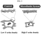
- the present inventors found that during this cytoskeletal reorganization process, the density of actin, which is a constituent monomer of microfilament, another component of the cytoskeleton, increases, and the structure becomes dense, and resultantly it is confirmed that the characteristic phenotype of cells without senescence is reproduced.
- senescence-related phenotypes such as, for example, the size of the cell, the level of reactive oxygen species, the expression levels of senescence-related proteins or genes, and actin density, have been changed to substantially inhibit, delay, or induce reverse aging.
- the "predetermined parameters" refers to a series of hydrodynamic variables that can affect the amount of energy applied when the cell collides with the impact surface, such as, for example, a flow rate, density, or viscosity of the fluid containing the cells, and a diameter of a microchannel.
- the parameter may be determined by one or more factors selected from a density, a flow rate, a characteristic length, and a viscosity coefficient of a fluid containing the cells.
- characteristic length means a length that has the greatest influence on the flow, and may be, for example, the diameter of a microchannel.
- Equation 1 ⁇ is the density of the fluid, V is the velocity of the fluid, D is the characteristic length of the fluid, and ⁇ is the viscosity coefficient.
- Equation 1 is an equation for deriving a Reynolds number.
- Reynolds number is a dimensionless number that quantitatively expresses a ratio of "inertial force” and “viscous force” in a fluid and may define flow conditions affected by various variables comprehensively.
- the flowing may be performed under the condition that the parameter (Re) having a value of less than 500.
- the "Re" may be from about 1 to 500, from about 10 to 500, from about 10 to 450, from about 10 to 400, from about 10 to 350, from about 10 to 300, from about 10 to 290, from about 20 to 290, from about 30 to 290, from about 40 to 290, from about 50 to 290, from about 60 to 290, from about 70 to 290, from about 80 to 290, from about 90 to 290, from about 100 to 290, from about 100 to 280, from about 110 to 280, from about 120 to 280, from about 130 to 280, from about 130 to 270, from about 170 to 260, from about 180 to 260, from about 190 to 260, from about 200 to 260, from about 200 to 255, from about 205 to 255, from about 210 to 255, from about 210 to 250, from about 220 to 250, from about 225 to
- the distance from the point where a cell starts to flow in the microchannel to the impact surface may be from about 0.1 mm to about 50 mm.
- the distance may be from about 0.1 mm to about 50 mm, from about 1 mm to about 50 mm, from about 0.1 mm to about 45 mm, from about 0.1 mm to about 40 mm, from about 1 mm to about 35 mm, from about 1 mm to about 30 mm, from about 1 mm to about 25 mm, from about 1 mm to about 20 mm, from about 2 mm to about 50 mm, from about 2 mm to about 40 mm, from about 2 mm to about 30 mm, from about 2 mm to about 30 mm, from about 3 mm to about 50 mm, from about 3 mm to about 40 mm, from about 3 mm to about 30 mm, from about 3 mm to about 25 mm, from about 3 mm to about 20 mm, from about 3 mm to about 15 mm, from about 5
- the method of the present invention may be applied without limitation to any cell that has a cytoskeleton.
- the cell may be, for example, a fibroblast, a stem cell including a mesenchymal stem cell, an embryonic stem cell, an induced pluripotent stem cell, or a combination thereof; an immune cell including a T cell, an NK cell, a B cell, a dendritic cell, a macrophage, or a combination thereof; a precursor cell of the fibroblast, the stem cell, or the immune cell; or a combination thereof, and may be any cell for research and therapeutics.
- the cell may be a stem cell or a precursor cell.
- precursor cell refers to a unipotent cell that is only partially differentiated and shares a majority phenotype with stem cells.
- stem cell is an undifferentiated cell before differentiation into each cell constituting a tissue, and refers to cells having the ability to differentiate into a specific cell under a specific differentiation stimulus.
- the stem cells unlike differentiated cells in which cell division is stopped, can produce the same cells as themselves by cell division (self-renewal), and when a differentiation stimulus is applied, the stem cells have the plasticity of differentiation that they can be differentiated into various cells depending on the nature of the stimulus.
- the stem cells used in the present invention have characteristics of stem cells, that is, undifferentiated, indefinite proliferation, and differentiation ability into specific cells, and any cell capable of inducing differentiation into a tissue to be regenerated may be used without limitation.
- the stem cells are mesenchymal stem cells.
- mesenchymal stem cells refers to stem cells having multipotency capable of differentiation into adipocytes, osteocytes, chondrocytes, muscle cells, nerve cells, or cardiomyocytes.
- the mesenchymal stem cells may be identified through their vortex shape and expression levels of the basic cell surface markers CD73(+), CD105(+), CD34(-), CD45(-) and also has a function of regulating the immune response along with multipotency.
- the mesenchymal stem cells may be umbilical cord-derived mesenchymal stem cells (WJ-MSC).
- WJ-MSC umbilical cord-derived mesenchymal stem cells
- cellular senescence refers to the expression of a natural aging phenomenon in which the function of a living organ deteriorates over time is expressed at the cellular level. For example, it means the stop or significant delay of growth and division due to various physical, chemical, and biological stresses (e.g., high oxygen conditions during successive passages and in vitro culture) that cells receive from the inside or outside.
- undifferentiated cells such as stem cells
- cellular senescence is a concept including a process in which stem cells lose their undifferentiated phenotype and lose their multipotent or pluripotent properties.
- the term “inhibition, delay, or reverse of cellular senescence” refers to including a series of processes of artificially temporarily inhibiting cellular senescence, delaying the rate of senescence, or reversibly recovering the biological function of cells lost due to aging to have a phenotype similar to non-senescent young cells. Accordingly, the term “inhibition of cellular senescence” may also be expressed as “delaying cellular senescence” or "inducing reverse aging of cells”.
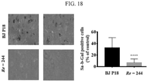
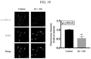
- the size of the cells decreases, the actin density of the cytoskeleton increases, the reactive oxygen species decreases, and the expression of senescence-related proteins or genes, including ⁇ -galactosidase and ⁇ -H2AX is significantly inhibited, to effectively inhibit, delay, or reverse the cellular senescence progression.
- the present invention provides a cell in which senescence is inhibited by the method described above.
- the cells whose senescence is inhibited by the method of the present invention have high activity and proliferative capacity compared with the number of passages, and thus they can be usefully used as effective cell therapeutics or research cells even after long-term culture.
- stem cells may further increase their values as therapeutic cells for various degenerative diseases caused by irreversible tissue loss, by maintaining undifferentiated state and maintaining multipotency.
- the cell may be a cell in which the activity or expression of a protein selected from the ⁇ -galactosidase, ⁇ -H2AX, and a combination thereof is inhibited.
- inhibition of activity or expression means that the activity or expression level of a specific protein or gene encoding the same is reduced. Therefore, inhibition of the activity or expression of a specific protein or gene means that the activity or expression of the protein or gene is reduced to such an extent that detection of the protein or gene is impossible or they are present at an insignificant level, and their biological functions are significantly reduced.
- cells in which the activity or expression of the senescence-related protein or gene is inhibited by the method of the present invention has a decrease in activity or expression of the protein or gene by about 10% or more, a decrease of about 20% or more, a decrease of about 30% or more, a decrease of about 40% or more, a decrease of about 50% or more, or a decrease of about 60% or more, compared with a control cell to which the method of the present invention is not applied, but the range outside this is not excluded.
- the cell of the present invention in which the activity or expression of the senescence-related protein or gene is significantly inhibited is a novel cell having a new protein expression profile that does not exist before.
- the present invention provides a method for maintaining or enhancing the multipotency of a stem cell including:
- the present invention provides a device for inhibiting, delaying, or reversing senescence of an isolated cell, including:
- the device further includes a second inlet port through which an additional fluid containing or not containing a cell may be injected simultaneously or sequentially with the injection of the fluid containing the cell.
- the additional fluid may be a cell-free buffer solution.
- each of the first and the second inlet ports may have a separate connecting portion connected to the microchannel.
- the two connecting portions may be joined at one point before reaching the microchannel and connected to the microchannel through one flow path.
- a culture solution containing 1% P/S and 10% FBS was added to ⁇ -MEM in a 100 mm culture dish (SPL), and each 1 ⁇ 10 6 cells was seeded, and cultured for 48 hours before use in the experiment.
- the cell culture medium was removed from the cells cultured in a 100 mm culture dish, washed 2 to 3 times with 1X PBS, 0.1% trypsin/EDTA 1 ml was added, and incubated for 3 minutes in an incubator of 5% CO 2 and 37°C. Then, a serum-free medium was added thereto and centrifuged at 1,000 rpm for 3 minutes to remove a supernatant. 300 ⁇ l of Trizol (Invitrogen) was added to the tube from which the supernatant has been removed, and RNA was extracted using direct-zol RNA Miniprep plus (Zymo, #R2070S) kit.
- Trizol Invitrogen
- RNA concentration was measured using a Nanodrop spectrophotometer (IMPEN, NanoPhotometer N60/N50), RNA was quantified, and then cDNA was synthesized using 2 ⁇ g of total RNA and M-MLV reverse transcriptase. qPCR was analyzed using 2 ⁇ SYBR Green, and mRNA expression was calculated using GAPDH as a reference value.
- IMPEN Nanodrop spectrophotometer
- composition of the SA- ⁇ -gal staining solution was as follows: 200 mM citric acid/phosphoric acid, 100 mM K4[Fe(CN)6] ⁇ 3H 2 O, 100 Mm K3[Fe(CN) 6 ], 5M Nacl, 1M MgCl 2 , X-gal 50 mg/ml.
- SA- ⁇ -gal positive cells are shown in blue.
- H2DCFDA 2',7'-dichlorodihydrofluorescein diacetate
- the solution was added so that the final concentration of H2DCFDA was 10 ⁇ M, and the cells were incubated at 37 °C, under 5% CO 2 for 30 minutes. After discarding the culture medium to which 10 ⁇ M H2DCFDA was added, the resultant was washed once or twice with 1 ⁇ PBS, and the umbilical cord-derived mesenchymal stem cells were directly observed using a fluorescence microscope (Nikon Eclipse TE2000-E).
- the cells were cultured in a 100 mm culture dish (SPL) until the proliferation state reaches 80%, the culture medium was removed therefrom, and 1 ml of 1 ⁇ PBS (Veratech) was added thereto and then, as washed twice at 100 rpm for 5 minutes. After that, 1 ml of 0.1% trypsin/EDTA was added, and the resultant was incubated for 3 minutes in an incubator at 37 °C under 5% CO 2 . After adding serum-free medium and centrifuging at 1,000 rpm for 3 minutes, the supernatant was removed. After discarding the fixative after the first centrifugation, 1 ml of 1 ⁇ PBS was added, and the resultant was washed twice at 100 rpm for 5 minutes.
- SPL 100 mm culture dish
- the obtained cells of 10 passages were performed under the following conditions using the NeonTM Transfection System (MPK5000, InvitrogenTM). That is, the cells were stimulated with a voltage of 1650 V and a pulse width of 10 ms. Then, the obtained umbilical cord-derived mesenchymal stem cells were seeded in a 100 mm culture dish with the number of 1 ⁇ 10 6 cells, cultured in an incubator for 2 to 3 days, and the cells were separated and stored at -20 °C.
- MPK5000 NeonTM Transfection System
- InvitrogenTM the NeonTM Transfection System
- the obtained umbilical cord-derived mesenchymal stem cells were seeded in a 100 mm culture dish with the number of 1 ⁇ 10 6 cells, cultured in an incubator for 2 to 3 days, and the cells were separated and stored at -20 °C.
- Exome sequencing was performed to selectively analyze only the Exon region within the gene by the NovaSeq 6000 analysis method (Macrogen) to analyze gene expressions of control cells that were the umbilical cord-derived mesenchymal stem cells of 10 passages were not treated, the cells that the electroporation method was applied, and the cells that passed through the microfluidic device according to the method of the present invention.
- Macrogen the NovaSeq 6000 analysis method
- FIG. 1 is a schematic view showing a structure of a microfluidic device according to an embodiment, and a process of recovering the cells by applying an impact to the cells after flowing through them to cause deformation.
- the microfluidic device includes one or more syringes (Syringe 10 ml LUER Lock, 302149, BD) for injecting target cells and a fluid such as a buffer solution and the like into the microfluidic device, one or more syringe pumps (Chemyx Fusion 100 Tuch) for pushing the one or more syringes at a constant speed for injecting the cells and/or the buffer solution in each syringe into the microfluidic device at the constant speed, a luer stub adaptor (LS25, luer stub, 25 ga (red) ⁇ 0.5 in (12 mm) for connecting the syringe and a power source providing power to the syringe pump, a tube (PEEK Tubing, 1/32" OD (outer diameter) ⁇ 0.020" ID (inner diameter)) a camera (Optinityauto focus HD camera, KCS-50F) for photographing movements and
- the syringes may further be provided for separately injecting a target cell and buffer solution, or alternatively, one syringe may be used for simultaneously injecting a fluid containing the target cell into the microfluidic device.
- the other elements of the microfluidic device may further be provided or excluded, if necessary, and the diameter and length of the tube may appropriately adjusted according to an example.
- the operation steps of the microfluidic device configured as described above may be divided into the following three steps: First, the cells and a phosphate buffered aqueous solution (PBS) are respectively put into two syringes (each about 10 ml capacity), and then, each syringe is connected to a pump (Stage I in FIG. 1 ).
- PBS phosphate buffered aqueous solution
- the pumps connected to each syringe were operated by setting a flow into a Reynolds number (Re) of, for example, 1 to 500. Accordingly, the cells and PBS contained in the syringes may be injected into the microchannels in the microfluidic device and flow at a constant speed along the microchannels (Stage II of FIG. 1 ).
- the cells moving at a constant speed along the microchannels are crashed into an inner wall extending perpendicularly to the cell-moving direction, that is, an impact surface, and thus receive a physical impact of an appropriate force, and then, the cells deformed thereby are recovered through the outlet port (Stage III of FIG. 1 ).
- mice In order to evaluate in vivo wound healing effect, 6 weeks old BALB/c nude female mice (CAnN.Cg-Foxn1nu/CrljOri SPF/VAF Immunodeficient mice) were obtained from OrientBio Inc. Ltd. (Seongnam-si, Gyeonggi-do, Korea). The experiments were performed under recognition of the Institute of Animal's Control and Utilization Committee (IACUC) of Konkuk University (Recognition No.: KU20132). For being appropriately prepared for experiments, the mice were breeded for 1 week before the experiment in a room for which temperature and humidity were well controlled under a 12 hours' light and 12 hours' dark condition. The mice were divided to 6 groups as below:
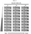
- the cell proliferation rate decreased by 50% or more, compared with all the other cells. In other words, when Re was too high, there might be a higher risk of cell lysis and the like than the anti-aging effect caused by the cell deformation.
- ROS reactive oxygen species
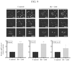
- FIG. 9 the immunohistochemistry (IHC) staining result of FIG. 9 also confirmed that expression levels of Oct4, Sox2, and SSEA-4 at Re of 244 significantly increased, compared with those of the control group.
- a representative undifferentiated marker, Oct4 exhibited 2.5 times or more increase in an expression level of mRNA, compared with that of the control group.
- the experiment results exhibited that when a physical impact was applied to the cells in the method of the present invention, the stem cells continued to be undifferentiated and thus maintained stemness. In other words, the cellular senescence was inhibited.
- a cell cycle is divided into a cell division phase and an interphase for preparing the cell division.
- the interphase which occupies about 90% of the cell cycle, is divided again into G1, S, and G2 phases.
- cells live through a life of G1, S, G2, and M (mitosis) in order but in particular, have various interphase durations depending on the cells.
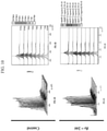
- FACS fluorescence-activated cell sorting
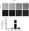
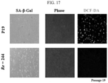
- the cells were compared and experimented by using X-gal and DCF-DA staining methods in order to examine levels of ⁇ -galactosidase, one of cellular senescence markers, and intracellular ROS, and the results are shown in FIGs. 15 and 16 .
- cytoskeleton The reorganization of cytoskeleton has been reported to regulate various activities such as cell survival, proliferation, death, cell motility, and protein selection ( FEBS Letters 582 (2008) 2120-2127 ).
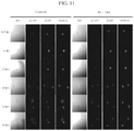
- FIG. 12 is a photograph analyzing the expression patterns of actinin and tubulin in the cells by immunohistochemical staining.
- the aforementioned results support that when a certain physical impact was applied to cells according to the method of the present invention, a temporary change occurred in cytoskeleton structures of the cells, and accordingly, the cellular senescence was inhibited, delayed, or reversibly reversed through the reorganization of the cytoskeleton inevitably followed thereby.
- FIG. 20 is a Venn diagram showing that when cells obtained by passing 10 passage cells of the umbilical cord-derived mesenchymal stem cells through the microfluidic device of the present invention or by applying electroporation to the 10 passage cells were analyzed with respect of genetic mutation and compared with cells of the control group having no treatment were analyzed, 94.9% of mutations between the control group and the microfluidic device-applied group of the present invention, 95.3% of mutations between the control group and the electroporation group were identical, and accordingly, 95% or more between the genetic mutations between all the groups were shared.
- FIG. 20 is a Venn diagram showing that when cells obtained by passing 10 passage cells of the umbilical cord-derived mesenchymal stem cells through the microfluidic device of the present invention or by applying electroporation to the 10 passage cells were analyzed with respect of genetic mutation and compared with cells of the control group having no treatment were analyzed, 94.9% of mutations between the control group and the microfluidic device-applied group of the
- a bioinformatics analysis was performed by using the microfluidic device according to the present invention to analyze differences in gene expression patterns in cells subjected respectively with electroporation and with a physical impact to 7 passage cells of umbilical cord-derived mesenchymal stem cells, and also in the control group cells having no treatment.
- RNA was respectively extracted from the three types of cells and then, used to analyze total mRNA in a NextSeq 500/550 method. Subsequently, in order to compare expression differences of the analyzed RNA, genes having a significant difference in expression levels were selected through a z-score and a p-value respectively from the control and the experiment groups, and the results are shown in FIGS. 22 and 23 .
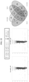
- FIG. 22 is a view showing the number of each gene in a Venn diagram after analyzing up-regulated genes, which show significantly increased expression, and down-regulated genes, which show significantly decreased expression, in the electroporation cells (EP) and the cells (HP) to which the microfluidic device of the present invention was applied, by bioinformatics.
- the number of genes (HP-UP) showing increased expression in the cells (HP) subjected with a physical impact according to the present invention was 198 in total, and of which 174 genes (EP-UP) exhibited increased expression in the cells (EP) subjected with electroporation, and accordingly, these 174 genes exhibited increased expression in both cells.
- genes (HP-DOWN) exhibiting significantly decreased expression in the cells (HP) subjected with a physical impact according to the method of the present invention was 236 in total, of which 149 genes also were commonly expressed in the cells (EP) subjected with electroporation.
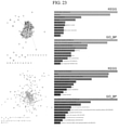
- FIG. 23 is graphs (right) showing patterns of the genes (HP-UP) with increased expression and the genes (HP-DOWN) with decreased expression in the cells passing through the microfluidic device by the method of the present invention by utilizing the databases of Kyoto Encyclopedia of Genes and Genomes (KEGG) and The Gene Ontology Biological Process (GO_BP) and perspective views showing positions of the genes in the Suite by utilizing the GO_BP.
- KEGG Kyoto Encyclopedia of Genes and Genomes
- GO_BP Gene Ontology Biological Process
- genes showing increased expression in FIG. 22 are genes highly related to a cell cycle or DNA repair, and, as schematically shown at the upper left of FIG. 23 , genes that express proteins mainly present inside cells, in cell membranes, etc.
- the two graphs at the lower right of FIG. 23 show genes with decreased expression in FIG. 22 , which are mainly related to an extracellular matrix (ECM) related to collagen, integrin, and the like.
- ECM extracellular matrix
- genes that express proteins mainly located in a matrix outside cells that is, in the extracellular matrix (ECM)
- ECM extracellular matrix
- genes with reduced expression are mainly related to cellular senescence
- Stefanie Sudhop et al. Age related changes in cell stiffness of tendon stem/progenitor cells and a rejuvenating effect of ROCK-inhibition, Biochemical and Biophysical Research Communications 509 2019 839-844 ; Pinar Zorlutuna et al., Effect of cellular and ECM aging on human iPSC-derived cardiomyocyte performance, maturity and senescence, Biomaterials 268 2021 120554 ; Toshie Tsuchiya et al., FGF-2 suppresses cellular senescence of human mesenchymal stem cells by down-regulation of TGF-ß2, Biochemical and Biophysical Research Communications 359 2007 108-114 ; Ting-Hein Lee et al., MFG-E8 mediates arterial aging by
- the cellular senescence can be inhibited, delayed, or reversed by applying a physical impact to a cell according to the method of the present invention.
- genes with different expressions in the cells subjected to a physical impact through the microfluidic device of the present invention were examined in more detail, by using a Heatmap analysis method, with respect to a DNA repair system, a cell cycle, a p53 mechanism related to cellular senescence, and the like, and the results are shown in FIG. 24 .
- genes with increased expression in the cells subjected with a physical impact according to the present invention although some of the genes exhibited increased expression toward an aging direction, but most of them exhibited increased expression toward a reverse aging (anti-aging) direction, increased or decreased expression toward a cell cycle-activating direction, and decreased expression of aging-inducing genes such as TP53.
- BER base excision repair
- MR mismatch repair
- NER nucleotide excision repair
- DSBR double-strand break repair
- the above cells are filled in the culture dish in a specific density, while maintaining a portion unoccupied.
- the dishes are cultured in a condition for cell culture for a period of time, and observed for changes in the unoccupied regions.
- wound healing effects are exhibited.
- the in vitor wound healing effect is one of functional models of evaluation for migration of cells due to proliferation. Accordingly, if a cell ages, the wound healing effect reduces as the migration or proliferation of cells reduce.
- the cells of 18 passages (P18), not treated by the physical impact according to the method of the present invention exhibit reduced effect of wound healing, i.e., reduced migration, during the same period, compared to the other two cells. That is, it is possible to assume that the senescence of a cell may be suppressed, delayed, or reversed by applying a physical impact to the cell in accordance with the method of the present invention.
- P6 mesenchymal stem cells of 6 passages
- P11 mesenchymal stem cells of 11 passages
- P18 mesenchymal stem cells of 18 passages
- the cells passing through the microfluidic device of the present invention which were aged mesenchymal stem cells of 18 passages subjected to a physical impact through the microfluidic device of the present invention, exhibited more excellent wound healing effect than young mesenchymal stem cells of 11 passages.
- the mesenchymal stem cells of 18 passages (P18) subjected with a physical impact through the microfluidic device of the present invention still showed a wide wound site even on the 9 th day after the wounding and greatly low wound healing activity, compared with the cells subjected with a physical impact according to the present invention.
- FIG. 26 is a graph showing relative wound areas of the tissues of FIG. 25 over time.
- the wound area of the tissues treatd with the cells passing through the microfluidic device according to the present invention was similar to those treated with the cells of P6 or P18 on the 3 rd day from the wounding, and accordingly, in all these tissues, the wound areas did not almost decrease.
- the wound areas of all the tissues were greatly reduced from the 6 th day after the wounding, and among them, the tissue treated with the cells passing through the microfluidic device of the present invention, when compared with the control group, or the groups treated with P11, or P18 cells, exhibited a little larger wound area than that treated with P6, but 25% or more smaller wound areas than the other groups, and thus excellent wound healing activity.
- all the tissues exhibited greatly decreased wound areas, but among them, the tissue treated with the cells passing through the microfluidic device of the present invention along with the tissue treated with the P6 cells exhibited greatly reduced wound areas down to greater than or equal to about 1/3 of those of the other tissues.
- the cells subjected with a physical impact through the microfluidic device of the present invention even though very aged cells of 18 passages, were reversely aged and turned out to have similarly excellent wound healing activity to that of young cells of 6 passages.
- the cells passing through the microfluidic device of the present invention turned out to be reversely aged.
- FIG. 27 As shown from FIG. 26 , since wound healing activity of all cells was activated from the 6 th day of the wounding, H&E staining was performed to check whether or not intracellular tissues were necrotic due to inflammation of each cell, and the results are shown in FIG. 27 . As shown from FIG. 27 , on the 6 th day of the wounding, the tissue necrosis treated with the young cells of P6 and the cells according to the present invention was relatively very lower than that of the other cells.
- MT Masson-Trichome staining was performed in order to check tissue-to-tissue binding ability of the tissues with collagen and the like on the 6 th day of the wounding, and the results are shown in FIG. 28 .
- the tissues treated with the cells according to the present invention and P6 cells had a narrow gap between the tissues and collagen, which shows excellent binding ability.
- the tissues treated with P11 and P18 cells exhibit a wide gap between the tissues and collagen, and particularly, the tissue treated with P18 cells had a very wider gap.
- the tissues treated with the cells treated by a physical impact through the microfluidic device according to the present invention exhibits not so narrow a gap as that of P6 but had a much narrower gap between the tissues and collagen than P11, and accordingly, exhibited improved binding ability.
- the cells whose senescence is inhibited, delayed, or reversed may maintain the same or similar activity as or to the younger cells.
Landscapes
- Health & Medical Sciences (AREA)
- Life Sciences & Earth Sciences (AREA)
- Engineering & Computer Science (AREA)
- Chemical & Material Sciences (AREA)
- Biomedical Technology (AREA)
- Wood Science & Technology (AREA)
- Zoology (AREA)
- Organic Chemistry (AREA)
- Bioinformatics & Cheminformatics (AREA)
- Biotechnology (AREA)
- Genetics & Genomics (AREA)
- General Health & Medical Sciences (AREA)
- Developmental Biology & Embryology (AREA)
- General Engineering & Computer Science (AREA)
- Biochemistry (AREA)
- Microbiology (AREA)
- Cell Biology (AREA)
- Hematology (AREA)
- Rheumatology (AREA)
- Sustainable Development (AREA)
- Dispersion Chemistry (AREA)
- Clinical Laboratory Science (AREA)
- Mechanical Engineering (AREA)
- Analytical Chemistry (AREA)
- Chemical Kinetics & Catalysis (AREA)
- Micro-Organisms Or Cultivation Processes Thereof (AREA)
Applications Claiming Priority (2)
| Application Number | Priority Date | Filing Date | Title |
|---|---|---|---|
| KR20200094616 | 2020-07-29 | ||
| KR1020210098400A KR102577297B1 (ko) | 2020-07-29 | 2021-07-27 | 미세유체 시스템, 이를 이용한 세포의 노화 억제, 지연, 또는 역행 방법, 및 상기 방법으로 얻어진 세포 |
Publications (1)
| Publication Number | Publication Date |
|---|---|
| EP3945132A1 true EP3945132A1 (de) | 2022-02-02 |
Family
ID=77104002
Family Applications (1)
| Application Number | Title | Priority Date | Filing Date |
|---|---|---|---|
| EP21188286.5A Pending EP3945132A1 (de) | 2020-07-29 | 2021-07-28 | Mikrofluidisches system, verfahren zur hemmung, verzögerung oder umkehrung der zellulären seneszenz unter verwendung eines mikrofluidischen systems und so erhaltene zelle |
Country Status (3)
| Country | Link |
|---|---|
| US (1) | US11951479B2 (de) |
| EP (1) | EP3945132A1 (de) |
| CN (1) | CN114058574B (de) |
Families Citing this family (2)
| Publication number | Priority date | Publication date | Assignee | Title |
|---|---|---|---|---|
| JP2023149142A (ja) * | 2022-03-30 | 2023-10-13 | 地方独立行政法人東京都立産業技術研究センター | 角化細胞培養基板、角化細胞培養基板の製造方法、角化細胞の培養方法、培養表皮組織及び培養表皮組織の製造方法 |
| FR3140171A1 (fr) * | 2022-09-23 | 2024-03-29 | Centre National De La Recherche Scientifique | Dispositif et procede pour la caracterisation de cellules soumises a une contrainte physique |
Citations (5)
| Publication number | Priority date | Publication date | Assignee | Title |
|---|---|---|---|---|
| WO2006007701A1 (en) * | 2004-07-16 | 2006-01-26 | Simon Fraser University | Microfluidic device and method of using same |
| CN201138312Y (zh) * | 2007-08-29 | 2008-10-22 | 高霞 | 一种细胞破碎装置 |
| EP3556845A1 (de) * | 2018-04-20 | 2019-10-23 | Cellix Limited | Verfahren und vorrichtung zur transfektion von zellen |
| CN110934879A (zh) * | 2019-08-22 | 2020-03-31 | 中国科学院上海生命科学研究院 | 靶向组织微环境中衰老细胞的抗衰老药物d/a及其应用 |
| WO2020117856A1 (en) * | 2018-12-04 | 2020-06-11 | Cellfe, Inc. | Methods and systems for intracellular delivery |
Family Cites Families (7)
| Publication number | Priority date | Publication date | Assignee | Title |
|---|---|---|---|---|
| US20080131323A1 (en) * | 2004-10-13 | 2008-06-05 | Carnegie Mellon University | Apparatuses, Systems, and Methods Utilizing Laminar Flow Interface Control and for Controlling Laminar Flow Interface |
| DE102004053596B4 (de) * | 2004-11-05 | 2006-10-12 | Fraunhofer-Gesellschaft zur Förderung der angewandten Forschung e.V. | Verfahren und Vorrichtungen zur Bearbeitung einzelner biologischer Zellen |
| US9014801B2 (en) * | 2008-03-19 | 2015-04-21 | The Board Of Trustees Of The University Of Illinois | Electromagnetic field therapy delays cellular senescence and death by enhancement of the heat shock response |
| KR20120070219A (ko) | 2010-12-21 | 2012-06-29 | 인제대학교 산학협력단 | 세포 반응 감지 시스템 및 세포 반응 감지 방법 |
| WO2014130419A1 (en) * | 2013-02-19 | 2014-08-28 | Board Of Regents, The University Of Texas System | Devices and methods for the prevention and treatment of neuromas |
| KR102209234B1 (ko) | 2018-10-02 | 2021-02-01 | 주식회사 스템온 | 세포의 텔로미어를 신장시키는 방법 |
| US20240122794A1 (en) * | 2022-10-12 | 2024-04-18 | Softwave Tissue Regeneration Technologies, Inc. | Acoustic shock wave or pressure pulse treatment and methods of use for anti-aging |
-
2021
- 2021-07-28 EP EP21188286.5A patent/EP3945132A1/de active Pending
- 2021-07-29 US US17/389,138 patent/US11951479B2/en active Active
- 2021-07-29 CN CN202110865875.8A patent/CN114058574B/zh active Active
Patent Citations (5)
| Publication number | Priority date | Publication date | Assignee | Title |
|---|---|---|---|---|
| WO2006007701A1 (en) * | 2004-07-16 | 2006-01-26 | Simon Fraser University | Microfluidic device and method of using same |
| CN201138312Y (zh) * | 2007-08-29 | 2008-10-22 | 高霞 | 一种细胞破碎装置 |
| EP3556845A1 (de) * | 2018-04-20 | 2019-10-23 | Cellix Limited | Verfahren und vorrichtung zur transfektion von zellen |
| WO2020117856A1 (en) * | 2018-12-04 | 2020-06-11 | Cellfe, Inc. | Methods and systems for intracellular delivery |
| CN110934879A (zh) * | 2019-08-22 | 2020-03-31 | 中国科学院上海生命科学研究院 | 靶向组织微环境中衰老细胞的抗衰老药物d/a及其应用 |
Non-Patent Citations (12)
| Title |
|---|
| DOUGLAS E. VAUGHAN ET AL.: "PAI-1-regulated extracellular proteolysis governs senescence and survival in Klotho mice", PNAS, vol. 111, no. 19, 13 May 2014 (2014-05-13), pages 7090 - 7095 |
| FEBS LETTERS, vol. 582, 2008, pages 2120 - 2127 |
| IRIT SAGI ET AL.: "The ECM path of senescence in aging: components and modifiers", THE FEBS JOURNAL, vol. 287, 2020, pages 2636 - 2646 |
| LESTER F. LAU ET AL.: "The Matricellular Protein CCN1/CYR61 Induces Fibroblast Senescence and Restricts Fibrosis in Cutaneous Wound Healing", NATURE CELL BIOLOGY, 1 January 2011 (2011-01-01) |
| MARCO DEMARIA ET AL.: "Unmasking Transcriptional Heterogeneity in Senescent Cells", CURRENT BIOLOGY, vol. 27, 11 September 2017 (2017-09-11), pages 2652 - 2660 |
| MICHELLE R. DAWSON ET AL.: "Senescent mesenchymal stem cells remodel extracellular matrix driving breast cancer cells to a more-invasive phenotype", JOURNAL OF CELL SCIENCE, vol. 133, 2020 |
| NATURE PROTOCOLS, vol. 4, no. 12, 2009, pages 1798 |
| PINAR ZORLUTUNA ET AL.: "Effect of cellular and ECM aging on human iPSC-derived cardiomyocyte performance, maturity and senescence", BIOMATERIALS, vol. 268, 2021, pages 120554 |
| RUI-MING LIU ET AL.: "Serpine 1 induces alveolar type II cell senescence through activating p53-p21-Rb pathway in fibrotic lung disease", AGING CELL, vol. 16, 2017, pages 1114 - 1124 |
| STEFANIE SUDHOP ET AL.: "Age related changes in cell stiffness of tendon stem/progenitor cells and a rejuvenating effect of ROCK-inhibition", BIOCHEMICAL AND BIOPHYSICAL RESEARCH COMMUNICATIONS, vol. 509, 2019, pages 839 - 844, XP085579701, DOI: 10.1016/j.bbrc.2019.01.027 |
| TING-HEIN LEE ET AL.: "MFG-E8 mediates arterial aging by promoting the proinflammatory phenotype of vascular smooth muscle cells", JOURNAL OF BIOMEDIAL SCIENCE, vol. 26, 2019, pages 61 |
| TOSHIE TSUCHIYA ET AL.: "FGF-2 suppresses cellular senescence of human mesenchymal stem cells by down-regulation of TGF-R.2", BIOCHEMICAL AND BIOPHYSICAL RESEARCH COMMUNICATIONS, vol. 359, 2007, pages 108 - 114, XP022103150, DOI: 10.1016/j.bbrc.2007.05.067 |
Also Published As
| Publication number | Publication date |
|---|---|
| CN114058574A (zh) | 2022-02-18 |
| US11951479B2 (en) | 2024-04-09 |
| CN114058574B (zh) | 2025-07-08 |
| US20220032304A1 (en) | 2022-02-03 |
Similar Documents
| Publication | Publication Date | Title |
|---|---|---|
| Hong et al. | Topically delivered adipose derived stem cells show an activated-fibroblast phenotype and enhance granulation tissue formation in skin wounds | |
| EP3862009A1 (de) | Zusammensetzung mit induzierten exosomen zur regeneration der haut und wundheilung | |
| Liu et al. | Direct comparison of the potency of human mesenchymal stem cells derived from amnion tissue, bone marrow and adipose tissue at inducing dermal fibroblast responses to cutaneous wounds | |
| EP2614145B1 (de) | Matrizen für gewebespezifische differenzierung und ihre verwendung | |
| EP2554660A1 (de) | Bandscheiben-nukleus-pulposus-stamm-/vorläuferzelle, verfahren zu ihrer kultivierung und anwendung davon | |
| JP6243738B2 (ja) | 羊膜間葉系幹細胞の調製方法および単離された羊膜間葉系幹細胞集団 | |
| US8771677B2 (en) | Colony-forming unit cell of human chorion and method to obtain and use thereof | |
| JP2022527998A (ja) | 高機能な製造されたabcb5+間葉系幹細胞 | |
| Ko et al. | Combined systemic and local delivery of stem cell inducing/recruiting factors for in situ tissue regeneration | |
| KR20160042816A (ko) | 상처 치유 및 조직 공학 | |
| EP3656867A1 (de) | Verfahren zur abtastung von anti-aging-substanzen | |
| US11951479B2 (en) | Microfluidic system, method for inhibiting, delaying, or reversing cellular senescence using microfluidic system, and cell obtained therefrom | |
| US20200129433A1 (en) | Stem cell-derived microvesicles with enhanced efficacy, use thereof, and method for enhancing efficacy | |
| Zhang et al. | Human dental pulp stem cell-derived exosomes decorated titanium scaffolds for promoting bone regeneration | |
| KR20200047330A (ko) | 효능이 증진된 줄기세포 유래 미세소포, 이의 용도 및 효능강화 방법 | |
| US9950016B2 (en) | Cell secreted proteins for the treatment of myocardial infarction | |
| JP7421049B2 (ja) | 糸球体ポドサイトの誘導方法、及び該誘導方法を用いた多能性幹細胞からのポドサイトの製造方法 | |
| KR20190047880A (ko) | 에티오나마이드를 이용한 줄기세포 이동성 향상 방법 | |
| Hung et al. | Paracrine-mediated rejuvenation of aged mesenchymal stem cells is associated with downregulation of the autophagy-lysosomal pathway | |
| US10017817B2 (en) | Skin activation by acceleration of PDGF-BB activity | |
| US20200102547A1 (en) | Reprosomes, as exosomes capable of inducing reprogramming of cells and preparation method thereof | |
| KR102577297B1 (ko) | 미세유체 시스템, 이를 이용한 세포의 노화 억제, 지연, 또는 역행 방법, 및 상기 방법으로 얻어진 세포 | |
| US20130005037A1 (en) | Method of treating myocardial injury | |
| Shao et al. | Human growth hormone-overexpressing adipose-derived stem cells enhance fibroblast activity and accelerate burn wound healing via ERK pathway therapeutic potential of ADSCs in burn wound repair | |
| US20200190475A1 (en) | Methods for identifying and isolating cardiac stem cells and methods for making and using them |
Legal Events
| Date | Code | Title | Description |
|---|---|---|---|
| PUAI | Public reference made under article 153(3) epc to a published international application that has entered the european phase |
Free format text: ORIGINAL CODE: 0009012 |
|
| STAA | Information on the status of an ep patent application or granted ep patent |
Free format text: STATUS: THE APPLICATION HAS BEEN PUBLISHED |
|
| AK | Designated contracting states |
Kind code of ref document: A1 Designated state(s): AL AT BE BG CH CY CZ DE DK EE ES FI FR GB GR HR HU IE IS IT LI LT LU LV MC MK MT NL NO PL PT RO RS SE SI SK SM TR |
|
| STAA | Information on the status of an ep patent application or granted ep patent |
Free format text: STATUS: REQUEST FOR EXAMINATION WAS MADE |
|
| 17P | Request for examination filed |
Effective date: 20220801 |
|
| RBV | Designated contracting states (corrected) |
Designated state(s): AL AT BE BG CH CY CZ DE DK EE ES FI FR GB GR HR HU IE IS IT LI LT LU LV MC MK MT NL NO PL PT RO RS SE SI SK SM TR |