EP3955257A1 - Procédé et système de mesure d'un niveau d'anxiété combiné et/ou corrélé avec un niveau d'état de conscience modifié et/ou un niveau de douleur - Google Patents
Procédé et système de mesure d'un niveau d'anxiété combiné et/ou corrélé avec un niveau d'état de conscience modifié et/ou un niveau de douleur Download PDFInfo
- Publication number
- EP3955257A1 EP3955257A1 EP20191014.8A EP20191014A EP3955257A1 EP 3955257 A1 EP3955257 A1 EP 3955257A1 EP 20191014 A EP20191014 A EP 20191014A EP 3955257 A1 EP3955257 A1 EP 3955257A1
- Authority
- EP
- European Patent Office
- Prior art keywords
- data
- loa
- level
- treatment
- lop
- Prior art date
- Legal status (The legal status is an assumption and is not a legal conclusion. Google has not performed a legal analysis and makes no representation as to the accuracy of the status listed.)
- Withdrawn
Links
- 208000019901 Anxiety disease Diseases 0.000 title claims abstract description 511
- 230000036506 anxiety Effects 0.000 title claims abstract description 487
- 208000002193 Pain Diseases 0.000 title claims abstract description 455
- 230000036407 pain Effects 0.000 title claims abstract description 429
- 238000005259 measurement Methods 0.000 title claims abstract description 227
- 238000000034 method Methods 0.000 title claims abstract description 199
- 230000002596 correlated effect Effects 0.000 title claims abstract description 64
- 230000000875 corresponding effect Effects 0.000 claims abstract description 76
- 238000011282 treatment Methods 0.000 claims description 713
- 230000033001 locomotion Effects 0.000 claims description 96
- 230000000694 effects Effects 0.000 claims description 80
- 210000004556 brain Anatomy 0.000 claims description 70
- 238000012544 monitoring process Methods 0.000 claims description 70
- 230000029058 respiratory gaseous exchange Effects 0.000 claims description 69
- 210000004761 scalp Anatomy 0.000 claims description 59
- 230000000241 respiratory effect Effects 0.000 claims description 50
- 230000002526 effect on cardiovascular system Effects 0.000 claims description 40
- 230000003387 muscular Effects 0.000 claims description 36
- 230000036760 body temperature Effects 0.000 claims description 27
- 238000002567 electromyography Methods 0.000 claims description 23
- 210000001652 frontal lobe Anatomy 0.000 claims description 23
- 230000001936 parietal effect Effects 0.000 claims description 23
- 210000001152 parietal lobe Anatomy 0.000 claims description 21
- 206010053694 Saccadic eye movement Diseases 0.000 claims description 19
- 230000004434 saccadic eye movement Effects 0.000 claims description 19
- 230000002776 aggregation Effects 0.000 claims description 16
- 238000004220 aggregation Methods 0.000 claims description 16
- 238000012986 modification Methods 0.000 claims description 15
- 230000004048 modification Effects 0.000 claims description 15
- 238000001514 detection method Methods 0.000 claims description 12
- 230000004931 aggregating effect Effects 0.000 claims description 11
- QVGXLLKOCUKJST-UHFFFAOYSA-N atomic oxygen Chemical compound [O] QVGXLLKOCUKJST-UHFFFAOYSA-N 0.000 claims description 8
- 229910052760 oxygen Inorganic materials 0.000 claims description 8
- 239000001301 oxygen Substances 0.000 claims description 8
- 230000001419 dependent effect Effects 0.000 claims 1
- 230000004044 response Effects 0.000 description 126
- 230000000147 hypnotic effect Effects 0.000 description 114
- 238000010494 dissociation reaction Methods 0.000 description 82
- 230000005593 dissociations Effects 0.000 description 82
- 238000002474 experimental method Methods 0.000 description 82
- 238000011156 evaluation Methods 0.000 description 75
- 230000000144 pharmacologic effect Effects 0.000 description 50
- 230000008859 change Effects 0.000 description 41
- 238000004458 analytical method Methods 0.000 description 37
- 238000000537 electroencephalography Methods 0.000 description 35
- 238000011457 non-pharmacological treatment Methods 0.000 description 33
- 230000008569 process Effects 0.000 description 33
- 238000012545 processing Methods 0.000 description 32
- 230000001939 inductive effect Effects 0.000 description 28
- 230000006870 function Effects 0.000 description 27
- 230000002829 reductive effect Effects 0.000 description 27
- 210000003491 skin Anatomy 0.000 description 26
- 230000001149 cognitive effect Effects 0.000 description 25
- 230000001965 increasing effect Effects 0.000 description 25
- 230000008447 perception Effects 0.000 description 25
- 206010039897 Sedation Diseases 0.000 description 21
- 238000011458 pharmacological treatment Methods 0.000 description 21
- 230000036280 sedation Effects 0.000 description 21
- 239000002249 anxiolytic agent Substances 0.000 description 20
- 230000000949 anxiolytic effect Effects 0.000 description 20
- 230000037324 pain perception Effects 0.000 description 20
- 230000007423 decrease Effects 0.000 description 17
- 230000009467 reduction Effects 0.000 description 17
- 239000000932 sedative agent Substances 0.000 description 17
- 238000010521 absorption reaction Methods 0.000 description 16
- 238000004364 calculation method Methods 0.000 description 16
- 230000004424 eye movement Effects 0.000 description 15
- 238000012360 testing method Methods 0.000 description 14
- 230000000202 analgesic effect Effects 0.000 description 12
- 239000008280 blood Substances 0.000 description 12
- 210000004369 blood Anatomy 0.000 description 12
- 230000003247 decreasing effect Effects 0.000 description 12
- 230000008921 facial expression Effects 0.000 description 12
- 230000001624 sedative effect Effects 0.000 description 12
- 239000000853 adhesive Substances 0.000 description 11
- 230000001070 adhesive effect Effects 0.000 description 11
- 230000005484 gravity Effects 0.000 description 11
- 230000001976 improved effect Effects 0.000 description 11
- 238000002203 pretreatment Methods 0.000 description 11
- 208000000094 Chronic Pain Diseases 0.000 description 10
- 229940035676 analgesics Drugs 0.000 description 10
- 239000000730 antalgic agent Substances 0.000 description 10
- 230000002996 emotional effect Effects 0.000 description 10
- 210000003128 head Anatomy 0.000 description 10
- 238000010801 machine learning Methods 0.000 description 10
- 230000009466 transformation Effects 0.000 description 10
- 229940005530 anxiolytics Drugs 0.000 description 9
- 210000001061 forehead Anatomy 0.000 description 9
- 230000002093 peripheral effect Effects 0.000 description 9
- 238000011084 recovery Methods 0.000 description 9
- 238000002560 therapeutic procedure Methods 0.000 description 9
- 238000004448 titration Methods 0.000 description 9
- 230000002567 autonomic effect Effects 0.000 description 8
- 230000000638 stimulation Effects 0.000 description 8
- 238000001356 surgical procedure Methods 0.000 description 8
- 238000000844 transformation Methods 0.000 description 8
- 230000003321 amplification Effects 0.000 description 7
- 238000013473 artificial intelligence Methods 0.000 description 7
- 210000003205 muscle Anatomy 0.000 description 7
- 238000003199 nucleic acid amplification method Methods 0.000 description 7
- 238000012800 visualization Methods 0.000 description 7
- 230000003444 anaesthetic effect Effects 0.000 description 6
- 230000036772 blood pressure Effects 0.000 description 6
- 238000002570 electrooculography Methods 0.000 description 6
- 230000001815 facial effect Effects 0.000 description 6
- 230000003993 interaction Effects 0.000 description 6
- 238000013341 scale-up Methods 0.000 description 6
- 230000001225 therapeutic effect Effects 0.000 description 6
- 230000001960 triggered effect Effects 0.000 description 6
- 206010001854 Altered state of consciousness Diseases 0.000 description 5
- 230000009286 beneficial effect Effects 0.000 description 5
- 210000003414 extremity Anatomy 0.000 description 5
- 210000002414 leg Anatomy 0.000 description 5
- 238000010606 normalization Methods 0.000 description 5
- 238000007781 pre-processing Methods 0.000 description 5
- 230000002265 prevention Effects 0.000 description 5
- 229940125723 sedative agent Drugs 0.000 description 5
- 231100000430 skin reaction Toxicity 0.000 description 5
- 238000013519 translation Methods 0.000 description 5
- 210000003484 anatomy Anatomy 0.000 description 4
- 229940035674 anesthetics Drugs 0.000 description 4
- 230000008901 benefit Effects 0.000 description 4
- 230000035565 breathing frequency Effects 0.000 description 4
- 238000006243 chemical reaction Methods 0.000 description 4
- 230000001684 chronic effect Effects 0.000 description 4
- 238000004590 computer program Methods 0.000 description 4
- 239000003814 drug Substances 0.000 description 4
- 239000003193 general anesthetic agent Substances 0.000 description 4
- 230000003304 psychophysiological effect Effects 0.000 description 4
- 238000007670 refining Methods 0.000 description 4
- 230000036387 respiratory rate Effects 0.000 description 4
- 230000011218 segmentation Effects 0.000 description 4
- 208000024891 symptom Diseases 0.000 description 4
- 230000001131 transforming effect Effects 0.000 description 4
- 210000005166 vasculature Anatomy 0.000 description 4
- 206010013470 Dissociative states Diseases 0.000 description 3
- 238000001467 acupuncture Methods 0.000 description 3
- 230000007177 brain activity Effects 0.000 description 3
- 239000003795 chemical substances by application Substances 0.000 description 3
- 229940079593 drug Drugs 0.000 description 3
- 238000001914 filtration Methods 0.000 description 3
- 230000006698 induction Effects 0.000 description 3
- 239000004615 ingredient Substances 0.000 description 3
- 230000007774 longterm Effects 0.000 description 3
- 230000001537 neural effect Effects 0.000 description 3
- 210000002569 neuron Anatomy 0.000 description 3
- 230000003287 optical effect Effects 0.000 description 3
- 238000006213 oxygenation reaction Methods 0.000 description 3
- 238000001671 psychotherapy Methods 0.000 description 3
- 230000001953 sensory effect Effects 0.000 description 3
- 230000020341 sensory perception of pain Effects 0.000 description 3
- 230000001629 suppression Effects 0.000 description 3
- 238000011477 surgical intervention Methods 0.000 description 3
- 230000001755 vocal effect Effects 0.000 description 3
- 208000000044 Amnesia Diseases 0.000 description 2
- 208000031091 Amnestic disease Diseases 0.000 description 2
- GQPLMRYTRLFLPF-UHFFFAOYSA-N Nitrous Oxide Chemical compound [O-][N+]#N GQPLMRYTRLFLPF-UHFFFAOYSA-N 0.000 description 2
- 206010041349 Somnolence Diseases 0.000 description 2
- 208000005298 acute pain Diseases 0.000 description 2
- 230000006978 adaptation Effects 0.000 description 2
- 230000002411 adverse Effects 0.000 description 2
- 230000006986 amnesia Effects 0.000 description 2
- VIROVYVQCGLCII-UHFFFAOYSA-N amobarbital Chemical compound CC(C)CCC1(CC)C(=O)NC(=O)NC1=O VIROVYVQCGLCII-UHFFFAOYSA-N 0.000 description 2
- 210000004727 amygdala Anatomy 0.000 description 2
- 230000003190 augmentative effect Effects 0.000 description 2
- 229940125717 barbiturate Drugs 0.000 description 2
- 229940049706 benzodiazepine Drugs 0.000 description 2
- 150000001557 benzodiazepines Chemical class 0.000 description 2
- 230000006998 cognitive state Effects 0.000 description 2
- 238000005314 correlation function Methods 0.000 description 2
- 238000003745 diagnosis Methods 0.000 description 2
- 201000010099 disease Diseases 0.000 description 2
- 208000037265 diseases, disorders, signs and symptoms Diseases 0.000 description 2
- 238000002565 electrocardiography Methods 0.000 description 2
- 230000008451 emotion Effects 0.000 description 2
- 238000000605 extraction Methods 0.000 description 2
- 210000000744 eyelid Anatomy 0.000 description 2
- 230000036541 health Effects 0.000 description 2
- 230000000977 initiatory effect Effects 0.000 description 2
- 230000000670 limiting effect Effects 0.000 description 2
- 238000002690 local anesthesia Methods 0.000 description 2
- 230000028161 membrane depolarization Effects 0.000 description 2
- 239000000203 mixture Substances 0.000 description 2
- 230000010004 neural pathway Effects 0.000 description 2
- 210000000118 neural pathway Anatomy 0.000 description 2
- 230000002085 persistent effect Effects 0.000 description 2
- 230000010344 pupil dilation Effects 0.000 description 2
- 230000011514 reflex Effects 0.000 description 2
- 230000035807 sensation Effects 0.000 description 2
- 230000002123 temporal effect Effects 0.000 description 2
- 210000001103 thalamus Anatomy 0.000 description 2
- 230000007704 transition Effects 0.000 description 2
- 238000011144 upstream manufacturing Methods 0.000 description 2
- SFLSHLFXELFNJZ-QMMMGPOBSA-N (-)-norepinephrine Chemical compound NC[C@H](O)C1=CC=C(O)C(O)=C1 SFLSHLFXELFNJZ-QMMMGPOBSA-N 0.000 description 1
- DIWRORZWFLOCLC-HNNXBMFYSA-N (3s)-7-chloro-5-(2-chlorophenyl)-3-hydroxy-1,3-dihydro-1,4-benzodiazepin-2-one Chemical compound N([C@H](C(NC1=CC=C(Cl)C=C11)=O)O)=C1C1=CC=CC=C1Cl DIWRORZWFLOCLC-HNNXBMFYSA-N 0.000 description 1
- 208000009132 Catalepsy Diseases 0.000 description 1
- 206010012289 Dementia Diseases 0.000 description 1
- 208000012661 Dyskinesia Diseases 0.000 description 1
- 241000084490 Esenbeckia delta Species 0.000 description 1
- 206010020772 Hypertension Diseases 0.000 description 1
- PIWKPBJCKXDKJR-UHFFFAOYSA-N Isoflurane Chemical compound FC(F)OC(Cl)C(F)(F)F PIWKPBJCKXDKJR-UHFFFAOYSA-N 0.000 description 1
- YQEZLKZALYSWHR-UHFFFAOYSA-N Ketamine Chemical compound C=1C=CC=C(Cl)C=1C1(NC)CCCCC1=O YQEZLKZALYSWHR-UHFFFAOYSA-N 0.000 description 1
- NZXKDOXHBHYTKP-UHFFFAOYSA-N Metohexital Chemical compound CCC#CC(C)C1(CC=C)C(=O)NC(=O)N(C)C1=O NZXKDOXHBHYTKP-UHFFFAOYSA-N 0.000 description 1
- 206010049816 Muscle tightness Diseases 0.000 description 1
- 206010028813 Nausea Diseases 0.000 description 1
- 206010062519 Poor quality sleep Diseases 0.000 description 1
- 229910003798 SPO2 Inorganic materials 0.000 description 1
- 101100478210 Schizosaccharomyces pombe (strain 972 / ATCC 24843) spo2 gene Proteins 0.000 description 1
- 208000013738 Sleep Initiation and Maintenance disease Diseases 0.000 description 1
- 208000032140 Sleepiness Diseases 0.000 description 1
- 239000000219 Sympatholytic Substances 0.000 description 1
- IUJDSEJGGMCXSG-UHFFFAOYSA-N Thiopental Chemical compound CCCC(C)C1(CC)C(=O)NC(=S)NC1=O IUJDSEJGGMCXSG-UHFFFAOYSA-N 0.000 description 1
- 206010047853 Waxy flexibility Diseases 0.000 description 1
- 238000001793 Wilcoxon signed-rank test Methods 0.000 description 1
- 210000001015 abdomen Anatomy 0.000 description 1
- 230000003044 adaptive effect Effects 0.000 description 1
- 230000036626 alertness Effects 0.000 description 1
- 229940126675 alternative medicines Drugs 0.000 description 1
- 229960001301 amobarbital Drugs 0.000 description 1
- 239000000935 antidepressant agent Substances 0.000 description 1
- 229940005513 antidepressants Drugs 0.000 description 1
- 229940125715 antihistaminic agent Drugs 0.000 description 1
- 239000000739 antihistaminic agent Substances 0.000 description 1
- 210000000617 arm Anatomy 0.000 description 1
- 230000003542 behavioural effect Effects 0.000 description 1
- 230000002146 bilateral effect Effects 0.000 description 1
- 230000015572 biosynthetic process Effects 0.000 description 1
- 230000004397 blinking Effects 0.000 description 1
- 229940054025 carbamate anxiolytics Drugs 0.000 description 1
- 150000004657 carbamic acid derivatives Chemical class 0.000 description 1
- 230000000747 cardiac effect Effects 0.000 description 1
- 238000009225 cognitive behavioral therapy Methods 0.000 description 1
- 230000003920 cognitive function Effects 0.000 description 1
- 230000000295 complement effect Effects 0.000 description 1
- 229940126676 complementary medicines Drugs 0.000 description 1
- 239000000470 constituent Substances 0.000 description 1
- 230000008602 contraction Effects 0.000 description 1
- 238000012937 correction Methods 0.000 description 1
- 210000004207 dermis Anatomy 0.000 description 1
- DPYMFVXJLLWWEU-UHFFFAOYSA-N desflurane Chemical compound FC(F)OC(F)C(F)(F)F DPYMFVXJLLWWEU-UHFFFAOYSA-N 0.000 description 1
- 229960003537 desflurane Drugs 0.000 description 1
- 238000013461 design Methods 0.000 description 1
- 238000011161 development Methods 0.000 description 1
- 230000018109 developmental process Effects 0.000 description 1
- 238000010586 diagram Methods 0.000 description 1
- 229960003529 diazepam Drugs 0.000 description 1
- AAOVKJBEBIDNHE-UHFFFAOYSA-N diazepam Chemical compound N=1CC(=O)N(C)C2=CC=C(Cl)C=C2C=1C1=CC=CC=C1 AAOVKJBEBIDNHE-UHFFFAOYSA-N 0.000 description 1
- 238000009826 distribution Methods 0.000 description 1
- 210000001513 elbow Anatomy 0.000 description 1
- 230000006397 emotional response Effects 0.000 description 1
- JPGQOUSTVILISH-UHFFFAOYSA-N enflurane Chemical compound FC(F)OC(F)(F)C(F)Cl JPGQOUSTVILISH-UHFFFAOYSA-N 0.000 description 1
- 229960000305 enflurane Drugs 0.000 description 1
- 230000007613 environmental effect Effects 0.000 description 1
- 210000002615 epidermis Anatomy 0.000 description 1
- NPUKDXXFDDZOKR-LLVKDONJSA-N etomidate Chemical compound CCOC(=O)C1=CN=CN1[C@H](C)C1=CC=CC=C1 NPUKDXXFDDZOKR-LLVKDONJSA-N 0.000 description 1
- 229960001690 etomidate Drugs 0.000 description 1
- 230000004399 eye closure Effects 0.000 description 1
- 230000000193 eyeblink Effects 0.000 description 1
- 206010016256 fatigue Diseases 0.000 description 1
- 210000000245 forearm Anatomy 0.000 description 1
- 239000003205 fragrance Substances 0.000 description 1
- 210000005153 frontal cortex Anatomy 0.000 description 1
- 210000005200 frontal scalp Anatomy 0.000 description 1
- 210000004326 gyrus cinguli Anatomy 0.000 description 1
- 229960003132 halothane Drugs 0.000 description 1
- BCQZXOMGPXTTIC-UHFFFAOYSA-N halothane Chemical compound FC(F)(F)C(Cl)Br BCQZXOMGPXTTIC-UHFFFAOYSA-N 0.000 description 1
- 210000004247 hand Anatomy 0.000 description 1
- 230000004886 head movement Effects 0.000 description 1
- 210000002837 heart atrium Anatomy 0.000 description 1
- 230000000004 hemodynamic effect Effects 0.000 description 1
- 125000005842 heteroatom Chemical group 0.000 description 1
- 230000036571 hydration Effects 0.000 description 1
- 238000006703 hydration reaction Methods 0.000 description 1
- 239000003326 hypnotic agent Substances 0.000 description 1
- 238000007654 immersion Methods 0.000 description 1
- 238000002347 injection Methods 0.000 description 1
- 239000007924 injection Substances 0.000 description 1
- 206010022437 insomnia Diseases 0.000 description 1
- 238000001990 intravenous administration Methods 0.000 description 1
- 229960002725 isoflurane Drugs 0.000 description 1
- 229960003299 ketamine Drugs 0.000 description 1
- 210000003127 knee Anatomy 0.000 description 1
- 230000008449 language Effects 0.000 description 1
- 230000002045 lasting effect Effects 0.000 description 1
- 239000003589 local anesthetic agent Substances 0.000 description 1
- 229960004391 lorazepam Drugs 0.000 description 1
- 230000007257 malfunction Effects 0.000 description 1
- 239000003550 marker Substances 0.000 description 1
- 210000001595 mastoid Anatomy 0.000 description 1
- 238000007620 mathematical function Methods 0.000 description 1
- 230000006996 mental state Effects 0.000 description 1
- 229960002683 methohexital Drugs 0.000 description 1
- RFKMCNOHBTXSMU-UHFFFAOYSA-N methoxyflurane Chemical compound COC(F)(F)C(Cl)Cl RFKMCNOHBTXSMU-UHFFFAOYSA-N 0.000 description 1
- 229960002455 methoxyflurane Drugs 0.000 description 1
- DDLIGBOFAVUZHB-UHFFFAOYSA-N midazolam Chemical compound C12=CC(Cl)=CC=C2N2C(C)=NC=C2CN=C1C1=CC=CC=C1F DDLIGBOFAVUZHB-UHFFFAOYSA-N 0.000 description 1
- 229960003793 midazolam Drugs 0.000 description 1
- 239000003607 modifier Substances 0.000 description 1
- 210000000663 muscle cell Anatomy 0.000 description 1
- 230000004118 muscle contraction Effects 0.000 description 1
- 230000017311 musculoskeletal movement, spinal reflex action Effects 0.000 description 1
- 210000003928 nasal cavity Anatomy 0.000 description 1
- 230000008693 nausea Effects 0.000 description 1
- 230000000926 neurological effect Effects 0.000 description 1
- 230000004751 neurological system process Effects 0.000 description 1
- 230000007935 neutral effect Effects 0.000 description 1
- 239000001272 nitrous oxide Substances 0.000 description 1
- 229960001730 nitrous oxide Drugs 0.000 description 1
- 229960002748 norepinephrine Drugs 0.000 description 1
- SFLSHLFXELFNJZ-UHFFFAOYSA-N norepinephrine Natural products NCC(O)C1=CC=C(O)C(O)=C1 SFLSHLFXELFNJZ-UHFFFAOYSA-N 0.000 description 1
- 230000001473 noxious effect Effects 0.000 description 1
- 229940005483 opioid analgesics Drugs 0.000 description 1
- 238000005457 optimization Methods 0.000 description 1
- 210000005199 parietal scalp Anatomy 0.000 description 1
- 230000036961 partial effect Effects 0.000 description 1
- 238000000554 physical therapy Methods 0.000 description 1
- 230000001766 physiological effect Effects 0.000 description 1
- 230000035479 physiological effects, processes and functions Effects 0.000 description 1
- 230000006461 physiological response Effects 0.000 description 1
- 230000006343 physiological stress response Effects 0.000 description 1
- 210000002442 prefrontal cortex Anatomy 0.000 description 1
- OLBCVFGFOZPWHH-UHFFFAOYSA-N propofol Chemical compound CC(C)C1=CC=CC(C(C)C)=C1O OLBCVFGFOZPWHH-UHFFFAOYSA-N 0.000 description 1
- 229960004134 propofol Drugs 0.000 description 1
- 238000002106 pulse oximetry Methods 0.000 description 1
- 238000000718 qrs complex Methods 0.000 description 1
- 238000011002 quantification Methods 0.000 description 1
- 238000009877 rendering Methods 0.000 description 1
- 230000002336 repolarization Effects 0.000 description 1
- 238000011160 research Methods 0.000 description 1
- 230000004202 respiratory function Effects 0.000 description 1
- 238000009531 respiratory rate measurement Methods 0.000 description 1
- 230000004043 responsiveness Effects 0.000 description 1
- 230000033764 rhythmic process Effects 0.000 description 1
- 238000005096 rolling process Methods 0.000 description 1
- 238000005070 sampling Methods 0.000 description 1
- 238000012216 screening Methods 0.000 description 1
- 230000009155 sensory pathway Effects 0.000 description 1
- 229960002078 sevoflurane Drugs 0.000 description 1
- DFEYYRMXOJXZRJ-UHFFFAOYSA-N sevoflurane Chemical compound FCOC(C(F)(F)F)C(F)(F)F DFEYYRMXOJXZRJ-UHFFFAOYSA-N 0.000 description 1
- 230000037321 sleepiness Effects 0.000 description 1
- 230000003238 somatosensory effect Effects 0.000 description 1
- 230000002269 spontaneous effect Effects 0.000 description 1
- 230000003068 static effect Effects 0.000 description 1
- 238000000528 statistical test Methods 0.000 description 1
- 239000002438 stress hormone Substances 0.000 description 1
- 229910052717 sulfur Inorganic materials 0.000 description 1
- 230000001502 supplementing effect Effects 0.000 description 1
- 230000008093 supporting effect Effects 0.000 description 1
- 230000009747 swallowing Effects 0.000 description 1
- 210000000106 sweat gland Anatomy 0.000 description 1
- 230000002889 sympathetic effect Effects 0.000 description 1
- 230000000948 sympatholitic effect Effects 0.000 description 1
- 238000003786 synthesis reaction Methods 0.000 description 1
- XLOMZPUITCYLMJ-UHFFFAOYSA-N thiamylal Chemical compound CCCC(C)C1(CC=C)C(=O)NC(=S)NC1=O XLOMZPUITCYLMJ-UHFFFAOYSA-N 0.000 description 1
- 229960001166 thiamylal Drugs 0.000 description 1
- 229960003279 thiopental Drugs 0.000 description 1
- 230000008448 thought Effects 0.000 description 1
- 210000001519 tissue Anatomy 0.000 description 1
- 208000037816 tissue injury Diseases 0.000 description 1
- 230000000287 tissue oxygenation Effects 0.000 description 1
- 230000000007 visual effect Effects 0.000 description 1
Images









Classifications
-
- A—HUMAN NECESSITIES
- A61—MEDICAL OR VETERINARY SCIENCE; HYGIENE
- A61B—DIAGNOSIS; SURGERY; IDENTIFICATION
- A61B5/00—Measuring for diagnostic purposes; Identification of persons
- A61B5/24—Detecting, measuring or recording bioelectric or biomagnetic signals of the body or parts thereof
- A61B5/316—Modalities, i.e. specific diagnostic methods
- A61B5/369—Electroencephalography [EEG]
-
- G—PHYSICS
- G16—INFORMATION AND COMMUNICATION TECHNOLOGY [ICT] SPECIALLY ADAPTED FOR SPECIFIC APPLICATION FIELDS
- G16H—HEALTHCARE INFORMATICS, i.e. INFORMATION AND COMMUNICATION TECHNOLOGY [ICT] SPECIALLY ADAPTED FOR THE HANDLING OR PROCESSING OF MEDICAL OR HEALTHCARE DATA
- G16H50/00—ICT specially adapted for medical diagnosis, medical simulation or medical data mining; ICT specially adapted for detecting, monitoring or modelling epidemics or pandemics
- G16H50/20—ICT specially adapted for medical diagnosis, medical simulation or medical data mining; ICT specially adapted for detecting, monitoring or modelling epidemics or pandemics for computer-aided diagnosis, e.g. based on medical expert systems
-
- G—PHYSICS
- G16—INFORMATION AND COMMUNICATION TECHNOLOGY [ICT] SPECIALLY ADAPTED FOR SPECIFIC APPLICATION FIELDS
- G16H—HEALTHCARE INFORMATICS, i.e. INFORMATION AND COMMUNICATION TECHNOLOGY [ICT] SPECIALLY ADAPTED FOR THE HANDLING OR PROCESSING OF MEDICAL OR HEALTHCARE DATA
- G16H20/00—ICT specially adapted for therapies or health-improving plans, e.g. for handling prescriptions, for steering therapy or for monitoring patient compliance
-
- A—HUMAN NECESSITIES
- A61—MEDICAL OR VETERINARY SCIENCE; HYGIENE
- A61B—DIAGNOSIS; SURGERY; IDENTIFICATION
- A61B5/00—Measuring for diagnostic purposes; Identification of persons
- A61B5/01—Measuring temperature of body parts ; Diagnostic temperature sensing, e.g. for malignant or inflamed tissue
-
- A—HUMAN NECESSITIES
- A61—MEDICAL OR VETERINARY SCIENCE; HYGIENE
- A61B—DIAGNOSIS; SURGERY; IDENTIFICATION
- A61B5/00—Measuring for diagnostic purposes; Identification of persons
- A61B5/02—Detecting, measuring or recording for evaluating the cardiovascular system, e.g. pulse, heart rate, blood pressure or blood flow
- A61B5/024—Measuring pulse rate or heart rate
-
- A—HUMAN NECESSITIES
- A61—MEDICAL OR VETERINARY SCIENCE; HYGIENE
- A61B—DIAGNOSIS; SURGERY; IDENTIFICATION
- A61B5/00—Measuring for diagnostic purposes; Identification of persons
- A61B5/08—Measuring devices for evaluating the respiratory organs
- A61B5/0816—Measuring devices for examining respiratory frequency
-
- A—HUMAN NECESSITIES
- A61—MEDICAL OR VETERINARY SCIENCE; HYGIENE
- A61B—DIAGNOSIS; SURGERY; IDENTIFICATION
- A61B5/00—Measuring for diagnostic purposes; Identification of persons
- A61B5/16—Devices for psychotechnics; Testing reaction times ; Devices for evaluating the psychological state
- A61B5/163—Devices for psychotechnics; Testing reaction times ; Devices for evaluating the psychological state by tracking eye movement, gaze, or pupil change
-
- A—HUMAN NECESSITIES
- A61—MEDICAL OR VETERINARY SCIENCE; HYGIENE
- A61B—DIAGNOSIS; SURGERY; IDENTIFICATION
- A61B5/00—Measuring for diagnostic purposes; Identification of persons
- A61B5/16—Devices for psychotechnics; Testing reaction times ; Devices for evaluating the psychological state
- A61B5/165—Evaluating the state of mind, e.g. depression, anxiety
-
- A—HUMAN NECESSITIES
- A61—MEDICAL OR VETERINARY SCIENCE; HYGIENE
- A61B—DIAGNOSIS; SURGERY; IDENTIFICATION
- A61B5/00—Measuring for diagnostic purposes; Identification of persons
- A61B5/24—Detecting, measuring or recording bioelectric or biomagnetic signals of the body or parts thereof
- A61B5/25—Bioelectric electrodes therefor
- A61B5/279—Bioelectric electrodes therefor specially adapted for particular uses
- A61B5/291—Bioelectric electrodes therefor specially adapted for particular uses for electroencephalography [EEG]
-
- A—HUMAN NECESSITIES
- A61—MEDICAL OR VETERINARY SCIENCE; HYGIENE
- A61B—DIAGNOSIS; SURGERY; IDENTIFICATION
- A61B5/00—Measuring for diagnostic purposes; Identification of persons
- A61B5/24—Detecting, measuring or recording bioelectric or biomagnetic signals of the body or parts thereof
- A61B5/316—Modalities, i.e. specific diagnostic methods
- A61B5/389—Electromyography [EMG]
-
- A—HUMAN NECESSITIES
- A61—MEDICAL OR VETERINARY SCIENCE; HYGIENE
- A61B—DIAGNOSIS; SURGERY; IDENTIFICATION
- A61B5/00—Measuring for diagnostic purposes; Identification of persons
- A61B5/72—Signal processing specially adapted for physiological signals or for diagnostic purposes
- A61B5/7235—Details of waveform analysis
- A61B5/7264—Classification of physiological signals or data, e.g. using neural networks, statistical classifiers, expert systems or fuzzy systems
-
- A—HUMAN NECESSITIES
- A61—MEDICAL OR VETERINARY SCIENCE; HYGIENE
- A61B—DIAGNOSIS; SURGERY; IDENTIFICATION
- A61B5/00—Measuring for diagnostic purposes; Identification of persons
- A61B5/74—Details of notification to user or communication with user or patient; User input means
- A61B5/742—Details of notification to user or communication with user or patient; User input means using visual displays
-
- G—PHYSICS
- G16—INFORMATION AND COMMUNICATION TECHNOLOGY [ICT] SPECIALLY ADAPTED FOR SPECIFIC APPLICATION FIELDS
- G16H—HEALTHCARE INFORMATICS, i.e. INFORMATION AND COMMUNICATION TECHNOLOGY [ICT] SPECIALLY ADAPTED FOR THE HANDLING OR PROCESSING OF MEDICAL OR HEALTHCARE DATA
- G16H20/00—ICT specially adapted for therapies or health-improving plans, e.g. for handling prescriptions, for steering therapy or for monitoring patient compliance
- G16H20/70—ICT specially adapted for therapies or health-improving plans, e.g. for handling prescriptions, for steering therapy or for monitoring patient compliance relating to mental therapies, e.g. psychological therapy or autogenous training
Definitions
- the present invention is in a field of a computer-implemented method and system for automatically measuring a level of anxiety, a level of a modified state of consciousness, and/or a level of pain in a subject.
- prior art methods for assessment of the level of anxiety a subject is experiencing have important drawbacks and are not able to provide for an efficient, reliable, robust, objective and reproducible indication of the level of anxiety as experienced by a subject.
- This problem is immediately apparent when the subject is not capable of providing for a reliable or clear description of the level of anxiety experienced, such as for example in the case of non-verbal children, persons with a disability, .... Further, similarly, similarly, similarly, similarly, there is a need for an improved measurement of the level of pain as perceived by a subject.
- anxiety and pain and the treatment thereof have an impact on medical outcomes and patient recovery.
- Anxiety has an impact on health-related effects such as for example fatigue, nausea, insomnia, pain, for example as experienced by a subject before, during and/or after a treatment, such as for example a treatment including surgery and/or any other suitable medical and/or non-medical treatment. Pain measurement can be used to screen for potential diseases or medical problems, to perform a triage by determining the urgency of the needs of one patient over the other, can be used to determine whether it is safe to engage and/or continue with a medical treatment, etc..
- a computer-implemented method for measurement of a level of anxiety combined with and/or correlated with a level of a modified state of consciousness, and/or a level of pain comprising the steps of:
- Measuring the LoA combined with DoS, the LoA combined with LoP or the LoA, LoP and DoS all three in combination it for example becomes possible to identify how the LoA influences the LoP or vice versa, how the LoA is modulated by a modified state of consciousness, how the ability to reach a particular modified state of consciousness is influenced by the LoA and/or the LoP, etc..
- measuring the LoA, LoP and/or DoS in a combined way is specifically advantageous as for example, a part of the LoA could be triggered by the LoP.
- This improved, more robust and reliable measurement of LoA, LoP and DoS helps in determining a more accurate diagnosis, an improved and safer treatment plan, and an improved evaluation of the effectiveness of treatment.
- a clear allocation can be made to the impact of anxiety, pain, and a modified state of consciousness, there the risk of administering a pain treatment when an anxiety treatment is needed or vice versa is reduced.
- the risk for overdosing and/or underdosing is reduced, for example as this measurement allows to make a clear decision on whether to use anxiolytics to treat anxiety and/or to use analgesics to treat pain and/or to use sedatives to modify the state of consciousness, and to optimize their dosing in function of the measured combination of LoA, LoP and/or DoS.
- such a measurement is not limited to optimization of pharmacological treatments, but is also useful when optimizing non-pharmacological treatments, and/or any combination of pharmacological and/or non-pharmacological treatments directed at treating anxiety, pain, and/or modifying a state of consciousness, or any other symptom related to anxiety, pain, and/or a specific state of consciousness, or any other suitable treatment.
- this method is limited to merely obtaining information, such as data and physical quantities from the living human body and is not directed to finding any significant deviation, i.e. a symptom during a comparison of the collected data with standard values and is not directed to the attribution of such a deviation to a particular clinical picture, i.e. the deductive medical decision phase.
- the method is performed on measured data, received, after it has been collected, or in other words, the method excludes a step of collection of data practiced on the human body.
- the computer-implemented method only concerns obtaining information, for measurement of the level of anxiety as experienced by a subject, which is a psychophysiological quality of the subject, for the measurement of the level of pain as perceived by the subject and/or for the measurement of a level of modified state of consciousness.
- the computer-implemented method excludes any steps for curing of a disease or malfunction of the body, nor any step relating to a therapeutic effect on the body of the subject.
- the level of anxiety as measured comprises at least a level of anxiety experienced by a subject as measured at the level of the cortex of the brain of the subject or in other words comprising a cognitive dimension.
- Anxiety being a response involving a psychophysiological and emotional aspect, clearly involves such a cognitive dimension. It has been shown based on the experiments above that anxiety as measured above is able to capture this cognitive dimension and is not limited to measurement of only non-cognitive parameters.
- Anxiety is for example associated with increased activity in the amygdala, and the activity of the amygdala can be influenced by activity in the higher brain structure, for example including the anterior cingulate cortex and the pre-frontal cortex responsible for focused attention. It is thus clear that anxiety as experienced by a subject comprises a cognitive dimension, optionally in addition to any other suitable dimensions.
- the level of pain relates to the perceived level of pain, or in other words the consciously perceived level of pain by a subject as measured at the level of the cortex of the brain of the subject.
- the level of pain thus corresponds to the consciously perceived level of pain perception of the subject.
- the level of pain as perceived by the subject is thus not directly correlated to the intensity of the external pain stimulus itself, but relates to the level of pain perception.
- a local anesthetic, and/or IV analgesics dosages could be increased based on a combination of an objectivized DoS, a LoP as perceived by the subject, a LoA as experienced by the subject and not on a subjective estimation of patient's state of consciousness, pain and anxiety, for example by an anesthesiologist before, during and after a surgical treatment.
- Measurement of any combination of DoS, LoP and/or LoA also allows for an anxiety and/or a pain diagnostic for non-verbal patients (children, adults who are disabled, patients with dementia, ).
- the measured combination of any of DoS, LoP and/or LoA is for example also a good indicator of the need or not for more sedation and/or analgesics and/or anxiolytics.
- a safe and reliable dose or titration of sedatives and/or analgesics and/or anxiolytics can be realized, with a reduced risk of overdosing and/or negative collateral effects of the analgesic or sedative or anxiolytic treatment.
- the modulation of the LoA by DoS can be determined.
- anxiety is inversely correlated with a modified state of consciousness or in other words a depth of state, such as for example corresponding to a depth of dissociation of the subject, a level of absorption of the subject, etc.
- a depth of state such as for example corresponding to a depth of dissociation of the subject, a level of absorption of the subject, etc.
- the LoA could be decreased by modifying the level of consciousness.
- a modified level of consciousness associated with a deeper depth of state such as for example a deeper depth of dissociation, a higher level of absorption, etc. as further detailed seems to decrease the level of anxiety experienced by a subject.
- measurement of the LoA combined with DoS might enable to determine which part of anxiety could be treated by modifying the state of consciousness of the subject, for example modifying the DoS, such as for example modifying the level of absorption, depth of dissociation, ... and additionally it might allow to differentiate between immediate and long-term effect of such changes to the depth of state to reduce anxiety, such as for example when used non-pharmacological treatments such as for example in psychotherapies, ...
- LoP is modulated by DoS and/or it may be possible to determine how LoP is modulated by LoA and/or vice versa.
- This is useful as an increased level of anxiety before, during and/or after a treatment session might increase the level of pain as perceived by a subject and can even trigger pain perception. This is important as for example anxiety plays a role in chronic pain.
- Further anxiety is a risk factor for developing persistent/chronic pain following acute pain, and the embodiment above could thus for example be useful for determining a high risk group with respect to development of chronic pain and/or to reduce the risk of developing chronic pain.
- the LoP has an impact on the LoA as pain is an important anxiety inducing event..
- the value for NLoA would provide for a value of LoA at a predetermined reference DoS and/or LoP of interest, independently of the current DoS / LoP and/or any changes to the current DoS / LoP, thereby for example providing an indicator for the equivalent amount of anxiety that would be experienced when the subject would be at, or return to a normal awake state of consciousness and/or a painless state, when this reference DoS is the normal awake state and/or the reference LoP corresponds to a painless state.
- the value for NLoP would provide for a value of LoP at a predetermined reference DoS and/or LoA of interest, independently of the current DoS / LoA and/or any changes to the current DoS / LoA, thereby for example providing an indicator for the equivalent amount of pain that would be consciously perceived when the subject would be, or return to a normal awake state of consciousness and/or in a state without anxiety, when this reference DoS is the normal awake state and/or the reference LoA is a state without anxiety.
- LoP is too high
- anxiety becomes difficult to manage and when LoA is too high pain becomes difficult to manage and might peak naturally.
- anxiety and/or pain treatments might experience difficulty in managing the LoA and/or LoP to desired levels.
- LoA and/or LoP are too high, it might be difficult to reach a desired modified level of consciousness, and for example no, insufficient, or too limited depth of state might be reached.
- LoA, DoS and/or LoP only need to be measured respectively at at least two different points in time, and thus the LoA / DoS / LoP must not necessarily be measured concurrently.
- the method comprises the step of:
- the predetermined range could be defined by one or more suitable limit values, and/or one or more desired ranges, thresholds, limits, ... which define the predetermined range for the LoP and/or LoA and/or DoS , as the range in which an anxiety management treatment, a pain management treatment and/or treatment modifying the state of consciousness still remains effective.
- the range could be defined in such a way that outside this range an anxiety management treatment, a pain-reducing treatment and/or treatment modifying the state of consciousness is no longer effective or is too difficult to manage.
- the predetermined range could, according to some embodiments, be automated, dynamic, adaptive, etc.
- the predetermined range is adapted based on for example earlier measured values of a subject (such as for example a reference range measured during a calibration phase, etc.) or population (such as for example predetermined ranges based on population characteristics, etc.).
- the treatment is an anxiety-reducing treatment and the method comprises the step of:
- the anxiety treatment could for example be an anxiety-reducing treatment, or alternatively an anxiety-controlling or anxiety-preventing treatment that controls or prevents any undesired levels of anxiety, or any other suitable anxiety treatment.
- the pain treatment could for example be a pain-reducing treatment, or alternatively a pain-controlling or pain-preventing treatment that controls or prevents any undesired levels of pain, or any other suitable pain treatment.
- a computer-implemented method for determining a treatment and/or determining an adjustment to a treatment and/or determining an intensity of a treatment and/or determining the efficacy of a treatment and/or determining the choice of a treatment comprising the steps of:
- EEG electrodes there is also present a ground electrode and/or a reference electrode.
- the data from the EEG electrodes can be processed independently, which means that there is no need to provide for calculations on the interaction and/or interrelation of the signals of two or more EEG electrode.
- a scaled and/or indexed LoA may for example be referred to as a level of anxiety index or LoAI.
- a scaled and/or indexed LoP may for example be referred to as a level of pain index or LoPI.
- a scaled and/or indexed DoS may for example be referred to as a Depth of State index or DoSI.
- a computer-implemented method for measuring and/or monitoring and/or predicting an evolution and/or aggregating a level of anxiety combined with and/or correlated with a level of pain, and/or a level of modified state of consciousness of a subject comprising the method for measurement of a level of anxiety combined with and/or correlated with a level of modified state of consciousness and a level of pain according to the first aspect of the invention applied on a subject by receiving the measured data of a subject.
- the measured data of the subject could for example be real-time data as being received from suitable sensors, however, alternative embodiments are possible in which historic data, previously stored data, ... and/or any combination thereof is used for measurement of a level of anxiety, a level of pain and/or a level of modified state of consciousness.
- the treatment could for example comprise an anxiety treatment or a pain treatment, such as for example an anxiety-reducing treatment or a pain-reducing treatment.
- a surgical treatment or any other medical intervention during which one or more anxiety or pain inducing acts are performed such as for example a needle puncture, placement of a port of a catheter, an incision, introduction of a wire, etc..
- the desired treatment-induced evolution will comprise a reduction of the LoA or LoP, however in alternative treatments, it might be more important to monitor the LoA or LoP for any undesired changes during the treatment, such that for example the need for extra anxiety or pain management measures could be reliably assessed.
- a desired treatment induced evolution of DoS might comprise a desired change to the DoS by the treatment, or the prevention of an undesired change to the DoS by the treatment.
- theta-alpha frequency band, ta comprises one or more of the following:
- the delta-theta frequency band consists of a combination of at least part of a delta frequency band and at least part of a theta frequency band.
- the delta-theta (dt) frequency band comprises, and preferably consists of a combination of at least part of the delta frequency band and at least part of the theta frequency band.
- such a delta-theta frequency band comprising and/or consisting of a combination of two brain wave frequencies, or at least a part thereof, is different from a frequency band only comprising a single brain wave frequency band and different from a frequency band comprising more than two brain wave frequency bands.
- theta-alpha frequency band consists of a combination of at least part of a theta frequency band and at least part of an alpha frequency band.
- theta-alpha (ta) frequency band comprises, and preferably consists of a combination of at least part of the theta frequency band and at least part of the alpha frequency band.
- such a theta-alpha frequency band comprising and/or consisting of a combination of two brain wave frequencies, or at least a part thereof, is different from a frequency band only comprising a single brain wave frequency band and different from a frequency band comprising more than two brain wave frequency bands.
- the method comprises steps of:
- the level of anxiety is measured by means of a combination of a specific signal extracted from EEG data, which is more directly linked to the cognitive dimension of anxiety, and one or more specific signals like heart rate, heart rate variability, breathing frequency, breathing variability, oxygen saturation, ... and/or other suitable specific embodiments of physiological parameters as mentioned below, which are more directly linked to the autonomic-emotional dimension provides for a specifically advantageous multidimensional measurement of the level of anxiety as experienced by a subject.
- a specific combination of heart rate and heart rate variability data provides for a particularly advantageous and simple measurement with an improved correlation with the level of anxiety as experienced by a subject.
- cardiovascular data and/or combination thereof is used, such as for example cardiac activity data, hemodynamic activity data, ....
- the method comprises steps of:
- the method comprises steps of:
- the method comprises steps of:
- the method comprises steps of:
- the EMG data is captured at two or more electrode locations such that global muscle tension can be determined and locally isolated artefacts can be detected and removed.
- the method comprises steps of:
- the LoA is measured taken into account the multidimensional aspect of anxiety.
- the treatment is an anxiety treatment and the method comprises the step of:
- the anxiety treatment could for example be an anxiety-reducing treatment, or alternatively an anxiety-controlling or anxiety-preventing treatment that controls or prevents any undesired levels of anxiety, or any other suitable anxiety treatment.
- the treatment is an anxiety treatment and the method comprises the step of:
- the anxiety treatment could for example be an anxiety-reducing treatment, or alternatively an anxiety-controlling or anxiety-preventing treatment that controls or prevents any undesired levels of anxiety, or any other suitable anxiety treatment.
- a computer-implemented method for determining a treatment and/or determining an adjustment to a treatment and/or determining an intensity of a treatment and/or determining the efficacy of a treatment and/or determining the choice of a treatment comprising the steps of:
- a system configured to measure a LoA combined with and/or correlated with a DoS and/or a LoP according to the method according to any previous aspect and/or embodiments, the system comprising:
- the monitoring apparatus comprises, for obtaining measured data, one or more of:
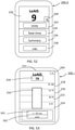
- GUI graphical user interface
- GUI GUI
- further data such as for example ECG data, cardiovascular data, respiratory data, ocular movement data, EOG data, muscular activity data, EMG data, body temperature data, ...
- any embodiments defined with reference to LoA, LoP and/or DoS also refer to similar embodiments based on parameters derived from LoA, LoP and/or DoS, such as for example LoAI, LoPI, DoSI, LoA(I)S, LoP(I)S, etc..
- an experiment including a hypnotic treatment session was set up.
- any other suitable treatment session is used and/or in which no use is made of a treatment session.
- This experiment resulted in a study with 12 healthy subjects, of which 10 were included in the subsequent analysis.
- the experiment comprised measurement of a level of anxiety, a level of pain, and a modified state of consciousness, by self-reporting of the subject using a validated questionnaire.
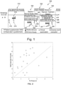
- FIG. 1 shows the different phases of the experiment.
- clinical examination and an adverse event evaluation was performed as part of a screening and/or the end of study, EOS, in order to assure the safety of the subjects undergoing the experiment and to assure a high level of quality with respect to the self-reported data.
- a first step 512 the subjects were asked to provide self-reported values for rating parameters as explained in further detail below.
- step 514 by using a stair-case method, the subjects received a sequence of transcutaneous electrical stimulus, or in other words a pain stimulus, of increasing intensity, so that in step 516 the subject could indicate when the perceived level of pain corresponded to a level of pain with a value of 8 on a VAS pain scale.
- the intensity of the pain stimulus is calibrated for each subject individually using the stair-case method, by asking the subjects to indicate when they judged the perceived level of pain to corresponding to a value 8 on the pain scale, which corresponds to a level of pain qualified as "significantly painful and demanding some effort to tolerate”.
- a pain scale for self-reporting of pain was determined by means of a Visual Analogue Scale or VAS, which is a measurement instrument that tries to measure a characteristic or attitude that is believed to range across a continuum of values and cannot easily be directly measured.
- VAS scale ranging from a value of 0 for "no pain” up to and including 10 for "severe or extreme amount of pain”.
- NRS-11 Numeric Rating Scale
- step 512, and 516 of the calibration phase 510 Similar as for the self-reported level of pain, at step 512, and 516 of the calibration phase 510, and as will be described in further detail below at further steps during the experiment, such as for example step 534 of the normal awake state 530 and step 544 of the hypnotic treatment session 540, patients were also asked to provide self-reported values for rating their experienced level of anxiety. According to this embodiment a VAS scale ranging from a value of 0 for "no anxiety" up to and including 10 for "severe or extreme amount of anxiety". It is clear that alternative embodiments for self-reporting values are possible.
- the calibration phase 510 was followed by a first phase 520, which was labeled as test phase 520 in Fig. 1 , which comprises a first session 530 in a normal awake state of the subject.
- a first phase 520 which was labeled as test phase 520 in Fig. 1 , which comprises a first session 530 in a normal awake state of the subject.
- this first session 530 there were generated one or more pain stimuli, as calibrated during the previous calibration phase.
- the pain stimuli when applied, provided for identical pain conditions, for example by means of transcutaneous electricals stimuli of identical intensity.
- suitable sensors are configured to monitor and/or register physiological signals generated by the subject for provision to a suitable processor to generate measurement data corresponding to the state and/or activity of the body or bodily functions of the subject.
- physiological sensors provide an objective measure of physiological signals.
- Such physiological sensors provide for signals, that, when suitably processed, provide for measurement data based on recordings of electrical signals produced by the brain, the heart, the muscles, the skin, ... of the subject.
- Embodiments of such physiological sensors are for example sensors for measuring physiological signals configured to measure respiration, electrocardiography or ECG, electromyography or EMG, Blood Pressure or BP, Blood Volume Pressure or BVP, electrooculography or EOG, electroencephalography or EEG, .
- Electroencephalography or EEG is an electrophysiological monitoring method to record electrical activity of the brain, and can be referred to as a cognitive, electrophysiological signal, and is related to corticoidal signals.
- EMG electrophysiological signals
- EDA muscle activity
- EKG ECG
- ECG electrophysiological signals
- BVP blood pressure
- Respiration sensors which measure for example the rate of respiration, depth of breath, effort, temperature, air pressure during inhalation and/or exhalation, ....
- electroencephalogram (EEG) data cardiovascular data (heart rate, heart rate variability, ...), electromyogram (EMG) data, respiratory data (respiration rate, respiration variability, predetermined respiration patterns, effort, temperature, and air pressure (inhalation/exhalation)), electrooculography or EOG and body temperature, were collected as measured data from each subject, however it is clear that alternative embodiments are possible comprising the monitoring of alternative cognitive electrophysiological and/or autonomic physiological signals as for example described in further detail below.
- the subject was asked to provide self-reported values for the level of anxiety, the level of pain, modified level of consciousness, etc. for example by means a suitable VAS patient questionnaire during step 534.
- the test phase 520 subsequently comprises second session 540, which comprises an automated hypnotic treatment session 548 that was delivered using a system that comprises a virtual reality headset as will also be described in further detail below.
- second session 540 which comprises an automated hypnotic treatment session 548 that was delivered using a system that comprises a virtual reality headset as will also be described in further detail below.
- the embodiment of the system also referred to as a hypnotic package, is referred to as Sedakit, as manufactured by Oncomfort.
- the Sedakit was configured to the Aqua 30 minutes module embodiment of an automated hypnotic treatment session, which subjected the subject to an automatic hypnotic treatment session for a time period of 30 minutes at step 548.
- electroencephalogram EEG
- cardiovascular data muscular activity data such as for example electromyogram or EMG data
- respiratory data respiratory data (respiration rate, respiration variability, predetermined respiration patterns, effort, temperature, and air pressure (inhalation/exhalation)
- ocular movement data such as for example electrooculography or EOG data and body temperature data
- each subject was exposed to one or more transcutaneous painful stimuli, which also qualify as anxiety inducing events.
- a sequence of a plurality of pain stimuli such as for example shown as three painful stimuli 542.
- the painful stimuli 542 that are applied during the hypnotic treatment session 538 were also qualified as 8 on the VAS pain scale during the calibration phase.
- the pain stimuli when applied, provided for identical pain conditions, for example by means of transcutaneous electricals stimuli of identical intensity. It is however clear that alternative embodiments of the experiment are possible, in which a different number of pain stimuli and/or pain stimuli of other suitable intensities and/or varying intensities or any other suitable types of anxiety inducing stimuli are applied.
- the calibrated pain stimuli 532 of a predetermined intensity, which were associated by the subject with a predetermined level of pain during the calibration phase 510, are applied at suitable times, with suitable intervals, during a time period of for example 30 minutes.
- the external pain stimulus was provided in the form of an electric transcutaneous stimulus being provided at the fore-arm during the experiment, however it is clear that alternative embodiments of a suitable pain stimulus are possible.
- each subject was asked to rate the level of anxiety as they experienced it, the level of pain as they experienced it and the depth of dissociation, etc. during the hypnotic treatment session 548 by means of a patient questionnaire.
- the subject is placed in a similar context.
- the subject is placed in a similar position, for example laying on chair.
- the subject is experiencing similar sounds 537, 547, such as for example sounds of a hospital environment.
- there may be made use of an observer's questionnaire for example assure the quality of the experiment and/or to provide for additional and/or alternative ratings for one or more of the self-reported ratings of pain, anxiety, dissociation, etc..
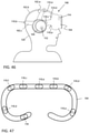
- EEG electrodes were used that were placed so as to cover the scalp and so as to detect local voltage fluctuations resulting from localized regions of the brain.
- a high density EEG was employed with 256 sensors or channels, however it is clear that alternative embodiments with a different number of sensors are possible.
- electrodes were placed according to the predefined layout for a 256-channel Hydrocel Geodesic Sensor Network. It is further known to a person skilled in the art that EEG electrodes mainly measure neural activity that occurs at the upper layers of the brain, or in other words the neural activity of the cortex of the brain.
- a pair of surface EMG sensors were placed on a leg of each patient to as to detect the electric potential generated by muscle cells when then muscles become activated.
- EMG sensors are provided at any other suitable location, such as for example the face, arm, .... It is however clear that according to alternative embodiments use can be made of other types of suitable EEG electrodes or sensors, EMG electrodes or sensors and/or any other suitable type of electrode or sensor for measuring EEG and/or EMG signals.
- the experiment 500 comprises a test phase 520 with a "normal awake state” 530 and a "Hypnotic treatment session" for which both self-reported data with respect to the level of anxiety, the level of pain and a level of dissociation were collected in steps 538, 548 for each subject that was subjected to one or more calibrated pain stimuli, which also qualify as calibrated anxiety inducing events.
- the level of dissociation is an exemplary metric for quantifying a modified level of consciousness, such as for example resulting from a hypnotic treatment session, however it is clear that alternative embodiments are possible of a level of modified consciousness, such as for example a level of absorption, etc..
- Table 1 shows a Wilcoxon signed-rank test of the self-reported data for subjects during the experiment of FIG. 1 during the normal awake state 530 and during the hypnotic treatment session 540: Self-reported data 534, 544 Normal awake state 530 Hypnotic treatment session 540 p-value Adjusted p-value (Bonferroni correction) Mean ⁇ SD Median [IQR] Mean ⁇ SD Median [IQR] Self-reported level of Anxiety 4.92 (1.74) 3.16 (1.77) 0.0004 0.0024 5.55 [1.8] 3.1 [2.95] Self-reported level of Pain 4.21 (2.27) 1.39 (1.50) 0.0059 0.0296 4.35 [2.22] 1.2 [1.65] Self-reported level of Dissociation 4.11 (3.13) 7.52 (1.43) 0.0032 0.0162 4.95 [5.72] 8 [0.9]
- the self-reported level of Anxiety is significantly different in both sessions 530, 540.
- the mean of the value for self-reported anxiety in the normal awake state 530 is 4.92, while in the hypnotic treatment session 540 it was reported to be 3.16. In other words, a reduction of more than 30% with respect to the normal awake state.
- the self-reported level of anxiety corresponds to a level of anxiety as experienced by the subject, as clearly, the self-reported values for the level of anxiety triggered by an identical external pain stimulus are modulated by the modified state of consciousness caused by the hypnotic treatment session.
- the self-reported data for the level of anxiety are an indicator for the level of anxiety as experienced by the subject, or in other words the level of experienced anxiety.
- the self-reported data relating to anxiety are triggered by the painful stimuli
- these painful stimuli can be considered as an anxiety inducing event, or in other words an adverse situation at a specific moment in time, and thus the self-reported data relates to the level of anxiety of the subject as experienced with respect to the painful, anxiety inducing stimuli.
- the experiment was set up with a first phase in the normal awake state 530 with increased levels of anxiety as compared to prior to the experiment, and still during the subsequent second phase in during the hypnotic treatment session 540, again reduced levels of anxiety are reported.
- a predetermined rest time between the normal awake state 530 and the hypnotic treatment session 540 was included.
- the self-reported level of pain are significantly different in both sessions 530, 540.
- the mean of the value for self-reported pain in the normal awake state 530 is 4.21, while in the hypnotic treatment session 540 it was reported to be 1.39. In other words, a reduction of more than 50% with respect to the normal awake state.
- the self-reported level of pain corresponds to a level of pain as perceived by the subject, as clearly the self-reported values for the level of pain correlated to an identical external pain stimulus are modulated by the modified state of consciousness caused by the hypnotic treatment session.
- the self-reported data for the level of pain are an indicator for the level of pain as consciously perceived by the subject, or in other words the level of perceived pain.
- this level of perceived pain is such that it is subject to modulation by means of a modification or change of the level of consciousness of a subject, such as for example by means of a change to the level of dissociation of the subject.
- the self-reported level of anxiety thus corresponds to the level of anxiety as experienced by the subject, and is thus not directly correlated to the intensity of the external pain stimulus, or in other words the intensity of the anxiety inducing event itself, but relates to the level of anxiety as cognitively experienced by the subject.
- the self-reported level of anxiety clearly relates to the experience of anxiety as it appears in the cognitive experience and optionally the autonomic physiological experience of the subject.
- the self-reported level of pain thus corresponds to the consciously perceived level of pain perception of the subject, and is thus not directly correlated to the intensity of the external pain stimulus itself, but relates to the conscious level of pain perception.
- the perception of pain as it appears in the conscious perception consciousness of the subject, typically associated with the cortex, after potential modulation and/or suppression by for example the thalamus and/or other downstream and/or upstream neural pathways.
- the r is the general correlation coefficient.
- the rs is the spearman correlation coefficient. It can take a range of values from +1 to -1.
- a value of 0 indicates that there is no association between the two variables.
- a value greater than 0 indicates a positive association; that is, as the value of one variable increases, so does the value of the other variable.
- a value less than 0 indicates a negative association; that is, as the value of one variable increases, the value of the other variable decreases.
- the p is the p-value associated with the statistical test to evaluate whether the correlation is statistically significant and not just a coincidence. Generally, a p-value smaller than 0.05 is considered as statistically significant. When a p-value is closer to zero, this shows a higher statistical relevance, or in other words a better correlation.
- Results from the analysis of the EEG measurement data generated during experiment 500 in combination with the self-reported data allowed associations to made between localized brain structures/networks and the experience of anxiety, the perception of pain and/or a modified state of consciousness, such as for example dissociation. Identification of those localized structures/networks associated with the experience of anxiety, perception of pain and modified state of consciousness allows for an objective combined measurement of the level of anxiety, the level of pain as perceived by the subject, or in other words the level of pain perception of the subject and/or of the modifications to the state of consciousness of the subject, for example during a hypnotic treatment session or any other suitable treatment session.
- Such frequency bands could for example comprise a combination of at least two, or exactly two types of brainwaves, such as for example a delta-theta frequency band, or a theta-alpha frequency band.
- a delta-theta frequency band for example consists of a combination of at least part of a theta frequency band and at least part of an alpha frequency band.
- a theta-alpha frequency band for example consists of a combination of at least part of a theta frequency band and at least part of an alpha frequency band.
- delta-theta frequency band for example consists of a combination of at least part of a delta frequency band and at least part of a theta frequency band.
- a delta-theta frequency band for example consists of a combination of at least part of a delta frequency band and at least part of a theta frequency band.
- the frequency bands could for example comprise a combination of at least two, or exactly two types of brainwaves, such as for example the delta-theta frequency band comprising a combination of the two brainwaves delta and theta and the theta-alpha frequency band comprising a combination of the two brainwaves theta and alpha.
- any suitable combination of at least two of the self-reported level of anxiety, the self-reported level of dissociation and/or the self-reported level of pain are significantly different during the hypnosis treatment session 540 with respect to the normal awake state 530.
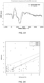
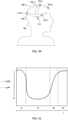
- the early component refers to the power(dt) decrease that was measured in a time period from 0.1 to 0.5 sec after the pain stimulus onset and/or the onset of any other suitable anxiety inducing event.
- Late component refers to the power(ta) increase that was measured in a time period from 0.38 to 0.78 sec after the pain stimulus onset.
- the power was calculated from -0.5s to 1.5s from with reference to the stimulus onset, or in other words the power was calculated in a time window of 2s including the stimulus onset.
- a time window of at least 0.1s for example a time window in the range of 0,5s up to and including 60s, or any other suitable time window is used, at any other suitable point in time with respect to the stimulus onset, as long as the time window includes at least a portion at a point in time after the stimulus onset.
- the power can be continuously calculated based on overlapping time windows of for example 2s, or of any other suitable time period, such as for example at least 0.1s, or in the range of 0.5s up to and including 60s, ...
- results are the first identifying location-specific brain frequency band differences between an automated hypnosis treatment session and a normal awake state, especially for a brain frequency band comprising and/or consisting of a combination of at least part of two brain waves such as for example theta and alpha brainwaves, or delta and theta brain waves. It is clear, that these results are also the first at identifying this for any other suitable treatment sessions, for example pharmacological and/or non-pharmacological treatment sessions, and/or in which no use is made of a treatment session.
- brain frequency band differences especially a brain frequency band comprising a combination of two brain waves, with respect to different levels of anxiety as experienced by the subject and/or levels of pain as perceived by the subject.
- a frequency band comprising and/or consisting of a combination of two brain wave frequencies, or at least a part thereof is different from a frequency band only comprising a single brain wave frequency band and different from a frequency band comprising more than two brain wave frequency bands.
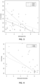
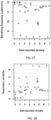
- the P-power (dt) calculated from the combined brainwave frequency band delta-theta surprisingly provides for a substantially higher correlation to the self-reported level of anxiety of the each of its composing individual brainwave frequency bands, i.e. delta and theta.
- the self-reported level of anxiety was correlated (spearman) with P-power (theta), i.e.
- the above experimental data generated during the experiment 500 which comprises both measurement data, such as EEG data, as well as self-reported data, and which could also be referred to as response data, confirms the existence of a correlation between self-reported level of anxiety and a power of a specific combined brain wave frequency band at a specific location, P-power (dt), associated with a delta-theta frequency band, dt, which according to this embodiment extends from 1Hz up to and including 6Hz, also referred to as a group 8 indicator.
- the frequency band comprises a combination of both delta brainwaves and theta brainwaves.
- an alternative external pain and/or anxiety stimulus could be provided. It is clear that instead of and/or in addition to measurement of the level of anxiety in reaction to an external pain and/or anxiety stimulus, the experimental data can also be used for measurement of a level of anxiety caused by internal events or states of the subject, which for example could relate to anxiety experienced, which is not caused by an external event, such as an external pain and/or anxiety stimulus, being applied. It is clear that alternative embodiments of the experiment are possible, in which the P-power(dt) is determined for any other suitable frequency band comprising a combination of both delta brain waves and theta brainwaves, or at least part thereof, such as for example any suitable frequency band extending in the frequency range of 0Hz up to and including 8 Hz.
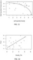
- the above experimental data generated during the experiment 500 which comprises both measurement data, such as EEG data, as well as self-reported data, and which could be referred to as response data, confirms the existence of a correlation between self-reported pain and a power, power (ta) associated with a theta-alpha frequency band, ta, which according to this embodiment extends from 6Hz up to and including 12Hz, also referred to as a group 4 indicator.
- the frequency band comprises a combination of both theta brainwaves and alpha brainwaves. It is clear from the experimental data above that measured data received from the C, P and F EEG electrodes all show a certain level of correlation of power (ta) with the self-reported measure of pain.
- the correlation is highest at the C EEG electrode, it is clear that according to alternative embodiments, the distribution of the level of the correlations amongst different EEG electrodes might be different. However, it is clear that according to alternative embodiments an alternative external pain stimulus could be provided. It is clear that instead of and/or in addition to measurement of the level of pain in reaction to an external pain stimulus, the experimental data can also be used for measurement of a level of pain caused by internal events or states of the subject, which for example could relate to pain experienced, which is not caused by an external event, such as an external pain stimulus, being applied.
- the power(ta) is determined for any other suitable frequency band comprising a combination of both theta brain waves and alpha brainwaves, such as for example any suitable band extending in the frequency range of 4Hz up to and including 12 Hz.
- the Level of Anxiety is an indicator of the strength of the psychophysiological response to an anxiety inducing event or situation. It is clear that such a level of anxiety as experienced by the subject can be modulated by for example its emotional, psychological, cognitive state, such as for example the state of consciousness of the subject.
- the LoA is thus an indicator for the level of anxiety as it is experienced of the subject and can thus be used as an indicator of the degree to which the subject experiences anxiety, for example on a cognitive, emotional and/or physical level.
- the Level of Pain, or LoP is an indicator of the level or degree to which a subject is aware and receptive to pain. In other words, it refers to the level of pain as consciously perceived by the subject.
- Such a level of pain perception by the subject can be modulated by for example its emotional, psychological, cognitive state, such as for example the state of consciousness of the subject.
- the LoP is thus an indicator for the subjective pain perception of the subject and can thus be used as an indicator of the degree to which the subject experiences pain, for example on an emotional, cognitive and/or physical level.
- anxiety includes a cognitive experience and optionally also a physiological experience by the subject.
- the level of anxiety as experienced by the subject is a subjective, psychophysiological experience that is influenced to varying degrees by for example biological, psychological, cognitive, physiological, neurophysiological and social factors.
- the experience that something causes anxiety includes for example cognitive features, such as for example intensity, quality, ... of an anxiety stimulus and/or affective emotional aspects.
- anxiety as experienced and purely autonomic physiological stress responses are different, and can for example occur independently of each other.
- a Level of Anxiety Index refers to a standardized index indicating the level of anxiety of which the subject is at least cognitively experiencing and optionally physiologically experiencing.
- the degree to which the subject experiences anxiety at a cognitive dimension optionally supplemented by for example an autonomic sensory and/or an emotional level as described in further detail below.
- This LoAI for example varies systematically with the intensity of anxiety as experienced by the subject, independently of factors not affecting the experience of anxiety.
- a first limit of the LoAI for example a lower limit of the LoAI, may for example indicate that the subject experiences no anxiety.
- a second limit of the LoAl for example an upper limit of the LoAI, may for example indicate that the subject experiences the strongest/worst possible experience of anxiety.
- pain in the context of this description is an aversive sensory and emotional experience typically caused by, or resembling that caused by, actual or potential tissue injury. It is thus clear that pain is experienced as a conscious perception by the subject.
- the level of pain as experienced by the subject is a subjective experience that is influenced to varying degrees by for example biological, psychological and social factors.
- the conscious perception that something causes pain includes for example sensory-discriminative features, such as for example intensity, quality and location, ... and/or affective emotional aspects. It should further be noted that in this way pain as consciously perceived and nociception are different phenomena, which can occur independently of each other.
- a conscious pain experience is not necessarily implied.
- a Level of Pain Index, or LoPI, or Pain index refers to a standardized index indicating the level pain of which the subject is consciously aware. In other words, the degree to which the subject consciously perceives pain at for example a sensory and/or an emotional level.
- This LoPI for example varies systematically with pain intensity as perceived by the subject, independently of factors not affecting pain perception.
- a first limit of the LoPI for example a lower limit of the LoPI, may for example indicate that the subject experiences no conscious perception of pain.
- a second limit of the LoPI for example an upper limit of the LoPI, may for example indicate that the subject experiences the strongest possible conscious perception of pain.
- such a computer-implemented method for an objective measurement of a level of anxiety experienced by a subject, a level of modified consciousness in a subject and/or a level of pain 300 as consciously perceived by a subject is shown in FIG. 5 .
- such a computer-implemented method 300 for an objective measurement of a level of anxiety or LoA as experienced by a subject, level of pain or LoP as perceived by a subject and a level of modified state of consciousness or DoS in a subject for example comprises the steps of receiving EEG data 302, extracting P-power(dt) 304 and determining a level of anxiety, LoA, 306.
- measured data comprising electroencephalogram, EEG, data collected from one or more P-EEG electrodes.
- a group 8 indicator corresponding to: a power, P-power (dt), associated with a delta-theta frequency band, dt, within a delta-theta frequency range.
- a level of anxiety LoA, which is a value indicative for the level of anxiety as experienced by the subject.
- the method could for example comprise additional steps, such as for example after the step of receiving EEG data or any other suitable data, pre-processing the EEG data or other data, subsequently extracting relevant features, thereby allowing the determination of for example group 8 indicators or other suitable group indicators, and subsequently normalization and/or classification, thereby for example determining LoA or an indexed and/or scaled version of LoA, such as for example LoAI as will be explained in further detail below. It is however clear that still further alternative embodiments are possible.
- the LoA is correlated to the group 8 indicator, P-power(dt), or in other words an increase in P-power(dt) corresponds to an increase in the LoA, and a decrease in P-power(dt) corresponds to an decrease in the LoA as experienced by the subject.
- P-power(dt) values mentioned above were calculated from - 0.5s to 1.5s from with reference to the stimulus onset, or in other words the power was calculated in a time window of 2s including the stimulus onset, according to an embodiment where there was an external pain and/or anxiety stimulus.
- alternative suitable time windows are being used, and/or in which the time window is not related to a specific external stimulus, such that for example the power can be continuously calculated based on overlapping time windows.
- the method shown in FIG. 5 which for example comprises the steps of receiving EEG data 302, extracting power(ta) 314 and determining a level of pain, LoP, 316.
- measured data comprising electroencephalogram, EEG, data collected from one or more EEG electrodes.
- a group 4 indicator corresponding to: a power, power (ta), associated with a theta-alpha frequency band, ta, within a theta-alpha frequency range.
- a level of pain, LoP which is a value indicative for the level of pain in the subject.
- the method could for example comprise additional steps, such as for example after the step of receiving EEG data or any other suitable data, pre-processing the EEG data or other data, subsequently extracting relevant features, thereby allowing the determination of for example group 4 indicators or other suitable group indicators, and subsequently normalization and/or classification, thereby for example determining LoP or an indexed and/or scaled version of LoP, such as for example LoPI as will be explained in further detail below. It is however clear that still further alternative embodiments are possible.
- step 318 there could be correlated a plurality of LoA, LoP and/or DoS measurements and/or determining a correlation between a plurality of LoA, LoP and/or DoS measurements as will be explained in further detail below, for example with reference to the embodiments of FIG. 10 - FIG. 16 , etc..
- the LoP is inversely correlated to the group 4 indicator, power(ta), or in other words an increase in power(ta) corresponds to a decrease in the LoP, and a decrease in power(ta) corresponds to an increase in the LoP as consciously perceived by the subject.
- response data for example representing a subject's response to the treatment session can also be used to determine the state of consciousness of the subject, in particular a level of a modified state of consciousness.
- the method comprises further steps for measuring a depth of state, DoS by means of a group 1 indicator and optionally a group 2 indicator.
- measured data at step 302 comprising EEG data comprising data collected from at least one frontal (F) EEG electrode located on the scalp anatomical region corresponding to a frontal lobe of the subject.
- This then allows the extraction of a group 1 indicator at step 308 based on, a power, F-power(dt), associated with a delta-theta frequency band, dt, within a delta-theta frequency range extracted from the at least one F- EEG electrode data.
- a DoS Based on this group 1 indicator, there can then be determined a DoS.
- DoS Based on the group 1 indicator, there can then be determined a depth of state, DoS, which is a value indicative for the level of a modified state of consciousness.
- determining a plurality of LoA and/or LoP at different DoS; and/or a plurality of LoP at different LoA; and/or a plurality of LoA at different LoP; etc. from measurement data and/or measurement data comprising simultaneous measurement of LoA and/or LoP and DoS obtained, preferably, but not necessarily under identical pain conditions and/or under identical anxiety inducing events.
- identical pain conditions do not necessarily mean constant pain conditions, it only means that at the time of measurement of the LoA and/or LoP and DoS are measured, the anxiety and/or pain conditions are identical, substantially identical and/or similar to.
- the measurement of LoA, LoP and/or DoS is not performed simultaneous. Based on such measurements there can then for example be determined the modulation of LoA and/or LoP by DoS, by evaluating the evolution of the LoA and/or LoP at different DoS and/or the evolution of the LoA and/or LoP with respect to the evolution of DoS from the measurement data obtained, preferably, but not necessarily under identical pain and/or anxiety conditions.
- the LoP can be adjusted based on said DoS and/or said LoA to a level of normalized pain, NLoP, which is a value indicative for the level of pain in the subject at a predetermined reference DoS and/or a predetermined reference LoA.
- the LoA can be adjusted based on said DoS and/or said LoP to a level of normalized anxiety, NLoA, which is a value indicative for the level of anxiety in the subject at a predetermined reference DoS and/or at a predetermined reference LoP.
- the reference DoS could for example be that of a normal awake state
- the reference LoP could for example be a level of pain where no or a minimal amount of pain is perceived
- the reference LoA could for example be a level of anxiety where no or a minimal level of anxiety is experienced, such as for example shown in FIG. 7 - FIG.
- these can be replaced by the non-indexed and/or scaled values such as DoS, LoA, NLoA, LoP, NLoP, etc. or vice versa.
- the value for NLoP would provide for a value of LoP at a predetermined reference DoS of interest, independently of the current DoS and/or any changes to the current DoS, thereby for example providing an indicator for the equivalent amount of pain that would be consciously perceived when the subject would be, or return to a normal awake state of consciousness, when this reference DoS is the normal awake state such as for example shown in Fig. 7-9 .
- NLoP or NLoAI can be calculated as follows.
- a DoSI of 50% is measured and a LoPI or LoAI of 15%, this refers to a particular point which is part of a predetermined correlation between DoS and LoP or DoS and LoA, for example determined by measuring LoPI or LoAI at a plurality of different DoSI under identical pain and/or anxiety conditions, however it is clear that alternative embodiments are possible.
- the NLoPI or NLoAI is 40% at the reference DoSI, which according to this embodiment is set to 100%. This means that when the DoSI would be changed from the measured DoSI of 50% to the reference DoSI of 100%, the level of pain perceived by the subject or the level of anxiety experienced by the subject, for example under the current pain and/or anxiety conditions or in other words when the pain and/or anxiety conditions would remain identical or unchanged, would rise from the measured 15% to 40%.
- the reference DoSI which according to this embodiment is set to 100%.
- the LoA based on said DoS is adjusted to a level of normalized anxiety, NLoA, which is a value indicative for the level of anxiety in the subject at a predetermined reference DoS.
- NLoA a level of normalized anxiety
- the LoA is also influenced by the LoP, and therefor similarly the LoA could be adjusted, based on said LoP, instead of and/or in addition to the adjustment based on the DoS, to a level of normalized anxiety, NLoA, which is a value indicative for the level of anxiety in the subject at a predetermined reference LoP, and optionally at a predetermined reference DoS.
- the LoP may be modulated by the LoA and the DoS as shown in further detail below
- this will for example help to determine the modulation of LoP by DoS and/or LoA, by evaluating the evolution of the LoP at different DoS and/or LoA and/or the evolution of the LoP with respect to the evolution of DoS and/or LoA from the measurement data.
- FIG. 10 shows an exemplary embodiment of a method in which a plurality of LoA and LoP are correlated, based on the measurement data from the embodiment of the experiment of FIG. 1 .
- This correlation thus functions in such a way that the LoA increases as LoP increases and can even trigger pain perception.
- a combined measurement of LoA and LoP and the calculation of any corresponding correlations thus allows to determine which part of the LoP is associated with the LoA and could for example be treated by an anxiety management treatment.
- a combined measurement of LoA and LoP and the calculation of any corresponding correlations allows to determine which part of the LoA is associated with the LoP and could for example be treated by a pain management treatment.
- pain can be a causes of anxiety and vice versa
- the combined measurement of LoA and LoP and optionally the calculation of any correlations therebetween make it possible to determine the root cause, modulator, aggravating factor, etc. of anxiety and/or pain and to treat it effectively by means of a suitable treatment, for example an anxiety management treatment when the LoA is the main contributing component, or a pain management treatment when the LoP is the main contributing component, and/or a suitable weighted mix of an anxiety and pain management treatment, depending on the respective weights in the contribution of the LoA and/or the LoP and their respective correlation.
- FIG. 11 shows an exemplary embodiment of a method in which a plurality of LoA and DoS are correlated, based on the measurement data from the embodiment of the experiment of FIG. 1 .
- there is an inverse correlation between the DoS and the LoA which means that an increase in the depth of dissociation decreases the level of anxiety.
- the combined measurement of LoA and DoS and optionally their correlation for example enables to determine which part of the LoA could be managed by means of a treatment modifying the DoS, such as for example a treatment modifying the depth of dissociation, the level of absorption, etc.
- a treatment modifying the DoS such as for example a treatment modifying the depth of dissociation, the level of absorption, etc.
- it might even be possible to determine and/or differentiate between short-term and long-term effects of for example treatments involving a modification of the DoS for example by means of managing the depth of dissociation, the level of absorption, etc. for example by means of suitable pharmacological and/or non-pharmacological treatments, such as for example psychotherapy, etc. on a change, for example a reduction, in the level of anxiety (e.g. use in psychotherapies , ).
- FIG. 12 shows an alternative embodiment of a method in which a plurality of LoA and LoP are correlated, based on the measurement data from the embodiment of the experiment of FIG. 1 .
- the level of anxiety and the level of pain are not measured simultaneously.
- the LoA was measured before the test phase 520, for example at step 512 of the embodiment of FIG. 1
- the LoP was measure during the test session, preferably during and/or after painful, anxiety inducing stimulation, such as for example at step 534 and/or 544.
- the LoA is for example measured before a treatment and the LoP during and/or after the treatment, and/or any other suitable embodiment in which the LoA and the LoP are measured at different moments in time.
- a higher level of anxiety before the test phase 520, or any other treatment session is associated with a higher level of pain during the test phase 520, or any other suitable treatment session.
- Such type of correlation could be useful in measuring, managing and/or predicting, post-treatment levels of pain based on the LoA measured before the treatment and optionally combined with LoP measured before the treatment. According to a particular embodiment, this would for example enable to measure, manage and/or predict post-intervention pain.
- the pre-treatment measurement of a LoA and optionally the LoP could for example enable to determine subjects and/or a population for which an anxiety management treatment, before engaging in another treatment that comprises potential anxiety inducing stimuli, so that by managing the pre-treatment level of anxiety, the level of pain during and/or after the treatment could be reduced.
- anxiety is also a risk factor for developing persistent and/or chronic pain following an acute pain and/or anxiety inducing event.
- the combined, non-simultaneous measurement of LoA and LoP and any correlation, for example pre-treatment, could determine high risk subjects and/or a high risk group of subjects, with an increased risk of post-treatment chronic pain, and could potentially enable a reduction of this risk of developing chronic pain, for example by means of a suitable anxiety management treatment.
- FIG. 13 shows an exemplary embodiment of a method in which a plurality of LoA and DoS are correlated, based on the measurement data from the embodiment of the experiment of FIG. 1 .
- the LoA and DoS are not measure simultaneously.
- the LoA is measured before the treatment session
- the DoS is measured during the treatment session 540 of the test phase 520.
- a higher level of anxiety before the treatment session is associated by means of the correlation with a lower level of dissociation during the treatment session.
- the level of anxiety before the treatment session limits the maximum level of and/or the maximum modification of the DoS that can subsequently be achieved during the treatment session involving a modified state of consciousness, such as for example the hypnotic treatment session 540 of the embodiment of Fig.1 or any other suitable embodiment of a treatment session.
- the non-simultaneous combined measurement of the LoA and DoS and its associated correlation enables for example to determine more objectively subjects with a too high anxiety level that could for example need of an anxiety treatment in order to reduce the LoA to within a predetermined desired range, before engaging in a further treatment session, for example a treatment involving a modified state of consciousness or any other suitable treatment.
- this thus means that when a pre-treatment LoA of for example 6 or higher is measured, it has no sense of initiating the treatment session involving a modified state of consciousness as it will not be able to modify the DoS, which according to this embodiment is embodied as the depth of dissociation.
- the pre-treatment LoA will need to be reduced, until a suitably high associated DoSLimit is reached, for example by means of a suitable anxiety management treatment.
- a desired DoS is to be reached, for example by means of a suitable treatment
- the LoA associated with the DoSLimit will provide for a maximum level of the LoA that is acceptable when it is desired to reach his DoS.
- LoA and/or LoP associated with a DoSLimit will define suitable limit values and/or ranges for a respectively desired LoA, LoP and/or DoS.
- a LoA is measured associated with a LoPLimit that is higher than a desired LoP
- a treatment adjusting the LoP will need to be adjusted, such as for example a suitable pain management treatment, such that a suitable associated LoALimit can be attained, of which the desired LoA falls inside its range and/or limits.
- Fig 14 shows still a further embodiment in which the level of pain and the level of anxiety are correlated.
- the LoA and the LoP are simultaneously measured.
- the data is split into data points associated with a high level of dissociation indicated with a circle and data points associated with a low level of dissociation, which is an embodiment of DoS.
- Both the data with a low DoS as the data with a high DoS have a good correlation.
- this could for example be used for reducing the impact of the LoA on the LoP by for example increasing the depth of dissociation, or any other suitable DoS, or vice-versa, as the depth of dissociation then supports an enhanced effect of either LoP or LoA.
- the measured LoA doesn't change considerably in both states shown in FIG. 15 and FIG. 16 .
- the LoP is clearly reduced at higher depth of dissociation such as shown in FIG.
- the negative impact of the LoA on the LoP is reduced at a higher depth of dissociation, or any other suitable embodiment of a DoS.
- This could for example be used in an advantageous way by determining subjects and/or populations that could benefit from a treatment aimed at raising the depth of dissociation in order to indirectly manage pain, by reducing the any negative impact of the LoA.
- a representation could for example be integrated in an embodiment of a GUI, similar as will be described below, and will for example allow a practitioner to assess during a treatment which involves a modified level of consciousness, how the level of anxiety or the level of pain will evolve after the treatment when the subject returns to a normal awake state, and/or vice versa.
- the reference DoSI could for example be implemented as a suitable, modifiable input parameter.
- Fig. 9 This is for example illustrated by means of Fig. 9 , in which in a modified state of consciousness, for example the result of non-pharmacological treatment for modifying the level of consciousness as explained with reference to FIG. 1 , however it is clear that alternative embodiments might be possible, for example involving pharmacological treatments modifying the state of consciousness, the DoSI has been reduced to 50% and there is measured a LoA of 25% and a LoPI of 15%, similar as in the example above with respect to FIG. 8 . In a subsequent phase of the treatment, as shown, the subject is allowed to awaken and the measured DoSI rises until in the normal awake state it reaches for example 100%.
- the measured LoAI will also rise from 25% until it reaches 50% in the normal awake state and the measured LoPI will also rise from 15% until it reaches 40% in the normal awake state.
- the NLoAI for example calculated as described above to be 50%
- the NLoPI for example calculated as described above to be 40%
- NLoAI will remain constant at 50% and NLoPI remains constant at 40% independent of the changes of any DoSI.
- calculation of NLoA and/or NLoP based on a predetermined reference DoS may allow for a comparison or use of the NLoA and/or NLoP between subjects/populations independent of their DoS, for example in the context of developing and/or determining suitable scales and/or indexes for LoA, LoP and/or DoS. It is however clear that still further alternative embodiments and uses of NLoA and NLoP are possible.
- the NLoA is a value indicative for the level of anxiety in the subject at a predetermined reference LoP and/or a predetermined reference DoS
- the NLoP is a value indicative for the level of pain in the subject at a predetermined reference DoS and/or a predetermined reference LoA.
- the NLoAI after the treatment might for example remain close to or equal the minimum LoA as attained during the treatment, which for example could be a treatment involving a modified state of consciousness, however alternative treatments, such as for example any suitable anxiety management treatments might be possible.
- LoA when for example returning to a normal awake state of consciousness after the treatment might be lower than it was before the treatment.
- the LoAI which is reduced during the treatment, would thus remain at such a lower level as compared to before the treatment.
- the NLoAI after the treatment, for the reference DoSI could for example be calculated based on the LoAI before the treatment and the minimum value for DoSI and/or LoAI as attained during the treatment, or in any other suitable way.
- a further derived index is referred to as the Recovery coefficient.
- this will for example allow for the calculation of the NLoA based on such a recovery coefficient.
- Min(LoA) is the minimum level of LoA reached during the treatment session.
- a subject that reached a DoS of 50% during the treatment session will have a LoA after the session, which as shown in the embodiment of FIG. 54 , at the reference DoS of 100% will be equal to the NLoA and which could be computed as Min(LoA) multiplied by the recovery coefficient correlated to this DoS of 50% during the treatment.
- NLoA will thus be Min(LoA) * 1.4.
- the full line curve shows the such a correlation for an alternative treatment, and it is clear that for such a treatment, for a subject, that reached a DoS lower than 40% during the treatment session, the LoA after the session, or in other words the NLoA at the reference DoS could be computed as Min(LoA)*1, as the recovery coefficient is approximately 1.
- the LoA will not increase or remain substantially the same as the minimum LoA attained during the treatment.
- this computer-implemented measurement of the LoA combined with DoS and/or LoP based on measurement data could be applied to measure, monitor and/or predict an evolution, such as for example a treatment induced evolution of the LoA, DoS and/or LoP.
- a treatment such as for example an anxiety management treatment, a pain management treatment, or any other suitable treatment
- the effect of this treatment on the LoA, DoS and/or LoP may be monitored by such an embodiment of the method.
- Such treatments might for example comprise any suitable treatment, such as non-pharmacological treatments, such as for example non-pharmacological anxiety treatments, non-pharmacological sedation, hypnosis, other evidence-based psychological and/or mind/body interventions, etc.
- FIG. 6 shows an embodiment of a computer-implemented method for measuring and/or monitoring a LoA, a DoS and/or LoP in a subject before, during and/or after a treatment 400.
- the embodiment of the method 400 shown in FIG. 6 comprises the steps of:
- the treatment, and its associated chronology has been schematically illustrated by means of arrow 408 in the embodiment of FIG. 6 , which could for example represent a singular treatment session with a well-defined start and end-point, however it is clear that alternative embodiments are possible in which the treatment for example has continuous and/or chronic nature without a well-defined start and end-point, or in which the treatment has an intermittent nature and comprises a sequence of a plurality of treatment sessions with optional breaks or pauses in between these treatment sessions, or any other suitable type of treatment comprising a chronological nature, for example comprising a sequence of treatment phases, steps, etc.
- the LoA, DoS and/or LoP is measured, for example with the embodiment of the method for measurement of DoS and LoP 300 according to the embodiment shown in FIG. 5 .
- This first measurement of LoA, DoS and/or LoP could for example be a measurement of the LoA, combined with DoS and/or LoP before the start of a treatment session and could, according to some embodiments be considered as a reference, initial, baseline, ... LoA, DoS and/or LoP respectively that serves as a predetermined reference level for subsequent measurements of LoA, DoS and/or LoP.
- This first measurement of LoA, DoS and/or LoP could for example be a measurement of the LoA, combined with DoS and/or LoP before the start of a treatment session and could, according to some embodiments be considered as a reference, initial, baseline, ... LoA, DoS and/or LoP respectively that serves as a predetermined reference level for subsequent measurements of LoA, DoS and/or LoP.
- the second, subsequent measurement of LoA, DoS and/or LoP is for example also measured with the method for measurement of LoA, DoS and/or LoP 300 shown in FIG. 5 .
- the treatment-induced evolution of the LoA, DoS and/or LoP based on a comparison of the first LoA, DoS and/or LoP and a respective second LoA, DoS and/or LoP, measured at steps 402 and 404.
- the LoA, the LoP and/or the DoS there is measured and/or monitored and/or predicted and/or aggregated the LoA, the LoP and/or the DoS at at least two different points in time, for example before, during and/or after the treatment; and/or over a period of time.
- the LoA, the LoP and/or the DoS are measured and/or monitored at at least two different points in time during any suitable time period.
- the treatment-induced evolution of the LoA, the LoP and DoS is determined based on a respective comparison of the LoA, the LoP and DoS measured for example during and/or after the treatment with the LoA, the LoP and DoS respectively measured before the treatment.
- comparisons could be performed to determine any suitable evolution of the LoA, the LoP and DoS during any suitable time period.
- the LoA, the LoP and/or the DoS could also be predicted.
- a suitable plurality of historical data points with respect to LoA, LoP and/or DoS could provide for a suitable trend analysis, for example based on a trend correlating to the a running average, mean, or any other suitable trend of the historical data points, thereby allowing prediction of future, expected or trending data points. It is clear that in this way a prediction of an evolution of the LoA, the LoP and the DoS can be accomplished.
- Predictive analysis of the historical data points could be done by means of any suitable method, for example based on predetermined correlations between the evolution of LoA, LoP and/or DoS during different phases or time periods of a particular treatment, or by means of self-learning, self-optimizing algorithms, artificial intelligence algorithms, etc. which have been trained on historical data sets in order to detect repeating and/or predictable patterns and/or correlations, thereby allowing for an assessment of detectable patterns and/or correlations in the measured data, thereby allowing for a determination of current and predictive trends based on the measured data.
- a level of anxiety score or LoAS there may be aggregated a level of anxiety score or LoAS, a level of pain score or LoPS and/or a Depth of State Score or DoSS based on an aggregation of respectively at least two LoA, at least two LoP and/or at least two DoS measured at at least two different points in time.
- the aggregation of respectively at least two LoA, at least two LoP and/or at least two DoS measured could be performed over a predetermined period of time.
- Such aggregated parameters such as LoAS related to anxiety measurement could be useful in determining the impact of a chronic state of anxiety, which might be related to a personality trait and/or an anxiety disorder.
- An aggregated parameter such as a LoPS related to pain measurement could be useful in determining the impact of chronic pain, in which for example during a longer period, of for example several hours and/or days the level of pain is continuously measured, and the aggregated LoPS is able to provide for an indicator of the impact of a constant, chronic level of pain as perceived by a subject as opposed to an instantaneous peak of pain.
- This is useful as even moderate or low levels of anxiety and/or pain, when experienced chronically, may have a high impact on the quality of life of subjects, while for example short, infrequent bursts of anxiety and/or pain, even when they are intense can be tolerated more easily.
- an aggregated DoSS may be useful in identifying the level of safety and stability associated with a modified level of consciousness during a particular time period. For example, during surgery, it is valuable to ascertain whether the modified level of consciousness has been established in a sufficiently stable way, and is not the result of a temporary, short term, evolution of the level of consciousness. It is clear that such aggregated forms could also be useful for any other parameters related to the level of pain and the state of consciousness as detailed in this description, and/or their scaled and/or indexed versions, such as for example LoA(I), LoP(I), DoS(I), NLoP(I), DoD(I), DoH(I), etc.
- the LoA(I)S, LoP(I)S, DoS(I), NLoP(I)S, DoD(I)S, DoH(I)S, etc. could for example comprise any suitable embodiment of aggregation of the respective LoA(I),LoP(I), DoS(I), NLoP(I), DoD(I), DoH(I), etc. values over a predetermined time period, such as for example a sum, mean, average, etc.
- the value could for example be presented in a GUI of a system similarly as described below, for example presenting the aggregated value, optionally in relation to the time period during which the LoA(I), LoP(I), DoS(I), NLoP(I), DoD(I), DoH(I), etc. was aggregated.
- the treatment-induced evolution of the LoA combined with DoS and/or LoP can be determined based on a respective comparison of the LoA combined with DoS and/or LoP determined during and/or after a treatment, with the LoA combined with DoS and/or LoP determined before a treatment.
- the treatment induced evolution of the LoA is determined by a comparison of the LoA determined during and after the treatment according to this embodiment, and/or that the treatment induced evolution of the DoS, similarly is determined by a comparison of DoS determined during and after the treatment, and/or that the treatment induced evolution of the LoP is determined by a comparison of the LoP determined during and after the treatment.
- the pain management treatment when the treatment is a pain management treatment, the pain management treatment could be replaced and/or combined with an anxiety management treatment; and/or when the treatment is an anxiety management treatment, the anxiety management treatment could be replaced and/or combined with a pain management treatment, when the LoP and/or the LoA is outside the predetermined range and/or until the LoP and/or the LoA is inside the predetermined range.
- the anxiety management treatment it is beneficial to replace and or combine the pain management treatment with an anxiety management treatment, at least until the anxiety management treatment was able to provide for a treatment induced evolution of the LoA, for example by suitably reducing the LoA to within predetermined limits, such that the LoA is again in the predetermined range in which a pain management treatment is known to be effective for managing the LoP upon which the pain management treatment can be continued and/or resumed and optionally the anxiety management treatment can be stabilized, reduced and/or ended.
- the anxiety management treatment can be replaced with a pain management treatment when the LoP and/or the LoA is outside a predetermined range and/or until the LoP and/or the LoA is inside the predetermined range.
- a pain management treatment when for example the LoP is exceeds a predetermined limit and/or threshold for any anxiety management treatment to be effective, for example first the LoP is reduced till the predetermined limit is no longer exceeded, or until it is within any suitable predetermined range for the anxiety management treatment to be effective in managing the LoA again.
- any suitable combination of an anxiety management treatment and/or a pain management treatment and/or a treatment involving a modification of the state of consciousness are used in order to ensure that LoP and/or LoA and/or DoS are respectively managed such that they become and/or remain within a respective predetermined range for enabling any further treatment to be efficient.
- the treatment when the treatment is a treatment modifying the state of consciousness it might for example be beneficial to replace and/or combine the treatment modifying the state of consciousness with an anxiety and/or pain reducing treatment when the LoP and/or LoA is outside a predetermined range and/or until the LoP and/or the LoA is inside the predetermined range.
- the pain management treatment when the treatment is a pain management treatment, can be replaced and/or combined with a treatment modifying the state of consciousness when the DoS is outside a predetermined range and/or until the DoS is inside the predetermined range.
- the treatment modifying the state of consciousness can be replaced and/or combined with a pain management treatment when the LoP is outside a predetermined range and/or until the LoP is inside the predetermined range.
- the efficacy of a pain management treatment such as for example a pain-reducing, pain-controlling, pain-preventing, or any other suitable pain management treatment can be determined and/or monitored based on the treatment-induced evolution of the NLoP.
- the efficacy of an anxiety-reducing treatment can be determined and/or monitored based on the treatment-induced evolution of the NLoA.
- the treatment is a treatment modifying the state of consciousness for reducing anxiety, such as for example a non-pharmacological treatment such as a hypnosis treatment session, a mind-body treatment, cognitive behavioral therapy, etc. , or a pharmacological treatment, for example a sedative treatment
- a treatment-induced evolution of the DoS and/or the LoA based on a comparison of for example the DoS and/or the LoA measured during and/or after the treatment modifying the state of consciousness with the DoS and/or the LoA measured before the treatment modifying the state of consciousness.
- the treatment is a treatment modifying the state of consciousness for reducing pain, such as for example a non-pharmacological treatment such as a hypnosis treatment session, or a pharmacological treatment, for example a sedation treatment
- a treatment modifying the state of consciousness for reducing pain such as for example a non-pharmacological treatment such as a hypnosis treatment session, or a pharmacological treatment, for example a sedation treatment
- the DoS at at least two different points in time, before, during and/or after the treatment modifying the state of consciousness
- there can be determined the treatment-induced evolution of the DoS and/or the LoP based on a comparison of the DoS and/or the LoP measured during and/or after the treatment modifying the state of consciousness with the DoS and/or the LoP measured before the treatment modifying the state of consciousness.
- a treatment and/or determining an adjustment to a treatment and/or determining an intensity of a treatment based on the determined and/or monitored NLoA and/or NLoP and/or DoSLimit and/or LoPLimit and/or LoALimit respectively.
- NLoA and/or the NLoP are possible. According to further embodiments this for example allows to determine a treatment and/or an adjustment to a treatment and/or an intensity of a treatment and/or the efficacy of a treatment and/or the choice of a treatment based on the measured and/or monitored and/or predicted and/or aggregated NLoA and/or NLoP and/or DoSLimit and/or LoPLimit and/or LoALimit.
- a treatment-induced evolution of the LoA which for example corresponds to a reduction of the second LoA measured at step 304 as compared to the first LoA measured at step 302 and treatment-induced evolution of the DoS that for example corresponds to a decrease of the second DoS measured at step 304 as compared to the first DoS measured at step 302, is able to confirm the effectiveness of the treatment.
- the value of the treatment-induced evolution of the DoS and LoA also provides for an indication of the intensity of the treatment.
- an anxiety management treatment reduces a first LoA measurement with a first value at step 402 to second value, that corresponds to a 50% reduction of the first value, at a second measurement at step 404, which thus corresponds to a treatment-induced evolution of the LoA of -50% or in other words a change in the LoA corresponding to a reduction of 50% with respect to the measurement at step 302 before the treatment.
- this treatment is adapted or another treatment is applied, which only causes a treatment induced evolution of the LoA of -25%, it is clear that this latter treatment has an intensity that is lower than the former.
- the treatment-induced evolution of the LoA corresponds to no change to the LoA, or in other words, that the treatment at that point in time is for example not effective in reducing the LoA and might need adaptation or change to another type of treatment.
- a treatment induced evolution of the DoS could also be quantified to indicate the intensity of the treatment, preferably in combination with the LoA.
- the treatment comprises the application of anxiety inducing stimuli, such as for example during a medical treatment involving an injection, administration of local sedation, etc.
- the anxiety management treatment for example in the form of pharmacological and/or non-pharmacological anxiety management treatment is performing effectively.
- a treatment-induced evolution of the LoP that for example corresponds to a reduction and/or the absence of an undesired increase of the second LoP measured at step 304 as compared to the first LoP measured at step 302 and treatment-induced evolution of the DoS that for example corresponds to an decrease of the second DoS measured at step 304 as compared to the first DoS measured at step 302, is able to confirm the effectiveness of the treatment.
- the value of the treatment-induced evolution of the DoS and LoP also provides for an indication of the intensity of the treatment.
- a pain reducing treatment reduces a first LoP measurement with a first value at step 402 to second value, that corresponds to a 50% reduction of the first value, at a second measurement at step 404, which thus corresponds to a treatment-induced evolution of the LoP of -50% or in other words a change in the LoP corresponding to a reduction of 50% with respect to the measurement at step 302 before the treatment.
- this treatment is adapted or another treatment is applied, which only causes a treatment induced evolution of the LoP of -25%, it is clear that this latter treatment has an intensity that is lower than the former.
- the treatment-induced evolution of the LoP corresponds to no change to the LoP, or in other words, that the treatment at that point in time is for example not effective in reducing the LoP and might need adaptation or change to another type of treatment.
- a treatment induced evolution of the DoS could also be quantified to indicate the intensity of the treatment, preferably in combination with the LoP.
- the treatment comprises the application of painful stimuli, such as for example during a surgical treatment
- the LoP as measured before the treatment, and/or the DoS attained during a particular phase of the treatment does not change, does not increase and/or does not increase outside a predetermined desired range, during the treatment, especially during and/or after the application of the painful stimuli
- the pain treatment for example in the form of pharmacological and/or non-pharmacological sedation is performing effectively.
- the measurement of a further LoA combined with and/or correlated with a DoS and/or LoP at step 404, at a later point in time than at step 402, could be repeated or could be performed continuously, which allows for a repeated and/or continuous monitoring of the treatment-induced evolution of the LoA, DoS and/or LoP, or in other words the comparison of two LoA, DoS and/or LoP respectively measured at two different points in time.
- any suitable plurality of LoA, DoS and/or LoP measurements at different points in time, before, during and/or after the treatment are used in order to determine and/or monitor a treatment-induced evolution of the LoA, DoS and/or LoP, based on a comparison of these at least two LoA, DoS and/or LoP measured at at least two different points in time. It is thus clear that such a treatment induced evolution of the LoA, DoS and/or LoP is indicative of a treatment induced change, if any, to the LoA, DoS and/or LoP.
- the LoP can optionally be determined by means of additional indicators, such as for example a group 3 indicator or a group 5 indicator as described in further detail below.
- a group 3 indicator in the time domain for example by means of a suitable event related potential, or ERP, in response to the calibrated pain stimulus.
- ERP event related potential
- Such a time-domain EEG analysis showed an overall decreased in the ERP components during the hypnotic treatment session 540 as compare to the normal awake state 530.
- MSPA mean signal peak-to-peak amplitude
- the time window could for example be set from 300ms to 350ms after the painful stimulus onset, which is represented as 0ms on the x-axis.
- the MSPA is measured on an ongoing basis, for example by means of a suitable, rolling time window, not linked or triggered by an external pain stimulus.
- the MSPA is equal to the peak-to-peak amplitude of the signal. In case there are multiple peaks in the signal in the time window for measurement of the MSPA, the MSPA will be calculated in function of the mean or average of the individual peak-to-peak amplitudes of the plurality of these peaks of the signals.
- the study based on this experiment is thus showing statistically significant MSPA changes at those specific locations of the EEG measurement data, when comparing a modified state of consciousness for example during a non-pharmacologically, e.g. hypnotically induced treatment session, with a normal awake state 530, and which correlate to the different level of pain perceived in reaction to a calibrated painful stimulus.
- the MSPA is equal to the peak-to-peak amplitude of the signal.
- the MSPA will be calculated in function of the mean or average of the individual peak-to-peak amplitudes of the plurality of these peaks of the signals.
- the time window for measurement could for example be in the range of 0.01s to 10s, such as for example in the range of 0.1s to 5s.
- the study of the experiment 500 mentioned above thus shows significant MSPA changes in the measured EEG data at those specific EEG sensor locations when comparing a modified state of consciousness during a hypnotic treatment session 540, or any other suitable non-pharmacological or pharmacological treatment inducing a modified state of consciousness, with a normal awake state 530, or in other words when comparing different levels or depths of dissociation or DoD, which is an embodiment of different depth of states or DoS. It is clear that this also means that significant MSPA changes in the measured EEG data have been observed when comparing different levels of pain as perceived by the subject, as the level of pain perceived by the subject during the normal awake state 530 differed from the level of pain perceived by the subject during the hypnotic treatment session 540.
- the insights of the experiment 500 above allow for an alternative embodiment of a computer-implemented method 300 for measurement of a level of pain comprising the further step of extracting from the EEG data, a group 3 indicator corresponding to a mean signal peak-to-peak amplitude, MSPA. Then at a subsequent step the LoP is determined based on the group 4 indicator and at least the group 3 indicator. It is clear that similarly an alternative embodiment of the computer-implemented method 400 for determining and/or monitoring a level of anxiety, a level of modified consciousness and/or a level of pain of a subject, comprising this alternative method for measurement of a level of anxiety, a level of modified consciousness and/or a level of pain is possible.
- a computer-implemented method 300 for measurement of a level of pain comprising the further step of receiving measured data comprising muscular activity data, such as for example electromyography, EMG, data, for example collected from one or more EMG electrodes or any other suitable electrode.
- EMG electrodes could for example be positioned at one or more of the following positions: a leg, face, arm, or any other suitable position, ....
- a group 5 indicator corresponding to a mean signal peak-to-peak amplitude EMG-MSPA. Subsequently then determining the LoP based on the group 4 indicator and at least the group 5 indicator.
- the LoP is determined based on the group 4 indicator, the group 3 indicator, and the group 5 indicator.
- the method for measuring the LoA, DoS and/or LoP 300 comprises the step of receiving the measured data of the subject comprising the electroencephalogram, EEG, data collected from one or more of the following EEG electrodes:
- Electroencephalography is a technique well known in the art for recording electrical activity of the brain.
- EEG Electrodes placed along the scalp at certain locations measure voltage fluctuations resulting from electrical impulses within the neurons of the brain, mainly in the region of the brain close to the scalp, including the cortex.
- EEG measurement data reflects how different neurons/ populations of neurons in the brain networks communicate with each other by means of electrical impulses.
- Electroencephalography produces an electroencephalogram.
- the electroencephalogram which thus forms the EEG data, may be refined to a particular electrode location, such as the frontal, parietal or central electrodes mentioned above. From the EEG data there may for example be extracted a frequency-based parameter and/or a phase-based parameter, and/or amplitude-based parameter.
- Frequency-based parameters may for example be obtained by a frequency-based analysis of EEG measurement data.
- a frequency based analysis of EEG measurement data may for example be used to measure the power contained in a specific signal frequency band, where power refers to a square of amplitude of the frequency domain signals typically within a specified in a frequency band or range, such as for example a frequency band spanning or within a delta-theta frequency range, or a frequency band spanning or within a theta-alpha frequency range.
- the "delta-theta" (dt) frequency band refers for example to a band of frequencies in a range encompassing both delta and theta brain waves.
- the delta-theta (dt) frequency band is typically greater than 0 Hz and equal to or less than 8 Hz.
- the theta brain waves are preferably slow theta brain wave, for example greater than 3 Hz and equal to or less than 6 Hz.
- the width of the delta-theta (dt) frequency band may be at least 2 Hz.
- Most preferably the delta-theta (dt) frequency band is greater than or equal to 1 Hz and less than or equal to 6 Hz.
- the delta-theta (dt) frequency band comprises and preferably consists of a combination of at least part of the delta frequency band and at least part of the theta frequency band.
- the "theta-alpha" (ta) frequency band refers to a band of frequencies in a frequency range encompassing both theta and alpha brain waves.
- the theta-alpha (ta) frequency range is typically greater than or equal to 4 Hz and equal to or less than 12 Hz.
- the theta brain waves are preferably fast theta brain waves, for example greater than 6 Hz and equal to or less than 8 Hz.
- the width of the theta-alpha (ta) frequency band may be at least 2 Hz. Most preferably the theta-alpha (ta) frequency band is greater than 6 Hz and less than or equal to 12 Hz.
- the theta-alpha (ta) frequency band comprises and preferably consists of a combination of at least part of the theta frequency band and at least part of the alpha frequency band.
- a phase-based parameter may be obtained by phase-based analysis of EEG measurement data.
- An amplitude-based parameter may be obtained by amplitude -based analysis of EEG measurement data, such as for example a mean signal peak-to-peak amplitude or MSPA in a time domain.
- the signal peak amplitude is the maximum excursion of the signal wave from the zero point.
- the signal peak-to-peak amplitude is the distance from a negative peak to a positive peak.
- a subject may experience during the hypnotic treatment session 540 an increased activity in the frontal cortex and/or in the frequency range of delta waves and a decreased activity in the frontal and/or occipital network and/or in the frequency range of alpha waves, as compared to the activity during the normal awake state 530.
- the EEG data may be acquired with one or more EEG electrodes placed on the scalp in the anatomical regions corresponding to the position of the frontal lobe, the parietal lobe and/or both precentral and postcentral gyrus.
- a frontal EEG electrode or F-EEG electrode is located on the scalp anatomical region corresponding to the frontal lobe. It is configured for attachment to or contact with the scalp anatomical region corresponding to the frontal lobe. It records F-EEG electrode data.
- the frontal lobe refers to the anatomical part of the brain. The region of the frontal lobe may be along the sagittal plane, more preferable to in middle of the scalp anatomical region corresponding to the frontal lobe, along the midsagittal plane.
- a parietal EEG electrode or P-EEG electrode is located on the scalp anatomical region corresponding to the parietal lobe. It is configured for attachment to or contact with the scalp anatomical region corresponding to the parietal lobe. It records P-EEG electrode data.
- the parietal lobe refers to the anatomical part of the brain. The region of the parietal lobe may be along the sagittal plane, more preferable to in middle of the scalp anatomical region corresponding to the parietal lobe, along the midsagittal plane.
- a central EEG electrode or C-EEG electrode is located on the scalp anatomical region corresponding to the precentral and postcentral gyrus. It is configured for attachment to or contact with the scalp anatomical region corresponding to the precentral and postcentral gyrus. It records C-EEG electrode data.
- the precentral and postcentral gyrus refers to the anatomical part of the brain.
- the region of the precentral and postcentral gyrus may be along the sagittal plane, more preferable to in middle of the scalp anatomical region corresponding to the precentral and postcentral gyrus, along the midsagittal plane.
- EEG electrodes are arranged at the above positions, and or additional suitable scalp anatomical regions.
- one localized EEG electrode may be used, such as for example positioned at the scalp anatomical region of the frontal lobe.
- a second electrode, or ground electrode may be employed for common mode rejection, i.e. to dissociate relevant information from noise; this can be placed anywhere on the scalp, for instance in another region of the scalp, in a bilateral position.
- a third electrode, or reference electrode may be employed to compute voltage differences. This third electrode can be placed anywhere on the scalp, for instance at an electrically neutral location.
- the measured data may comprise one or more measured data components, such as for example EEG data, which is a measured data component, such as for example also EMG data, ECG data, EOG data, ....
- the embodiments of the experiment 500, method for measurement of the LoA combined with DoS and LoP 300, etc. may in addition to measured data optionally also take into account observational data and/or self-reported data.
- these methods may make use of response data that may comprise measured data and optionally observational data and/or self-reported data.
- the response data may comprise measured data and/or observational data and/or self-reported data.
- the response data contains one or more data components, each component being derived from a single measurement (e.g. EEG, EMG, EDA, ECG, EOG), single observation (e.g. movement, skin color), or single self-reported event such as for example a self-reported level of anxiety, a self-reported level of pain or a self-reported level of dissociation as mentioned above.
- the measured data is data obtained from the subject using one or more devices such as an electrode and/or sensor, such as for example a transducer, camera, motion sensor, ... disposed in relation to the subject.
- Observational data is then data obtained from a non-self, or hetero, observation of the subject.
- Self-reported data is data or information reported by the subject, for example before, during or after the measurement of the LoA, LoP, DoS and/or the treatment.
- the response data may comprise electrical activity data.
- Electrical activity data is measured data.
- Electrical activity data is any data derived from a measurement obtained by an electrical electrode.
- the electrode is typically placed on the skin.
- the data is captured by at least 2 electrodes (e.g. 2, 3, 4, 5 or more).
- Examples of electrode-captured data include electroencephalogram (EEG) data, electromyography (EMG) data, electrodermal activity (EDA) data, galvanic skin response (GSR), electrocardiogram (ECG) data, electrooculogram (EOG) data.
- EEG electroencephalogram
- EMG electromyography
- EDA electrodermal activity
- GSR galvanic skin response
- ECG electrocardiogram
- EEG electrooculogram
- the response data may comprise physiological data.
- Physiological data is measured data.
- Physiological data is data derived from a measurement on the subject obtained by a device usually containing a transducer, camera, motion sensor.
- Physiological data excludes the above-mentioned electrical activity data.
- Examples of physiological data include pulse rate data, cardiovascular data (e.g. heart rate, heart rate variability data), blood pressure data, respiratory (rate) data (e.g. respiratory rate, respiratory rate variability data), brain oxygenation data, blood O2 saturation data, regional and/or central blood O2 saturation data, body temperature data, photoplethysmogram (PPG) data.
- Blood oxygen saturation may be, for instance SpO2, SvO2, and the like.
- Any physiological data component e.g. pulse rate data
- physiological data could be derived from electrical activity data, such as for example cardiovascular data could for example be derived from ECG data, etc.
- the LoP can further be determined in function of such response data, as during the experiment it was detected that:
- the response data may comprise motion-tracking data.
- Motion-tracking data is measured data.
- the motion tracking data is data derived from a movement of the body or of a part of the body.
- motion tracking data is measured by one or more motion sensors (e.g. a 2- or 3-axis accelerometer, gyroscope, one or more cameras).
- the motion-tracking data may comprise body one or more of body tracking data (e.g. head, limb (arms legs hands), bodily shaking) and/or eye tracking data.
- the motion may be spontaneous and/or as a result of a stimulation.
- the response data may comprise facial expression data.
- Facial-expression data is measured data.
- the facial expression data is data relating an emotion and/or pain perception.
- facial expression data is measured by one or more cameras directed at the face (or EMG sensors).
- the response data may comprise observational data.
- the observational data is data observed about the subject by another person.
- the observational data is data observed by the user (e.g. care provider such as a physician, or medical assistant, or non-medical assistant (e.g. friend, relative, helper)), or by a stakeholder associated with the user (e.g. hospital), or obtained from a database (e.g. medical records).
- the observed data may comprise one or more observed data components (e.g. a subject's movements is an observed data component).
- Observational data may include one or more of subject's movements/ lack of movement; procedural events; clinical observation (skin color, groaning or verbalization of discomfort...); age, type of surgery, ethnic origin, language; dosages of sedation drugs.
- the response data may comprise self-reported data.
- the self-reported data is data reported by the subject.
- the self-reported data may for example be provided during a treatment session.
- the self-reported data is provided before, during and/or after the treatment session.
- the self-reported data may comprise one or more self-reported data components (e.g. level of pain during measurement, or during treatment is a self-reported data component).
- Self-reported data may include one or more of a self-reported level of pain, for example before, during and/or after treatment, a self-reported level of dissociation before, during and/or after treatment, estimated duration of the procedure, recall of the events during the session, ....
- the muscular activity data comprises EMG measured data
- EMG data may be acquired using two or more EMG electrodes placed on the skin, preferably on limbs or face; or derived from another sensor like EEG. It represents electrical activity data from the subject muscle tissue. It may comprise a frequency-based parameter and/or a phase-based parameter and/or an amplitude-based parameter.
- EMG measured data might for example be extracted from any suitable electrodes, such as for example from signals measured by EEG electrodes.
- Cardiovascular measurement data may be acquired using two or more electrocardiogram (ECG) electrodes placed on the skin using peripheral or precordial placement, or using any other placement where ECG data can be derived.
- ECG electrocardiogram
- cardiovascular data may be acquired using a photoplethysmogram (PPG) sensor placed on the skin. PPG detects blood volume changes vasculature below the skin.
- PPG photoplethysmogram
- cardiovascular data may comprise heart rate, heart rate variability, derived metrics, phase-based parameters, frequency-based parameters.
- ECG measured data might for example be extracted from any suitable electrodes, such as for example from signals measured by EEG electrodes.
- Respiratory measurement data may be acquired using one or more force/movement transducer (e.g. chest band) or air pressure sensor, or derived from a PPG sensor or from ECG electrodes placed at location where respiratory rate is detectable.
- the respiratory data may comprise one or more of respiration rate, respiration rate variability (also known as respiration variability), inhalation pressure, exhalation pressure, respiratory rhythm, respiratory depth, respiratory pattern and derived indexes. Changes of respiration patterns may for example also indicate changes to the LoP.
- Respiratory data may be known herein as respiratory rate data.
- the eye movement measurement data or eye tracking measurement data may include one or more of pupil dilation, eye fixation, blink rate or frequency, eye-EOG, eye movement, blink closing duration, blink flurries, eye closure rate, eyelid distance change (with respect to the normal eyelid distance).
- Pupil dilation can give information about the activity in the coeruleus, the main site of norepinephrine (stress hormones) synthesis in the brain, as well as the state of sleepiness/ alertness.
- Eye movement data such as for example electrooculogram (EOG) measurement data may be acquired using two or more EOG electrodes placed on the skin around the eye ( e.g. above and below, or to the left and right of the eye).
- EOG records eye movements based on the corneo-retinal standing potential that exists between the front and the back of the human eye.
- EOG data may comprise eye movements/fixation, eye blinks, synchronization of both eye's movements.
- Skin galvanic measurement data may be acquired using two or more conductance-measuring electrodes placed on the skin.
- the data represents skin conductivity and is linked to changes in sweat glands activity that are reflective of sympathetic activity.
- Skin galvanic data may comprise conductivity variability, peak amplitude/latency, trends.
- SpO2 measurement data is a measure of blood oxygen saturation. It may be measured using a number of techniques such as pulse oximetry. SpO2 data may comprise timeline values. It is a key parameter for anesthesiologists since it is linked to respiratory function and it reflects tissue oxygenation.
- the body motion tracking measurement data may include an indication of increased or reduced body movement.
- a change in motion of the head may be a sign of lack of concentration in the tasks/ low immersiveness/ distraction during passive "deepening" phase of the treatment session. It is clear that alternative embodiments are possible, for example when instructions and/or a treatment session request for interaction resulting in body motion, then the lack of detection of such body motion could be an indicator of the lack of absorption in this task.
- the response data may comprise measured data that comprises EEG data, and optionally muscular activity data, such as for example EMG data, and cardiovascular data, such as for example ECG data, and eye movement data, such as for example EOG or eye tracking data, observational data that comprises patient's observed movement or lack of movement and dosages of received medication and/or self-reported data that comprises self-reported consciously perceived level of pain ratings, level of dissociation ratings, optionally amnesia ratings, ....
- the response data comprises EEG data.
- the self-reported data (if any) comprises ratings for a level of anxiety, a level of pain and optionally a depth of state, such as for example a level of dissociation., a level of absorption, etc.
- cardiovascular data and/or respiratory data could also be derived from motion tracking data and/or data generated by one or more cameras directed at the face.
- response data further comprises ECG and/or cardiovascular data and/or respiratory data and/or muscular activity data and/or EOG data and/or body temperature data, from which the LoA can be determined.
- the measured data comprises EEG data that comprises data collected from the F-EEG electrode and/or P-EEG electrode and/or C-EEG electrode, and the group 4 indicator is determined by:
- the EEG data comprises data collected from the C-EEG electrode and optionally from P-EEG electrode and optionally from the F-EEG electrode, and the determining comprises:
- the EEG data comprises data collected from the C-EEG electrode and from P-EEG electrode and optionally from the F-EEG electrode, and the determining comprises:
- the EEG data may comprise data collected from the C-EEG electrode and the P-EEG electrode and the F-EEG electrode, and the determining comprises:
- EEG data collected from any suitable combination of one or more of the above-mentioned EEG electrodes, and/or at least one other EEG electrode are possible. It is thus clear that still further embodiments of the group 4 indicator are possible in which EEG electrode data from one or more electrodes at any suitable scalp location is used to extract a power (ta) or in other words a power associated with a theta-alpha frequency band that is indicative for the LoP consciously experienced by the subject.
- a value of power (ta) compared with a reference value is further indicative of the LoP experienced by a subject.
- a value of said F-power (ta) compared with a reference value is further indicative of the LoP experienced by a subject
- a value of said P-power (ta) compared with a reference value is further indicative of the LoP experienced by a subject
- a value of said C-power (ta) compared with a reference value is further indicative of the LoP experienced by a subject.
- an increase of power(ta), for example of F-power (ta) and/or P-power (ta) and/or C-power (ta) compared with a respective reference value(s) is indicative of a reduced LoP and vice versa.
- an increase of power(ta), for example F-power (ta), P-power (ta), and/or C-power (ta), compared with a reference value is further also indicative of a lower state of consciousness, for example associated with a higher level of dissociation, of a subject and/or vice versa.
- reference data can be used to scale and/or index the measured data, such as for example power(ta) or any other measured data and/or the parameters determined from it, such as for example the LoP.
- reference data comprising measured data of a subject and/or a population correlated to at least one predetermined reference LoP is received.
- This could for example be reference data generated by the subject itself, such as a process similar as the calibration phase or the normal awake state of the experiment, for example prior to a treatment session.
- the reference data and the correlations could result from separate and distinct subject or population-based reference measurements. There can then be determined at least one correlation of at least one predetermined reference LoP and at least one group indicator extracted from the reference data.
- one or more particular values of the group 4 indicator power(ta) can be correlated with one or more corresponding particular reference values for LoP. These correlations then allow to scale and/or index any subsequently measurements of such a group indicator, such as for example the group 4 indicator power(ta). It is clear that similarly for other group indicators there can be scaled and/or indexed a subsequently measured LoP with respect to the at least one predetermined reference LoP by means of said at least one correlation.
- the LoP is also based on a group 3 indicator.
- the EEG data comprises data collected from the C-EEG electrode, and/or P-EEG electrode and/or F-EEG electrode, and the determining comprises:
- the group 3 indicator there can be made use of reference data to scale and/or index the measured data for determining the LoP.
- a change of said C-MSPA and/or P-MSPA and/or F-MSPA compared with a respective reference value(s) is indicative of a change to the LoP of the subject.
- a reduction of said C-MSPA and/or of said P-MSPA and/or of said F-MSPA is indicative of a decrease in the LoP of the subject and an increase with respect to the reference value is thus indicative for an increase in the LoP.
- a change to the LoP can also be indicative for a corresponding change to the state of consciousness of the subject. It is clear that similar as described above with respect to group 4 indicator 4, also for group 3 indicator, and/or any suitable combination of group indicators and/or other group indicators, there can be scaled and/or indexed a subsequently measured LoP with respect to the at least one predetermined reference LoP by means of said at least one correlation, which could be subject based or population based.
- the EEG data comprises data collected from the C-EEG electrode and optionally from P-EEG electrode and optionally from the F-EEG electrode, and the determining comprises:
- the EEG data may comprise data collected from the C-EEG electrode and from P-EEG electrode and optionally from the F-EEG electrode, and the determining comprises:
- the EEG data may comprise data collected from the C-EEG electrode and the P-EEG electrode and the F-EEG electrode, and the determining comprises:
- the LoP is also based on a group 5 indicator.
- the measured data may further comprise electromyography, EMG, data, for example collected from one or more EMG electrodes, or derived from any other suitable electrode, such as for example EEG electrodes.
- EMG electromyography
- data for example collected from one or more EMG electrodes, or derived from any other suitable electrode, such as for example EEG electrodes.
- group 5 indicator which is for example a mean signal peak-to-peak amplitude, or EMG-MSPA, wherein the EMG-MSPA, or Group 5 indicator, is indicative of the level of pain the subject consciously perceives.
- pain perception for example under identical pain conditions, can also be an indicator of the state of consciousness of a subject.
- a change of said EMG-MSPA compared with a reference value is further indicative of a change to the LoP of the subject.
- a reduction of said EMG-MSPA compared with a reference value is further indicative of a reduced LoP of the subject. It is clear that an increase compared to a reference value, is thus indicative of an increase of the LoP.
- a change to the LoP can also be indicative for a corresponding change to the state of consciousness of the subject.
- a level of pain or LoP of the subject may be determined from a Group 4 indicator and optionally, a Group 3 indicator and/or a Group 5 indicator.
- the level of pain, LoP may be determined from:
- the level of pain LoP may be determined from:
- the level of pain may be determined from:
- the LoA can optionally be determined by means of additional indicators, such as for example a group 6, 7, 9, 10 and/or 11 indicator as described in further detail below.
- any other suitable group indicator in the time domain for example by means of a suitable event related potential, or ERP, in response to the calibrated pain and/or anxiety inducing stimulus and/or any other suitable group indicator in the frequency domain, similar as described above.
- ERP event related potential
- the group 8 indicator is based on measured data comprising EEG data, which can be referred to as a cognitive, electrophysiological signal, and is related to corticoidal signals, and is related to the cognitive dimension of the level of anxiety as experienced by the subject.
- the group 6, 7, 9, 10 and/or 11 indicators are all related to non-cognitive physiological and/or non-cognitive electrophysiological signals for generating measured data, such as for example cardiovascular data, ECG data, respiratory data, ocular movement data, EOG data, muscular activity data, EMG data, body temperature data, etc.
- a combination of the group 8 indicator for measuring the cognitive dimension of the level of anxiety and optionally, additionally and/or preferably at least one non-cognitive physiological group indicator such as for example group 6, 7, 9, 10 and/or 11 as described in further detail below provides for measuring a combination of this cognitive dimension of anxiety with the autonomic dimension, thereby allowing for measurement of the multi-dimensional aspect of the level of anxiety as experienced by a subject.
- the measured data may comprise cardiovascular data.
- cardiovascular data may for example be acquired using two or more electrocardiogram (ECG) electrodes placed on the skin using peripheral or precordial placement, or using any other placement where ECG data can be derived.
- ECG electrocardiogram
- ECG detects electrical activity of the heart.
- cardiovascular data may be acquired using a photoplethysmogram (PPG) sensor placed on the skin. PPG detects blood volume changes vasculature below the skin, from which for example heart rate, heart rate variability, etc. can be derived.
- PPG photoplethysmogram
- Cardiovascular data may alternatively, or in addition be acquired from at least one other suitable electrode, such as for example one or more EEG electrodes, etc., configured to detect a signal from which cardiovascular data, such as heart rate, heart rate variability, ... can be detected and/or extracted.
- Cardiovascular data may comprise heart rate, heart rate variability, derived metrics, phase-based parameters, frequency-based parameters.
- ECG measured data might for example be extracted from any suitable electrodes, such as for example from signals measured by EEG electrodes.
- cardiovascular data may be received from any suitable cardiovascular data capturing unit.
- a group 7 indicator based on a heart rate and/or heart rate variability.
- a specific combination of heart rate and heart rate variability is used to extract the group 7 indicator.
- the LoA is determined based on said group 8 indicator and at least the group 7 indicator.
- Fig. 37 The correlation between a specific combination of heart rate or HR and heart rate variability HRV providing for a group 7 indicator is shown in Fig. 37 for the measurement data according to the embodiment explained above with reference to Fig. 1 .
- Fig. 23 details an embodiment, in which use is made of ECG data to obtain the HR and HRV.
- the HR and HRV are determined by alternative means, and/or are present in the measured data obtained from the experiment directly. According to the embodiment of Fig.
- the ECG signal is for example first pre-processed to remove any noise and artefacts.
- any useful characteristic features of the ECG signal can be extracted such as for example, the P wave, which represents the depolarization of the atria; the QRS complex, which represents the depolarization of the ventricles; and/or the T wave, which represents the repolarization of the ventricles, as known to the man skilled in the art.
- the R peaks of each ECG signal are detected, and the R-R interval, i.e. the interval between the R peaks of two consecutive heart-beat ECG signals is determined, and based on this R-R interval the heart rate is calculated.
- the HRV could for example be determined based on the standard deviation of the normal R-R interval in a particular time period.
- FIG. 24 shows an exemplary heart rate variability for a subject with a higher level of anxiety than in FIG. 25
- FIG. 25 shows a heart rate variability for a subject with a lower level of anxiety than in FIG. 24
- t ⁇ t where:
- a time-frequency analysis of the HRV could be performed in order to extract from the HRV signal a signal correlated to the breathing rate, which could also be used for determining a group indicator, as will be described in further detail below.
- the HR and HRV can be extracted from the measured data.
- FIG. 24 and FIG. 25 there is a clear effect of the breathing frequency that is recognizable as a sinusoidal pattern with peaks and valleys, of which one cycle corresponds to a breathing cycle, in the HRV signal. It is clear, that when a subject is experiencing a higher level of anxiety as in FIG. 24 , the breathing rate is less stable or comprises a higher level of variability then at a lower level of anxiety as shown in FIG. 25 .
- a computer-implemented method 300 for measurement of a level of anxiety comprising the further step of receiving measured data further comprising respiratory data. From this respiratory data there can then be extracted a group 6 indicator based on respiration rate and/or a respiration variability and/or a predetermined respiration pattern and/or oxygen saturation. This then allows to determine the LoA based on the group 8 indicator and at least the group 6 indicator, which similar as described above provides for a multi-dimensional measurement of the LoA.
- FIG. 27 shows correlation between self-reported anxiety and respiration rate also referred to as breathing frequency for the measured data from the experiment of Fig. 1 . It is clear that the respiration rate or the breathing frequency, typically refers to the number of breathing cycles per minute.
- respiration rate variability such as for example the standard deviation of the Inter-cycles interval or ICI
- the respiration rate variability is a measure for the regularity and/or irregularity of successive breathing cycles over a certain period of time.
- FIG. 29 shows an exemplary embodiment of a time domain respiratory data signal, which for example shows flow and/or pressure variations during a few successive breathing cycles, in which a breathing cycle comprises a combination of a successive exhalation and inhalation.
- FIG. 29 shows such respiratory data during a time period of about 1 minute.
- FIG. 30 and FIG. 31 show the respiratory data signals of an exemplary subject that participated in the experiment of FIG. 1 .
- FIG. 30 and FIG. 31 show the respiratory data signals of an exemplary subject that participated in the experiment of FIG. 1 .
- a lower level of anxiety also correlates to a more regular breathing cycle, in other words there is less variability regarding the duration and/or amplitude of successive breathing cycles, the duration and/or amplitude of successive inhalation periods, the duration and/or amplitude of successive exhalation periods, etc..
- the exhalation duration is longer than the inhalation duration, while at a higher level of anxiety this is no longer always the case.
- the LoA is calculated based on the group 8 indicator, in combination with an embodiment the group 6 indicator and the group 7 indicator, or in other words: LoA ⁇ f ( group 8 indicator ,group 7 indicator, group 6 indicator )
- a combined factor taking into account the influence of the respiration rate or BR on the heart rate and/or the heart rate variability, so similar as explained above such a combined HR-BR factor, such as for example could be included in the formula for calculation of the LoA, such as for example ⁇ i f i (HR s,[t',t] ,BR s,[t',t] ) and/or ⁇ i f i (HRV s,[t',t] , BR s,[t',t] ), for example as part of the group 7 indicator and/or the group 6 indicator.
- determining a group 6 indicator from the respiratory data for example it is possible to take into account data with respect to the volume and/or depth of breathing, which could for example be directly measured, and/or indirectly derived from data relating to oxygen saturation, such as for example by means an SpO2 sensor, or from the amount of belly and/or chest movement during breathing.
- a person with a higher level of anxiety has been detected to have a shallower breath, with a reduced volume of breathing, then when a subject is experiencing a lower level of anxiety.
- This is for example also illustrated in the embodiment of FIG. 30 and FIG. 31 as the peak to peak amplitude of the signal of FIG. 31 is larger than that of FIG. 30 .
- still further alternative embodiments are possible, such as for example the detection of a reduced flow, volume and/or pressure of air during an inhalation and/or an exhalation, etc.
- a computer-implemented method 300 for measurement of a level of anxiety comprising the further step of receiving measured data relating to a autonomic physiological parameter for providing a multi-dimensional measurement of the LoA similar as described above and as will be explained in more detail below.
- the LoA can be determined based on the group 8 indicator and at least one or more of a group 6, 7, 9, 10 and/or 11 indicator.
- the received measured data further comprises ocular movement data, and there can be extracted a group 9 indicator based on an ocular movement rate and/or ocular movement amplitude and/or predetermined ocular movement patterns from the ocular movement data. This then allows to determine the LoA based on the group 8 indicator and at least the group 9 indicator, and optionally any other suitable autonomic physiological group indicator.
- the ocular movement data was captured as EOG data and the group 9 indicator is based on the detection of saccadic eye movements from the ocular movement data.
- FIG. 32 and FIG. 33 show an embodiment of ocular movement data of a single subject that was captured during the experiment of FIG. 1 in the form of EOG data signals.
- the time period of the signal shown is about 30 seconds.
- a saccadic eye movement is detected.
- the frequency of saccadic eye movements is higher when there is a higher level of anxiety.
- the number and/or frequency of saccadic eye movements, and or any other suitable parameter related to saccadic eye movements is extracted from ocular movement data, such as for example EOG data, on which a suitable embodiment of a group 9 indicator can be based for determining the LoA.
- FIG. 34 shows an alternative, exemplary embodiment of time domain ocular movement data, in the form of an EOG data signal, averaged for a plurality of subjects, which is time-locked to a painful stimulus, which serves as an anxiety inducing stimulus.
- an event-related potential or ERP for the EOG data signal which is time-locked to the painful stimuli during the experiment of FIG. 1 .
- the time-domain signal can for example be analyzed, during a specific time window, as schematically shown by means of the dot striped box, which is for example approximately 2 seconds, during which for example a peak amplitude, a peak to peak amplitude, and/or any other suitable amplitude based parameter with respect to the ocular movement data signal can be determined.
- the full line graph shows the averaged EOG signal of all subjects during the experiment of FIG. 1 during the normal awake state 530
- the striped graph shows the averaged EOG signal for all subjects during the hypnotic treatment session 540. It is clear that in the time window, the peak to peak amplitude of the eye movement data signal is larger for the signal with a higher level of anxiety.
- the negative peak amplitude of the EOG signal or in other words the minimum value of the EOG signal in the time window shows a clear difference at different levels of anxiety.
- the average value of the amplitude of the EOG data signal during the time window was for example -17.8 mV during the normal awake state 530 which corresponds to a higher level of anxiety, while it was only -7.6 mV during the hypnotic treatment session 540.
- a suitable embodiment of a group 9 indicator for determining the LoA for example based on the amplitude of the ocular movement data.
- the amplitude of the EOG data, and or any other suitable parameter related to ocular movement is extracted from ocular movement data, such as for example EOG data, on which a suitable embodiment of a group 9 indicator can be based for determining the LoA, such as for example ocular movement rate, ocular movement amplitude, predetermined ocular movement patterns, detection of saccadic eye movements, one or more characteristics of saccadic eye movement patterns, such as for example frequency, amplitude, direction and/or predetermined saccadic eye movement patterns.
- the EOG data could be captured by a suitable sensor for electrooculography, which is a technique for measuring the corneo-retinal standing potential that exists between the front and the back of the human eye.
- the resulting signal is called the electrooculogram or EOG.
- An EOG allows to record eye movements.
- saccadic eye movements There are four basic types of eye movements: saccadic eye movements, smooth pursuit eye movements, vergence eye movements, and vestibulo-ocular eye movements.
- Saccadic eye movements are rapid, ballistic movements of the eyes that abruptly change the point of fixation. It has been found that the frequency and/or number in predetermined time frame of saccadic eye movements increases when a higher level of anxiety is experienced by a subject.
- the received measured data further comprises muscular activity data, such as for example electromyography, EMG, data from which a group 10 indicator based on muscular activity, such as for example electromyographic, EMG, activity can be extracted, which allows to determine the LoA based on said group 8 indicator and at least the group 10 indicator, and optionally any other suitable group indicator.
- EMG electromyography
- FIG. 35 shows an embodiment of muscular activity data of a single subject that was captured during the experiment of FIG. 1 in the form of EMG data signals, of for example mastoid muscles or any other suitable muscles; in the time domain.
- the EMG signal in the dot striped line is associated with a higher level of anxiety, while the EMG signal in the full line is associated with a lower level of anxiety.
- the peak to peak amplitude, of the EMG signal is higher at higher levels of anxiety, or in other words, there is more muscular activity, and/or a higher level of muscular contraction when the subject experiences higher levels of anxiety. It is thus clear that, for example based on such differences in the amplitude of the EMG signal, a suitable embodiment of the group 10 indicator could be determined for determining the LoA. It is however clear that further embodiments are possible in which a group 10 indicator is based on any suitable parameters of muscular activity data, for example extracted from an EMG signal, such as for example the amplitude, frequency, variability, predetermined patterns, etc.
- the received measured data further comprises body temperature data from which a group 11 indicator based on body temperature can be extracted so that the LoA based on said group 8 indicator and at least the group 11 indicator or any other suitable indicator.
- the method for measuring the LoA 300 comprises the step of receiving the measured data of the subject comprising the electroencephalogram, EEG, data collected from one or more of the following EEG electrodes:
- a value of P-power (dt) compared with a reference value is further indicative of the LoA experienced by a subject.
- a value of said P-power (dt) compared with a reference value is further indicative of the LoA experienced by a subject.
- an increase of P-power (dt) compared with a respective reference value(s) is indicative of an increased LoA and vice versa.
- reference data can be used to scale and/or index the measured data, such as for example P-power(dt) or any other measured data and/or the parameters determined from it, such as for example the LoA.
- reference data comprising measured data of a subject and/or a population correlated to at least one predetermined reference LoA is received.
- This could for example be reference data generated by the subject itself, such as a process similar as the calibration phase or the normal awake state of the experiment, for example prior to a treatment session.
- the reference data and the correlations could result from separate and distinct subject or population-based reference measurements.
- At least one correlation of at least one predetermined reference LoA and at least one group indicator extracted from the reference data can then be determined at least one correlation of at least one predetermined reference LoA and at least one group indicator extracted from the reference data.
- one or more particular values of the group 8 indicator P-power(dt) can be correlated with one or more corresponding particular reference values for LoA.
- These correlations then allow to scale and/or index any subsequently measurements of such a group indicator, such as for example the group 8 indicator P-power(dt). It is clear that similarly for other group indicators there can be scaled and/or indexed a subsequently measured LoA with respect to the at least one predetermined reference LoA by means of said at least one correlation.
- any of the other group indicators such as the group 6, 7, 9, 10, and/or 11 indicator there can be made use of reference data to scale and/or index the measured data for determining the LoA. It is clear that similar as described above with respect to group 8 indicator, also for any the group 6, 7, 9, 10, and/or 11 indicators, and/or any suitable combination of group indicators and/or other group indicators, there can be scaled and/or indexed a subsequently measured LoA with respect to the at least one predetermined reference LoA by means of said at least one correlation, which could be subject based or population based.
- the reference data is received comprising measured data of a subject and/or a population correlated to at least one predetermined reference LoA, combined with DoS and/or LoP respectively. From the reference data there is determined at least one correlation of at least one predetermined reference LoA, combined with DoS and/or LoP respectively and at least one respective group indicator extracted. A subsequently measured LoA can then be scaled and/or indexed combined with DoS and/or LoP with respect to the at least one predetermined reference LoA, combined with DoS and/or LoP respectively by means of said at least one respective correlation, and optionally determining a level of anxiety index or LoAI, and a Depth of State index or DoSI and/or a level of pain index or LoPI therefrom.
- a level of anxiety index or LoAI and a Depth of State index or DoSI and/or a level of pain index or LoPI therefrom.
- indexes which are standardized indexes indicating the level of anxiety as experienced by the subject.
- an index is standardized or normalized as compared to a reference.
- a reference could for example be subject-based or population based.
- Indexes described herein include a level of anxiety index (LoAI).
- LoAI level of anxiety index
- a level of anxiety index is a standardized index of the level of anxiety (LoA) indicating the level of anxiety as experienced by the subject. Such an index also allows to determine how much the LoA has been modified, for example by a treatment session, in particular by a pharmacological, and/or non-pharmacological treatment session as compared to a reference state.
- An index for example one or more of LoAI, may have a scale with a first and a second limit.
- the first limit may represent a lowest level of anxiety, or alternatively a lowest level of anxiety associated with no anxiety and the second limit may represent the highest level of anxiety when compared to a reference state.
- the first limit may represent an initial state or a reference state of the subject and/or population.
- the first limit may represent a normal level of anxiety and the second limit may represent a highest level of anxiety.
- the first limit may represent a normal level of anxiety, and the second limit may represent a significantly increased level of anxiety.
- the first and second limits may be numerically represented e.g. 0 to 20, 20 to 0, 0 to 60, 60 to 0, 0 to 100, 100 to 0, 0 to 1, 1 to 0 respectively.
- the second limit may be higher than the first limit, or the first limit may be higher than the second limit.
- the choice of a higher number for the second limit may depend on the subject or user perspective. An anesthetist may have a preference that the first limit is higher (e.g. 1, 60, 100, 100%) than the second limit (e.g. 0, 0%).
- a subject with an index for example a LoAI which remains close to the first limit, for instance, can be safely approached for further treatments, for example as long as the level of anxiety remains sufficiently low.
- the index may have a scale between the first and second limits; the scale may be linear, logarithmic, or other.
- the scale may be continuous, categorical, discrete, percentage, ratio.
- the index may be indicative of a number of different levels of anxiety or different LoA, for the subject.
- the number of levels may be any, for instance 3 to 10, preferably 4 to 8 levels. There may be 4, 6, or 8 levels.
- the levels may be divided within the first and second limits. The division may be even (linear), logarithmic, or according to another scheme.
- the lowest level e.g. 1st level
- the highest level e.g. 4th level
- the 4 levels may be 100-76 (1st level), 75 to 51 (2nd level), 50 to 26 (3rd level), 25 to 0 (4th level). It is appreciated that the end points of the levels may allow a continuous numerical scale (not interrupted) into the next level.
- the 8 levels may be 100-87.5 (1st level), 87.5 to 75 (2nd level), 75 to 62.5 (3rd level), 62.5 to 50 (4th level), 50 to 37.5 (5th level), 37.5 to 25 (6th level), 25 to 12.5 (7th level), 12.5 to 0 to (8th level).
- the skilled person would be able to determine the extent of the scale (e.g. 100 to 0, or 60 to 0), the number of levels within the scale (e.g. 4, 6 or 8), the type of scale (e.g. linear, logarithmic) and the boundaries between the levels according to the situation.
- the extent of the scale e.g. 100 to 0, or 60 to 0
- the number of levels within the scale e.g. 4, 6 or 8
- the type of scale e.g. linear, logarithmic
- the index may be a subject-based index in which state of each subject is compared to a reference state of the subject itself or of a group of subjects, or one or more particular populations.
- the index may be a population-based index in which the state of each subject is compared to a population-based reference. It is clear that alternatively, the index may also be scaled according to an absolute scale. It is clear that any suitable combination of relative, absolute, individual and/or population-based could be used to scale the index.
- a reference state e.g. a reference value for LoA
- a reference value for LoA is for example measured for the subject before the treatment session.
- real-time values for one or more of LoA is measured for the subject.
- real-time values for one or more of LoAI is calculated.
- a reference state e.g. a reference value for one or more of LoA is for example drawn from a database.
- the database for example comprises reference values that are based on population studies.
- the reference value may vary according to certain factors include age, gender, ethnicity, intervention characteristics hence, the reference value may be adapted according to the subject and/or the user.
- real-time values for one or more of LoA is measured for the subject.
- real time values for one or more of LoAI is calculated.
- the LoA could be scaled according to a subject-based or population-based absolute scale, such that no further reference measurement is required, and that for example measurement of LoA to determine real-time values could be performed without the need for any treatment session.
- a subject-based or population-based absolute scale it is possible to measure LoA and/or LoAI in any suitable context.
- the response data may be transformed into an index e.g. into a LoAI using an evaluation protocol.
- the evaluation protocol may comprise use of one or more of a mathematical (e.g. statistical) model, trained machine-leaning model, mathematical index, reference data.
- the evaluation protocol takes as input the response data, and outputs the index.
- the evaluation protocol may include a step of extracting from the response data of the group 8 indicator and optionally one or more of the Group 6, 7, 9, 10 and/or 11 indicator.
- the measured data may comprise one or more data components.
- a data component is attributed to a single measurement data, for example EEG data, ECG data, ... , a single observation data, for example movement, skin color, ..., or a single self-reported event data, for example a self-reported level of anxiety, ....
- the evaluation protocol may weight the data components of the response data equally or differently.
- the weighting given to a data component depends, amongst other things, on relevance and precision of the component. For instance, the evaluation protocol may give EEG data a higher rating, reflecting its high relevance and precision.
- the evaluation protocol may use data components which have less relevance and precision, that are still indicative enough for the situation.
- the response data may comprise a smaller number of data components that have high relevance and precision that are indicative enough for the situation.
- the response data may comprise a larger number of data components that have lower relevance and precision that are indicative enough for the situation.
- the weighting given to the different group indicators mentioned throughout this description for calculation of the LoA could be adapted in order to adjust and/or fine-tune the calculation of the LoA depending on for example the desired weight and/or effect of the different dimensions of the LoA.
- the weighting could be configured to provide a greater weight to the group 8 indicator, when the LoA is measured in a context where the cognitive dimension of LoA outweighs that of the other dimensions of LoA.
- the group 8 indicator could be provided with a lower weight, and/or be outweighed by other group indicators in a context in which the combination of the cognitive dimension of the LoA with other dimensions of the LoA is more important.
- the evaluation protocol may be refined as more subjects are evaluated using the method.
- a method of refining the evaluation protocol may comprise:
- a method of refining the evaluation protocol may comprise:
- the evaluation protocol may additionally or alternatively be refined using analytics.
- the data collected before, during and/or after one or more previous measurements, and/or one or more previous treatment sessions may contribute a refinement of the evaluation protocol.
- the evaluation protocol is created and/or refined, it may be apparent that one or more data components of the response data become redundant in measuring, or in other words determining, and/or monitoring the level of Anxiety, or for example the modification of the Level of Anxiety by means of a pharmacological and/or non-pharmacological treatment, for example involving an anxiety treatment, a treatment involving the modification of the state of consciousness of a subject, etc..
- a pharmacological and/or non-pharmacological treatment for example involving an anxiety treatment, a treatment involving the modification of the state of consciousness of a subject, etc.
- at least one, preferably all, of the measured data, observation data and response data are used to create and/or refine the evaluation protocol.
- only measured data is used in the method for measuring the LoA.
- An embodiment of an evaluation protocol for determining a LoAI is set out below. Generally speaking, it may for example comprise 5 principal steps:
- epochs sets of x seconds time windows
- artifact rejection ocular, muscular, 10.1.
- baseline adjustment For time domain analysis of EEG, ECG, EMG, EOG, body temperature, ... signals, there is typically peak detection to for example determine the time period between peaks, peak amplitude, latency (in form of a time series if multiple epochs), and mathematical transformations performed (e.g.: mean or change variable if multiple epochs.
- the time window for measurement could for example be in the range of 0.01s to 10s, such as for example in the range of 0.1s to 5s.
- a frequency domain analysis of for example EEG signals there is typically a time to frequency transform (e.g. Fourier transform), relevant frequency bands (e.g. delta-theta (dt)) are extracted, amplitude/power and phase for each frequency band (and each epoch) are determined, and mathematical transformations performed (e.g.: mean if multiple epochs).
- peaks can be detected, time interval between peaks, mathematical transformations performed (e.g.: mean if multiple epochs).
- At least one electrode delta-theta frequency band related amplitude/power, delta-theta frequency band related phase, ECG R-R peak interval, ....
- ⁇ i is an optional parameter that evolves with the population size and characteristics, and may be determined using learning processes based on response data and/or independently measured data of the LoA
- f i is a function determined using learning processes based on response data and/or independently measured data of the LoA
- F i , s ,[ t ', t ] is the value(s) of the feature obtained from point 2 from time t' to time t (t' ⁇ t) and for the subjects.
- the above-mentioned [t' - t] interval means that the value of LoA at time t may be computed based on features extracted from several F i,s,t values. As example, taking the mean and the standard deviation from the past x milli-seconds leads to a more sensitive and more robust determination of LoA. It is clear that the [t' - t] interval thus refers to the time window mentioned above.
- step 4 there is a comparison of values with reference values
- index is a subject-based index (each subject is its own control)
- ⁇ r LoA s , t S ⁇ LoA s , p * ⁇ LoA s , t ⁇ t + K
- S is a scale-up parameter
- ⁇ is a parameter accounting for the population characteristics (age, sex, ethnicity, 10) determined using learning processes based on response data and/or independently measured data of the LoA
- LoA s , p * is the individual/subject-based reference state
- LoA p * is the absolute/population-based reference state using learning processes based on response data and/or independently measured data of the LoA
- K is a "translation" parameter allowing to adjust thresholds based on user preference.
- step 5 the LoAI index is generated.
- the value obtained in step 4 is compared with a predefined scale depending on subject/intervention characteristics determined following process described below in the section "refinement of the evaluation protocol", for example using trained machine-learning models and/or artificial intelligence, based on response data and/or independently measured data and/or reference data.
- the index may be a population-based index, or in other words an absolute scale, in which a subject is compared with a population and/or any other suitable absolute reference.
- the first limit may indicate that the level of anxiety corresponds to no anxiety being experienced or being free from any anxiety as determined by the population.
- the second limit may indicate that the level of anxiety corresponds to experiencing the highest possible level of anxiety as determined by the population.
- Intermediate values represent intermediate levels of anxiety.
- the index may be a subject-based index, or in other words a relative scale, in which each subject is compared with itself.
- the first limit may indicate that the level of anxiety, corresponds to no anxiety being experienced or being free from any anxiety as determined by the subject itself.
- the second limit may indicate that the level of anxiety corresponds to experiencing the highest possible level of anxiety as determined by the subject itself.
- the method and system may determine an expected LoAI and/or ratio, for example for a point in time before, during and/or after the treatment session, for example based on the particular phase, acts of treatment already performed, etc. before, during and/or after the treatment session.
- an expected LoAI may be determined from the phase, for example one of phases (i) to (iv), as will be described in further detail below, of the hypnotic session, and for example from the position within such a phase.
- an expected LoAI may be determined from the dose, titration, and/or timing of the administration of anxiolytic.
- the expected LoAI may for example be determined from population data or subject data, namely from historical and/or precedence data.
- a computer-implemented method for measuring, or in other words determining, and/or monitoring a level of anxiety of a subject which comprises the method for measurement of a level of anxiety, according to one or more of the embodiments as described above, applied on a subject by receiving the measured data of a subject.
- a method for determining and/or monitoring a level of anxiety in a subject at at least two different points in time for example before, during and/or after a treatment.
- a LoA at at least two different points in time, for example before, during and/or after the treatment.
- an evolution for example a treatment-induced evolution of the LoA.
- comparisons could be performed to determine any suitable evolution of the LoA during any suitable time period. Additionally and/or alternatively according to some embodiments the LoA could also be predicted.
- a suitable plurality of historical data points with respect to LoA could provide for a suitable trend analysis, for example based on a trend correlating to the a running average, mean, or any other suitable trend of the historical data points, thereby allowing prediction of future, expected or trending data points. It is clear that in this way a prediction of an evolution of the LoA can be accomplished. Predictive analysis of the historical data points, could be done by means of any suitable method, for example based on predetermined correlations between the evolution of LoA during different phases or time periods of a particular treatment, or by means of self-learning, self-optimizing algorithms, artificial intelligence algorithms, etc.
- a level of anxiety score or LoAS based on an aggregation of at least two LoA determined at at least two different points in time.
- Such aggregated parameters such as LoAS related to anxiety measurement could be useful in determining the impact of chronic anxiety, in which for example during a longer period, of for example several hours and/or days, the level of anxiety is continuously measured, and the aggregated LoAS is able to provide for an indicator of the impact of a constant, chronic level of anxiety as experienced by a subject as opposed to an instantaneous peak of anxiety.
- the LoA(I)S could for example comprise any suitable embodiment of aggregation of the LoA(I) values over a predetermined time period, such as for example a sum, mean, average, etc. and the value could for example be presented in a GUI of a system similarly as described below, for example presenting the aggregated value, optionally in relation to the time period during which the LoA(I) was aggregated.
- a pharmacological and/or non-pharmacological treatment is used to reduce a particular level of anxiety the subject is experiencing for example by means of an anxiolytic
- the goal of the treatment is to reduce the LoA, and that such a reduction in the LoA, for example during and/or after the treatment, corresponds to a desired treatment-induced evolution of the LoA.
- certain other treatments for example in a context of a sedative treatment, for example prior to a chirurgical treatment, a dental treatment, etc. , it could be desired to retain the LoA as experienced in the normal awake state in which the subject does not undergo the treatment, also during the treatment.
- the desired treatment induced evolution of the LoA is the prevention of an increase of the LoA, or the prevention of an increase of the LoA beyond an undesirable threshold.
- the treatment-induced evolution of the LoA can for example be based on a comparison of the LoA measured before and during the treatment, or measured before and after the treatment, etc..
- the efficacy of such an anxiety treatment can be determining and/or monitored based on the treatment-induced evolution of the LoA.
- this treatment-induced evolution of the LoA allows to optimizing and/or adapting an anxiety treatment, such as for example an anxiety-reducing, anxiety-controlling, anxiety-preventing, or any other suitable anxiety treatment.
- a treatment and/or determining an adjustment to a treatment and/or determining an intensity of a treatment there could be determining a treatment and/or determining an adjustment to a treatment and/or determining an intensity of a treatment. For example, a higher value for the LoA could lead to a higher dosage of a sedative or anxiolytics. According to another example, if a particular anxiety treatment doesn't result in a decrease of the LoA, doesn't prevent an increase of the LoA, then an alternative treatment could be initiated.
- the desired level of titration of a pharmacological anxiety treatment, or the desired intensity of a non-pharmacological treatment, such as for example the depth of hypnosis, the intensity of a non-pharmacological anxiety management treatment could for example be increased or decreased based on a desired LoA or treatment induced change thereto. According to still further embodiments, there based on a measured and/or desired LoA, there could be made a decision to choose and/or adapt and/or replace specific anxiety management treatment and/or any combination and/or mix of anxiety management treatments, such as for example one or more pharmacological and/or non-pharmacological anxiety management treatments.
- indexes which are standardized indexes indicating the level or degree to which subject's level of pain or state of consciousness has been modified by the treatment session.
- an index is standardized or normalized as compared to a reference. As mentioned above such a reference could for example be subject-based or population based.
- Indexes described herein include depth of hypnosis index (DoHI), depth of state index (DoSI), depth of dissociation index (DoDI), level of pain index (LoPI), a level of anxiety index (LoAI)
- a level of anxiety index is a standardized index of the level of anxiety (LoA) indicating the level of anxiety as experienced by the subject. Such an index also allows to determine how much the LoA has been modified, for example by a treatment session, in particular by a pharmacological, and/or non-pharmacological treatment session as compared to a reference state.
- a level of pain index is a standardized index of the level of pain (LoP) indicating the level of pain as consciously perceived by the subject. Such an index also allows to determine how much the LoP has been modified, for example by a treatment session, in particular by a pharmacological, and/or non-pharmacological treatment session as compared to a reference state.
- a depth of state index is a standardized index of the depth of state (DoS) indicating the level or degree to which the state of consciousness of the subject has been modified, for example by means of a treatment session that is non-pharmacological (e.g. hypnotic) treatment session and/or by a pharmacological treatment session, as compared to a reference state.
- a depth of hypnosis index is a standardized index of the depth of hypnosis (DoH) indicating the level or degree to which the state of consciousness of the subject has been modified by a treatment session that is non-pharmacological (e.g. hypnotic), as compared to a reference state.
- a depth of dissociation index is a standardized index of the depth of dissociation (DoD) indicating the level or degree to which dissociation of the subject has been modified by the treatment session, in particular by a non-pharmacological treatment session as compared to a reference state.
- An index for example one or more of DoHI, DoSI, DoDI, LoPI, LoAI may have a scale with a first and a second limit.
- the first limit may represent a lowest level of pain, or alternatively a lowest level of pain associated with no pain, or a normal awake state of consciousness and the second limit may represent the highest level of pain, or the deepest modified state of consciousness when compared to a reference state.
- the first limit may represent an initial state or a reference state of the subject and/or population.
- the first limit may represent a normal level of pain or a normal awake state or only a lightly modified state of consciousness and the second limit may represent a highest level of pain or a fully or deeply modified state of consciousness.
- the first limit may represent a normal level of pain, and the second limit may represent a significantly increased level of pain.
- the first limit may represent a light level of trance and the second limit may represent a deep level of trance or vice versa.
- the first and second limits may be numerically represented e.g. 0 to 20, 20 to 0, 0 to 60, 60 to 0, 0 to 100, 100 to 0, 0 to 1, 1 to 0 respectively.
- the second limit may be higher than the first limit, or the first limit may be higher than the second limit.
- the choice of a higher number for the second limit may depend on the subject or user perspective. An anesthetist may have a preference that the first limit is higher (e.g. 1, 60, 100, 100%) than the second limit (e.g. 0, 0%).
- DoS in which a normal awake state or a state with a high / increased / maximum level of consciousness, corresponds to a DoS of for example 100%, which is then an embodiment of the first limit and in which a deeply dissociated state / immersive state or a state with a low / decreased / minimum level of consciousness corresponds to a DoS of for example 0%, which is then an embodiment of the second limit.
- a subject with an index, for example DoSI, DoHI, DoDI towards the second limit and a LoPI and/or LoAI which remains closer to the first limit, for instance, can be safely operated on by surgery when the perception of the level of pain, the level anxiety and/or the state of consciousness has been sufficiently modified in order to attain the desired analgesic, anxiolytic and/or anesthetic effect.
- an index for example DoSI, DoHI, DoDI towards the second limit and a LoPI and/or LoAI which remains closer to the first limit, for instance
- the index may have a scale between the first and second limits; the scale may be linear, logarithmic, or other.
- the scale may be continuous, categorical, discrete, percentage, ratio.
- the index e.g. one or more of DoHI, DoSI, DoDI, LoPI, LoAI may be indicative of a number of different levels of consciousness, such as for example a depth of state (DoSI), a depth of dissociation (DoDI), or may be indicative of a number of different levels of pain (LoPI), or may be indicative of a number of different levels of anxiety (LoAI) for the subject.
- the number of levels may be any, for instance 3 to 10, preferably 4 to 8 levels. There may be 4, 6, or 8 levels.
- the levels may divided be within the first and second limits. The division may be even (linear), logarithmic, or according to another scheme. The lowest level (e.g.
- 1st level may correlate with or contain the first limit
- the highest level e.g. 4th level
- the 4 levels may be 100-76 (1st level), 75 to 51 (2nd level), 50 to 26 (3rd level), 25 to 0 (4th level). It is appreciated that the end points of the levels may allow a continuous numerical scale (not interrupted) into the next level.
- the 8 levels may be 100-87.5 (1st level), 87.5 to 75 (2nd level), 75 to 62.5 (3rd level), 62.5 to 50 (4th level), 50 to 37.5 (5th level), 37.5 to 25 (6th level), 25 to 12.5 (7th level), 12.5 to 0 to (8th level).
- the skilled person would be able to determine the extent of the scale (e.g. 100 to 0, or 60 to 0), the number of levels within the scale (e.g. 4, 6 or 8), the type of scale (e.g. linear, logarithmic) and the boundaries between the levels according to the situation.
- the index may be a subject-based index in which state of each subject is compared to a reference state of the subject itself, or of a group of subjects, or one or more particular populations.
- the index may be a population-based index in which the state of each subject is compared to a population-based reference. It is clear that alternatively, the index may also be scaled according to an absolute scale. It is clear that any suitable combination of relative, absolute, individual and/or population-based could be used to scale the index.
- a reference state e.g. a reference value for one or more of LoA, LoP, DoS, DoD, DoH is for example measured for the subject before the treatment session.
- real-time values for one or more of LoA, LoP, DoS, DoD, DoH is measured for the subject.
- real-time values for one or more of LoAI, LoPI, DoSI, DoDI, DoHI is calculated.
- a reference state e.g. a reference value for one or more of LoA, LoP, DoS, DoD, DoH is for example drawn from a database.
- the database for example comprises reference values that are based on population studies.
- the reference value may vary according to certain factors include age, gender, ethnicity, intervention characteristics hence, the reference value may be adapted according to the subject and/or the user.
- real-time values for one or more of LoA, LoP, DoS, DoD, DoH is measured for the subject.
- the LoP and/or the LoA could be scaled according to a subject-based of population-based absolute scale, such that no further reference measurement is required, and that for example measurement of LoP and/or LoA to determine real-time values could be performed without the need for any treatment session.
- a subject-based or absolute and/or population-based scale it is possible to measure LoP and/or LoA in any suitable context.
- the response data may be transformed into an index e.g. into a LoAI, LoPI, DoDI, DoHI, DoSI using an evaluation protocol.
- the evaluation protocol may comprise use of one or more of a mathematical (e.g. statistical) model, trained machine-leaning model, mathematical index, reference data.
- the evaluation protocol takes as input the response data, and outputs the index.
- the evaluation protocol may include a step of extracting from the response data one or more of:
- the measured data may comprise one or more data components.
- a data component is attributed to a single measurement data, for example EEG data, EMG data, muscular activity data, EDA data, ECG data, cardiovascular data, respiratory data, ocular movement data, EOG data, body temperature data .. , a single observation data, for example movement, skin color, ... , or a single self-reported event data, for example a self-reported level of anxiety, a self-reported level of pain perception, a self-reported level of dissociation, ...
- the evaluation protocol may weight the data components of the response data equally or differently.
- the weighting given to a data component depends, amongst other things, on relevance and precision of the component. For instance, the evaluation protocol may give EEG data may have a higher rating, reflecting its high relevance and precision.
- the evaluation protocol may use data components which have less relevance and precision, that are still indicative enough for the situation.
- the response data may comprise a smaller number of data components that have high relevance and precision that are indicative enough for the situation.
- the response data may comprise a larger number of data components that have lower relevance and precision that are indicative enough for the situation.
- the evaluation protocol may be refined as more subjects are evaluated using the method.
- a method of refining the evaluation protocol may comprise:
- a method of refining the evaluation protocol may comprise:
- the evaluation protocol may additionally or alternatively be refined using analytics.
- the data collected before, during and/or after one or more previous measurements, and/or one or more previous treatment sessions may contribute a refinement of the evaluation protocol.
- the evaluation protocol is created and/or refined, it may be apparent that one or more data components of the response data become redundant in determining and/or monitoring the level of Pain, or for example the modification of the Level of Pain by means of a pharmacological and/or non-pharmacological treatment, for example involving a modification of the state of consciousness of a subject.
- at least one, preferably all of the measured data, observation data and response data are used to create and/or refine the evaluation protocol.
- only measured data is used in the method for measuring the LoP.
- An embodiment of an evaluation protocol for determining a LoPI is set out below. Generally speaking, it may for example comprise 5 principal steps:
- epochs sets of x seconds time windows
- artifact rejection ocular, muscular, Certainly, and baseline adjustment.
- time domain analysis of EEG signals there is typically peak detection to determine peak amplitude and latency (in form of a time series if multiple epochs), and mathematical transformations performed (e.g.: mean or change variable if multiple epochs).
- peaks are detected, mean signal peak-to-peak amplitude (MSPA) and MSPA latency are determined, mathematical transformations performed (e.g.: mean if multiple epochs).
- MSPA mean signal peak-to-peak amplitude
- At least one of the following features for at least one electrode MSPA amplitude, MSPA power, MSPA latency, theta-alpha (ta) frequency band related amplitude/power, theta-alpha (ta)frequency band related phase, EMG MSPA, EMG MSPA latency.
- MSPA amplitude MSPA power
- MSPA latency MSPA latency
- theta-alpha (ta) frequency band related amplitude/power theta-alpha (ta)frequency band related phase
- EMG MSPA EMG MSPA latency.
- There may be a normalization step for instance to correct for the time of intervention to ensure that every subject contributes similarly.
- ⁇ i is an optional parameter that evolves with the population size and characteristics, and may be determined using learning processes based on response data and/or independently measured data of the LoP
- f i is a function determined using learning processes based on response data and/or independently measured data of the LoP
- F i , s ,[ t ', t ] is the value(s) of the feature obtained from point 2 from time t' to time t (t' ⁇ t) and for the subjects.
- the above-mentioned [t' - t] interval means that the value of LoP at time t may be computed based on features extracted from several F i , s , t values. As example, taking the mean and the standard deviation from the past x milli-seconds leads to a more sensitive and more robust determination of LoP. It is clear that the [t' - t] interval thus refers to the time-window mentioned above.
- step 4 there is a comparison of values with reference values
- index is a subject-based index (each subject is its own control)
- ⁇ r LoP s , t S ⁇ LoP s , p * ⁇ LoP s , t ⁇ t + K
- ⁇ a LoP s , t S ⁇ LoP p * ⁇ LoP s , t ⁇ t + K
- S is a scale-up parameter
- ⁇ is a parameter accounting for the population characteristics (age, sex, ethnicity, 8) determined using learning processes based on response data and/or independently measured data of the LoP
- LoP s is the individual/subject-based reference state
- LoP p * is the absolute/population-based reference state using learning processes based on response data and/or independently measured data of the LoP
- K is a "translation" parameter allowing to adjust thresholds based on user preference.
- step 5 the LoPI index is generated.
- the value obtained in step 4 is compared with a predefined scale depending on subject/intervention characteristics determined following process described below in the section "refinement of the evaluation protocol", for example using trained machine-learning models and/or artificial intelligence, based on response data and/or independently measured data and/or reference data.
- the index may be a population-based index, or in other words an absolute scale, in which a subject is compared with a population and/or any other suitable absolute reference.
- the first limit may indicate that the level of pain corresponds to no pain being perceived or being free from any pain as determined by the population.
- the second limit may indicate that the level of pain corresponds to perceiving the highest possible level of pain as determined by the population.
- Intermediate values represent intermediate levels of pain.
- the index may be a subject-based index , or in other words a relative scale, in which each subject is compared with itself.
- the first limit may indicate that the level of pain, corresponds to no pain being perceived or being free from any pain as determined by the subject itself.
- the second limit may indicate that the level of pain corresponds to perceiving the highest possible level of pain as determined by the subject itself.
- the evaluation protocol may include a step of extracting from the response data one or more of the Group 1 indicator, Group 2 indicator for determining embodiments of DoS(I), DoD(I), and/or DoH(I).
- An example of an evaluation protocol for determining a DoDI is set out below. Generally speaking, it may comprise 5 principal steps:
- the digitised response data may undergo a pre-processing protocol including one or more of re-referencing to average reference, bandpass and/or notch filtering to exclude low and high frequency artifacts, signal segmentation into so called epochs, artifact rejection (ocular, muscular, ...), and baseline adjustment.
- Segmentation into epochs refers to a procedure in which specific time-windows are extracted from the continuous EEG signal. These time windows are called “epochs", and usually are time-locked with respect an event, e.g. electrical stimulus. ERP or MSPA are measured based on epochs.
- time domain analysis of EEG signals there is typically peak detection to determine peak amplitude and latency (in form of a time series if multiple epochs), and mathematical transformations performed (e.g.: mean or change variable if multiple epochs).
- a frequency domain analysis of EEG signals there is typically a time to frequency transform (e.g. Fourier transform), relevant frequency bands (e.g. delta-theta (td)) are extracted, amplitude/power and phase for each frequency band (and each epoch) are determined, and mathematical transformations performed (e.g: mean if multiple epochs).
- delta-theta (td) frequency band related amplitude/power delta-theta (td) frequency band related phase
- EEG mean signal peak-to-peak amplitude (MSPA) EEG MSPA latency.
- MSPA EEG mean signal peak-to-peak amplitude
- ⁇ i is an optional parameter that evolves with the population size and characteristics, and may be determined using learning processes based on response data and/or independently measured data of the DoD
- f i is a function determined using learning processes based on response data and/or independently measured data of the DoD
- F i , s ,[ t ', t ] is the value(s) of the feature obtained from point 2 from time t' to time t (t' ⁇ t) and for the subjects.
- the upper [t' - t] interval means that the value of dissociation at time t may be computed based on features extracted from several F i,s,t values. As example, taking the mean and the standard deviation from the past x milli-seconds leave to a more sensitive and more robust determination of DoDI. It is clear that the [t' - t] interval thus refers to the time window mentioned above.
- step 4 there is a comparison of values with reference values
- index is a subject-based index (each subject is its own control)
- ⁇ r DoD s , t S ⁇ DoD s , p * ⁇ DoD s , t ⁇ t + K
- S is a scale-up parameter
- ⁇ is a parameter accounting for the population characteristics (age, sex, ethnicity, 8) determined using learning processes based on response data and/or independently measured data of the DoD
- DoD s is the individual/subject-based reference state
- DoD p * is the absolute/population-based reference state determined using learning processes based on response data and/or independently measured data of the DoD
- K is a "translation" parameter allowing to adjust thresholds based on user preference.
- step 5 the DoDI index is generated.
- the value obtained in step 4 is compared with a predefined scale depending on subject/intervention characteristics determined following process described in the refine protocol below (Using trained machine-learning models and/or artificial intelligence, based on collected measured, observed and self-reported data / reference data).
- the index may be a population-based index_(absolute scale, a subject is compared with a population).
- the first limit may represent "no dissociation", or full attention regarding stimulus coming from the actual environment and/or regarding internal event.
- the second limit may represent "the most complete dissociation", or complete inattention regarding stimulus coming from the actual environment and/or regarding internal event; the subject has fully a feeling of being present in an alternative reality.
- Intermediate values represent intermediate levels of dissociation which are intermediate levels of inattention regarding stimulus coming from the actual environment and/or regarding internal event.
- the index may be a subject-based index (relative scale, each subject compared with itself).
- the first limit may indicate that the subject is dissociated as initially (reference state).
- the subject's degree of inattention regarding stimulus coming from the actual environment and/or regarding internal event is the same as before the treatment session.
- the second limit may indicate that the subject is fully dissociated.
- the subject's degree of inattention regarding stimulus coming from the actual environment and/or regarding internal event is maximal (for itself); the subject has fully a feeling of being present in an alternative reality.
- Intermediate values represent intermediate levels of dissociation which are intermediate levels of inattention regarding stimulus coming from the actual environment and/or regarding internal event.
- steps 1 and 2 generate one or more groups indicators 1 to 2 from the response data.
- steps 1 and 2 may comprise 5 principal steps:
- the evaluation protocol for determining a DoHI may be based on one or more of DoDI already generated. Steps 1 and 2 may be replaced with steps of obtaining one or more of DoDI, and normalising them.
- step 2 the normalise step is a transformation to bring one or more of DoDI to a comparable scale.
- ⁇ i is an optional parameter that evolves with the population size and characteristics, and may be determined using learning processes based on response data and/or computed DoD, DoH and/or independently measured data of the DoD, DoH, f i is a function determined using learning processes based on response data and/or computed DoD, DoH and/or independently measured data of the DoD, DoH, F i,s, [ t',t ] is the value(s) of the feature obtained from point 2 from time t' to time t (t' ⁇ t) and for the subjects. It is clear that the [t' - t] interval thus refers to the time window mentioned above.
- S is a scale-up parameter
- ⁇ is a parameter accounting for the population characteristics (age, sex, ethnicity, 8) determined using learning processes based on response data and/or computed DoD
- DoH and/or independently measured data of the DoD, DoH, , DoH s , p * is the individual/subject-based reference state
- DoH p * is the absolute/population-based reference state determined using learning processes based on response data and/or computed DoD
- K is a "translation" parameter allowing to adjust thresholds based on user preference.
- step 5 the DoHI index is generated.
- the value obtained in step 4 is compared with a predefined scale depending on subject/intervention characteristics determined following process described below in the section “refinement of the evaluation protocol", (Using trained machine-learning models and/or artificial intelligence, based on collected measured, observed and self-reported data / reference data).
- the index may be a population-based index_(absolute scale, a subject is compared with a population).
- the first limit may indicate that the subject level of consciousness is similar to the level of consciousness generally observed for normal awake subjects.
- the second limit may represent the deepest modified state of consciousness, or completely reduced perception of peripheral stimuli and a subject's modified perception of the self and the self in the environment.
- Intermediate values represent intermediate levels of modified state of consciousness.
- the index may be a subject-based index (relative scale, each subject compared with itself).
- the first limit may indicate that the subject state of consciousness is not (or not significantly) modified as compared to the reference state; Both the subject perception of peripheral stimuli and perception of the self and the self in the environment are not modified as compared to the reference state.
- the second limit may indicate that the subject state of consciousness is fully/deeply modified as compared to the reference state.
- Intermediate values represent intermediate levels of modified state of consciousness.
- steps 1 and 2 generate one or more groups indicators 1 to 2 from the response data.
- steps 1 and 2 may comprise 5 principal steps:
- the evaluation protocol for determining a DoSI may be based on one or more of DoDI already generated. Steps 1 and 2 may be replaced with steps of obtaining one or more of DoDI, and normalising them.
- the evaluation protocol for determining a DoSI may be based on one or more of DoDI already generated. Steps 1 and 2 may be replaced with steps of obtaining one or more of DoDI, and normalising them.
- the normalise step is a transformation to bring one or more of DoDI, measure of state of consciousness induced pharmacologically, measure of state of consciousness induced by other treatments to a comparable scale.
- ⁇ i is an optional parameter that evolves with the population size and characteristics, using learning processes based on response data and/or computed one or more of DoD, DoH, DoS, measure of state of consciousness induced pharmacologically, measure of state of consciousness induced by other treatments and/or independently measured data of the one or more of DoD, DoH, DoS, measure of state of consciousness induced pharmacologically, measure of state of consciousness induced by other treatments
- f i is a function determined using learning processes based on response data and/or computed one or more of DoD, DoH, DoS, measure of state of consciousness induced pharmacologically, measure of state of consciousness induced by other treatments and/or independently measured data of the one or more of DoD, DoH, measure of state of consciousness induced pharmacologically, measure of state of consciousness induced by other treatments
- F i,s ,[ t ', t ] is the value(s) of the feature obtained from point 2 from time t' to time t (
- step 5 the DoSI index is generated.
- the value obtained in step 4 is compared with a predefined scale depending on subject/intervention characteristics determined following process described below in the section “refinement of the evaluation protocol", (Using trained machine-learning models and/or artificial intelligence, based on collected measured, observed and self-reported data / reference data).
- the index may be a population-based index_(absolute scale, a subject is compared with a population).
- the first limit may represent "no modified state of consciousness".
- the first limit may indicate that the subject level of consciousness is similar to the level of consciousness generally observed for normal awake subjects.
- the second limit may represent the deepest modified state of consciousness, or completely reduced perception of peripheral stimuli and a subject's modified perception of the self and the self in the environment.
- Intermediate values represent intermediate levels of modified state of consciousness.
- DoSI can be a measure of DoHI optionally with measure of state of consciousness induced pharmacologically and optionally with a measure of state of consciousness induced by other treatments.
- the index may be a subject-based index (relative scale, each subject compared with itself).
- the first limit may indicate that the subject state of consciousness is not (or not significantly) modified as compared to the reference state; Both the subject perception of peripheral stimuli and perception of the self and the self in the environment are not modified as compared to the reference state.
- the second limit may indicate that the subject state of consciousness is fully/deeply modified as compared to the reference state. Intermediate values represent intermediate levels of modified state of consciousness.
- DoSI can be a measure of DoHI optionally with measure of state of consciousness induced pharmacologically and optionally with a measure of state of consciousness induced by other treatments.
- Model parameters ( ⁇ i , f i , ⁇ ) are evaluated based on measured data, and self-reported or independently measured DoD(I), DoH(I) and/or DoSI, using mathematical and/or statistical and/or machine learning processes.
- Population-based thresholds and/or reference state are computed based on measured data and/or observed data and/or self-reported and/or computed DoD(I), DoH(I), DoS(I) and/or independently measured DoD(I), DoH(I) and DoSI, using mathematical and/or statistical and/or machine learning processes. They should further be validated for convergence and stability.
- the response data is transformed into the DoS(I) and/or DoH(I) using an evaluation protocol.
- the evaluation protocol may comprise use of one or more of a mathematical (e.g. statistical) model, trained machine-leaning model, mathematical index, reference data.
- the evaluation protocol takes as input the response data, and outputs the DoSI and/or DoHI.
- the measured data may comprise one or more data components.
- a data component is attributed to a single measurement (e.g. EEG, EMG, EDA, or ECG), single observation (e.g. movement, skin colour), or single self-reported event.
- the evaluation protocol may weight the data components of the response data equally or differently. The weighting given to a data component depends, amongst other things, on relevance and precision of the component. In this way it is clear that as explained above with respect to the group 1 indicator, when P-Power(dt) is used in the evaluation protocol for determining DoS(I), DoD(I) and/or DoH(I), then it is always in combination with F-power(dt) and with a lower weight than F-power(dt).
- the method and system may determine an expected LoAI, LoPI, DoSI, DoDI or DoHI and/or ratio, for example for a point in time before, during and/or after the treatment session, for example based on the particular phase, acts of treatment already performed, etc. before, during and/or after the treatment session.
- an expected LoAI, LoPI, DoSI, DoDI and/or DoHI may be determined from the phase, for example one of phases (i) to (iv), as will be described in further detail below, of the hypnotic session, and for example from the position within such a phase.
- an expected LoAI, LoPI, DoSI, DoDI may be determined from the dose, titration, timing of the administration of the sedative, anxiolytic, analgesic, etc..
- the expected LoAI, LoPI, DoSI, DoDI and/or DoHI may for example be determined from population data or subject data, namely from historical and/or precedence data.
- a difference between the measured LoA, LoP, DoS, DoD, DoH and the expected LoA, LoP, DoS, DoD, DoH provides the user with guidance for corrective measures with respect to the treatment.
- a computer-implemented method for determining and/or monitoring a level of anxiety and/or a level of pain of a subject which comprises the method for measurement of a level of modified consciousness and a level of anxiety and a level of pain, according to one or more of the embodiments as described above, applied on a subject by receiving the measured data of a subject.
- a method for determining and/or monitoring a level of pain and/or a level of anxiety in a subject before, during and/or after a treatment there is measured a LoP and/or LoA at at least two different points in time, before, during and/or after the treatment. Based on a comparison of at least two LoP and/or LoA measured at at least two different points in time, there is then determined and/or monitored a treatment-induced evolution of the LoP and/or LoA respectively.
- some treatments such as for example pain treatments, in which for example a pharmacological and/or non-pharmacological treatment is used to reduce a particular level of pain the subject is experiencing for example by means of a sedative
- the goal of the treatment is to reduce the LoP, and that such a reduction in the LoP, for example during and/or after the treatment, corresponds to a desired treatment-induced evolution of the LoP.
- certain other treatments for example in a context of an anesthetic treatment, for example prior to a chirurgical treatment, it could be desired to retain the LoP as experienced in the normal awake state, in which the subject does not undergo the chirurgical treatment, also during the chirurgical treatment.
- the desired treatment induced evolution of the LoP is the prevention of an increase of the LoP, or the prevention of an increase of the LoP beyond an undesirable limit or outside a predetermined desired range.
- the treatment-induced evolution of the LoP can for example be based on a comparison of the LoP measured before and during the treatment, or measured before and after the treatment, etc.. It is thus clear that, according to an embodiment in which the treatment is a pain reducing treatment, then the efficacy of such a pain treatment, such as for example a pain-reducing, pain-controlling, pain-preventing, or any other suitable pain treatment, can be determining and/or monitored based on the treatment-induced evolution of the LoP.
- this treatment-induced evolution of the LoP allows to optimizing and/or adapting a pain treatment, such as for example a pain-reducing, pain-controlling, pain-preventing, or any other suitable pain treatment.
- a pain treatment such as for example a pain-reducing, pain-controlling, pain-preventing, or any other suitable pain treatment.
- similar embodiments of a desired treatment-induced evolution of the LoA can for example be allow to optimize and/or adapt an anxiety management treatment, such as for example an anxiety-reducing, anxiety -controlling, anxiety -preventing, or any other suitable anxiety management treatment.
- the desired level of titration of a pharmacological pain and/or anxiety management treatment, or the desired intensity of a non-pharmacological treatment, such as for example the depth of hypnosis, the level of absorption, etc. could for example be increased or decreased based on a desired LoA, LoP or a treatment induced change thereto.
- FIG. 36 shows a schematic illustration of Group indicators 1 and 2 related to DoS, and 3 to 5 related to LoP, and 6 to 11 related to LoA, as extracted from the measured data.
- the links to anxiety and the use for determination of LoA the links to pain and the use for determination of LoP
- the links to the modified state of consciousness such as for example a depth of dissociation, a level of absorption, etc. ; which, according to such an embodiment are all linked to a pharmacologically and/or non-pharmacologically induced modified state of consciousness (SoC).
- SoC pharmacologically and/or non-pharmacologically induced modified state of consciousness
- SoC modified state of consciousness
- SoC in the context of a non-pharmacological treatment, making use of hypnosis, is linked to Depth of Hypnosis.
- Depth of State is linked to Depth of Hypnosis, optionally with a measure of the modified state of consciousness induced pharmacologically, and optionally with a measure of the modified state of induced by other treatments.
- DoS is linked to alternative and/or additional parameters configured for indicating a modified state of consciousness, such as for example a level of absorption of a subject, etc..
- any suitable combination of embodiments of the LoA, LoP and/or DoS provides for a suitable derived index, for example by means of a calculation in which each of the parameters is provided with suitable weights, and which for example could provide for a suitable indicator or signal, for example a signal that it is safe to perform a medical intervention, a signal that it is necessary to modify the current treatment in order to guarantee the subjects safety, etc.
- the method and system may provide an output to a graphical user interface (GUI).
- GUI graphical user interface
- the system may comprise a GUI.
- GUI graphical user interface
- the system may comprise a graphical user interface, GUI, configured to indicate numerically and/or graphically one or more of:
- the output to the GUI may additionally or alternatively show one or more current data components (e.g. one or more measured data components, one or more observed data components, and/or one or more self-reported data components).
- current data components include those listed elsewhere herein, preferably one or more of EEG data, cardiovascular data, respiratory data, muscular activity data, ocular movement data, body temperature data, ....
- the output to the GUI may comprise a graphical indicator wherein a position and/or color on the screen provide the user (e.g. doctor) an indication of the state of the subject and if necessary steps to be taken.
- a green color may indicate a positive status in terms of patient experience of signal for the user.
- a red color may indicate a negative status in terms of patient experience of signal for the user.
- a left position may indicate a positive status in terms of patient experience of signal for the user.
- a right position may indicate a negative status in terms of patient experience of signal for the user.
- an upper position may indicate a positive status in terms of patient experience of signal for the user.
- a lower position may indicate a negative status in terms of patient experience of signal for the user.
- the output to the GUI may additionally or alternatively show one or more derived indexes.
- a derived index is an index derived from one or more data components and/or from another index.
- a derived index might be based on a ratio of LoAI, LoPI and/or DoSI. These derived indexes may be based on collected data as observed for the subject, optionally refined using analytics (i.e. historical/population data).
- a graphical output may comprise a timeline and a marker on the timeline indicating one or more of the LoAI, NLoAI, LoPI, NLoPI, DoSI, DoDI, DoHI, ... and/or a ratio thereof.
- One or more of the LoAI, NLoAI, LoPI, NLoPI, DoSI, DoDI, DoHI, ... and/or a ratio thereof may be on scale with a first and second limit e.g. 0 to 1, 0 to 100.
- the GUI may show one or more of the current LoAI, NLoAI, LoPI, NLoPI, DoSI, DoDI, DoHI on one or more scales between the first and second limits; the scale may be linear, logarithmic, or other.
- the scale may be continuous, categorical, discreet, percentage, ratio.
- FIG. 38 shows a GUI (200, a) that is a graphical display of current status of DoSI, LoPI and LoAI as separate bar charts (252, 254, 255 respectively).
- the DoSI (y-axis) bar chart displayed is divided into 4 zones: 0-25% (224), 26-50% (222), 51-75% (220), 76-100% (218); these may optionally be colored to indicate a level of safety (e.g.
- the 100-76% (218, red) zone may indicate that the subject is conscious, alert, agitated if provoked: wait for hypnotic session to advance and optionally administer analgesic.
- the 75-50% (220, orange) zone may indicate that the subject is conscious and calm, but not dissociated, not yet ready.
- the 50-26% (222, light green) zone may indicate that the subject is in a light dissociated state, not yet ready.
- the 0-25% (224, deep green) zone may indicate that the subject is deep in the dissociated state/immersive state.
- the units of percent and the extent of the scale of 0 to 100% is exemplary, and other units (e.g. unitless) and scales (e.g. 0 to 1, 0 to 40, 0 to 50, 0 to 60 etc., linear, logarithmic) are within the scope of the disclosure.
- the height of the bar (256) is indicative of the DoSI at the indicated time (261).
- a numerical display (258) also indicates the DoSI.
- Current units (percent) are indicated (260), that can be changed (e.g. to unitless and/or other units) with a units button (262). Forwards (264, a) and backwards (264, b) buttons allow the display to scroll back and forth between current DoSI and previous readings.
- the percentages on the LoPI bar and the LoAI bar might scale similarly as for a pain scale and/or an anxiety scale respectively, as mentioned above, in which for example 0% corresponds to no pain or no anxiety and 100% corresponds to the most severe pain possible or the most severe anxiety possible.
- the LoPI and the LoAI (y-axis) bar chart displayed is divided into 4 zones: 0-25% (224), 26-50% (222), 51-75% (220), 76-100% (218); these may optionally be colored to indicate a level of safety (e.g. 76-100% - red, 51-75% - orange, 26-50% - light green, 0-25% - deep green.
- the 100-76% (218, red) zone may indicate that in his perception the subject is experiencing very high, undesirable amounts of pain or anxiety respectively, which might for example indicate the need to optionally administer analgesic or anxiolytic respectively.
- the 75-50% (220, orange) zone may indicate that the subject is perceiving a level of pain or anxiety that is uncomfortable.
- the 50-26% (222, light green) zone may indicate that the subject is perceiving a light level of pain or anxiety.
- the 0-25% (224, deep green) zone may indicate that the subject is not perceiving any pain or anxiety or a level of pain or anxiety that is not perceived as uncomfortable. It is appreciated that the units of percent and the extent of the scale of 0 to 100% is exemplary, and other units (e.g.
- the height of the bar (256) is indicative of the LoPI or LoAI at the indicated time (261).
- a numerical display (258) also indicates the LoPI or LoAI.
- Current units (percent) are indicated (260), that can be changed (e.g. to unitless and/or other units) with a units button (262).
- Forwards (264, a) and backwards (264, b) buttons allow the display to scroll back and forth between current LoPI or LoAI and previous readings.
- the percentages on the LoPI bar or the LoAI bar might scale similarly as for a pain scale as mentioned above, in which for example 0% corresponds to no pain and 100% corresponds to the most severe pain or anxiety possible.
- FIG. 39A and FIG. 38B shows a GUI (200, b) that is graphical display of current status of LoPI, LoAI and DoSI as separate scroll charts (266-a 266-b, 266-c respectively).
- the LoPI scroll chart displayed is divided into 4 zones: 0-25% (224), 26-50% (222), 51-75% (220), 76-100% (218); these may optionally be colored to indicate a level of safety (e.g. 76-100% - red, 51-75% - orange, 26-50% - light green, 0-25% - deep green as in FIG. 38 .
- the LoPI scroll chart (266-a) contains a stationary time axis (268) along which the LoPI reading (210) at the indicated time (261) slides (up and down).
- a numerical display (258) also indicates the LoPI.
- the time axis (268) remains static, while the background scrolls in the direction of the time (typically from right to left).
- Historical or trending datapoints (244) are indicated.
- Current units (percent) are indicated (260) that can be changed (e.g. to unitless and/or other units) with a 'units' button (262). Forwards (264, a) and backwards (264, b) buttons allow the display to scroll back and forth between current LoPI and previous readings.
- buttons (272) allows the user to choose from a selection of scroll charts for display: buttons for LoPI (274), LoAI (276) and DoSI (280) are selected (grey background), and the selected scroll charts are displayed (266-a, 266-b, 266-c respectively). Unselected are buttons for EMG (278) and DI-1 (derived index-1, 272)).
- button (270) is unselected in FIG. 39A and is selected in FIG. 39B ; button (270) toggles on or off a display of expected LoPI (216) for the subject that is superimposed on the scroll chart; current LoPI (210) and historical LoPI (214) can be visually compared with the expected LoPI (216).
- FIG. 39B the same GUI as FIG. 39A are shown, and button (270) is unselected in FIG. 39A and is selected in FIG. 39B ; button (270) toggles on or off a display of expected LoPI (216) for the subject that is superimposed on the scroll chart; current LoPI (210) and historical LoPI (214) can
- FIG. 40B shows a GUI (200, c) that is graphical display of a progress of a treatment session over time (x-axis).
- the current LoAI (210) of the subject at the current point of time shown as a circle (210) along a time axis (212) that advances in the direction of arrow (213), for example as the treatment session progresses.
- the LoAI graph y-axis displayed is divided into 4 zones: 0-25% (224), 26-50% (222), 51-75% (220), 76-100% (218); these may optionally be colored to indicate a level of safety (e.g.
- LoPI there could for example be represented LoPI in FIG. 40A and FIG.40B .
- Historical or trending LoAI for the subject during the treatment session is shown as a dashed line (214).
- Current units (percent) are indicated (260); the box (260) may also serve as a button to cycle between different units (e.g. to unitless and/or other units).
- FIG. 40B the same graph as FIG. 40A is shown; button (270) is unselected in FIG. 40A and is selected in FIG. 40B .
- Button (270) toggles on or off a display of expected LoPI (216) for the subject that is superimposed on the graph; current LoPI (210) and historical LoPI (214) can be visually compared with the expected LoPI (216).
- FIG. 41 shows a GUI (200, d) that contains a "speedometer" scale of the current LoAI (210) of the subject shown as a pointer.
- Current scale units (percent) are indicated (260), that can be changed (e.g. to unitless and/or other units) with a units button (262).
- the LoAI speedometer scale is divided into 4 zones: 0-25% (224), 26-50% (222), 51-75% (220), 76-100% (218); these may optionally be colored to indicate a level of safety (e.g. 76-100% - red, 51-75% - orange, 26-50% - light green, 0-25% - deep green as in FIG. 38 .
- FIG. 42 shows a GUI (200, d2) that contains a numerical display of the current LoAI (230) of the subject. Current units (percent) are indicated (260), that can be changed (e.g. to unitless and/or other units) with a units button (262). Buttons (234, 236, 238) are displayed from which the user can select one or more options to change the display of the GUI and for further information.
- FIG. 43 shows a GUI (200, e) wherein different indexes are represented. External point of each axis represents the maximal scale value for this index. Chart gives an overview of the values of different indexes.
- FIG. 44 shows a GUI (200, f) displaying current LoAI for the subject (240), for example during a therapeutic session, an anesthetics, analgesic treatment, etc. over time.
- Historical or trending datapoints (244) are also shown, and an average of the historical or trending data points is shown as a dashed line (242).
- FIG. 45 shows a GUI (200, g) displaying an average measure per index (LoPI, DoSI, LoAI) compared to the rest of the population (analytics), for example at the same point in time of the treatment session.
- FIG. 52 shows a GUI (200, h), similar to that one of FIG. 42 and similar references have been used to indicate similar features, but now an LoAIS that contains a numerical display of the aggregated LoAIS (230) of the subject is shown instead of the current LoAI.
- Current units (percent) are indicated (260), that can be changed (e.g. to unitless and/or other units) with a units button (262).
- Buttons (234, 236, 238) are displayed from which the user can select one or more options to change the display of the GUI and for further information.
- Button 234 labeled "Total time” could for example be used to output and/or input an indication of the time during which LoAI has been aggregated.
- the button labeled "Summary” could for example be use to output a graphical diagram, such as a curve of the LoA(I) and the LoA(I)S during the period of time it has been aggregated, such as for example shown in FIG. 54 .
- FIG. 53 shows an embodiment of a GUI (200, i) similar to the embodiment of the GUI (200, a) of FIG. 38 and similar elements are indicated with similar references. However instead of a graphical display of current status of DoSI, LoPI and LoAI as bar charts (252, 254, 255 respectively) in FIG. 38 , now this embodiment shows a graphical display of the aggregated LoAIS barchart 255. It is clear that alternative embodiments are possible, similar to FIG. 52 and FIG. 53 , wherein one or more of LoA(I)S, LoP(I)S and/or DoS(I)S is show in a numerical and/or graphical way, alone and/or in combination.
- the LoAIS (y-axis) bar chart displayed is divided into 4 zones: 0-25% (224), 26-50% (222), 51-75% (220), 76-100% (218); these may optionally be colored to indicate a level of safety (e.g. 76-100% - red, 51-75% - orange, 26-50% - light green, 0-25% - deep green.
- the height of the bar (256) is indicative of the aggregated LoAIS, which as aggregated during an indicated time period (261). According to the embodiment shown, this means that the LoAl was aggregated during a time period of 30 minutes for calculating the displayed LoAIS, however it is clear that alternative time periods are possible.
- a numerical display (258) also indicates the LoAIS. Current units (percent) are indicated (260), that can be changed (e.g. to unitless and/or other units) with a units button (262).
- FIG. 54 shows an embodiment of a GUI (200, j), similar to that of FIG. 44 , which for example also shows the measured LoAI for the subject (240) for example during the time period over which the LoAI is aggregated to calculate the LoAIS or any other suitable time period, such as for example during a therapeutic session, an anesthetics, analgesic treatment, etc..
- the dashed line represents the evolution of LoAI over time, for example by means an average of the historical or trending data points (242), or by means of any other suitable embodiment, for example making use of any historical, current or trending datapoints of the LoAI.
- the full line shows the evolution over time of the aggregated LoAIS 246, which is an aggregation over a predetermined time period of the LoAI.
- LoA(I)S increases more rapidly when LoA(I) is higher, and the LoA(I)S increases more slowly as the LoA(I) is lower.
- similar alternative embodiments of such a GUI are possible for displaying LoP(I) and LoP(I)S, or DoS(I) and DoS(I)S, alone and/or in combination.
- FIG. 57 shows a further embodiment of a GUI (200, k), similar to that of FIG 52 and similar to that one of FIG. 42 and similar references have been used to indicate similar features, but now a derived index referred to as a "Risk index" is shown instead of a LoAI(S).
- the "Risk Index” contains a display, which by means of suitable labels and/or categories indicates a level of Risk for example, for engaging in and/or continuing a treatment based on the measured LoA, LoP and/or DoS and/or any correlations thereof.
- Current units categories
- are indicated (260) that can be changed (e.g. to percent to show a numerical value, unitless and/or other units) with a units button (262).
- Buttons (234, 236, 238) are displayed from which the user can select one or more options to change the display of the GUI and for further information.
- Button 234 labeled "LoPI” could for example be used to output a numerical or graphical indication of the LoPI.
- the button 236 labeled “LoAI” could for example be use to output a numerical or graphical indication of the LoAI.
- the button 238 labeled "LoPI” could for example be use to output a numerical or graphical indication of the LoPI. In this way the buttons allow access to a more detailed analysis of the constituent components of the derived indicator in the form of the Risk Index.
- derived indicators are possible such as for example any suitable index, based on LoA(I)(S), LoP(I)(S), DoS(I)(S) and/or any suitable correlations thereof.
- a derived index such as a risk index, comfort index, etc. could provide for a single and simple visualization of the patient's overall comfort and/or safety.
- Such a derived index is advantageous as it provides, for example to the anesthesiologist or any other suitable operator a single, simple indicator, that combines and/or correlates LoA, LoP and/or DoS, for measuring a more general state of the subject.
- Such an indicator and/or aggregated indicator relating to a general risk score, a general comfort score, etc.
- an anesthesiologist for example to have an idea of the overall state of the patient prior to and/or during a treatment.
- Such a simple general index might suffice in most situations and/or when urgent assessments need to be made.
- access to the individual components and/or the corresponding desired ranges for components such as LoP, LoA and/or DoS, for example in the form of limits and/or thresholds is also provided to the health care professional, such as for example by means of the buttons as explained above, and/or by means of a concurrent display of these components.
- Such derived and/or aggregated indexes may be computed based on one or more of LoA, LoP and DoS, and/or any correlations, aggregation, indexing, range detection, limit detection, etc. thereof, for example by means of a suitable evaluation protocol.
- the evaluation protocol may dynamically weight LoA, LoP and DoS with different weights depending on for example the relevant population, the treatment, the type of intervention, the individual current values of LoA, LoP and DoS and/or current evolution of LoA, LoP and DoS, etc..
- the evaluation function might give a higher weight to the LoA for the current computation of the derived and/or aggregated index.
- the evaluation function could assess whether LoA, LoP and DoS, are all within pre-defined desired ranges, in which it is safe to engage and/or continue the treatment.
- Comfort Index, Safety Index, or any other suitable index is presented graphically and/or numerically, similarly as explained above with respect to the different embodiments of the GUI.
- a system may be provided for measuring and/or monitoring a level of anxiety combined with and/or correlated with a level of pain and/or a modified state of consciousness of a subject, the system comprising:
- a system configured to measure a level of anxiety combined with and/or correlated with a level of pain and/or a modified state of consciousness according to the method for measuring the LoA, LoP and/or DoS as described above.
- a monitoring apparatus configured to obtain measured data comprising electroencephalogram, EEG, data collected from one or more suitable electrodes.
- the EEG data comprises data, collected from at least one of: at least one parietal (P) electrode configured for collection of P-EEG data from the scalp anatomical region corresponding to a parietal lobe of a subject; and at least one frontal (F) electrode configured for collection of F-EEG data from the scalp anatomical region corresponding to a frontal lobe of a subject.
- It further comprises a controller module configured for receiving the measured data from the monitoring apparatus.
- the controller module is configured for extracting from the EEG data collected from the P-EEG data, a group 8 indicator based on a power, P-power (dt), associated with a delta-theta frequency band, dt, within a delta-theta frequency range.
- the controller module is further configured for extracting from the EEG data, a group 1 indicator based on, a power, F-power(dt), associated with a delta-theta frequency band, dt, within a delta-theta frequency range extracted from the at least one F- EEG electrode data; and/or optionally for extracting from the EEG data, a group 4 indicator corresponding to a power, power (ta), associated with a theta-aipha frequency band, ta, within a theta-alpha frequency range.
- the controller module is configured for determining, based on said group 8 indicator, a level of anxiety, LoA, which is a value indicative for the level of anxiety in the subject.
- the controller module is further configured for determining, based on said group 1 indicator, a depth of state, DoS, which is a value indicative for the level of a modified state of consciousness; and/or determining, based on said group 4 indicator, a level of pain, LoP, which is a value indicative for the level of pain in the subject.
- the LoP could be further determined by means of group 3 and/or group 5 indicators.
- the LoA could be further determined by means of a group 6, 7, 9, 10, and/or 11 group indicator in combination with the group 8 indicator.
- the monitoring apparatus comprises, for obtaining measured data, one or more parietal (P) electrodes configured for collection of P-EEG data from the scalp anatomical region corresponding to a parietal lobe of the subject, and one or more frontal (F) electrodes configured for collection of F-EEG data from the scalp anatomical region corresponding to a frontal lobe of the subject.
- the monitoring apparatus comprises one or more central (C) electrodes configured for collection of C-EEG data from the scalp anatomical region corresponding to a precentral and postcentral gyrus of the subject, and optionally, one or more further electrodes configured for collection of electroencephalogram, EEG, data.
- the monitoring apparatus comprises one or more of the following: a respiratory data capturing unit; a cardiovascular data capturing unit; an ocular movement data capturing unit; a muscular activity data capturing unit; optionally a body temperature data capturing unit. It is clear, as will be explained in further detail below, that still further embodiments of the system are possible.
- the monitoring apparatus may be configured to capture response data of the subject, in particular measured data of the subject.
- the monitor apparatus comprises one or more units, each unit containing, depending on the type of unit one or more electrodes, sensors, cameras that capture measured data.
- the measured data component of each unit may or may not result from a processing of a signal captured by the one or more electrodes, sensors, cameras.
- the monitoring apparatus may comprise an electroencephalogram (EEG) capturing unit.
- EEG electroencephalogram
- the response data i.e. measured data comprises outputted EEG data.
- EEG capturing unit may comprise at least two (e.g. 2, 3, 4, 5 or more), preferably a plurality of electrodes configured for acquiring electrical activity data from the subject brain.
- the electrodes may be configured for placement at predetermined positions across the subject's cephalic region; for instance to the frontal, parietal, and/or occipital region.
- An exemplary electrode configuration is 2 frontal electrodes, 2 lateral electrodes, 1 occipital electrode with respect to the head.
- alternative embodiments are possible comprising other electrodes at other scalp locations.
- the EEG capturing unit may comprise at least three electrodes:
- the electrodes may be integrated into a wearable device e.g. wearable headset.
- the electrodes may be fixed to or detachable from the wearable device.
- the electrodes may be dry-contact electrodes.
- the electrodes may be held in place be gravity, or by a force (e.g. applied by a spring or elastic) or by adhesive.
- the electrodes may be reusable or one-time use.
- the electrodes may be integrated into a head-strap or fixing band.
- the electrodes may be integrated into a mask portion of a media renderer, as explained in further detail below.
- the electroencephalogram (EEG) capturing unit typically includes an amplifier for amplification of the detected signal. Signals are digitized for processing by a digital-to-analogue converter for processing by the processing unit. The sampling rate is typically at least 2.5 times higher than the highest frequency of interest.
- the digital-to-analogue converter may be separate or built into the processing unit.
- the monitoring apparatus may comprise a muscular movement data capturing unit, such as for example an electromyography (EMG) capturing unit.
- the response data i.e. measured data comprises outputted EMG data.
- EMG capturing unit may comprise at least one (e.g. 1, 2, 3, 4, 5 or more), preferably a plurality of electrodes configured for acquiring electrical activity data from the subject muscle tissue. Where an EEG capturing unit is present, the EMG capturing unit may share at least one electrode with the EEG capturing unit. Similarly, where an EEG capturing unit is present, an ECG capturing unit may share at least one electrode with the EEG capturing unit.
- the electrodes may be configured for placement at predetermined positions across the subject's cranial and/or facial region; preferably to the frontal region.
- the electrodes may be integrated into a wearable device e.g. wearable headset.
- the electrodes may be fixed to or detachable from the wearable device.
- the electrodes may be dry-contact electrodes.
- the electrodes may be held in place be gravity, or by a force (e.g. applied by a spring or elastic) or by adhesive.
- the electrodes may be reusable or one-time use.
- the electrodes may be integrated into a head-strap or fixing band.
- the electrodes may be integrated into a mask portion of the media renderer.
- the EMG capturing unit typically includes an amplifier for amplification of the detected signal. Signals are digitized for processing by a digital-to-analogue converter for processing by the processing unit.
- the digital-to-analogue converter may be separate or built into the processing unit.
- the monitoring apparatus may comprise a respiratory data (RD) capturing unit.
- the response data i.e. measured data comprises outputted respiratory data.
- RD capturing unit may comprise at least one (e.g. 1, 2, 3, 4, 5 or more) sensor(s) configured for acquiring RD from the subject.
- the respiratory data may comprise one or more of respiration rate, respiration rate variability, inhalation pressure.
- the RD capturing unit may comprise one or more photoplethysmogram (PPG) sensors.
- PPG photoplethysmogram
- a PPG sensor detects blood volume changes in vasculature below the skin; the sensor is typically optical.
- the respiration rate and respiration rate variability may be determined from the PPG sensor.
- a PPG sensor may be placed at temple area and/or forehead area.
- the respiration rate and respiration rate variability may be determined from the PPG sensor.
- One or more of the sensor(s) may be configured for placement at predetermined positions across the subject's cranial and/or facial region.
- the sensor(s) may be integrated into a wearable device e.g. wearable headset.
- the sensor(s) may be fixed to or detachable from the wearable device.
- the sensor(s) may be dry-contact sensor(s) and/or electrode(s).
- the sensor(s) may be held in place be gravity, or by a force (e.g. applied by a spring or elastic) or by adhesive.
- the sensor(s) may be reusable or one-time use.
- the sensor(s) may be integrated into a head-strap or fixing band.
- the sensor(s) may be integrated into a mask portion of the media renderer e.g. so as to place PPG sensors at the forehead and/or temple region.
- the RD capturing unit may comprise a wearable chest band containing a force transducer that measures the expansion and contraction of the chest; the respiration rate and respiration rate variability may be determined from the wearable chest band sensor.
- the RD capturing unit may comprise an airways air pressure detector that measures the air pressure (e.g. during inhalation or exhalation).
- the RD capturing unit typically includes an amplifier for amplification of the detected signal. Signals are digitized for processing by a digital-to-analogue converter for processing by the processing unit.
- the digital-to-analogue converter may be separate or built into the processing unit.
- the monitoring apparatus may comprise a cardiovascular data capturing unit, which according to an embodiment for example comprises a heart rate (HR) capturing unit.
- the response data i.e. measured data comprises outputted HR data.
- HR capturing unit may comprise at least one (e.g. 1, 2, 3, 4, 5 or more) sensor(s) and/or electrode(s) configured for acquiring heart data from the subject.
- the HR capturing unit may comprise one or more photoplethysmogram (PPG) sensors that detects blood volume changes vasculature below the skin; the sensor is typically optical.
- a PPG sensor may be placed at temple area and/or forehead area.
- the peaks of a captured PPG wave can be used to estimate the heart rate, heart rate variability as well as heartbeat interval, and blood pressure (two PPG sensors).
- the HR capturing unit may comprise one or more ECG electrodes, or any other suitable electrodes configured for acquiring electrical activity data from the subject heart. Where an EEG or EMG capturing unit is present, the HR capturing unit may share an electrode with the EEG or EMG capturing unit.
- One or more of the sensor(s) and/or electrode(s) may be configured for placement at predetermined positions across the subject's cranial and/or facial region; they may be placed bilaterally. Where the sensors are PPG sensors, may be configured for placement at the forehead and/or temple region.
- the sensor(s) and/or electrode(s) may be integrated into a wearable device e.g. wearable headset.
- the sensor(s) and/or electrode(s) may be fixed to or detachable from the wearable device.
- the sensor(s) and/or electrode(s) may be dry-contact sensor(s) and/or electrode(s).
- the sensor(s) and/or electrode(s) may be held in place be gravity, or by a force (e.g. applied by a spring or elastic) or by adhesive.
- the sensor(s) and/or electrode(s) may be reusable or one-time use.
- the sensor(s) and/or electrode(s) may be integrated into a head-strap or fixing band.
- the sensor(s) and/or electrode(s) may be integrated into a mask portion of the media renderer e.g. so as to place PPG sensors at the forehead and/or temple region.
- the HR capturing unit typically includes an amplifier for amplification of the detected signal. Signals are digitized for processing by a digital-to-analogue converter for processing by the processing unit.
- the digital-to-analogue converter may be separate or built into the processing unit.
- the monitoring apparatus may comprise an ocular movement data capturing unit, which according to an embodiment comprises an electrooculography (EOG) capturing unit.
- the response data i.e. measured data comprises outputted EOG data.
- EOG capturing unit may comprise at least one (e.g. 1, 2, 3, 4, 5 or more), preferably a plurality of electrodes configured for acquiring electrical activity data from around the subject eye. Where an EEG or EMG capturing unit is present, the EOG capturing unit may share an electrode with the EEG or EMG capturing unit.
- the electrode(s) may be configured for placement at predetermined positions across the subject's cranial and/or facial region; preferably to the around (e.g. above and below, to left and right of an eye).
- the electrode(s) may be integrated into a wearable device e.g. wearable headset.
- the electrode(s) may be fixed to or detachable from the wearable device.
- the electrode(s) may be dry-contact electrode(s).
- the electrode(s) may be held in place be gravity, or by a force (e.g. applied by a spring or elastic) or by adhesive.
- the electrode(s) may be reusable or one-time use.
- the electrode(s) may be integrated into a head-strap or fixing band.
- the electrode(s) may be integrated into a mask portion of the media renderer.
- the EOG capturing unit typically includes an amplifier for amplification of the detected signal. Signals are digitized for processing by a digital-to-analogue converter for processing by the processing unit.
- the digital-to-analogue converter may be separate or built into the processing unit.
- the monitoring apparatus may comprise an electrodermal activity (EDA) capturing unit.
- the response data i.e. measured data comprises outputted EDA data.
- EDA capturing unit may comprise at least one (e.g. 1, 2, 3, 4, 5 or more), preferably a plurality of electrodes configured for acquiring electrical activity data from the subject skin tissue. Where an EEG or EMG capturing unit is present, the EDA capturing unit may share an electrode with the EEG or EMG capturing unit.
- the electrode(s) may be configured for placement at predetermined positions across the subject's cranial and/or facial region; preferably to the frontal region.
- the electrode(s) may be integrated into a wearable device e.g. wearable headset.
- the electrode(s) may be fixed to or detachable from the wearable device.
- the electrode(s) may be held in place be gravity, or by a force (e.g. applied by a spring or elastic) or by adhesive.
- the electrode(s) may be reusable or one-time use.
- the electrode(s) may be integrated into a head-strap or fixing band.
- the electrode(s) may be integrated into a mask portion of the media renderer.
- the EDA capturing unit typically includes an amplifier for amplification of the detected signal. Signals are digitized for processing by a digital-to-analogue converter for processing by the processing unit.
- the digital-to-analogue converter may be separate or built into the processing unit.
- the EDA capturing unit may be configured for measurement of galvanic skin response.
- the response data i.e. measured data comprises outputted GSR data.
- EDA capturing unit may comprise at least one (e.g. 1, 2, 3, 4, 5 or more), preferably a plurality of GSR electrodes configured for acquiring galvanic skin response. Where an EEG or EMG capturing unit is present, the EDA capturing unit may share a localized or reference electrode with the EEG or EMG capturing unit for acquiring galvanic skin response.
- the electrode(s) may be configured for placement at predetermined positions across the subject's cranial and/or facial region; preferably to the frontal or forehead region.
- the GSR electrode(s) may be integrated into a wearable device e.g.
- the GSR electrodes may be fixed to or detachable from the wearable device.
- the GSR electrodes may be dry-contact electrode(s).
- the GSR electrode(s) may be held in place be gravity, or by a force (e.g. applied by a spring or elastic) or by adhesive.
- the GSR electrode(s) may be reusable or one-time use.
- the GSR electrode(s) may be integrated into a head-strap or fixing band.
- the GSR electrode(s) may be integrated into a mask portion of the media renderer.
- the monitoring apparatus may comprise an electrocardiogram (ECG) capturing unit.
- the response data i.e. measured data comprises outputted heart rate or ECG data.
- ECG capturing unit may comprise at least one (e.g. 1, 2, 3, 4, 5 or more), preferably a plurality of electrodes configured for acquiring electrical activity data from the subject heart. Where an EEG, EMG, or EDA capturing unit is present, the ECG capturing unit may share an electrode with the EEG, EMG, or EDA capturing unit.
- the electrodes being configured for placement at predetermined positions across the subject's chest, or any other suitable location where the heart ECG data can be captured.
- the electrodes may be integrated into a wearable device e.g. wearable headset.
- the electrodes may be fixed to or detachable from the wearable device.
- the electrodes may be held in place by gravity, or by a force (e.g. applied by a spring or elastic) or by adhesive.
- the electrodes may be reusable or one-time use.
- An electrode may be integrated into a head-strap or fixing band.
- An electrode may be integrated into a portion of the media renderer.
- the monitoring apparatus may further comprise a physiological monitoring unit.
- the response data i.e. measured data comprises outputted physiological data.
- the physiological monitoring unit may comprise at least one (e.g. 1, 2, 3, 4 or more) sensor for acquiring subject's physiological data, in particular a component of the physiological data.
- the physiological data may relate to one or more of the following: pulse rate, heart rate, heart rate variation, blood pressure, respiration rate, respiration rate variability, inhalation pressure, exhalation pressure, brain oxygenation, blood O2 saturation (SpO2), regional and/or central blood O2 saturation, skin conductance, galvanic skin response, body temperature.
- the physiological data may relate to one or more of the following: respiration rate, respiration rate variability, inhalation pressure, heart rate.
- the monitoring apparatus may comprise a SpO2 capturing unit.
- the response data i.e. measured data comprises outputted SpO2 data.
- SpO2 capturing unit capturing unit may comprise at least one (e.g. 1, 2, 3, 4, 5 or more) sensor(s) configured for capturing SpO2 (blood oxygen saturation) data from the subject.
- the SpO2 data capturing unit may comprise one or more sensors e.g. photoplethysmogram (PPG) sensor; the sensor is typically optical.
- a sensor may be placed at temple area and/or forehead area.
- the sensor may be configured for placement at the forehead and/or temple region.
- the sensor(s) may be integrated into a wearable device e.g. wearable headset.
- the sensor(s) may be fixed to or detachable from the wearable device.
- the sensor(s) may be dry-contact sensor(s).
- the sensor(s) may be held in place be gravity, or by a force (e.g. applied by a spring or elastic) or by adhesive.
- the sensor(s) may be reusable or one-time use.
- the sensor(s) may be integrated into a head-strap or fixing band.
- the sensor(s) may be integrated into a mask portion of the media renderer e.g. so as to place sensor(s) at the forehead and/or temple region.
- the SpO2 capturing unit capturing unit typically includes an amplifier for amplification of the detected signal. Signals are digitized for processing by a digital-to-analogue converter for processing by the processing unit.
- the digital-to-analogue converter may be separate or built into the processing unit.
- the one or more sensors may be integrated into a wearable device e.g. wearable headset.
- the one or more sensors may be fixed to or detachable from the wearable device.
- the one or more sensors may be held in place be gravity, or by a force (e.g. applied by a spring or elastic) or by adhesive.
- the one or more sensors may be reusable or one-time use.
- the one or more sensors may be integrated into a head-strap.
- the sensors may be integrated into a mask portion of a media renderer as explained in further detail below.
- the monitoring apparatus may further comprise a body motion tracking unit.
- the response data i.e. measured data comprises outputted body motion tracking data.
- the body motion tracking unit may comprise at least one (e.g. 1, 2, 3, 4 or more) motion sensor for acquiring subject's body motion, in particular a component of the body motion tracking data. Examples of motion sensors include a 2- or 3-axis accelerometer, a gyroscope, one or more cameras, magnetic/induction transducers.
- the body motion includes movements of the head, limb (arms, legs, hands, knee, elbow).
- the body motion tracking unit may be a head movement tracking unit.
- the one or more motion sensors may be integrated into a wearable device e.g. wearable headset.
- the one or more motion sensors may be fixed to or detachable from the wearable device.
- the one or more motion sensors may be held in place be gravity, or by a force (e.g. applied by a spring or elastic) or by adhesive.
- the one or more motion sensors may be reusable or one-time use.
- the one or more motion sensors may be integrated into a head-strap.
- the motion sensors may be integrated into a mask portion of the media renderer.
- the monitoring apparatus may further comprise an eye-tracking unit.
- the response data i.e. measured data comprises outputted eye tracking data.
- the eye tracking unit may comprise at least one (e.g. 1, 2, 3, 4 or more) camera for monitoring movement of one or both eyes of the subject.
- the eye tracking unit may comprise at least one light source (e.g. visible, infrared) configured to illuminate the eye. Captures images may be analyzed using eye tracking software to determine subject's focus of attention, drowsiness, consciousness or other mental states.
- the one or more cameras may be integrated into a wearable device e.g. wearable headset.
- the one or more cameras may be fixed to or detachable from the wearable device.
- the one or more cameras may be integrated into a mask portion of the media renderer.
- the monitoring apparatus may further comprise a facial expression capturing unit.
- the response data i.e. measured data comprises outputted facial expression data - e.g. emotions, nociception.
- the facial expression capturing unit may comprise at least one (e.g. 1, 2, 3, 4 or more) camera for monitoring facial expressions of the subject.
- the facial expression capturing unit may comprise at least one light source (e.g. visible, infrared) configured to illuminate the face. Captures images may be analyzed using facial expression recognition software to determine subject's facial expressions and responses, e.g. pain or (dis)comfort.
- the one or more cameras may be integrated into a wearable device e.g. wearable headset.
- the one or more cameras may be fixed to or detachable from the wearable device.
- the one or more cameras may be integrated into a portion of the media renderer.
- the monitoring apparatus may comprise an EEG capturing unit, an EMG capturing unit, an EDA capturing unit, an ECG capturing unit, a physiological monitoring unit, a head tracking unit, an eye tracking unit and a face capturing unit.
- the monitoring apparatus may comprise an EEG capturing unit, an EMG capturing unit, a HR capturing unit, and/or a respiratory data capturing unit.
- the system may further comprise a media renderer that presents a treatment session to the subject.
- the treatment session may contain hypnosis and/or other evidence-based psychological and/or mind/body intervention.
- the media render may comprise a screen (e.g. LED, LCD, projector) on which moving images are displayed. The images immerse the attention of the subject and/or control the subject's experience and physiological response.
- the media render may comprise a sound transducer (e.g. earphone, headphone, speaker) to which audio is passed (e.g. music, dialogue, sound effects).
- the media renderer is integrated as a wearable device, e.g. headset.
- the media rendered may be provided as a virtual reality headset, as an augmented reality headset, or mixed reality headset.
- the media renderer is integrated into a wearable device that provides virtual/ augmented/ mixed/ etc. reality; which typically includes a display or projector, stereo sound (mono, stereo, multidimensional), and head motion tracking sensors (e.g. gyroscopes, accelerometers, structured light systems, etc.).
- suitable headsets are those supplied by Oculus (e.g. Oculus Rift, Oculus Go), Pico (e.g. G2 4K, G2 Pro), LG electronics (e.g. LG 360 VR), HTC (e.g.
- the media renderer may be expanded by also rendering somatosensory content, such as vibrations (e.g. vibration modules equipped in a seat), and/or olfactory content (e.g. releasing one or more fragrances into the nasal cavity).
- somatosensory content such as vibrations (e.g. vibration modules equipped in a seat), and/or olfactory content (e.g. releasing one or more fragrances into the nasal cavity).
- the media renderer may be equipped with a computing unit for executing or playing data, or it may receive data from an external media server (i.e. streaming).
- the media renderer may be integrated into a wearable device, e.g. headset.
- the monitoring apparatus may be integrated into the wearable device.
- One or more electrodes and/or one or more sensors, and/or one or more cameras of the monitoring apparatus may be integrated into the wearable device
- FIG. 46 depicts an embodiment of a wearable device (100) comprising a media rendered integrated into a virtual reality viewer (104).
- the wearable device (100) further comprises a plurality of elasticated straps (102,a,b,c) that hold an eye mask (104) supporting a virtual reality viewer and headphones (104) in position on the head (120) of the subject.
- the wearable device (100) which for example do not comprise the eye mask such as for example shown in Fig. 49 and/or the headphones such as for example shown in FIG. 50 , but only the electrodes and sensors.
- the elasticated straps (102,a,b,c) also hold a plurality of electrodes and sensors in position on the head (120) of the subject. Depicted are electrodes (110, a (central (C) EEG electrode), b (parietal (P) EEG electrode), c (temporal), d (frontal (F) EEG electrode)) for measurement of EEG data; sensor (112) for measurement of heart rate, heart rate variability, SPO2; and camera (114) for capture of eye movement and facial expression.
- FIG. 47 is a view of a face-contacting edge of the embodiment of a mask disposed with electrodes (116, a, b, c, d) for measurement of EEG data; electrode (118) for measurement of skin conductance.
- FIG. 19 is a view of a face-contacting edge of an embodiment of the mask (104) disposed with
- system and/or wearable device may make use of any other suitable configuration and/or arrangement of EEG electrodes, such as for example devices comprising one or more electrodes, for example two, three, four or more electrodes arranged at one or more scalp locations referred to in the international 10-20 system, or any other suitable device comprising one or more EEG electrodes. It is further also clear that the system and/or wearable device may make use of any other and/or further suitable configuration and/or arrangement of sensors and/or electrodes, such as for example a body temperature sensor, respiratory data sensor, etc.
- the controller module may be configured for receiving measured data from the monitoring apparatus, and transforming the response data comprising the measured data into one or more of (N)LoA(I)(S), (N)LoP(I)(S), DoS(I)(S), DoD(I), DoH(I),... .
- the evaluation protocol may comprise use of one or more of a mathematical (e.g. statistical) model, trained machine-leaning model, mathematical index, reference data.
- the controller module may be configured for receiving measured data from the monitoring apparatus, and transforming the response data comprising the measured data into the LoA, which is the level of anxiety as experienced by the subject, the LoPI, which is representing the level of pain as perceived by the subject, and/or a DoSI, DoDI and/or DoHI representing a measure of the non-pharmacological and/or pharmacological modified state of consciousness of the subject, using an evaluation protocol as described above.
- the evaluation protocol may for example comprise use of one or more of a mathematical (e.g. statistical) model, trained machine-leaning model, mathematical index, reference data.
- the controller module typically comprises a circuit (e.g. microprocessor) configured to perform processing steps and memory.
- the controller module may or may not be integrated into a wearable device.
- the method may be a computer implemented method.
- a computer readable medium having stored thereon instructions which when executed by a computing device or system cause the computing device or system to perform the method as described herein.
- a data stream which is representative of a computer program or computer program product having instructions which when executed by a computing device or system cause the computing device or system to perform the method as described herein.
- the method and system described herein enable the therapist to quantify the patients level of anxiety, the level of pain as consciously perceived by the subject, and optionally a state of consciousness, a hypnotic state, ... and/or enable optimal and potentially individual titration of initial dosages of analgesics, anesthetics, anxiolytics; ... hypnotics, and/or optimize pharmacological and/or non-pharmacological sedation (VR therapy titration); and/or provide safer progress by Quantifying, Visualizing, Trending and potentially Predicting the patient's physiological reaction / consciously perceived level of pain during clinical, medical, surgical or therapeutic interventions; and/or increase subject safety; and/or increase subject amnesia of medical intervention.
- a computer-implement method described herein for determining a level of sedation of a subject induced pharmacologically and/or non-pharmacologically.
- the sedation may for example be for a surgical intervention and/or for example replacing and/or supplementing a pharmacological anesthetic with a non-pharmacological anesthetic.
- the terms "one or more” or “at least one”, such as one or more or at least one member(s) of a group of members, is clear per se , by means of further exemplification, the term encompasses inter alia a reference to any one of said members, or to any two or more of said members, such as, e.g., any ⁇ 3, ⁇ 4, ⁇ 5, ⁇ 6 or ⁇ 7 etc. of said members, and up to all said members.
- the inventors have found that for the first time a strong association between response data in particular measured data collected from at least one EEG electrode, such as for example at least one of a frontal-EEG electrode, a parietal -EEG electrode and central-EEG electrode and the LoP of the subject and at an association between data from a parietal -EEG electrode and the LoA and an association between a frontal -EEG electrode and a level of modified consciousness, for example during a pharmacological and/or non-pharmacological treatment session to modify the state of consciousness of the subject.
- an objective measure of a level of pain and/or a level of anxiety of the subject can be determined in real time.
- State of consciousness refers to a measurement of a subject's wakefulness and/or self-awareness as well as environmental awareness determining the subject's presence sensation, arousability (emotional response) and (physical) responsiveness to an internal or external stimulus or stimuli, characterized by the physiological activity and/or by a neurological signature.
- a specific modified state of consciousness for example where the level of consciousness is different or modified with respect to a normal awake state, the subject is for example more relaxed, perceives less or no pain, is more dissociated, is more prone to suggestions, and/or is optionally sedated.
- subject's cognitive functions may be absorbed in a specific task, and brain processes which usually happen together are separated.
- the creation of this dissociative state allows a reduced perception of peripheral stimuli and a subject's modified perception of the self and the self in the environment.
- Subject's physiology is usually modified (less involuntary movement i.e. swallowing, limb movement, eye blinking, higher stability of vital signs, larger respiratory patterns and improved oxygenation rates).
- a subject In such a specific modified state of consciousness, a subject might experience partial or total catalepsy as well as disconnect from the environment (i.e. not hear/ respond to verbal command). Senses may be under/overactivated according to requirements. For example, a subject could be induced into such a specific modified state of consciousness, in which the dissociative state suppresses the conscious perception of pain sufficiently so that it is possible to safely undergo a surgical intervention, for instance.
- the state of consciousness induced non-pharmacologically can be determined from one or more of a depth of dissociation (DoD) of the subject during the treatment session as discussed later below.
- DoD depth of dissociation
- a treatment session is applied to the subject to modify the state of consciousness of the subject.
- the treatment session may include one or more of hypnosis and/or other evidence-based psychological and/or mind/body intervention (i.e. non-pharmacological treatment), administration of active pharmacological ingredient (i.e. pharmacological treatment), other treatments (e.g. acupuncture, mechanical treatment, other non-pharmacological treatment).
- hypnosis a degree to which the state of consciousness of the subject has been modified by hypnosis
- DoH depth of hypnosis
- DoS depth of state
- a non-pharmacological treatment includes for example hypnosis but also other treatments which are used to potentialize the hypnosis session and/or improve dissociation/ a modified level of absorption, a modified state of consciousness/ therapeutic impact, mind-body interventions, etc..
- pharmacological treatment sessions may comprise administration of an anesthetic, analgesic and/or anxiolytic.
- anesthetics include:
- the term "subject” refers to the beneficiary of the therapeutic session.
- the "user” refers to a person or persons operating the method or system.
- the user may be a care provider such as a physician, or medical assistant, or non-medical assistant (e.g. friend, relative, helper) of the subject.
- the user may be the subject, for instance, where a treatment is self-administered at home.
- Hypnosis is one way of inducing an altered state of consciousness in a subject.
- the hypnosis therapy may be medical (clinical) hypnosis therapy, or home hypnosis therapy, or hypnosis therapy provided in any care or wellness environment.
- the hypnosis therapy may be extramural hypnosis therapy that is not provided in a care institution ( i.e. not provided in a clinic, hospital, care center). While immersed in the altered state of consciousness, the subject's self-perception and the peripheral awareness are affected, changing the subject's experience of his/her sensation, perception, and thoughts, and making the subject prone to follow suggestions. Subject is absorbed and dissociated from reality.
- An embodiment of a hypnotic treatment session typically comprises 4 sequential phases as exemplified in FIG. 51 , (i) an induction phase, (ii) a deepening phase, (iii) a transition phase, and/or (iv) a re-alerting phase.
- an induction phase a deepening phase
- a transition phase a transition phase
- a re-alerting phase a re-alerting phase.
- the hypnotic treatment session may be delivered by a hypnotist, more preferably it is presented using a media renderer such as a virtual reality head set for a fully immersive effect as described below in more detail.
- a stored hypnotic treatment session may be played through the media renderer, the content of the treatment session may be specific to the treatment goals.
- the subject can better manage anxiety and/or pain as well as other symptoms/ issues.
- the subject can for example undergo medical interventions (e.g. invasive procedures) in the absence or with a lower dose of a pharmacological (anesthetic/analgesic/anxiolytic) agent.
Landscapes
- Health & Medical Sciences (AREA)
- Life Sciences & Earth Sciences (AREA)
- Engineering & Computer Science (AREA)
- Medical Informatics (AREA)
- Public Health (AREA)
- General Health & Medical Sciences (AREA)
- Biomedical Technology (AREA)
- Pathology (AREA)
- Physics & Mathematics (AREA)
- Biophysics (AREA)
- Heart & Thoracic Surgery (AREA)
- Molecular Biology (AREA)
- Surgery (AREA)
- Animal Behavior & Ethology (AREA)
- Veterinary Medicine (AREA)
- Psychiatry (AREA)
- Psychology (AREA)
- Physiology (AREA)
- Child & Adolescent Psychology (AREA)
- Developmental Disabilities (AREA)
- Hospice & Palliative Care (AREA)
- Social Psychology (AREA)
- Primary Health Care (AREA)
- Epidemiology (AREA)
- Cardiology (AREA)
- Educational Technology (AREA)
- Pulmonology (AREA)
- Artificial Intelligence (AREA)
- Data Mining & Analysis (AREA)
- Databases & Information Systems (AREA)
- Evolutionary Computation (AREA)
- Fuzzy Systems (AREA)
- Mathematical Physics (AREA)
- Computer Vision & Pattern Recognition (AREA)
- Signal Processing (AREA)
- Measurement And Recording Of Electrical Phenomena And Electrical Characteristics Of The Living Body (AREA)
- Measurement Of The Respiration, Hearing Ability, Form, And Blood Characteristics Of Living Organisms (AREA)
Priority Applications (7)
| Application Number | Priority Date | Filing Date | Title |
|---|---|---|---|
| EP20191014.8A EP3955257A1 (fr) | 2020-08-13 | 2020-08-13 | Procédé et système de mesure d'un niveau d'anxiété combiné et/ou corrélé avec un niveau d'état de conscience modifié et/ou un niveau de douleur |
| KR1020237005135A KR20230051194A (ko) | 2020-08-13 | 2021-08-13 | 수정된 의식 상태 수준 및/또는 통증 수준과 결합 및/또는 상관된 불안 수준의 측정을 위한 방법 및 시스템 |
| EP21749760.1A EP4196990A1 (fr) | 2020-08-13 | 2021-08-13 | Procédé et système de mesure d'un niveau d'anxiété associé et/ou corrélé à un niveau d'un état de conscience modifié et/ou d'un niveau de douleur |
| PCT/EP2021/072568 WO2022034205A1 (fr) | 2020-08-13 | 2021-08-13 | Procédé et système de mesure d'un niveau d'anxiété associé et/ou corrélé à un niveau d'un état de conscience modifié et/ou d'un niveau de douleur |
| US18/014,653 US20230248294A1 (en) | 2020-08-13 | 2021-08-13 | A method and system for measurement of a level of anxiety combined with and/or correlated with a level of a modified state of consciousness and/or a level of pain |
| CN202180055284.2A CN116250043A (zh) | 2020-08-13 | 2021-08-13 | 用于测量与改变的意识状态水平和/或疼痛水平组合和/或相关联的焦虑水平的方法和系统 |
| JP2023509843A JP2023537996A (ja) | 2020-08-13 | 2021-08-13 | 修正された意識状態のレベルおよび/または疼痛のレベルと組み合わせたおよび/または相関した不安のレベルを測定するための方法およびシステム |
Applications Claiming Priority (1)
| Application Number | Priority Date | Filing Date | Title |
|---|---|---|---|
| EP20191014.8A EP3955257A1 (fr) | 2020-08-13 | 2020-08-13 | Procédé et système de mesure d'un niveau d'anxiété combiné et/ou corrélé avec un niveau d'état de conscience modifié et/ou un niveau de douleur |
Publications (1)
| Publication Number | Publication Date |
|---|---|
| EP3955257A1 true EP3955257A1 (fr) | 2022-02-16 |
Family
ID=72266101
Family Applications (2)
| Application Number | Title | Priority Date | Filing Date |
|---|---|---|---|
| EP20191014.8A Withdrawn EP3955257A1 (fr) | 2020-08-13 | 2020-08-13 | Procédé et système de mesure d'un niveau d'anxiété combiné et/ou corrélé avec un niveau d'état de conscience modifié et/ou un niveau de douleur |
| EP21749760.1A Pending EP4196990A1 (fr) | 2020-08-13 | 2021-08-13 | Procédé et système de mesure d'un niveau d'anxiété associé et/ou corrélé à un niveau d'un état de conscience modifié et/ou d'un niveau de douleur |
Family Applications After (1)
| Application Number | Title | Priority Date | Filing Date |
|---|---|---|---|
| EP21749760.1A Pending EP4196990A1 (fr) | 2020-08-13 | 2021-08-13 | Procédé et système de mesure d'un niveau d'anxiété associé et/ou corrélé à un niveau d'un état de conscience modifié et/ou d'un niveau de douleur |
Country Status (6)
| Country | Link |
|---|---|
| US (1) | US20230248294A1 (fr) |
| EP (2) | EP3955257A1 (fr) |
| JP (1) | JP2023537996A (fr) |
| KR (1) | KR20230051194A (fr) |
| CN (1) | CN116250043A (fr) |
| WO (1) | WO2022034205A1 (fr) |
Cited By (2)
| Publication number | Priority date | Publication date | Assignee | Title |
|---|---|---|---|---|
| CN116269392A (zh) * | 2023-05-22 | 2023-06-23 | 华南理工大学 | 一种多参数耦合的应激水平评估方法及其系统 |
| TWI882769B (zh) * | 2024-04-15 | 2025-05-01 | 臺北醫學大學 | 辨識與預後系統及其運作方法 |
Families Citing this family (1)
| Publication number | Priority date | Publication date | Assignee | Title |
|---|---|---|---|---|
| CN117257303B (zh) * | 2023-11-21 | 2024-02-06 | 北京视友科技有限责任公司 | 一种焦虑检测方法、装置、电子设备及存储介质 |
Family Cites Families (14)
| Publication number | Priority date | Publication date | Assignee | Title |
|---|---|---|---|---|
| WO2007143663A2 (fr) * | 2006-06-05 | 2007-12-13 | The Regents Of The University Of California | Procédé eeg quantitatif pour identifier des individus SUSCEPTIBLES DE PRÉSENTER des effets SECONDAIRES néfastes DûS à LA Consommation d'antidépresseurs |
| EP2442714A1 (fr) * | 2009-06-15 | 2012-04-25 | Brain Computer Interface LLC | Batterie de test d'interface cerveau-ordinateur pour l'évaluation physiologique de la santé du système nerveux |
| CN102946797B (zh) * | 2009-08-14 | 2016-12-07 | D·伯顿 | 麻醉和意识深度监测系统 |
| KR20150085007A (ko) * | 2012-11-13 | 2015-07-22 | 엘마인다 리미티드 | 시공간 분할을 이용한 신경 생리학적 데이터 분석 |
| WO2014151874A1 (fr) * | 2013-03-14 | 2014-09-25 | Accendowave Incorporated | Systèmes, méthodes et dispositifs pour évaluer et traiter la douleur, la gêne et l'anxiété |
| WO2016130843A1 (fr) * | 2015-02-11 | 2016-08-18 | Biosensor, Inc. | Procédés et systèmes de neuromodulation thérapeutique |
| SG11202107624YA (en) * | 2019-03-29 | 2021-08-30 | Agency Science Tech & Res | Identifying and extracting electroencephalogram signals |
| US11609633B2 (en) * | 2020-12-15 | 2023-03-21 | Neurable, Inc. | Monitoring of biometric data to determine mental states and input commands |
| US20220233253A1 (en) * | 2021-01-22 | 2022-07-28 | Ethicon Llc | Situation adaptable surgical instrument control |
| US20230104450A1 (en) * | 2021-10-05 | 2023-04-06 | Koa Health B.V. | Continuous Monitoring To Detect Changes in a User's Mental State to Implement Stepped Care |
| US20240415433A1 (en) * | 2021-12-30 | 2024-12-19 | Neumora Therapeutics, Inc. | Systems and methods for detecting anxiety using eeg |
| EP4544568A1 (fr) * | 2022-06-21 | 2025-04-30 | Mako Surgical Corporation | Dispositifs, systèmes, et procédés pour prédire une durée chirurgicale, et optimiser des procédures et des résultats médicaux |
| US20240087548A1 (en) * | 2022-09-09 | 2024-03-14 | Stephane Guetin | Tempo-Based Music Modulator |
| US20250235265A1 (en) * | 2024-01-22 | 2025-07-24 | Mako Surgical Corporation | Systems, devices, and methods for predicting total knee arthroplasty and partial knee arthroplasty procedures |
-
2020
- 2020-08-13 EP EP20191014.8A patent/EP3955257A1/fr not_active Withdrawn
-
2021
- 2021-08-13 CN CN202180055284.2A patent/CN116250043A/zh active Pending
- 2021-08-13 WO PCT/EP2021/072568 patent/WO2022034205A1/fr not_active Ceased
- 2021-08-13 KR KR1020237005135A patent/KR20230051194A/ko not_active Withdrawn
- 2021-08-13 JP JP2023509843A patent/JP2023537996A/ja active Pending
- 2021-08-13 US US18/014,653 patent/US20230248294A1/en active Pending
- 2021-08-13 EP EP21749760.1A patent/EP4196990A1/fr active Pending
Non-Patent Citations (1)
| Title |
|---|
| YPPARILA H: "Depth of sedation in intensive care patients. A neurophysiological study", 14 June 2004, DISSERTATION,, PAGE(S) 104PP, XP007917111 * |
Cited By (3)
| Publication number | Priority date | Publication date | Assignee | Title |
|---|---|---|---|---|
| CN116269392A (zh) * | 2023-05-22 | 2023-06-23 | 华南理工大学 | 一种多参数耦合的应激水平评估方法及其系统 |
| CN116269392B (zh) * | 2023-05-22 | 2023-07-18 | 华南理工大学 | 一种多参数耦合的应激水平评估方法及其系统 |
| TWI882769B (zh) * | 2024-04-15 | 2025-05-01 | 臺北醫學大學 | 辨識與預後系統及其運作方法 |
Also Published As
| Publication number | Publication date |
|---|---|
| EP4196990A1 (fr) | 2023-06-21 |
| CN116250043A (zh) | 2023-06-09 |
| WO2022034205A1 (fr) | 2022-02-17 |
| JP2023537996A (ja) | 2023-09-06 |
| US20230248294A1 (en) | 2023-08-10 |
| KR20230051194A (ko) | 2023-04-17 |
Similar Documents
| Publication | Publication Date | Title |
|---|---|---|
| US12383187B2 (en) | Method and system for monitoring a level of non-pharmacologically-induced modified state of consciousness | |
| EP3954290A1 (fr) | Procédé et système pour mesurer un niveau d'anxiété | |
| EP1495715B1 (fr) | Appareil et méthode basé sur la combinaison de trois signaux physiologiques afin d' évaluer l' analgésie pendant une anesthésie or une sedation | |
| US20230293100A1 (en) | A method and system for monitoring a level of pain | |
| IL204642A (en) | Methods and apparatus for monitoring consciousness | |
| US20230248294A1 (en) | A method and system for measurement of a level of anxiety combined with and/or correlated with a level of a modified state of consciousness and/or a level of pain | |
| Chockboondee et al. | Long and short durations of binaural beats differently affect relaxation: a study of HRV and BRUMS | |
| US20210100491A1 (en) | System for use in improving cognitive function | |
| TWI494082B (zh) | 多維麻醉深度信號監控方法 | |
| Demaree et al. | Quantitative electroencephalographic analyses of cardiovascular regulation in low-and high-hostile men | |
| US20230284961A1 (en) | A method and system for monitoring a level of modified consciousness and a level of pain | |
| RU2807285C1 (ru) | Способ оценки адекватности анестезии | |
| Moradi et al. | The Effect of Theta Binaural Beats on Executive Functions, Heart Rate Variability, Self-Rated Stress and Tiredness | |
| Wyckoff et al. | Sleep better, live better | |
| Kasai et al. | Assessment of somatosensory sensitivity and activity of the electroencephalogram using body surface electrical stimulation in virtual reality environments | |
| Longo | The use of biofeedback, CES, brain mapping and neurofeedback with youth who have sexual behavior problems. | |
| Jang¹ et al. | and physical pain in healthy | |
| Lonishin | EEG changes and C-Trend parameters in healthy patients during induction of anesthesia | |
| Datar et al. | Based on EEG Signal | |
| Molisso | Neurophysiological and neuropsychological evaluation of stress on health care professionals during Covid-19 emergency | |
| TWM451978U (zh) | 多維麻醉深度信號擷取裝置 |
Legal Events
| Date | Code | Title | Description |
|---|---|---|---|
| PUAI | Public reference made under article 153(3) epc to a published international application that has entered the european phase |
Free format text: ORIGINAL CODE: 0009012 |
|
| STAA | Information on the status of an ep patent application or granted ep patent |
Free format text: STATUS: THE APPLICATION HAS BEEN PUBLISHED |
|
| AK | Designated contracting states |
Kind code of ref document: A1 Designated state(s): AL AT BE BG CH CY CZ DE DK EE ES FI FR GB GR HR HU IE IS IT LI LT LU LV MC MK MT NL NO PL PT RO RS SE SI SK SM TR |
|
| STAA | Information on the status of an ep patent application or granted ep patent |
Free format text: STATUS: THE APPLICATION IS DEEMED TO BE WITHDRAWN |
|
| 18D | Application deemed to be withdrawn |
Effective date: 20220817 |