EP3955819B1 - Medizinische bildgebungssysteme und verfahren mit automatischer korrektur der bildqualität basierend auf der protokollanalyse von medizinprodukten - Google Patents
Medizinische bildgebungssysteme und verfahren mit automatischer korrektur der bildqualität basierend auf der protokollanalyse von medizinprodukten Download PDFInfo
- Publication number
- EP3955819B1 EP3955819B1 EP20719431.7A EP20719431A EP3955819B1 EP 3955819 B1 EP3955819 B1 EP 3955819B1 EP 20719431 A EP20719431 A EP 20719431A EP 3955819 B1 EP3955819 B1 EP 3955819B1
- Authority
- EP
- European Patent Office
- Prior art keywords
- image acquisition
- acquisition device
- image
- parameters
- range
- Prior art date
- Legal status (The legal status is an assumption and is not a legal conclusion. Google has not performed a legal analysis and makes no representation as to the accuracy of the status listed.)
- Active
Links
Images
Classifications
-
- A—HUMAN NECESSITIES
- A61—MEDICAL OR VETERINARY SCIENCE; HYGIENE
- A61B—DIAGNOSIS; SURGERY; IDENTIFICATION
- A61B6/00—Apparatus or devices for radiation diagnosis; Apparatus or devices for radiation diagnosis combined with radiation therapy equipment
- A61B6/52—Devices using data or image processing specially adapted for radiation diagnosis
- A61B6/5211—Devices using data or image processing specially adapted for radiation diagnosis involving processing of medical diagnostic data
-
- A—HUMAN NECESSITIES
- A61—MEDICAL OR VETERINARY SCIENCE; HYGIENE
- A61B—DIAGNOSIS; SURGERY; IDENTIFICATION
- A61B6/00—Apparatus or devices for radiation diagnosis; Apparatus or devices for radiation diagnosis combined with radiation therapy equipment
- A61B6/54—Control of apparatus or devices for radiation diagnosis
-
- A—HUMAN NECESSITIES
- A61—MEDICAL OR VETERINARY SCIENCE; HYGIENE
- A61B—DIAGNOSIS; SURGERY; IDENTIFICATION
- A61B6/00—Apparatus or devices for radiation diagnosis; Apparatus or devices for radiation diagnosis combined with radiation therapy equipment
- A61B6/58—Testing, adjusting or calibrating thereof
- A61B6/586—Detection of faults or malfunction of the device
-
- G—PHYSICS
- G06—COMPUTING OR CALCULATING; COUNTING
- G06F—ELECTRIC DIGITAL DATA PROCESSING
- G06F11/00—Error detection; Error correction; Monitoring
- G06F11/07—Responding to the occurrence of a fault, e.g. fault tolerance
- G06F11/0703—Error or fault processing not based on redundancy, i.e. by taking additional measures to deal with the error or fault not making use of redundancy in operation, in hardware, or in data representation
- G06F11/0706—Error or fault processing not based on redundancy, i.e. by taking additional measures to deal with the error or fault not making use of redundancy in operation, in hardware, or in data representation the processing taking place on a specific hardware platform or in a specific software environment
- G06F11/0736—Error or fault processing not based on redundancy, i.e. by taking additional measures to deal with the error or fault not making use of redundancy in operation, in hardware, or in data representation the processing taking place on a specific hardware platform or in a specific software environment in functional embedded systems, i.e. in a data processing system designed as a combination of hardware and software dedicated to performing a certain function
-
- G—PHYSICS
- G06—COMPUTING OR CALCULATING; COUNTING
- G06F—ELECTRIC DIGITAL DATA PROCESSING
- G06F11/00—Error detection; Error correction; Monitoring
- G06F11/07—Responding to the occurrence of a fault, e.g. fault tolerance
- G06F11/0703—Error or fault processing not based on redundancy, i.e. by taking additional measures to deal with the error or fault not making use of redundancy in operation, in hardware, or in data representation
- G06F11/079—Root cause analysis, i.e. error or fault diagnosis
-
- G—PHYSICS
- G06—COMPUTING OR CALCULATING; COUNTING
- G06F—ELECTRIC DIGITAL DATA PROCESSING
- G06F11/00—Error detection; Error correction; Monitoring
- G06F11/30—Monitoring
- G06F11/3003—Monitoring arrangements specially adapted to the computing system or computing system component being monitored
- G06F11/3013—Monitoring arrangements specially adapted to the computing system or computing system component being monitored where the computing system is an embedded system, i.e. a combination of hardware and software dedicated to perform a certain function in mobile devices, printers, automotive or aircraft systems
-
- G—PHYSICS
- G06—COMPUTING OR CALCULATING; COUNTING
- G06F—ELECTRIC DIGITAL DATA PROCESSING
- G06F11/00—Error detection; Error correction; Monitoring
- G06F11/30—Monitoring
- G06F11/3058—Monitoring arrangements for monitoring environmental properties or parameters of the computing system or of the computing system component, e.g. monitoring of power, currents, temperature, humidity, position, vibrations
-
- G—PHYSICS
- G06—COMPUTING OR CALCULATING; COUNTING
- G06F—ELECTRIC DIGITAL DATA PROCESSING
- G06F11/00—Error detection; Error correction; Monitoring
- G06F11/30—Monitoring
- G06F11/34—Recording or statistical evaluation of computer activity, e.g. of down time, of input/output operation ; Recording or statistical evaluation of user activity, e.g. usability assessment
- G06F11/3466—Performance evaluation by tracing or monitoring
-
- G—PHYSICS
- G06—COMPUTING OR CALCULATING; COUNTING
- G06T—IMAGE DATA PROCESSING OR GENERATION, IN GENERAL
- G06T5/00—Image enhancement or restoration
-
- G—PHYSICS
- G06—COMPUTING OR CALCULATING; COUNTING
- G06T—IMAGE DATA PROCESSING OR GENERATION, IN GENERAL
- G06T5/00—Image enhancement or restoration
- G06T5/20—Image enhancement or restoration using local operators
-
- G—PHYSICS
- G06—COMPUTING OR CALCULATING; COUNTING
- G06T—IMAGE DATA PROCESSING OR GENERATION, IN GENERAL
- G06T7/00—Image analysis
- G06T7/0002—Inspection of images, e.g. flaw detection
-
- G—PHYSICS
- G16—INFORMATION AND COMMUNICATION TECHNOLOGY [ICT] SPECIALLY ADAPTED FOR SPECIFIC APPLICATION FIELDS
- G16H—HEALTHCARE INFORMATICS, i.e. INFORMATION AND COMMUNICATION TECHNOLOGY [ICT] SPECIALLY ADAPTED FOR THE HANDLING OR PROCESSING OF MEDICAL OR HEALTHCARE DATA
- G16H30/00—ICT specially adapted for the handling or processing of medical images
- G16H30/20—ICT specially adapted for the handling or processing of medical images for handling medical images, e.g. DICOM, HL7 or PACS
-
- G—PHYSICS
- G16—INFORMATION AND COMMUNICATION TECHNOLOGY [ICT] SPECIALLY ADAPTED FOR SPECIFIC APPLICATION FIELDS
- G16H—HEALTHCARE INFORMATICS, i.e. INFORMATION AND COMMUNICATION TECHNOLOGY [ICT] SPECIALLY ADAPTED FOR THE HANDLING OR PROCESSING OF MEDICAL OR HEALTHCARE DATA
- G16H40/00—ICT specially adapted for the management or administration of healthcare resources or facilities; ICT specially adapted for the management or operation of medical equipment or devices
- G16H40/40—ICT specially adapted for the management or administration of healthcare resources or facilities; ICT specially adapted for the management or operation of medical equipment or devices for the management of medical equipment or devices, e.g. scheduling maintenance or upgrades
-
- G—PHYSICS
- G06—COMPUTING OR CALCULATING; COUNTING
- G06T—IMAGE DATA PROCESSING OR GENERATION, IN GENERAL
- G06T2207/00—Indexing scheme for image analysis or image enhancement
- G06T2207/10—Image acquisition modality
- G06T2207/10072—Tomographic images
- G06T2207/10081—Computed x-ray tomography [CT]
-
- G—PHYSICS
- G06—COMPUTING OR CALCULATING; COUNTING
- G06T—IMAGE DATA PROCESSING OR GENERATION, IN GENERAL
- G06T2207/00—Indexing scheme for image analysis or image enhancement
- G06T2207/10—Image acquisition modality
- G06T2207/10072—Tomographic images
- G06T2207/10088—Magnetic resonance imaging [MRI]
-
- G—PHYSICS
- G06—COMPUTING OR CALCULATING; COUNTING
- G06T—IMAGE DATA PROCESSING OR GENERATION, IN GENERAL
- G06T2207/00—Indexing scheme for image analysis or image enhancement
- G06T2207/10—Image acquisition modality
- G06T2207/10072—Tomographic images
- G06T2207/10104—Positron emission tomography [PET]
-
- G—PHYSICS
- G06—COMPUTING OR CALCULATING; COUNTING
- G06T—IMAGE DATA PROCESSING OR GENERATION, IN GENERAL
- G06T2207/00—Indexing scheme for image analysis or image enhancement
- G06T2207/10—Image acquisition modality
- G06T2207/10072—Tomographic images
- G06T2207/10108—Single photon emission computed tomography [SPECT]
-
- G—PHYSICS
- G06—COMPUTING OR CALCULATING; COUNTING
- G06T—IMAGE DATA PROCESSING OR GENERATION, IN GENERAL
- G06T2207/00—Indexing scheme for image analysis or image enhancement
- G06T2207/10—Image acquisition modality
- G06T2207/10116—X-ray image
-
- G—PHYSICS
- G06—COMPUTING OR CALCULATING; COUNTING
- G06T—IMAGE DATA PROCESSING OR GENERATION, IN GENERAL
- G06T2207/00—Indexing scheme for image analysis or image enhancement
- G06T2207/10—Image acquisition modality
- G06T2207/10132—Ultrasound image
-
- G—PHYSICS
- G06—COMPUTING OR CALCULATING; COUNTING
- G06T—IMAGE DATA PROCESSING OR GENERATION, IN GENERAL
- G06T2207/00—Indexing scheme for image analysis or image enhancement
- G06T2207/20—Special algorithmic details
- G06T2207/20081—Training; Learning
-
- G—PHYSICS
- G06—COMPUTING OR CALCULATING; COUNTING
- G06T—IMAGE DATA PROCESSING OR GENERATION, IN GENERAL
- G06T2207/00—Indexing scheme for image analysis or image enhancement
- G06T2207/20—Special algorithmic details
- G06T2207/20084—Artificial neural networks [ANN]
-
- G—PHYSICS
- G06—COMPUTING OR CALCULATING; COUNTING
- G06T—IMAGE DATA PROCESSING OR GENERATION, IN GENERAL
- G06T2207/00—Indexing scheme for image analysis or image enhancement
- G06T2207/30—Subject of image; Context of image processing
- G06T2207/30004—Biomedical image processing
-
- G—PHYSICS
- G06—COMPUTING OR CALCULATING; COUNTING
- G06T—IMAGE DATA PROCESSING OR GENERATION, IN GENERAL
- G06T2207/00—Indexing scheme for image analysis or image enhancement
- G06T2207/30—Subject of image; Context of image processing
- G06T2207/30168—Image quality inspection
Definitions
- the following relates generally to the imaging arts, medical imaging device optimization arts, medical imaging device auto-correction arts, and related arts.
- Medical imaging is a crucial part of medical services which are critical for correct inference of many critical medical conditions.
- imaging modalities that have specific purposes and are used for targeting different body parts (for example, X-ray for bones and magnetic resonance imaging (MRI) for soft tissue, etc.).
- MRI magnetic resonance imaging
- a commonality among these diagnostic imaging modalities is the generation of a reconstructed image of a portion of a patient which is displayed on a display and that is used by clinicians for clinical analysis of the image for purposes such as clinical screening or diagnosis, assessment or monitoring of a medical condition, or so forth.
- the acquisition of the image with high image quality play a pivotal role in correct clinical assessment for the patient.
- radiation detector pixel size impacts image resolution
- sensor characteristics such as sensitivity, linearity, detector uniformity across an array, and so forth
- impacts diverse image quality metrics such as contrast and image uniformity
- radiation source metrics such as brightness and beam shape (impacts image brightness and uniformity); magnetic field gradient uniformity in magnetic resonance imaging (MRI) scanners (impacts numerous image quality metrics)
- robotic patient support/transport hardware where vibrations or other motion from such hardware can introduce blurring or motion artifacts in the image
- the sensor data are recorded in an ongoing machine log, and similarly maintenance or servicing performed on the imaging device is recorded in a servicing log.
- These logs are typically used in scheduling preventative or precautionary maintenance, and may also be referenced by a servicing technician to diagnose a device malfunction reported by the user of the imaging device.
- image quality degradation over time due to hardware degradation is identified and remediated.
- a document US 2016/120497 A1 relates to a radiographic imaging apparatus that includes a radiation scanner; and a workstation configured to control the radiation scanner.
- the workstation is configured to output analyzed data for a functional error of at least one of the radiation scanner and the workstation in graphics, and output analyzed data for an item of the functional error in response to an input of a user.
- Another document US 2015/227838 A1 discloses a method of building a model for predicting failure of a machine, including parsing daily machine event logs of one or more machines to extract data for a plurality of features, parsing service notifications for the one or more machine to extract failure information data, creating bags from the daily machine event log data and failure information data for multiple instance learning by grouping daily event log data into the bags based on a predetermined predictive interval, labeling each bag with a known failure as positive, and bags without known failures as negative, where a bag is a set of feature vectors and an associated label, where each feature vector is an n-tuple of features, transforming the multiple instance learning bags into a standard classification task form, selecting a subset of features from the plurality of features, and training a failure prediction model using the selected subset of features.
- WO 2019/011765 A1 discloses a predictive maintenance alerting device that comprises a server computer operatively connected with an electronic network to receive time stamped machine log data and time stamped service log data from a medical imaging device, and to transmit maintenance alerts to a service center.
- the predictive maintenance alerting method includes deriving features from the received log data, and applying a set of models of component groups to the derived features to generate the maintenance alerts.
- Each model may comprise a heterogeneous model including a machine learned analytical model representing the component group with embedded statistical remaining useful lifetime models of the components of the component group.
- Each component may belong to a single component group.
- Some derived features may be built failure mode features or built failure resolution features.
- a device for optimizing an image acquisition device includes at least one electronic processor operatively connected to read a machine log of the image acquisition device.
- a non-transitory computer readable medium stores instructions readable and executable by the at least one electronic processor to perform an image acquisition method.
- the method includes: extracting logged parameters of the image acquisition device from the machine log of the image acquisition device; identifying one or more out-of-range parameters of the image acquisition device from the logged parameters extracted from the machine log of the image acquisition device; automatically tuning one or more electrical or mechanical settings of the image acquisition device on the basis of the one or more out-of-range parameters to transform the image acquisition device into a tuned image acquisition device; controlling the tuned image acquisition device to acquire one or more images of a patient; applying a service recommender model to the one or more out-of-range parameters to generate service recommendations for the image acquisition device; and displaying the service recommendations generated by the service recommender model on a display device, and wherein the service recommender model comprises a machine learned model trained on training data comprising sets of logged parameters labeled with image quality values for images acquired by the image acquisition device using the sets of logged parameters.
- an image acquisition method includes: extracting logged parameters of an image acquisition device from a machine log of the image acquisition device; identifying one or more out-of-range parameters of the image acquisition device from the logged parameters extracted from the machine log of the image acquisition device; tuning one or more electrical or mechanical settings of the image acquisition device on the basis of the one or more out-of-range parameters to transform the image acquisition device into a tuned image acquisition device; controlling the tuned image acquisition device to acquire one or more images of a patient; computing one or more image quality metrics for the acquired one or more images; identifying one or more out-of-range image quality metrics from the computed one or more image quality metrics; selecting one or more image filters on the basis of the one or more out-of-range image quality metrics and on the further basis of the one or more out-of-range parameters of the image acquisition device; and applying the selected one or more image filters to the acquired one or more images to generate one or more improved images of a patient.
- One advantage resides in identifying causes of imaging device degradation.
- Another advantage resides in providing an improved medical imaging device which provides automatic tuning of hardware components of the imaging device.
- Another advantage resides in providing automatic servicing recommendations such as recommending replacing components of an imaging device that are adversely impacting image quality, in which the replacement recommendations are delayed by the hardware tuning thereby extending useful service life of components.
- Another advantage resides in extending the useful service life of components of an imaging device by acquiring images with image quality adjusted by post-acquisition image processing applied on the basis of a state of the component as automatically monitored in a machine log of the imaging device.
- a given embodiment may provide none, one, two, more, or all of the foregoing advantages, and/or may provide other advantages as will become apparent to one of ordinary skill in the art upon reading and understanding the present disclosure.
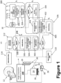
- FIGURE 1 diagrammatically shows an illustrative device for optimizing an image acquisition device according to one aspect.
- machine log data are leveraged to tune imaging device hardware for improved image acquisition.
- an artificial intelligence (AI) system is trained to identify faulty machine parameters based on log data.
- the log patterns that serve as input to the AI may be handcrafted and/or machine learned log patterns. If a faulty machine parameter value is identified by the AI (e.g., a machine parameter value is identified by the AI as associated with significantly degraded image quality), then a machine learning (ML) model is used to adjust (i.e. tune) one or more hardware settings of the imaging device to improve the image.
- the image is then acquired (including any image reconstruction processing) using the tuned imaging device.
- image quality metrics are computed for the resulting image. These image quality metrics are input along with the image, and optionally also along with faulty machine parameter values identified by the AI, to an AI system trained to apply filter(s) or other post-acquisition image processing to improve the image.
- the ML system that performs the hardware tuning may also be trained to optimally identify service recommendations.
- log patterns may be used to identify degradation of the X-ray tube in a CT scanner. Initially, this will be compensated by adjusting the X-ray tube operating parameters to maintain image quality. Secondarily, as the log patterns indicate progressively more degradation of the X-ray tube the ML may additionally output a recommendation to replace the X-ray tube within a certain timeframe.
- the recommendation to service or replace a hardware component may be delayed to the extent that the tuning ML can tune machine parameter(s) (and/or the post-acquisition image processing AI can post-process images) to compensate for degradation of the hardware component. This can extend the effective service life of the hardware component between replacements/servicing compared with a service recommender system that addresses degradation of hardware components only by recommending service or replacement.
- the AI system that performs the post-processing may issue servicing recommendations based on trained responses to the input image quality metrics.
- analysis of recorded log data of the imaging device by the AI does not record any parameters of the imaging device that are likely to be significantly degrading image quality, and the imaging device is acquiring and generating data without any artifacts.
- the AI detects parameters in log files that degrade image quality, and acquired image has artifacts or other degradation.
- the ML adjusts one or more hardware component parameters, thus tuning the hardware of the imaging device and correcting the subsequently acquired image.
- the ML may optionally also propose field corrective action(s) that needs to be carried out at site by equipment supplier (e.g., OEM) with specific input about which part of the imaging device is to be repaired or replaced.
- the image acquisition device 12 can be a Magnetic Resonance (MR) image acquisition device, a Computed Tomography (CT) image acquisition device; a positron emission tomography (PET) image acquisition device; a single photon emission computed tomography (SPECT) image acquisition device; an X-ray image acquisition device; an ultrasound (US) image acquisition device; or a medical imaging device of another modality.
- the imaging device 12 may also be a hybrid imaging device such as a PET/CT or SPECT/CT imaging system.
- FIGURE 1 also shows an electronic processor 20 , typically a server computer 19 or a plurality of server computers, e.g. interconnected to form a server cluster, cloud computing resource, or so forth), although the electronic processor 20 may be alternatively or additionally embodied as a workstation computer 18 , or more generally a computer.
- the illustrative workstation 18 comprises a computer or other electronic data processing device with typical components, such as at least one microprocessor, at least one user input device (e.g., a mouse, a keyboard, a trackball, and/or the like) 22 , and a display device 24 (e.g. an LCD display, plasma display, cathode ray tube display, and/or so forth).
- the display device 24 can be a separate component from the workstation 18.
- the workstation 18 may optionally operate as a user interface or controller for the medical imaging device 12.
- the electronic processor 20 is operatively connected with one or more non-transitory storage media 26.
- the non-transitory storage media 26 may, by way of non-limiting illustrative example, include one or more of a magnetic disk, RAID, or other magnetic storage medium; a solid state drive, flash drive, electronically erasable read-only memory (EEROM) or other electronic memory; an optical disk or other optical storage; various combinations thereof; or so forth; and may be for example a network storage accessible by the server 19 , an internal hard drive of the workstation 18 , various combinations thereof, or so forth.
- EEROM electronically erasable read-only memory
- any reference to a non-transitory medium or media 26 herein is to be broadly construed as encompassing a single medium or multiple media of the same or different types.
- the electronic processor 20 may be embodied as a single electronic processor or as two or more electronic processors (e.g. the server 19 and processor of the workstation 18 ).
- the non-transitory storage media 26 stores instructions executable by the at least one electronic processor 20.
- the instructions include instructions to identify problems with the image acquisition device 12 that dilutes image quality, and providing a recommendation to remedy the identified problems.
- the instructions automatically implement the recommendation, e.g. by automatically tuning one or more settings of hardware of the imaging device 12.
- a machine log 30 is recorded for the imaging device 12 and stored on the non-transitory storage medium 26.
- the machine log 30 stores various information pertaining to operation of the medical imaging device 12. For example, the machine log 30 stores sensor readings acquired by sensors that monitor parameters of hardware of the imaging device 12.
- Such sensors may, for example, include (by way of non-limiting illustration): temperature sensors monitoring temperature of various components; electrical sensors monitoring electric current, voltage, resistance, or so forth at various points in electric circuitry of hardware components; flux and/or intensity sensors measuring optical light, electron beam energy, or other types of outputs; position sensors measuring positions of robotic hardware (rotating gantry, patient positioning system, et cetera); and so forth.
- the machine log 30 may store information on imaging sessions, e.g. start/end times, imaging sequences executed by the imaging device 12 , patient position (e.g. from robotic positioning system), X-ray tube operating parameters during a CT scan, and/or so forth.
- the machine log 30 may store imaging device calibration information, such as counts/locations of dead detector pixels, detector dark current readings, and/or so forth.
- the machine log 30 may store servicing information on servicing operations performed on the imaging device 12 , such as timestamped service calls, service summaries entered by servicing technicians, and/or so forth.
- the specific information that is logged is dependent upon the imaging modality, the specific make/model of imaging device, the configuration of the specific machine logging system, and other factors.
- the machine log 30 may be logically divided into multiple logs, such as a sensor(s) log(s), a service log, and an imaging sessions log (again, by way of non-limiting illustrative example).
- the machine log 30 is to be understood as broadly encompassing such a plurality of machine logs.
- the machine log 30 may be stored locally, e.g. in a database at the hospital where the imaging device 12 is located, and/or may be stored (e.g. backed up) at a remote location, e.g.
- the at least one electronic processor 20 is in operative communication with the image acquisition device 12 , and is also in operative communication to read the machine log 30.
- Such operative communication with the machine log 30 may be direct, e.g. the illustrative server 19 may receive sensor data from the imaging device 12 and directly maintain the machine log 30 , or indirect, e.g. the illustrative server 19 may download a most recent portion of the machine log 30 stored at the manufacturer's server.
- the device 10 is configured as described above to perform an image acquisition method or process 100.
- the non-transitory storage medium 26 stores instructions which are readable and executable by the at least one electronic processor 20 (e.g. the workstation 18 and/or the server 19 ) to perform disclosed operations including performing the image acquisition method or process 100.
- the method 100 may be performed at least in part by cloud processing.
- an illustrative embodiment of the image acquisition method 100 is diagrammatically shown as a flowchart.
- log parameters of the image acquisition device 12 are analyzed to identify faulty parameters.
- a faulty parameter is one whose value is suspected, e.g. based on AI analysis as in illustrative embodiments disclosed herein, to be causing or contributing to a significant degradation in image quality.
- one or more electrical or mechanical settings of the imaging device 12 are tuned to compensate for the faulty parameter and an image is acquired with the tuned parameters.
- quality measures of the acquired image are analyzed to identify faulty quality measures.
- the image is post-processed and the quality measures of the acquired image are analyzed to identify faulty quality measures.
- an image with an accepted quality is acquired.
- a recommendation to correct for the faulty image quality measures is output, where a corresponding correction action can be performed at 114 .
- log parameters P stored in the machine log 30 of the image acquisition device 12 are extracted by the at least one electronic processor 20 .
- the log parameters P indicate a current state of components of the image acquisition device 12 .
- Some of these parameters may impact image quality metrics such as, for example, sharpness/blur, contrast, distortion, vignetting (e.g., reduction in brightness/saturation of image toward the periphery as compared to middle portion of the image), chromatic aberration, noise, change in dynamic range, and presence of a vertical or horizontal line (e.g., thick or thin).
- Degradation in one or more of the logged parameters may, directly or indirectly, lead to undesired artifacts or other degradation in the image (which may be quantifiable by one or more of the just-mentioned image quality metrics).
- the type of modality of the image acquisition device 12 is identified, and relevant parameters are extracted (at 202 ) from the machine log 30 using, e.g., natural language processing (NLP).
- NLP natural language processing
- an instance of the method 100 is executed for a single imaging device, in which case the imaging modality may be known a priori and the instructions stored on the non-transitory storage medium 26 which are executed may be modality-specific - in this case there is no need for a step of identifying the imaging modality).
- these parameters can be analyzed statistically if the desired acceptable ranges are known in advance, e.g., from manuals or by using a supervised learning method. From a list of "n" parameters, the most suspect parameters can be identified in, for example, a random forest analysis. The identified lists of suspect parameters are candidates for tuning of the components of the image acquisition device 12.
- one or more out-of-range (i.e., faulty or suspect) parameters P k of the image acquisition device 12 extracted from the machine log 30 of the image acquisition device are identified.
- the out-of-range parameters P k can be identified by a ML model implemented on the at least one electronic processor 20.
- the ML model is trained on training data comprising sets of logged parameters labelled with image quality values for images acquired by the image acquisition device having the sets of logged parameters (e.g., stored in the machine log 30 and/or the non-transitory storage media 26 ).
- one or more electrical or mechanical settings of the image acquisition device 12 are tuned on the basis of the one or more out-of-range parameters P k to transform the image acquisition device into a tuned image acquisition device.
- the source for the imaging generation could be, for example, an X-ray tube, or a magnet in the case of an MRI, or ultrasound transducers as in an ultrasound (US) machine.
- Medical imaging devices of certain modalities may not have an imaging generation source - for example, a PET scanner detects 511 keV gamma rays emitted by a radiopharmaceutical administered to the patient).
- Each of these sources have mechanical/electrical parameters which has an ability to affect the image quality based on tuning constraints.
- a correction factor is determined that is responsible for the correcting the image in case of degradation.
- a radiation detector array of a CT scanner, PET scanner, SPECT camera, or the like has a range of logged parameters that can impact image quality, such as dark current levels for the pixels of the detector array, sensitivity calibration values for sensor pixels, and/or so forth. Settings that might be tuned to compensate for high dark current might, for example, include adjustment of a bias voltage on a detector pixel.
- the tuning at operation 302 includes applying a tuning model (e.g., stored in the non-transitory computer readable media 26 ) to the one or more out-of-range parameters P k to generate tuned values for the one or more electrical or mechanical settings of the image acquisition device 12.
- the one or more electrical or mechanical settings are adjusted to the tuned values generated by the tuning model.
- the tuning model comprises a machine learned tuning model trained on historical training data comprising sets of logged parameters (e.g., stored in the non-transitory storage media 26 ) labelled with image quality values (generated either automatically as image quality metrics and/or manually labeled by expert clinicians) for images acquired by the image acquisition device using the sets of logged parameters, with the images further labeled with the values of the electrical and/or mechanical settings of the imaging device 12 which are contemplated to be tuned by the ML tuning model.
- the training optimizes the ML to select tuned value(s) for electrical and/or mechanical settings of the imaging device 12 that yield the highest image quality labels in the historical training set for images with similar out-of-range parameters. In one example, this adjustment process is automatically performed by the at least one electronic processor 20.
- the tuned values generated by the tuning model can be displayed on the display device 24 for review and approval by a user before the adjustment process is performed by the at least one electronic processor 20.
- the tuning model could be otherwise implemented, e.g. as an analytic function dependent on the one or more out-of-range parameters P k .
- the identification of the modality type is performed based on the log file analysis using NLP which provides the classification of the modality type. (As previously mentioned, in some embodiments the method 100 is modality-specific in which case the step of identifying the modality type is omitted).
- the trained ML models search for parameters in the log file that are associated with the modality (e.g., for X-ray tube).
- the extracted parameters P k are fed to the trained ML models, which outputs tuned values for one or more electrical and/or mechanical settings of one or more components of the image acquisition device 12 (e.g., mechanical/ electrical) so as to compensate for image degradation caused by the out-of-range parameter(s) P k . In some embodiments, this is done automatically by the electronic processor 20 (e.g., if the electronic processor 20 includes or communicates with the workstation 18 serving as the controller for the imaging device 12 ).
- the tuned value of a setting may instead be initially proposed to the user, for example by being displayed on the display 24 of the workstation 18 along with a user dialog via which the user may select (via the user input device 22 ) to accept or reject the tuning operation.
- the setting is only adjusted to the tuned value if the user selects to accept the tuning operation.
- the tuning operation is typically recorded in the machine log 30 in common with most significant events occurring in or to the imaging device 12.
- the tuned image acquisition device 12 is controlled by the at least one electronic processor 20 to acquire one or more images of a patient (e.g., at 304 ).
- the image acquisition method 100 can proceed directly to operation 112 , in which a recommendation to correct for the faulty image quality measures is output.
- Operation 112 includes applying a service recommender model (e.g., stored in the non-transitory storage media 26 ) to the one or more out-of-range parameters P k to generate service recommendations for the image acquisition device 12.
- the service recommender model comprises a machine learned model trained on training data comprising sets of logged parameters labelled with image quality values for images acquired by the image acquisition device 12 using the sets of logged parameters P.
- the service recommender model is applied by the at least one electronic processor 20 to the one or more out-of-range parameters and to one or more image quality metrics generated for the acquired one or more images to generate the service recommendations for the image acquisition device 12.
- the service recommendations can be displayed on the display device 24.
- the recommendation to replace a component can be delayed by the tuning, which can effectively extend the useful service life while still scheduling timely replacement to avoid unnecessary shutdowns.
- the service recommendations can include a recommendation to order one or more replacement parts for the image acquisition device 12.
- the recommendation as displayed on the display device 24 can include a link or button to allow the user to order the needed replacements parts (e.g., by linking the user with a third-party parts ordering service or with a company that manufactures the needed replacement parts).
- the user can also be able to enter shipping information (e.g., a shipping address, a delivery date, a priority shipping level, and so forth).
- the parts order can include a priority and timing (e.g., if the part is needed immediately).
- the priority and timing of the ordered replacement parts based on which specific parameters are below the designated thresholds, a type of artifacts detected in the image, filters used to remedy artifacts in the images, and so forth.
- a service scheduler can be implemented in the workstation 18 or the server 19 to prioritize the orders based on a schedule service appointment, a service appointment of nearby imaging systems 12, and so forth.
- the image acquisition method 100 proceeds to operation 106.
- one or more image quality metrics are computed or estimated (e.g., by the at least one electronic processor 20 ) for the image(s) acquired at operation 304.
- the computed image quality metrics may include, by way of non-limiting illustrative examples, one or more of sharpness, blur, contrast, distortion, vignetting, chromatic aberration, noise, change in dynamic range, and/or presence of a vertical or horizontal line.
- one or more out-of-range image quality metrics are identified (e.g., by the at least one electronic processor 20 ) from the computed one or more image quality metrics.
- the post-processing operations are performed.
- one or more image filters are selected on the basis of the one or more out-of-range image quality metrics, and are applied to the image to improve its image quality.
- the one or more image filters are selected on the basis of the one or more out-of-range image quality metrics, and on the further basis of the one or more out-of-range parameters of the image acquisition device 12.
- the selection 502 may employ an AI algorithm trained on historical post-processed images labelled with pre-post-processing image quality metrics (and also with out-of-range parameters of the image acquisition device 12 , for embodiments in which the post-processing operations are also selected on that basis).
- the training optimizes the AI to select the post-processing operation(s) that yield the highest improvement in image quality metrics going from the pre-processed to post-processed image in the historical training set for images with similar out-of-range parameters.
- the selected one or more image filters are applied (e.g., by the at least one electronic processor 20 ) to the acquired one or more images (at 304 ) to generate one or more improved images of a patient (e.g., at operation 110 ).
- the image quality metrics are computed for the post-processed images, and checked at 506 to determine whether any image quality metrics are still faulty (e.g. out of an acceptable range).
- any image quality metrics remain faulty then this is input to the recommender 112 to generate a servicing recommendation.
- the ML models stored in the non-transitory storage media 26 can be, for example, Generative Adversarial Networks (GANs).
- GANs Generative Adversarial Networks
- the AI algorithms described above can be based on the models, along with Deep Learning models, GAN models, statistical models, and so forth. Inputs to these types of networks can include one or more corrupted images, along with list of parameters and correction algorithms, The ML models use these inputs to learn and generate appropriate correction techniques and recommendations.
Landscapes
- Engineering & Computer Science (AREA)
- Health & Medical Sciences (AREA)
- Physics & Mathematics (AREA)
- Theoretical Computer Science (AREA)
- Life Sciences & Earth Sciences (AREA)
- Medical Informatics (AREA)
- General Physics & Mathematics (AREA)
- Biomedical Technology (AREA)
- Public Health (AREA)
- General Health & Medical Sciences (AREA)
- Quality & Reliability (AREA)
- General Engineering & Computer Science (AREA)
- Nuclear Medicine, Radiotherapy & Molecular Imaging (AREA)
- Radiology & Medical Imaging (AREA)
- Molecular Biology (AREA)
- Heart & Thoracic Surgery (AREA)
- Pathology (AREA)
- Surgery (AREA)
- Animal Behavior & Ethology (AREA)
- Optics & Photonics (AREA)
- High Energy & Nuclear Physics (AREA)
- Veterinary Medicine (AREA)
- Biophysics (AREA)
- Computer Vision & Pattern Recognition (AREA)
- Computing Systems (AREA)
- Epidemiology (AREA)
- Primary Health Care (AREA)
- Business, Economics & Management (AREA)
- General Business, Economics & Management (AREA)
- Computer Hardware Design (AREA)
- Mathematical Physics (AREA)
- Apparatus For Radiation Diagnosis (AREA)
Claims (11)
- Vorrichtung zum Optimieren einer Bilderfassungsvorrichtung (12), wobei die Vorrichtung Folgendes beinhaltet:mindestens einen elektronischen Prozessor (20), der betriebsmäßig so angeschlossen ist, dass er ein Maschinenprotokoll (30) der Bilderfassungsvorrichtung liest; undein nicht-transitorisches computerlesbares Medium (26), das Anweisungen speichert, die von dem mindestens einen elektronischen Prozessor lesbar und ausführbar sind, um ein Bilderfassungsverfahren (100) durchzuführen, das Folgendes beinhaltet:Extrahieren von protokollierten Parameter der Bilderfassungsvorrichtung aus dem Maschinenprotokoll der Bilderfassungsvorrichtung;Identifizieren eines oder mehrerer außerhalb des Bereichs liegender Parameter der Bilderfassungsvorrichtung anhand der protokollierten Parameter, die aus dem Maschinenprotokoll der Bilderfassungsvorrichtung extrahiert wurden;automatisches Abstimmen einer oder mehrerer elektrischer oder mechanischer Einstellungen der Bilderfassungsvorrichtung auf der Grundlage des einen oder der mehreren außerhalb des Bereichs liegenden Parameter, um die Bilderfassungsvorrichtung in eine abgestimmte Bilderfassungsvorrichtung umzuwandeln;Steuern der abgestimmten Bilderfassungsvorrichtung zur Erfassung eines oder mehrerer Bilder eines Patienten;und dadurch gekennzeichnet, dass das Bilderfassungsverfahren (100) weiter Folgendes umfasst:Anwenden eines Wartungsempfehlungsmodells auf den einen oder die mehreren außerhalb des Bereichs liegenden Parameter, um Wartungsempfehlungen für die Bilderfassungsvorrichtung zu erzeugen; undAnzeigen der von dem Wartungsempfehlungsmodell erzeugten Wartungsempfehlungen auf einer Anzeigevorrichtung (24), undwobei das Wartungsempfehlungsmodell ein maschinengelerntes Modell umfasst, das auf Trainingsdaten trainiert wurde, die Sätze protokollierter Parameter umfassen, die mit Bildqualitätswerten für Bilder gekennzeichnet sind, die von der Bilderfassungsvorrichtung unter Verwendung der Sätze protokollierter Parameter erfasst wurden.
- Vorrichtung nach Anspruch 1, wobei die Abstimmung Folgendes beinhaltet:Anwenden eines Abstimmungsmodells auf den einen oder die mehreren außerhalb des Bereichs liegenden Parameter, um abgestimmte Werte für die eine oder die mehreren elektrischen oder mechanischen Einstellungen der Bilderfassungsvorrichtung (12) zu erzeugen; undautomatisches Anpassen der einen oder mehreren elektrischen oder mechanischen Einstellungen an die durch das Abstimmungsmodell erzeugten abgestimmten Werte,wobei das Abstimmungsmodell ein maschinengelerntes Abstimmungsmodell umfasst, das auf Trainingsdaten trainiert wurde, die Sätze protokollierter Parameter umfassen, die mit Bildqualitätswerten für Bilder gekennzeichnet sind, die von der Bilderfassungsvorrichtung unter Verwendung der Sätze protokollierter Parameter erfasst wurden.
- Vorrichtung nach einem der Ansprüche 1 bis 2, wobei die Identifizierung des einen oder der mehreren außerhalb des Bereichs liegenden Parameter Folgendes beinhaltet:Identifizieren mindestens eines außerhalb des Bereichs liegenden Parameters durch Anwendung eines maschinellen Lernmodells auf Daten des Maschinenprotokolls;wobei das maschinelle Lernmodell auf Trainingsdaten trainiert wird, die Sätze protokollierter Parametern umfassen, die mit Bildqualitätswerten für Bilder gekennzeichnet sind, die von der Bilderfassungsvorrichtung mit den Sätzen protokollierter Parametern erfasst wurden.
- Vorrichtung nach Anspruch 1, wobei das Anwenden Folgendes umfasst:
Anwenden des Wartungsempfehlungsmodells auf den einen oder die mehreren außerhalb des Bereichs liegenden Parameter und auf eine oder mehrere Bildqualitätsmetriken, die für die erfassten ein oder mehreren Bilder erzeugt wurden, um die Wartungsempfehlungen für die Bilderfassungsvorrichtung zu erzeugen. - Vorrichtung nach einem der Ansprüche 1-4, wobei das Bilderfassungsverfahren (100) weiter Folgendes beinhaltet:Berechnen einer oder mehrerer Bildqualitätsmetriken für die erfassten ein oder mehreren Bilder;Identifizieren einer oder mehrerer außerhalb des Bereichs liegender Bildqualitätsmetriken anhand der berechneten einen oder mehreren Bildqualitätsmetriken;Auswählen eines oder mehrerer Bildfilter auf der Grundlage der einen oder mehreren außerhalb des Bereichs liegenden Bildqualitätsmetriken; undAnwenden des ausgewählten einen oder der mehreren Bildfilter auf die erfassten einen oder mehreren Bilder, um ein oder mehrere verbesserte Bilder eines Patienten zu erzeugen.
- Vorrichtung nach Anspruch 5, wobei das Auswählen Folgendes umfasst:
Auswählen des einen oder der mehreren Bildfilter auf der Grundlage des einen oder der mehreren außerhalb des Bereichs liegenden Bildqualitätsmetriken und auf der weiteren Grundlage des einen oder der mehreren außerhalb des Bereichs liegenden Parameter der Bilderfassungsvorrichtung. - Vorrichtung nach einem der Ansprüche 5-6, wobei die berechneten Bildqualitätsmetriken eines oder mehrere von Schärfe, Unschärfe, Kontrast, Verzerrung, Vignettierung, chromatische Aberration, Rauschen, Änderung des Dynamikbereichs und/oder Vorhandensein einer vertikalen oder horizontalen Linie beinhalten.
- Vorrichtung nach einem der Ansprüche 1-7, weiter beinhaltend:
die Bilderfassungsvorrichtung (12), wobei die Bilderfassungsvorrichtung eine Magnetresonanz (MR)-Bilderfassungsvorrichtung, eine Computertomographie (CT)-Bilderfassungsvorrichtung, eine Positronen-Emissions-Tomographie (PET)-Bilderfassungsvorrichtung, eine Einzelphotonen-Emissionscomputertomographie (SPECT)-Bilderfassungsvorrichtung, eine Röntgenbilderfassungsvorrichtung oder eine Ultraschall (US)-Bilderfassungsvorrichtung ist. - Bilderfassungsverfahren (100), das Folgendes beinhaltet:Extrahieren von protokollierten Parametern einer Bilderfassungsvorrichtung (12) aus einem Maschinenprotokoll (30) der Bilderfassungsvorrichtung;Identifizieren eines oder mehrerer außerhalb des Bereichs liegender Parameter der Bilderfassungsvorrichtung anhand der protokollierten Parameter, die aus dem Maschinenprotokoll der Bilderfassungsvorrichtung extrahiert wurden;Abstimmen einer oder mehrerer elektrischer oder mechanischer Einstellungen der Bilderfassungsvorrichtung auf der Grundlage des einen oder der mehreren außerhalb des Bereichs liegenden Parameter, um die Bilderfassungsvorrichtung in eine abgestimmte Bilderfassungsvorrichtung umzuwandeln;Steuern der abgestimmten Bilderfassungsvorrichtung zur Erfassung eines oder mehrerer Bilder eines Patienten; unddadurch gekennzeichnet, dass das Bilderfassungsverfahren weiter Folgendes beinhaltet:Berechnen einer oder mehrerer Bildqualitätsmetriken für die erfassten ein oder mehreren Bilder;Identifizieren einer oder mehrerer außerhalb des Bereichs liegender Bildqualitätsmetriken anhand der berechneten einen oder mehreren Bildqualitätsmetriken;Auswählen eines oder mehrerer Bildfilter auf der Grundlage der einen oder mehreren außerhalb des Bereichs liegenden Bildqualitätsmetriken und auf der weiteren Grundlage des einen oder der mehreren außerhalb des Bereichs liegenden Parameter der Bilderfassungsvorrichtung; undAnwenden des ausgewählten einen oder der mehreren Bildfilter auf die erfassten einen oder mehreren Bilder, um ein oder mehrere verbesserte Bilder eines Patienten zu erzeugen.
- Verfahren (100) nach Anspruch 9, wobei die Abstimmung Folgendes beinhaltet:Anwenden eines Abstimmungsmodells auf den einen oder die mehreren außerhalb des Bereichs liegenden Parameter, um abgestimmte Werte für die eine oder die mehreren elektrischen oder mechanischen Einstellungen der Bilderfassungsvorrichtung (12) zu erzeugen; undautomatisches Anpassen der einen oder mehreren elektrischen oder mechanischen Einstellungen an die durch das Abstimmungsmodell erzeugten abgestimmten Werte,wobei das Abstimmungsmodell ein maschinengelerntes Abstimmungsmodell umfasst, das auf Trainingsdaten trainiert wurde, die Sätze protokollierter Parameter umfassen, die mit Bildqualitätswerten für Bilder gekennzeichnet sind, die von der Bilderfassungsvorrichtung unter Verwendung der Sätze protokollierter Parameter erfasst wurden.
- Verfahren (100) nach einem der Ansprüche 9-10, das weiter Folgendes beinhaltet:Anwenden eines Wartungsempfehlungsmodells auf den einen oder die mehreren außerhalb des Bereichs liegenden Parameter, um Wartungsempfehlungen für die Bilderfassungsvorrichtung zu erzeugen; undAnzeigen der von dem Wartungsempfehlungsmodell erzeugten Wartungsempfehlungen auf einer Anzeigevorrichtung (24),wobei das Wartungsempfehlungsmodell ein maschinengelerntes Modell umfasst, das auf Trainingsdaten trainiert wurde, die Sätze protokollierter Parameter umfassen, die mit Bildqualitätswerten für Bilder gekennzeichnet sind, die von der Bilderfassungsvorrichtung unter Verwendung der Sätze protokollierter Parameter erfasst wurden; undwobei die Wartungsempfehlungen Bestellung eines oder mehrerer Ersatzteile für die Bilderfassungsvorrichtung basierend auf dem einen oder den mehreren außerhalb des Bereichs liegenden Parameter beinhalten, wobei die Wartungsempfehlungen basierend auf einer Dringlichkeit für das eine oder die mehreren Ersatzteile priorisiert werden.
Applications Claiming Priority (2)
| Application Number | Priority Date | Filing Date | Title |
|---|---|---|---|
| US201962834988P | 2019-04-17 | 2019-04-17 | |
| PCT/EP2020/060667 WO2020212470A1 (en) | 2019-04-17 | 2020-04-16 | Medical imaging systems and methods with auto-correction of image quality-based on the log analysis of medical devices |
Publications (2)
| Publication Number | Publication Date |
|---|---|
| EP3955819A1 EP3955819A1 (de) | 2022-02-23 |
| EP3955819B1 true EP3955819B1 (de) | 2024-08-07 |
Family
ID=70289812
Family Applications (1)
| Application Number | Title | Priority Date | Filing Date |
|---|---|---|---|
| EP20719431.7A Active EP3955819B1 (de) | 2019-04-17 | 2020-04-16 | Medizinische bildgebungssysteme und verfahren mit automatischer korrektur der bildqualität basierend auf der protokollanalyse von medizinprodukten |
Country Status (4)
| Country | Link |
|---|---|
| US (1) | US12357260B2 (de) |
| EP (1) | EP3955819B1 (de) |
| CN (1) | CN113747838A (de) |
| WO (1) | WO2020212470A1 (de) |
Families Citing this family (13)
| Publication number | Priority date | Publication date | Assignee | Title |
|---|---|---|---|---|
| WO2020212470A1 (en) * | 2019-04-17 | 2020-10-22 | Koninklijke Philips N.V. | Medical imaging systems and methods with auto-correction of image quality-based on the log analysis of medical devices |
| US20210406112A1 (en) * | 2020-06-29 | 2021-12-30 | International Business Machines Corporation | Anomaly classification in information technology environments |
| EP3996105A1 (de) * | 2020-11-09 | 2022-05-11 | Koninklijke Philips N.V. | Mehrphasige lebensdauerüberwachungsvorrichtung |
| US20240029875A1 (en) * | 2020-12-15 | 2024-01-25 | Koninklijke Philips N.V. | System and method to recommend service action for predictive maintenance |
| EP4050565B1 (de) * | 2021-02-26 | 2024-12-04 | Siemens Healthineers AG | Verfahren zur iterativen rekonstruierung von bildern unter verwendung von tiefenlernen |
| GB202104680D0 (en) * | 2021-03-31 | 2021-05-12 | Cancer Research Tech Ltd | Determination of spectral filters for spectral imaging |
| WO2022207516A1 (en) * | 2021-04-01 | 2022-10-06 | Koninklijke Philips N.V. | Systems and methods for self-sensing mobile medical devices to auto-correct an output |
| CN113849370A (zh) * | 2021-09-24 | 2021-12-28 | 武汉联影医疗科技有限公司 | 监控参数的调整方法、装置、计算机设备和存储介质 |
| GB2624749A (en) * | 2022-09-19 | 2024-05-29 | Bright Stewart | System and methods for monitoring medical equipment by using cloud-based telemetry |
| US12475553B2 (en) * | 2022-09-19 | 2025-11-18 | GE Precision Healthcare LLC | Machine learning image analysis based on explicit equipment parameters |
| EP4462442A1 (de) * | 2023-05-11 | 2024-11-13 | Koninklijke Philips N.V. | Bewertung einer leistungsmetrik für eine komponente eines medizinischen bildgebungssystems |
| US20250391015A1 (en) * | 2024-06-21 | 2025-12-25 | GE Precision Healthcare LLC | Scanner fault prediction via image-based deep learning |
| CN118658602B (zh) * | 2024-08-21 | 2024-11-29 | 江苏爱星信息科技有限公司 | 医学影像设备日志数据智能化分析预警方法 |
Family Cites Families (17)
| Publication number | Priority date | Publication date | Assignee | Title |
|---|---|---|---|---|
| US5600574A (en) | 1994-05-13 | 1997-02-04 | Minnesota Mining And Manufacturing Company | Automated image quality control |
| US7577246B2 (en) * | 2006-12-20 | 2009-08-18 | Nice Systems Ltd. | Method and system for automatic quality evaluation |
| US20120177173A1 (en) * | 2011-01-07 | 2012-07-12 | General Electric Company | Method and apparatus for reducing imaging artifacts |
| WO2014043623A1 (en) * | 2012-09-17 | 2014-03-20 | Siemens Corporation | Log-based predictive maintenance |
| KR20160050761A (ko) * | 2014-10-31 | 2016-05-11 | 삼성전자주식회사 | 방사선 촬영 장치 및 방사선 촬영 장치의 제어방법 |
| DE102015204449A1 (de) | 2015-03-12 | 2016-09-15 | Siemens Healthcare Gmbh | Verfahren zum Bestimmen eines Röntgenröhrenstromprofils, Computerprogramm, Datenträger sowie Röntgenbildaufnahmevorrichtung |
| US20180095653A1 (en) * | 2015-08-14 | 2018-04-05 | Martin Hasek | Device, method and graphical user interface for handwritten interaction |
| US10387765B2 (en) * | 2016-06-23 | 2019-08-20 | Siemens Healthcare Gmbh | Image correction using a deep generative machine-learning model |
| US10242443B2 (en) * | 2016-11-23 | 2019-03-26 | General Electric Company | Deep learning medical systems and methods for medical procedures |
| US11003988B2 (en) * | 2016-11-23 | 2021-05-11 | General Electric Company | Hardware system design improvement using deep learning algorithms |
| DE102017203333B4 (de) * | 2017-03-01 | 2025-08-28 | Siemens Healthineers Ag | Verfahren zum Erzeugen einer Liste von Untersuchungsschritten und Verfahren zur Anpassung eines Untersuchungsprotokolls während einer medizinischen Bildgebungsuntersuchung |
| WO2019011765A1 (en) | 2017-07-10 | 2019-01-17 | Koninklijke Philips N.V. | PREDICTIVE MAINTENANCE FOR LARGE MEDICAL IMAGING SYSTEMS |
| JP2019058073A (ja) * | 2017-09-25 | 2019-04-18 | オリンパス株式会社 | 画像処理装置、細胞認識装置、細胞認識方法および細胞認識プログラム |
| US11517197B2 (en) * | 2017-10-06 | 2022-12-06 | Canon Medical Systems Corporation | Apparatus and method for medical image reconstruction using deep learning for computed tomography (CT) image noise and artifacts reduction |
| US11497459B2 (en) * | 2018-01-26 | 2022-11-15 | General Electric Company | Methods and system for optimizing an imaging scan based on a prior scan |
| GB2584986B (en) * | 2019-03-19 | 2023-07-26 | Sony Interactive Entertainment Inc | System and camera device for capturing images |
| WO2020212470A1 (en) * | 2019-04-17 | 2020-10-22 | Koninklijke Philips N.V. | Medical imaging systems and methods with auto-correction of image quality-based on the log analysis of medical devices |
-
2020
- 2020-04-16 WO PCT/EP2020/060667 patent/WO2020212470A1/en not_active Ceased
- 2020-04-16 US US17/600,806 patent/US12357260B2/en active Active
- 2020-04-16 CN CN202080029127.XA patent/CN113747838A/zh active Pending
- 2020-04-16 EP EP20719431.7A patent/EP3955819B1/de active Active
Also Published As
| Publication number | Publication date |
|---|---|
| EP3955819A1 (de) | 2022-02-23 |
| CN113747838A (zh) | 2021-12-03 |
| US12357260B2 (en) | 2025-07-15 |
| US20220165395A1 (en) | 2022-05-26 |
| WO2020212470A1 (en) | 2020-10-22 |
Similar Documents
| Publication | Publication Date | Title |
|---|---|---|
| EP3955819B1 (de) | Medizinische bildgebungssysteme und verfahren mit automatischer korrektur der bildqualität basierend auf der protokollanalyse von medizinprodukten | |
| US10796181B2 (en) | Machine learning based method and system for analyzing image artifacts and imaging system failure | |
| US11333732B2 (en) | Automatic artifact detection and pulse sequence modification in magnetic resonance imaging | |
| JP6907410B2 (ja) | 磁気共鳴イメージングデータ内の動きアーティファクトの深層学習に基づく処理 | |
| JP7309605B2 (ja) | ディープラーニング医療システムおよび画像収集のための方法 | |
| EP3652748B1 (de) | Prädiktive wartung für grosse medizinische bildgebungssysteme | |
| US7885384B2 (en) | System and method to manage maintenance of a radiological imaging system | |
| US20190266436A1 (en) | Machine learning in an imaging modality service context | |
| US20220375088A1 (en) | Magnetic resonance (mr) image artifact determination using texture analysis for image quality (iq) standardization and system health prediction | |
| EP4035601B1 (de) | Verfahren und systeme zur überwachung von auf röntgenröhren bezogenen ereignissen | |
| WO2022229005A1 (en) | Systems and methods to improve x-ray tube filament failure prediction | |
| EP4429553B1 (de) | Nutzungsbasiertes verschleissmodell und verbesserte lebensdauer für röntgenvorrichtungen | |
| WO2022128704A1 (en) | System and method to recommend service action for predictive maintenance | |
| EP3688769A1 (de) | Automatisierte assistenz von personal und qualitätssicherung auf basis von echtzeitarbeitsflussanalyse | |
| EP4244865A1 (de) | System und verfahren zur automatisierten oder halbautomatischen identifizierung von defekten zonen für wartungsfälle | |
| JP2025513681A (ja) | ニューラルネットワーク生成画像評価を用いた医用画像の監視 | |
| US20240008834A1 (en) | Imaging failure image management apparatus, operation method and operation program of imaging failure image management apparatus, and radiography system | |
| WO2024008599A1 (en) | System and method to improve image quality and prevent rescans by analyzing cross-hospital context and scan settings | |
| JP6457112B2 (ja) | 医療データ取得システムについて品質評価を決定するための方法および装置 | |
| EP4160273A1 (de) | Detektion von röntgenstrahlung | |
| EP4276497A1 (de) | Detektion und sammlung von röntgendetektor-artefaktdaten durch das erfassungssystem zur unterstützung von kundendienst und entwicklung | |
| WO2022084127A1 (en) | Multi-criteria fair queueing of alerts |
Legal Events
| Date | Code | Title | Description |
|---|---|---|---|
| STAA | Information on the status of an ep patent application or granted ep patent |
Free format text: STATUS: UNKNOWN |
|
| STAA | Information on the status of an ep patent application or granted ep patent |
Free format text: STATUS: THE INTERNATIONAL PUBLICATION HAS BEEN MADE |
|
| PUAI | Public reference made under article 153(3) epc to a published international application that has entered the european phase |
Free format text: ORIGINAL CODE: 0009012 |
|
| STAA | Information on the status of an ep patent application or granted ep patent |
Free format text: STATUS: REQUEST FOR EXAMINATION WAS MADE |
|
| 17P | Request for examination filed |
Effective date: 20211117 |
|
| AK | Designated contracting states |
Kind code of ref document: A1 Designated state(s): AL AT BE BG CH CY CZ DE DK EE ES FI FR GB GR HR HU IE IS IT LI LT LU LV MC MK MT NL NO PL PT RO RS SE SI SK SM TR |
|
| DAV | Request for validation of the european patent (deleted) | ||
| DAX | Request for extension of the european patent (deleted) | ||
| REG | Reference to a national code |
Ref legal event code: R079 Free format text: PREVIOUS MAIN CLASS: A61B0006000000 Ipc: A61B0006580000 Ref country code: DE Ref document number: 602020035276 Country of ref document: DE |
|
| GRAP | Despatch of communication of intention to grant a patent |
Free format text: ORIGINAL CODE: EPIDOSNIGR1 |
|
| STAA | Information on the status of an ep patent application or granted ep patent |
Free format text: STATUS: GRANT OF PATENT IS INTENDED |
|
| RIC1 | Information provided on ipc code assigned before grant |
Ipc: G16H 30/20 20180101ALI20240214BHEP Ipc: G06F 11/34 20060101ALI20240214BHEP Ipc: G06F 11/30 20060101ALI20240214BHEP Ipc: G16H 40/40 20180101ALI20240214BHEP Ipc: A61B 6/00 20060101ALI20240214BHEP Ipc: G06F 11/07 20060101ALI20240214BHEP Ipc: A61B 6/58 20240101AFI20240214BHEP |
|
| INTG | Intention to grant announced |
Effective date: 20240306 |
|
| GRAS | Grant fee paid |
Free format text: ORIGINAL CODE: EPIDOSNIGR3 |
|
| GRAA | (expected) grant |
Free format text: ORIGINAL CODE: 0009210 |
|
| STAA | Information on the status of an ep patent application or granted ep patent |
Free format text: STATUS: THE PATENT HAS BEEN GRANTED |
|
| AK | Designated contracting states |
Kind code of ref document: B1 Designated state(s): AL AT BE BG CH CY CZ DE DK EE ES FI FR GB GR HR HU IE IS IT LI LT LU LV MC MK MT NL NO PL PT RO RS SE SI SK SM TR |
|
| REG | Reference to a national code |
Ref country code: GB Ref legal event code: FG4D |
|
| REG | Reference to a national code |
Ref country code: CH Ref legal event code: EP |
|
| REG | Reference to a national code |
Ref country code: IE Ref legal event code: FG4D |
|
| REG | Reference to a national code |
Ref country code: DE Ref legal event code: R096 Ref document number: 602020035276 Country of ref document: DE |
|
| REG | Reference to a national code |
Ref country code: DE Ref legal event code: R084 Ref document number: 602020035276 Country of ref document: DE |
|
| REG | Reference to a national code |
Ref country code: LT Ref legal event code: MG9D |
|
| REG | Reference to a national code |
Ref country code: NL Ref legal event code: MP Effective date: 20240807 |
|
| PG25 | Lapsed in a contracting state [announced via postgrant information from national office to epo] |
Ref country code: NO Free format text: LAPSE BECAUSE OF FAILURE TO SUBMIT A TRANSLATION OF THE DESCRIPTION OR TO PAY THE FEE WITHIN THE PRESCRIBED TIME-LIMIT Effective date: 20241107 |
|
| REG | Reference to a national code |
Ref country code: AT Ref legal event code: MK05 Ref document number: 1710022 Country of ref document: AT Kind code of ref document: T Effective date: 20240807 |
|
| PG25 | Lapsed in a contracting state [announced via postgrant information from national office to epo] |
Ref country code: NL Free format text: LAPSE BECAUSE OF FAILURE TO SUBMIT A TRANSLATION OF THE DESCRIPTION OR TO PAY THE FEE WITHIN THE PRESCRIBED TIME-LIMIT Effective date: 20240807 Ref country code: PL Free format text: LAPSE BECAUSE OF FAILURE TO SUBMIT A TRANSLATION OF THE DESCRIPTION OR TO PAY THE FEE WITHIN THE PRESCRIBED TIME-LIMIT Effective date: 20240807 Ref country code: FI Free format text: LAPSE BECAUSE OF FAILURE TO SUBMIT A TRANSLATION OF THE DESCRIPTION OR TO PAY THE FEE WITHIN THE PRESCRIBED TIME-LIMIT Effective date: 20240807 Ref country code: GR Free format text: LAPSE BECAUSE OF FAILURE TO SUBMIT A TRANSLATION OF THE DESCRIPTION OR TO PAY THE FEE WITHIN THE PRESCRIBED TIME-LIMIT Effective date: 20241108 Ref country code: PT Free format text: LAPSE BECAUSE OF FAILURE TO SUBMIT A TRANSLATION OF THE DESCRIPTION OR TO PAY THE FEE WITHIN THE PRESCRIBED TIME-LIMIT Effective date: 20241209 |
|
| PG25 | Lapsed in a contracting state [announced via postgrant information from national office to epo] |
Ref country code: BG Free format text: LAPSE BECAUSE OF FAILURE TO SUBMIT A TRANSLATION OF THE DESCRIPTION OR TO PAY THE FEE WITHIN THE PRESCRIBED TIME-LIMIT Effective date: 20240807 |
|
| PG25 | Lapsed in a contracting state [announced via postgrant information from national office to epo] |
Ref country code: LV Free format text: LAPSE BECAUSE OF FAILURE TO SUBMIT A TRANSLATION OF THE DESCRIPTION OR TO PAY THE FEE WITHIN THE PRESCRIBED TIME-LIMIT Effective date: 20240807 |
|
| PG25 | Lapsed in a contracting state [announced via postgrant information from national office to epo] |
Ref country code: IS Free format text: LAPSE BECAUSE OF FAILURE TO SUBMIT A TRANSLATION OF THE DESCRIPTION OR TO PAY THE FEE WITHIN THE PRESCRIBED TIME-LIMIT Effective date: 20241207 Ref country code: AT Free format text: LAPSE BECAUSE OF FAILURE TO SUBMIT A TRANSLATION OF THE DESCRIPTION OR TO PAY THE FEE WITHIN THE PRESCRIBED TIME-LIMIT Effective date: 20240807 |
|
| PG25 | Lapsed in a contracting state [announced via postgrant information from national office to epo] |
Ref country code: HR Free format text: LAPSE BECAUSE OF FAILURE TO SUBMIT A TRANSLATION OF THE DESCRIPTION OR TO PAY THE FEE WITHIN THE PRESCRIBED TIME-LIMIT Effective date: 20240807 |
|
| PG25 | Lapsed in a contracting state [announced via postgrant information from national office to epo] |
Ref country code: RS Free format text: LAPSE BECAUSE OF FAILURE TO SUBMIT A TRANSLATION OF THE DESCRIPTION OR TO PAY THE FEE WITHIN THE PRESCRIBED TIME-LIMIT Effective date: 20241107 Ref country code: ES Free format text: LAPSE BECAUSE OF FAILURE TO SUBMIT A TRANSLATION OF THE DESCRIPTION OR TO PAY THE FEE WITHIN THE PRESCRIBED TIME-LIMIT Effective date: 20240807 |
|
| PG25 | Lapsed in a contracting state [announced via postgrant information from national office to epo] |
Ref country code: RS Free format text: LAPSE BECAUSE OF FAILURE TO SUBMIT A TRANSLATION OF THE DESCRIPTION OR TO PAY THE FEE WITHIN THE PRESCRIBED TIME-LIMIT Effective date: 20241107 Ref country code: PT Free format text: LAPSE BECAUSE OF FAILURE TO SUBMIT A TRANSLATION OF THE DESCRIPTION OR TO PAY THE FEE WITHIN THE PRESCRIBED TIME-LIMIT Effective date: 20241209 Ref country code: PL Free format text: LAPSE BECAUSE OF FAILURE TO SUBMIT A TRANSLATION OF THE DESCRIPTION OR TO PAY THE FEE WITHIN THE PRESCRIBED TIME-LIMIT Effective date: 20240807 Ref country code: NO Free format text: LAPSE BECAUSE OF FAILURE TO SUBMIT A TRANSLATION OF THE DESCRIPTION OR TO PAY THE FEE WITHIN THE PRESCRIBED TIME-LIMIT Effective date: 20241107 Ref country code: NL Free format text: LAPSE BECAUSE OF FAILURE TO SUBMIT A TRANSLATION OF THE DESCRIPTION OR TO PAY THE FEE WITHIN THE PRESCRIBED TIME-LIMIT Effective date: 20240807 Ref country code: LV Free format text: LAPSE BECAUSE OF FAILURE TO SUBMIT A TRANSLATION OF THE DESCRIPTION OR TO PAY THE FEE WITHIN THE PRESCRIBED TIME-LIMIT Effective date: 20240807 Ref country code: IS Free format text: LAPSE BECAUSE OF FAILURE TO SUBMIT A TRANSLATION OF THE DESCRIPTION OR TO PAY THE FEE WITHIN THE PRESCRIBED TIME-LIMIT Effective date: 20241207 Ref country code: HR Free format text: LAPSE BECAUSE OF FAILURE TO SUBMIT A TRANSLATION OF THE DESCRIPTION OR TO PAY THE FEE WITHIN THE PRESCRIBED TIME-LIMIT Effective date: 20240807 Ref country code: GR Free format text: LAPSE BECAUSE OF FAILURE TO SUBMIT A TRANSLATION OF THE DESCRIPTION OR TO PAY THE FEE WITHIN THE PRESCRIBED TIME-LIMIT Effective date: 20241108 Ref country code: FI Free format text: LAPSE BECAUSE OF FAILURE TO SUBMIT A TRANSLATION OF THE DESCRIPTION OR TO PAY THE FEE WITHIN THE PRESCRIBED TIME-LIMIT Effective date: 20240807 Ref country code: ES Free format text: LAPSE BECAUSE OF FAILURE TO SUBMIT A TRANSLATION OF THE DESCRIPTION OR TO PAY THE FEE WITHIN THE PRESCRIBED TIME-LIMIT Effective date: 20240807 Ref country code: BG Free format text: LAPSE BECAUSE OF FAILURE TO SUBMIT A TRANSLATION OF THE DESCRIPTION OR TO PAY THE FEE WITHIN THE PRESCRIBED TIME-LIMIT Effective date: 20240807 Ref country code: AT Free format text: LAPSE BECAUSE OF FAILURE TO SUBMIT A TRANSLATION OF THE DESCRIPTION OR TO PAY THE FEE WITHIN THE PRESCRIBED TIME-LIMIT Effective date: 20240807 |
|
| PG25 | Lapsed in a contracting state [announced via postgrant information from national office to epo] |
Ref country code: DK Free format text: LAPSE BECAUSE OF FAILURE TO SUBMIT A TRANSLATION OF THE DESCRIPTION OR TO PAY THE FEE WITHIN THE PRESCRIBED TIME-LIMIT Effective date: 20240807 Ref country code: SM Free format text: LAPSE BECAUSE OF FAILURE TO SUBMIT A TRANSLATION OF THE DESCRIPTION OR TO PAY THE FEE WITHIN THE PRESCRIBED TIME-LIMIT Effective date: 20240807 |
|
| PG25 | Lapsed in a contracting state [announced via postgrant information from national office to epo] |
Ref country code: EE Free format text: LAPSE BECAUSE OF FAILURE TO SUBMIT A TRANSLATION OF THE DESCRIPTION OR TO PAY THE FEE WITHIN THE PRESCRIBED TIME-LIMIT Effective date: 20240807 |
|
| PG25 | Lapsed in a contracting state [announced via postgrant information from national office to epo] |
Ref country code: CZ Free format text: LAPSE BECAUSE OF FAILURE TO SUBMIT A TRANSLATION OF THE DESCRIPTION OR TO PAY THE FEE WITHIN THE PRESCRIBED TIME-LIMIT Effective date: 20240807 |
|
| PG25 | Lapsed in a contracting state [announced via postgrant information from national office to epo] |
Ref country code: SK Free format text: LAPSE BECAUSE OF FAILURE TO SUBMIT A TRANSLATION OF THE DESCRIPTION OR TO PAY THE FEE WITHIN THE PRESCRIBED TIME-LIMIT Effective date: 20240807 |
|
| REG | Reference to a national code |
Ref country code: DE Ref legal event code: R097 Ref document number: 602020035276 Country of ref document: DE |
|
| PLBE | No opposition filed within time limit |
Free format text: ORIGINAL CODE: 0009261 |
|
| STAA | Information on the status of an ep patent application or granted ep patent |
Free format text: STATUS: NO OPPOSITION FILED WITHIN TIME LIMIT |
|
| PGFP | Annual fee paid to national office [announced via postgrant information from national office to epo] |
Ref country code: DE Payment date: 20250428 Year of fee payment: 6 |
|
| 26N | No opposition filed |
Effective date: 20250508 |
|
| PG25 | Lapsed in a contracting state [announced via postgrant information from national office to epo] |
Ref country code: SE Free format text: LAPSE BECAUSE OF FAILURE TO SUBMIT A TRANSLATION OF THE DESCRIPTION OR TO PAY THE FEE WITHIN THE PRESCRIBED TIME-LIMIT Effective date: 20240807 |
|
| REG | Reference to a national code |
Ref country code: CH Ref legal event code: H13 Free format text: ST27 STATUS EVENT CODE: U-0-0-H10-H13 (AS PROVIDED BY THE NATIONAL OFFICE) Effective date: 20251125 |
|
| PG25 | Lapsed in a contracting state [announced via postgrant information from national office to epo] |
Ref country code: LU Free format text: LAPSE BECAUSE OF NON-PAYMENT OF DUE FEES Effective date: 20250416 |
|
| PG25 | Lapsed in a contracting state [announced via postgrant information from national office to epo] |
Ref country code: MC Free format text: LAPSE BECAUSE OF FAILURE TO SUBMIT A TRANSLATION OF THE DESCRIPTION OR TO PAY THE FEE WITHIN THE PRESCRIBED TIME-LIMIT Effective date: 20240807 |
|
| GBPC | Gb: european patent ceased through non-payment of renewal fee |
Effective date: 20250416 |
|
| REG | Reference to a national code |
Ref country code: BE Ref legal event code: MM Effective date: 20250430 |
|
| PG25 | Lapsed in a contracting state [announced via postgrant information from national office to epo] |
Ref country code: GB Free format text: LAPSE BECAUSE OF NON-PAYMENT OF DUE FEES Effective date: 20250416 |
|
| PG25 | Lapsed in a contracting state [announced via postgrant information from national office to epo] |
Ref country code: FR Free format text: LAPSE BECAUSE OF NON-PAYMENT OF DUE FEES Effective date: 20250430 |
|
| PG25 | Lapsed in a contracting state [announced via postgrant information from national office to epo] |
Ref country code: BE Free format text: LAPSE BECAUSE OF NON-PAYMENT OF DUE FEES Effective date: 20250430 |
|
| PG25 | Lapsed in a contracting state [announced via postgrant information from national office to epo] |
Ref country code: CH Free format text: LAPSE BECAUSE OF NON-PAYMENT OF DUE FEES Effective date: 20250430 |
|
| PG25 | Lapsed in a contracting state [announced via postgrant information from national office to epo] |
Ref country code: IT Free format text: LAPSE BECAUSE OF FAILURE TO SUBMIT A TRANSLATION OF THE DESCRIPTION OR TO PAY THE FEE WITHIN THE PRESCRIBED TIME-LIMIT Effective date: 20240807 |
|
| PG25 | Lapsed in a contracting state [announced via postgrant information from national office to epo] |
Ref country code: IE Free format text: LAPSE BECAUSE OF NON-PAYMENT OF DUE FEES Effective date: 20250416 |
|
| PG25 | Lapsed in a contracting state [announced via postgrant information from national office to epo] |
Ref country code: RO Free format text: LAPSE BECAUSE OF FAILURE TO SUBMIT A TRANSLATION OF THE DESCRIPTION OR TO PAY THE FEE WITHIN THE PRESCRIBED TIME-LIMIT Effective date: 20240807 |