EP3957247A1 - Verfahren und system zur bestimmung des schweregrades von myokardialer ischämie basierend auf der schätzung von hämodynamischen parametern - Google Patents
Verfahren und system zur bestimmung des schweregrades von myokardialer ischämie basierend auf der schätzung von hämodynamischen parametern Download PDFInfo
- Publication number
- EP3957247A1 EP3957247A1 EP21191188.8A EP21191188A EP3957247A1 EP 3957247 A1 EP3957247 A1 EP 3957247A1 EP 21191188 A EP21191188 A EP 21191188A EP 3957247 A1 EP3957247 A1 EP 3957247A1
- Authority
- EP
- European Patent Office
- Prior art keywords
- cardiac
- pressure
- left heart
- value
- ventricle
- Prior art date
- Legal status (The legal status is an assumption and is not a legal conclusion. Google has not performed a legal analysis and makes no representation as to the accuracy of the status listed.)
- Granted
Links
- GDOPTJXRTPNYNR-UHFFFAOYSA-N CC1CCCC1 Chemical compound CC1CCCC1 GDOPTJXRTPNYNR-UHFFFAOYSA-N 0.000 description 1
Images



Classifications
-
- A—HUMAN NECESSITIES
- A61—MEDICAL OR VETERINARY SCIENCE; HYGIENE
- A61B—DIAGNOSIS; SURGERY; IDENTIFICATION
- A61B5/00—Measuring for diagnostic purposes; Identification of persons
- A61B5/72—Signal processing specially adapted for physiological signals or for diagnostic purposes
- A61B5/7271—Specific aspects of physiological measurement analysis
- A61B5/7278—Artificial waveform generation or derivation, e.g. synthesizing signals from measured signals
-
- A—HUMAN NECESSITIES
- A61—MEDICAL OR VETERINARY SCIENCE; HYGIENE
- A61B—DIAGNOSIS; SURGERY; IDENTIFICATION
- A61B5/00—Measuring for diagnostic purposes; Identification of persons
- A61B5/48—Other medical applications
- A61B5/4842—Monitoring progression or stage of a disease
-
- A—HUMAN NECESSITIES
- A61—MEDICAL OR VETERINARY SCIENCE; HYGIENE
- A61B—DIAGNOSIS; SURGERY; IDENTIFICATION
- A61B5/00—Measuring for diagnostic purposes; Identification of persons
- A61B5/02—Detecting, measuring or recording for evaluating the cardiovascular system, e.g. pulse, heart rate, blood pressure or blood flow
- A61B5/02007—Evaluating blood vessel condition, e.g. elasticity, compliance
-
- A—HUMAN NECESSITIES
- A61—MEDICAL OR VETERINARY SCIENCE; HYGIENE
- A61B—DIAGNOSIS; SURGERY; IDENTIFICATION
- A61B5/00—Measuring for diagnostic purposes; Identification of persons
- A61B5/02—Detecting, measuring or recording for evaluating the cardiovascular system, e.g. pulse, heart rate, blood pressure or blood flow
- A61B5/021—Measuring pressure in heart or blood vessels
-
- A—HUMAN NECESSITIES
- A61—MEDICAL OR VETERINARY SCIENCE; HYGIENE
- A61B—DIAGNOSIS; SURGERY; IDENTIFICATION
- A61B5/00—Measuring for diagnostic purposes; Identification of persons
- A61B5/24—Detecting, measuring or recording bioelectric or biomagnetic signals of the body or parts thereof
- A61B5/316—Modalities, i.e. specific diagnostic methods
- A61B5/318—Heart-related electrical modalities, e.g. electrocardiography [ECG]
- A61B5/327—Generation of artificial ECG signals based on measured signals, e.g. to compensate for missing leads
-
- A—HUMAN NECESSITIES
- A61—MEDICAL OR VETERINARY SCIENCE; HYGIENE
- A61B—DIAGNOSIS; SURGERY; IDENTIFICATION
- A61B5/00—Measuring for diagnostic purposes; Identification of persons
- A61B5/24—Detecting, measuring or recording bioelectric or biomagnetic signals of the body or parts thereof
- A61B5/316—Modalities, i.e. specific diagnostic methods
- A61B5/318—Heart-related electrical modalities, e.g. electrocardiography [ECG]
- A61B5/346—Analysis of electrocardiograms
- A61B5/349—Detecting specific parameters of the electrocardiograph cycle
- A61B5/36—Detecting PQ interval, PR interval or QT interval
-
- A—HUMAN NECESSITIES
- A61—MEDICAL OR VETERINARY SCIENCE; HYGIENE
- A61B—DIAGNOSIS; SURGERY; IDENTIFICATION
- A61B5/00—Measuring for diagnostic purposes; Identification of persons
- A61B5/24—Detecting, measuring or recording bioelectric or biomagnetic signals of the body or parts thereof
- A61B5/316—Modalities, i.e. specific diagnostic methods
- A61B5/318—Heart-related electrical modalities, e.g. electrocardiography [ECG]
- A61B5/346—Analysis of electrocardiograms
- A61B5/349—Detecting specific parameters of the electrocardiograph cycle
- A61B5/366—Detecting abnormal QRS complex, e.g. widening
-
- A—HUMAN NECESSITIES
- A61—MEDICAL OR VETERINARY SCIENCE; HYGIENE
- A61B—DIAGNOSIS; SURGERY; IDENTIFICATION
- A61B5/00—Measuring for diagnostic purposes; Identification of persons
- A61B5/24—Detecting, measuring or recording bioelectric or biomagnetic signals of the body or parts thereof
- A61B5/316—Modalities, i.e. specific diagnostic methods
- A61B5/318—Heart-related electrical modalities, e.g. electrocardiography [ECG]
- A61B5/367—Electrophysiological study [EPS], e.g. electrical activation mapping or electro-anatomical mapping
-
- A—HUMAN NECESSITIES
- A61—MEDICAL OR VETERINARY SCIENCE; HYGIENE
- A61B—DIAGNOSIS; SURGERY; IDENTIFICATION
- A61B5/00—Measuring for diagnostic purposes; Identification of persons
- A61B5/74—Details of notification to user or communication with user or patient; User input means
- A61B5/742—Details of notification to user or communication with user or patient; User input means using visual displays
- A61B5/743—Displaying an image simultaneously with additional graphical information, e.g. symbols, charts, function plots
-
- G—PHYSICS
- G16—INFORMATION AND COMMUNICATION TECHNOLOGY [ICT] SPECIALLY ADAPTED FOR SPECIFIC APPLICATION FIELDS
- G16H—HEALTHCARE INFORMATICS, i.e. INFORMATION AND COMMUNICATION TECHNOLOGY [ICT] SPECIALLY ADAPTED FOR THE HANDLING OR PROCESSING OF MEDICAL OR HEALTHCARE DATA
- G16H50/00—ICT specially adapted for medical diagnosis, medical simulation or medical data mining; ICT specially adapted for detecting, monitoring or modelling epidemics or pandemics
- G16H50/30—ICT specially adapted for medical diagnosis, medical simulation or medical data mining; ICT specially adapted for detecting, monitoring or modelling epidemics or pandemics for calculating health indices; for individual health risk assessment
-
- G—PHYSICS
- G16—INFORMATION AND COMMUNICATION TECHNOLOGY [ICT] SPECIALLY ADAPTED FOR SPECIFIC APPLICATION FIELDS
- G16H—HEALTHCARE INFORMATICS, i.e. INFORMATION AND COMMUNICATION TECHNOLOGY [ICT] SPECIALLY ADAPTED FOR THE HANDLING OR PROCESSING OF MEDICAL OR HEALTHCARE DATA
- G16H50/00—ICT specially adapted for medical diagnosis, medical simulation or medical data mining; ICT specially adapted for detecting, monitoring or modelling epidemics or pandemics
- G16H50/50—ICT specially adapted for medical diagnosis, medical simulation or medical data mining; ICT specially adapted for detecting, monitoring or modelling epidemics or pandemics for simulation or modelling of medical disorders
Definitions
- the disclosure herein generally relates to myocardial ischemia disease, and, more particularly, to method and system for determining myocardial ischemia severity based on hemodynamic parameters estimation.
- Myocardial ischemia leads to sudden cardiac death due to narrowing of coronary artery causing poor oxygen deprivation in cardiac muscles. Many patients suffer from myocardial ischemia due to smoking, diabetes, hypertension, and the like. Early detection of myocardial ischemia provides opportunity for a wide range of effective therapies such as surgical revascularization, neural stimulation, and drug delivery to reduce cardiac workload or to improve cardiac circulation. In recent years, computer simulations and mathematical models have provided substantial insights for electrophysiological behavior to detect abnormalities in myocardial ischemia. Varying ischemia conditions in cardiac contractility results inefficient pumping in heart muscles and thus hampers hemodynamic equilibrium. Further, any computer models to determine ischemic progression provides dual effect of change in electrophysiology and hemodynamics as the disease manifests.
- ECG electrocardiogram
- a method for determining myocardial ischemia severity based on hemodynamic parameter estimation is provided.
- the system includes for determining myocardial ischemia severity based on hemodynamic parameter estimation is provided.
- the method includes receiving a plurality of Electrophysiology (EP) signals from a heart surface model as an input.
- Each Electrophysiology (EP) signal from the plurality of Electrophysiology (EP) signals corresponds to cardiac transmembrane potential (TMP) giving rise to cardiac contraction.
- TMP cardiac transmembrane potential
- a Forward Electrophysiology signal from the plurality of Electrophysiology (EP) signals is generated by a cardiac source module.
- the Forward Electrophysiology signal are processed to generate a single lead ECG template, wherein the single lead ECG template comprises at least one parameter comprising: (i) a auricular depolarization (PQ) segment, (ii) a ventricular depolarization (QRS) segment, (iii) a ventricular repolarization (ST) segment and combination thereof.
- a hemodynamic module a plurality of hemodynamic parameters based on the single lead ECG template is estimated.
- the plurality of hemodynamic parameters comprises a left heart atrium compliance function C la ( t ) and a left heart ventricle compliance function C lv ( t ).
- a plurality of cardiac pressure-volume loop variables is estimated based on atleast one of (i) the plurality of hemodynamic parameters, and (ii) pressure variation associated with cardiac excitation.
- myocardial ischemia severity of the heart surface model is determined based on at least one of (i) a scar tissue size, (ii) a velocity reduction value of the cardiac affected region, (iii) the transmembrane potential (TMP) amplitude and repolarization time, (iv) the single lead ECG template and (v) the plurality of cardiac pressure-volume loop variables.
- the myocardial ischemia severity includes one of moderate ischemia, severe ischemia and silent ischemia.
- the system of the cardiac source module is coupled with the hemodynamic module to determine cardiac transmembrane potential (TMP) of the heart surface model through a contractility function.
- the left heart atrium compliance function C la ( t ) is computed based on atleast one of (i) a minimum value of the left heart atrium, (ii) a maximum value of the left heart atrium, (iii) a left heart atrium activation function ( A la ), and (iv) a time delay in firing between the left heart atrium and the left heart ventricle.
- the left heart atrium activation function ( A la ) is computed based on left heart atrium activation time analogous to the auricular depolarization (PQ) segment and the time duration of the cardiac cycle.
- the left heart ventricle compliance function C lv ( t ) is computed based on the end systolic compliance and the left heart ventricle activation function ( A lv ( t )) .
- the left heart ventricle activation function ( A lv ( t )) is computed based on an end systolic and diastolic time duration of the cardiac cycle analogous to (i) the ventricular depolarization (QRS) segment, and (ii) the ventricular repolarization (ST) segment.
- the plurality of cardiac pressure-volume loop variables includes at least one of (i) a dynamic change observed in a systemic artery pressure, (ii) a dynamic change observed in a left heart ventricle pressure and (iii) a dynamic change observed in a right ventricle pressure.
- the dynamic change observed in the systemic artery pressure is estimated based on at least one of (i) a systemic artery compliance, (ii) the left heart ventricle pressure, (iii) a systemic ventricle pressure, (iv) a systemic artery pressure,(v)a resistance value observed in systemic vessels, and (vi) a resistance value observed in aortic vessel.
- the dynamic change observed in the left heart ventricle pressure is estimated based on at least one of (i) the left heart ventricle compliance function, (ii) the left heart ventricle pressure, (iii) a pulmonary vein pressure, (iv) the systemic artery pressure, (v) a resistance value observed in mitral vessel, and (vi) the resistance value observed in aortic vessel.
- the dynamic change observed in the right heart ventricle pressure is estimated based on at least one of (i) the right heart ventricle compliance function, (ii) a systemic vein pressure (iii) the right heart ventricle pressure, (iv) a resistance value observed in tricuspid vessel, (v) a pulmonary artery pressure, and (vi) a resistance value observed in pulmonary valve.
- the moderate myocardial ischemia is determined if (i) the scar tissue size varies between a first threshold value and a second threshold value, (ii) the velocity reduction value of cardiac affected region is equal to a velocity value, and (iii) the cardiac transmembrane potential (TMP) amplitude and repolarization time range between a first repolarization time value and a second repolarization time value.
- TMP cardiac transmembrane potential
- the severe myocardial ischemia is determined if (i) the scar tissue size varies between a first predetermined value and a second predetermined value, (ii) the velocity reduction value of cardiac affected region is equal to a velocity value, and (iii) the cardiac transmembrane potential (TMP) amplitude and repolarization time range between a first transmembrane potential amplitude value and a second transmembrane potential amplitude value.
- TMP cardiac transmembrane potential
- the silent myocardial ischemia is determined if (i) the scar tissue size varies between a first predefined value and a second predefined value, (ii) the velocity reduction value of cardiac affected region is equal to a velocity value, and (iii) the cardiac transmembrane potential (TMP) amplitude and repolarization time range between a first transmembrane potential value and a second transmembrane potential value.
- TMP cardiac transmembrane potential
- a method for determining myocardial ischemia severity based on hemodynamic parameter estimation includes receiving a plurality of Electrophysiology (EP) signals from a heart surface model as an input.
- Each Electrophysiology (EP) signal from the plurality of Electrophysiology (EP) signals corresponds to cardiac transmembrane potential (TMP) giving rise to cardiac contraction.
- TMP cardiac transmembrane potential
- a Forward Electrophysiology signal from the plurality of Electrophysiology (EP) signals is generated by a cardiac source module.
- the Forward Electrophysiology signal are processed to generate a single lead ECG template, wherein the single lead ECG template comprises at least one parameter comprising: (i) a auricular depolarization (PQ) segment, (ii) a ventricular depolarization (QRS) segment, (iii) a ventricular repolarization (ST) segment and combination thereof.
- a hemodynamic module a plurality of hemodynamic parameters based on the single lead ECG template is estimated.
- the plurality of hemodynamic parameters comprises a left heart atrium compliance function C la ( t ) and a left heart ventricle compliance function C lv (t).
- a plurality of cardiac pressure-volume loop variables is estimated based on atleast one of (i) the plurality of hemodynamic parameters, and (ii) pressure variation associated with cardiac excitation.
- myocardial ischemia severity of the heart surface model is determined based on at least one of (i) a scar tissue size, (ii) a velocity reduction value of the cardiac affected region, (iii) the transmembrane potential (TMP) amplitude and repolarization time, (iv) the single lead ECG template and (v) the plurality of cardiac pressure-volume loop variables.
- the myocardial ischemia severity includes one of moderate ischemia, severe ischemia and silent ischemia.
- the method of the cardiac source module is coupled with the hemodynamic module to determine cardiac transmembrane potential (TMP) of the heart surface model through contractility function.
- the left heart atrium compliance function C la ( t ) is computed based on atleast one of (i) a minimum value of the left heart atrium, (ii) a maximum value of the left heart atrium, (iii) a left heart atrium activation function ( A la ), and (iv) a time delay in firing between the left heart atrium and the left heart ventricle.
- the left heart atrium activation function ( A la ) is computed based on the left heart atrium activation time analogous to the auricular depolarization (PQ) segment and the time duration of the cardiac cycle.
- the left heart ventricle compliance function C lv ( t ) is computed based on the end systolic compliance and the left heart ventricle activation function ( A lv ( t )) .
- the left heart ventricle activation function ( A lv ( t )) is computed based on systolic and diastolic time duration of the cardiac cycle analogous to (i) the ventricular depolarization (QRS) segment, and (ii) the ventricular repolarization (ST) segment.
- the plurality of cardiac pressure-volume loop variables includes at least one of (i) a dynamic change observed in a systemic artery pressure, (ii) a dynamic change observed in a left heart ventricle pressure and (iii) a dynamic change observed in a right ventricle pressure.
- the dynamic change observed in the systemic artery pressure is estimated based on at least one of (i) left heart ventricle pressure is estimated based on at least one of (i) the left heart ventricle compliance function, (ii) the left heart ventricle pressure, (iii) a pulmonary vein pressure, (iv) the systemic artery pressure, (v) a resistance value observed in mitral vessel, and (vi) the resistance value observed in aortic vessel.
- the dynamic change observed in the right heart ventricle pressure is estimated based on at least one of (i) the right heart ventricle compliance function, (ii) a systemic vein pressure (iii) the right heart ventricle pressure, (iv) a resistance value observed in tricuspid vessel, (v) a pulmonary artery pressure, and (vi) a resistance value observed in pulmonary valve.
- the moderate myocardial ischemia is determined if (i) the scar tissue size varies between a first threshold value and a second threshold value, (ii) the velocity reduction value of cardiac affected region is equal to a velocity value, and (iii) the cardiac transmembrane potential (TMP) amplitude and repolarization time range between a first repolarization time value and a second repolarization time value.
- TMP cardiac transmembrane potential
- the severe myocardial ischemia is determined if (i) the scar tissue size varies between a first predetermined value and a second predetermined value, (ii) the velocity reduction value of cardiac affected region is equal to a velocity value, and (iii) the cardiac transmembrane potential (TMP) amplitude and repolarization time range between a first transmembrane potential amplitude value and a second transmembrane potential amplitude value.
- TMP cardiac transmembrane potential
- the silent myocardial ischemia is determined if (i) the scar tissue size varies between a first predefined value and a second predefined value, (ii) the velocity reduction value of cardiac affected region is equal to a velocity value, and (iii) the cardiac transmembrane potential (TMP) amplitude and repolarization time range between a first transmembrane potential value and a second transmembrane potential value.
- TMP cardiac transmembrane potential
- a non-transitory machine readable information storage mediums comprising one or more instructions, which when executed by one or more hardware processors perform actions includes receiving a plurality of Electrophysiology (EP) signals from a heart surface model as an input.
- Each Electrophysiology (EP) signal from the plurality of Electrophysiology (EP) signals corresponds to cardiac transmembrane potential (TMP) giving rise to cardiac contraction.
- TMP cardiac transmembrane potential
- a Forward Electrophysiology signal from the plurality of Electrophysiology (EP) signals is generated by a cardiac source module.
- the Forward Electrophysiology signal are processed to generate a single lead ECG template, wherein the single lead ECG template comprises at least one parameter comprising: (i) a auricular depolarization (PQ) segment, (ii) a ventricular depolarization (QRS) segment, (iii) a ventricular repolarization (ST) segment and combination thereof.
- a hemodynamic module a plurality of hemodynamic parameters based on the single lead ECG template is estimated.
- the plurality of hemodynamic parameters comprises a left heart atrium compliance function C la ( t ) and a left heart ventricle compliance function C lv ( t ).
- a plurality of cardiac pressure-volume loop variables is estimated based on atleast one of (i) the plurality of hemodynamic parameters, and (ii) pressure variation associated with cardiac excitation.
- myocardial ischemia severity of the heart surface model is determined based on at least one of (i) a scar tissue size, (ii) a velocity reduction value of the cardiac affected region, (iii) the transmembrane potential (TMP) amplitude and repolarization time, (iv) the single lead ECG template and (v) the plurality of cardiac pressure-volume loop variables.
- the myocardial ischemia severity includes one of moderate ischemia, severe ischemia and silent ischemia.
- the method of the cardiac source module is coupled with the hemodynamic module to determine cardiac transmembrane potential (TMP) of the heart surface model through contractility function.
- the left heart atrium compliance function C la ( t ) is computed based on atleast one of (i) a minimum value of the left heart atrium, (ii) a maximum value of the left heart atrium, (iii) a left heart atrium activation function ( A la ), and (iv) a time delay in firing between the left heart atrium and the left heart ventricle.
- the left heart atrium activation function ( A la ) is computed based on the left heart atrium activation time analogous to the auricular depolarization (PQ) segment and the time duration of the cardiac cycle.
- the left heart ventricle compliance function C lv ( t ) is computed based on the end systolic compliance and the left heart ventricle activation function ( A lv ( t )) .
- the left heart ventricle activation function ( A lv ( t )) is computed based on systolic and diastolic time duration of the cardiac cycle analogous to (i) the ventricular depolarization (QRS) segment, and (ii) the ventricular repolarization (ST) segment.
- the plurality of cardiac pressure-volume loop variables includes at least one of (i) a dynamic change observed in a systemic artery pressure, (ii) a dynamic change observed in a left heart ventricle pressure and (iii) a dynamic change observed in a right ventricle pressure.
- the dynamic change observed in the systemic artery pressure is estimated based on at least one of (i) left heart ventricle pressure is estimated based on at least one of (i) the left heart ventricle compliance function, (ii) the left heart ventricle pressure, (iii) a pulmonary vein pressure, (iv) the systemic artery pressure, (v) a resistance value observed in mitral vessel, and (vi) the resistance value observed in aortic vessel.
- the dynamic change observed in the right heart ventricle pressure is estimated based on at least one of (i) the right heart ventricle compliance function, (ii) a systemic vein pressure (iii) the right heart ventricle pressure, (iv) a resistance value observed in tricuspid vessel, (v) a pulmonary artery pressure, and (vi) a resistance value observed in pulmonary valve.
- the moderate myocardial ischemia is determined if (i) the scar tissue size varies between a first threshold value and a second threshold value, (ii) the velocity reduction value of cardiac affected region is equal to a velocity value, and (iii) the cardiac transmembrane potential (TMP) amplitude and repolarization time range between a first repolarization time value and a second repolarization time value.
- TMP cardiac transmembrane potential
- the severe myocardial ischemia is determined if (i) the scar tissue size varies between a first predetermined value and a second predetermined value, (ii) the velocity reduction value of cardiac affected region is equal to a velocity value, and (iii) the cardiac transmembrane potential (TMP) amplitude and repolarization time range between a first transmembrane potential amplitude value and a second transmembrane potential amplitude value.
- TMP cardiac transmembrane potential
- the silent myocardial ischemia is determined if (i) the scar tissue size varies between a first predefined value and a second predefined value, (ii) the velocity reduction value of cardiac affected region is equal to a velocity value, and (iii) the cardiac transmembrane potential (TMP) amplitude and repolarization time range between a first transmembrane potential value and a second transmembrane potential value.
- TMP cardiac transmembrane potential
- Embodiments herein provide a method and system for determining myocardial ischemia severity based on hemodynamic parameters estimation.
- the method disclosed enables assessing progression of myocardial ischemic severity based on change occurred in cardiac ejection fraction.
- the present disclosure is a multi-model simulation of myocardial ischemia to assess disease progression with change in ischemic size and myocardial electrical propagation by observing the changes in hemodynamic parameters.
- the cardiac multi-model is coupling of a cardiac source model with a hemodynamic module to determine cardiac action transmembrane potential (TMP) of the heart surface model through contractility function.
- TMP cardiac action transmembrane potential
- the cardiac disease has a high variable manifestation due to difference in location and extent of damaged area, thus hampering the understanding of disease progression and stratification.
- Varying myocardial ischemia conditions are assessed based on the morphological changes occurred in a ventricular repolarization (ST) segment of the ECG template. Additionally, the present disclosure provides the assessment of disease progression based on various parameters such as ejection fraction, contractility, blood pressure and thereof for ischemic manifestation which leads to cardiac or heart failure. Three different conditions of myocardial ischemia have been simulated to determine disease progression, by way of experimental results and such results shall not be construed as limiting the scope of the present disclosure.
- FIG. 1 through FIG. 7C where similar reference characters denote corresponding features consistently throughout the figures, there are shown preferred embodiments and these embodiments are described in the context of the following exemplary system and/or method.
- FIG. 1 illustrates an exemplary block diagram of a system for determining myocardial ischemia severity based on hemodynamic parameters estimation, in accordance with some embodiments of the present disclosure.
- the system 100 includes processor (s) 104, communication interface (s), alternatively referred as or input/output (I/O) interface(s) 106, and one or more data storage devices or memory 102 operatively coupled to the processor (s) 104.
- the system 100, with the processor(s) is configured to execute functions of one or more functional blocks of the system 100.
- the processor (s) 104 can be one or more hardware processors 104.
- the one or more hardware processors 104 can be implemented as one or more microprocessors, microcomputers, microcontrollers, digital signal processors, central processing units, state machines, logic circuitries, and/or any devices that manipulate signals based on operational instructions.
- the processor(s) 104 is configured to fetch and execute computer-readable instructions stored in the memory.
- the system 100 can be implemented in a variety of computing systems, such as laptop computers, notebooks, 10 hand-held devices, workstations, mainframe computers, servers, a network cloud, and the like.
- the I/O interface(s) 106 can include a variety of software and hardware interfaces, for example, a web interface, a graphical user interface, and the like and can facilitate multiple communications within a wide variety of networks N/W and protocol types, including wired networks, for example, LAN, cable, etc., and wireless networks, such as WLAN, cellular, or satellite.
- the I/O interface (s) 106 can include one or more ports for connecting a number of devices (nodes) of the system 100 to one another or to another server.
- the memory 102 may include any computer-readable medium known in the art including, for example, volatile memory, such as static random access memory (SRAM) and dynamic random access memory (DRAM), and/or non-volatile memory, such as read only memory (ROM), erasable programmable ROM, flash memories, hard disks, optical disks, and magnetic tapes.
- volatile memory such as static random access memory (SRAM) and dynamic random access memory (DRAM)
- non-volatile memory such as read only memory (ROM), erasable programmable ROM, flash memories, hard disks, optical disks, and magnetic tapes.
- ROM read only memory
- erasable programmable ROM erasable programmable ROM
- flash memories hard disks
- optical disks optical disks
- magnetic tapes magnetic tapes
- the modules 108 can be an Integrated Circuit (IC) (not shown), external to the memory 102, implemented using a Field-Programmable Gate Array (FPGA) or an Application-Specific Integrated Circuit (ASIC).
- IC Integrated Circuit
- FPGA Field-Programmable Gate Array
- ASIC Application-Specific Integrated Circuit
- the names (or expressions or terms) of the modules of functional block within the modules 108 referred herein, are used for explanation and are not construed to be limitation(s).
- the modules 108 includes the cardiac source module 110 for processing a plurality of Electrophysiology (EP) signals received from a heart surface model as an input, and the hemodynamic module 112 for estimating a plurality of hemodynamic parameters based on processing the plurality of Electrophysiology (EP) signals received from the cardiac source module 110.
- the cardiac source module 110 and the hemodynamic module 112 are coupled through a contractility function which in turn determines the compliance function of auricles and ventricles which brings pumping function of the heart source model.
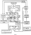
- FIG. 2 illustrates an example schematic diagram for determining severity of myocardial ischemia based on hemodynamic parameters estimation using the system of FIG. 1 , in accordance with some embodiments of the present disclosure.
- FIG. 2 includes the cardiac source module 110 and the hemodynamic module 112.
- the cardiac source module 110 comprises a heart surface potential and a body surface potential.
- the heart surface potential uses myocyte model defining cardiac action potential or a mathematical equivalent approximating the cardiac transmembrane potential (TMP).
- the body surface potential is calculated by feeding the cardiac transmembrane potential (TMP) through a cardiac propagation model such as monodomain or bidomain equations and boundary conditions through proper torso coupling.
- FEM Finite element method
- BEM Boundary element method
- ST ventricular repolarization
- QRS ventricular depolarization
- the hemodynamic module 112 estimates the output received from the cardiac source model 110 for determining the myocardial ischemic severity.
- the hemodynamic module 112 consists of a simulation model of patient's heart or heart source model comprising four chambers with a systemic circulation, and a pulmonic circulation along with baroreflex auto regulation and the like.
- the heart chambers have been modeled as compliant vessels.
- the pumping of the heart surface model is triggered through an autonomous contractility function derived from the cardiac source module 110.
- the simulated body surface potential (BSP) of the cardiac source module 110 drives the hemodynamic module 112, which modeled as four chambered heart with the pulmonic circulation and the systemic circulation.
- BSP body surface potential
- the integrated multi-model considers cellular to organ level manifestation of myocardial ischemia to simulate healthy heart dynamics and varying conditions of myocardial ischemia.
- the ground truth data of a healthy cardiac is generated based on all the varying conditions of ECG, blood pressure, left ventricle dynamics, ejection fraction and Photoplethysmogram (PPG) signal based on medical observations for close match.
- PPG Photoplethysmogram
- FIG. 3 illustrates a flow diagram for determining myocardial ischemia severity based on hemodynamic parameters estimation using the system of FIG. 1 , in accordance with some embodiments of the present disclosure.
- the system 100 comprises one or more data storage devices or the memory 102 operatively coupled to the processor(s) 104 and is configured to store instructions for execution of steps of the method 300 by the processor(s) or one or more hardware processors 104.
- the steps of the method 300 of the present disclosure will now be explained with reference to the components or blocks of the system 100 as depicted in FIG. 1 and FIG. 2 and the steps of flow diagram as depicted in FIG. 3 .
- process steps, method steps, techniques or the like may be described in a sequential order, such processes, methods and techniques may be configured to work in alternate orders. In other words, any sequence or order of steps that may be described does not necessarily indicate a requirement that the steps to be performed in that order.
- the steps of processes described herein may be performed in any order practical. Further, some steps may be performed simultaneously.
- the one or more hardware processors 104 receive a plurality of Electrophysiology (EP) signals from a heart surface model as an input.
- Each Electrophysiology (EP) signal from the plurality of Electrophysiology (EP) signals corresponds to cardiac action transmembrane potential (TMP) giving rise to cardiac contraction.
- TMP cardiac action transmembrane potential
- the system 100 can be used, for example an anatomical model of patient's heart referred as simulated heart surface model for determining the myocardial ischemic severity.
- the system 100 receives the plurality of Electrophysiology (EP) signals from a plurality of points of the heart surface model with its associated location information to create a diagnostic map of the heart surface model of the cardiac source module 110.
- the one or more hardware processors 104 generate, via the cardiac source module 110, a Forward Electrophysiology signal from the plurality of Electrophysiology (EP) signal.
- Electrocardiogram is based on a biophysical model that connects cardiac transmembrane potential (TMP) of representative myocytes on the heart surface model to electrocardiogram (ECG) signal on the surface of body. Geometrical parameters related to atria, ventricle and torso are reconstructed from magnetic resonance imaging.
- TMP cardiac transmembrane potential
- ECG electrocardiogram
- the one or more hardware processors 104 process the Forward Electrophysiology signal, to generate a single lead ECG template, wherein the single lead ECG template comprises at least one parameter comprising: (i) a auricular depolarization (PQ) segment, (ii) a ventricular depolarization (QRS) segment, (iii) a ventricular repolarization (ST) segment and combination thereof.
- the cardiac source module 110 is expressed as equivalent double layer (EDL) of sources on the closed surface of the atrium and the ventricles.
- TMP cardiac transmembrane potential
- the simulated patients heart surface is divided into a triangular mesh of 1500 elements or nodes, where each node poses an equivalent source which is proportional to the cardiac transmembrane potential (TMP) of the nearest myocyte.
- time course of strength of the equivalent double layer (EDL) is an analytical function represented as sigmoid curve expressed as product of logistics function involving markers for the timing of the ventricular depolarization (QRS) segment and the ventricular repolarization (ST) segment for approximating the cardiac transmembrane potential (TMP).
- the timing of local depolarization at node 'n' is denoted as ' ⁇ '.
- the interval ⁇ ⁇ - ⁇ is taken as a measure of the local action potential duration.
- TMP cardiac transmembrane potential
- the one or more hardware processors 104 estimate, via the hemodynamic module 112, a plurality of hemodynamic parameters based on the single lead ECG template, wherein the plurality of hemodynamic parameters comprises a left heart atrium compliance function C la ( t ) and a left heart ventricle compliance function C lv ( t ).
- the generated single lead ECG signal served as the driving signal to the hemodynamic module 112 for estimating the left heart atrium compliance function C la ( t ) and the left heart ventricle compliance function C lv ( t ).
- the hemodynamic module 112 consists of simulated heart surface model which includes four chambers with the systemic circulation and the pulmonic circulation along the baroflex auto regulation and the like.
- the vasculature of major vessels is modeled as combination of resistive and capacitive tube.
- all the major heart valves have been modeled to work in synchronized manner corresponding to auricular depolarization and ventricular repolarization of the heart chambers, thereby bringing the pulsatile effect with pressure gradient generation and volumetric change in the blood flow.
- the coupling of cardiac source module 110 and the hemodynamic module 112 enables to determine the compliance of the atrium and the ventricles for the pumping action of the heart surface model.
- the driving signal lead ECG template received from the cardiac source module 110 is decomposed into its characteristic constituents such as the auricular depolarization (PQ) segment, the ventricular depolarization (QRS) segment and the ventricular repolarization (ST) segment. Changes encoded to modulate compliance function and timing information to control synchronized operation of the four heart chambers.
- PQ auricular depolarization
- QRS ventricular depolarization
- ST ventricular repolarization
- the left heart atrium activation function is computed based on the left heart atrium activation time analogous to the auricular depolarization (PQ) segment and the time duration of the cardiac cycle as described below in equation (5),
- a la ⁇ 0 0 ⁇ t ⁇ T a 1 ⁇ cos 2 ⁇ t ⁇ T a T ⁇ T a T a ⁇ t ⁇ T
- T a is the left heart atrium activation time which is analogous to the (PQ) segment
- T is the time duration of the cardiac cycle.
- the left heart ventricle compliance function C lv ( t ) is computed based on the end systolic compliance and the left heart ventricle activation function.
- the product of end systolic compliance and the left heart ventricle activation function as described below in equation (6), C lv t C es , lv ⁇ A lv t where, C es,lv is the end systolic compliance, and A lv ( t ) is the activation function for the left heart ventricle.
- the left heart ventricle activation function is computed based on systolic and diastolic time duration of the cardiac cycle analogous to (i) the ventricular depolarization (QRS) segment, and (ii) the ventricular repolarization (ST) segment as described below in equation (7),
- a lv ⁇ 1 ⁇ cos t T s ⁇ 2 0 ⁇ t ⁇ T s 1 ⁇ cos ( t ⁇ T s / T D ⁇ T s ⁇ 2 T s ⁇ t ⁇ T d 0 T d ⁇ t ⁇ T
- T s and T d are the systolic and diastolic time duration of the cardiac cycle analogous to the ventricular depolarization (QRS) segment and the ventricular repolarization (ST) segment associated with the single lead ECG template.
- the one or more hardware processors 104 estimate a plurality of cardiac pressure-volume loop variables based on atleast one of (i) the plurality of hemodynamic parameters, and (ii) pressure variation associated with cardiac excitation.
- the plurality of cardiac pressure-volume loop variables includes at least one of (i) a dynamic change observed in a systemic artery pressure, (ii) a dynamic change observed in a left heart ventricle pressure and (iii) a dynamic change observed in a right ventricle pressure.
- the dynamic change observed in the left heart ventricle pressure ( ⁇ lv ) is estimated based on at least one of (i) the left heart ventricle compliance function, (ii) the left heart ventricle pressure, (iii) a pulmonary vein pressure, (iv) the systemic artery pressure, (v) a resistance value observed in mitral vessel, and (vi) the resistance value observed in aortic vessel which is described below in equation (9),
- P lv ⁇ C lv t ⁇ C lv t p lv + 1 C lv t ⁇ p pv ⁇ p lv R Mi ⁇ p lv ⁇ p sa R
- Ao ⁇ ⁇ lv is the pressure variation observed in left ventricle expressed as the dynamic change in the left ventricle compliance function, pressure gradients, valves and vessel resistance
- C lv ( t ) is the left heart ventricle compliance function
- p lv is the left heart ventric
- ⁇ rv is the pressure variation in right ventricle during the cardiac cycle expressed as the dynamic changes in the right ventricle compliance, pressure gradient, systemic vein pressure, pulmonary artery pressure and vessel resistance
- C rv ( t ) is the right heart ventricle compliance function
- the hemodynamic module 112 determines the plurality of hemodynamic parameters such as the arterial blood pressure, the left heart ventricle, the end systolic and diastolic volume (ESV, EDV), the ejection fraction (EF), the cardiac output (CO), the stroke volume (SV) and the end systolic and end diastolic pressure volume ratio (ESPVR, EDPVR) can be calculated which reveals concise information related to the state of heart and cardiovascular system.
- ESV end systolic and diastolic volume
- EF ejection fraction
- CO cardiac output
- SV stroke volume
- ESPVR end systolic and end diastolic pressure volume ratio
- the one or more hardware processors 104 determine myocardial ischemic severity of the heart surface model based on at least one of (i) a scar tissue size, (ii) a velocity reduction value of cardiac affected region, (iii) the transmembrane potential (TMP) amplitude and repolarization time, (iv) the single lead ECG template and, (v) the plurality of cardiac pressure-volume loop variables.
- the myocardial ischemic severity includes one of moderate ischemia, severe ischemia and silent ischemia.
- the simulated myocardial ischemia as an occlusion in the left anterior descending artery (LAD), effecting apical anterior and anterio-septal area of the heart surface model.
- LAD left anterior descending artery
- Cellular etiology of the myocardial ischemia suggests variational effects in ionic concentration at cell level which manifests itself in the form of action potential or cardiac transmembrane potential (TMP) on the cardiac surface model 110.
- TMP cardiac transmembrane potential
- the pathophysiology of ischemic effect can be simulated by inducing the following conditions:
- Moderate myocardial ischemia severity is determined based on the scar tissue size, the velocity reduction value of cardiac affected region and the cardiac transmembrane potential (TMP) amplitude and repolarization time.
- the scar tissue size for moderate myocardial ischemia varies between a first threshold value and a second threshold value, wherein the first threshold value serving as a minimum value of 25mm and the second threshold value serving as a maximum value of about 35mm.
- the velocity reduction value of cardiac affected region is equal to a predetermined value, wherein this velocity value of about 50%.
- the cardiac transmembrane potential (TMP) amplitude and repolarization time ranges between a first repolarization time value and a second repolarization time value, wherein the first repolarization time value serving as a minimum value of about 18% and the second repolarization time value serving as a maximum value of about 24%.
- the method determines the cardiac source module 110 belongs to the moderate myocardial ischemia based on the specified conditions.
- Severe myocardial ischemia severity is determined based on the scar tissue size, the velocity reduction value of cardiac affected region and the cardiac transmembrane potential (TMP) amplitude and repolarization time.
- the scar tissue size for severe myocardial ischemia varies between a first predetermined value and a second predetermined value, wherein the first predetermined value is 40mm and the second predetermined value is 55mm.
- the velocity reduction value of cardiac affected region is equal to a velocity value, wherein this velocity value is of about 50%.
- the cardiac transmembrane potential (TMP) amplitude and repolarization time ranges between a between a first transmembrane potential amplitude value and a second transmembrane potential amplitude value, wherein the first transmembrane potential amplitude value is minimum value of 30 % and the second transmembrane potential amplitude value is maximum value of about 38%.
- the method determines the cardiac source module 110 belongs to the moderate myocardial ischemia based on the specified conditions
- Silent myocardial ischemia severity is determined based on the scar tissue size, the velocity reduction value of cardiac affected region and the cardiac transmembrane potential (TMP) amplitude and repolarization time.
- the scar tissue size for silent myocardial ischemia varies between a first predefined value and a second predefined value, wherein the first predefined value is 12mm and the second predefined value is 16mm.
- the velocity reduction value of cardiac affected region is equal to a velocity value, wherein the velocity value is of about 50%.
- the cardiac transmembrane potential (TMP) amplitude and repolarization time ranges between a first transmembrane potential value and a second transmembrane potential value, wherein the first transmembrane potential value is minimum of about 12% and the second transmembrane potential value is a maximum value of about 17%.
- the method determines the cardiac source module 110 belongs to the moderate myocardial ischemia based on the specified conditions. Further, the silent myocardial ischemia where pathological manifestation happens only during stress or exercise conditions.
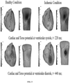
- FIG. 5 illustrates ventricular systole and diastole myocardial conditions of healthy conditions and ischemic conditions, in accordance with some embodiments of the present disclosure. More specifically, FIG. 5 depicts myocardial ischemia conditions of healthy cardiac and ischemic affected myocardium. Two separate instances represent the ventricular depolarization (QRS) segment at 220ms during the time of QRS complex generation and the ventricular repolarization (ST) segment at 440ms associated with the ECG template.
- QRS ventricular depolarization
- ST ventricular repolarization
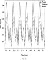
- FIG. 6A illustrates simulated ECG signals showing myocardial ischemic severity, in accordance with some embodiments of the present disclosure.
- a simulated single lead ECG template captures large changes in the ventricular repolarization (ST) segment correlates with the ventricular repolarization phase.
- transmural myocardial ischemia results in the ventricular repolarization (ST) segments often with 'Q' wave inversion.
- Change in the ventricular repolarization (ST) segment refers to generic weakness in the myocardium around the affected area and the effect magnifies with increase in size of scar tissue.
- FIG. 6B illustrates pressure volume loop showing left heart ventricle with myocardial ischemic severity in accordance with some embodiments of the present disclosure.
- the weakness observed in turn effects the pumping function, resulting in the left heart ventricle cardiac pressure-volume loop variables as shown.
- the cardiac pressure-volume loop variables of the moderate and the severe ischemia in comparison to the healthy pressure volume shows reduction in cardiac output and ejection fraction and a general trend of systolic dysfunction, commonly related with ischemia.
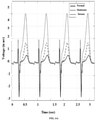
- FIG. 6C illustrates simulated PPG signals showing myocardial ischemic severity in accordance with some embodiments of the present disclosure.
- Simulated photoplethysmogram (PPG) signal shown in figure reveals interesting insights on myocardial ischemia conditions.
- PPG photoplethysmogram
- PPG photoplethysmogram
- PPG photoplethysmogram
- Morphological changes seen in these type of signals and when correlated with ischemic behavior can aid in early screening of myocardial ischemic conditions.
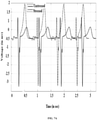
- FIG. 7A illustrates simulated ECG signals for silent myocardial ischemia showing stressed and destressed condition in accordance with some embodiments of the present disclosure.
- Ischemia is generally associated with chest pain and pathological changes in ventricular repolarization (ST) segment of the single lead ECG template which are easy symptoms for diagnosis.
- ST ventricular repolarization
- ischemic episodes are asymptomatic or silent in as many as 80% of cases where there is no associated symptom manifestation observed during daily ambulatory activity but results in maximum cases of sudden cardiac death due to lack of manifestation at diagnosis.
- Silent ischemia can only be detected under stress testing, where the hidden disease conditions gets reflected in the ECG morphology.
- Table 1 provides different simulated conditions with the plurality of hemodynamic parameters.
- the single lead ECG template was simulated at heart rate of 92 beats/min, during the experiments conducted by the embodiments of the present disclosure.
- Blood pressure of the silent myocardial ischemia case under stressed condition revealed a lower diastolic range compared to unstressed, which is another indication of electrical conduction disorder in myocardium.
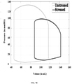
- FIG. 7B illustrates pressure volume loop showing left heart ventricle for silent myocardial ischemia showing stressed and destressed condition in accordance with some embodiments of the present disclosure.
- Stress scenario is simulated with an increased heart rate, as evident during exercise or stress condition.
- ECG during normal condition is similar to the healthy template, irrespective of small scar area simulation.
- As the heart rate is increased ECG morphology changes from normal to pathological.
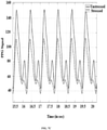
- FIG.7C similar observations were made in Pressure-Volume (PV) loop dynamics and PPG generation where unstressed condition replicates normal behavior, whereas under stress, EF decreases significantly.
- PV Pressure-Volume
- the embodiments of present disclosure herein address unresolved problem of myocardial ischemia severity.
- the embodiment thus provides determining myocardial ischemia based on hemodynamic parameters estimation.
- the embodiments herein further provide a cardiac model to capture the changes occurred in Electrophysiology (EP) signal from the plurality of Electrophysiology (EP) signals of the heart dynamics during myocardial ischemia.
- the changes occurred in the cardiac source module 110 serve as driving signal to the hemodynamic module 112 of cardiovascular system. This determines disease progression and its manifestation not only at the electric conduction level but also at the mechanical functioning of heart. Reflection of change in electric parameters over the hemodynamic variables like ejection fraction aids in a holistic understanding of the disease progression and analysis.
- the present disclosure is an efficient simulation platform that enables study of physiological parameters and deeper understanding of myocardial disease progression.
- the virtual simulation method serves as digital twin offers to the patient and the care givers / doctors to implement digital therapy/ medication to find the best possible prescription. This in turn provides different analysis in stress condition.
- the hardware device can be any kind of device which can be programmed including e.g. any kind of computer like a server or a personal computer, or the like, or any combination thereof.
- the device may also include means which could be e.g. hardware means like e.g. an application-specific integrated circuit (ASIC), a field-programmable gate array (FPGA), or a combination of hardware and software means, e.g.
- ASIC application-specific integrated circuit
- FPGA field-programmable gate array
- the means can include both hardware means and software means.
- the method embodiments described herein could be implemented in hardware and software.
- the device may also include software means.
- the embodiments may be implemented on different hardware devices, e.g. using a plurality of CPUs.
- the embodiments herein can comprise hardware and software elements.
- the embodiments that are implemented in software include but are not limited to, firmware, resident software, microcode, etc.
- the functions performed by various components described herein may be implemented in other components or combinations of other components.
- a computer-usable or computer readable medium can be any apparatus that can comprise, store, communicate, propagate, or transport the program for use by or in connection with the instruction execution system, apparatus, or device.
- a computer-readable storage medium refers to any type of physical memory on which information or data readable by a processor may be stored.
- a computer-readable storage medium may store instructions for execution by one or more processors, including instructions for causing the processor(s) to perform steps or stages consistent with the embodiments described herein.
- the term "computer- readable medium" should be understood to include tangible items and exclude carrier waves and transient signals, i.e., be non-transitory. Examples include random access memory (RAM), read-only memory (ROM), volatile memory, nonvolatile memory, hard drives, CD ROMs, DVDs, flash drives, disks, and any other known physical storage media.
Landscapes
- Health & Medical Sciences (AREA)
- Life Sciences & Earth Sciences (AREA)
- Engineering & Computer Science (AREA)
- Public Health (AREA)
- Medical Informatics (AREA)
- Cardiology (AREA)
- General Health & Medical Sciences (AREA)
- Pathology (AREA)
- Biomedical Technology (AREA)
- Veterinary Medicine (AREA)
- Surgery (AREA)
- Animal Behavior & Ethology (AREA)
- Molecular Biology (AREA)
- Heart & Thoracic Surgery (AREA)
- Physics & Mathematics (AREA)
- Biophysics (AREA)
- Physiology (AREA)
- Vascular Medicine (AREA)
- Epidemiology (AREA)
- Databases & Information Systems (AREA)
- Data Mining & Analysis (AREA)
- Primary Health Care (AREA)
- Artificial Intelligence (AREA)
- Computer Vision & Pattern Recognition (AREA)
- Psychiatry (AREA)
- Signal Processing (AREA)
- Nuclear Medicine, Radiotherapy & Molecular Imaging (AREA)
- Radiology & Medical Imaging (AREA)
- Measurement And Recording Of Electrical Phenomena And Electrical Characteristics Of The Living Body (AREA)
- Measuring Pulse, Heart Rate, Blood Pressure Or Blood Flow (AREA)
Applications Claiming Priority (1)
| Application Number | Priority Date | Filing Date | Title |
|---|---|---|---|
| IN202021036165 | 2020-08-21 |
Publications (3)
| Publication Number | Publication Date |
|---|---|
| EP3957247A1 true EP3957247A1 (de) | 2022-02-23 |
| EP3957247B1 EP3957247B1 (de) | 2024-02-14 |
| EP3957247C0 EP3957247C0 (de) | 2024-02-14 |
Family
ID=77338495
Family Applications (1)
| Application Number | Title | Priority Date | Filing Date |
|---|---|---|---|
| EP21191188.8A Active EP3957247B1 (de) | 2020-08-21 | 2021-08-13 | Verfahren und system zur bestimmung des schweregrades von myokardialer ischämie basierend auf der schätzung von hämodynamischen parametern |
Country Status (2)
| Country | Link |
|---|---|
| US (1) | US12285266B2 (de) |
| EP (1) | EP3957247B1 (de) |
Families Citing this family (1)
| Publication number | Priority date | Publication date | Assignee | Title |
|---|---|---|---|---|
| CN115530841B (zh) * | 2022-11-03 | 2025-08-12 | 浙江大学 | 一种无创心肌跨膜电位重建及异常点定位方法 |
Citations (3)
| Publication number | Priority date | Publication date | Assignee | Title |
|---|---|---|---|---|
| US20120157822A1 (en) * | 2009-06-24 | 2012-06-21 | Cortius Holding B.V. | Inverse Imaging of Electrical Activity of a Heart Muscle |
| US20150042646A1 (en) * | 2013-08-09 | 2015-02-12 | Dorin Comaniciu | System and Method for Patient Specific Planning and Guidance of Electrophysiology Interventions |
| US20190357854A1 (en) * | 2009-04-22 | 2019-11-28 | Streamline Automation, Llc | Cardiovascular state determination apparatus and method of use thereof |
Family Cites Families (3)
| Publication number | Priority date | Publication date | Assignee | Title |
|---|---|---|---|---|
| USRE38119E1 (en) | 1989-01-23 | 2003-05-20 | Mirowski Family Ventures, LLC | Method and apparatus for treating hemodynamic disfunction |
| US7215997B2 (en) * | 2003-12-22 | 2007-05-08 | Cardiac Pacemakers, Inc. | Dynamic device therapy control for treating post myocardial infarction patients |
| US7367951B2 (en) * | 2005-01-27 | 2008-05-06 | Medtronic, Inc. | System and method for detecting cardiovascular health conditions using hemodynamic pressure waveforms |
-
2021
- 2021-08-13 EP EP21191188.8A patent/EP3957247B1/de active Active
- 2021-08-17 US US17/445,292 patent/US12285266B2/en active Active
Patent Citations (3)
| Publication number | Priority date | Publication date | Assignee | Title |
|---|---|---|---|---|
| US20190357854A1 (en) * | 2009-04-22 | 2019-11-28 | Streamline Automation, Llc | Cardiovascular state determination apparatus and method of use thereof |
| US20120157822A1 (en) * | 2009-06-24 | 2012-06-21 | Cortius Holding B.V. | Inverse Imaging of Electrical Activity of a Heart Muscle |
| US20150042646A1 (en) * | 2013-08-09 | 2015-02-12 | Dorin Comaniciu | System and Method for Patient Specific Planning and Guidance of Electrophysiology Interventions |
Non-Patent Citations (1)
| Title |
|---|
| VAN DAM P M ET AL: "Simulating ECG changes during acute myocardial ischemia", COMPUTERS IN CARDIOLOGY, 2007, IEEE, PISCATAWAY, NJ, USA, 30 September 2007 (2007-09-30), pages 325 - 328, XP031404713, ISBN: 978-1-4244-2533-4 * |
Also Published As
| Publication number | Publication date |
|---|---|
| US12285266B2 (en) | 2025-04-29 |
| US20220104763A1 (en) | 2022-04-07 |
| EP3957247B1 (de) | 2024-02-14 |
| EP3957247C0 (de) | 2024-02-14 |
Similar Documents
| Publication | Publication Date | Title |
|---|---|---|
| US11957438B2 (en) | Method and apparatus for left ventricular end diastolic pressure measurement | |
| Seif et al. | Bedside ultrasound in resuscitation and the rapid ultrasound in shock protocol | |
| Costachescu et al. | The hemodynamically unstable patient in the intensive care unit: hemodynamic vs. transesophageal echocardiographic monitoring | |
| Rosenberg et al. | Noninvasive assessment of hemodynamics: an emphasis on bioimpedance cardiography | |
| Suess et al. | Hemodynamic monitoring for the evaluation and treatment of shock: what is the current state of the art? | |
| Benington et al. | Emerging trends in minimally invasive haemodynamic monitoring and optimization of fluid therapy | |
| Lee et al. | The Swan-Ganz catheter remains a critically important component of monitoring in cardiovascular critical care | |
| EP3957247B1 (de) | Verfahren und system zur bestimmung des schweregrades von myokardialer ischämie basierend auf der schätzung von hämodynamischen parametern | |
| CN121335664A (zh) | 基于来自机械循环支持装置的信号来确定心脏收缩性的系统及方法 | |
| Singh et al. | Perioperative intravascular fluid assessment and monitoring: a narrative review of established and emerging techniques | |
| Leeper | Right ventricular failure | |
| CN107106015A (zh) | 心脏状态监视系统 | |
| Wajdan et al. | Automatic detection of valve events by epicardial accelerometer allows estimation of the left ventricular pressure trace and pressure–displacement loop area | |
| Loosen et al. | Preload Responsiveness in Patients With Acute Respiratory Distress Syndrome Managed With Extracorporeal Membrane Oxygenation | |
| Michaels et al. | Invasive left ventricular energetics during enhanced external counterpulsation | |
| US12605137B2 (en) | Volume management apparatus, device, and storage medium for emergency treatment | |
| EP4408262B1 (de) | Lap-signalverarbeitung zur automatischen berechnung des a/v-verhältnisses | |
| EP4289358B1 (de) | Schätzung des blutdrucks einer person unter verwendung eines ekg-gesteuerten kardiovaskulären modells | |
| Young et al. | Heart failure with preserved left ventricular function: diagnostic and therapeutic challenges in patients with diastolic heart failure | |
| Khraim et al. | A novel non-invasive assessment of cardiac hemodynamics in patients with heart failure and atrial fibrillation | |
| US11241198B2 (en) | Method and apparatus for synchronizing impedance cardiography with electrocardiography to lower patient auxiliary current | |
| Saha | Influence of the left atrium dysfunction on the left ventricular function in heart failure with preserved ejection fraction | |
| Bukhari | Estimation of Serum Potassium and Calcium Concentrations from Electrocardiographic Depolarization and Repolarization Waveforms | |
| Hossain | Computational analysis of prognosis of myocardial infarction in left ventricle | |
| Laverde | Analysis of thoracic and intra-gastric cardiac vibration signals for the monitoring of heart failure |
Legal Events
| Date | Code | Title | Description |
|---|---|---|---|
| PUAI | Public reference made under article 153(3) epc to a published international application that has entered the european phase |
Free format text: ORIGINAL CODE: 0009012 |
|
| STAA | Information on the status of an ep patent application or granted ep patent |
Free format text: STATUS: THE APPLICATION HAS BEEN PUBLISHED |
|
| AK | Designated contracting states |
Kind code of ref document: A1 Designated state(s): AL AT BE BG CH CY CZ DE DK EE ES FI FR GB GR HR HU IE IS IT LI LT LU LV MC MK MT NL NO PL PT RO RS SE SI SK SM TR |
|
| STAA | Information on the status of an ep patent application or granted ep patent |
Free format text: STATUS: REQUEST FOR EXAMINATION WAS MADE |
|
| 17P | Request for examination filed |
Effective date: 20220817 |
|
| RBV | Designated contracting states (corrected) |
Designated state(s): AL AT BE BG CH CY CZ DE DK EE ES FI FR GB GR HR HU IE IS IT LI LT LU LV MC MK MT NL NO PL PT RO RS SE SI SK SM TR |
|
| GRAP | Despatch of communication of intention to grant a patent |
Free format text: ORIGINAL CODE: EPIDOSNIGR1 |
|
| STAA | Information on the status of an ep patent application or granted ep patent |
Free format text: STATUS: GRANT OF PATENT IS INTENDED |
|
| INTG | Intention to grant announced |
Effective date: 20230905 |
|
| RIN1 | Information on inventor provided before grant (corrected) |
Inventor name: SINHA, ANIRUDDHA Inventor name: ROY, DIBYENDU Inventor name: MAZUMDER, OISHEE |
|
| GRAS | Grant fee paid |
Free format text: ORIGINAL CODE: EPIDOSNIGR3 |
|
| GRAA | (expected) grant |
Free format text: ORIGINAL CODE: 0009210 |
|
| STAA | Information on the status of an ep patent application or granted ep patent |
Free format text: STATUS: THE PATENT HAS BEEN GRANTED |
|
| AK | Designated contracting states |
Kind code of ref document: B1 Designated state(s): AL AT BE BG CH CY CZ DE DK EE ES FI FR GB GR HR HU IE IS IT LI LT LU LV MC MK MT NL NO PL PT RO RS SE SI SK SM TR |
|
| REG | Reference to a national code |
Ref country code: GB Ref legal event code: FG4D |
|
| REG | Reference to a national code |
Ref country code: CH Ref legal event code: EP |
|
| REG | Reference to a national code |
Ref country code: DE Ref legal event code: R096 Ref document number: 602021009337 Country of ref document: DE |
|
| REG | Reference to a national code |
Ref country code: IE Ref legal event code: FG4D |
|
| U01 | Request for unitary effect filed |
Effective date: 20240214 |
|
| U07 | Unitary effect registered |
Designated state(s): AT BE BG DE DK EE FI FR IT LT LU LV MT NL PT SE SI Effective date: 20240223 |
|
| PG25 | Lapsed in a contracting state [announced via postgrant information from national office to epo] |
Ref country code: IS Free format text: LAPSE BECAUSE OF FAILURE TO SUBMIT A TRANSLATION OF THE DESCRIPTION OR TO PAY THE FEE WITHIN THE PRESCRIBED TIME-LIMIT Effective date: 20240614 |
|
| PG25 | Lapsed in a contracting state [announced via postgrant information from national office to epo] |
Ref country code: GR Free format text: LAPSE BECAUSE OF FAILURE TO SUBMIT A TRANSLATION OF THE DESCRIPTION OR TO PAY THE FEE WITHIN THE PRESCRIBED TIME-LIMIT Effective date: 20240515 |
|
| PG25 | Lapsed in a contracting state [announced via postgrant information from national office to epo] |
Ref country code: HR Free format text: LAPSE BECAUSE OF FAILURE TO SUBMIT A TRANSLATION OF THE DESCRIPTION OR TO PAY THE FEE WITHIN THE PRESCRIBED TIME-LIMIT Effective date: 20240214 Ref country code: RS Free format text: LAPSE BECAUSE OF FAILURE TO SUBMIT A TRANSLATION OF THE DESCRIPTION OR TO PAY THE FEE WITHIN THE PRESCRIBED TIME-LIMIT Effective date: 20240514 |
|
| PG25 | Lapsed in a contracting state [announced via postgrant information from national office to epo] |
Ref country code: ES Free format text: LAPSE BECAUSE OF FAILURE TO SUBMIT A TRANSLATION OF THE DESCRIPTION OR TO PAY THE FEE WITHIN THE PRESCRIBED TIME-LIMIT Effective date: 20240214 |
|
| PG25 | Lapsed in a contracting state [announced via postgrant information from national office to epo] |
Ref country code: RS Free format text: LAPSE BECAUSE OF FAILURE TO SUBMIT A TRANSLATION OF THE DESCRIPTION OR TO PAY THE FEE WITHIN THE PRESCRIBED TIME-LIMIT Effective date: 20240514 Ref country code: NO Free format text: LAPSE BECAUSE OF FAILURE TO SUBMIT A TRANSLATION OF THE DESCRIPTION OR TO PAY THE FEE WITHIN THE PRESCRIBED TIME-LIMIT Effective date: 20240514 Ref country code: IS Free format text: LAPSE BECAUSE OF FAILURE TO SUBMIT A TRANSLATION OF THE DESCRIPTION OR TO PAY THE FEE WITHIN THE PRESCRIBED TIME-LIMIT Effective date: 20240614 Ref country code: HR Free format text: LAPSE BECAUSE OF FAILURE TO SUBMIT A TRANSLATION OF THE DESCRIPTION OR TO PAY THE FEE WITHIN THE PRESCRIBED TIME-LIMIT Effective date: 20240214 Ref country code: GR Free format text: LAPSE BECAUSE OF FAILURE TO SUBMIT A TRANSLATION OF THE DESCRIPTION OR TO PAY THE FEE WITHIN THE PRESCRIBED TIME-LIMIT Effective date: 20240515 Ref country code: ES Free format text: LAPSE BECAUSE OF FAILURE TO SUBMIT A TRANSLATION OF THE DESCRIPTION OR TO PAY THE FEE WITHIN THE PRESCRIBED TIME-LIMIT Effective date: 20240214 |
|
| PG25 | Lapsed in a contracting state [announced via postgrant information from national office to epo] |
Ref country code: PL Free format text: LAPSE BECAUSE OF FAILURE TO SUBMIT A TRANSLATION OF THE DESCRIPTION OR TO PAY THE FEE WITHIN THE PRESCRIBED TIME-LIMIT Effective date: 20240214 |
|
| PG25 | Lapsed in a contracting state [announced via postgrant information from national office to epo] |
Ref country code: PL Free format text: LAPSE BECAUSE OF FAILURE TO SUBMIT A TRANSLATION OF THE DESCRIPTION OR TO PAY THE FEE WITHIN THE PRESCRIBED TIME-LIMIT Effective date: 20240214 |
|
| U20 | Renewal fee for the european patent with unitary effect paid |
Year of fee payment: 4 Effective date: 20240826 |
|
| PG25 | Lapsed in a contracting state [announced via postgrant information from national office to epo] |
Ref country code: SM Free format text: LAPSE BECAUSE OF FAILURE TO SUBMIT A TRANSLATION OF THE DESCRIPTION OR TO PAY THE FEE WITHIN THE PRESCRIBED TIME-LIMIT Effective date: 20240214 |
|
| PG25 | Lapsed in a contracting state [announced via postgrant information from national office to epo] |
Ref country code: CZ Free format text: LAPSE BECAUSE OF FAILURE TO SUBMIT A TRANSLATION OF THE DESCRIPTION OR TO PAY THE FEE WITHIN THE PRESCRIBED TIME-LIMIT Effective date: 20240214 |
|
| PG25 | Lapsed in a contracting state [announced via postgrant information from national office to epo] |
Ref country code: SK Free format text: LAPSE BECAUSE OF FAILURE TO SUBMIT A TRANSLATION OF THE DESCRIPTION OR TO PAY THE FEE WITHIN THE PRESCRIBED TIME-LIMIT Effective date: 20240214 |
|
| PG25 | Lapsed in a contracting state [announced via postgrant information from national office to epo] |
Ref country code: SM Free format text: LAPSE BECAUSE OF FAILURE TO SUBMIT A TRANSLATION OF THE DESCRIPTION OR TO PAY THE FEE WITHIN THE PRESCRIBED TIME-LIMIT Effective date: 20240214 Ref country code: SK Free format text: LAPSE BECAUSE OF FAILURE TO SUBMIT A TRANSLATION OF THE DESCRIPTION OR TO PAY THE FEE WITHIN THE PRESCRIBED TIME-LIMIT Effective date: 20240214 Ref country code: RO Free format text: LAPSE BECAUSE OF FAILURE TO SUBMIT A TRANSLATION OF THE DESCRIPTION OR TO PAY THE FEE WITHIN THE PRESCRIBED TIME-LIMIT Effective date: 20240214 Ref country code: CZ Free format text: LAPSE BECAUSE OF FAILURE TO SUBMIT A TRANSLATION OF THE DESCRIPTION OR TO PAY THE FEE WITHIN THE PRESCRIBED TIME-LIMIT Effective date: 20240214 |
|
| REG | Reference to a national code |
Ref country code: DE Ref legal event code: R097 Ref document number: 602021009337 Country of ref document: DE |
|
| PLBE | No opposition filed within time limit |
Free format text: ORIGINAL CODE: 0009261 |
|
| STAA | Information on the status of an ep patent application or granted ep patent |
Free format text: STATUS: NO OPPOSITION FILED WITHIN TIME LIMIT |
|
| 26N | No opposition filed |
Effective date: 20241115 |
|
| PG25 | Lapsed in a contracting state [announced via postgrant information from national office to epo] |
Ref country code: MC Free format text: LAPSE BECAUSE OF FAILURE TO SUBMIT A TRANSLATION OF THE DESCRIPTION OR TO PAY THE FEE WITHIN THE PRESCRIBED TIME-LIMIT Effective date: 20240214 |
|
| PG25 | Lapsed in a contracting state [announced via postgrant information from national office to epo] |
Ref country code: IE Free format text: LAPSE BECAUSE OF NON-PAYMENT OF DUE FEES Effective date: 20240813 |
|
| U20 | Renewal fee for the european patent with unitary effect paid |
Year of fee payment: 5 Effective date: 20250825 |
|
| PGFP | Annual fee paid to national office [announced via postgrant information from national office to epo] |
Ref country code: GB Payment date: 20250826 Year of fee payment: 5 |
|
| PGFP | Annual fee paid to national office [announced via postgrant information from national office to epo] |
Ref country code: CH Payment date: 20250901 Year of fee payment: 5 |
|
| PG25 | Lapsed in a contracting state [announced via postgrant information from national office to epo] |
Ref country code: CY Free format text: LAPSE BECAUSE OF FAILURE TO SUBMIT A TRANSLATION OF THE DESCRIPTION OR TO PAY THE FEE WITHIN THE PRESCRIBED TIME-LIMIT; INVALID AB INITIO Effective date: 20210813 |
|
| PG25 | Lapsed in a contracting state [announced via postgrant information from national office to epo] |
Ref country code: HU Free format text: LAPSE BECAUSE OF FAILURE TO SUBMIT A TRANSLATION OF THE DESCRIPTION OR TO PAY THE FEE WITHIN THE PRESCRIBED TIME-LIMIT; INVALID AB INITIO Effective date: 20210813 |