EP3958729B1 - Dispositif de surveillance d'un système pulmonaire d'un sujet - Google Patents
Dispositif de surveillance d'un système pulmonaire d'un sujet Download PDFInfo
- Publication number
- EP3958729B1 EP3958729B1 EP20723804.9A EP20723804A EP3958729B1 EP 3958729 B1 EP3958729 B1 EP 3958729B1 EP 20723804 A EP20723804 A EP 20723804A EP 3958729 B1 EP3958729 B1 EP 3958729B1
- Authority
- EP
- European Patent Office
- Prior art keywords
- light
- cavity
- light signal
- gas
- wavelength
- Prior art date
- Legal status (The legal status is an assumption and is not a legal conclusion. Google has not performed a legal analysis and makes no representation as to the accuracy of the status listed.)
- Active
Links
Images



Classifications
-
- A—HUMAN NECESSITIES
- A61—MEDICAL OR VETERINARY SCIENCE; HYGIENE
- A61B—DIAGNOSIS; SURGERY; IDENTIFICATION
- A61B5/00—Measuring for diagnostic purposes; Identification of persons
- A61B5/0002—Remote monitoring of patients using telemetry, e.g. transmission of vital signals via a communication network
- A61B5/0026—Remote monitoring of patients using telemetry, e.g. transmission of vital signals via a communication network characterised by the transmission medium
- A61B5/0028—Body tissue as transmission medium, i.e. transmission systems where the medium is the human body
-
- A—HUMAN NECESSITIES
- A61—MEDICAL OR VETERINARY SCIENCE; HYGIENE
- A61B—DIAGNOSIS; SURGERY; IDENTIFICATION
- A61B1/00—Instruments for performing medical examinations of the interior of cavities or tubes of the body by visual or photographical inspection, e.g. endoscopes; Illuminating arrangements therefor
- A61B1/267—Instruments for performing medical examinations of the interior of cavities or tubes of the body by visual or photographical inspection, e.g. endoscopes; Illuminating arrangements therefor for the respiratory tract, e.g. laryngoscopes, bronchoscopes
- A61B1/2676—Bronchoscopes
-
- A—HUMAN NECESSITIES
- A61—MEDICAL OR VETERINARY SCIENCE; HYGIENE
- A61B—DIAGNOSIS; SURGERY; IDENTIFICATION
- A61B5/00—Measuring for diagnostic purposes; Identification of persons
- A61B5/0033—Features or image-related aspects of imaging apparatus, e.g. for MRI, optical tomography or impedance tomography apparatus; Arrangements of imaging apparatus in a room
- A61B5/0036—Features or image-related aspects of imaging apparatus, e.g. for MRI, optical tomography or impedance tomography apparatus; Arrangements of imaging apparatus in a room including treatment, e.g., using an implantable medical device, ablating, ventilating
-
- A—HUMAN NECESSITIES
- A61—MEDICAL OR VETERINARY SCIENCE; HYGIENE
- A61B—DIAGNOSIS; SURGERY; IDENTIFICATION
- A61B5/00—Measuring for diagnostic purposes; Identification of persons
- A61B5/0059—Measuring for diagnostic purposes; Identification of persons using light, e.g. diagnosis by transillumination, diascopy, fluorescence
- A61B5/0075—Measuring for diagnostic purposes; Identification of persons using light, e.g. diagnosis by transillumination, diascopy, fluorescence by spectroscopy, i.e. measuring spectra, e.g. Raman spectroscopy, infrared absorption spectroscopy
-
- A—HUMAN NECESSITIES
- A61—MEDICAL OR VETERINARY SCIENCE; HYGIENE
- A61B—DIAGNOSIS; SURGERY; IDENTIFICATION
- A61B5/00—Measuring for diagnostic purposes; Identification of persons
- A61B5/0059—Measuring for diagnostic purposes; Identification of persons using light, e.g. diagnosis by transillumination, diascopy, fluorescence
- A61B5/0077—Devices for viewing the surface of the body, e.g. camera, magnifying lens
-
- A—HUMAN NECESSITIES
- A61—MEDICAL OR VETERINARY SCIENCE; HYGIENE
- A61B—DIAGNOSIS; SURGERY; IDENTIFICATION
- A61B5/00—Measuring for diagnostic purposes; Identification of persons
- A61B5/0059—Measuring for diagnostic purposes; Identification of persons using light, e.g. diagnosis by transillumination, diascopy, fluorescence
- A61B5/0082—Measuring for diagnostic purposes; Identification of persons using light, e.g. diagnosis by transillumination, diascopy, fluorescence adapted for particular medical purposes
- A61B5/0084—Measuring for diagnostic purposes; Identification of persons using light, e.g. diagnosis by transillumination, diascopy, fluorescence adapted for particular medical purposes for introduction into the body, e.g. by catheters
-
- A—HUMAN NECESSITIES
- A61—MEDICAL OR VETERINARY SCIENCE; HYGIENE
- A61B—DIAGNOSIS; SURGERY; IDENTIFICATION
- A61B5/00—Measuring for diagnostic purposes; Identification of persons
- A61B5/0059—Measuring for diagnostic purposes; Identification of persons using light, e.g. diagnosis by transillumination, diascopy, fluorescence
- A61B5/0082—Measuring for diagnostic purposes; Identification of persons using light, e.g. diagnosis by transillumination, diascopy, fluorescence adapted for particular medical purposes
- A61B5/0084—Measuring for diagnostic purposes; Identification of persons using light, e.g. diagnosis by transillumination, diascopy, fluorescence adapted for particular medical purposes for introduction into the body, e.g. by catheters
- A61B5/0086—Measuring for diagnostic purposes; Identification of persons using light, e.g. diagnosis by transillumination, diascopy, fluorescence adapted for particular medical purposes for introduction into the body, e.g. by catheters using infrared radiation
-
- A—HUMAN NECESSITIES
- A61—MEDICAL OR VETERINARY SCIENCE; HYGIENE
- A61B—DIAGNOSIS; SURGERY; IDENTIFICATION
- A61B5/00—Measuring for diagnostic purposes; Identification of persons
- A61B5/02—Detecting, measuring or recording for evaluating the cardiovascular system, e.g. pulse, heart rate, blood pressure or blood flow
- A61B5/021—Measuring pressure in heart or blood vessels
- A61B5/0215—Measuring pressure in heart or blood vessels by means inserted into the body
- A61B5/02154—Measuring pressure in heart or blood vessels by means inserted into the body by optical transmission
-
- A—HUMAN NECESSITIES
- A61—MEDICAL OR VETERINARY SCIENCE; HYGIENE
- A61B—DIAGNOSIS; SURGERY; IDENTIFICATION
- A61B5/00—Measuring for diagnostic purposes; Identification of persons
- A61B5/02—Detecting, measuring or recording for evaluating the cardiovascular system, e.g. pulse, heart rate, blood pressure or blood flow
- A61B5/024—Measuring pulse rate or heart rate
- A61B5/02416—Measuring pulse rate or heart rate using photoplethysmograph signals, e.g. generated by infrared radiation
- A61B5/02427—Details of sensor
-
- A—HUMAN NECESSITIES
- A61—MEDICAL OR VETERINARY SCIENCE; HYGIENE
- A61B—DIAGNOSIS; SURGERY; IDENTIFICATION
- A61B5/00—Measuring for diagnostic purposes; Identification of persons
- A61B5/08—Measuring devices for evaluating the respiratory organs
-
- A—HUMAN NECESSITIES
- A61—MEDICAL OR VETERINARY SCIENCE; HYGIENE
- A61B—DIAGNOSIS; SURGERY; IDENTIFICATION
- A61B5/00—Measuring for diagnostic purposes; Identification of persons
- A61B5/08—Measuring devices for evaluating the respiratory organs
- A61B5/0803—Recording apparatus specially adapted therefor
-
- A—HUMAN NECESSITIES
- A61—MEDICAL OR VETERINARY SCIENCE; HYGIENE
- A61B—DIAGNOSIS; SURGERY; IDENTIFICATION
- A61B5/00—Measuring for diagnostic purposes; Identification of persons
- A61B5/08—Measuring devices for evaluating the respiratory organs
- A61B5/082—Evaluation by breath analysis, e.g. determination of the chemical composition of exhaled breath
-
- A—HUMAN NECESSITIES
- A61—MEDICAL OR VETERINARY SCIENCE; HYGIENE
- A61B—DIAGNOSIS; SURGERY; IDENTIFICATION
- A61B5/00—Measuring for diagnostic purposes; Identification of persons
- A61B5/103—Measuring devices for testing the shape, pattern, colour, size or movement of the body or parts thereof, for diagnostic purposes
- A61B5/11—Measuring movement of the entire body or parts thereof, e.g. head or hand tremor or mobility of a limb
- A61B5/113—Measuring movement of the entire body or parts thereof, e.g. head or hand tremor or mobility of a limb occurring during breathing
-
- A—HUMAN NECESSITIES
- A61—MEDICAL OR VETERINARY SCIENCE; HYGIENE
- A61B—DIAGNOSIS; SURGERY; IDENTIFICATION
- A61B5/00—Measuring for diagnostic purposes; Identification of persons
- A61B5/145—Measuring characteristics of blood in vivo, e.g. gas concentration or pH-value ; Measuring characteristics of body fluids or tissues, e.g. interstitial fluid or cerebral tissue
- A61B5/1495—Calibrating or testing of in-vivo probes
-
- A—HUMAN NECESSITIES
- A61—MEDICAL OR VETERINARY SCIENCE; HYGIENE
- A61B—DIAGNOSIS; SURGERY; IDENTIFICATION
- A61B5/00—Measuring for diagnostic purposes; Identification of persons
- A61B5/24—Detecting, measuring or recording bioelectric or biomagnetic signals of the body or parts thereof
- A61B5/25—Bioelectric electrodes therefor
- A61B5/251—Means for maintaining electrode contact with the body
- A61B5/256—Wearable electrodes, e.g. having straps or bands
-
- A—HUMAN NECESSITIES
- A61—MEDICAL OR VETERINARY SCIENCE; HYGIENE
- A61B—DIAGNOSIS; SURGERY; IDENTIFICATION
- A61B5/00—Measuring for diagnostic purposes; Identification of persons
- A61B5/74—Details of notification to user or communication with user or patient; User input means
- A61B5/746—Alarms related to a physiological condition, e.g. details of setting alarm thresholds or avoiding false alarms
-
- A—HUMAN NECESSITIES
- A61—MEDICAL OR VETERINARY SCIENCE; HYGIENE
- A61B—DIAGNOSIS; SURGERY; IDENTIFICATION
- A61B5/00—Measuring for diagnostic purposes; Identification of persons
- A61B5/74—Details of notification to user or communication with user or patient; User input means
- A61B5/7465—Arrangements for interactive communication between patient and care services, e.g. by using a telephone network
-
- G—PHYSICS
- G01—MEASURING; TESTING
- G01N—INVESTIGATING OR ANALYSING MATERIALS BY DETERMINING THEIR CHEMICAL OR PHYSICAL PROPERTIES
- G01N21/00—Investigating or analysing materials by the use of optical means, i.e. using sub-millimetre waves, infrared, visible or ultraviolet light
- G01N21/17—Systems in which incident light is modified in accordance with the properties of the material investigated
- G01N21/25—Colour; Spectral properties, i.e. comparison of effect of material on the light at two or more different wavelengths or wavelength bands
- G01N21/31—Investigating relative effect of material at wavelengths characteristic of specific elements or molecules, e.g. atomic absorption spectrometry
- G01N21/39—Investigating relative effect of material at wavelengths characteristic of specific elements or molecules, e.g. atomic absorption spectrometry using tunable lasers
-
- A—HUMAN NECESSITIES
- A61—MEDICAL OR VETERINARY SCIENCE; HYGIENE
- A61B—DIAGNOSIS; SURGERY; IDENTIFICATION
- A61B2560/00—Constructional details of operational features of apparatus; Accessories for medical measuring apparatus
- A61B2560/02—Operational features
- A61B2560/0223—Operational features of calibration, e.g. protocols for calibrating sensors
-
- A—HUMAN NECESSITIES
- A61—MEDICAL OR VETERINARY SCIENCE; HYGIENE
- A61B—DIAGNOSIS; SURGERY; IDENTIFICATION
- A61B2562/00—Details of sensors; Constructional details of sensor housings or probes; Accessories for sensors
- A61B2562/02—Details of sensors specially adapted for in-vivo measurements
- A61B2562/0233—Special features of optical sensors or probes classified in A61B5/00
-
- G—PHYSICS
- G01—MEASURING; TESTING
- G01N—INVESTIGATING OR ANALYSING MATERIALS BY DETERMINING THEIR CHEMICAL OR PHYSICAL PROPERTIES
- G01N21/00—Investigating or analysing materials by the use of optical means, i.e. using sub-millimetre waves, infrared, visible or ultraviolet light
- G01N21/17—Systems in which incident light is modified in accordance with the properties of the material investigated
- G01N21/47—Scattering, i.e. diffuse reflection
- G01N21/4738—Diffuse reflection, e.g. also for testing fluids, fibrous materials
- G01N21/474—Details of optical heads therefor, e.g. using optical fibres
- G01N2021/4742—Details of optical heads therefor, e.g. using optical fibres comprising optical fibres
-
- G—PHYSICS
- G01—MEASURING; TESTING
- G01N—INVESTIGATING OR ANALYSING MATERIALS BY DETERMINING THEIR CHEMICAL OR PHYSICAL PROPERTIES
- G01N21/00—Investigating or analysing materials by the use of optical means, i.e. using sub-millimetre waves, infrared, visible or ultraviolet light
- G01N21/01—Arrangements or apparatus for facilitating the optical investigation
- G01N21/03—Cuvette constructions
- G01N21/031—Multipass arrangements
-
- G—PHYSICS
- G01—MEASURING; TESTING
- G01N—INVESTIGATING OR ANALYSING MATERIALS BY DETERMINING THEIR CHEMICAL OR PHYSICAL PROPERTIES
- G01N21/00—Investigating or analysing materials by the use of optical means, i.e. using sub-millimetre waves, infrared, visible or ultraviolet light
- G01N21/17—Systems in which incident light is modified in accordance with the properties of the material investigated
- G01N21/25—Colour; Spectral properties, i.e. comparison of effect of material on the light at two or more different wavelengths or wavelength bands
- G01N21/31—Investigating relative effect of material at wavelengths characteristic of specific elements or molecules, e.g. atomic absorption spectrometry
- G01N21/314—Investigating relative effect of material at wavelengths characteristic of specific elements or molecules, e.g. atomic absorption spectrometry with comparison of measurements at specific and non-specific wavelengths
- G01N21/3151—Investigating relative effect of material at wavelengths characteristic of specific elements or molecules, e.g. atomic absorption spectrometry with comparison of measurements at specific and non-specific wavelengths using two sources of radiation of different wavelengths
Definitions
- the pulmonary transition from intrauterine environment to normal postnatal air breathing may cause pulmonary diseases that are increasingly prevalent with decreasing gestational age of the newborn infant.
- pulmonary diseases that are increasingly prevalent with decreasing gestational age of the newborn infant.
- the number of infants needing respiratory support in neonatal care units will increase.
- Abnormal pulmonary aeration is common in postnatal lung diseases, such as respiratory distress syndrome due to surfactant deficiency and bronchopulmonary dysplasia.
- respiratory distress syndrome due to surfactant deficiency and bronchopulmonary dysplasia.
- bronchopulmonary dysplasia Despite modern respiratory support methods and minimally invasive surfactant therapy, there is still a risk of a life-threatening complication, such as pneumothorax.
- lung aeration problems are partially diagnosed with pulmonary X-ray, which unfortunately only gives a snap-shot of the current condition and when repeated causes a load of harmful radiation, increasing the risk of malignancy development later in life.
- Another method is positive transillumination where doctors shine a strong light through the affected side of the newborn's chest while in a darkened room. This procedure is done to show free air in the area surrounding the lungs (pleural cavity). Positive transillumination is analysed visually and is not used for continuous monitoring of the pulmonary system.
- EP3 338 077 A1 describes a device and system for measuring free gas in a cavity of a subject and a control unit for evaluating the detected transmitted light for determining a concentration of the free gas.
- the following disclosure focuses on examples of the present disclosure applicable for monitoring a pulmonary system of a subject, for example by arranging a light source or an optical member in a cavity, such as in the trachea, or in the digestive system, such as in the oesophagus or the intestines, such as parts of the digestive tract and detecting the transmitted light using a detector, for example one or a plurality of detectors arranged outside the human body.
- this is advantageous for detecting weak signals due to the light passing a long path through the tissue enclosing the cavity.
- both the detector and the light source may be arranged dermally.
- the light source is then configured to transmit light to the cavity and the detector is configured for detecting light scattering back which has passed through the cavity at least once.
- Such arrangements are described in EP1871221 .
- a light signal, such as an absorption spectrum, related to the free gas may be used for monitoring a pulmonary system to determining a status of the pulmonary system, such as a physiological status of the pulmonary system.
- Physiological problems may, for example, be aeration problems and/or pulmonary complications, such as lung aeration problems, more particularly diagnosis of pneumothorax and/or atelectasis.
- the light source is arranged to transmit light to the tissue with losses as low as possible and then detect the light at the surface of the skin.
- the total attenuation factor may be reduced to about 0.001 which is about 1000 times larger than what was previously be achieved.
- a free gas, or a mixture of gases, in a cavity of the pulmonary system may be monitored for determining the status of the pulmonary system, such as the physiological status of the pulmonary system.
- GASMAS technology may be applied for obtaining the concentration and/or distribution of the free gas.
- the disclosed devices, systems and methods utilize a member for introducing the light sources for injecting the light into the tissue, for example, a bronchoscope, a nasogastric feeding tube, endoscope, a tracheal tube, a colonoscope or similar introducing members.
- a member for introducing the light sources for injecting the light into the tissue for example, a bronchoscope, a nasogastric feeding tube, endoscope, a tracheal tube, a colonoscope or similar introducing members.
- the light from the optical fibre arrangement is distributed over a larger distance or area by a light diffusing material.
- the transmitted light is detected at the skin surface by one or a plurality of detectors positioned at the skin over the cavity such as the lung.
- the detectors may be configured to be arranged at the skin surface or other means, such as optical fibres, could be used to collect the light at the skin surface and direct the collected light to a detector.
- a plurality of detectors may be used in parallel or sequentially to detect gases or a distribution of gases, or gas concentrations in different parts of the bronchial tree or lung of a patient.
- the detection is made frequency- and/or phase-sensitively.
- the light, such as laser light, from the light source may be wavelength modulated at a selected frequency, and synchronous intensity variations may be detected when the modulation is conducted around a gas absorption wavelength.
- modulation is conducted close to a gas absorption wavelength, the intensity of the detected light will quickly change at small variations of the wavelength, as described in S. Svanberg, Gas in Scattering Media Absorption Spectroscopy - from Basic Studies to Biomedical Applications, Lasers and Photonics Reviews 7, 779 (2013 ).
- a skin area may be detected by an imaging sensor, such as a digital camera, with high intensity dynamic both at an absorbing wavelength and at a close non-absorbing wavelength.
- the two images may then be compared, for example by division or subtraction, whereby the areas affected by gases may be visualized.
- the path length through the gas is unknown; this need to be accounted for when performing a gas concentration measurement.
- Different method may be used for handling the problems of an unknown path length, these are discussed in, for example L. Mei, G. Somesfalean and S. Svanberg, Pathlength Determination for Gas in Scattering Media Absorption Spectroscopy, Sensors 14, 3871 (2014 )
- the gas concentration such as for oxygen, nitric oxide (NO), and carbon dioxide, may then be directly calculated using the approximated path length.
- This method works best when the light absorption and the light scattering in the tissue is the same for both measurements which is the case when the wavelengths used for the measurements are close.
- oxygen is normally monitored around some of the sharp components in the oxygen molecule's A-band about 760 nm.
- Water vapour has a strong absorption around, for example 935 nm, but the difference in wavelength compared to oxygen may need to be corrected for due to differences in the optical properties at the different wavelengths. Therefore, the weaker absorption wavelength for water vapour at around 820 nm may be a better choice.
- Fig. 1 One example of an implementation is illustrated in Fig. 1 .
- the patient 1 in this example is a preterm born infant.
- the infant is connected to a medical ventilator 2 due to, under the circumstances common disorder, respiratory distress syndrome (RDS).
- RDS respiratory distress syndrome
- control unit or a data processing device may be implemented by special-purpose software (or firmware) run on one or more general-purpose or special-purpose computing devices.
- each "element” or “means” of such a computing device refers to a conceptual equivalent of a method step; there is not always a one-to-one correspondence between elements/means and particular pieces of hardware or software routines.
- One piece of hardware sometimes comprises different means/elements.
- a processing unit serves as one element/means when executing one instruction, but serves as another element/means when executing another instruction.
- one element/means may be implemented by one instruction in some cases, but by a plurality of instructions in some other cases.
- the system memory may include computer storage media in the form of volatile and/or non-volatile memory such as read only memory (ROM), random access memory (RAM) and flash memory.
- the special-purpose software may be stored in the system memory, or on other removable/non-removable volatile/non-volatile computer storage media which is included in or accessible to the computing device, such as magnetic media, optical media, flash memory cards, digital tape, solid state RAM, solid state ROM, etc.
- the data processing device 10 may include one or more communication interfaces, such as a serial interface, a parallel interface, a USB interface, a wireless interface, a network adapter, etc., as well as one or more data acquisition devices, such as an A/D converter.
- the special-purpose software may be provided to the control unit or data processing device on any suitable computerreadable medium, including a record medium and a read-only memory.
- controller 9 may also be used to control the distribution of medicaments to the patient 1.
- a compact diffuse multi-pass cell made from a porous material, such as ceramic may be used.
- the porous material may be enclosed in a compact gas cell, see T. Svensson, E. Adolfsson, M. Lewander, C.T. Xu and S. Svanberg, Disordered, Strongly Scattering Porous Materials as Miniature Multi-pass Gas Cells, Phys. Rev. Lett. 107, 143901 (2011 ).
- the main part of the individually frequency marked light is led through optical fibre 11 down through the, in this example used, endotracheal tube 3.
- the injected light transmitted from the end of the optical fibre 11, such as through the diffusor may be transmitted to the tissue without passing any air in the trachea. If the light passes through air, the air may give some background signal in light detected by the detectors 14.
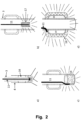
- an inflatable cuff or balloon made from optically scattering material and with reflecting material on the inner walls may be used to have as much light as possible to be directly transmitted into the tissue, as seen in Fig. 2A .
- Fig 2A gives good positioning of the optical fibre for monitoring the upper part of the lungs.
- the light injection may be made deeper into the bronchial tree as illustrated in Fig. 3 .
- more than one, such as at least two, positions are used for light injection.
- the measurements for the different locations need to be performed sequentially.
- more than one controllable standard fibre may be used.
- the detector or detectors 14 is/are adapted to be arranged against the skin.
- the detector should have a surface sized to detect the light transmitted through the tissue, for example in the range 0.25cm 2 to 5cm 2 , such as 1cm 2 .
- the detector may be made from different materials, such as germanium.
- an analogue lock-in-amplifier may be used.
- the analogue lock-in-amplifier may be connected to the control unit 8.
- Fig. 3 is illustrating an example wherein diffused light is injected using a working channel of a bronchoscope.
- the light sources are connected to the drive unit 7, which is controlled by the control unit 8.
- the measured value of the free gas in the lungs may be used for affecting the controller 9 for controlling the setting of the medical ventilator 2.
- the measured value by the control unit 8 may be used for administration of a medicament 10.
- a small portion of the light to be injected may be diverted optically, such as by a fibre, to a calibration unit 13.
- a detector 14 is configured to be positioned at a skin location at the chest of the patient for detecting the transmitted diffused light.
- the detected light carries information about the gas concentration in the lungs or gas distribution in the lung tissue, such as oxygen, nitric oxide (NO), and carbon dioxide concentration or distribution.
- Figs. 2A to 2D are illustrating different examples of coupling the light from the optical fibre to the tissue for measuring lung function.
- Fig. 2B is illustrating an example of an optical fibre 11 brought down a trachea tube, such as an endotracheal tube, to its end.
- a light diffusor 17 is arranged as an ending to the optical fibre 11.
- Fig. 2C is illustrating an example of how light in the fibre 11 may be connected to the end section 18 of a trachea tube, such as an endotracheal tube.
- the end section is made from a non-absorbing, but strongly light scattering, material.
- the laser light source is applied via the oesophagus rather than the trachea.
- the laser light source may be combined with the nasogastric feeding tube that is used in most infants in neonatal intensive care.
- the use of the nasogastric feeding tube for light application is advantageous because most infants already need to have such a device inserted, so no additional device needs to be introduced. It is also advantageous since the oesophagus environment is less sensitive to infection. It is also advantageous due to that the oesophagus is normally mostly collapsed, so there is potentially not a need for an expanding cuff to create good optical contact between the light source and the tissue. Further, lower parts of the lungs may also be more easily reached.
- the light source should be positioned at a point in the oesophagus that represents a position close to the lungs.
- an optical fibre that guides the laser light is combined with the nasogastric feeding tube, so that the optical fibre goes along the tube to an adequate position along the tube.
- Nasogastric feeding tubes have marks that indicate how deep the tube is inserted, and these marks can be used to determine the position of the light source in the oesophagus.
- the distal end of the optical fibre is terminated with a light diffusor that distributes the light over a larger area than would be provided by the fibre tip only.
- the diffusor is implemented by designing the tube wall in front of the fibre tip with light scattering properties so that the light scatters over an area along the tube corresponding to the desired characteristics of the diffusor.
- the diffusor is manufactured as a separate component that is embedded in the tube wall similarly to the optical fibre.
- the diffusor can be made part of an expandable cuff similar to the diffusor described in connection with the trachea herein above.
- the optical fibre may also be positioned in the nasogastric tube in such a way, that it may be sequentially moved to different positions along the tube for facilitating, e.g. a full tomographic view of the gas distribution, such as oxygen, nitric oxide (NO), and carbon dioxide distribution, using multiple detectors 14.
- the gas distribution such as oxygen, nitric oxide (NO), and carbon dioxide distribution
- a plurality of optical fibres may be used in parallel at different positions for facilitating, e.g. a full tomographic view of the gas distribution, such as oxygen, nitric oxide (NO), and carbon dioxide distribution, using multiple detectors 14.
- the gas distribution such as oxygen, nitric oxide (NO), and carbon dioxide distribution
- Figure 4A illustrates an optical fibre 41 combined with a nasogastric feeding tube 46 inserted into the oesophagus 42.
- the relation with the trachea 43 and the lungs 44 is also shown.
- At the distal end of the optical fibre 41 there may be a light diffusor 45.
- the upper part of the stomach 47 is also shown.
- a fibre arrangement connected to a light source may be used but a light source may, in some examples, be adapted to be directly arranged on the skin surface, such as a LED, laser, and waveguide.
Landscapes
- Health & Medical Sciences (AREA)
- Life Sciences & Earth Sciences (AREA)
- Physics & Mathematics (AREA)
- General Health & Medical Sciences (AREA)
- Pathology (AREA)
- Engineering & Computer Science (AREA)
- Surgery (AREA)
- Biomedical Technology (AREA)
- Heart & Thoracic Surgery (AREA)
- Medical Informatics (AREA)
- Molecular Biology (AREA)
- Animal Behavior & Ethology (AREA)
- Biophysics (AREA)
- Public Health (AREA)
- Veterinary Medicine (AREA)
- Physiology (AREA)
- Pulmonology (AREA)
- Cardiology (AREA)
- Spectroscopy & Molecular Physics (AREA)
- Optics & Photonics (AREA)
- Radiology & Medical Imaging (AREA)
- Nuclear Medicine, Radiotherapy & Molecular Imaging (AREA)
- Analytical Chemistry (AREA)
- Otolaryngology (AREA)
- Computer Networks & Wireless Communication (AREA)
- Chemical & Material Sciences (AREA)
- Biochemistry (AREA)
- General Physics & Mathematics (AREA)
- Immunology (AREA)
- Dentistry (AREA)
- Oral & Maxillofacial Surgery (AREA)
- Vascular Medicine (AREA)
- Nursing (AREA)
- Investigating Or Analysing Materials By Optical Means (AREA)
- Measurement Of The Respiration, Hearing Ability, Form, And Blood Characteristics Of Living Organisms (AREA)
Claims (13)
- Dispositif de surveillance d'un système pulmonaire d'un sujet, ledit dispositif comprenant :un élément optique (11) destiné à émettre un signal lumineux à travers une cavité dudit système pulmonaire dudit sujet, où ledit signal lumineux comprend au moins une longueur d'onde, où ladite au moins une longueur d'onde se trouve dans une fenêtre optique pour tissu ;une unité de détecteur (14), où ledit détecteur (14) est configuré pour être positionné sur une surface cutanée afin de détecter ledit signal lumineux transmis à travers ladite cavité dudit sujet ;où ledit élément optique (11) et ladite unité de détecteur (14) sont configurés pour être positionnés de sorte que ledit signal lumineux est transmis depuis ledit élément optique (11) à travers ladite cavité pour être détecté par ledit détecteur (14) ; etune unité de commande (8) destinée à évaluer ledit signal lumineux détecté afin de déterminer un état physiologique dudit système pulmonaire dudit sujet en évaluant une évolution dans le temps d'une intensité dudit signal lumineux détecté transmis à travers ladite cavité ;caractérisé en ce que ladite unité de commande (8) est en outre configurée pour associer ladite évolution dans le temps d'une intensité dudit signal lumineux détecté transmis à travers ladite cavité à une variation de volume de ladite cavité.
- Dispositif selon la revendication 1, dans lequel ledit signal lumineux détecté est associé à la même longueur d'onde au fil du temps, de telle sorte que ladite au moins une longueur d'onde dudit signal lumineux émis est une seule longueur d'onde.
- Dispositif selon l'une quelconque des revendications 1 et 2, dans lequel ladite longueur d'onde n'est pas associée à une bande d'absorption d'un gaz libre.
- Dispositif selon la revendication 1, dans lequel ladite variation dudit volume est une mesure qualitative.
- Dispositif selon l'une quelconque des revendications 1 à 4, où le dispositif est en outre configuré de telle sorte que ladite variation de volume est détectée en obtenant un premier signal de transmission transmis à travers une première cavité, telle qu'un premier poumon, et un second signal de transmission transmis à travers une seconde cavité, telle qu'un second poumon, les premier et second signaux de transmission sont ensuite comparés pour détecter une évolution de volume d'une desdites cavités par rapport à l'autre.
- Dispositif selon l'une quelconque des revendications 1 à 2 et 4 à 5, dans lequel ladite au moins une longueur d'onde dudit signal lumineux émis est associée à une bande d'absorption d'un gaz libre dans le système pulmonaire.
- Dispositif selon l'une quelconque des revendications 1 à 6, dans lequel ledit élément optique (11) est conçu pour être agencé sur une surface cutanée du sujet afin d'émettre ledit signal lumineux en direction de ladite cavité.
- Dispositif selon l'une quelconque des revendications 6 et 7, dans lequel ladite unité de commande (8) est configurée pour détecter en outre ledit état physiologique dudit système pulmonaire en obtenant une concentration et/ou une distribution dudit gaz libre à partir dudit signal lumineux détecté transmis à travers ladite cavité et en évaluant des évolutions de ladite concentration et/ou de ladite distribution dans le temps.
- Dispositif selon la revendication 8, dans lequel ladite unité de commande est configurée pour détecter ledit état physiologique dudit système pulmonaire en corrélant ladite intensité dudit signal lumineux détecté transmis à travers ladite cavité et ladite concentration et/ou ladite distribution dudit gaz libre, dans le temps.
- Dispositif selon l'une quelconque des revendications 8 et 9, dans lequel le signal lumineux comprend au moins deux longueurs d'onde différentes.
- Dispositif selon la revendication 10, dans lequel au moins une desdites longueurs d'onde est associée à une bande d'absorption d'un gaz de référence.
- Dispositif selon l'une quelconque des revendications 6 à 11, dans lequel le gaz libre est un gaz physiologique ou un mélange de gaz, tels que l'un quelconque parmi l'oxygène, le monoxyde d'azote (NO), le dioxyde de carbone, des gaz d'anesthésie et de la vapeur d'eau.
- Dispositif selon l'une quelconque des revendications 1 à 12, dans lequel l'unité de commande (8) est configurée pour commander un ventilateur médical (2) sur la base dudit état physiologique détecté dudit système pulmonaire.
Priority Applications (1)
| Application Number | Priority Date | Filing Date | Title |
|---|---|---|---|
| EP25184346.2A EP4595867A3 (fr) | 2019-04-26 | 2020-04-23 | Dispositif de surveillance d'un système pulmonaire d'un sujet |
Applications Claiming Priority (2)
| Application Number | Priority Date | Filing Date | Title |
|---|---|---|---|
| SE1950510 | 2019-04-26 | ||
| PCT/EP2020/061326 WO2020216839A1 (fr) | 2019-04-26 | 2020-04-23 | Dispositif de surveillance d'un système pulmonaire d'un sujet |
Related Child Applications (1)
| Application Number | Title | Priority Date | Filing Date |
|---|---|---|---|
| EP25184346.2A Division EP4595867A3 (fr) | 2019-04-26 | 2020-04-23 | Dispositif de surveillance d'un système pulmonaire d'un sujet |
Publications (3)
| Publication Number | Publication Date |
|---|---|
| EP3958729A1 EP3958729A1 (fr) | 2022-03-02 |
| EP3958729B1 true EP3958729B1 (fr) | 2025-07-02 |
| EP3958729C0 EP3958729C0 (fr) | 2025-07-02 |
Family
ID=70483100
Family Applications (2)
| Application Number | Title | Priority Date | Filing Date |
|---|---|---|---|
| EP25184346.2A Pending EP4595867A3 (fr) | 2019-04-26 | 2020-04-23 | Dispositif de surveillance d'un système pulmonaire d'un sujet |
| EP20723804.9A Active EP3958729B1 (fr) | 2019-04-26 | 2020-04-23 | Dispositif de surveillance d'un système pulmonaire d'un sujet |
Family Applications Before (1)
| Application Number | Title | Priority Date | Filing Date |
|---|---|---|---|
| EP25184346.2A Pending EP4595867A3 (fr) | 2019-04-26 | 2020-04-23 | Dispositif de surveillance d'un système pulmonaire d'un sujet |
Country Status (5)
| Country | Link |
|---|---|
| US (1) | US20220202311A1 (fr) |
| EP (2) | EP4595867A3 (fr) |
| CN (2) | CN120130941A (fr) |
| AU (1) | AU2020262302A1 (fr) |
| WO (1) | WO2020216839A1 (fr) |
Families Citing this family (3)
| Publication number | Priority date | Publication date | Assignee | Title |
|---|---|---|---|---|
| SE1500335A1 (sv) * | 2015-08-17 | 2017-02-18 | Utrustning och metod för intern laserbelysning för medicinskgasanalys | |
| US12336805B2 (en) | 2021-01-20 | 2025-06-24 | Vivonics, Inc. | System and method for detecting and/or monitoring the presence of at least one of pneumothorax, hemopneumothorax, or hemothorax in a living subject using one or more light sources and one or more light detectors |
| SE544842C2 (en) * | 2021-01-27 | 2022-12-13 | Gpx Medical Ab | A method and device for rescaling a signal to remove an absorption offset from an optical measurement |
Citations (1)
| Publication number | Priority date | Publication date | Assignee | Title |
|---|---|---|---|---|
| EP3338077A1 (fr) * | 2015-08-17 | 2018-06-27 | GPX Medical AB | Système et procédé d'analyse de gaz interne à base de laser dans un corps d'un être humain |
Family Cites Families (12)
| Publication number | Priority date | Publication date | Assignee | Title |
|---|---|---|---|---|
| US5005573A (en) * | 1990-07-20 | 1991-04-09 | Buchanan Dale C | Endotracheal tube with oximetry means |
| JPH0979980A (ja) * | 1995-09-13 | 1997-03-28 | Yazaki Corp | ガス検出器及びガス検出装置 |
| SE0200782D0 (sv) * | 2002-03-14 | 2002-03-14 | Astrazeneca Ab | Method of analysing a pharmaceutical sample |
| JP5583340B2 (ja) * | 2005-04-18 | 2014-09-03 | ガスポロックス エイビー | 体内腔部ガス測定装置及び測定方法 |
| JP2008107337A (ja) * | 2006-09-27 | 2008-05-08 | National Institute Of Advanced Industrial & Technology | ガス検出装置 |
| US8881724B2 (en) * | 2006-10-19 | 2014-11-11 | The General Electric Company | Device and method for graphical mechanical ventilator setup and control |
| US20080275355A1 (en) * | 2007-05-04 | 2008-11-06 | Ekips Technologies, Inc. | Method For Diagnosing An Infectioin Condition |
| US20120183949A1 (en) * | 2011-01-19 | 2012-07-19 | Searete Llc, A Limited Liability Corporation Of The State Of Delaware | Method, device, or system using lung sensor for detecting a physiological condition in a vertebrate subject |
| WO2013016639A1 (fr) * | 2011-07-28 | 2013-01-31 | Brigham And Women's Hospital, Inc. | Systèmes et procédés pour mesures par résonance magnétique portables de propriétés pulmonaires |
| RU2653633C2 (ru) * | 2012-03-01 | 2018-05-11 | Конинклейке Филипс Н.В. | Датчик определения концентрации газа |
| US20150305612A1 (en) * | 2014-04-23 | 2015-10-29 | Mark Hunter | Apparatuses and methods for registering a real-time image feed from an imaging device to a steerable catheter |
| US10478375B2 (en) * | 2015-09-25 | 2019-11-19 | Peter Antros | Pulmonary expansion therapy devices |
-
2020
- 2020-04-23 CN CN202510312520.4A patent/CN120130941A/zh active Pending
- 2020-04-23 US US17/594,706 patent/US20220202311A1/en active Pending
- 2020-04-23 EP EP25184346.2A patent/EP4595867A3/fr active Pending
- 2020-04-23 WO PCT/EP2020/061326 patent/WO2020216839A1/fr not_active Ceased
- 2020-04-23 AU AU2020262302A patent/AU2020262302A1/en not_active Abandoned
- 2020-04-23 CN CN202080031658.2A patent/CN113766870B/zh active Active
- 2020-04-23 EP EP20723804.9A patent/EP3958729B1/fr active Active
Patent Citations (1)
| Publication number | Priority date | Publication date | Assignee | Title |
|---|---|---|---|---|
| EP3338077A1 (fr) * | 2015-08-17 | 2018-06-27 | GPX Medical AB | Système et procédé d'analyse de gaz interne à base de laser dans un corps d'un être humain |
Non-Patent Citations (1)
| Title |
|---|
| BEEK J F ET AL: "Physics in Medicine & Biology", 30 June 1997 (1997-06-30), XP093266908, Retrieved from the Internet <URL:https://iopscience.iop.org/article/10.1088/0031-9155/42/11/018/pdf> * |
Also Published As
| Publication number | Publication date |
|---|---|
| WO2020216839A1 (fr) | 2020-10-29 |
| EP3958729A1 (fr) | 2022-03-02 |
| US20220202311A1 (en) | 2022-06-30 |
| EP4595867A3 (fr) | 2026-02-11 |
| CN113766870B (zh) | 2025-02-25 |
| AU2020262302A1 (en) | 2021-11-18 |
| EP4595867A2 (fr) | 2025-08-06 |
| CN113766870A (zh) | 2021-12-07 |
| CN120130941A (zh) | 2025-06-13 |
| EP3958729C0 (fr) | 2025-07-02 |
Similar Documents
| Publication | Publication Date | Title |
|---|---|---|
| US12178548B2 (en) | System and method for laser based internal analysis of gases in a body of a human | |
| EP3958729B1 (fr) | Dispositif de surveillance d'un système pulmonaire d'un sujet | |
| US4697593A (en) | Method and apparatus for measuring blood oxygen levels | |
| US7992573B2 (en) | Optically guided system for precise placement of a medical catheter in a patient | |
| US6526297B1 (en) | Method and apparatus for quantifying the hypnotic component of the depth of anesthesia by monitoring changes in optical scattering properties of brain tissue | |
| US20080039715A1 (en) | Three-dimensional optical guidance for catheter placement | |
| US20100006103A1 (en) | Endotracheal-oximeter device, system and method of using same | |
| JP2013176636A (ja) | 体腔のガス測定装置及び測定方法 | |
| US20080045822A1 (en) | Optical Fibre Catheter Pulse Oximeter | |
| Svanberg et al. | Changes in pulmonary oxygen content are detectable with laser absorption spectroscopy: proof of concept in newborn piglets | |
| JP3553595B2 (ja) | 遠隔感知圧力測定カテーテル装置 | |
| JP2008536557A5 (fr) | ||
| Yamamori et al. | A flow-through capnometer for obstructive sleep apnea | |
| JP4886698B2 (ja) | 医学的カテーテルの患者内精密配置用の光学的にガイドされるシステム |
Legal Events
| Date | Code | Title | Description |
|---|---|---|---|
| STAA | Information on the status of an ep patent application or granted ep patent |
Free format text: STATUS: UNKNOWN |
|
| STAA | Information on the status of an ep patent application or granted ep patent |
Free format text: STATUS: THE INTERNATIONAL PUBLICATION HAS BEEN MADE |
|
| PUAI | Public reference made under article 153(3) epc to a published international application that has entered the european phase |
Free format text: ORIGINAL CODE: 0009012 |
|
| STAA | Information on the status of an ep patent application or granted ep patent |
Free format text: STATUS: REQUEST FOR EXAMINATION WAS MADE |
|
| 17P | Request for examination filed |
Effective date: 20211126 |
|
| AK | Designated contracting states |
Kind code of ref document: A1 Designated state(s): AL AT BE BG CH CY CZ DE DK EE ES FI FR GB GR HR HU IE IS IT LI LT LU LV MC MK MT NL NO PL PT RO RS SE SI SK SM TR |
|
| DAV | Request for validation of the european patent (deleted) | ||
| DAX | Request for extension of the european patent (deleted) | ||
| RAP3 | Party data changed (applicant data changed or rights of an application transferred) |
Owner name: NEOLA MEDICAL AB |
|
| STAA | Information on the status of an ep patent application or granted ep patent |
Free format text: STATUS: EXAMINATION IS IN PROGRESS |
|
| 17Q | First examination report despatched |
Effective date: 20230726 |
|
| GRAP | Despatch of communication of intention to grant a patent |
Free format text: ORIGINAL CODE: EPIDOSNIGR1 |
|
| STAA | Information on the status of an ep patent application or granted ep patent |
Free format text: STATUS: GRANT OF PATENT IS INTENDED |
|
| INTG | Intention to grant announced |
Effective date: 20250414 |
|
| GRAS | Grant fee paid |
Free format text: ORIGINAL CODE: EPIDOSNIGR3 |
|
| GRAA | (expected) grant |
Free format text: ORIGINAL CODE: 0009210 |
|
| STAA | Information on the status of an ep patent application or granted ep patent |
Free format text: STATUS: THE PATENT HAS BEEN GRANTED |
|
| AK | Designated contracting states |
Kind code of ref document: B1 Designated state(s): AL AT BE BG CH CY CZ DE DK EE ES FI FR GB GR HR HU IE IS IT LI LT LU LV MC MK MT NL NO PL PT RO RS SE SI SK SM TR |
|
| REG | Reference to a national code |
Ref country code: GB Ref legal event code: FG4D |
|
| REG | Reference to a national code |
Ref country code: CH Ref legal event code: EP |
|
| REG | Reference to a national code |
Ref country code: DE Ref legal event code: R096 Ref document number: 602020053769 Country of ref document: DE |
|
| REG | Reference to a national code |
Ref country code: IE Ref legal event code: FG4D |
|
| U01 | Request for unitary effect filed |
Effective date: 20250702 |
|
| U07 | Unitary effect registered |
Designated state(s): AT BE BG DE DK EE FI FR IT LT LU LV MT NL PT RO SE SI Effective date: 20250708 |
|
| PG25 | Lapsed in a contracting state [announced via postgrant information from national office to epo] |
Ref country code: IS Free format text: LAPSE BECAUSE OF FAILURE TO SUBMIT A TRANSLATION OF THE DESCRIPTION OR TO PAY THE FEE WITHIN THE PRESCRIBED TIME-LIMIT Effective date: 20251102 |
|
| PG25 | Lapsed in a contracting state [announced via postgrant information from national office to epo] |
Ref country code: NO Free format text: LAPSE BECAUSE OF FAILURE TO SUBMIT A TRANSLATION OF THE DESCRIPTION OR TO PAY THE FEE WITHIN THE PRESCRIBED TIME-LIMIT Effective date: 20251002 |
|
| PG25 | Lapsed in a contracting state [announced via postgrant information from national office to epo] |
Ref country code: HR Free format text: LAPSE BECAUSE OF FAILURE TO SUBMIT A TRANSLATION OF THE DESCRIPTION OR TO PAY THE FEE WITHIN THE PRESCRIBED TIME-LIMIT Effective date: 20250702 |
|
| PG25 | Lapsed in a contracting state [announced via postgrant information from national office to epo] |
Ref country code: GR Free format text: LAPSE BECAUSE OF FAILURE TO SUBMIT A TRANSLATION OF THE DESCRIPTION OR TO PAY THE FEE WITHIN THE PRESCRIBED TIME-LIMIT Effective date: 20251003 |
|
| PG25 | Lapsed in a contracting state [announced via postgrant information from national office to epo] |
Ref country code: CZ Free format text: LAPSE BECAUSE OF FAILURE TO SUBMIT A TRANSLATION OF THE DESCRIPTION OR TO PAY THE FEE WITHIN THE PRESCRIBED TIME-LIMIT Effective date: 20250702 |
|
| PG25 | Lapsed in a contracting state [announced via postgrant information from national office to epo] |
Ref country code: PL Free format text: LAPSE BECAUSE OF FAILURE TO SUBMIT A TRANSLATION OF THE DESCRIPTION OR TO PAY THE FEE WITHIN THE PRESCRIBED TIME-LIMIT Effective date: 20250702 |
|
| PG25 | Lapsed in a contracting state [announced via postgrant information from national office to epo] |
Ref country code: RS Free format text: LAPSE BECAUSE OF FAILURE TO SUBMIT A TRANSLATION OF THE DESCRIPTION OR TO PAY THE FEE WITHIN THE PRESCRIBED TIME-LIMIT Effective date: 20251002 |
|
| PG25 | Lapsed in a contracting state [announced via postgrant information from national office to epo] |
Ref country code: ES Free format text: LAPSE BECAUSE OF FAILURE TO SUBMIT A TRANSLATION OF THE DESCRIPTION OR TO PAY THE FEE WITHIN THE PRESCRIBED TIME-LIMIT Effective date: 20250702 |
|
| PG25 | Lapsed in a contracting state [announced via postgrant information from national office to epo] |
Ref country code: SM Free format text: LAPSE BECAUSE OF FAILURE TO SUBMIT A TRANSLATION OF THE DESCRIPTION OR TO PAY THE FEE WITHIN THE PRESCRIBED TIME-LIMIT Effective date: 20250702 |
|
| PG25 | Lapsed in a contracting state [announced via postgrant information from national office to epo] |
Ref country code: SK Free format text: LAPSE BECAUSE OF FAILURE TO SUBMIT A TRANSLATION OF THE DESCRIPTION OR TO PAY THE FEE WITHIN THE PRESCRIBED TIME-LIMIT Effective date: 20250702 |