EP3960102A1 - Cathéter transseptal - Google Patents
Cathéter transseptal Download PDFInfo
- Publication number
- EP3960102A1 EP3960102A1 EP20193373.6A EP20193373A EP3960102A1 EP 3960102 A1 EP3960102 A1 EP 3960102A1 EP 20193373 A EP20193373 A EP 20193373A EP 3960102 A1 EP3960102 A1 EP 3960102A1
- Authority
- EP
- European Patent Office
- Prior art keywords
- catheter
- transseptal
- septum
- distal end
- dilation device
- Prior art date
- Legal status (The legal status is an assumption and is not a legal conclusion. Google has not performed a legal analysis and makes no representation as to the accuracy of the status listed.)
- Withdrawn
Links
- 230000010339 dilation Effects 0.000 claims description 29
- 230000007480 spreading Effects 0.000 claims description 7
- 210000005246 left atrium Anatomy 0.000 description 4
- 210000005245 right atrium Anatomy 0.000 description 4
- 238000000034 method Methods 0.000 description 3
- 230000007704 transition Effects 0.000 description 3
- FAPWRFPIFSIZLT-UHFFFAOYSA-M Sodium chloride Chemical compound [Na+].[Cl-] FAPWRFPIFSIZLT-UHFFFAOYSA-M 0.000 description 2
- 230000003213 activating effect Effects 0.000 description 2
- 230000004913 activation Effects 0.000 description 2
- 239000002872 contrast media Substances 0.000 description 2
- 210000003709 heart valve Anatomy 0.000 description 2
- 238000013152 interventional procedure Methods 0.000 description 2
- 239000000463 material Substances 0.000 description 2
- 230000008439 repair process Effects 0.000 description 2
- 238000002679 ablation Methods 0.000 description 1
- 210000003157 atrial septum Anatomy 0.000 description 1
- 230000000747 cardiac effect Effects 0.000 description 1
- 230000000916 dilatatory effect Effects 0.000 description 1
- 238000002592 echocardiography Methods 0.000 description 1
- 238000002594 fluoroscopy Methods 0.000 description 1
- 210000004491 foramen ovale Anatomy 0.000 description 1
- 238000002513 implantation Methods 0.000 description 1
- 238000003780 insertion Methods 0.000 description 1
- 230000037431 insertion Effects 0.000 description 1
- 238000001361 intraarterial administration Methods 0.000 description 1
- 239000002184 metal Substances 0.000 description 1
- 210000004115 mitral valve Anatomy 0.000 description 1
- 238000001356 surgical procedure Methods 0.000 description 1
- 230000002861 ventricular Effects 0.000 description 1
Images



Classifications
-
- A—HUMAN NECESSITIES
- A61—MEDICAL OR VETERINARY SCIENCE; HYGIENE
- A61B—DIAGNOSIS; SURGERY; IDENTIFICATION
- A61B17/00—Surgical instruments, devices or methods
- A61B17/34—Trocars; Puncturing needles
- A61B17/3478—Endoscopic needles, e.g. for infusion
-
- A—HUMAN NECESSITIES
- A61—MEDICAL OR VETERINARY SCIENCE; HYGIENE
- A61B—DIAGNOSIS; SURGERY; IDENTIFICATION
- A61B17/00—Surgical instruments, devices or methods
- A61B17/00234—Surgical instruments, devices or methods for minimally invasive surgery
- A61B2017/00238—Type of minimally invasive operation
- A61B2017/00243—Type of minimally invasive operation cardiac
- A61B2017/00247—Making holes in the wall of the heart, e.g. laser Myocardial revascularization
-
- A—HUMAN NECESSITIES
- A61—MEDICAL OR VETERINARY SCIENCE; HYGIENE
- A61M—DEVICES FOR INTRODUCING MEDIA INTO, OR ONTO, THE BODY; DEVICES FOR TRANSDUCING BODY MEDIA OR FOR TAKING MEDIA FROM THE BODY; DEVICES FOR PRODUCING OR ENDING SLEEP OR STUPOR
- A61M25/00—Catheters; Hollow probes
- A61M25/0067—Catheters; Hollow probes characterised by the distal end, e.g. tips
- A61M25/0082—Catheter tip comprising a tool
- A61M25/0084—Catheter tip comprising a tool being one or more injection needles
- A61M2025/0089—Single injection needle protruding axially, i.e. along the longitudinal axis of the catheter, from the distal tip
Definitions
- TP transseptal puncture
- a device which comprises a catheter and a hollow needle arranged in a lumen of the catheter, the hollow needle comprising a proximal area and a distal area which is more flexible than the proximal area.
- the device is introduced into the right atrium by passing the needle over the guidewire.
- the guidewire is partially retracted into the needle while a distal end of the guidewire remains in the needle and in a patient's body. A hole is then punctured in the intra-arterial septum with the needle.
- a passage through the septum sometimes has to be enlarged in order, for example, to move a sheath through the septum, which serves to introduce further instruments or prostheses.
- the catheter must also be passed through an occluder or a surgically placed patch that penetrates an opening in the septum.
- the present invention seeks to provide an improved transseptal catheter.
- transseptal catheter having the features of claim 1.
- This transseptal catheter has a cone area in the area of its distal end. This taper area extends the outside dimension of the distal end of the catheter.
- a dilating device is provided adjacent to and proximal to this cone region.
- the transseptal catheter can first be introduced with its distal end into a relatively small puncture opening of the septum.
- the cone area widens the septum or an occluder radially to such an extent that the dilation device can enter the opening of the septum.
- the dilatation device is activated in order to additionally radially widen the puncture opening within the septum or the occluder.
- the catheter can have a distal end that is adapted for piercing the septum or the occluder and is formed on a catheter body that forms the cone area and holds the dilation device.
- the catheter body is formed by a unitary structure extending from the distal to the proximal end and having the distal end adapted for piercing. This end is usually formed tapering like a needle.
- the corresponding catheter body usually has two lumens, namely a first lumen, which extends from the proximal to the distal end and is suitable, for example, for passing a guide wire through.
- the guide wire can be pushed through the dilation device to the desired position on the left side of the septum and used to introduce other instruments or prostheses, eg a heart valve, into the left atrium.
- the embodiment discussed here is usually provided together with a sheath which, during delivery of the variant, prevents the distal end of the catheter body from damaging the vessel used for delivery on its way into the heart.
- the sheath may be curved or steerable at its distal end to enhance the desired positioning of the transseptal catheter on the septum.
- the sluice usually follows the usual and known specifications.
- the end adapted for piercing the septum or an occluder is provided on a puncture body.
- This puncture body is relatively movable in a dilatation body and usually forms the first lumen for passing the guide wire.
- a dilatation body is understood to be a body that forms the cone area and holds the dilation device.
- the dilatation body is then advanced, usually relative to the puncture body, in order to widen the septum or the occluder over the cone area and to position the dilatation device at the level of the septum.
- the dilation device is then activated in order to radially expand the septum or the occluder.
- the puncture body usually remains stationary after it has been pierced and when the dilatation body is introduced, and it guides the forward movement of the dilation body.
- the dilation device is formed by a mechanical spreading device.
- This spreading device has two holding sections which can be moved towards one another in the longitudinal direction of the catheter.
- Spreading segments which are connected to the respective holding sections, extend between these holding sections and in the longitudinal direction of the catheter.
- the holding sections can, for example, be mounted or movably guided on the outer circumference of the catheter.
- one of the holding sections can be firmly connected to the outer circumference of the catheter, while another interacts with a pipe or hose that can be pushed over the catheter and with which the two holding sections can be moved toward one another. This is done after appropriate positioning of the spreader in the area of the septum as previously described.
- the expansion segments are expanded radially by the holding sections moving towards one another.
- the corresponding mechanical expansion device can be formed, for example, by a sleeve, in which the expansion segments are formed by slitting the sleeve material between the two holding sections.
- the spreading device can be made of plastic or metal.
- the previously mentioned first lumen which is also referred to below as the catheter lumen, is usually designed as a continuous catheter lumen from the proximal to the distal end of the transseptal catheter.
- the dilation device can be shaped as a balloon.
- a second lumen is formed for activating the balloon, which usually communicates with a connection, for example for saline solution and/or contrast medium, which extends radially from the transseptal catheter at its proximal end.
- the dilation device according to the present invention is a device whose radial dimension in the collapsed state is usually not greater than the largest radial dimension of the cone area, so that the collapsed dilation device can be introduced into the puncture opening widened by the cone area and placed at the level of the septum.
- the dilation device In the expanded state, the dilation device protrudes radially beyond the cone area.
- the dilation device in the expanded state can be larger radially by a factor of 2 to 20 than the largest dimension of the cone area.
- the present invention proposes a transseptal catheter that can puncture the septum or the occluder and, after the puncture, can radially expand the opening created by the puncturing, for which purpose the septum and/or the occluder is mechanically passed through the cone area and then specified by the Actuation of the dilator is expanded radially.
- a balloon can be used as a dilation device. When activated, the dilation device usually acts purely radially on the septum or the occluder. The radial expansion with axial insertion of the cone area should be kept to a minimum. This applies in particular when the passage between the right and left atrium has to take place through an occluder that penetrates and closes the septum.
- the 1 shows a catheter arrangement with a transseptal catheter identified by reference numeral 2, which guides a guide wire 6 in its first lumen 4, which is shown in accordance with FIG 1 a distal end 8 of the catheter 2 surpasses.
- the catheter 2 is accommodated in a sheath 10 and protrudes beyond a proximal end 12 of the sheath 10.
- a proximal end of the catheter 2 is identified by reference numeral 14.
- the guide wire 6 protrudes from the catheter lumen 4 there.
- Immediately in front of the proximal end 14 branches off a connection with a second, in the 2 and 3 lumen of the catheter 2 identified by reference numeral 18 . This second lumen serves to activate a balloon 20, which is provided as an exemplary embodiment of a dilation device.
- a cone region 22 Distally in front of the balloon 20 there is a cone region 22 which distally transitions without a step into the outer diameter of a tip 24 which is shaped distally to the distal end 8 tapering to a point.
- the transition between the outer diameter of the tip 24 and the cone area can be concavely curved.
- a step-free, smooth transition between the two surfaces of the tip 24 on the one hand and the cone area 22 on the other hand is essential.
- the opposite end of the cone portion 22 has an annular surface 26 which extends strictly radially.
- the balloon 20 is located immediately adjacent to this annular surface 26 and proximally behind the cone region 22.
- FIG. 2b This one is in Figure 2b shown in its expanded state.
- saline solution and/or contrast medium is usually introduced into the catheter 2 through the second lumen 18, as a result of which the balloon is expanded radially.
- the radial widening can take place up to an outside diameter D, which is usually larger by a factor of 2 to 20 than the largest radial outside diameter d of the cone region 22 .
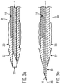
- the catheter lumen 4 is formed continuously in a uniform catheter body identified by reference numeral 28, this catheter lumen 4 is in the variant according to the Figures 3a, 3b formed by a puncture body 30 which is guided in a guide lumen 32 of a dilatation body identified by reference numeral 34 , which in the present case forms the cone region 22 and the second lumen 18 and holds the balloon 20 .
- the distal end of this dilation body 34 ends bluntly.
- the puncturing body 30 is retracted relative to the dilatation body 34 so that a puncture tip 36 formed by the puncturing body 30 is located within the dilatation body 34 .
- the puncturing body 30 is advanced so that the puncturing tip 36 protrudes beyond the distal end of the dilatation body 34 .
- the figure 4 12 shows a spreading device 40 which has two holding sections 42 which are spaced apart from one another in the longitudinal direction of the catheter 2 and which, in the exemplary embodiment shown, are designed as sleeves around the outer circumference of the catheter body identified by reference numeral 28 .
- This catheter body 28 forms - as in the embodiment Figure 2a, 2b - the catheter lumen 4 off.
- the holding section 42 shown on the right is firmly connected to the catheter body 28 .
- the outer circumference of the catheter body 28 merges into the outer circumference of the holding section 42 via a ramp surface that forms the cone area 22 . In this way, the radius widens uniformly up to the outside diameter of the holding section 42 .
- Between the two holding sections 42 are spreader segments 44, which figure 4 are already slightly expanded radially by pushing the two holding sections 42 onto one another.
- the expansion segments 44 can be formed, for example, by longitudinal slots in an initially uniform sleeve material, which forms the two holding sections 42 at the ends.
- the inside figure 4 left holding portion 42 is slidably held on the outer periphery of catheter body 28 .
- This left-hand holding section 42 can be displaced in the direction of the right-hand holding section 42 by means of a further activation catheter, which is guided over the outer circumference of the catheter body 28 and which rests against the free end face of the holding section 42 .
- a further activation catheter which is guided over the outer circumference of the catheter body 28 and which rests against the free end face of the holding section 42 .
- the individual expansion segments widen further radially.
- the dilation device is activated when the septum is approximately at the middle level of the expansion segments 44 .
Landscapes
- Health & Medical Sciences (AREA)
- Surgery (AREA)
- Life Sciences & Earth Sciences (AREA)
- Biomedical Technology (AREA)
- Nuclear Medicine, Radiotherapy & Molecular Imaging (AREA)
- Engineering & Computer Science (AREA)
- Pathology (AREA)
- Heart & Thoracic Surgery (AREA)
- Medical Informatics (AREA)
- Molecular Biology (AREA)
- Animal Behavior & Ethology (AREA)
- General Health & Medical Sciences (AREA)
- Public Health (AREA)
- Veterinary Medicine (AREA)
- Media Introduction/Drainage Providing Device (AREA)
Priority Applications (1)
| Application Number | Priority Date | Filing Date | Title |
|---|---|---|---|
| EP20193373.6A EP3960102A1 (fr) | 2020-08-28 | 2020-08-28 | Cathéter transseptal |
Applications Claiming Priority (1)
| Application Number | Priority Date | Filing Date | Title |
|---|---|---|---|
| EP20193373.6A EP3960102A1 (fr) | 2020-08-28 | 2020-08-28 | Cathéter transseptal |
Publications (1)
| Publication Number | Publication Date |
|---|---|
| EP3960102A1 true EP3960102A1 (fr) | 2022-03-02 |
Family
ID=72290917
Family Applications (1)
| Application Number | Title | Priority Date | Filing Date |
|---|---|---|---|
| EP20193373.6A Withdrawn EP3960102A1 (fr) | 2020-08-28 | 2020-08-28 | Cathéter transseptal |
Country Status (1)
| Country | Link |
|---|---|
| EP (1) | EP3960102A1 (fr) |
Citations (9)
| Publication number | Priority date | Publication date | Assignee | Title |
|---|---|---|---|---|
| US20020169377A1 (en) * | 2000-04-13 | 2002-11-14 | Khairkhahan Alexander K. | Method and apparatus for accessing the left atrial appendage |
| US20090287087A1 (en) * | 2006-11-22 | 2009-11-19 | Broncus Technologies, Inc. | Devices for creating passages and sensing for blood vessels |
| US20100010303A1 (en) * | 2008-07-09 | 2010-01-14 | Ethicon Endo-Surgery, Inc. | Inflatable access device |
| US20110046660A1 (en) * | 2009-02-13 | 2011-02-24 | Intrapace, Inc. | Endoscopic Forceps With Removable Handle |
| US20120191181A1 (en) * | 2007-04-27 | 2012-07-26 | Kassab Ghassan S | Systems and methods for localization of a puncture site relative to a mammalian tissue of interest |
| US20120289776A1 (en) * | 2011-05-13 | 2012-11-15 | Keast Thomas M | Methods and devices for diagnosing, monitoring, or treating medical conditions through an opening through an airway wall |
| US20130281978A1 (en) * | 2006-12-20 | 2013-10-24 | Onset Medical Corporation | Expandable trans-septal sheath |
| US20170105761A1 (en) | 2015-10-14 | 2017-04-20 | Transseptal Solutions Ltd. | Fossa ovalis penetration |
| US20190269392A1 (en) * | 2011-02-10 | 2019-09-05 | Corvia Medical, Inc. | Apparatus and methods to create and maintain an intra-atrial pressure relief opening |
-
2020
- 2020-08-28 EP EP20193373.6A patent/EP3960102A1/fr not_active Withdrawn
Patent Citations (9)
| Publication number | Priority date | Publication date | Assignee | Title |
|---|---|---|---|---|
| US20020169377A1 (en) * | 2000-04-13 | 2002-11-14 | Khairkhahan Alexander K. | Method and apparatus for accessing the left atrial appendage |
| US20090287087A1 (en) * | 2006-11-22 | 2009-11-19 | Broncus Technologies, Inc. | Devices for creating passages and sensing for blood vessels |
| US20130281978A1 (en) * | 2006-12-20 | 2013-10-24 | Onset Medical Corporation | Expandable trans-septal sheath |
| US20120191181A1 (en) * | 2007-04-27 | 2012-07-26 | Kassab Ghassan S | Systems and methods for localization of a puncture site relative to a mammalian tissue of interest |
| US20100010303A1 (en) * | 2008-07-09 | 2010-01-14 | Ethicon Endo-Surgery, Inc. | Inflatable access device |
| US20110046660A1 (en) * | 2009-02-13 | 2011-02-24 | Intrapace, Inc. | Endoscopic Forceps With Removable Handle |
| US20190269392A1 (en) * | 2011-02-10 | 2019-09-05 | Corvia Medical, Inc. | Apparatus and methods to create and maintain an intra-atrial pressure relief opening |
| US20120289776A1 (en) * | 2011-05-13 | 2012-11-15 | Keast Thomas M | Methods and devices for diagnosing, monitoring, or treating medical conditions through an opening through an airway wall |
| US20170105761A1 (en) | 2015-10-14 | 2017-04-20 | Transseptal Solutions Ltd. | Fossa ovalis penetration |
Non-Patent Citations (1)
| Title |
|---|
| GUIDO RUSSO ET AL.: "Transseptal Puncture: A Step-by Step Procedural Guide", CARDIAC INTERVENTIONS TODAY, vol. 13, no. 3, May 2019 (2019-05-01), pages S 22 |
Similar Documents
| Publication | Publication Date | Title |
|---|---|---|
| DE69825200T2 (de) | Kathetersystem zum Anbringen eines Stent | |
| DE69703681T2 (de) | Katheter zur Gefässneubildung | |
| EP0339460B1 (fr) | Jeu d'instruments médicaux d'introduction | |
| DE2238722C3 (de) | Katheter | |
| DE602004003142T2 (de) | Infusionsgerät | |
| DE69734672T2 (de) | Gallenkatheter zum auswechseln durch einen einzelnen bediener | |
| DE68918632T3 (de) | Führungsdrahtverlängerung mit selbstverriegelbarer, lösbarer Verbindung. | |
| DE8910603U1 (de) | Vorrichtung zum Ausbringen von Blutgerinnseln aus Arterien und Venen | |
| DE69123982T2 (de) | Dehnung aufrechterhaltendes führungselement sowie dilatator | |
| EP1356779B1 (fr) | Dispositif d'ablation cardiaque pour la création d'une lésion circulaire dans en infundibulum | |
| DE69635967T2 (de) | Schnellaustausch-Stentanbringungsballonkatheter | |
| EP1606006B1 (fr) | Dispositif pour introduire un objet dans un vaisseau du corps | |
| DE69833882T2 (de) | Medizinischer transplantatverbinder oder stopfen sowie verfahren zu ihrer herstellung | |
| DE69219878T2 (de) | Vorrichtung zur Aufweiten einer Stenose | |
| DE60221289T2 (de) | Zuführsystem für medizinische vorrichtungen | |
| DE69632852T2 (de) | Teleskopischer katheter | |
| DE69201874T2 (de) | Intravaskulärer mehrlumiger Katheter für das Implantieren mit Hilfe eines Tunnels. | |
| DE3853057T2 (de) | Entfernbare endoarterielle-Vorrichtung, bestimmt für die Reparatur von Ablösungen in arteriellen Wänden. | |
| DE202017007707U1 (de) | Punktionsvorrichtung und Verankerungsvorrichtung | |
| EP2542162B1 (fr) | Valvulotome guidé par un câble de guidage ou un cathéter | |
| EP0433717A1 (fr) | Jeu d'instruments pour ponction | |
| EP1303231B1 (fr) | Dispositif d'ancrage vasculaire | |
| DE102012204166A1 (de) | Mikrozugangs-Ausrüstung, welche eine angeschrägte Nadel umfasst | |
| EP3072463A1 (fr) | Kit médical ou set pour traiter un organe creux malade | |
| WO2000067673A1 (fr) | Dispositif pour l'implantation de tuteurs intravasculaires |
Legal Events
| Date | Code | Title | Description |
|---|---|---|---|
| PUAI | Public reference made under article 153(3) epc to a published international application that has entered the european phase |
Free format text: ORIGINAL CODE: 0009012 |
|
| STAA | Information on the status of an ep patent application or granted ep patent |
Free format text: STATUS: THE APPLICATION HAS BEEN PUBLISHED |
|
| AK | Designated contracting states |
Kind code of ref document: A1 Designated state(s): AL AT BE BG CH CY CZ DE DK EE ES FI FR GB GR HR HU IE IS IT LI LT LU LV MC MK MT NL NO PL PT RO RS SE SI SK SM TR |
|
| STAA | Information on the status of an ep patent application or granted ep patent |
Free format text: STATUS: THE APPLICATION IS DEEMED TO BE WITHDRAWN |
|
| 18D | Application deemed to be withdrawn |
Effective date: 20220903 |