EP3985128A1 - Isotherme nukleinsäureamplifikationsverfahren zur diagnose von infektionskrankheiten am ort des bedarfs - Google Patents
Isotherme nukleinsäureamplifikationsverfahren zur diagnose von infektionskrankheiten am ort des bedarfs Download PDFInfo
- Publication number
- EP3985128A1 EP3985128A1 EP20201885.9A EP20201885A EP3985128A1 EP 3985128 A1 EP3985128 A1 EP 3985128A1 EP 20201885 A EP20201885 A EP 20201885A EP 3985128 A1 EP3985128 A1 EP 3985128A1
- Authority
- EP
- European Patent Office
- Prior art keywords
- chamber
- amplification
- enzymes
- reaction
- enzyme
- Prior art date
- Legal status (The legal status is an assumption and is not a legal conclusion. Google has not performed a legal analysis and makes no representation as to the accuracy of the status listed.)
- Withdrawn
Links
- 238000003199 nucleic acid amplification method Methods 0.000 title claims abstract description 180
- 238000003745 diagnosis Methods 0.000 title description 8
- 208000032163 Emerging Communicable disease Diseases 0.000 title 1
- 230000003321 amplification Effects 0.000 claims abstract description 174
- 102000004190 Enzymes Human genes 0.000 claims abstract description 170
- 108090000790 Enzymes Proteins 0.000 claims abstract description 170
- 238000000034 method Methods 0.000 claims abstract description 139
- 239000000523 sample Substances 0.000 claims abstract description 102
- 150000007523 nucleic acids Chemical class 0.000 claims abstract description 61
- 102000039446 nucleic acids Human genes 0.000 claims abstract description 49
- 108020004707 nucleic acids Proteins 0.000 claims abstract description 49
- 102000018120 Recombinases Human genes 0.000 claims abstract description 45
- 108010091086 Recombinases Proteins 0.000 claims abstract description 45
- 238000006243 chemical reaction Methods 0.000 claims description 139
- 238000004020 luminiscence type Methods 0.000 claims description 49
- 230000009089 cytolysis Effects 0.000 claims description 46
- 239000003795 chemical substances by application Substances 0.000 claims description 44
- 238000001514 detection method Methods 0.000 claims description 43
- 238000001917 fluorescence detection Methods 0.000 claims description 38
- 108020004414 DNA Proteins 0.000 claims description 30
- 239000012472 biological sample Substances 0.000 claims description 30
- 230000000694 effects Effects 0.000 claims description 24
- 239000000126 substance Substances 0.000 claims description 21
- 239000012530 fluid Substances 0.000 claims description 20
- ZKHQWZAMYRWXGA-UHFFFAOYSA-N Adenosine triphosphate Natural products C1=NC=2C(N)=NC=NC=2N1C1OC(COP(O)(=O)OP(O)(=O)OP(O)(O)=O)C(O)C1O ZKHQWZAMYRWXGA-UHFFFAOYSA-N 0.000 claims description 17
- 230000003287 optical effect Effects 0.000 claims description 16
- 230000002441 reversible effect Effects 0.000 claims description 15
- 108091028043 Nucleic acid sequence Proteins 0.000 claims description 14
- 108010092799 RNA-directed DNA polymerase Proteins 0.000 claims description 14
- 239000003153 chemical reaction reagent Substances 0.000 claims description 13
- 239000000203 mixture Substances 0.000 claims description 13
- 239000013641 positive control Substances 0.000 claims description 13
- 238000011069 regeneration method Methods 0.000 claims description 12
- 230000002934 lysing effect Effects 0.000 claims description 11
- 230000008929 regeneration Effects 0.000 claims description 11
- 102000053602 DNA Human genes 0.000 claims description 10
- 108010014303 DNA-directed DNA polymerase Proteins 0.000 claims description 9
- 102000016928 DNA-directed DNA polymerase Human genes 0.000 claims description 9
- 239000012634 fragment Substances 0.000 claims description 9
- 230000003213 activating effect Effects 0.000 claims description 7
- 238000000605 extraction Methods 0.000 claims description 7
- 108010014594 Heterogeneous Nuclear Ribonucleoprotein A1 Proteins 0.000 claims description 6
- 230000000903 blocking effect Effects 0.000 claims description 6
- 239000007853 buffer solution Substances 0.000 claims description 6
- 230000002401 inhibitory effect Effects 0.000 claims description 6
- 239000003638 chemical reducing agent Substances 0.000 claims description 5
- 108020004682 Single-Stranded DNA Proteins 0.000 claims description 4
- ZKHQWZAMYRWXGA-KQYNXXCUSA-J ATP(4-) Chemical compound C1=NC=2C(N)=NC=NC=2N1[C@@H]1O[C@H](COP([O-])(=O)OP([O-])(=O)OP([O-])([O-])=O)[C@@H](O)[C@H]1O ZKHQWZAMYRWXGA-KQYNXXCUSA-J 0.000 claims description 3
- 102100034343 Integrase Human genes 0.000 claims 2
- 241001678559 COVID-19 virus Species 0.000 abstract description 25
- 238000011901 isothermal amplification Methods 0.000 abstract description 12
- 238000002405 diagnostic procedure Methods 0.000 abstract description 6
- 239000012678 infectious agent Substances 0.000 abstract description 5
- 239000013615 primer Substances 0.000 description 59
- 238000003556 assay Methods 0.000 description 52
- 108090000623 proteins and genes Proteins 0.000 description 39
- 102000004169 proteins and genes Human genes 0.000 description 34
- 108091032973 (ribonucleotides)n+m Proteins 0.000 description 32
- 238000012360 testing method Methods 0.000 description 31
- 230000008901 benefit Effects 0.000 description 23
- 108060004795 Methyltransferase Proteins 0.000 description 19
- 108091093088 Amplicon Proteins 0.000 description 17
- 241000700605 Viruses Species 0.000 description 16
- 230000035945 sensitivity Effects 0.000 description 16
- 238000003753 real-time PCR Methods 0.000 description 14
- 102100034349 Integrase Human genes 0.000 description 13
- 238000010438 heat treatment Methods 0.000 description 13
- 239000002773 nucleotide Substances 0.000 description 13
- 125000003729 nucleotide group Chemical group 0.000 description 12
- 238000003752 polymerase chain reaction Methods 0.000 description 12
- 238000004891 communication Methods 0.000 description 11
- 230000001965 increasing effect Effects 0.000 description 11
- 239000000463 material Substances 0.000 description 11
- WYURNTSHIVDZCO-UHFFFAOYSA-N Tetrahydrofuran Chemical compound C1CCOC1 WYURNTSHIVDZCO-UHFFFAOYSA-N 0.000 description 10
- PEDCQBHIVMGVHV-UHFFFAOYSA-N Glycerine Chemical compound OCC(O)CO PEDCQBHIVMGVHV-UHFFFAOYSA-N 0.000 description 9
- 239000000872 buffer Substances 0.000 description 9
- 239000011777 magnesium Substances 0.000 description 9
- 241000588724 Escherichia coli Species 0.000 description 8
- 238000013461 design Methods 0.000 description 8
- JDDUQGRUPLKDNT-IDTAVKCVSA-N (2r,3r,4s,5s)-2-(6-aminopurin-9-yl)-5-(2-methylpropylsulfanylmethyl)oxolane-3,4-diol Chemical compound O[C@@H]1[C@H](O)[C@@H](CSCC(C)C)O[C@H]1N1C2=NC=NC(N)=C2N=C1 JDDUQGRUPLKDNT-IDTAVKCVSA-N 0.000 description 7
- 108091034117 Oligonucleotide Proteins 0.000 description 7
- 238000004458 analytical method Methods 0.000 description 7
- 108010052305 exodeoxyribonuclease III Proteins 0.000 description 7
- 208000015181 infectious disease Diseases 0.000 description 7
- 229920001223 polyethylene glycol Polymers 0.000 description 7
- 230000003612 virological effect Effects 0.000 description 7
- 241000959949 Deinococcus geothermalis Species 0.000 description 6
- 238000007397 LAMP assay Methods 0.000 description 6
- 102000011931 Nucleoproteins Human genes 0.000 description 6
- 108010061100 Nucleoproteins Proteins 0.000 description 6
- 230000027455 binding Effects 0.000 description 6
- 210000004027 cell Anatomy 0.000 description 6
- 238000011161 development Methods 0.000 description 6
- 230000018109 developmental process Effects 0.000 description 6
- 230000006870 function Effects 0.000 description 6
- 230000002779 inactivation Effects 0.000 description 6
- 239000002987 primer (paints) Substances 0.000 description 6
- 230000008569 process Effects 0.000 description 6
- 230000002829 reductive effect Effects 0.000 description 6
- -1 DNA and RNA Chemical class 0.000 description 5
- JLCPHMBAVCMARE-UHFFFAOYSA-N [3-[[3-[[3-[[3-[[3-[[3-[[3-[[3-[[3-[[3-[[3-[[5-(2-amino-6-oxo-1H-purin-9-yl)-3-[[3-[[3-[[3-[[3-[[3-[[5-(2-amino-6-oxo-1H-purin-9-yl)-3-[[5-(2-amino-6-oxo-1H-purin-9-yl)-3-hydroxyoxolan-2-yl]methoxy-hydroxyphosphoryl]oxyoxolan-2-yl]methoxy-hydroxyphosphoryl]oxy-5-(5-methyl-2,4-dioxopyrimidin-1-yl)oxolan-2-yl]methoxy-hydroxyphosphoryl]oxy-5-(6-aminopurin-9-yl)oxolan-2-yl]methoxy-hydroxyphosphoryl]oxy-5-(6-aminopurin-9-yl)oxolan-2-yl]methoxy-hydroxyphosphoryl]oxy-5-(6-aminopurin-9-yl)oxolan-2-yl]methoxy-hydroxyphosphoryl]oxy-5-(6-aminopurin-9-yl)oxolan-2-yl]methoxy-hydroxyphosphoryl]oxyoxolan-2-yl]methoxy-hydroxyphosphoryl]oxy-5-(5-methyl-2,4-dioxopyrimidin-1-yl)oxolan-2-yl]methoxy-hydroxyphosphoryl]oxy-5-(4-amino-2-oxopyrimidin-1-yl)oxolan-2-yl]methoxy-hydroxyphosphoryl]oxy-5-(5-methyl-2,4-dioxopyrimidin-1-yl)oxolan-2-yl]methoxy-hydroxyphosphoryl]oxy-5-(5-methyl-2,4-dioxopyrimidin-1-yl)oxolan-2-yl]methoxy-hydroxyphosphoryl]oxy-5-(6-aminopurin-9-yl)oxolan-2-yl]methoxy-hydroxyphosphoryl]oxy-5-(6-aminopurin-9-yl)oxolan-2-yl]methoxy-hydroxyphosphoryl]oxy-5-(4-amino-2-oxopyrimidin-1-yl)oxolan-2-yl]methoxy-hydroxyphosphoryl]oxy-5-(4-amino-2-oxopyrimidin-1-yl)oxolan-2-yl]methoxy-hydroxyphosphoryl]oxy-5-(4-amino-2-oxopyrimidin-1-yl)oxolan-2-yl]methoxy-hydroxyphosphoryl]oxy-5-(6-aminopurin-9-yl)oxolan-2-yl]methoxy-hydroxyphosphoryl]oxy-5-(4-amino-2-oxopyrimidin-1-yl)oxolan-2-yl]methyl [5-(6-aminopurin-9-yl)-2-(hydroxymethyl)oxolan-3-yl] hydrogen phosphate Polymers Cc1cn(C2CC(OP(O)(=O)OCC3OC(CC3OP(O)(=O)OCC3OC(CC3O)n3cnc4c3nc(N)[nH]c4=O)n3cnc4c3nc(N)[nH]c4=O)C(COP(O)(=O)OC3CC(OC3COP(O)(=O)OC3CC(OC3COP(O)(=O)OC3CC(OC3COP(O)(=O)OC3CC(OC3COP(O)(=O)OC3CC(OC3COP(O)(=O)OC3CC(OC3COP(O)(=O)OC3CC(OC3COP(O)(=O)OC3CC(OC3COP(O)(=O)OC3CC(OC3COP(O)(=O)OC3CC(OC3COP(O)(=O)OC3CC(OC3COP(O)(=O)OC3CC(OC3COP(O)(=O)OC3CC(OC3COP(O)(=O)OC3CC(OC3COP(O)(=O)OC3CC(OC3COP(O)(=O)OC3CC(OC3COP(O)(=O)OC3CC(OC3CO)n3cnc4c(N)ncnc34)n3ccc(N)nc3=O)n3cnc4c(N)ncnc34)n3ccc(N)nc3=O)n3ccc(N)nc3=O)n3ccc(N)nc3=O)n3cnc4c(N)ncnc34)n3cnc4c(N)ncnc34)n3cc(C)c(=O)[nH]c3=O)n3cc(C)c(=O)[nH]c3=O)n3ccc(N)nc3=O)n3cc(C)c(=O)[nH]c3=O)n3cnc4c3nc(N)[nH]c4=O)n3cnc4c(N)ncnc34)n3cnc4c(N)ncnc34)n3cnc4c(N)ncnc34)n3cnc4c(N)ncnc34)O2)c(=O)[nH]c1=O JLCPHMBAVCMARE-UHFFFAOYSA-N 0.000 description 5
- 239000002585 base Substances 0.000 description 5
- 230000015572 biosynthetic process Effects 0.000 description 5
- 230000003197 catalytic effect Effects 0.000 description 5
- 238000011109 contamination Methods 0.000 description 5
- 238000005516 engineering process Methods 0.000 description 5
- 230000003278 mimic effect Effects 0.000 description 5
- 239000000047 product Substances 0.000 description 5
- 238000003860 storage Methods 0.000 description 5
- 238000003786 synthesis reaction Methods 0.000 description 5
- YLQBMQCUIZJEEH-UHFFFAOYSA-N tetrahydrofuran Natural products C=1C=COC=1 YLQBMQCUIZJEEH-UHFFFAOYSA-N 0.000 description 5
- 238000012546 transfer Methods 0.000 description 5
- 208000025721 COVID-19 Diseases 0.000 description 4
- 208000035473 Communicable disease Diseases 0.000 description 4
- DRBBFCLWYRJSJZ-UHFFFAOYSA-N N-phosphocreatine Chemical compound OC(=O)CN(C)C(=N)NP(O)(O)=O DRBBFCLWYRJSJZ-UHFFFAOYSA-N 0.000 description 4
- 208000002606 Paramyxoviridae Infections Diseases 0.000 description 4
- 239000002202 Polyethylene glycol Substances 0.000 description 4
- 102000001218 Rec A Recombinases Human genes 0.000 description 4
- 108010055016 Rec A Recombinases Proteins 0.000 description 4
- 239000011942 biocatalyst Substances 0.000 description 4
- 238000006073 displacement reaction Methods 0.000 description 4
- 239000000284 extract Substances 0.000 description 4
- 230000002349 favourable effect Effects 0.000 description 4
- 238000004108 freeze drying Methods 0.000 description 4
- 238000003780 insertion Methods 0.000 description 4
- 230000037431 insertion Effects 0.000 description 4
- UEGPKNKPLBYCNK-UHFFFAOYSA-L magnesium acetate Chemical compound [Mg+2].CC([O-])=O.CC([O-])=O UEGPKNKPLBYCNK-UHFFFAOYSA-L 0.000 description 4
- 238000004519 manufacturing process Methods 0.000 description 4
- 230000035484 reaction time Effects 0.000 description 4
- 230000001105 regulatory effect Effects 0.000 description 4
- 230000008685 targeting Effects 0.000 description 4
- 208000035657 Abasia Diseases 0.000 description 3
- 241000894006 Bacteria Species 0.000 description 3
- 241000711573 Coronaviridae Species 0.000 description 3
- FYYHWMGAXLPEAU-UHFFFAOYSA-N Magnesium Chemical compound [Mg] FYYHWMGAXLPEAU-UHFFFAOYSA-N 0.000 description 3
- 241000315672 SARS coronavirus Species 0.000 description 3
- 239000000654 additive Substances 0.000 description 3
- 238000001816 cooling Methods 0.000 description 3
- 230000009260 cross reactivity Effects 0.000 description 3
- 230000001419 dependent effect Effects 0.000 description 3
- VHJLVAABSRFDPM-QWWZWVQMSA-N dithiothreitol Chemical compound SC[C@@H](O)[C@H](O)CS VHJLVAABSRFDPM-QWWZWVQMSA-N 0.000 description 3
- 241001493065 dsRNA viruses Species 0.000 description 3
- 239000000975 dye Substances 0.000 description 3
- 238000005485 electric heating Methods 0.000 description 3
- 230000002255 enzymatic effect Effects 0.000 description 3
- 230000009088 enzymatic function Effects 0.000 description 3
- 238000006911 enzymatic reaction Methods 0.000 description 3
- 239000007850 fluorescent dye Substances 0.000 description 3
- 208000037797 influenza A Diseases 0.000 description 3
- 238000012792 lyophilization process Methods 0.000 description 3
- 239000012139 lysis buffer Substances 0.000 description 3
- 229910052749 magnesium Inorganic materials 0.000 description 3
- 230000035772 mutation Effects 0.000 description 3
- 230000001717 pathogenic effect Effects 0.000 description 3
- 239000008188 pellet Substances 0.000 description 3
- 125000005642 phosphothioate group Chemical group 0.000 description 3
- 238000000746 purification Methods 0.000 description 3
- 239000011541 reaction mixture Substances 0.000 description 3
- 230000006798 recombination Effects 0.000 description 3
- 238000005215 recombination Methods 0.000 description 3
- 238000000611 regression analysis Methods 0.000 description 3
- 210000003296 saliva Anatomy 0.000 description 3
- 238000013022 venting Methods 0.000 description 3
- 238000012800 visualization Methods 0.000 description 3
- KDCGOANMDULRCW-UHFFFAOYSA-N 7H-purine Chemical compound N1=CNC2=NC=NC2=C1 KDCGOANMDULRCW-UHFFFAOYSA-N 0.000 description 2
- QTBSBXVTEAMEQO-UHFFFAOYSA-M Acetate Chemical compound CC([O-])=O QTBSBXVTEAMEQO-UHFFFAOYSA-M 0.000 description 2
- 241000203069 Archaea Species 0.000 description 2
- 102000004420 Creatine Kinase Human genes 0.000 description 2
- 108010042126 Creatine kinase Proteins 0.000 description 2
- 230000006820 DNA synthesis Effects 0.000 description 2
- 241000287828 Gallus gallus Species 0.000 description 2
- 108010070675 Glutathione transferase Proteins 0.000 description 2
- 102100029100 Hematopoietic prostaglandin D synthase Human genes 0.000 description 2
- 241000342334 Human metapneumovirus Species 0.000 description 2
- VEXZGXHMUGYJMC-UHFFFAOYSA-N Hydrochloric acid Chemical compound Cl VEXZGXHMUGYJMC-UHFFFAOYSA-N 0.000 description 2
- 102000004317 Lyases Human genes 0.000 description 2
- 108090000856 Lyases Proteins 0.000 description 2
- 101710175625 Maltose/maltodextrin-binding periplasmic protein Proteins 0.000 description 2
- 241000714177 Murine leukemia virus Species 0.000 description 2
- 208000037847 SARS-CoV-2-infection Diseases 0.000 description 2
- 101500025257 Severe acute respiratory syndrome coronavirus 2 RNA-directed RNA polymerase nsp12 Proteins 0.000 description 2
- 102000006943 Uracil-DNA Glycosidase Human genes 0.000 description 2
- 108010072685 Uracil-DNA Glycosidase Proteins 0.000 description 2
- HDRRAMINWIWTNU-NTSWFWBYSA-N [[(2s,5r)-5-(2-amino-6-oxo-3h-purin-9-yl)oxolan-2-yl]methoxy-hydroxyphosphoryl] phosphono hydrogen phosphate Chemical compound C1=2NC(N)=NC(=O)C=2N=CN1[C@H]1CC[C@@H](COP(O)(=O)OP(O)(=O)OP(O)(O)=O)O1 HDRRAMINWIWTNU-NTSWFWBYSA-N 0.000 description 2
- 239000002253 acid Substances 0.000 description 2
- 230000009471 action Effects 0.000 description 2
- 239000003513 alkali Substances 0.000 description 2
- 239000012491 analyte Substances 0.000 description 2
- 230000001580 bacterial effect Effects 0.000 description 2
- 238000005842 biochemical reaction Methods 0.000 description 2
- 230000036760 body temperature Effects 0.000 description 2
- 238000004113 cell culture Methods 0.000 description 2
- 239000013043 chemical agent Substances 0.000 description 2
- 102000021178 chitin binding proteins Human genes 0.000 description 2
- 108091011157 chitin binding proteins Proteins 0.000 description 2
- 238000003776 cleavage reaction Methods 0.000 description 2
- 239000002299 complementary DNA Substances 0.000 description 2
- 230000003247 decreasing effect Effects 0.000 description 2
- 238000010586 diagram Methods 0.000 description 2
- 229940125532 enzyme inhibitor Drugs 0.000 description 2
- 239000002532 enzyme inhibitor Substances 0.000 description 2
- 238000002474 experimental method Methods 0.000 description 2
- 238000002866 fluorescence resonance energy transfer Methods 0.000 description 2
- 230000007062 hydrolysis Effects 0.000 description 2
- 238000006460 hydrolysis reaction Methods 0.000 description 2
- 238000000126 in silico method Methods 0.000 description 2
- 238000010348 incorporation Methods 0.000 description 2
- 230000002458 infectious effect Effects 0.000 description 2
- 208000037798 influenza B Diseases 0.000 description 2
- 230000005764 inhibitory process Effects 0.000 description 2
- 230000000977 initiatory effect Effects 0.000 description 2
- 230000010354 integration Effects 0.000 description 2
- 230000002452 interceptive effect Effects 0.000 description 2
- 230000009545 invasion Effects 0.000 description 2
- 238000002372 labelling Methods 0.000 description 2
- 238000011068 loading method Methods 0.000 description 2
- 229940069446 magnesium acetate Drugs 0.000 description 2
- 235000011285 magnesium acetate Nutrition 0.000 description 2
- 239000011654 magnesium acetate Substances 0.000 description 2
- 238000002156 mixing Methods 0.000 description 2
- 244000052769 pathogen Species 0.000 description 2
- 229950007002 phosphocreatine Drugs 0.000 description 2
- UEZVMMHDMIWARA-UHFFFAOYSA-M phosphonate Chemical group [O-]P(=O)=O UEZVMMHDMIWARA-UHFFFAOYSA-M 0.000 description 2
- BASFCYQUMIYNBI-UHFFFAOYSA-N platinum Chemical compound [Pt] BASFCYQUMIYNBI-UHFFFAOYSA-N 0.000 description 2
- 230000004952 protein activity Effects 0.000 description 2
- 230000004853 protein function Effects 0.000 description 2
- 238000001742 protein purification Methods 0.000 description 2
- 238000011897 real-time detection Methods 0.000 description 2
- 230000010076 replication Effects 0.000 description 2
- 238000010839 reverse transcription Methods 0.000 description 2
- 238000012552 review Methods 0.000 description 2
- 230000007017 scission Effects 0.000 description 2
- 238000012216 screening Methods 0.000 description 2
- 238000000926 separation method Methods 0.000 description 2
- 238000013207 serial dilution Methods 0.000 description 2
- 239000000243 solution Substances 0.000 description 2
- 241000894007 species Species 0.000 description 2
- 239000000758 substrate Substances 0.000 description 2
- 239000013589 supplement Substances 0.000 description 2
- 238000005382 thermal cycling Methods 0.000 description 2
- 239000001226 triphosphate Substances 0.000 description 2
- 235000011178 triphosphate Nutrition 0.000 description 2
- 210000002700 urine Anatomy 0.000 description 2
- 102000040650 (ribonucleotides)n+m Human genes 0.000 description 1
- OAKPWEUQDVLTCN-NKWVEPMBSA-N 2',3'-Dideoxyadenosine-5-triphosphate Chemical compound C1=NC=2C(N)=NC=NC=2N1[C@H]1CC[C@@H](CO[P@@](O)(=O)O[P@](O)(=O)OP(O)(O)=O)O1 OAKPWEUQDVLTCN-NKWVEPMBSA-N 0.000 description 1
- UPPBKCOGWJTQMJ-UHFFFAOYSA-N 2-amino-2-(hydroxymethyl)propane-1,3-diol;2-[methyl-(n'-phosphonocarbamimidoyl)amino]acetic acid Chemical compound OCC(N)(CO)CO.OCC(N)(CO)CO.OC(=O)CN(C)C(\N)=N\P(O)(O)=O UPPBKCOGWJTQMJ-UHFFFAOYSA-N 0.000 description 1
- ASJSAQIRZKANQN-CRCLSJGQSA-N 2-deoxy-D-ribose Chemical compound OC[C@@H](O)[C@@H](O)CC=O ASJSAQIRZKANQN-CRCLSJGQSA-N 0.000 description 1
- 101150095491 AACS gene Proteins 0.000 description 1
- 108010011170 Ala-Trp-Arg-His-Pro-Gln-Phe-Gly-Gly Proteins 0.000 description 1
- 241000205042 Archaeoglobus fulgidus Species 0.000 description 1
- 241000711404 Avian avulavirus 1 Species 0.000 description 1
- 244000063299 Bacillus subtilis Species 0.000 description 1
- 235000014469 Bacillus subtilis Nutrition 0.000 description 1
- 108091026890 Coding region Proteins 0.000 description 1
- 229920000742 Cotton Polymers 0.000 description 1
- 241000272779 Cygnus olor Species 0.000 description 1
- 230000005778 DNA damage Effects 0.000 description 1
- 231100000277 DNA damage Toxicity 0.000 description 1
- 239000003155 DNA primer Substances 0.000 description 1
- 102000052510 DNA-Binding Proteins Human genes 0.000 description 1
- 101710116602 DNA-Binding protein G5P Proteins 0.000 description 1
- 229920002307 Dextran Polymers 0.000 description 1
- 238000009007 Diagnostic Kit Methods 0.000 description 1
- 101100300807 Drosophila melanogaster spn-A gene Proteins 0.000 description 1
- 102100038132 Endogenous retrovirus group K member 6 Pro protein Human genes 0.000 description 1
- 101800001466 Envelope glycoprotein E1 Proteins 0.000 description 1
- 101710091045 Envelope protein Proteins 0.000 description 1
- 241000206602 Eukaryota Species 0.000 description 1
- 229920001917 Ficoll Polymers 0.000 description 1
- 238000001327 Förster resonance energy transfer Methods 0.000 description 1
- 241000701063 Gallid alphaherpesvirus 1 Species 0.000 description 1
- 101001057037 Gallus gallus Adenylate kinase isoenzyme 1 Proteins 0.000 description 1
- 241000193385 Geobacillus stearothermophilus Species 0.000 description 1
- 101000686179 Haemophilus influenzae (strain ATCC 51907 / DSM 11121 / KW20 / Rd) Protein RecA Proteins 0.000 description 1
- 241000701149 Human adenovirus 1 Species 0.000 description 1
- 241001135572 Human adenovirus E4 Species 0.000 description 1
- 241000711467 Human coronavirus 229E Species 0.000 description 1
- 241000482741 Human coronavirus NL63 Species 0.000 description 1
- 241001428935 Human coronavirus OC43 Species 0.000 description 1
- 241000713772 Human immunodeficiency virus 1 Species 0.000 description 1
- 241001429370 Human rhinovirus A16 Species 0.000 description 1
- 241001325456 Human rhinovirus B5 Species 0.000 description 1
- 241000134304 Influenza A virus H3N2 Species 0.000 description 1
- 208000025370 Middle East respiratory syndrome Diseases 0.000 description 1
- 241000127282 Middle East respiratory syndrome-related coronavirus Species 0.000 description 1
- 229910019142 PO4 Inorganic materials 0.000 description 1
- 108091005804 Peptidases Proteins 0.000 description 1
- BELBBZDIHDAJOR-UHFFFAOYSA-N Phenolsulfonephthalein Chemical compound C1=CC(O)=CC=C1C1(C=2C=CC(O)=CC=2)C2=CC=CC=C2S(=O)(=O)O1 BELBBZDIHDAJOR-UHFFFAOYSA-N 0.000 description 1
- 108091000080 Phosphotransferase Proteins 0.000 description 1
- 241000425347 Phyla <beetle> Species 0.000 description 1
- 229920002534 Polyethylene Glycol 1450 Polymers 0.000 description 1
- 229920002560 Polyethylene Glycol 3000 Polymers 0.000 description 1
- 229920002594 Polyethylene Glycol 8000 Polymers 0.000 description 1
- 206010036790 Productive cough Diseases 0.000 description 1
- 239000004365 Protease Substances 0.000 description 1
- 102000001708 Protein Isoforms Human genes 0.000 description 1
- 108010029485 Protein Isoforms Proteins 0.000 description 1
- 101710188315 Protein X Proteins 0.000 description 1
- CZPWVGJYEJSRLH-UHFFFAOYSA-N Pyrimidine Chemical compound C1=CN=CN=C1 CZPWVGJYEJSRLH-UHFFFAOYSA-N 0.000 description 1
- 239000013616 RNA primer Substances 0.000 description 1
- 239000013614 RNA sample Substances 0.000 description 1
- 238000011529 RT qPCR Methods 0.000 description 1
- 101710162453 Replication factor A Proteins 0.000 description 1
- 101710176758 Replication protein A 70 kDa DNA-binding subunit Proteins 0.000 description 1
- 241000725643 Respiratory syncytial virus Species 0.000 description 1
- 101710176276 SSB protein Proteins 0.000 description 1
- 101000667982 Severe acute respiratory syndrome coronavirus 2 Envelope small membrane protein Proteins 0.000 description 1
- 101710126859 Single-stranded DNA-binding protein Proteins 0.000 description 1
- 241000191967 Staphylococcus aureus Species 0.000 description 1
- 108020000005 Sucrose phosphorylase Proteins 0.000 description 1
- 101800001690 Transmembrane protein gp41 Proteins 0.000 description 1
- 238000009825 accumulation Methods 0.000 description 1
- 239000008351 acetate buffer Substances 0.000 description 1
- 230000000996 additive effect Effects 0.000 description 1
- 238000001042 affinity chromatography Methods 0.000 description 1
- 150000001412 amines Chemical class 0.000 description 1
- 239000000427 antigen Substances 0.000 description 1
- 102000036639 antigens Human genes 0.000 description 1
- 108091007433 antigens Proteins 0.000 description 1
- 238000003149 assay kit Methods 0.000 description 1
- 238000007068 beta-elimination reaction Methods 0.000 description 1
- 230000002457 bidirectional effect Effects 0.000 description 1
- 238000002306 biochemical method Methods 0.000 description 1
- 238000005415 bioluminescence Methods 0.000 description 1
- 230000029918 bioluminescence Effects 0.000 description 1
- 239000008280 blood Substances 0.000 description 1
- 210000004369 blood Anatomy 0.000 description 1
- 206010006451 bronchitis Diseases 0.000 description 1
- 210000004899 c-terminal region Anatomy 0.000 description 1
- 239000003054 catalyst Substances 0.000 description 1
- 230000001413 cellular effect Effects 0.000 description 1
- 230000008859 change Effects 0.000 description 1
- 238000007385 chemical modification Methods 0.000 description 1
- 238000012411 cloning technique Methods 0.000 description 1
- 230000000295 complement effect Effects 0.000 description 1
- 238000013329 compounding Methods 0.000 description 1
- 150000001875 compounds Chemical class 0.000 description 1
- 239000000470 constituent Substances 0.000 description 1
- SUYVUBYJARFZHO-RRKCRQDMSA-N dATP Chemical compound C1=NC=2C(N)=NC=NC=2N1[C@H]1C[C@H](O)[C@@H](COP(O)(=O)OP(O)(=O)OP(O)(O)=O)O1 SUYVUBYJARFZHO-RRKCRQDMSA-N 0.000 description 1
- SUYVUBYJARFZHO-UHFFFAOYSA-N dATP Natural products C1=NC=2C(N)=NC=NC=2N1C1CC(O)C(COP(O)(=O)OP(O)(=O)OP(O)(O)=O)O1 SUYVUBYJARFZHO-UHFFFAOYSA-N 0.000 description 1
- RGWHQCVHVJXOKC-SHYZEUOFSA-J dCTP(4-) Chemical compound O=C1N=C(N)C=CN1[C@@H]1O[C@H](COP([O-])(=O)OP([O-])(=O)OP([O-])([O-])=O)[C@@H](O)C1 RGWHQCVHVJXOKC-SHYZEUOFSA-J 0.000 description 1
- HAAZLUGHYHWQIW-KVQBGUIXSA-N dGTP Chemical compound C1=NC=2C(=O)NC(N)=NC=2N1[C@H]1C[C@H](O)[C@@H](COP(O)(=O)OP(O)(=O)OP(O)(O)=O)O1 HAAZLUGHYHWQIW-KVQBGUIXSA-N 0.000 description 1
- NHVNXKFIZYSCEB-XLPZGREQSA-N dTTP Chemical compound O=C1NC(=O)C(C)=CN1[C@@H]1O[C@H](COP(O)(=O)OP(O)(=O)OP(O)(O)=O)[C@@H](O)C1 NHVNXKFIZYSCEB-XLPZGREQSA-N 0.000 description 1
- 238000013500 data storage Methods 0.000 description 1
- URGJWIFLBWJRMF-JGVFFNPUSA-N ddTTP Chemical compound O=C1NC(=O)C(C)=CN1[C@@H]1O[C@H](COP(O)(=O)OP(O)(=O)OP(O)(O)=O)CC1 URGJWIFLBWJRMF-JGVFFNPUSA-N 0.000 description 1
- 238000000354 decomposition reaction Methods 0.000 description 1
- 238000004925 denaturation Methods 0.000 description 1
- 230000036425 denaturation Effects 0.000 description 1
- 230000001627 detrimental effect Effects 0.000 description 1
- 238000012631 diagnostic technique Methods 0.000 description 1
- 238000010790 dilution Methods 0.000 description 1
- 239000012895 dilution Substances 0.000 description 1
- 239000000539 dimer Substances 0.000 description 1
- BFMYDTVEBKDAKJ-UHFFFAOYSA-L disodium;(2',7'-dibromo-3',6'-dioxido-3-oxospiro[2-benzofuran-1,9'-xanthene]-4'-yl)mercury;hydrate Chemical compound O.[Na+].[Na+].O1C(=O)C2=CC=CC=C2C21C1=CC(Br)=C([O-])C([Hg])=C1OC1=C2C=C(Br)C([O-])=C1 BFMYDTVEBKDAKJ-UHFFFAOYSA-L 0.000 description 1
- 239000003814 drug Substances 0.000 description 1
- 238000005401 electroluminescence Methods 0.000 description 1
- 230000002708 enhancing effect Effects 0.000 description 1
- 230000005284 excitation Effects 0.000 description 1
- 238000011049 filling Methods 0.000 description 1
- 238000003306 harvesting Methods 0.000 description 1
- 239000000383 hazardous chemical Substances 0.000 description 1
- 238000003018 immunoassay Methods 0.000 description 1
- 230000006872 improvement Effects 0.000 description 1
- 239000012535 impurity Substances 0.000 description 1
- 238000001727 in vivo Methods 0.000 description 1
- 238000011065 in-situ storage Methods 0.000 description 1
- 238000011534 incubation Methods 0.000 description 1
- 230000036512 infertility Effects 0.000 description 1
- 208000037802 influenza A (H3N2) Diseases 0.000 description 1
- 239000004615 ingredient Substances 0.000 description 1
- 239000003112 inhibitor Substances 0.000 description 1
- 230000003993 interaction Effects 0.000 description 1
- 239000000543 intermediate Substances 0.000 description 1
- 230000002427 irreversible effect Effects 0.000 description 1
- 238000002955 isolation Methods 0.000 description 1
- 230000033001 locomotion Effects 0.000 description 1
- 230000007774 longterm Effects 0.000 description 1
- 238000000504 luminescence detection Methods 0.000 description 1
- 229910001425 magnesium ion Inorganic materials 0.000 description 1
- 230000001404 mediated effect Effects 0.000 description 1
- 239000012528 membrane Substances 0.000 description 1
- 230000002503 metabolic effect Effects 0.000 description 1
- 244000005700 microbiome Species 0.000 description 1
- 239000003068 molecular probe Substances 0.000 description 1
- 238000012544 monitoring process Methods 0.000 description 1
- 239000000178 monomer Substances 0.000 description 1
- 239000002086 nanomaterial Substances 0.000 description 1
- 239000013642 negative control Substances 0.000 description 1
- 239000002777 nucleoside Substances 0.000 description 1
- 238000005457 optimization Methods 0.000 description 1
- 239000003960 organic solvent Substances 0.000 description 1
- 238000013021 overheating Methods 0.000 description 1
- 239000003973 paint Substances 0.000 description 1
- 230000002085 persistent effect Effects 0.000 description 1
- 229960003531 phenolsulfonphthalein Drugs 0.000 description 1
- NBIIXXVUZAFLBC-UHFFFAOYSA-K phosphate Chemical compound [O-]P([O-])([O-])=O NBIIXXVUZAFLBC-UHFFFAOYSA-K 0.000 description 1
- 239000010452 phosphate Substances 0.000 description 1
- 102000020233 phosphotransferase Human genes 0.000 description 1
- 238000005424 photoluminescence Methods 0.000 description 1
- 229910052697 platinum Inorganic materials 0.000 description 1
- 238000006116 polymerization reaction Methods 0.000 description 1
- 229920001184 polypeptide Polymers 0.000 description 1
- 230000002265 prevention Effects 0.000 description 1
- 238000007639 printing Methods 0.000 description 1
- 108090000765 processed proteins & peptides Proteins 0.000 description 1
- 102000004196 processed proteins & peptides Human genes 0.000 description 1
- 238000012545 processing Methods 0.000 description 1
- 230000001915 proofreading effect Effects 0.000 description 1
- 230000000069 prophylactic effect Effects 0.000 description 1
- 229940021993 prophylactic vaccine Drugs 0.000 description 1
- 238000003908 quality control method Methods 0.000 description 1
- 238000010791 quenching Methods 0.000 description 1
- 230000000171 quenching effect Effects 0.000 description 1
- 230000002285 radioactive effect Effects 0.000 description 1
- 239000012857 radioactive material Substances 0.000 description 1
- 239000011535 reaction buffer Substances 0.000 description 1
- 230000036632 reaction speed Effects 0.000 description 1
- 230000009257 reactivity Effects 0.000 description 1
- 101150079601 recA gene Proteins 0.000 description 1
- 238000004064 recycling Methods 0.000 description 1
- 238000011160 research Methods 0.000 description 1
- 230000000241 respiratory effect Effects 0.000 description 1
- 238000005096 rolling process Methods 0.000 description 1
- 238000011309 routine diagnosis Methods 0.000 description 1
- 150000003839 salts Chemical class 0.000 description 1
- 238000007789 sealing Methods 0.000 description 1
- 239000007787 solid Substances 0.000 description 1
- 230000007928 solubilization Effects 0.000 description 1
- 238000005063 solubilization Methods 0.000 description 1
- 125000006850 spacer group Chemical group 0.000 description 1
- 230000002269 spontaneous effect Effects 0.000 description 1
- 210000003802 sputum Anatomy 0.000 description 1
- 208000024794 sputum Diseases 0.000 description 1
- 241000114864 ssRNA viruses Species 0.000 description 1
- 230000000087 stabilizing effect Effects 0.000 description 1
- 230000004936 stimulating effect Effects 0.000 description 1
- 238000006467 substitution reaction Methods 0.000 description 1
- 230000008093 supporting effect Effects 0.000 description 1
- 239000004094 surface-active agent Substances 0.000 description 1
- 230000031068 symbiosis, encompassing mutualism through parasitism Effects 0.000 description 1
- 230000009466 transformation Effects 0.000 description 1
- 238000000844 transformation Methods 0.000 description 1
- PIEPQKCYPFFYMG-UHFFFAOYSA-N tris acetate Chemical compound CC(O)=O.OCC(N)(CO)CO PIEPQKCYPFFYMG-UHFFFAOYSA-N 0.000 description 1
- 241000701161 unidentified adenovirus Species 0.000 description 1
- 241001430294 unidentified retrovirus Species 0.000 description 1
- 238000010200 validation analysis Methods 0.000 description 1
- XLYOFNOQVPJJNP-UHFFFAOYSA-N water Substances O XLYOFNOQVPJJNP-UHFFFAOYSA-N 0.000 description 1
Images





Classifications
-
- B—PERFORMING OPERATIONS; TRANSPORTING
- B01—PHYSICAL OR CHEMICAL PROCESSES OR APPARATUS IN GENERAL
- B01L—CHEMICAL OR PHYSICAL LABORATORY APPARATUS FOR GENERAL USE
- B01L3/00—Containers or dishes for laboratory use, e.g. laboratory glassware; Droppers
- B01L3/50—Containers for the purpose of retaining a material to be analysed, e.g. test tubes
- B01L3/502—Containers for the purpose of retaining a material to be analysed, e.g. test tubes with fluid transport, e.g. in multi-compartment structures
-
- C—CHEMISTRY; METALLURGY
- C12—BIOCHEMISTRY; BEER; SPIRITS; WINE; VINEGAR; MICROBIOLOGY; ENZYMOLOGY; MUTATION OR GENETIC ENGINEERING
- C12Q—MEASURING OR TESTING PROCESSES INVOLVING ENZYMES, NUCLEIC ACIDS OR MICROORGANISMS; COMPOSITIONS OR TEST PAPERS THEREFOR; PROCESSES OF PREPARING SUCH COMPOSITIONS; CONDITION-RESPONSIVE CONTROL IN MICROBIOLOGICAL OR ENZYMOLOGICAL PROCESSES
- C12Q1/00—Measuring or testing processes involving enzymes, nucleic acids or microorganisms; Compositions therefor; Processes of preparing such compositions
- C12Q1/68—Measuring or testing processes involving enzymes, nucleic acids or microorganisms; Compositions therefor; Processes of preparing such compositions involving nucleic acids
- C12Q1/6806—Preparing nucleic acids for analysis, e.g. for polymerase chain reaction [PCR] assay
-
- C—CHEMISTRY; METALLURGY
- C12—BIOCHEMISTRY; BEER; SPIRITS; WINE; VINEGAR; MICROBIOLOGY; ENZYMOLOGY; MUTATION OR GENETIC ENGINEERING
- C12Q—MEASURING OR TESTING PROCESSES INVOLVING ENZYMES, NUCLEIC ACIDS OR MICROORGANISMS; COMPOSITIONS OR TEST PAPERS THEREFOR; PROCESSES OF PREPARING SUCH COMPOSITIONS; CONDITION-RESPONSIVE CONTROL IN MICROBIOLOGICAL OR ENZYMOLOGICAL PROCESSES
- C12Q1/00—Measuring or testing processes involving enzymes, nucleic acids or microorganisms; Compositions therefor; Processes of preparing such compositions
- C12Q1/68—Measuring or testing processes involving enzymes, nucleic acids or microorganisms; Compositions therefor; Processes of preparing such compositions involving nucleic acids
- C12Q1/6844—Nucleic acid amplification reactions
-
- B—PERFORMING OPERATIONS; TRANSPORTING
- B01—PHYSICAL OR CHEMICAL PROCESSES OR APPARATUS IN GENERAL
- B01L—CHEMICAL OR PHYSICAL LABORATORY APPARATUS FOR GENERAL USE
- B01L2200/00—Solutions for specific problems relating to chemical or physical laboratory apparatus
- B01L2200/06—Fluid handling related problems
- B01L2200/0689—Sealing
-
- B—PERFORMING OPERATIONS; TRANSPORTING
- B01—PHYSICAL OR CHEMICAL PROCESSES OR APPARATUS IN GENERAL
- B01L—CHEMICAL OR PHYSICAL LABORATORY APPARATUS FOR GENERAL USE
- B01L2200/00—Solutions for specific problems relating to chemical or physical laboratory apparatus
- B01L2200/10—Integrating sample preparation and analysis in single entity, e.g. lab-on-a-chip concept
-
- B—PERFORMING OPERATIONS; TRANSPORTING
- B01—PHYSICAL OR CHEMICAL PROCESSES OR APPARATUS IN GENERAL
- B01L—CHEMICAL OR PHYSICAL LABORATORY APPARATUS FOR GENERAL USE
- B01L2300/00—Additional constructional details
- B01L2300/08—Geometry, shape and general structure
- B01L2300/0832—Geometry, shape and general structure cylindrical, tube shaped
-
- B—PERFORMING OPERATIONS; TRANSPORTING
- B01—PHYSICAL OR CHEMICAL PROCESSES OR APPARATUS IN GENERAL
- B01L—CHEMICAL OR PHYSICAL LABORATORY APPARATUS FOR GENERAL USE
- B01L2400/00—Moving or stopping fluids
- B01L2400/04—Moving fluids with specific forces or mechanical means
- B01L2400/0475—Moving fluids with specific forces or mechanical means specific mechanical means and fluid pressure
- B01L2400/0478—Moving fluids with specific forces or mechanical means specific mechanical means and fluid pressure pistons
-
- B—PERFORMING OPERATIONS; TRANSPORTING
- B01—PHYSICAL OR CHEMICAL PROCESSES OR APPARATUS IN GENERAL
- B01L—CHEMICAL OR PHYSICAL LABORATORY APPARATUS FOR GENERAL USE
- B01L2400/00—Moving or stopping fluids
- B01L2400/06—Valves, specific forms thereof
- B01L2400/0677—Valves, specific forms thereof phase change valves; Meltable, freezing, dissolvable plugs; Destructible barriers
- B01L2400/0683—Valves, specific forms thereof phase change valves; Meltable, freezing, dissolvable plugs; Destructible barriers mechanically breaking a wall or membrane within a channel or chamber
Definitions
- the present invention relates to a method for fast isothermal amplification of nucleic acids, including DNA and RNA, using thermostable enzymes.
- the invention relates to diagnostic methods for rapidly diagnosing, for example, an infectious agent, in a biological probe of interest.
- the invention further relates to a handheld and portable diagnostic system for performing the amplification method in a non-laboratory environment. Further provided are suitable enzyme sequences, kits and uses of the method and the system.
- Nucleic acid detecting techniques like molecular real-time PCR assays are usually very sensitive, and more specific than but they have the huge advantage to require highly-equipped laboratories and well-trained personnel. Therefore, new portable diagnostic solutions having a good sensitivity and sensibility and being able to provide reliable results in situ at the place of testing are urgently needed.
- PCR suffers from certain drawbacks, including the inherently necessary need for iterative rounds of thermal cycling and the shift between different temperatures (repeated cycles of two or three temperature-dependent steps during the amplification process) and the use of high (>90°C) temperatures. These drawbacks have led to the development of alternative amplification methods.
- NASBA nucleic acid sequence-based amplification
- LAMP loop-mediated isothermal amplification
- HDA helicase-dependent amplification
- RCA rolling circle amplification
- MDA multiple displacement amplification
- RPA recombinase polymerase amplification
- SIBA strand invasion based amplification
- NASBA, RPA and HDA rely on the use of target-specific probes to provide for higher specificity of the reaction, this may not simultaneously be positive for sensitivity.
- LAMP uses additional primers instead of probes together with a strand displacement polymerase ( Notomi et al., 2000, Nucleic Acids Research 28: e63 ) and thereby achieves a high degree of analytical sensitivity and specificity, on the other hand, there is also the risk that the use of numerous large primers needed may result in false-positive results - an outcome definitely to be avoided during diagnostic applications, particularly for diagnosing infectious diseases, where a false-positive result may have a detrimental outcome.
- LAMP reactions require high temperatures not as such practicable for portable devices, particularly diagnostic devices for use in a home environment due to regulatory and practical issues.
- RT-LAMP assays have a run time of about 30 minutes and need devices to maintain an amplification temperature of 65°C. Consequently, LAMP will not be the method of choice in case a diagnostic nucleic acid amplification result is needed in a short time in a handheld device, which has to fulfill certain regulatory requirements to be suitable for non-laboratory use.
- RPA and SIBA technology both rely on the use of a recombinase during the binding and amplification process.
- a nucleoprotein complex constituted by oligonucleotide primers and the recombinase proteins is formed for RPA- and SIBA-type nucleic acid amplification strategies, these complexes facilitating the primer binding to the template DNA.
- a certain advantage is the fact the recombinase can tolerate at least one incorrect base without preventing strand invasion necessary to start the reaction. Overall, it has been shown that an RPA amplicon can tolerate up to 9 mismatches across all primers and the probe sequences (cf.
- RPA may be a suitable technique, still, a technique with certain draw backs when not performed in the highly controlled laboratory environment and conducted and supervised by experts, but when performed in the "auto-mode" specialized to perform best in a portable device to be used by any person even in the home environment, or in testing stations under varying external conditions.
- a central and crucial prerequisite for such a ready-to use portable test kit is the stability of the inevitably necessary working horses in such a kit: the stability of the enzymes used to perform the diagnostic test to guarantee its functionality. This requires the possibility to transport and store the device including the test reagents at ambient temperature without cooling steps not easily possible and to provide a new enzymes and suitable reaction buffers and methods being more stable to perform a diagnosis in short time with both high sensitivity and sensibility
- thermostable enzymes as catalysts
- an isothermal method of nucleic acid amplification of at least one target sequence comprising the steps of: (a) providing (i) at least one recombinase, at least one single-strand binding protein, at least one, and preferably at least two target sequence specific primers, at least one DNA polymerase, and optionally: providing at least one reverse transcriptase and/or at least one energy regeneration enzyme, together with suitable reaction components, wherein (ii) either at least one co-factor essential for the activity of at least one of the enzymes is not provided; or wherein at least one inhibitory agent blocking the activity of the at least one recombinase is provided; (b) providing at least one biological sample to be analyzed for the presence of the at least one target sequence and optionally providing a preferably sterile extraction kit to obtain the biological sample; (c) optionally: providing at least one probe and optionally providing at least one enzyme activating the probe so that a detectable signal is generated
- a method wherein at least one, preferably at least two of the enzymes provided is independently selected from the group consisting of SEQ ID NO: 1 to 8, or a catalytically active fragment thereof, or a sequence having at least 90%, 91%, 92%, 93%, 94%, 95%, 96%, 97%, 98%, or at least 99% to the respective reference sequence, or a nucleic acid sequence encoding the same, or any functional equivalent of such a thermostable enzyme.
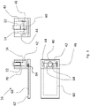
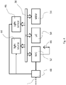
- a system for detecting at least one target sequence in a biological sample comprising: a lysis chamber (12), an amplification chamber (14) and a luminescence, preferably a fluorescence detection device (16), wherein the lysis chamber (12) contains a lysing fluid, the amplification chamber (14) contains a mixture that comprises the set of enzymes and the suitable reaction components as defined in claims 1 to 10, and the luminescence, preferably the fluorescence detection device (16) comprises a detection chamber (42) configured to receive the amplification chamber (14) or the contents of the amplification chamber, a light source (44), an optical sensor (46), an energy supply means (48), a wireless data interface (52) and a controller (50), wherein the system is configured to perform a method according to the first aspect above.
- kits for performing a method according to the first aspect above comprising (i) at least one recombinase, at least one single-strand binding protein, at least one DNA polymerase, and optionally: at least one reverse transcriptase and/or at least one energy regeneration enzyme, together with suitable reaction components, wherein (ii) at least one, preferably at least two target sequence specific primers; (iii) suitable reaction components, including at least one co-factor for at least one enzyme, dNTPs or a mixture of dNTPs and ddNTPs, at least one buffer system, at least one reducing agent, and adenosine triphosphate; (iv) at least one sterile set for extracting a biological probe to be analyzed and comprising at least one component configured to be applied to a system as defined in claim 11, preferably into the lysis chamber (12) of the system; (v) optionally: at least one probe and optionally at least one enzyme activating the probe
- thermostable enzyme as defined above, or a use of at least one nucleic acid sequence encoding the same, optionally for use in combination with a kit as defined in the above aspect, for performing an isothermal method of nucleic acid amplification of at least one target sequence as defined in the first aspect above.
- nucleic acid amplification techniques including isothermal amplification strategies
- the present inventors now provide a new isothermal nucleic acid amplification strategy using a new set of thermostable enzymes perfectly interacting in an isothermal assay, like RPA, and having several advantages over commonly used enzymes, as less additives are needed and the thermostable enzymes allow optimum performance in portable amplification devices under field conditions, or in a home environment, outside a fully equipped laboratory.
- an isothermal method of nucleic acid amplification of at least one target sequence preferably in a biological sample, the method comprising the steps of: (a) providing (i) at least one recombinase, optionally: at least one recombinase-assisting factor, and at least one single-strand binding protein, at least one, and preferably at least two target sequence specific primers, at least one DNA polymerase, preferably
- an isothermal DNA/RNA amplification method of the prior art usually starts with the binding of a phage-derived T4 UvsX protein as central recombinase, assisted by the T4 UvsY acting as loading factor, to the primers (a forward and a reverse primer) to form a nucleoprotein filament starting the reaction and guaranteeing accessibility of the target nucleic acid to be amplified.
- the resulting complex so to say "searches" for homologous sequences in a duplex DNA usually forming the target. Once the homology is located, the complex invades the double-stranded (ds) DNA, forming a so-called D-loop structure.
- This D-loop is double-stranded where the primer hybridises with the template strand, initiating a strand exchange reaction, whereas the other side of the D-loop remains single-stranded.
- This unwound complementary strand is stabilised by a single-strand binding (SSB) protein, where usually a T4-derived gp32 protein is used. Subsequently, the recombinase disassembles from the nucleoprotein filament and becomes immediately available to initiate another strand displacement reaction with a new primer etc. pp.
- SSB single-strand binding
- Primer incorporation allows the DNA polymerase, where usually a Bsu ( Bacillus subtilis-derived ) or Sau ( Staphylococcus aureus-derived) polymerase, usually the large fragment of a polymerase I, is used, to initiate the synthesis from the free 3'-OH at the end of the primer. As the polymerisation continues, the two parental strands will thus continue to separate. Incorporation of both forward and reverse primers then enables strand synthesis to occur in both directions simultaneously, and ultimately results in the exponential accumulation of amplified duplex DNA, consisting of the sequence between the forward and reverse primers.
- Bsu Bacillus subtilis-derived
- Sau Staphylococcus aureus-derived
- RPA was initially demonstrated to be a nucleic acid amplification method for DNA.
- RPA was later also shown to be a suitable reaction for RNA as template by addition of an additional enzyme, a reverse transcriptase (e.g., from Murine Leukemia virus (MuLV) reverse transcriptase) in the same reaction compartment.
- a reverse transcriptase e.g., from Murine Leukemia virus (MuLV) reverse transcriptase
- the recommended RPA amplicon length should be below 500 nucleotides for efficient amplification.
- Most presently published RPA papers have applied amplicon lengths between 100 and 250 nucleotides, which usually incur fast and efficient amplification.
- Shorter PCR primers can also be used in the RPA reaction but may decrease the reaction speed and sensitivity, which is to be avoided in diagnostic settings.
- the general amplicon length which can be influenced by the corresponding primer design, will usually range from about 50 to about 400 base pairs (bps), preferably 100 to about 200 bp, and most preferably about 120 to about 150 bps.
- An amplicon length below 200 bps is considered to be favorable for diagnostic purposes as the length of the amplicon has an influence on the efficiency of the amplicon amplification and, therefore, is directly linked to the analytical sensitivity of the reaction. Inefficient longer amplicons also result in slightly longer reaction times, which generally should be avoided.
- at least one, at least two, at least three, at least four or even all enzymes of the method of the present invention are thermostable enzymes.
- RPA assays were established with phage derived enzymes with a focus on the functionality, i.e., the specific catalytic activity, of these enzymes.
- "Standard" RPA enzymes even nowadays, are usually derived from mesophilic phages like T4 or T6 (cf. Li et al., 2019, supra), i.e. phages specific for the gut bacterium Escherichia coli and thus mesophilic in the sense that the bacterium and thus the phage lifecycle is adapted to standard temperatures (around body temperature). Still, these enzymes have certain disadvantages as these enzymes are not specifically thermostable, even though these enzymes due to their functionality are commonly used in methods requiring temperatures above 40°C.
- Thermostability or “thermostable” as used herein is meant to describe the quality of folded protein or enzyme to resist irreversible change in its chemical or physical structure, for an enzyme its three-dimensional folded state active as biocatalyst, by resisting decomposition or polymerization, at a high relative temperature above standard temperature, wherein the relevant reference temperature representing a standard temperature is 37°C.
- thermostable enzyme as used herein can thus be characterized by showing denaturation only at temperatures above around 70°C, 71 °C, 72°C, 73°C, 74°C and preferably above 75°C and/or an optimum activity temperature at 37°C to about 85°C, preferably of about 38°C to about 80°C, and most preferably from about 40°C to about 65°C.
- the at least one enzyme will be perfectly suited for a method performed in a portable diagnostic device as disclosed herein to provide fast and reliable results.
- the thermostable enzyme may have an optimum pH at or around a physiological pH, but this is not strict prerequisite.
- the optimum pH will be in a range from about pH 6.0 to about pH 9.0, more preferably in a range from about pH 6.5 to about pH 8.5, and most preferably in a range from about 7.0 to 8.0. It should be guaranteed that the enzymes chosen to interact in the same reaction environment will be provided in a suitable buffer system so that all enzymes can be active and will exert their catalytic activity.
- the huge advantage of the enzymes as used in the methods as disclosed herein is the fact that these enzymes will have optimum activity, meaning optimum catalytic activity, above a standard (body) temperature of 37°C so that these enzymes will perform particularly well when used in a detection device as disclosed herein configured to perform a diagnostic nucleic acid amplification reaction in a portable handheld device also under home environment conditions.
- the thermostable enzymes as identified, tested and used herein will have a particular performance even after freeze-drying and further under the reaction conditions in a handheld device.
- thermostable enzymes offer many advantages for their applications in industrial applications: i) At high temperature, viscosity is decreased, which increases the mass transfer and facilitates the mixing of reaction components. ii) Sterility requirements are not as stringent due to the low probability of contamination by thermostable microorganisms from the environment. iii) Furthermore, thermostable enzymes have little activity at low temperature, which allows to stop the reaction just by cooling. iv) Thermostable proteins are more resistant to the addition of denaturing agents (organic solvents, high and low pH). v) Furthermore, the purification of thermostable proteins is easier compared to their mesophilic counterparts as a single step of heat treatment is sufficient for protein purification.
- thermostable enzymes suitable to be used in a combined setting for an isothermal nucleic acid amplification reaction, preferably an isothermal diagnostic nucleic acid amplification reaction performed in a portable device.
- Functionally equivalent orthologous, homologous and paralogous isoforms of the sequences specifically disclosed herein, or catalytically active fragment thereof, may be used as well.
- thermostable proteins offer these specific advantages: (i) production costs for thermostable proteins can be reduced compared to mesophilic proteins; (ii) as thermostable proteins can be purified in a special manner, the use of an affinity tag may, hence, no longer be necessary. Consequently, an influence of the tag on protein function can be avoided; (iii) due to an increased stability of the thermostable biocatalysts short-term storage before lyophilization at 4°C is possible. Hence, the addition of supplements (e.g. glycerol) negatively interfering with the lyophilization process and complicated in the subsequent can be avoided.
- supplements e.g. glycerol
- this allows a much easier temperature control of an isothermal nucleic acid amplification reaction in comparison to a standard RPA reaction, for instance. Further, the reaction can be performed in a better controllable manner and the enzyme stability after storage and reconstitution - an important prerequisite for enzymatic activity and thus a reliable and fast test result - can be guaranteed. Finally, the reaction additives can be drastically reduced further improving the reaction. In contrast to an RPA, for example, the addition of a crowding agent necessarily used in standard RPA assays can be avoided or reduced to a minimum.
- no crowding agent is separately added to an isothermal nucleic acid amplification reaction according to the present invention going beyond inevitable impurities of a crowding agent (e.g., PEG or glycerol) as used during the lyophilization process.
- a crowding agent e.g., PEG or glycerol
- thermostable enzyme as disclosed herein, a thermostable functional equivalent from another organism, or a recombinantly modified mutant thereof, may be used as well without deviating from the gist of the present invention.
- a “biological sample” is to be understood broadly and relates to any material isolated from a living organism, which contains genetic material from this organism and/or (additionally) from a pathogen (a pathogenic prokaryote or eukaryote) or virus (with an RNA or a DNA genome, single, or double-stranded, or a retrovirus) having infected or infiltrated the organism, or living in symbiosis with this organism etc.
- a biological sample includes, for example, swab and saliva samples, including nasopharyngeal and oropharyngeal swab or saliva, but also sputum, blood, urine and stool and the like.
- a biological sample to be tested in a non-laboratory environment will be a swab, saliva, urine, or stool etc. which can be retrieved without the assistance of trained medical personnel.
- Suitable reaction components for enzymatic reactions and the concentration and compounding thereof, in case the enzyme and the reaction these suitable reaction components are intended for is known, can easily be determined by the skilled person. Certain aspects of particular relevance for the methods of the present invention will be disclosed in detail in the following.
- a "starting reagent" as used herein according to the various methods, can be particularly suitable, as this reagent allows an exact and controllable start of the reaction.
- a starting reagent can be an essential co-factor of at least one enzyme, without which the enzymatic reaction cannot start.
- the starting reagent may be magnesium, or a salt thereof or a magnesium containing solution, for example, magnesium acetate.
- the concentration can vary and can be between around 5 nM and 20 mM.
- the starting reagent may be provided in an activatable form, in a separate compartment, or in a heat activatable manner so that the start of the reaction can be tightly controlled.
- the starting reagent will be provided in a manner that the reaction can be started with a simple action of the user (click on a button, or automatic start after application of a test sample).
- a nucleic acid amplification reaction will require dNTPs (nucleotide triphosphates), for example, dATP, dGTP, dCTP, and dTTP.
- dNTPs nucleotide triphosphates
- ATP, GTP, CTP, and UTP may also be included for synthesis of RNA primers.
- ddNTPs ddATP, ddTTP, ddGTP and ddGTP
- the dNTP may be used at a concentration of between 1 ⁇ M to 200 ⁇ M of each NTP species.
- a mixture of dNTP and ddNTP may be used with ddNTP concentrations at 1/100 to 1/1000 of that of the dNTP (1 ⁇ M to 200 ⁇ M).
- the use of UTP may be particularly suitable in case a carry-over prevention kit (e.g., Thermo Fisher using a so-called AmpErase) is used.
- a suitable, usually recombinant, uracil N-glycosylase (UNG) can be used to prevent that an amplification product is again amplified in a subsequent reaction. To this end, the reaction can perform faster and false positive results can be avoided.
- a reducing agent may be used, wherein the reducing agents to be used includes dithiothreitol (DTT).
- DTT dithiothreitol
- the DTT concentration may be between 1 mM and 10 mM.
- the methods as disclosed herein are not only suitable to amplify DNA, but also to amplify RNA and thus to detect RNA virus material in a biological sample of interest.
- a probe may be used in the methods as disclosed herein, particularly in those methods performed on the diagnostic device as disclosed and claimed herein, which is specifically configured to comprise different light sources and light sensors to detect at least one probe-mediated signal, and preferably at least two different probes: one associated with the target nucleic acid to be amplified, and one associated with a positive control.
- the use of reliable positive controls, particularly in the field of diagnosis, more particularly the diagnosis of infections agents like SARS-CoV-2, is very helpful and not included in presently available test kits, as the positive control allows a conclusion whether the reaction as such worked. This is of particular importance and a huge advantage in case the test is not performed by trained experts in a laboratory, but in a home environment, or in test stations.
- probe as used herein broadly refers to a molecule or atom used in molecular biology, and particularly in the methods as disclosed herein, to study the properties or, for diagnostic setting, the quantity of another molecule or structure of interest.
- probes can be easily combined with the methods as disclosed herein (cf. Molecular Probe Techniques, Tree Number(s) E05.601, Unique ID D015336, RDF Unique Identifier, http://id.nlm.nih.gov/mesh/D015336).
- probes for using the methods as disclosed herein on the specific device as disclosed herein, probes, optionally activatable probes, providing a detectable and/or quantifiable signal may be preferred, e.g. Forster/fluorescence resonance energy transfer (FRET) probes etc.
- FRET Fluorescence resonance energy transfer
- this may allow a rapid diagnostic result.
- this offers the advantage to include a positive control associated with one probe, and a different probe associated with the actual target sequence, which may drastically increase the reliability of a diagnostic result.
- any luminescent detection system may be used to detect an amplification result of interest fast and reliably.
- Luminescence is the umbrella term for any kind of spontaneous emission of light by a substance not resulting from heat.
- Luminescence comprises various types of luminescence, wherein the most relevant for the purpose of the present invention are chemiluminescence, including bioluminescence and electroluminescence as well as photoluminescence, including fluorescence and phosphorescence, as a device as disclosed herein can be equipped appropriately to detect several kinds of luminescent probes or analytes easily.
- Probes that can be incorporated during the RPA reaction include, for example, a commercially available TwistAmp TM exo probe (typically having a length of about between 46 and 52 nucleotides) and the TwistAmp ® fpg probe(typically between 32 and 35 nucleotides), These probes are used for fluorogenic real-time detection.
- These two probes are usually labelled with a fluorophore, a quencher (e.g., a BlackHole Quencher) that is in close proximity to the fluorophore, to temporarily deter the fluorescent signal, and a blocker (e.g.C3-spacer, a phosphate, a biotin-TEG or an amine) at the 3'-end serving to prevent polymerase extension from the 3'-end (see again Li et al., Analyst, 2019 , supra, particularly Fig. 3A and B ).
- a quencher e.g., a BlackHole Quencher
- a blocker e.g.C3-spacer, a phosphate, a biotin-TEG or an amine
- the real-time detection is based on cleavage of fluorogenic probes at an abasic site (also known as an apurinic/apyrimidinic site that is a location in DNA (less often in RNA), which has neither a purine nor a pyrimidine base) between the fluorophor and the quencher.
- the abasic site can either be tetrahydrofuran (THF) or a dSpacer (a derivative of the THF) or a dR group (the deoxyribose of the abasic site via a C-O-C linker).
- the TwistAmp TM exo probe cleaves the TwistAmp TM exo probe at a THF or a dSpacer site, while the E. coli glycosylase/lyase E. coli cleaves the TwistAmp TM fpg probe at the dR position.
- the TwistAmp ® exo probe can serve as a forward primer.
- the TwistAmp TM fpg probe cannot serve as a primer due to different catalytic mode (beta-elimination) of the E.
- probes or labels for marking a nucleic acid base or a nucleotide of interest with a detectable label including probes not necessitating the addition of at least one activating enzyme, are imaginable as long as there is at least one suitable light source and a cognate detection device specifically detecting the presence of the probe and/or label by producing a detectable signal.
- a further luminescent luminophore dye may be used (cf. Roy et al., Biosens Bioelectron. 2016 Dec 15; doi: 10.1016/j.bios.2016.06.065 .).
- All primers or probes as used in the methods as disclosed herein can be naturally occurring DNA and/or RNA sequences, or synthetic primers or probes including naturally occurring bases and/or backbones, or synthetic elements including labels covalently and/or non-covalently attached to the primer or probe, or including at least one phosphothioate backbone for increasing the stability of the primer or probe, or to improve the amplification of DNA sequences by DNA polymerases with proofreading activity ( Skerra, Nucleic Acids Res 20, 3551-3554, doi:10.1093/nar/20.14.3551 (1992 )), and any combination thereof.
- the device according to the present invention in its overall configuration can be easily adapted to be suitable to detect various fluorophores, chromophores, luminophores or other probes and/or analytes or dyes to be visualized by a light source.
- the visualization will be one caused by a pH drop occurring during the amplification reaction, which - provided that suitable pH-sensitive dyes are added to the reaction mixture, for example, phenol red or crescol red - allow a colorimetric distinction between positive and negative results with the naked eye.
- a “crowding agent” as used herein is meant to specify any agent assisting in providing an optimum reaction environment for an enzymatic reaction.
- the mode of action of a crowding agent is the creation of smaller reaction compartments so that the probability of a reaction between a substrate and the cognate enzyme is increased.
- Crowding agents usually actively added to a RPA reaction include, for example, polyethylene glycol (PEG), dextran and ficoll.
- the crowding agent may usually be at a concentration of between 1% to 12% by volume or 1% to 12% by weight of the reaction.
- PEGs While all PEGs are useful, preferred PEGs were disclosed to include PEG1450, PEG3000, PEG8000, PEG10000, PEG compound molecular weight 15000-to 20,000 (also known as Carbowax 20M), and a combination thereof. Still, the addition of an additional agent like a crowding agent adds more complexity to a reaction.
- nanostructured elements may be used in the reaction mixtures of the present invention to further optimize the amplification result, for example, ZnO nanostructures ( Ma et al., Virus Res., 2017, 232: 34-40. doi: 10.1016/j.virusres.2017.01.021 .).
- the buffer solution in an isothermal method of the present invention may be a Tris-HCI buffer, a Tris-Acetate buffer, or a combination thereof, but other buffer systems are suitable as well.
- the buffers may be present at a concentration of between around 10 nm to 100 mM, which will depend on the buffer system used and the pH of the reaction to be achieved. The latter is influenced by the optimum activity of an enzyme of interest, as it is known to the skilled person.
- thermostable enzymes as tested for the purpose of the present invention were chosen in a way to have a rather homogenous optimum pH activity range between around pH 6.0 to about 9.0, preferably in the range of pH 7.0 and 8,5.
- the buffered pH may thus be between 6.0 to 9.0.
- the buffer may further contain Mg ions (e. g., in the form of Mg Acetate) at a concentration between 5 nm to 100 mM with a concentration of between 5 to 15 mM being preferred.
- Mg concentration is 10 mM (Mg concentration or Mg Acetate concentration).
- an RPA reaction may be incubated between 5 minutes and 16 hours, such as between 15 minutes and 3 hours or between 30 minutes and 2 hours. The incubation may be performed until a desired degree of amplification is achieved (cf. EP 2 336 361 A2 ).
- the huge advantage of the methods as disclosed herein is the fact that these methods are extremely fast in amplification in view of the special configuration of the thermostable biochemical components used in a specific portable system or device combined with certain detection techniques as enabled by at least one luminescence, preferably at least one fluorescence signal as detectable by the portable system.
- the method after a few minutes, e.g., after 2 min, after 3 min, after 4 min, after 5 min, after 6 min, after 7 min, after 8 min, after 9 min, or at latest after around 10 min provides a reliable result for a positive control and, in turn, for a biological sample to be analyzed as positive or negative.
- At least one recombinase-assisting enzyme or factor may additionally be provided, preferably before step (d).
- a recombinase-assisting enzyme or factor (a chemical)
- the recombinase and/or the chemical factor activates, stabilizes or otherwise enhances the activity of the recombinase.
- UvsY is such a recombinase-assisting enzyme or loading factor helping to start nucleoprotein filament building, or to stabilize the nucleoprotein complex with recombinase any kind (Li et al, supra , or EP 1 759 012 A2 ).
- Another recombinase-assisting enzyme of interest may be one that can promote efficient disassembly of recombinase/dsDNA complexes after DNA synthesis initiation.
- auxiliary enzymes include those that are capable of stimulating 3' to 5' disassembly and those capable of supporting 5' to 3' disassembly.
- Further recombinase-assisting enzymes may include several polymerases that can displace RecA or an equivalent, preferably thermostable, recombinase in the 3' to 5' direction and can stimulate 3' to 5' disassembly of recombinase-dsDNA complexes.
- DNA polymerases include preferably thermostable equivalents of E.
- recombinase-assisting enzymes include a class of enzymes called helicases which can be used to promote the disassembly of a recombinase from dsDNA. These promote disassembly in both the 5' to 3' and 3' to 5' directions. Helicases are essential components of the recombination process in vivo and function to move the branch points of recombination intermediates from one place to another, to separate strands, and to disassemble and recycle components bound to DNA. Still further recombinase-assisting enzymes include E. coli RecG homologoues and preferably thermostable enzymes. RecG can stimulate disassembly of branch structures. In its natural environment, this enzyme functions to reverse replication forks at sites of DNA damage by unwinding both leading and lagging strands driving the replication fork back to generate a 4-way junction.
- sucrose phosphorylase may be provided as additional recombinase-assisting enzyme (see Hoser et al., supra).
- the central thermostable enzyme of the methods as disclosed herein is a recombinase, preferably a thermostable recombinase (cf. SEQ ID NO: 1).
- a recombinase is an enzyme that can coat single-stranded DNA (ssDNA) to form filaments, which can then scan double-stranded DNA (dsDNA) for regions of sequence homology.
- ssDNA single-stranded DNA
- dsDNA double-stranded DNA
- the nucleoprotein filament comprising the recombinase agent
- Recombinases known for long include the E.
- RecA protein or any homologous protein or protein complex from any phyla. These RecA homologues are generally named Rad51 after the first member of this group to be identified.
- Other recombinase agents may be utilized in place of RecA, for example as RecT or RecO. Recombinase agents generally require the presence of ATP, or other nucleoside triphosphates and their analogues to be active.
- thermostable recombinase agents is used in a reaction environment under suitable reaction conditions in/under which regeneration of targeting sites can occur shortly following a round of D-loop stimulated synthesis. This will avoid a stalling of amplification or inefficient linear amplification of ssDNA caused by oscillating single-sided synthesis from one end to the other.
- At least one and preferably at least two, and most preferably all of the enzymes provided may be independently selected from the group consisting of SEQ ID NO: 1 to 8, or a catalytically active fragment thereof, or a sequence having at least 90%, 91%, 92%, 93%, 94%, 95%, 96%, 97%, 98%, or at least 99% to the respective reference sequence, or a nucleic acid sequence encoding the same.
- thermostable enzyme or preferably a combination of at least two thermostable enzymes for use in a method of isothermal nucleic acid amplification, wherein the at least one enzyme is independently selected from the group consisting of SEQ ID NO: 1 to 8, or a catalytically active fragment thereof, or a sequence having at least 90%, 91%, 92%, 93%, 94%, 95%, 96%, 97%, 98%, or at least 99% to the respective reference sequence, or a nucleic acid sequence encoding the same.
- a "functional equivalent” in this context as used above thus is intended to refer to an enzyme, which is thermostable and has a comparable function, meaning an enzymatic function or catalytic activity, as the respective enzyme of any one of SEQ ID NOs: 1 to 8 a functional equivalent directly compared to.
- these functional equivalents may also be used according to the methods of the present invention.
- sequences of SEQ ID NOs: 1 to 8 were identified after in silico studies searching for candidates for temperature- and non-laboratory environment conductible isothermal methods for nucleic acid amplification.
- the aim was to obtain a set of enzymes working fast and reliable, but with a better robustness so that the reaction can be easily be conducted also by untrained personnel outside a lab.
- the candidates as identified showed a very promising enzymatic activity, if used alone, or in combination with standard enzymes used in RPA/SIBA reactions, but the enzymes and the reaction in toto was much better suited for the non-lab use.
- At least one energy regeneration enzyme system is provided.
- ATP as energy source for the amplification method as disclosed herein is relevant.
- An ATP regeneration system was earlier described as crucial for RPA reactions to permit persistent recombination reactions as recombinases have an extremely high rate of ATP hydrolysis when bound to nucleic acids (cf. EP1 789 012 A2 ).
- the UvsX protein was described to have a hydrolysis rate 10-20 times higher than recA and can consume 200 molecules of ATP per minute per monomer.
- a number of systems are available and are meanwhile routinely used for recycling energy in isothermal RPA reactions and similar reactions, as poor regeneration of ATP may reduce the reaction rate and likely stop the reaction before detectable levels of product have
- One of those systems is the creatine kinase/phosphocreatine system, or chicken myokinase, which converts a molecule of AMP and one of ATP to two molecules of ADP. ADP is then converted to ATP using the creatine kinase/phosphocreatine system (Li et al., supra ).
- a phosphocreatine di(tris) salt or buffer may be suitable (1 mM to 100 mM).
- the present inventors found that the provision of an energy-regeneration enzyme or system may be perfectly compatible with the methods as disclosed herein using thermostable enzymes. Still, it was also found that providing a simple surplus of ATP will perfectly suffice to perform a reliable amplification reaction in a single vial in short time providing a reliable result. Therefore, particularly if a kit and/or a diagnostic handheld system of the present invention is used to perform the methods as disclosed herein it may be preferable to provide a surplus of ATP in an amount above 1 mM to 100 mM. For preparative purposes (e.g., DNA synthesis), a higher concentration of ATP will be needed than for a diagnostic purpose, where a defined amount of a rather short fragment between two primers will be amplified.
- At least one enzyme comprises at least one tag and/or label.
- Protein tags i.e., artificial sequences added to the coding sequence of a protein or enzyme of interest, are well familiar to the skilled person in the relevant technical field of biotechnology and molecular biology. These tags are added to a protein to facilitate the purification after the recombinant production thereof (so-called affinity tag), the visualization, the solubility of a protein etc.
- every enzyme used according to the methods as claimed and disclosed herein may comprise at least one tag, either at the N-terminus, or at the C-terminus, or also within the protein sequence (without disturbing the function of the protein, preferably between to individual domains, of present), or any combination thereof (e.g., N- terminal and C-terminal tag, which will usually be different, e.g. two different affinity tags, or the combination of a solubility tag and a visualization tag etc.).
- tag or “protein tag” thus refers to a variety of different polypeptide sequences or the corresponding nucleic acid sequences encoding the same, which can be incorporated into a protein of interest at various positions, preferably at the N- and/or C-terminus, or between distinct functional domains, and fulfil several different functions.
- Protein tags can, for example, be differentiated into affinity tags such as chitin binding protein (CBP), maltose binding protein (MBP), Strep-tag and glutathione-S-transferase (GST) or His-Tag.
- affinity tags such as chitin binding protein (CBP), maltose binding protein (MBP), Strep-tag and glutathione-S-transferase (GST) or His-Tag.
- Affinity tags can be used for specifically purifying the protein of interest.
- protein tags are solubilization tags, which allow to obtain the protein of interest in its soluble form especially in bacterial cell culture.
- Epitope tags can be used for having a binding site for analytically antibodies and luminescence, preferably fluorescence tags offer the possibility to detect the protein of interest in the cell culture.
- a tag or label can also serve the purpose of labelling a protein or enzyme, or a fragment thereof, of interest to allow an easy identification. This may be of particular interest in diagnostic purposes.
- thermostable enzyme as disclosed herein will preferably not comprise an affinity tag at all, as the thermostable enzyme can be produced and purified without the use of affinity tags.
- affinity tags may have the disadvantage that the tag might have an impact on protein activity so it must be tested individually at which position the tag has only minor impact on protein activity.
- tags demand high material costs for affinity chromatography. Additionally, some tags may lead to protein inactivation. Even if cleavable tags may be used, this will cause additional efforts, as a suitable protease will have to be added, which will then have to be removed again in a separate process step.
- label can be a chemical modification or isotope substitution that makes possible the identification of a molecule, moiety or atom throughout its metabolic transformations or transport, or particularly during the course of a diagnostic or preparative assay of nucleic acid amplification.
- Standard suitable reaction conditions and concentrations of agents for the isothermal methods as disclosed herein are known to the skilled person and cancan be taken from, for example, Li et al., Analyst, 2019 , supra , whereas special conditions to be taken into consideration are disclosed herein for special embodiments.
- primers or a set of primers, or several set of primers, of a multiplex analysis is of interest, can be provided according to the methods as disclosed herein, wherein the primers are usually provided in an equimolar ration.
- RNA virus may be suitable to provide a higher ratio of one primer (depending on the fact whether it is a (+) or (-) ssRNA virus) to obtain more cDNA for the following amplification step, which will increase the performance of the reaction.
- the methods of the present invention may be used for a target sequence, which is a single stranded or double stranded (c)DNA and/or RNA nucleic acid sequence.
- the method can be used for all kinds of amplification reactions on all kind of nucleic acid material.
- the at least one primer is itself labelled with a detectable label.
- a fluorescence quencher is also considered a detectable label.
- the fluorescence quencher may be contacted to a fluorescent dye and the amount of quenching is detected.
- the detectable label should be such that it does not interfere with the amplification reaction.
- the primer is partially double stranded with an invading strand and a non-invading strand
- the detectable label should be attached in such a way so it would not interfere with the amplification reaction of the invading strand.
- the non-invading strand of a partially double stranded primer is not elongated so there are no limitations on the labelling of the non-invading strand with the sole exception being that the label on the non-invading strand should not interfere with the elongation reaction of the invading strand.
- Labelled primers offer the advantage of a more rapid detection of amplified product.
- the detection of unincorporated label that is, labelled oligonucleotides that have not been extended, will allow the monitoring of the status of the reaction.
- a double stranded primer may be labelled such that the separation of the two strands of the primer may be detected.
- the invading strand and the non-invading strands of a partially double stranded primer is separated. After this separation, the non-invading strand does not participate in the amplification reaction, which in turn may be used to detect and monitor the result of the amplification reaction on-line.
- At least one probe and/or label is provided, wherein the at least one probe and/or the at least one label comprise a directly or indirectly detectable moiety.
- the detectable label may be a fluorescent label or an enzyme and the label quencher (also referred to as the label inhibitor) may be a fluorescence quencher or an enzyme inhibitor.
- the label is detected by fluorescence or enzyme inhibition. The delectability of the label would be the fluorescence if a fluorescent label is used or enzyme activity if an enzyme is used.
- a separate probe is provided, which probe may be target sequence specific. Further, the probe may serve the purpose to provide a detectable or visible signal to monitor the result of the amplification reaction in a convenient manner.
- the probe may carry an activatable label or signal-giving component, which can be activated, for example, by the addition of an enzyme, for example an exonuclease III (Exo III, exo herein) as used in Twist-RPA reactions.
- the label may also be associated with at least one primer, in certain embodiments.
- the detectable moiety may be selected from the group consisting of a fluorochrome, an enzyme, a fluorescence quencher, an enzyme inhibitor, a radioactive label, a chromogenic agent, and a combination thereof, for a non-laboratory use of the methods of the present invention in a system as disclosed herein, the use of radioactive material and/or generally the use of hazardous substances (chemical and/or biochemical agents) should be avoided.
- a system for detecting at least one target sequence in a biological sample preferably a portable system, for example, a portable diagnostic system or device, said system comprising: a lysis chamber (12), an amplification chamber (14) and a luminescence, preferably a fluorescence detection device (16), wherein the lysis chamber (12) contains a lysing fluid, the amplification chamber (14) contains a mixture that comprises the set of enzymes and the suitable reaction components as defined in claims 1 to 10, and the luminescence, preferably the fluorescence detection device (16) comprises a detection chamber (42) configured to receive the amplification chamber (14) or the contents of the amplification chamber, a light source (44), an optical sensor (46), an energy supply means (48), a wireless data interface (52) and a controller (50), wherein the system is configured to perform a method according to any one of claims 1 to 10.
- the system may comprise a set of at least two distinct chambers (12, 14) that can be combined to form a single, fluid tight assembly (34), wherein a first chamber (12) comprises a first set of chemicals and/or agents, said first chamber being closed prior to use, and wherein a second chamber (14) comprises a second set of chemicals and/or agents that are at least in part distinct from the chemicals and/or agents of the first set, and wherein the first chamber comprises a lid (26), that can be opened when the first chamber (12) and the second chamber (14) are combined to form a single, fluid tight assembly (34), in order to allow the contents of the first chamber (12) to enter the second chamber (14).
- a first chamber (12) comprises a first set of chemicals and/or agents, said first chamber being closed prior to use
- a second chamber (14) comprises a second set of chemicals and/or agents that are at least in part distinct from the chemicals and/or agents of the first set
- the first chamber comprises a lid (26), that can be opened when the first chamber (12) and the
- control reaction e.g., a positive control
- kit components e.g., a standardized control
- the set of chambers can be even increased to provide a tightly controllable diagnostic device providing parallel multiplex analysis, and at the same time providing high hygienic standards.
- the system may additionally comprises a luminescence, preferably a fluorescence detection device (16), the luminescence, preferably the fluorescence detection device (16) comprising: a detection chamber (42) configure to receive an amplification chamber or the contents of the amplification chamber, a light source (44) configured to excite luminescence and in particular fluorescence, an optical sensor (46) configured to capture luminescence and in particular fluorescence, energy supply means (48), a wireless data interface (52) and a controller (50).
- a luminescence preferably a fluorescence detection device (16) comprising: a detection chamber (42) configure to receive an amplification chamber or the contents of the amplification chamber, a light source (44) configured to excite luminescence and in particular fluorescence, an optical sensor (46) configured to capture luminescence and in particular fluorescence, energy supply means (48), a wireless data interface (52) and a controller (50).
- This embodiment may be perfectly combined with the methods of the present invention, wherein a fluorescent, or an activatable fluorescent, probe or primer is used so that the amplification product will produce a detectable signal immediately showing a quantitative and qualitative result of the reaction.
- the system will preferably comprise a luminescence, preferably a fluorescence detection device allowing the identification of different luminescent signals so that at least two signals, e.g., a potential signal of a probe to be analyzed, and a signal related to a standardized positive control, can be detected.
- a system that comprises a lysis chamber, an amplification chamber and a luminescence, preferably a fluorescence detection device.
- the lysis chamber may contain a lysing fluid that causes lysing of the cells in a sample to thus release the nucleic acids (DNA or RNA) of a biological sample of interest.
- the lysing fluid may comprise an acid, e.g. HCI, K acetate or a weak alkali, and a surface active agent.
- the amplification chamber may preferably contain a mixture for performing an isothermal method of the present invention.
- a biological sample to be analyzed can then be provided to the chamber directly after the sample is obtain to guarantee (i) no contamination during transport and (ii) an immediate analysis of the sample.
- the amplification chamber may further contain exonuclease III in case an "exo probe" for real-time, fluorescence detection is used (cf. Examples below). Further, at least a second amplification chamber may be present.
- exonuclease III + probe setting as detailed herein turned out to be particularly efficient and fast: in real time settings, as they will be present in a portable system as disclosed herein for performing a diagnostic reaction using the methods as disclosed herein,
- the magnesium (Mg), for example magneisum acetate can be added to a lysis buffer or another buffer or a solid agent to be dissolved in a buffer, wherein it is important that the Mg is kept separately from the enzymes driving the amplification reaction so that the reaction can be started on demand by adding Mg in a controllable manner.
- applying a simple rotary motion, or pushing on a button placed on the exterior surface of the device may help the user to add the starting agent, including Mg, and all further necessary reagents to the reaction chamber for analyzing a biological sample of interest.
- the starting agent including Mg
- the reaction chamber for analyzing a biological sample of interest.
- no expert knowledge or skills of a trained molecular biologist will be required to start the reaction, which is a significant advantage over the current testing systems in highly-specialized laboratories.
- an internal positive control can be contained in the lysis buffer.
- the IPC can be provided and the control reaction can be conducted in a separate chamber.
- the kit will comprise a sterile set to extract a biological sample of interest, preferably in a non-invasive manner, so that the sample can then be transferred by the user/potential patient in the upper chamber of the device/system so that the biological sample can then be contacted with the lysis buffer etc. so that an inactivation of potentially contagious material, a release of genetic material, and an amplification reaction can be achieved/conducted within the device automatically.
- the combination of the lysis chamber and piston is arranged such that in the event of the piston moving into the lysis chamber pressure release is possible through venting.
- the venting means are configured to prevent content of either the lysis chamber or the amplification chamber or both from escaping out of the respective chamber or chambers.
- the system is configured to implement an isothermal amplification method such as RPA and SIBA and other methods, preferably isothermal methods.
- the amplification method (with a standard RPA or SIBA) was usually configured to be carried out in a temperature range between around 25°C and around 47°C. Still, certain practical issues showed that higher temperatures above 37°C may be preferable to obtain a faster reaction kinetics - and thus faster results, the latter being highly favorable for diagnostic applications. Therefore, a completely new design of the biochemical reaction turned out to be of importance, which would work at isothermal temperatures during amplification of around 37°C to about 85°C, preferably of about 39°C to about 60°C, and most preferably from about 40°C to about 55°C. The latter optimum range may be important due to practical reasons, simply in view of the fact that the system was additionally configured to work as a portable handheld device for which regulatory issues have to be taken into consideration to have a device safe in use.
- initial heating may be applied.
- electrical heat may be applied.
- the system may additionally, for portable use,
- the luminescence preferably the fluorescence detection device may comprise:
- the controller can be a microcontroller and/or a state machine.
- the luminescence preferably the fluorescence detection device may further comprise heating means that allow heating of an amplification chamber that is inserted in the luminescence, preferably the fluorescence detection device.
- the system can either be a point of care (POC) system wherein the luminescence, preferably the fluorescence detection device is arranged at a point of care, for instance in a medical doctor's office.
- POC point of care
- the system may be a personal system wherein the luminescence, preferably the fluorescence detection device is self-contained and mobile, in particular pocketable.
- a further aspect of the invention is a set of at least two distinct chambers that can be combined to form a single, fluid tight assembly, wherein a first chamber comprises a first set of chemicals and/or agents, said first chamber being closed prior to use, and wherein a second chamber comprises a second set of chemicals and/or agents that are at least in part distinct from the chemicals and/or agents of the first set, and wherein the first chamber comprises a lid, that can be opened when the first chamber and the second chamber are combined to form a single, fluid tight assembly, in order to allow the contents of the first chamber to enter the second chamber.
- each chamber is an integral unibody.
- the assembly of the first chamber and the second chamber is an integral unibody.
- the chambers can be connected by means of a fluid tight snap fit connection to thus form a single, fluid tight assembly.
- a fluid tight snap fit connection a screw lock connection similar to a Luer-lock can be provided for tightly connecting the chambers.
- a press fit connection between the chambers is another alternative.
- the advantage of a fluid tight assembly of at least two chambers is that the assembly can be disposed easily without the risk of contamination because the contents of the assembly is tightly secured within the assembly.
- the second chamber has transparent walls that allow excitation and detection of luminescence and in particular fluorescence.
- An assembly comprising two initially separate chambers and a luminescence, preferably a fluorescence detection device with a receptacle that can receive the assembly for luminescence detection makes it possible to perform a testing method that comprises two consecutive chemical or biochemical method steps - for instance lysis and amplification - and a luminescence testing step in a simple, clean and safe manner that minimizes the risk of contamination and infection while providing easy handling.
- a testing method that comprises two consecutive chemical or biochemical method steps - for instance lysis and amplification - and a luminescence testing step in a simple, clean and safe manner that minimizes the risk of contamination and infection while providing easy handling.
- the two chambers can be used for parallel amplification reactions for multiplexing purposes, or, in diagnostic settings, valuable controls can be included, or any combination thereof.
- the luminescence preferably the fluorescence detection device can be re-used because in use it is not contacted by the sample or any agents or chemicals since these are tightly enclosed in the assembly of chambers.
- the assembly of chambers and its contents can be safely disposed after use because the contents is reliably kept within the interior of the assembly.
- the system 10 for detecting a target analyte comprises a lysis chamber 12, an amplification chamber 14, a piston 18 and a luminescence, preferably a fluorescence detection device 16.
- the lysis chamber 12 contains a lysing fluid that causes lysing of cells or viruses in a sample to be tested.
- a lysing fluid that causes lysing of cells or viruses in a sample to be tested.
- nucleic acids such as DNA or RNA are released by way of breaking down the cells or viruses in the sample to be tested.
- Lysing can be achieved by a lysing fluid that comprises an acid such as hydrochloric acid or a weak alkali.
- the lysis chamber 12 has a lid 20 so lysis chamber 12 can be opened and a sample to be tested can be entered in the lysis chamber 12.
- the contents of the lysis chamber 12 is about 100 ⁇ l (max 550 ⁇ l), but can vary.
- the lysis chamber composition can also be of such a kind, if a biological sample of interest comprises potentially contagious material, that a rapid inactivation of an infectious/contagious material is achieved.
- Lid of the lysis chamber preferably is a membrane 20 that can be pierced by a cotton swab with a sample.
- the detection system 10 further comprises the amplification chamber 14.
- Amplification chamber 14 comprises a mixture of enzymes that are needed for a recombinase polymerase amplification (RPA).
- RPA recombinase polymerase amplification
- the mixture is provided in the form of a dry pellet 22 that is contained in the amplification chamber 14.
- the amplification chamber preferably is a cuvette that can be inserted in a receptacle 24 of the luminescence, preferably the fluorescence detection device 16.
- the receptacle 22 is part of a detection chamber 26 of the luminescence, preferably the fluorescence detection device 16.
- the amplification chamber 14 is dimensioned to allow inserting the lysis chamber 12 into the amplification chamber in a tight and defined manner.
- An abutment 28 limits the depth of insertion.
- the lysis chamber 12, the amplification chamber 14 and the piston 18 are configured to engage in a fluid-tight manner when fully inserted. Such fluid tight engagement can be achieved by means of a snap fit connection wherein an annular protrusion 30 of one of the lysis chamber 12 and the amplification chamber 14 engages in an annular groove of the respective other chamber. Likewise, an annular protrusion 32 of one of the piston 18 and the lysis chamber 12 engages in an annular groove of the respective other part. Annular protrusions 30 and 32 act as sealings and may be integrally formed with the rest of the respective chamber or piston.
- a screw lock connection similar to a Luer-lock can be provided for tightly connecting the lysis chamber 12 and the amplification chamber 14.
- venting means (not shown) are provided.
- the amplification chamber 14, the lysis chamber 12 and the piston 18 form a tight assembly 34 that can be handled as a single, fluid tight unit.
- Walls 36 of the amplification chamber 14 are transparent so as to allow light to enter the amplification chamber 14 and to exit the amplification chamber 14.
- the transparent walls 36 of the amplification chamber 14 make it possible to expose the content of amplification chamber 14 to exiting light that can cause a luminescence.
- the content of the amplification chamber is luminescent luminescence of the sample in the amplification chamber 14 can be detected through the transparent walls 36 of the amplification chamber 14.
- the pellet 26 that contains the mixture of enzymes of four recombinase polymerase amplification preferably comprises a recombinase, a single-stranded DNA-binding protein (SSB) and a strand-displacing polymerase, exonuclease III and in case RNA is to be detected, a reverse transcriptase.
- SSB single-stranded DNA-binding protein
- a sample to be tested first is filled into lysis chamber 12. After lysis, the cells in the biological sample to be tested, the entire content of lysis chamber 12 is transferred into the amplification chamber 14 by means of the piston 18 so that a recombinase polymerase amplification can occur in the amplification chamber 14.
- the amplification chamber 14 - typically between 10 to 15 minutes after filling in the content of the lysis chamber 12 into the amplification chamber 14 - the amplification chamber 14 - or alternatively only is content - can be entered into the luminescence, preferably the fluorescence detection device 16.
- the entire assembly 34 is inserted in the receptacle 22 of the luminescence, preferably the fluorescence detection device 16.
- a grip 38 is provided at a proximal end of the piston 18.
- a collar 40 is provided that forms a lid for the receptacle 22.
- the luminescence preferably the fluorescence detection device 16 comprises a detection chamber 42 that is configured to receive the amplification chamber 14 or the contents of the amplification chamber 14.
- the receptacle 22 is part of the detection chamber 42.
- a light source 44 and an optical sensor 46 are arranged within the detection chamber 42 or adjacent to the detection chamber 42 .
- the light source 44 is configured to illuminate the contents of the detection chamber 42 with a light that can cause luminescence in a sample to be tested during and after the sample has undergone recombinase polymerase amplification.
- the optical sensor 46 is arranged and configured to detect luminescence in the detection chamber 42 in case luminescence occurs.
- an energy supply 48 is provided.
- the energy supply 48 may comprise a battery, preferably a rechargeable battery.
- energy supply 48 may comprise a power interface for connecting the luminescence, preferably the fluorescence detection device 16 to an external power supply.
- the power interface may be wire-bound or wireless.
- the energy supply 48 may also comprise solar cells for providing photovoltaic power supply.
- the light source 44 and the optical sensor 46 are further connected to controller 50 that is configured to control the operation of the light source 44 and the optical sensor 46 and to further read out a sensor output signal provided by the optical sensor 46.
- Controller 50 can be a microcontroller or a state machine.
- the controller 50 is operatively connected to a wireless data interface 52 that is configured to allow for a data communication between the microcontroller 50 and an external device such as a mobile phone or another device for data communication and data processing.
- the wireless data interface 52 is operatively connected to the controller 50, to the energy supply 48 and to a data memory 54 and is configured to provide for energy harvesting data storage and data communication.
- the wireless interface 52 implements means for near field communication (NFC) and comprises a data bus such as a I2C data bus 56 for communication with the controller 50.
- NFC near field communication
- the wireless data interface 52 preferably is configured to allow bidirectional data communication so as to transmit data generated by the luminescence, preferably by the fluorescence detection device 16 to an external device and to receive the control commands and/or software updates from an external device so that the luminescence, preferably the fluorescence detection device 16 can be controlled and updated by way of an external device.
- the wireless data interface 52 may also implement WIFI-communication as an alternative to near field communication. Another alternative is Bluetooth-communication.
- the flat printed circuit board 60 can be part of a laminated card wherein the electronic components are arranged between two laminated cover sheets 62. Electric contacts 64 on the surface of one or both cover sheets 60 allow communication and energy transfer between the electronic components and further components such as the light source 44 or the optical sensor 46.
- the printed circuit board can be flexible, semi-flexible or a rigid-flexible board.
- the printed circuit board may be a conventional circuit board or a circuit board manufactured by printing or other additive methods, or combinations thereof.
- the printed circuit board 60 that can be laminated between two cover sheets 62 also comprises at least one antenna 66 for wireless data communication.
- heating means can be provided.
- the heating means may comprise an electric heating that is integrated into the luminescence, preferably into the fluorescence detection device 16 and that can heat the assembly 34 and its contents to thus start and/or promote the amplification process.
- the luminescence, preferably the fluorescence detection device 16 is configured to automatically start electric heating one the assembly 34 is inserted into the receptacle 22.
- the insertion of the assembly 34 can be detected by means of a dedicated sensor, for instance a switch, or by means of the optical sensor 46.
- the optical sensor 46 can sense that the assembly is inserted into the receptacle 22 because the collar 40 prevents external light from entering the detection chamber 42 once the assembly is inserted in the receptacle 22. Accordingly, insertion of the assembly 34 into the receptacle 22 causes a drop in light intensity sensed by the optical sensor 46. Such drop in light intensity can be detected and can cause a starting signal for the electric heating.
- the heating means may be provided by chemicals in the amplification chamber 14 that undergo an exothermal reaction once a sample is filled in the amplification chamber 14. It is also possible that the lysis chamber 12 comprises a first component and the amplification chamber 14 comprises a second component wherein the two components can react exothermically. The exothermal reaction begins when the contents of the lysis chamber 12 is transferred into the amplification chamber 14.
- the heating means may be provided by chemicals that undergo an exothermal reaction once a sample is introduced into the receptacle. These chemicals are arranged in proximity of the amplification chamber 14, in a separate sealed arrangement that contains the heating chemicals. Upon a trigger, the exothermic chemical reaction is started. This can be suitable when the system is used, for example, as a portable diagnostic tool.
- a fluorescence detection device together with the amplification chamber can be solar heated.
- temperature control means are provided that indicate potential overheating of the fluorescence detection device together with the amplification chamber.
- the temperature control means may comprise an ink or paint that changes its colour in case a predefined temperature is exceeded.
- an indicator that changes its colour at a certain temperature and that is applied to the outside of the fluorescence detection device may be a temperature control means.
- the detection chamber 24 is arranged in a detection chamber housing 70 that has outer dimension smaller than 10 cm by 10 cm by 4 cm.
- the volume of the entire luminescence, preferably of the fluorescence detection device 16 is smaller than 200 cm 2 , and even more preferred smaller than 100 cm 2 .
- the longest outer dimension is a least twice as long as the shortest outer dimension.
- the luminescence preferably the fluorescence detection device 16' may provide a second receptacle 68 wherein the lysis chamber 12 can be placed prior to the connection with the amplification chamber 14; see figure 5 .
- the second receptacle 68 can also host the amplification chamber 14 and the lysis chamber 12 prior to use.
- kits for performing a method as disclosed herein, preferably on a system as disclosed herein comprising (i) at least one recombinase, at least one single-strand binding protein, at least one DNA polymerase, and optionally: at least one reverse transcriptase and/or at least one energy regeneration enzyme, together with suitable reaction components, wherein (ii) at least one, preferably at least two target sequence specific primers; (iii) suitable reaction components, including at least one co-factor for at least one enzyme, dNTPs or a mixture of dNTPs and ddNTPs, at least one buffer system, at least one reducing agent, and adenosine triphosphate; (iv) at least one sterile set for extracting a biological probe to be analyzed and comprising at least one component configured to be applied to a system as disclosed herein, preferably into the lysis chamber (12) of the system; (v) optionally: at least one recombinase, at least one single-strand binding protein, at
- kits comprises optimized primers to detect the genetic material of an agent, usually an infectious agent, as target sequence in a highly specific manner.
- an agent usually an infectious agent
- the sterile extraction part of the kit will be configured so that the sample retrieval device will be compatible with at least one chamber, preferably the lysis chamber, of the system of the invention so that the sample retrieval device including the biological sample to be analyzed can be easily applied to the respective chamber of the device.
- thermostable enzyme of the methods of the present invention or a use of at least one nucleic acid sequence encoding the same, optionally for use in combination with a kit as defined above, for performing an isothermal method of nucleic acid amplification of at least one target sequence.
- the use of at least one thermostable enzyme as disclosed herein can be of particular interest in a variety of isothermal nucleic acid amplification assays performed under laboratory as well as under point-of-care and/or even under home environment conditions.
- Example 1 Establishing an RT-RPA for SARS-CoV-2
- RT-RPA real-time reverse transcription RPA
- RdRP RNA-dependent RNA polymerase
- E envelope protein
- N nucleocapsid
- RNA standards based on the RdRP -, E- and N-gene were synthesized by GenExpress (Berlin, Germany). Oligonucleotides for the RdRP and E genes were updated from our previous design for SARS-CoV-1 and MERS CoV (unpuplished data), whereas the N gene amplicon was modified from a previously published article ( Behrmann, O. et al.
- RT-RPA assay oligonucleotides Gene Oligonucl eotide Sequence 5'-3' RdRP Forward TATGCCATTAGTGCAAAGAATAGAGCTCGCAC (SEQ ID NO: X) Reverse CAACCACCATAGAATTTGCTTGTTCCAATTAC Exo-Probe E Forward GAAGAGACAGGTACGTTAATAGTTAATAGCGTA Reverse AAAAAGAAGGTTTT ACAAGACTCACGTT AACs A Exo-Probe N Forward CCTCTTCTCGTTCCTCATCACGTAGTCGCAAC Reverse AGTGACAGTTTGGCCTTGTTGTTGTTGGCCTT Exo-Probe BHQ1-dt (bT), Tetrahydrofuran (X), Fam-dT (fT), phosphothioate backbone (s) and PH: 3' phosphate to block elongation.
- RT-RPA a TwistAmp exo kit (TwistDx, Cambridge, UK) was used in combination with lyophilized RevertAid reverse transcriptase (Life Technologies, Darmstadt, Germany).
- the tube was closed, centrifuged, mixed, centrifuged and placed immediately into the T8 (Axxin, Fairfield, Australia) isothermal fluorescence reader.
- the reaction was incubated at 42°C for 15 minutes.
- a mixing step was conducted after 230 seconds for the RdRP and E RT-RPA assays and after 320 seconds for the N RT-RPA assay. All procedures were performed in a mobile suitcase laboratory ( Figure 6 , Bodo Koennecke, Berlin, Germany).
- the threshold time (TT) was calculated as the starting point of the amplification curve above the threshold of the negative control (water as template) in the 1 st derivative analysis in the T8 Desktop software (Axxin, Fairfield, Australia).
- the nucleic acids extracts were provided by Charotti Medical University (Berlin, Germany), Robert Koch Institute (Berlin, Germany), Austin Basal Institute (Berlin, Germany), Austin Basal, Middlebrook, Middlebrook, Middlebrook, Middlebrook, Middlebrook, Middlebrook, Middlebrook, Middlebrook, Middlebrook, Middlebrook, Middlebrook, Middlebrook, Middlebrook, Middlebrook, Middlebrook, Middlebrook, Middlebrook, Middlebrook, Middlebrook, Middlebrook, Middlebrook, Glasgow, Glasgow, Glasgow
- Friedrich-Loeffler-Institute Gareifswald-Insel Riems, Germany).
- RT-RPA-assays were validated with leftover RNA extracts from suspected COVID19 cases diagnosed at the Leipzig University Hospital by real-time RT-PCR (E based assay as screening test and RdRP as confirmatory test). A total of 18 positive and 18 negative samples (blinded) were tested. Diagnostic sensitivity and specificity, positive predictive value and negative predictive value for the RT-RPA assays were calculated using standard formulas against the real-time RT-PCR as reference test ( Altman, D. G. & Bland, J. M.: Diagnostic tests 2: Predictive values. BMJ 309, 102, doi:10.1136/bmj.309.6947.102 (1994 )).
- RNA extracts from patient materials were then tested with a real-time PCR assay combining commercial SARS-CoV-2-E oligonucleotides (TIB MOLBIOL GmbH, Berlin, Germany) and Superscript III Platinum One-Step qRT-PCR kits (Life Technologies, Darmstadt, Germany) using the following temperature profile: 50°C for 15 minutes, 95°C for 2 minutes followed by 40 cycles of 95°C for 15 seconds and 60°C for 30 seconds on a Agilent Technologies Stratagene Mx 3000p Real-Time PCR system.
- TIB MOLBIOL GmbH Berlin, Germany
- Superscript III Platinum One-Step qRT-PCR kits (Life Technologies, Darmstadt, Germany) using the following temperature profile: 50°C for 15 minutes, 95°C for 2 minutes followed by 40 cycles of 95°C for 15 seconds and 60°C for 30 seconds on a Agilent Technologies Stratagene Mx 3000p Real-Time PCR system.
- each assay was tested with a serial dilution of 10 6 -10 0 RNA molecules/reaction of the respective molecular RNA standard in five replicates.
- Molecular RNA standards with the concentration from 10 6 to 10 2 molecules/reaction were detected in all five RPA runs of the three RT-RPA-assays.
- the E and N RT-RPA identified 100 RNA molecules in 3/5 RT-RPA runs, while in RdRP-RT-RPA, all 5 runs were positive. Only the RdRP RT-RPA amplified one RNA copy in 2/5 RT-RPA runs. With this data set, probit regression analysis was performed and revealed a 95% detection limit of 2 RNA molecules for the RdRP RT-RPA assay, and 15 RNA molecules for E and N RT-RPA assays ( Figure 7 ).
- RdRP- and E- RT-RPA assays were thus able to amplify genomic RNA of both SARS-CoV-1 and -2, whereas the N- RT-RPA assay recognizes SARS-CoV-2 RNA only. None of the assays showed cross reactivity to nucleic acid extracts from other respiratory viruses listed in Table 2. Table 2: Viral genomes analyzed for the cross reactivity by the three SARS-CoV-2 RT-RPA assays.
- RNA samples which had been stored at -80°C were first tested by the three RT-RPA assays and retested by real-time RT-PCR to confirm previous results.
- the RT-RPA assay targeting the RdRP gene produced the best clinical performance ( Figure 8 ).
- All RT-RPA assays detected samples with a C T >30 at a maximum of 12 minutes (data not shown here, cf. Abd EI Wahed et al., supra ).
- three RT-RPA assays targeting the RdRP, E and N genes of the SARS-CoV-2 genome were evaluated.
- the RdRP RT-RPA assay produced sensitivity and specificity similar to a real-time RT-PCR within less than 15 minutes.
- the speed and the clinical accuracy of the RT-RPA assay make it an ideal candidate molecular assay at point-of-need.
- an isothermal nucleic acid amplification method can provide highly reliable results in the diagnosis of a SARS-CoV-2 infection, where real-time RT-PCR is currently the method of choice for molecular laboratory screening of samples from suspected COVID-19 patients.
- real-time RT-PCR is currently the method of choice for molecular laboratory screening of samples from suspected COVID-19 patients.
- state and commercial diagnostic laboratories in developed countries have all the infrastructure for real-time RT-PCR.
- reference laboratories are adequately equipped to perform real-time RT-PCR assays with high reliability though and alternative testing formats are thus urgently needed.
- virus inactivation and extraction are usually crucial steps in the diagnosis of SARS-CoV-2 in order to assure the safety of the healthcare worker or laboratory technician, sample inactivation is usually recommended in a BSL-2 cabinet and under field condition in a mobile Glove box.
- Several rapid and simple extraction procedures were developed for coronavirus extraction ( Fomsgaard & Rosenstierne, March 2020. Euro Surveill 25, doi:10.2807/1560-7917.ES.2020.25.14.2000398 ).
- Example 6 Analysis and test of thermostable biocatalysts useful in isothermal nucleic acid amplification assays
- thermostable enzymes having an enzymatic function allowing the performance of a reliable and fast isothermal amplification reaction.
- the thermostable nature of the enzymes has two major advantages in practice: the enzymes will not easily degrade before the test is performed and the reaction time can be increased as the enzymes will have optimum activity at temperatures above standard body temperature ( ⁇ 37°C), for which most enzymes are optimized for by nature, so that the amplification of a target DNA or RNA sequence - and in turn a diagnostic result - can be available much quicker.
- thermostable phage enzymes were rather cumbersome, as the relevant genomes are presently only poorly annotated.
- a complete set of enzymes having a functionality suitable for an isothermal amplification reaction and further having an optimum temperature above 37°C, and certain enzymes even having an optimum temperature between above 37°C and about 55°C, or even above, (meaning: the optimum temperature for the handheld diagnostic device) were identified.
- thermostable enzymes are predominantly derived from bacteria and/or archaea known to be thermostable. Further, in case a reverse transcriptase is needed for RNA targets, several commercially available thermostable enzymes are on the market, and one additional new virus-derived reverse transcriptase was identified.
- Organism Function SEQ ID NO: Q1IWF2/ Deinococcus geothermalis Recombinase 1 Q1IY76/ Deinococcus geothermalis RecR-like 2 Q1 J027/ Deinococcus geothermalis RecO-like 3 Q1 J1 N6/ Deinococcus geothermalis Single-strand binding/stabilizing protein 4 AAC37139.1/ Geobacillus stearothermophilus Polymerase 5 O40444/ HIV-1 O typ Reverse Transcriptase 6 Q1 J193/ Deinococcus geothermalis Poly-P Kinase 7 A0A075WCY2/ Archaeoglobus fulgidus Exonuclease III (Exo-III) 8
- the Deinococcus geothermalis enzymes and the further enzymes detailed in Table 4 above were found to have an optimum activity between 40°C and 60°C, or even above, which clearly shows that these enzymes are thermostable.
- the enzymes in view of their functionality and assisted by their thermostable nature, are suited to perform an isothermal amplification reaction even above the 42°C standard of a "classical" RPA reaction, and, therefore, is perfectly configured to perform better on the handheld diagnostic device as recently developed.
- a positive result could be detected via the cognate fluorescence channel already below 10 minutes. This taken together with the fact that a reliable positive control standard is simultaneously conducted further increasing the safety and reliability, paves the way for a new generation of diagnostics for SARS-CoV-2 and other infectious diseases.
- thermostable biocatalysts allows short-term storage before lyophilization at 4°C.
- supplements e.g. glycerol
- thermostable enzymes allows a less stringent temperature control, but simultaneously allows to decrease the reaction time whilst showing sensitivity and sensibility comparable to the RPA assays for SARS-CoV-2 as detailed below.
- thermostable enzymes will provide a higher stability of the reaction mix amenable to long term cold chain independent storage, and use at ambient temperatures in the range of about 30 to about 40°C.
- Example 7 Design of a mimic amplicon
- the probe sequence of the SARS CoV 2-RdRP assay detailed above was scrambled twice until it generated no more hits when tested by the BLAST algorithm.
- the flanking primers (SEQ ID NOs: 18 (forward) and 19 (reverse), respectively) of the SARS CoV 2- RdRP were placed up and downstream of the probe sequence (SEQ ID NO: 20) with a 5 nt spacer interspersed on each side of the probe sequence.
- the above mimic amplicon has been used as valuable internal positive control to test the efficiency of the amplification reactions using Twist RPA assays and the new experiments with optimized enzymes and reaction conditions and turned out to be highly specific and reliable.
- the mimic amplicon serves a valuable inhibition control meaning that the amplicon serves as process or amplification control added from the beginning to check whether the overall amplification parameters and perform well.
- a control as the mimic amplicaon just described is obviously of utmost importance.
Landscapes
- Chemical & Material Sciences (AREA)
- Life Sciences & Earth Sciences (AREA)
- Health & Medical Sciences (AREA)
- Organic Chemistry (AREA)
- Analytical Chemistry (AREA)
- Zoology (AREA)
- Proteomics, Peptides & Aminoacids (AREA)
- Engineering & Computer Science (AREA)
- Wood Science & Technology (AREA)
- Chemical Kinetics & Catalysis (AREA)
- General Health & Medical Sciences (AREA)
- Molecular Biology (AREA)
- Microbiology (AREA)
- Immunology (AREA)
- Biotechnology (AREA)
- Biophysics (AREA)
- Biochemistry (AREA)
- Bioinformatics & Cheminformatics (AREA)
- General Engineering & Computer Science (AREA)
- Physics & Mathematics (AREA)
- Genetics & Genomics (AREA)
- Hematology (AREA)
- Clinical Laboratory Science (AREA)
- Measuring Or Testing Involving Enzymes Or Micro-Organisms (AREA)
Priority Applications (7)
| Application Number | Priority Date | Filing Date | Title |
|---|---|---|---|
| EP20201885.9A EP3985128A1 (de) | 2020-10-14 | 2020-10-14 | Isotherme nukleinsäureamplifikationsverfahren zur diagnose von infektionskrankheiten am ort des bedarfs |
| EP21785897.6A EP4229218A1 (de) | 2020-10-14 | 2021-09-30 | Isotherme nukleinsäureamplifikationsverfahren zur diagnose am ort des bedarfs |
| US18/248,903 US20230383338A1 (en) | 2020-10-14 | 2021-09-30 | Isothermal nucleic acid amplification methods for point-of-need diagnosis |
| CN202180069674.5A CN116323000A (zh) | 2020-10-14 | 2021-09-30 | 用于即时需求诊断的等温核酸扩增方法 |
| JP2023547744A JP2023546284A (ja) | 2020-10-14 | 2021-09-30 | ポイントオブニード診断のための等温核酸増幅方法 |
| PCT/EP2021/077041 WO2022078775A1 (en) | 2020-10-14 | 2021-09-30 | Isothermal nucleic acid amplification methods for point-of-need diagnosis |
| ZA2023/03860A ZA202303860B (en) | 2020-10-14 | 2023-03-27 | Isothermal nucleic acid amplification methods for point-of-need diagnosis |
Applications Claiming Priority (1)
| Application Number | Priority Date | Filing Date | Title |
|---|---|---|---|
| EP20201885.9A EP3985128A1 (de) | 2020-10-14 | 2020-10-14 | Isotherme nukleinsäureamplifikationsverfahren zur diagnose von infektionskrankheiten am ort des bedarfs |
Publications (1)
| Publication Number | Publication Date |
|---|---|
| EP3985128A1 true EP3985128A1 (de) | 2022-04-20 |
Family
ID=72885474
Family Applications (1)
| Application Number | Title | Priority Date | Filing Date |
|---|---|---|---|
| EP20201885.9A Withdrawn EP3985128A1 (de) | 2020-10-14 | 2020-10-14 | Isotherme nukleinsäureamplifikationsverfahren zur diagnose von infektionskrankheiten am ort des bedarfs |
Country Status (1)
| Country | Link |
|---|---|
| EP (1) | EP3985128A1 (de) |
Cited By (1)
| Publication number | Priority date | Publication date | Assignee | Title |
|---|---|---|---|---|
| CN114686621A (zh) * | 2022-04-25 | 2022-07-01 | 南昌大学第一附属医院 | 一种新型冠状病毒检测试剂盒及基于磁珠富集结合rpa的检测方法 |
Citations (7)
| Publication number | Priority date | Publication date | Assignee | Title |
|---|---|---|---|---|
| EP1759012A2 (de) | 2004-06-01 | 2007-03-07 | ASM Scientific, Inc. | Rekombinase-polymerase-amplifikation |
| EP1789012A1 (de) | 2004-09-15 | 2007-05-30 | The Procter and Gamble Company | Nasswischlotionen mit partikelmaterial |
| US20110311971A1 (en) * | 2006-07-07 | 2011-12-22 | Brandeis University | Rt-late-pcr |
| CN107475456A (zh) * | 2017-09-27 | 2017-12-15 | 中国农业科学院兰州兽医研究所 | 基于等温逆转录重组酶聚合酶扩增法的pedv快速检测方法及其试剂盒 |
| CN108676920A (zh) * | 2018-07-07 | 2018-10-19 | 广东省实验动物监测所 | 一种快速检测小鼠诺如病毒引物、试剂盒及其rt-rpa方法 |
| CN105567871B (zh) * | 2016-01-15 | 2018-12-07 | 中国农业科学院兰州兽医研究所 | 快速检测高致病性猪繁殖与呼吸综合征病毒的rt-rpa检测试剂盒及其用途 |
| US10538760B2 (en) * | 2005-07-25 | 2020-01-21 | Alere San Diego, Inc. | Methods for multiplexing recombinase polymerase amplification |
-
2020
- 2020-10-14 EP EP20201885.9A patent/EP3985128A1/de not_active Withdrawn
Patent Citations (8)
| Publication number | Priority date | Publication date | Assignee | Title |
|---|---|---|---|---|
| EP1759012A2 (de) | 2004-06-01 | 2007-03-07 | ASM Scientific, Inc. | Rekombinase-polymerase-amplifikation |
| EP2336361A2 (de) | 2004-06-01 | 2011-06-22 | ASM Scientific, Inc. | Kit für Rekombinase-Polymerase-Amplifikation |
| EP1789012A1 (de) | 2004-09-15 | 2007-05-30 | The Procter and Gamble Company | Nasswischlotionen mit partikelmaterial |
| US10538760B2 (en) * | 2005-07-25 | 2020-01-21 | Alere San Diego, Inc. | Methods for multiplexing recombinase polymerase amplification |
| US20110311971A1 (en) * | 2006-07-07 | 2011-12-22 | Brandeis University | Rt-late-pcr |
| CN105567871B (zh) * | 2016-01-15 | 2018-12-07 | 中国农业科学院兰州兽医研究所 | 快速检测高致病性猪繁殖与呼吸综合征病毒的rt-rpa检测试剂盒及其用途 |
| CN107475456A (zh) * | 2017-09-27 | 2017-12-15 | 中国农业科学院兰州兽医研究所 | 基于等温逆转录重组酶聚合酶扩增法的pedv快速检测方法及其试剂盒 |
| CN108676920A (zh) * | 2018-07-07 | 2018-10-19 | 广东省实验动物监测所 | 一种快速检测小鼠诺如病毒引物、试剂盒及其rt-rpa方法 |
Non-Patent Citations (18)
| Title |
|---|
| ABD EL WAHED ET AL.: "Suitcase lab for rapid detection of SARS-CoV-2 based on recombinase polymerase amplification assay", J. CLIN. VIROL. |
| ALTMAN, D. G.BLAND, J. M.: "Diagnostic tests 2: Predictive values", BMJ, vol. 309, 1994, pages 102 |
| ANONYMOUS: "TwistAmp DNA Amplification Kits Combined Instruction Manual", 1 January 2018 (2018-01-01), XP055776794, Retrieved from the Internet <URL:https://www.twistdx.co.uk/docs/default-source/twistamp-manuals/ta01cmanual-combined-manual_revo_v1-3b.pdf?sfvrsn=e4b90efc_10> [retrieved on 20210216] * |
| BEHRMANN OLE ET AL: "Rapid detection of SARS-CoV-2 by low volume real-time single tube reverse transcription recombinase polymerase amplification using an exo probe with an internally linked quencher (exo-IQ) Running head: Rapid detection of SARS-CoV-2 by RPA", 8 May 2020 (2020-05-08), XP055776738, Retrieved from the Internet <URL:https://www.ncbi.nlm.nih.gov/pmc/articles/PMC7239256/pdf/hvaa116.pdf> [retrieved on 20210216] * |
| BEHRMANN, O. ET AL.: "Rapid detection of SARS-CoV-2 by low volume real-time single tube reverse transcription recombinase polymerase amplification using an exo probe with an internally linked quencher (exo-IQ", CLIN CHEM, vol. 16, 2020 |
| EL WAHED AHMED ABD ET AL: "Suitcase Lab for Rapid Detection of SARS-CoV-2 Based on Recombinase Polymerase Amplification Assay", ANALYTICAL CHEMISTRY, vol. 93, no. 4, 20 January 2021 (2021-01-20), pages 2627 - 2634, XP055776730, ISSN: 0003-2700, Retrieved from the Internet <URL:http://pubs.acs.org/doi/pdf/10.1021/acs.analchem.0c04779> DOI: 10.1021/acs.analchem.0c04779 * |
| FOMSGAARDROSENSTIERNE, EURO SURVEILL, vol. 25, March 2020 (2020-03-01) |
| HOSER ET AL., PLOS ONE, vol. 9, no. 11, pages e112656 |
| HUANG ET AL., MICROB BIOTECHNOL, vol. 13, 2020, pages 950 - 961 |
| LI ET AL., ANALYST, vol. 144, no. 31, 2019, pages 31 - 67 |
| LU ET AL., INT J MOL SCI, vol. 21, 2020 |
| MA ET AL., VIRUS RES., vol. 232, 2017, pages 34 - 40 |
| MIKESKA THOMAS ET AL: "Validation of a primer optimisation matrix to improve the performance of reverse transcription - quantitative real-time PCR assays", BMC RESEARCH NOTES, BIOMED CENTRAL LTD, GB, vol. 2, no. 1, 23 June 2009 (2009-06-23), pages 112, XP021060699, ISSN: 1756-0500, DOI: 10.1186/1756-0500-2-112 * |
| NOTOMI ET AL., NUCLEIC ACIDS RESEARCH, vol. 28, 2000, pages e63 |
| ROY ET AL., BIOSENS BIOELECTRON, 15 December 2016 (2016-12-15) |
| SKERRA, NUCLEIC ACIDS RES, vol. 20, 1992, pages 3551 - 3554 |
| ZANOLISPOTO, BIOSENSORS (BASEL, vol. 3, no. 1, 2013, pages 18 - 43 |
| ZHANG LU ET AL: "Point-of-care-testing of nucleic acids by microfluidics", TRAC TRENDS IN ANALYTICAL CHEMISTRY, vol. 94, 1 September 2017 (2017-09-01) - 1 September 2017 (2017-09-01), pages 106 - 116, XP085166157, ISSN: 0165-9936, DOI: 10.1016/J.TRAC.2017.07.013 * |
Cited By (1)
| Publication number | Priority date | Publication date | Assignee | Title |
|---|---|---|---|---|
| CN114686621A (zh) * | 2022-04-25 | 2022-07-01 | 南昌大学第一附属医院 | 一种新型冠状病毒检测试剂盒及基于磁珠富集结合rpa的检测方法 |
Similar Documents
| Publication | Publication Date | Title |
|---|---|---|
| Daher et al. | Recombinase polymerase amplification for diagnostic applications | |
| Shi et al. | Triggered isothermal PCR by denaturation bubble-mediated strand exchange amplification | |
| US9322053B2 (en) | Compositions and methods for monitoring in real time a nicking amplification reaction comprising a primer oligonucleotide with a 3′—terminal region comprising A2′—modified nucleotide | |
| US20260009088A1 (en) | Compositions and methods for enhancing and/or predicting dna amplification | |
| CN115515626A (zh) | 用于检测冠状病毒的组合物和方法 | |
| US11377683B2 (en) | Looped primer and loop-de-loop method for detecting target nucleic acid | |
| US9845495B2 (en) | Method and kit for detecting target nucleic acid | |
| DK2909339T3 (en) | Kits comprehensive controls for nucleic acid amplification | |
| US10358675B2 (en) | Oligonucleotides for controlling amplification of nucleic acids | |
| US10570441B2 (en) | Compositions and methods for detecting nicking enzyme and polymerase activity using a substrate molecule | |
| Lu et al. | Rapid and highly specific detection of communicable pathogens using one-pot loop probe-mediated isothermal amplification (oLAMP) | |
| UA47432C2 (uk) | Олігонуклеотидний праймер, призначений для ампліфікації нуклеїнової кислоти вірусу імунодефіциту людини типу 1 (віл-1) (варіанти), пара олігонуклеотидних праймерів (варіанти), набір олігонуклеотидних праймерів (варіанти), набір для виявлення нуклеїнової кислоти вірусу імунодефіциту людини віл-1 (варіанти), спосіб ампліфікації нуклеїнової кислоти вірусу імунодефіциту людини віл-1 | |
| EP3995592A1 (de) | Multiple isothermale nukleinsäure-amplifikationsverfahren zur bedarfspunktanalyse | |
| Wang et al. | Application of molecular beacons in real-time PCR | |
| US20230383338A1 (en) | Isothermal nucleic acid amplification methods for point-of-need diagnosis | |
| JP4162187B2 (ja) | ピロリン酸と核酸の定量方法およびその装置 | |
| Ding et al. | Fluorogenic bidirectional displacement probe-based real-time isothermal DNA amplification and specific visual detection of products | |
| US20230366038A1 (en) | Compositions and methods relating to loop mediated isothermal amplification (lamp) | |
| US20170283888A1 (en) | Use of rnase h for the selective amplification of viral dna | |
| US20240150819A1 (en) | Protected isothermal nucleic acid amplification (pina) methods for point-of-need diagnosis of emerging infectious diseases | |
| CN117858960A (zh) | 用于通过双靶标测定来检测丁型肝炎病毒的组合物和方法 | |
| CN118414438A (zh) | 使用内部定量标准物定量核酸序列的组合物、试剂盒和方法 | |
| Khan et al. | PACRAT: pathogen detection with aptamer-observed cascaded recombinase polymerase amplification–in vitro transcription | |
| US20240425911A1 (en) | Protractor isothermal nucleic acid detection methods | |
| EP4477663A1 (de) | Neue proteine zur isothermischen amplifikation |
Legal Events
| Date | Code | Title | Description |
|---|---|---|---|
| PUAI | Public reference made under article 153(3) epc to a published international application that has entered the european phase |
Free format text: ORIGINAL CODE: 0009012 |
|
| STAA | Information on the status of an ep patent application or granted ep patent |
Free format text: STATUS: THE APPLICATION HAS BEEN PUBLISHED |
|
| AK | Designated contracting states |
Kind code of ref document: A1 Designated state(s): AL AT BE BG CH CY CZ DE DK EE ES FI FR GB GR HR HU IE IS IT LI LT LU LV MC MK MT NL NO PL PT RO RS SE SI SK SM TR |
|
| STAA | Information on the status of an ep patent application or granted ep patent |
Free format text: STATUS: REQUEST FOR EXAMINATION WAS MADE |
|
| 17P | Request for examination filed |
Effective date: 20221020 |
|
| RBV | Designated contracting states (corrected) |
Designated state(s): AL AT BE BG CH CY CZ DE DK EE ES FI FR GB GR HR HU IE IS IT LI LT LU LV MC MK MT NL NO PL PT RO RS SE SI SK SM TR |
|
| STAA | Information on the status of an ep patent application or granted ep patent |
Free format text: STATUS: EXAMINATION IS IN PROGRESS |
|
| 17Q | First examination report despatched |
Effective date: 20230523 |
|
| STAA | Information on the status of an ep patent application or granted ep patent |
Free format text: STATUS: THE APPLICATION IS DEEMED TO BE WITHDRAWN |
|
| 18D | Application deemed to be withdrawn |
Effective date: 20231003 |