EP3998024A1 - Dispositif de traction à anneaux multiples détachable - Google Patents
Dispositif de traction à anneaux multiples détachable Download PDFInfo
- Publication number
- EP3998024A1 EP3998024A1 EP19937148.5A EP19937148A EP3998024A1 EP 3998024 A1 EP3998024 A1 EP 3998024A1 EP 19937148 A EP19937148 A EP 19937148A EP 3998024 A1 EP3998024 A1 EP 3998024A1
- Authority
- EP
- European Patent Office
- Prior art keywords
- mucosa
- rings
- lesion
- ring
- traction device
- Prior art date
- Legal status (The legal status is an assumption and is not a legal conclusion. Google has not performed a legal analysis and makes no representation as to the accuracy of the status listed.)
- Pending
Links
Images



Classifications
-
- A—HUMAN NECESSITIES
- A61—MEDICAL OR VETERINARY SCIENCE; HYGIENE
- A61B—DIAGNOSIS; SURGERY; IDENTIFICATION
- A61B17/00—Surgical instruments, devices or methods
- A61B17/02—Surgical instruments, devices or methods for holding wounds open, e.g. retractors; Tractors
- A61B17/0218—Surgical instruments, devices or methods for holding wounds open, e.g. retractors; Tractors for minimally invasive surgery
-
- A—HUMAN NECESSITIES
- A61—MEDICAL OR VETERINARY SCIENCE; HYGIENE
- A61B—DIAGNOSIS; SURGERY; IDENTIFICATION
- A61B17/00—Surgical instruments, devices or methods
- A61B17/02—Surgical instruments, devices or methods for holding wounds open, e.g. retractors; Tractors
- A61B17/0293—Surgical instruments, devices or methods for holding wounds open, e.g. retractors; Tractors with ring member to support retractor elements
-
- A—HUMAN NECESSITIES
- A61—MEDICAL OR VETERINARY SCIENCE; HYGIENE
- A61B—DIAGNOSIS; SURGERY; IDENTIFICATION
- A61B17/00—Surgical instruments, devices or methods
- A61B17/08—Wound clamps or clips, i.e. not or only partly penetrating the tissue ; Devices for bringing together the edges of a wound
- A61B17/083—Clips, e.g. resilient
-
- A—HUMAN NECESSITIES
- A61—MEDICAL OR VETERINARY SCIENCE; HYGIENE
- A61B—DIAGNOSIS; SURGERY; IDENTIFICATION
- A61B17/00—Surgical instruments, devices or methods
- A61B17/10—Surgical instruments, devices or methods for applying or removing wound clamps, e.g. containing only one clamp or staple; Wound clamp magazines
-
- A—HUMAN NECESSITIES
- A61—MEDICAL OR VETERINARY SCIENCE; HYGIENE
- A61B—DIAGNOSIS; SURGERY; IDENTIFICATION
- A61B17/00—Surgical instruments, devices or methods
- A61B17/00234—Surgical instruments, devices or methods for minimally invasive surgery
- A61B2017/00238—Type of minimally invasive operation
- A61B2017/00269—Type of minimally invasive operation endoscopic mucosal resection EMR
-
- A—HUMAN NECESSITIES
- A61—MEDICAL OR VETERINARY SCIENCE; HYGIENE
- A61B—DIAGNOSIS; SURGERY; IDENTIFICATION
- A61B17/00—Surgical instruments, devices or methods
- A61B17/00234—Surgical instruments, devices or methods for minimally invasive surgery
- A61B2017/00238—Type of minimally invasive operation
- A61B2017/00283—Type of minimally invasive operation with a device releasably connected to an inner wall of the abdomen during surgery, e.g. an illumination source
-
- A—HUMAN NECESSITIES
- A61—MEDICAL OR VETERINARY SCIENCE; HYGIENE
- A61B—DIAGNOSIS; SURGERY; IDENTIFICATION
- A61B17/00—Surgical instruments, devices or methods
- A61B2017/00526—Methods of manufacturing
-
- A—HUMAN NECESSITIES
- A61—MEDICAL OR VETERINARY SCIENCE; HYGIENE
- A61B—DIAGNOSIS; SURGERY; IDENTIFICATION
- A61B17/00—Surgical instruments, devices or methods
- A61B17/02—Surgical instruments, devices or methods for holding wounds open, e.g. retractors; Tractors
- A61B17/0218—Surgical instruments, devices or methods for holding wounds open, e.g. retractors; Tractors for minimally invasive surgery
- A61B2017/0225—Surgical instruments, devices or methods for holding wounds open, e.g. retractors; Tractors for minimally invasive surgery flexible, e.g. fabrics, meshes, or membranes
Definitions
- the present disclosure relates to a medical detachable multi-ring traction device.
- ESD endoscopic submucosal dissection
- the operator accurately determines the position of a lesion in a mucosa and incises the entire periphery of the mucosa surrounding the lesion.
- the operator holds the entirely-incised mucosa with a clip.
- the operator incises and dissects the submucosa while pulling a traction device coupled to the clip and lifting the mucosa.
- the mucosa being incised may cover the visual field or treatment field of the endoscope. Such a state may obstruct the treatment, increase the risk of perforation, and lengthen the treatment time.
- Patent Literature 1 discloses an example of technique that solves such a problem.
- Patent Literature 1 Japanese Laid-Open Patent Publication No. 2008-62004
- a detachable multi-ring traction device is inserted into a body through a forceps channel of an endoscope.
- the detachable multi-ring traction device is configured to be fixed by a clip to the lesion mucosa and a normal mucosa in order to pull the lesion mucosa, the normal mucosa opposing the lesion mucosa.
- the detachable multi-ring traction device includes toroidal rings each including a core curve (a large circle in plain view) and a meridional disk (a small circle as a cross-section), the rings being coupled to each other such that outer edges of two adjacent ones of the rings are in contact with each other and the rings are connected to each other in a diametrical direction of the core curves.
- a detachable multi-ring traction device according to an embodiment will now be described with reference to the drawings.
- the technical scope of the present disclosure is not limited to these embodiments but may be modified in various forms without changing the gist of the invention.
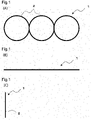
- Figs. 1(A) to 1(C) each show a detachable multi-ring traction device 1 (hereinafter simply referred to as device 1) of the present embodiment.
- Fig. 1(A) is a front view of the device 1.
- Fig. 1(B) is a plan view of the device 1.
- Fig. 1(C) is a right side view of the device 1.
- the back view would be depicted in the same manner as the front view.
- the bottom view would be depicted in the same manner as the plan view.
- the left side view would be depicted in the same manner as the right side view.
- the device 1 is configured to be inserted into a body through a forceps channel of a known endoscope (not shown). Further, when a lesion mucosa in the body is resected through an operation performed outside the body, the device 1 is configured to be fixed by a clip to the lesion mucosa and a normal mucosa in order to pull the lesion mucosa. The normal mucosa opposes the lesion mucosa.
- the device includes multiple (e.g., three to five) toroidal rings 2 each including a core curve and a meridional disk.
- the rings 2 are coupled to each other such that the outer edges of two adjacent ones of the rings 2 are in contact with each other and these rings 2 are connected to each other in the diametrical direction of the core curves.
- the rings 2 do not have to be circular.
- the rings 2 may be slightly distorted or may be oval.
- the ring 2 at one end is referred to as the first ring 2
- the ring 2 at the other end is referred to as the third ring 2
- the ring 2 at the center is referred to as the second ring 2.
- the rings 2 are made of the same material.
- the material of the rings 2 is preferably synthetic resin.
- the synthetic resin does not include rubber.
- the synthetic resin is preferably at least one selected from polyethylene, polypropylene, and polyvinyl chloride.
- the device 1, that is, the rings 2 connected to each other in a row are integrally molded by injection-molding synthetic resin (e.g., polyethylene) into a mold.
- synthetic resin e.g., polyethylene
- the device 1 may be formed as follows. That is, thread-like materials each having the diameter of the meridional disk of the ring 2 are used to form toroidal rings each having the diameter of the core curve. Then, ultrasonic welding is performed to connect a given number of the rings to each other.
- the following method may be employed. That is, thread-like materials each having the diameter of the meridional disk of the ring 2 are used to form toroidal rings each having the diameter of the core curve. Then, the rings are arranged in a row in the diametrical direction of the core curve so that adjacent ones of the rings are welded using ultrasonics.
- the device 1 preferably pulls lesion mucosae using the tensile strength of the mucosae, rather than actively pulling the lesion mucosae using the elasticity of rubber.
- the device 1 is made of plastic (synthetic resin), instead of elastic rubber.
- the plastic is deformable so as to be insertable into the forceps channel of the endoscope together with a hemostatic clip or grasping forceps and is deformable in a direction in which the plastic is folded such that the core curves of the ring 2 overlap each other.
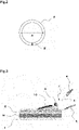
- Fig. 2 is an enlarged view of one of the rings 2 of the device 1.
- the dimension is changed such that the meridional disk (the thickness of the ring 2) becomes more noticeable.
- Diameter A of the core curve of each ring 2 is, for example, 5 mm to 10 mm and is preferably 6 mm to 8 mm.
- Diameter B of the meridional disk of each ring 2 is, for example, 0.1 mm to 0.5 mm and is preferably 0.2 mm to 0.4 mm.
- the connected rings 2 are bendable and deformable such that the rings 2 can pass through the forceps channel. Further, the connected rings 2 are extendable and deformable in the diametrical direction of each core curve. The rings 2 do not need to have rubber-like elasticity.
- Fig. 3 shows method 1 for using the device 1.
- the lower part of the figure shows a side cross-sectional view of the digestive tract (e.g., stomach or large intestine).
- the digestive tract includes a surface mucous layer S, a muscular layer M, and an inner serosa layer I that are arranged in order from the side of a lumen (the upper side in the figure).
- a lesion 10 remains in the surface mucous layer S.
- An operator uses a knife 5 (e.g., high-frequency knife) to make a cut in the surface mucous layer S so as to surround a lesion mucosa including the lesion 10, which needs to be resected.
- the cut typically has a substantially circular shape or an oval shape with an appropriate size. Normally, the cut is set to be wider than the lesion 10 by a certain width.
- the operator uses a clip 3 to fix the first ring 2 of the device 1 on the inner side of the lesion mucosa including the lesion 10, that is, on the inner side of the cut.
- the operator uses forceps 4 to pull the second ring 2 or the third ring 2 with a certain force.
- the operator detaches the lesion mucosa using the knife 5 while pulling the lesion mucosa using the device 1.
- Fig. 4 shows method 2 for using the device 1. Like or the same reference numerals are given to those components that are like or the same as the corresponding components of Fig. 3 , and detailed explanations are omitted.
- the operator uses the knife 5 to make a cut having a certain size in the surface mucous layer S of the lesion mucosa including the lesion 10. Then, the operator uses the clip 3 to fix the first ring 2 of the device 1 to the lesion mucosa, which needs to be resected. Subsequently, the operator uses a clip 3' to fix the third ring 2 of the device 1 to a normal mucosa S' (a mucosa that is not to be resected). The normal mucosa S' opposes the lesion mucosa.
- the operator draws air into an organ (a stomach in this example) with a certain pressure and produces a given pressure P. This separates the clips 3, 3' from each other and pulls the device 1. Further, the device 1 pulls the lesion 10. In this state, the operator uses the high-frequency knife 5 to cut the lower layer of the lesion mucosa and dissect the lesion mucosa.
- the operator may use the high-frequency knife 5 to cut the lower layer of the lesion mucosa while pulling the device 1 fixed to the clips 3, 3'.
- the operator After detaching the lesion mucosa including the lesion 10, the operator cuts the first ring 2 of the device 1. For example, the operator may cut the first ring 2 by strongly pulling the first ring 2 with forceps or may cut the first ring 2 using the high-frequency knife 5. Subsequently, the operator collects the lesion mucosa including the lesion 10 and performs, for example, pathological examination.
- the clip 3' left in the normal mucosa in the above-described operation naturally drops after a while and is then discharged out of the body.
- Figs. 5(A) to 5(C) show a procedure for attaching the device 1 to the grasping forceps.
- the operator folds the device 1 at the connection portions of the three rings 2 and then uses the grasping forceps to pinch all of the three rings 2 that have the same size as one ring 2 (refer to Fig. 5A ).
- the operator moves the device 1 through the forceps channel (refer to Figs. 5(B) and 5(C) ).
- the operator may use the grasping forceps to pinch only one ring 2 (e.g., first ring 2) and draw the device 1 into the body through the forceps channel.
- Figs. 6A to 6D show a situation in which the device 1 is pinched at the distal end of the hemostatic clip and drawn into the body.
- the operator hooks the first ring 2 of the device 1 on the basal end of the hemostatic clip that has been drawn from the distal end of a sheath (refer to Fig. 6A ).
- the operator draws the hemostatic clip into the sheath.
- This causes the device 1 to be inserted into the sheath (refer to Figs. 6(B), 6(C), and 6(D) ).
- the operator draws the device 1 into the body of a patient.
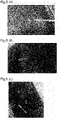
- the operator uses the high-frequency knife to cut the surrounding mucosa (lesion mucosa), including a focus, in a substantially oval form (refer to Fig. 7(A) ).
- the operator hooks the first ring 2 of the device 1 on the clip 3 to fix the clip 3 to the lesion mucosa 10 (refer to Fig. 7(B) ).
- the operator uses the clip 3' to fix the third ring 2 of the device 1 to the normal mucosa, which opposes the lesion mucosa.
- the device 1 connects between the clips 3, 3' with a certain tension (refer to Figs. 7(C) and 8(A) ).
- the operator applies a certain inner pressure to the inside of the digestive tract to inflate the digestive tract.
- This causes the device 1 to pull the lesion mucosa 10 (refer to Fig. 8(B) ).
- the operator uses the high-frequency knife to resect the exposed section of the lower layer of the exposed lesion mucosa 10 (refer to Fig. 8(C) ).
- the operator resects the entire lower layer of the lesion mucosa 10 to detach the lesion mucosa 10 from the digestive tract (refer to Fig. 9(A) ).
- the operator uses the high-frequency knife to cut the third ring 2, which is fixed to the normal mucosa by the clip 3' (refer to Fig. 9(B) ).
- This causes the device 1 and the lesion mucosa 10 to be detached from the digestive tract (refer to Fig. 9(C) ).
- the clip 3'left in the digestive tract is naturally discharged after certain days.
- the mucosa being incised does not cover the visual field or treatment field of the endoscope. This broadens the treatment field and allows for smooth and efficient incision of a lesion mucosa.
- the device 1 includes three rings 2.
- the number of rings 2 may be changed.
- the device 1 may include four or five rings 2.
- a device 1 including three to five rings 2 is suitable for ESD performed for a stomach.
- a ring at a certain position may be fixed to a clip without using the entire length of the rings.
- the first ring 2 may be fixed to the lesion mucosa 10 using the clip 3 and the second ring 2 may be fixed to the normal mucosa using the clip 3'.
Landscapes
- Health & Medical Sciences (AREA)
- Surgery (AREA)
- Life Sciences & Earth Sciences (AREA)
- Animal Behavior & Ethology (AREA)
- Veterinary Medicine (AREA)
- Public Health (AREA)
- Heart & Thoracic Surgery (AREA)
- General Health & Medical Sciences (AREA)
- Biomedical Technology (AREA)
- Molecular Biology (AREA)
- Medical Informatics (AREA)
- Engineering & Computer Science (AREA)
- Nuclear Medicine, Radiotherapy & Molecular Imaging (AREA)
- Surgical Instruments (AREA)
- Endoscopes (AREA)
- Catching Or Destruction (AREA)
- Friction Gearing (AREA)
- Devices For Conveying Motion By Means Of Endless Flexible Members (AREA)
- Chemical & Material Sciences (AREA)
- Chemical Kinetics & Catalysis (AREA)
- Vascular Medicine (AREA)
- Epidemiology (AREA)
Applications Claiming Priority (1)
| Application Number | Priority Date | Filing Date | Title |
|---|---|---|---|
| PCT/JP2019/027070 WO2021005698A1 (fr) | 2019-07-09 | 2019-07-09 | Dispositif de traction à anneaux multiples détachable |
Publications (2)
| Publication Number | Publication Date |
|---|---|
| EP3998024A1 true EP3998024A1 (fr) | 2022-05-18 |
| EP3998024A4 EP3998024A4 (fr) | 2023-03-22 |
Family
ID=74113959
Family Applications (1)
| Application Number | Title | Priority Date | Filing Date |
|---|---|---|---|
| EP19937148.5A Pending EP3998024A4 (fr) | 2019-07-09 | 2019-07-09 | Dispositif de traction à anneaux multiples détachable |
Country Status (7)
| Country | Link |
|---|---|
| US (2) | US20220287700A1 (fr) |
| EP (1) | EP3998024A4 (fr) |
| JP (1) | JP7376879B2 (fr) |
| KR (2) | KR20220035409A (fr) |
| CN (2) | CN120078461A (fr) |
| AU (2) | AU2019455874B2 (fr) |
| WO (1) | WO2021005698A1 (fr) |
Cited By (1)
| Publication number | Priority date | Publication date | Assignee | Title |
|---|---|---|---|---|
| WO2025190711A1 (fr) | 2024-03-12 | 2025-09-18 | Pinard Fabien | Dispositif médical endoscopique pour agir sur une muqueuse interne et comportant au moins un fil de traction cranté et un nœud auto-bloquant |
Family Cites Families (17)
| Publication number | Priority date | Publication date | Assignee | Title |
|---|---|---|---|---|
| JP4059656B2 (ja) * | 2001-03-07 | 2008-03-12 | オリンパス株式会社 | 生体組織のクリップ装置 |
| US20070250116A1 (en) * | 2006-04-25 | 2007-10-25 | Board Of Regents, The University Of Texas System | Tissue Approximator and Retractor Assistive Device |
| JP4472680B2 (ja) | 2006-09-09 | 2010-06-02 | 太一 坂本 | 医療用把持具及び医療用クリップ |
| JP5192771B2 (ja) * | 2006-11-28 | 2013-05-08 | 信行 櫻澤 | 粘膜牽引具 |
| US9101357B2 (en) * | 2007-06-08 | 2015-08-11 | Board Of Trustees Of The University Of Arkansas | Physiologic abdominal closure |
| WO2011126050A1 (fr) * | 2010-04-06 | 2011-10-13 | 日立化成工業株式会社 | Corps polymère moulé permettant d'exercer une traction sur un tissu biologique, élément de traction à usage médical l'utilisant et outil de traction à usage médical |
| JP5614833B2 (ja) | 2010-04-09 | 2014-10-29 | オカモト株式会社 | 組織結合用弾性体及びその製造方法 |
| JP5733754B2 (ja) * | 2011-07-12 | 2015-06-10 | 公益財団法人ヒューマンサイエンス振興財団 | 粘膜切開剥離術補助具 |
| EP3165183A4 (fr) * | 2014-07-01 | 2018-01-17 | Olympus Corporation | Outil de saisie de tissu |
| JP6936309B2 (ja) * | 2016-08-05 | 2021-09-15 | ボストン サイエンティフィック サイムド,インコーポレイテッドBoston Scientific Scimed,Inc. | 組織を牽引するためのシステム、装置、及び関連する方法 |
| CN106344083B (zh) * | 2016-10-29 | 2019-01-04 | 张强 | 一体套管式内镜治疗辅助管道装置 |
| JP6795410B2 (ja) * | 2017-01-23 | 2020-12-02 | 欣也 藤田 | 内視鏡用フード |
| EP3689254A4 (fr) * | 2017-09-29 | 2021-09-22 | Zeon Corporation | Pince de traction destinée à un endoscope |
| CN117814875A (zh) * | 2017-10-02 | 2024-04-05 | 波士顿科学国际有限公司 | 用于组织牵开的装置和方法 |
| EP4302708A3 (fr) * | 2017-11-08 | 2024-03-20 | Mayo Foundation for Medical Education and Research | Systèmes et procédés de dissection endoscopique de sous-muqueuse à l'aide de pinces hémostatiques à fixation magnétique |
| CN110731802A (zh) * | 2018-07-20 | 2020-01-31 | 南微医学科技股份有限公司 | 一种牵引装置 |
| CN109620320B (zh) * | 2019-02-15 | 2021-06-29 | 齐从虎 | 一种内镜下多环牵引器及其使用方法 |
-
2019
- 2019-07-09 EP EP19937148.5A patent/EP3998024A4/fr active Pending
- 2019-07-09 CN CN202510177446.XA patent/CN120078461A/zh active Pending
- 2019-07-09 KR KR1020227004037A patent/KR20220035409A/ko not_active Ceased
- 2019-07-09 CN CN201980098220.3A patent/CN114340511A/zh active Pending
- 2019-07-09 AU AU2019455874A patent/AU2019455874B2/en active Active
- 2019-07-09 JP JP2021530386A patent/JP7376879B2/ja active Active
- 2019-07-09 KR KR1020257005819A patent/KR20250033312A/ko active Pending
- 2019-07-09 WO PCT/JP2019/027070 patent/WO2021005698A1/fr not_active Ceased
- 2019-07-09 US US17/624,975 patent/US20220287700A1/en not_active Abandoned
-
2023
- 2023-05-02 AU AU2023202699A patent/AU2023202699B2/en active Active
-
2025
- 2025-02-26 US US19/064,118 patent/US20250213239A1/en active Pending
Cited By (2)
| Publication number | Priority date | Publication date | Assignee | Title |
|---|---|---|---|---|
| WO2025190711A1 (fr) | 2024-03-12 | 2025-09-18 | Pinard Fabien | Dispositif médical endoscopique pour agir sur une muqueuse interne et comportant au moins un fil de traction cranté et un nœud auto-bloquant |
| FR3160097A1 (fr) | 2024-03-12 | 2025-09-19 | Fabien PINARD | Dispositif médical endoscopique pour agir sur une muqueuse interne et comportant au moins un fil de traction cranté et un nœud auto-bloquant |
Also Published As
| Publication number | Publication date |
|---|---|
| CN114340511A (zh) | 2022-04-12 |
| AU2023202699B2 (en) | 2025-05-15 |
| JPWO2021005698A1 (fr) | 2021-01-14 |
| KR20250033312A (ko) | 2025-03-07 |
| CN120078461A (zh) | 2025-06-03 |
| AU2023202699A1 (en) | 2023-05-18 |
| AU2019455874A1 (en) | 2022-02-24 |
| WO2021005698A1 (fr) | 2021-01-14 |
| KR20220035409A (ko) | 2022-03-22 |
| JP7376879B2 (ja) | 2023-11-09 |
| EP3998024A4 (fr) | 2023-03-22 |
| US20250213239A1 (en) | 2025-07-03 |
| AU2019455874B2 (en) | 2023-02-09 |
| US20220287700A1 (en) | 2022-09-15 |
Similar Documents
| Publication | Publication Date | Title |
|---|---|---|
| JP5047297B2 (ja) | 粘膜切開剥離術補助具 | |
| JP4351458B2 (ja) | 内視鏡挿入システム | |
| US20170360423A1 (en) | Removable medical retractor tip | |
| CN100382770C (zh) | 内窥镜粘膜切除用治疗器及内窥镜粘膜切除装置 | |
| EP2679169A1 (fr) | Écarteur à trois dimensions | |
| JP2009183516A (ja) | Esd用処置具 | |
| JP7748943B2 (ja) | 組織牽引装置、システムおよび方法 | |
| US20190290325A1 (en) | Method for lifting mucosa | |
| US20250213239A1 (en) | Detachable multi-ring traction device | |
| JP2008511341A (ja) | オーバーチューブアセンブリ | |
| WO2016138027A1 (fr) | Conception d'anse pour traction améliorée | |
| JP2014171629A (ja) | 内視鏡装置用補助具及びこれを用いた内視鏡装置 | |
| JP6948637B2 (ja) | デタッチャブル・マルチリング・トラクション・デバイス | |
| JP6698107B2 (ja) | 2つの高周波メスを形成するためにチューブ内に開口部を通して導かれる切除ワイヤを有する内視鏡的切除装置、および経皮内視鏡的胃瘻造設術用のカテーテル | |
| US12016526B2 (en) | Mucous membrane lifting instrument and mucous membrane lifting method | |
| JP2002272677A (ja) | 湾曲圧排具 | |
| TWI820166B (zh) | 可拆式多環牽引裝置 | |
| JP3737540B2 (ja) | トロカール外套管およびトロカール | |
| CN112807049B (zh) | 一种止血夹和辅助系统 | |
| HK40123084A (en) | Detachable multi-ring traction device | |
| JP3759178B2 (ja) | トロカール管及びそれを備えたトロカール | |
| US20250312024A1 (en) | Medical devices, medical systems, and related methods for providing traction to tissue | |
| US20220273339A1 (en) | Cannula for preventing tumor seeding | |
| WO2025217106A1 (fr) | Systèmes, dispositifs et méthodes d'administration de dispositifs médicaux | |
| JP2007307001A (ja) | 粘膜切開剥離術補助具 |
Legal Events
| Date | Code | Title | Description |
|---|---|---|---|
| STAA | Information on the status of an ep patent application or granted ep patent |
Free format text: STATUS: THE INTERNATIONAL PUBLICATION HAS BEEN MADE |
|
| PUAI | Public reference made under article 153(3) epc to a published international application that has entered the european phase |
Free format text: ORIGINAL CODE: 0009012 |
|
| STAA | Information on the status of an ep patent application or granted ep patent |
Free format text: STATUS: REQUEST FOR EXAMINATION WAS MADE |
|
| 17P | Request for examination filed |
Effective date: 20220209 |
|
| AK | Designated contracting states |
Kind code of ref document: A1 Designated state(s): AL AT BE BG CH CY CZ DE DK EE ES FI FR GB GR HR HU IE IS IT LI LT LU LV MC MK MT NL NO PL PT RO RS SE SI SK SM TR |
|
| DAV | Request for validation of the european patent (deleted) | ||
| DAX | Request for extension of the european patent (deleted) | ||
| A4 | Supplementary search report drawn up and despatched |
Effective date: 20230221 |
|
| RIC1 | Information provided on ipc code assigned before grant |
Ipc: A61B 17/10 20060101ALI20230215BHEP Ipc: A61B 17/08 20060101ALI20230215BHEP Ipc: A61B 17/02 20060101AFI20230215BHEP |
|
| STAA | Information on the status of an ep patent application or granted ep patent |
Free format text: STATUS: EXAMINATION IS IN PROGRESS |
|
| 17Q | First examination report despatched |
Effective date: 20250204 |