EP4006165A1 - Détection de microréseau de sondes dépendant de la ligature multiplex - Google Patents
Détection de microréseau de sondes dépendant de la ligature multiplex Download PDFInfo
- Publication number
- EP4006165A1 EP4006165A1 EP20847306.6A EP20847306A EP4006165A1 EP 4006165 A1 EP4006165 A1 EP 4006165A1 EP 20847306 A EP20847306 A EP 20847306A EP 4006165 A1 EP4006165 A1 EP 4006165A1
- Authority
- EP
- European Patent Office
- Prior art keywords
- probe
- quenching
- primer
- detection
- nucleic acid
- Prior art date
- Legal status (The legal status is an assumption and is not a legal conclusion. Google has not performed a legal analysis and makes no representation as to the accuracy of the status listed.)
- Granted
Links
Images

Classifications
-
- C—CHEMISTRY; METALLURGY
- C12—BIOCHEMISTRY; BEER; SPIRITS; WINE; VINEGAR; MICROBIOLOGY; ENZYMOLOGY; MUTATION OR GENETIC ENGINEERING
- C12Q—MEASURING OR TESTING PROCESSES INVOLVING ENZYMES, NUCLEIC ACIDS OR MICROORGANISMS; COMPOSITIONS OR TEST PAPERS THEREFOR; PROCESSES OF PREPARING SUCH COMPOSITIONS; CONDITION-RESPONSIVE CONTROL IN MICROBIOLOGICAL OR ENZYMOLOGICAL PROCESSES
- C12Q1/00—Measuring or testing processes involving enzymes, nucleic acids or microorganisms; Compositions therefor; Processes of preparing such compositions
- C12Q1/68—Measuring or testing processes involving enzymes, nucleic acids or microorganisms; Compositions therefor; Processes of preparing such compositions involving nucleic acids
- C12Q1/6844—Nucleic acid amplification reactions
- C12Q1/6851—Quantitative amplification
Definitions
- the present invention relates to the field of nucleic acid detection, more particularly to the detection of multiplex ligation probe microarrays.
- nucleic acid detection is often required on the sample to be tested.
- commonly used detection methods include conventional PCR, quantitative PCR, probe detection, chip detection, sequencing and other technologies.
- primer pairs for amplification and probes for detection are present in the detection system.
- one side (or one end) of the probe in the solution has a fluorescent agent (fluorophore)
- the other side (or the other end) has a quencher.
- the probe When there is a nucleic acid sample matching the sequence of the probe in the solution, the probe is cleaved by the amplification enzyme during the PCR amplification of the sample, so that the fluorophore and the quencher will be separated from each other, resulting in a reduction or disappearance of the inhibitory effect, thereby allowing the fluorophore to generate a fluorescent signal.
- the current real-time fluorescent quantitative PCR still has some shortcomings, including: because the probe is labeled with a fluorophore and a quencher at the same time, the cost of probe synthesis is high; in order to make the probe in the solution cleaved by the amplification enzyme, amplification enzyme needs to have 5' ⁇ 3' exonuclease activity, however some amplification enzymes do not have 5' ⁇ 3' exonuclease activity and are therefore not suitable for qPCR.
- each sequence to be tested requires a fluorophore whose emission spectrum can be distinguished from other fluorophores
- the number of different nucleic acids that can be detected simultaneously in the same qPCR reaction is greatly limited, usually a qPCR reaction can only achieve 4-5 multiplex detection.
- a qPCR reaction can only achieve 4-5 multiplex detection.
- the corresponding optical detection equipment is complicated in design and expensive.
- the purpose of the present invention is to provide a quantitative detection method for a specific sequence by using only one pair of primers and multiple pairs of probes to perform multiplex ligation-dependent amplification.
- the detection system comprises:
- the first probe has the structure of Formula II: X 2 ′ - T 2 ′ wherein, X 2 ' is a reverse complement of the 5' end universal amplification primer; T 2 ' is a reverse complement of the specific sequence at the 5' end of a targeting gene.
- the length (nt) of X 2 ' is 15-50, preferably 19-36, more preferably 20-35.
- the length (nt) of T 2 ' is 15-60, 19-36, more preferably 20-35.
- the second probe has the structure of Formula III: T 1 ′ - P 1 ′ ⁇ X 1 ′ wherein, T 1 ' is a reverse complement of the specific sequence at the 3' end of a targeting gene;
- the length (nt) of T 1 ' is 15-60, preferably 19-36, more preferably 20-35.
- the length (nt) of P 1 ' is 10-500, preferably 19-100, more preferably 20-30.
- the length (nt) of X 1 ' is 15-50, preferably 19-36, more preferably 20-35.
- the ligase is selected from the group consisting of T4 ligase, NAD ligase (such as ligase-65), high temperature stable ligase (such as Taq DNA Ligase, HiFi Taq DNA Ligase, 9°N TM DNA Ligase), and a combination thereof.
- the third probe has the structure of Formula IV: X 2 ′ - T 2 ′ - T 1 ′ - P 1 ′ ⁇ X 1 ′ wherein, X 2 ' is a reverse complement of the 5' end universal amplification primer;
- the first detectable marker is a fluorophore.
- the fluorophore is selected from the group consisting of chemiluminescent label (luminescent labels).
- the fluorophore is selected from the group consisting of quantum dots.
- the ratio (Q1/Q0) of the number Q1 of the quenching amplification product within the effective hybridization distance to the surface probe to the number Q0 of the corresponding surface probe (Q1/Q0) is 0.5 to 100, preferably 1-10.
- the detection system further comprises one or more components selected from the group consisting of:
- the polymerase is selected from the group consisting of Taq enzyme, Pfu enzyme, Pwo enzyme, vent enzyme, KOD enzyme, superfi enzyme, and a combination thereof.
- the length (bp) of the amplification product amplified by the first primer and the second primer is 50-2000, preferably 75-500, more preferably 75-150.
- the quenching product capturing nucleic acids of different surface quantitation sub-detection regions are the same or different nucleic acid molecules.
- the first probe and/or the second probe includes DNA, RNA, and a combination thereof.
- the length (nt) of the first probe is 35-120, preferably 40-80, more preferably 45-65.
- the length (nt) of the second probe is 40-160, preferably 55-120, more preferably 65-90.
- the length (nt) of the third probe is 75-280, preferably 95-200, more preferably 110-155.
- the length of the quenching product capturing nucleic acid is ⁇ 150 nt, preferably ⁇ 100 nt, more preferably ⁇ 50 nt.
- the length (nt) of the quenching primer is 15-50, preferably 19-36, more preferably 20-35.
- the quenching primer capturing nucleic acid has a capture region that is substantially complementary or completely complementary to the quenching primer.
- the quenching product capturing nucleic acid has the following structure of Formula I: B 0 ⁇ B 1 ⁇ B 2 ⁇ B 3 wherein,
- the first detectable signal is located at B2 or B3, preferably at B2.
- B3 is none.
- one end or one side of the quenching primer capturing nucleic acid is connected with a first detectable marker (e.g., a fluorophore).
- a first detectable marker e.g., a fluorophore
- the other primer in the first primer and the second primer is a conventional primer or a universal primer.
- the number of the surface quantitative sub-detection region is m, m is a positive integer of ⁇ 1, and m ⁇ n.
- n is 2-500, preferably 5-250, more preferably 9-100 or 16-96.
- m is 2-500, preferably 5-250, more preferably 9-100 or 16-96.
- the same first detectable marker is used in at least ⁇ 2 (more preferably ⁇ 3) surface quantitation sub-detection regions.
- each of the surface quantitation sub-detection regions is used to detect different or the same target sequence.
- each of the surface quantitation sub-detection regions is used to detect different target sequences.
- step (d) the analysis includes comparing the detected signal of the first detectable marker with a standard curve.
- step (d) the analysis includes comparing the detected signal of the first detectable marker with the detected signal before amplification.
- the quantitative PCR detection is multiplex PCR detection.
- the first detectable marker of each surface quantitation sub-detection region is the same.
- the first detectable marker of each surface quantitation sub-detection region is different.
- kit for quantitative PCR detection comprising:
- the first container, the second container, the third container, the fourth container, and the fifth container are the same container or different containers.
- the inventors After extensive and in-depth research, the inventors have developed for the first time a multiplex ligation-dependent amplification using only one pair of primers and multiple pairs of probes. At the same time of amplification, in each amplification cycle, the corresponding probes immobilized on the surface can quantitatively read the amplification products to achieve quantitative detection of specific sequences. In addition, even if only one or two or a few detectable markers and corresponding quencher are used, the detection effect of the present invention will not be affected. Therefore, the manufacturing cost and the use cost are greatly reduced. The present invention has been completed on this basis.
- primer refers to an artificially synthesized short nucleic acid fragment (especially a DNA fragment) in the polymerase chain reaction that determines the initiation and termination positions required for amplification.
- Universal primer “unamplified primer” and “primer” are used interchangeably herein.
- quenching primer or “quenching primer of the present invention” are used interchangeably and refer to a primer with a quencher. It is understood that either primer (e.g., a first primer or a second primer) or both primers of a primer pair can be the quenching primer.
- quenching amplification product As used herein, the terms “quenching amplification product”, “amplification product”, “quenching product”, “quenching product of the present invention”, “quenching amplification product of the present invention” are used interchangeably and refer to an amplification product with a quencher.
- quenching product capturing nucleic acid As used herein, the terms “quenching product capturing nucleic acid”, “quenching product capturing nucleic acid of the present invention”, “surface probe”, “surface probe of the present invention” are used interchangeably and refer to a single-stranded nucleic acid molecule with a first detectable marker (such as a fluorophore) for capturing quenching amplification products and immobilized on the surface of a solid phase carrier. It should be understood that the quenching product capturing nucleic acids of the present invention are different from probes in the free state (which carry both a fluorophore and a quencher) in the prior art (e.g., fluorescence quantitative PCR).
- a first detectable marker such as a fluorophore
- the present invention provides a quantitative PCR detection system based on surface probes, and the detection system includes:
- the second primer may also have a quenching agent like the first primer, and the quenching amplification product produced by the second primer has corresponding different primer-capturing sub-detection regions on the surface.
- both the first primer and the second primer have a quenching agent, but they correspond to one or more sub-detection regions at the same time, that is, each sub-detection region corresponding to the quenching product generated by the amplification of the pair of primers is simultaneously immobilized with the corresponding two amplification product capturing nucleic acids.
- the first and second probes respectively have specific sequences T 2 ' and T 1 ' corresponding to the target nucleic acid sequence to be detected, and their design follows the traditional design principles of ligation probes, and its length is preferably 15-35 nucleotide units. A long specific sequence is beneficial to increase the specificity of recognition of the target to be detected.
- the corresponding second probe for detecting each nucleic acid to be tested has its specific barcode index sequence Pi', which respectively corresponds to the surface probe on a certain sub-detection region.
- the first probe may also have a complementary sequence corresponding to a surface probe, as long as the primer on its side is also a quenching primer.
- the same, usually the same two universal primers are used for specific amplification.
- the two primers increase the specificity of the amplification reaction and accelerate the formation of amplification products, and the surface probe on the solid surface corresponding to Pi' with the barcode index sequence generated by it ensures the specificity and signal of detection. If both primers in a primer pair are quenching primers, the corresponding two surface probes can be respectively immobilized in different surface quantitation sub-detection regions, or they can be mixed and fixed in the same surface quantification sub-detection region.
- the design of universal primers follows the traditional PCR primer design, and the length of the primer itself is preferably 20 - 35 nucleotide units. Short primers are beneficial to shorten the distance of the quenching amplification product from the fluorescent labels of the surface probe after hybridization with the surface probe.
- the label quenching agent is preferably at the 5' end of the quenching primer
- the label can be conjugated or synthesized at any position on the quenching primer as long as it does not interfere with the amplification of the chain reaction and after hybridization with the surface probe, it is close enough to the fluorescent labels of the surface probe (the distance of less than 25 nucleotide units).
- sequence P1' of the second probe is preferably completely complementary to the quenching product capturing nucleic acid on the surface, it can also be partially complementary, that is, it is complementary to a portion of the quenching amplification product generated by the quenching primer and the quenching product capturing nucleic acid (i.e., the surface probe).
- sequences of other amplification extensions with the amplification product should not be complementary to the surface probe and its other extensions should not exceed 150nt, and preferably should not exceed 120nt.
- the surface probes are immobilized on the surface quantitation sub-detection region of the solid phase carrier.
- the 5' end of the surface probe is closer to the solid surface and the 3' end is further away from the solid surface.
- the present invention provides a surface probe quantitative PCR method.
- the first probe and the second probe will be ligated into the third probe by the ligase.
- the core of the present invention is that the quenching amplification product hybridizes with the surface probe to gradually weaken the fluorescent signal of the surface probe to achieve real-time quantification of the target to be detected.
- the third probe is generated only when the target nucleic acid is present first.
- the surface probe of the sub-detection region corresponding to barcode index P1' does not have any sequence complementary to it and has a quencher, and the surface probe of the sub-detection region is in the light-emitting state; after the third probe is amplified, the amplification product contains both the quenching agent of the quenching primer and the barcode index P1' because of the amplification and extension reaction.
- the quenching amplification product When the quenching amplification product is hybridized with the surface probe, as a result, the fluorescence of the surface probe is weakened, and the degree of fluorescence reduction is directly related to the concentration of the quenching amplification product, that is, the speed of the amplification reaction, so the target nucleic acid can be quantified.
- a pair of "specific ligation probes" corresponding to the target namely the first probe and the second probe (X 2 '-T 2 ' and T 1 '-P 1 '-X 1 ') hybridize to the target, and the ligase links the two probes to form a sequence L 1 that can be amplified, that is, a third probe; if the detection target does not exist, its corresponding specific ligation probe, that is, the first probe and the second probe (X 2 '-T 2 ' and T 1 '-P 1 '-X 1 ') will not be connected, and thus will not be amplified.
- T 1 '-P 1 '-X 1 ' for each target to be detected, T 1 ' is the complementary sequence to the specific sequence of the target, P 1 ' (the sequence between T 1 ' and X 1 ' in the upper left of Figure 2 is not complementary to the target to be detected) is the marker (Barcode Index P 1 ') corresponding to a sub-detection region.
- the amplification product contains Barcode Index P 1 and a quenching agent, which can hybridize with the corresponding surface probe S 1 on the surface (S1 contains a sequence that is completely complementary to Pi).
- the quenching agent Q carried by the amplimer has a FRET reaction with the fluorophore F of S 1 , thus, the fluorescent signal of the surface probe is reduced.
- the weakening of the fluorescent signal directly corresponds to whether there is a target to be detected in the solution, and the intensity and speed of the signal weakening correspond to the initial concentration of the target to be detected.
- multiplex detection since the detection signal is derived from the surface probe, and because the physical position of the surface probe itself can be used to distinguish the corresponding target nucleic acid on it, multiplex detection can be achieved with only one label, without the multiple fluorophores of traditional multiplex qPCR. This method can be used for the quantitative detection of at least 1-100 target nucleic acids, or more than 100.
- the traditional multiplex qPCR technology is limited by the type of fluorescent agent and optical design, usually only 4-6 real-time quantitative detection can be performed, and very few can achieve 8.
- the number of multiples is not particularly limited, and can be any positive integer ⁇ 2, preferably 2-10000 or 2-500, preferably 3-250, more preferably 4-100 or 6-96 .
- the signal reading of the surface probe can be performed for every cycle (or every 1 or every 2 cycles) at a selected time and temperature during the amplification reaction.
- the surface probe of the sub-detection region corresponding to Barcode Index P 1 ' does not have any sequence complementary to it and has a quencher, and the surface probe of the sub-detection region is in a light-emitting state;
- the amplification product not only contains the quenching agent of the quenching primer, but also contains the Barcode Index P 1 because of the amplification and extension reaction.
- the quenching amplification product When the quenching amplification product is hybridized with the surface probe, as a result, the fluorescence of the surface probe is weakened, and the degree of fluorescence weakening is directly related to the concentration of the quenching amplification product, that is, the speed of the amplification reaction, so the target nucleic acid can be quantified.
- the present invention also provides a kit for quantitative PCR detection, which includes:
- kit of the present invention may also contain other compositions or components, such as standard curves, quality control samples, and the like.
- any two, three or all of the first, second, third, fourth, and fifth containers are the same container.
- first, second, third, fourth and fifth containers are different containers.
- all of the reagents themselves may be present in the first container.
- all reagents are stored in the first container in a lyophilized manner, the DNA to be detected is thawed in the corresponding buffer, and injected into the first container during the reaction.
- the present invention also provides a reaction device and a detection system for quantitative PCR based on surface probes.
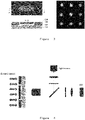
- a representative reaction device is shown in Figure 3 .
- the chip used for the reaction can be in any form, as long as the reaction chamber has an inner surface, a microarray (surface quantitation sub-detection region array) can be prepared in advance and subsequent fluorescence signal reading can be performed.
- a microarray surface quantitation sub-detection region array
- a typical design is a flat reaction chamber design.
- the reaction chamber is shown in Figure 3 and consists of three parts.
- the first part 101 is a piece of plastic or glass
- the second part 102 is another piece of flat plastic or glass, and combined with the third part 103, a double-sided tape with a thickness of 0.25 mm.
- the double-sided tape has a cut portion to form a reaction chamber.
- the reaction chamber can also be composed of only two parts: the first part already contains the grooves that constitute the reaction chamber, and the second part can be provided with single-sided adhesive, which can directly bond the first part, or it can also form a closed reaction chamber with the first part by means of ultrasonic fusion, thermal fusion, double-sided adhesive or ultraviolet curing adhesive.
- the chamber has inlet and outlet channels or inlets and outlets for the reaction solution to flow in. After the reaction solution is added, the valve on the channel can be closed or the inlet and outlet can be permanently closed.
- a single-sided adhesive 104 with a thickness of 0.1 mm is used to permanently close the entrance and exit.
- FIG. 3 shows a 3x3 microarray.
- the array is used to detect 3 different nucleic acids (2 target nucleic acids and 1 internal reference), and corresponding to each nucleic acid sequence to be detected, there are three sub-detection regions immobilized with surface probes corresponding to the nucleic acid to be detected.
- Detection using more than one sub-detection region for each nucleic acid sequence to be detected when the detection signal of one of the sub-detection regions is disturbed by air bubbles or impurities, there are still other spots with the same sequence to confirm the target nucleic acid to be detected.
- the number of sub-detection regions constituting the microarray can be 2-100, or more than 100.
- the material on one side of the reaction chamber must have sufficient transmittance in the relevant wavelength band (excitation and emission) of the fluorescent agent, with a transmittance of at least 80%, preferably more than 90%; at the same time, the lower the autofluorescence of the material on this side, the better.
- the first part of the reaction chamber is a glass slide with low autofluorescence (BOROFLOAT ® 33 from Schott Company), which has a transmittance of more than 90% to visible light, and its autofluorescence is much lower than that of ordinary glass materials.
- Surface nucleic acid probe sequences are typically coupled to the solid surface via a short linker.
- the linker can be oligonucleotide (oligo), polymer (polymer) (e.g. PEG), or a combination.
- the core of the present invention is that the hybridization of the quenching amplification product and the surface probe causes the fluorescence of the surface probe to be quenched due to the FRET reaction.
- the length of the conjugation linkers should not be less than 5 nm, preferably more than 10 nm, so that the surface probe available for hybridization is far enough away from the solid surface that the quenching amplification product can fully hybridize therewith.
- Solid surfaces are often surface chemically treated to generate reactive groups such as NHS esters, thioesters, etc., which can react with conjugation linkers.
- the link between the conjugation linkers and the solid surface is thermally stable and does not break or fall off due to the cooling and heating cycles of PCR.
- the surface probe is a synthetic sequence, and an amino group is synthesized at the 5' end that can be covalently coupled to the NHS ester on the surface of the glass slide.
- the sequence of the deoxyribonucleic acid is the same as the barcode index P 1 ' on the second probe of each target to be tested (that is, complementary to its corresponding amplification product), and is coupled to the fluorescent agent Cy3 at its 3' end.
- Surface probe arrays can employ conventional methods for generating microarrays.
- Scienion's non-contact spotting machine is used for spotting in this example.
- Touch-based spotting machines such as ArrayIt's SpotBot ® are also a common tool.
- the diameter of the array points i.e. each sub-detection region
- the edge-to-edge spacing of each point is about 100 microns.
- the total number of each surface probe should be equivalent to the number of the corresponding final amplification products in the solution near the array point, so that when the quenching amplification products in the solution near the array point increase due to amplification, on the array points, surface probes will gradually hybridize with the quenching amplification products, resulting in a gradual decrease in the fluorescent signal until almost complete annihilation at the end.
- the surface probe density per spot on the microarray should be higher than 500 fmole/cm 2 , preferably higher than 2000 fmole/cm 2 .
- the fluorescent agent on the surface probe Before amplification, there is no DNA sequence with a quenching agent to hybridize with the surface probe, the fluorescent agent on the surface probe is in a fully light-emitting state, and the fluorescence brightness of the array point is the highest.
- the quenching primer When the corresponding target nucleic acid exists and the third probe generated by ligase ligation is amplified, the quenching primer is extended (amplified), and the extended part contains the Barcode Index P i sequence, and its corresponding surface probe will hybridize to it, resulting in the quenching of the fluorescence of the surface probe, when the quenching amplification product reaches the maximum value, the fluorescence brightness of the array point reaches the minimum value.
- the self-fluorescence signal of the surface probe gradually decreases due to its hybridization. If the concentration of the initial target nucleic acid is relatively low, the growth of the quenching amplification product will be relatively late and slow, so the fluorescence decrease of the surface probe will also occur relatively late and slow. There is a one-to-one correspondence and monotonic decreasing relationship between the fluorescence brightness of the array point and the number of quenching amplification products, so the degree and speed of the fluorescence change of each array point can be used to quantify its corresponding initial target nucleic acid concentration.
- the heating and cooling cycle control of PCR can be performed on one or both sides of the reaction chamber simultaneously. Simultaneous temperature control on both sides can increase the rate of temperature regulation in the reaction chamber, thereby shortening the reaction time.
- the preset here is for the heating-cooling cycle only from one side. Whether it is single-sided or double-sided for temperature control, the thinner the reaction chamber, the faster the temperature adjustment, and the easier it is for the reaction chamber solution to reach thermal equilibrium at each temperature node. Therefore, the thickness of the reaction chamber is usually below 2mm, preferably below 1mm.
- the example uses a 0.25mm design.
- the thickness of the solid material on the temperature control side ensures the strength of the chip, and the thinner the material, the faster the heating and cooling cycle. In this example a 0.5mm polycarbonate sheet.
- black polycarbonate is used here to further reduce the background noise during fluorescence reading.
- thermoelectric module heats and cools a copper block, which has high thermal conductivity so that the reaction chamber can reach thermal equilibrium faster with each temperature change.
- the microarray fluorescence signal reading on the inner surface of the reaction chamber can use a common monochromatic fluorescence signal reading system.
- Figure 4 depicts a typical monochromatic fluorescence signal reading system.
- a fluorescence microscope (Olympus IX73) was used to collect the fluorescence signals of the surface probes on the microarray, the principle of which is consistent with the system described in the above figure.
- the method of the present invention is particularly advantageous for multiplex quantitative detection of DNA.
- each marker to be tested needs to be equipped with a different fluorescent agent, which makes the optical design of the device very complicated, and the spectrum is limited, and the multiplicity that can be achieved is limited.
- the present invention transfers the signal to be detected from the solution to the physical surface, and multiple positions can be spotted on the physical surface, and each spot can can design different surface probe sequences and solution probes (i.e., the first and second probes) for different targets to be tested, but each spot can use the same fluorophore, enabling multiple quantitative detection under one fluorophore. Ideally, it supports 2X-200X, and can also support multiple detections of 200-1,000X or 1,000-10,000X.
- the present invention can be used for the identification of microorganisms.
- quantitative detection can be performed by designing multiple surface probes and solution probes (i.e., first and second probes) against specific sequences of common viruses and bacteria to identify specific source of microbial infection.
- the invention can also identify the subtypes of microorganisms, for example, multiple surface probes can be designed for quantitative detection of specific sequences of different subtypes of HPV, and HPV samples can be typed in one reaction.
- RNA extracted from bordetella pertussis (purchased from ATCC, product number 9797DQ)
- a two-step linking reaction is used (three steps, i.e., annealing and linking in two steps are equally applicable).
- the Ligation reaction is set to:
- the reading of the fluorescent signal can be selected in the annealing or amplification (extension) period, preferably in the amplification period.
- the PCR temperature is set to:
- Amplification product length 118 bp
- X1 is a quenching primer, and the quencher is BHQ (Black hole quencher).
- T 1 '/T 2 ' the reverse complement containing the specific sequence of the toxin promoter region of bordetella pertussis
- P 1 ' Barcode Index 1
- P 1 ' GTA ACT CTA CAG ACG GAC GTA GTC A (SEQ ID NO.: 5) (25bp)
- the first probe (X2'-T2') is GTC GTG ACT GGG AAA ACC CTG GCG TTG AAA CAA GCC GAC GCA ACG (SEQ ID NO.: 6).
- the second probe (T1'-P1'-X1') is GAC AGG CTG TAC CTA CGG TTT AAG TAA CTC TAC AGA CGG ACG TAG TCA CCT GTG TGA AAT TGT TAT CCG CT (SEQ ID NO.:7).
- the surface probe is (NH 2 ) 12 -(PEG) 50 -GTA ACT CTA CAG ACG GAC GTA GTC A (SEQ ID NO.: 8) - Cy3.
- X1, X2 are universal primers, the same as the first target.
- X1 is a quenching primer
- the quencher is BHQ (Black hole quencher).
- T 1 '/T 2 ' contains the reverse complement of the BFLl-specific sequence
- P 1 ' Barcode Index 2
- P 1 ' CCC TCA ACG GTA TCG CGT CGG TTG C (SEQ ID NO.: 11) (25bp)
- the first probe (X2'-T2') is GTC GTG ACT GGG AAA ACC CTG GCG GTG GTA TCT GTA GGA CGC ACT GC (SEQ ID NO.: 12).
- the second probe (Tl'-Pl'-Xl') is AGA TAG TCC TGA GCC AGC CTG TC CCT CAA CGG TAT CGC GTC GGT TGC CCT GTG TGA AAT TGT TAT CCG CT (SEQ ID NO.: 13).
- the surface probe is (NH 2 ) 12 -(PEG) 50 - CCC TCA ACG GTA TCG CGT CGG TTG C (SEQ ID NO.: 14)- Cy3.
- Ligation reaction denaturation at 95°C for 30 seconds, reaction at 60°C for 15 minutes to complete annealing and ligation in one step.
- the reaction system includes HiFi Taq DNA Ligase Buffer (NEB #M0647), HiFi Taq DNA Ligase (NEB #M0647s) ligase, the first probe X 2 '-T 2 ' (2nM), the second probe X 1 '-P 1 '- T 1 ' (2nM).
- the target nucleic acid of the sample to be tested is added to the positive reaction; the target nucleic acid of the sample to be tested is not added to the negative reaction.
- the ligation reaction is terminated by heating at 95°C for 5 minutes, and the DNA polymerase is activated at the same time.
- the amplification reagent mixture comprises:
- reaction chip as shown in Figure 3 , including
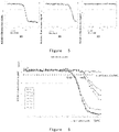
- FIG. 5 Each curve as shown in the figure is the fluorescence brightness curve of each detection point (sub-detection region) in each PCR cycle.
- Figure 5a shows the result of adding the target nucleic acid (10 5 copies) of the sample to be detected in the reaction. It can be seen that the fluorescence brightness of each detection point (3 in total) corresponding to the target to be detected gradually decreases with the amplification reaction progresses, indicating that the target to be tested is detected; the fluorescence intensity of the detection point crosses a set threshold near the 23rd cycle.
- Figure 5b shows the result of adding the target nucleic acid (500 copies) of the sample to be detected in the reaction. It can be seen that the fluorescence brightness of each detection point corresponding to the target to be detected (3 in total) gradually decreases with the progress of the amplification reaction, indicating that the target to be tested is detected; the fluorescence intensity of the detection point crosses the same set threshold around the 30th to 31st cycle.
- Figure 5c shows that the sample target to be detected is not added to the reaction. It can be seen that the fluorescence brightness of each detection point corresponding to the target to be detected (3 in total) has no obvious change with the progress of the amplification reaction, and has never exceeded the set threshold, indicating that the target to be tested is not detected.
- FIG. 6 is the verification experimental data of the multiplex reaction of the present invention for simultaneous real-time quantitative detection of multiple target sequences.
- Each curve shown in the figure is also the fluorescence brightness curve of each detection point (sub-detection region) in each PCR cycle.
- NTC No Template Control
- the first target is the pertussis target (2000copies) of the same experiment in Figure 5a .
- the fluorescence brightness of the detection point corresponding to the target TS1-A/B/C gradually decreased with the progress of the amplification reaction, indicating that the target to be tested is detected; the fluorescence intensity of the detection point has exceeded the set threshold around the 28th cycle.
- the reaction also contains 300 copies of the second target (TargetSubstrate2). It can be seen that the fluorescence brightness of each detection point (TS2-A/B/C) corresponding to the target to be tested gradually decreases with the progress of the amplification reaction.
- the same set thresholds are crossed around the 34th to 35th cycles.
- the results show that the method of the present invention can be used to detect the target nucleic acid to be detected, and the concentration of the target nucleic acid to be detected can be determined according to the fluorescence change speed of the detection point.
Landscapes
- Chemical & Material Sciences (AREA)
- Life Sciences & Earth Sciences (AREA)
- Organic Chemistry (AREA)
- Engineering & Computer Science (AREA)
- Zoology (AREA)
- Wood Science & Technology (AREA)
- Proteomics, Peptides & Aminoacids (AREA)
- Health & Medical Sciences (AREA)
- Biophysics (AREA)
- Chemical Kinetics & Catalysis (AREA)
- Immunology (AREA)
- Microbiology (AREA)
- Molecular Biology (AREA)
- Analytical Chemistry (AREA)
- Physics & Mathematics (AREA)
- Biotechnology (AREA)
- Biochemistry (AREA)
- Bioinformatics & Cheminformatics (AREA)
- General Engineering & Computer Science (AREA)
- General Health & Medical Sciences (AREA)
- Genetics & Genomics (AREA)
- Measuring Or Testing Involving Enzymes Or Micro-Organisms (AREA)
Applications Claiming Priority (2)
| Application Number | Priority Date | Filing Date | Title |
|---|---|---|---|
| CN201910701155.0A CN111621551B (zh) | 2019-07-31 | 2019-07-31 | 多重连接探针微阵列检测 |
| PCT/CN2020/103336 WO2021017955A1 (fr) | 2019-07-31 | 2020-07-21 | Détection de microréseau de sondes dépendant de la ligature multiplex |
Publications (3)
| Publication Number | Publication Date |
|---|---|
| EP4006165A1 true EP4006165A1 (fr) | 2022-06-01 |
| EP4006165A4 EP4006165A4 (fr) | 2023-10-04 |
| EP4006165B1 EP4006165B1 (fr) | 2026-04-29 |
Family
ID=72257793
Family Applications (1)
| Application Number | Title | Priority Date | Filing Date |
|---|---|---|---|
| EP20847306.6A Active EP4006165B1 (fr) | 2019-07-31 | 2020-07-21 | Détection de microréseau de sondes dépendant de la ligature multiplex |
Country Status (4)
| Country | Link |
|---|---|
| US (1) | US20220275435A1 (fr) |
| EP (1) | EP4006165B1 (fr) |
| CN (1) | CN111621551B (fr) |
| WO (1) | WO2021017955A1 (fr) |
Families Citing this family (6)
| Publication number | Priority date | Publication date | Assignee | Title |
|---|---|---|---|---|
| CN111621551B (zh) * | 2019-07-31 | 2021-10-26 | 深圳闪量科技有限公司 | 多重连接探针微阵列检测 |
| CN111394432B (zh) * | 2020-03-27 | 2023-03-24 | 深圳闪量科技有限公司 | 基于通用探针芯片的多重定量pcr检测系统 |
| CN114250224B (zh) * | 2021-12-16 | 2024-02-27 | 福建和瑞基因科技有限公司 | 一种用于提取或检测样本中小分子rna的核酸组合物及其试剂盒和方法 |
| CN114214393A (zh) * | 2021-12-30 | 2022-03-22 | 上海百傲科技股份有限公司 | 通用核酸微阵列芯片及检测方法 |
| CN114934102B (zh) * | 2022-03-08 | 2024-09-03 | 深圳闪量科技有限公司 | 基于二十重pcr的多种呼吸道病原体核酸同时检测用引物组及试剂盒 |
| CN116200542B (zh) * | 2023-01-06 | 2025-03-18 | 深圳闪量科技有限公司 | 同时检测犬多种病原体的引物池和试剂盒 |
Family Cites Families (15)
| Publication number | Priority date | Publication date | Assignee | Title |
|---|---|---|---|---|
| EP1130113A1 (fr) * | 2000-02-15 | 2001-09-05 | Johannes Petrus Schouten | Méthode d'amplification dépendant de ligatures multiples |
| US20030119004A1 (en) * | 2001-12-05 | 2003-06-26 | Wenz H. Michael | Methods for quantitating nucleic acids using coupled ligation and amplification |
| US20040110134A1 (en) * | 2002-12-05 | 2004-06-10 | Wenz H. Michael | Methods for quantitating nucleic acids using coupled ligation and amplification |
| US11001881B2 (en) * | 2006-08-24 | 2021-05-11 | California Institute Of Technology | Methods for detecting analytes |
| CN101586150B (zh) * | 2008-05-23 | 2016-09-28 | 陕西佰美基因股份有限公司 | 检测探针、通用寡核苷酸芯片及核酸检测方法及其用途 |
| US20100105032A1 (en) * | 2008-10-23 | 2010-04-29 | Tao Pan | Highly sensitive multiplex single nucleotide polymorphism and mutation detection using real time ligase chain reaction microarray |
| CA2757300C (fr) * | 2009-04-01 | 2018-01-09 | Dxterity Diagnostics Incorporated | Amplification de sonde dependante d'une ligature chimique |
| WO2012039529A1 (fr) * | 2010-09-20 | 2012-03-29 | Seegene, Inc. | Détection de séquences cibles d'acide nucléique par une activité exonucléolytique à l'aide de sondes immobilisées monomarquées sur une phase solide |
| CA2904181A1 (fr) * | 2013-03-13 | 2014-10-09 | Anahit Aghvanyan | Dosage immunologique sandwich comprenant un reactif d'ancrage |
| SG10201805453XA (en) * | 2013-04-25 | 2018-08-30 | Firefly Bioworks Inc | Multiplexed analysis of target nucleic acids |
| CN104357549A (zh) * | 2014-09-25 | 2015-02-18 | 徐州医学院 | 一种基于dna芯片的数字化定量检测核酸的方法 |
| CN108220399B (zh) * | 2016-12-14 | 2023-04-14 | 李保伟 | 一种基于通用探针技术的荧光定量pcr方法 |
| CN107267657A (zh) * | 2017-08-15 | 2017-10-20 | 江西贤聚景欣医药生物科技有限公司 | 检测靶核酸序列的引物、荧光探针及方法 |
| CN110016500A (zh) * | 2018-01-10 | 2019-07-16 | 深圳闪量科技有限公司 | 表面探针定量pcr方法 |
| CN111621551B (zh) * | 2019-07-31 | 2021-10-26 | 深圳闪量科技有限公司 | 多重连接探针微阵列检测 |
-
2019
- 2019-07-31 CN CN201910701155.0A patent/CN111621551B/zh active Active
-
2020
- 2020-07-21 US US17/631,558 patent/US20220275435A1/en active Pending
- 2020-07-21 WO PCT/CN2020/103336 patent/WO2021017955A1/fr not_active Ceased
- 2020-07-21 EP EP20847306.6A patent/EP4006165B1/fr active Active
Also Published As
| Publication number | Publication date |
|---|---|
| WO2021017955A1 (fr) | 2021-02-04 |
| CN111621551B (zh) | 2021-10-26 |
| US20220275435A1 (en) | 2022-09-01 |
| CN111621551A (zh) | 2020-09-04 |
| EP4006165B1 (fr) | 2026-04-29 |
| EP4006165A4 (fr) | 2023-10-04 |
Similar Documents
| Publication | Publication Date | Title |
|---|---|---|
| EP4006165B1 (fr) | Détection de microréseau de sondes dépendant de la ligature multiplex | |
| JP7210203B2 (ja) | リコンビナーゼポリメラーゼ増幅を多重化するための方法 | |
| US20120040853A1 (en) | Real time multiplex pcr detection on solid surfaces using double stranded nucleic acid specific dyes | |
| AU2006204087B2 (en) | Primer for nucleic acid detection | |
| EP2789689A1 (fr) | Amorces chimères avec des conformations en épingle à cheveux et leurs procédés d'utilisation | |
| KR19990063053A (ko) | 표적 핵산의 검출에 사용하기 위한 표지 프라이머 및 표적 핵산의 검출 | |
| KR101552669B1 (ko) | 오류 시그널이 배제되는 타겟 핵산서열의 실시간 멀티플렉싱 검출 | |
| US10287636B2 (en) | Method and rapid test for the detection of specific nucleic acid sequences | |
| EP4130294A1 (fr) | Système de test de pcr quantitative multiplex à base de puce de sonde universelle | |
| KR20120018734A (ko) | 헤파티티스 b 바이러스 검출용 키트 및 이를 이용한 헤파티티스 b 바이러스의 검출 방법 | |
| JP2023501219A (ja) | Lamp及びプローブ検出を使用する核酸検出法 | |
| US10370707B2 (en) | Multiplex probes | |
| JP5207355B2 (ja) | 標的ヌクレオチド配列を有する核酸の検出方法、プローブセット、及び核酸の識別方法 | |
| WO2003012142A1 (fr) | Detection d'acides nucleiques par acp en temps reel faisant intervenir des amorces chimeres arn-adn | |
| CN110016500A (zh) | 表面探针定量pcr方法 | |
| US7026120B2 (en) | Probes for detecting tumor cells | |
| CA2705852C (fr) | Amorce de transfert d'electrons photoinduit (pet) pour amplification d'acides nucleiques | |
| KR20250074714A (ko) | Rna를 이용한 폐암 변이 유전자의 대규모 다중 검출 방법 및 이를 이용한 폐암 변이 유전자의 대규모 다중 검출 키트 | |
| US20260117329A1 (en) | UTILIZATION OF dITP FOR PREFERENTIAL/SELECTIVE AMPLIFICATION OF RNA VERSUS DNA TARGETS BASED ON STRAND-SEPARATION TEMPERATURE |
Legal Events
| Date | Code | Title | Description |
|---|---|---|---|
| STAA | Information on the status of an ep patent application or granted ep patent |
Free format text: STATUS: THE INTERNATIONAL PUBLICATION HAS BEEN MADE |
|
| PUAI | Public reference made under article 153(3) epc to a published international application that has entered the european phase |
Free format text: ORIGINAL CODE: 0009012 |
|
| STAA | Information on the status of an ep patent application or granted ep patent |
Free format text: STATUS: REQUEST FOR EXAMINATION WAS MADE |
|
| 17P | Request for examination filed |
Effective date: 20220228 |
|
| AK | Designated contracting states |
Kind code of ref document: A1 Designated state(s): AL AT BE BG CH CY CZ DE DK EE ES FI FR GB GR HR HU IE IS IT LI LT LU LV MC MK MT NL NO PL PT RO RS SE SI SK SM TR |
|
| DAV | Request for validation of the european patent (deleted) | ||
| DAX | Request for extension of the european patent (deleted) | ||
| REG | Reference to a national code |
Ref country code: DE Ref country code: DE Ref legal event code: R079 Ref document number: 602020071302 Country of ref document: DE Free format text: PREVIOUS MAIN CLASS: C12Q0001680000 Ipc: C12Q0001685100 |
|
| A4 | Supplementary search report drawn up and despatched |
Effective date: 20230831 |
|
| RIC1 | Information provided on ipc code assigned before grant |
Ipc: C12Q 1/6851 20180101AFI20230825BHEP |
|
| RIC1 | Information provided on ipc code assigned before grant |
Ipc: C12Q 1/6851 20180101AFI20230912BHEP |
|
| GRAP | Despatch of communication of intention to grant a patent |
Free format text: ORIGINAL CODE: EPIDOSNIGR1 |
|
| STAA | Information on the status of an ep patent application or granted ep patent |
Free format text: STATUS: GRANT OF PATENT IS INTENDED |
|
| INTG | Intention to grant announced |
Effective date: 20251125 |
|
| GRAS | Grant fee paid |
Free format text: ORIGINAL CODE: EPIDOSNIGR3 |
|
| GRAA | (expected) grant |
Free format text: ORIGINAL CODE: 0009210 |
|
| STAA | Information on the status of an ep patent application or granted ep patent |
Free format text: STATUS: THE PATENT HAS BEEN GRANTED |
|
| AK | Designated contracting states |
Kind code of ref document: B1 Designated state(s): AL AT BE BG CH CY CZ DE DK EE ES FI FR GB GR HR HU IE IS IT LI LT LU LV MC MK MT NL NO PL PT RO RS SE SI SK SM TR |
|
| REG | Reference to a national code |
Ref country code: CH Ref legal event code: F10 Free format text: ST27 STATUS EVENT CODE: U-0-0-F10-F00 (AS PROVIDED BY THE NATIONAL OFFICE) Effective date: 20260429 |