EP4009283A1 - Reconstruction de géométrie enregistrée basée sur un instantané fluoroscopique constant - Google Patents
Reconstruction de géométrie enregistrée basée sur un instantané fluoroscopique constant Download PDFInfo
- Publication number
- EP4009283A1 EP4009283A1 EP21210115.8A EP21210115A EP4009283A1 EP 4009283 A1 EP4009283 A1 EP 4009283A1 EP 21210115 A EP21210115 A EP 21210115A EP 4009283 A1 EP4009283 A1 EP 4009283A1
- Authority
- EP
- European Patent Office
- Prior art keywords
- coordinates
- fluoroscopic images
- neural network
- artificial neural
- anatomical
- Prior art date
- Legal status (The legal status is an assumption and is not a legal conclusion. Google has not performed a legal analysis and makes no representation as to the accuracy of the status listed.)
- Granted
Links
Images



Classifications
-
- A—HUMAN NECESSITIES
- A61—MEDICAL OR VETERINARY SCIENCE; HYGIENE
- A61B—DIAGNOSIS; SURGERY; IDENTIFICATION
- A61B5/00—Measuring for diagnostic purposes; Identification of persons
- A61B5/0059—Measuring for diagnostic purposes; Identification of persons using light, e.g. diagnosis by transillumination, diascopy, fluorescence
- A61B5/0071—Measuring for diagnostic purposes; Identification of persons using light, e.g. diagnosis by transillumination, diascopy, fluorescence by measuring fluorescence emission
-
- G—PHYSICS
- G06—COMPUTING OR CALCULATING; COUNTING
- G06T—IMAGE DATA PROCESSING OR GENERATION, IN GENERAL
- G06T17/00—Three-dimensional [3D] modelling for computer graphics
- G06T17/20—Finite element generation, e.g. wire-frame surface description, tesselation
-
- G—PHYSICS
- G06—COMPUTING OR CALCULATING; COUNTING
- G06T—IMAGE DATA PROCESSING OR GENERATION, IN GENERAL
- G06T17/00—Three-dimensional [3D] modelling for computer graphics
-
- G—PHYSICS
- G06—COMPUTING OR CALCULATING; COUNTING
- G06F—ELECTRIC DIGITAL DATA PROCESSING
- G06F30/00—Computer-aided design [CAD]
-
- A—HUMAN NECESSITIES
- A61—MEDICAL OR VETERINARY SCIENCE; HYGIENE
- A61B—DIAGNOSIS; SURGERY; IDENTIFICATION
- A61B18/00—Surgical instruments, devices or methods for transferring non-mechanical forms of energy to or from the body
-
- A—HUMAN NECESSITIES
- A61—MEDICAL OR VETERINARY SCIENCE; HYGIENE
- A61B—DIAGNOSIS; SURGERY; IDENTIFICATION
- A61B5/00—Measuring for diagnostic purposes; Identification of persons
- A61B5/24—Detecting, measuring or recording bioelectric or biomagnetic signals of the body or parts thereof
- A61B5/25—Bioelectric electrodes therefor
- A61B5/279—Bioelectric electrodes therefor specially adapted for particular uses
- A61B5/28—Bioelectric electrodes therefor specially adapted for particular uses for electrocardiography [ECG]
- A61B5/283—Invasive
-
- A—HUMAN NECESSITIES
- A61—MEDICAL OR VETERINARY SCIENCE; HYGIENE
- A61B—DIAGNOSIS; SURGERY; IDENTIFICATION
- A61B5/00—Measuring for diagnostic purposes; Identification of persons
- A61B5/24—Detecting, measuring or recording bioelectric or biomagnetic signals of the body or parts thereof
- A61B5/316—Modalities, i.e. specific diagnostic methods
- A61B5/318—Heart-related electrical modalities, e.g. electrocardiography [ECG]
- A61B5/367—Electrophysiological study [EPS], e.g. electrical activation mapping or electro-anatomical mapping
-
- A—HUMAN NECESSITIES
- A61—MEDICAL OR VETERINARY SCIENCE; HYGIENE
- A61B—DIAGNOSIS; SURGERY; IDENTIFICATION
- A61B5/00—Measuring for diagnostic purposes; Identification of persons
- A61B5/72—Signal processing specially adapted for physiological signals or for diagnostic purposes
- A61B5/7235—Details of waveform analysis
- A61B5/7264—Classification of physiological signals or data, e.g. using neural networks, statistical classifiers, expert systems or fuzzy systems
-
- A—HUMAN NECESSITIES
- A61—MEDICAL OR VETERINARY SCIENCE; HYGIENE
- A61B—DIAGNOSIS; SURGERY; IDENTIFICATION
- A61B5/00—Measuring for diagnostic purposes; Identification of persons
- A61B5/74—Details of notification to user or communication with user or patient; User input means
- A61B5/742—Details of notification to user or communication with user or patient; User input means using visual displays
-
- A—HUMAN NECESSITIES
- A61—MEDICAL OR VETERINARY SCIENCE; HYGIENE
- A61B—DIAGNOSIS; SURGERY; IDENTIFICATION
- A61B6/00—Apparatus or devices for radiation diagnosis; Apparatus or devices for radiation diagnosis combined with radiation therapy equipment
- A61B6/48—Diagnostic techniques
- A61B6/486—Diagnostic techniques involving generating temporal series of image data
- A61B6/487—Diagnostic techniques involving generating temporal series of image data involving fluoroscopy
-
- A—HUMAN NECESSITIES
- A61—MEDICAL OR VETERINARY SCIENCE; HYGIENE
- A61B—DIAGNOSIS; SURGERY; IDENTIFICATION
- A61B6/00—Apparatus or devices for radiation diagnosis; Apparatus or devices for radiation diagnosis combined with radiation therapy equipment
- A61B6/52—Devices using data or image processing specially adapted for radiation diagnosis
- A61B6/5294—Devices using data or image processing specially adapted for radiation diagnosis involving using additional data, e.g. patient information, image labeling, acquisition parameters
-
- G—PHYSICS
- G06—COMPUTING OR CALCULATING; COUNTING
- G06N—COMPUTING ARRANGEMENTS BASED ON SPECIFIC COMPUTATIONAL MODELS
- G06N3/00—Computing arrangements based on biological models
- G06N3/02—Neural networks
- G06N3/04—Architecture, e.g. interconnection topology
- G06N3/0464—Convolutional networks [CNN, ConvNet]
-
- G—PHYSICS
- G06—COMPUTING OR CALCULATING; COUNTING
- G06N—COMPUTING ARRANGEMENTS BASED ON SPECIFIC COMPUTATIONAL MODELS
- G06N3/00—Computing arrangements based on biological models
- G06N3/02—Neural networks
- G06N3/08—Learning methods
-
- G—PHYSICS
- G06—COMPUTING OR CALCULATING; COUNTING
- G06N—COMPUTING ARRANGEMENTS BASED ON SPECIFIC COMPUTATIONAL MODELS
- G06N3/00—Computing arrangements based on biological models
- G06N3/02—Neural networks
- G06N3/08—Learning methods
- G06N3/09—Supervised learning
-
- G—PHYSICS
- G06—COMPUTING OR CALCULATING; COUNTING
- G06T—IMAGE DATA PROCESSING OR GENERATION, IN GENERAL
- G06T7/00—Image analysis
- G06T7/70—Determining position or orientation of objects or cameras
- G06T7/73—Determining position or orientation of objects or cameras using feature-based methods
- G06T7/74—Determining position or orientation of objects or cameras using feature-based methods involving reference images or patches
-
- G—PHYSICS
- G16—INFORMATION AND COMMUNICATION TECHNOLOGY [ICT] SPECIALLY ADAPTED FOR SPECIFIC APPLICATION FIELDS
- G16H—HEALTHCARE INFORMATICS, i.e. INFORMATION AND COMMUNICATION TECHNOLOGY [ICT] SPECIALLY ADAPTED FOR THE HANDLING OR PROCESSING OF MEDICAL OR HEALTHCARE DATA
- G16H30/00—ICT specially adapted for the handling or processing of medical images
- G16H30/40—ICT specially adapted for the handling or processing of medical images for processing medical images, e.g. editing
-
- A—HUMAN NECESSITIES
- A61—MEDICAL OR VETERINARY SCIENCE; HYGIENE
- A61B—DIAGNOSIS; SURGERY; IDENTIFICATION
- A61B18/00—Surgical instruments, devices or methods for transferring non-mechanical forms of energy to or from the body
- A61B2018/00315—Surgical instruments, devices or methods for transferring non-mechanical forms of energy to or from the body for treatment of particular body parts
- A61B2018/00345—Vascular system
- A61B2018/00351—Heart
-
- A—HUMAN NECESSITIES
- A61—MEDICAL OR VETERINARY SCIENCE; HYGIENE
- A61B—DIAGNOSIS; SURGERY; IDENTIFICATION
- A61B18/00—Surgical instruments, devices or methods for transferring non-mechanical forms of energy to or from the body
- A61B2018/00571—Surgical instruments, devices or methods for transferring non-mechanical forms of energy to or from the body for achieving a particular surgical effect
- A61B2018/00577—Ablation
-
- G—PHYSICS
- G06—COMPUTING OR CALCULATING; COUNTING
- G06T—IMAGE DATA PROCESSING OR GENERATION, IN GENERAL
- G06T2200/00—Indexing scheme for image data processing or generation, in general
- G06T2200/08—Indexing scheme for image data processing or generation, in general involving all processing steps from image acquisition to 3D model generation
-
- G—PHYSICS
- G06—COMPUTING OR CALCULATING; COUNTING
- G06T—IMAGE DATA PROCESSING OR GENERATION, IN GENERAL
- G06T2207/00—Indexing scheme for image analysis or image enhancement
- G06T2207/10—Image acquisition modality
- G06T2207/10064—Fluorescence image
-
- G—PHYSICS
- G06—COMPUTING OR CALCULATING; COUNTING
- G06T—IMAGE DATA PROCESSING OR GENERATION, IN GENERAL
- G06T2207/00—Indexing scheme for image analysis or image enhancement
- G06T2207/20—Special algorithmic details
- G06T2207/20081—Training; Learning
-
- G—PHYSICS
- G06—COMPUTING OR CALCULATING; COUNTING
- G06T—IMAGE DATA PROCESSING OR GENERATION, IN GENERAL
- G06T2207/00—Indexing scheme for image analysis or image enhancement
- G06T2207/20—Special algorithmic details
- G06T2207/20084—Artificial neural networks [ANN]
-
- G—PHYSICS
- G06—COMPUTING OR CALCULATING; COUNTING
- G06T—IMAGE DATA PROCESSING OR GENERATION, IN GENERAL
- G06T2207/00—Indexing scheme for image analysis or image enhancement
- G06T2207/30—Subject of image; Context of image processing
- G06T2207/30004—Biomedical image processing
- G06T2207/30048—Heart; Cardiac
-
- G—PHYSICS
- G06—COMPUTING OR CALCULATING; COUNTING
- G06T—IMAGE DATA PROCESSING OR GENERATION, IN GENERAL
- G06T2210/00—Indexing scheme for image generation or computer graphics
- G06T2210/41—Medical
-
- G—PHYSICS
- G06—COMPUTING OR CALCULATING; COUNTING
- G06T—IMAGE DATA PROCESSING OR GENERATION, IN GENERAL
- G06T2210/00—Indexing scheme for image generation or computer graphics
- G06T2210/56—Particle system, point based geometry or rendering
Definitions
- the present invention relates to medical devices, and in particular, but not exclusively to, generating anatomical maps.
- Magnetic location sensing is one of the methods known in the art.
- magnetic location sensing magnetic field generators are typically placed at known locations external to the patient.
- a magnetic field sensor within the distal end of the probe generates electrical signals in response to these magnetic fields, which are processed to determine the coordinate locations of the distal end of the probe.
- Cardiac arrhythmias and atrial fibrillation in particular, persist as common and dangerous medical ailments, especially in the aging population.
- Diagnosis and treatment of cardiac arrhythmias include mapping the electrical properties of heart tissue, especially the endocardium, and selectively ablating cardiac tissue by application of energy. Such ablation can cease or modify the propagation of unwanted electrical signals from one portion of the heart to another. The ablation process destroys the unwanted electrical pathways by formation of non-conducting lesions.
- Various energy delivery modalities have been disclosed for forming lesions, and include use of microwave, laser and more commonly, radiofrequency energies to create conduction blocks along the cardiac tissue wall.
- mapping followed by ablation electrical activity at points within the heart is typically sensed and measured by advancing a catheter containing one or more electrical sensors into the heart, and acquiring data at a multiplicity of points. These data are then utilized to select the endocardial target areas at which the ablation is to be performed.
- Electrode catheters have been in common use in medical practice for many years. They are used to stimulate and map electrical activity in the heart and to ablate sites of aberrant electrical activity. In use, the electrode catheter is inserted into a major vein or artery, e.g., femoral vein, and then guided into the chamber of the heart of concern.
- a typical ablation procedure involves the insertion of a catheter having a one or more electrodes at its distal end into a heart chamber.
- a reference electrode may be provided, generally taped to the skin of the patient or by means of a second catheter that is positioned in or near the heart.
- RF (radio frequency) current is applied between the catheter electrode(s) of the ablating catheter and an indifferent electrode (which may be one of the catheter electrodes), and current flows through the media between the electrodes, i.e., blood and tissue.
- the distribution of current may depend on the amount of electrode surface in contact with the tissue as compared to blood, which has a higher conductivity than the tissue. Heating of the tissue occurs due to its electrical resistance. The tissue is heated sufficiently to cause cellular destruction in the cardiac tissue resulting in formation of a lesion within the cardiac tissue which is electrically non-conductive. In some applications, irreversible electroporation may be performed to ablate the tissue.
- Electrode sensors in a cardiac chamber may detect far-field electrical activity, i.e., the ambient electrical activity originating away from the sensors, which can distort or obscure local electrical activity, i.e., signals originating at or near the sensors.
- far-field electrical activity i.e., the ambient electrical activity originating away from the sensors
- local electrical activity i.e., signals originating at or near the sensors.
- Commonly assigned U.S. Patent Application Publication No. 2014/0005664 of Govari et al. discloses distinguishing a local component in an intracardiac electrode signal, due to the tissue with which the electrode is in contact from a remote-field contribution to the signal, and explains that a therapeutic procedure applied to the tissue can be controlled responsively to the distinguished local component.
- a method for generating a three-dimensional (3D) anatomical map including applying a trained artificial neural network to (a) a set of two-dimensional (2D) fluoroscopic images of a body part of a living subject, and (b) respective first 3D coordinates of the set of 2D fluoroscopic images, yielding second 3D coordinates defining a 3D anatomical map, and rendering to a display the 3D anatomical map responsively to the second 3D coordinates.
- the set of 2D fluoroscopic images includes only two 2D fluoroscopic images.
- the set of 2D fluoroscopic images includes an anterior-posterior projection of the body part and a left anterior-oblique projection of the body part.
- the second 3D coordinates include one of more of the following mesh vertices of a 3D mesh, and a 3D point cloud.
- the method includes improving the 3D anatomical map responsively to signals received from electrodes of a catheter inserted into the body part of the living subject.
- first 3D coordinates and the second 3D coordinates are in a same coordinate space.
- the method includes training the artificial neural network to generate 3D anatomical maps responsively to training data including multiple sets of 2D fluoroscopic images of respective body parts of respective living subjects, respective 3D coordinates of the multiple sets of 2D fluoroscopic images, and respective 3D coordinates of multiple 3D anatomical maps of the respective body parts of the respective living subjects.
- the method includes inputting into the artificial neural network the multiple sets of 2D fluoroscopic images of respective body parts of respective living subjects and the respective 3D coordinates of the multiple sets of 2D fluoroscopic images, and iteratively adjusting parameters of the artificial neural network to reduce a difference between an output of the artificial neural network and a desired output, which includes the respective 3D coordinates of the multiple 3D anatomical maps.
- the method includes generating the multiple 3D anatomical maps of the training data responsively to signals received from electrodes of at least one catheter inserted into the body parts of the respective living subjects.
- each of the multiple sets of 2D fluoroscopic images includes only two 2D fluoroscopic images.
- the multiple sets of 2D fluoroscopic images include respective anterior-posterior projections and respective left anterior-oblique projections of the respective body parts.
- a medical system including a fluoroscopic imaging device configured to capture a set of two-dimensional (2D) fluoroscopic images of a body part of a living subject, a display, and processing circuitry configured to apply a trained artificial neural network to (a) a set of two-dimensional (2D) fluoroscopic images of a body part of a living subject, and (b) respective first 3D coordinates of the set of 2D fluoroscopic images, yielding second 3D coordinates of a 3D anatomical map, and render to the display the 3D anatomical map responsively to the second 3D coordinates.
- a fluoroscopic imaging device configured to capture a set of two-dimensional (2D) fluoroscopic images of a body part of a living subject
- processing circuitry configured to apply a trained artificial neural network to (a) a set of two-dimensional (2D) fluoroscopic images of a body part of a living subject, and (b) respective first 3D coordinates of the set of 2D fluoroscopic images, yielding second 3D coordinates of
- the set of 2D fluoroscopic images includes only two 2D fluoroscopic images.
- the set of 2D fluoroscopic images includes an anterior-posterior projection of the body part and a left anterior-oblique projection of the body part.
- the second 3D coordinates include one of more of the following mesh vertices of a 3D mesh, and a 3D point cloud.
- the system includes a catheter including electrodes and configured to be inserted into the body part of the living subject, the processing circuitry being configured to improve the 3D anatomical map responsively to signals received from the electrodes of the catheter.
- first 3D coordinates and the second 3D coordinates are in a same coordinate space.
- the fluoroscopic imaging device is configured to capture multiple sets of two-dimensional (2D) fluoroscopic images of respective body parts of respective living subjects
- the processing circuitry is configured to train the artificial neural network to generate 3D anatomical maps responsively to training data including the multiple sets of 2D fluoroscopic images of respective body parts of respective living subjects, respective 3D coordinates of the multiple sets of 2D fluoroscopic images, and respective 3D coordinates of multiple 3D anatomical maps of the respective body parts of the respective living subjects.
- the processing circuitry is configured to input into the artificial neural network the multiple sets of 2D fluoroscopic images of respective body parts of respective living subjects and the respective 3D coordinates of the multiple sets of 2D fluoroscopic images, and iteratively adjust parameters of the artificial neural network to reduce a difference between an output of the artificial neural network and a desired output, which includes the respective 3D coordinates of the multiple 3D anatomical maps.
- the system includes at least one catheter including electrodes and configured to be inserted into the body parts of the respective living subjects, the processing circuitry being configured to generate the multiple 3D anatomical maps of the training data responsively to signals received from the electrodes of the at least one catheter inserted into the body parts of the respective living subjects.
- each of the multiple sets of 2D fluoroscopic images includes only two 2D fluoroscopic images.
- the multiple sets of 2D fluoroscopic images include respective anterior-posterior projections and respective left anterior-oblique projections of the respective body parts.
- a software product including a non-transient computer-readable medium in which program instructions are stored, which instructions, when read by a central processing unit (CPU), cause the CPU to apply a trained artificial neural network to (a) a set of two-dimensional (2D) fluoroscopic images of a body part of a living subject, and (b) respective first 3D coordinates of the set of 2D fluoroscopic images, yielding second 3D coordinates of a 3D anatomical map, and render to a display the 3D anatomical map responsively to the second 3D coordinates.
- a trained artificial neural network to (a) a set of two-dimensional (2D) fluoroscopic images of a body part of a living subject, and (b) respective first 3D coordinates of the set of 2D fluoroscopic images, yielding second 3D coordinates of a 3D anatomical map, and render to a display the 3D anatomical map responsively to the second 3D coordinates.
- fluoroscopy Prior to the advent of mapping heart chambers using catheters, physicians would exclusively use fluoroscopy to guide catheters during procedures.
- the main disadvantage of fluoroscopy is the danger of radiation.
- fluoroscopy does have some advantages in that it provides a view of the various veins and the beating of the heart.
- catheter-based mapping is generally used to map heart chambers based on moving the catheter around a heart chamber to generate a three-dimensional (3D) anatomical map of the heart.
- Magnetic and/or impedance-based location tracking may be used to track the catheter without using fluoroscopy. Nevertheless, fluoroscopy may be used, for example, to introduce a sheath correctly.
- fluoroscopic images may be registered with the magnetic and/or impedance-based location tracking and rendered to a display along with the generated 3D map.
- mapping with a catheter may miss certain features such as veins, which are seen with fluoroscopy. For example, some people may have three pulmonary veins and others may have four. The physician could simply miss the fourth vein while moving the catheter and create an anatomical map without the fourth vein.
- Embodiments of the present invention solve the above problems by generating an initial 3D anatomical map from two two-dimensional (2D) fluoroscopic images (e.g., an anterior-posterior (AP) and a left anterior-oblique (LAO) projection or any suitable pair of fluoroscopic image projections) of a body part (e.g., heart chamber(s)) by using a trained artificial neural network (ANN).
- the initial 3D anatomical map may then be refined based on catheter-based mapping by moving a catheter around the body part (e.g., the heart chamber(s)).
- the ANN is trained to generate a 3D anatomical map from two 2D fluoroscopic images (e.g., an anterior-posterior (AP) and a left anterior-oblique (LAO) projection or any suitable pair of fluoroscopic image projections) and respective 3D coordinates of the 2D fluoroscopic images based on training data.
- the training data includes: (a) sets of 2D fluoroscopic images and respective 3D coordinates of the 2D fluoroscopic images from respective electrophysiological (EP) procedures as input to the ANN; and (b) carefully acquired catheter-based 3D anatomical maps from the respective EP procedures as the desired output of the ANN.
- parameters e.g., weights
- the ANN are amended so that the output of the ANN approaches the desired output within given limitations.
- the coordinates of the fluoroscopic images (used during training and application of the ANN) are generally registered with the magnetic and/or impedance-based location tracking system so that the 3D anatomic maps generated by the ANN may be rendered to a display according to the known registration.
- the registration i.e., the coordinates of the fluoroscopic images and the corresponding coordinates of the 3D anatomical maps) therefore forms part of the training process.
- the ANN receives two 2D fluoroscopic images (e.g., anterior-posterior (AP), left anterior-oblique (LAO) or any suitable pair of fluoroscopic image projections) and respective 3D coordinates of the two 2D fluoroscopic images as input, and outputs a 3D anatomical map with coordinates in the coordinate system of the magnetic and/or impedance-based location tracking system.
- the 3D anatomical map may be represented by mesh vertices of a 3D mesh or a 3D point cloud.
- the 3D point cloud may be used to generate an encapsulation mesh, for example, using a suitable algorithm such as "Marching cubes", which is a computer graphics algorithm, published in the 1987 SIGGRAPH proceedings by Lorensen and Cline, for extracting a polygonal mesh of an isosurface from a three-dimensional discrete scalar field.
- a suitable algorithm such as "Marching cubes", which is a computer graphics algorithm, published in the 1987 SIGGRAPH proceedings by Lorensen and Cline, for extracting a polygonal mesh of an isosurface from a three-dimensional discrete scalar field.
- Fig. 1 is a schematic view of a medical system 10 constructed and operative in accordance with an exemplary embodiment of the present invention.
- the medical system 10 is configured for performing catheterization procedures on a heart 12 of a living subject, which is constructed and operative in accordance with a disclosed embodiment of the invention.
- the system comprises a catheter 14, which is percutaneously inserted by an operator 16 through the patient's vascular system into a chamber or vascular structure of the heart 12.
- the operator 16 who is typically a physician, brings the catheter's distal end 18 into contact with the heart wall at an ablation target site.
- anatomic positional information i.e., of the distal end 18 of the catheter 14, and other functional images may then be prepared using processing circuitry 22 located in a console 24, according to any suitable methods, for example, the methods disclosed in U.S. Patent Nos. 6,226,542 , 6,301,496 , and 6,892,091 .
- One commercial product embodying elements of the system 10 is available as the CARTO ® 3 System, available from Biosense Webster, Inc., Irvine, CA, USA, which is capable of producing electroanatomic maps of the heart as required for the ablation. This system may be modified by those skilled in the art to embody the principles of embodiments of the present invention described herein.
- Areas determined to be abnormal can be ablated by application of thermal energy, e.g., by passage of radiofrequency electrical current through wires in the catheter 14 (or another catheter) to one or more electrodes 21 (only some labeled for the sake of simplicity) at the distal end 18 which apply the radiofrequency energy to the myocardium of the heart 12.
- the energy is absorbed in the tissue, heating (or cooling) it to a point (typically about 60 degrees Celsius) at which it permanently loses its electrical excitability.
- this procedure creates non-conducting lesions in the cardiac tissue, which disrupt the abnormal electrical pathway causing the arrhythmia.
- the principles of the invention can be applied to different heart chambers to treat many different cardiac arrhythmias.
- the catheter 14 typically comprises a handle 20, having suitable controls on the handle to enable the operator 16 to steer, position and orient the distal end 18 of the catheter 14 as desired for mapping and ablation.
- the distal portion of the catheter 14 contains position sensors (not shown) that provide signals to the processing circuitry 22, which computes a position of the distal end 18.
- Ablation energy and electrical signals can be conveyed to and from the heart 12 via a cable 34 to the console 24.
- Pacing signals and other control signals may be conveyed from the console 24 through the cable 34 and the electrodes 21 to the heart 12.
- Wire connections 35 link the console 24 with body surface electrodes 30 and other components of a positioning sub-system.
- the electrodes 21 and the body surface electrodes 30 may be used to measure tissue impedance at the ablation site as taught in U.S. Patent No. 7,536,218 .
- the console 24 typically contains one or more ablation power generators 25.
- the catheter 14 may be adapted to conduct ablative energy to the heart using any known ablation technique, e.g., radiofrequency energy, irreversible electroporation, ultra-sound energy, freezing technique and laser-produced light energy. Such methods are disclosed in U.S. Patent Nos. 6,814,733 , 6,997,924 , and 7,156,816 .
- the processing circuitry 22 may be an element of the positioning sub-system in the system 10 that measures location and orientation coordinates of the catheter 14.
- the positioning sub-system comprises a magnetic position tracking arrangement that determines the position and orientation of the catheter 14 by generating magnetic fields in a predefined working volume and sensing these fields at the catheter 14, using field generating coils 28.
- the positioning sub-system may employ impedance measurement, as taught, for example in U.S. Patent No. 7,756,576 , and U.S. Patent No. 7,536,218 .
- a fluoroscopic imaging device 37 has a C-arm 39, an x-ray source 41, an image intensifier module 43 and an adjustable collimator 45.
- a control processor (not shown), which may be located in the console 24, allows an operator to control the operation of the fluoroscopic imaging device 37, for example by setting imaging parameters, and controlling the collimator 45 to adjust the size and position of the field of view.
- the control processor may communicate with the fluoroscopic imaging device 37 via a cable 51 to enable and disable the x-ray source 41 or restrict its emissions to a desired region of interest by controlling the collimator 45, and to acquire image data from the image intensifier module 43.
- An optional display monitor 49 linked to the control processor, allows the operator 16 to view images produced by the fluoroscopic imaging device 37. When the display monitor 49 is not included, the fluoroscopic images may be viewed on a display 29, either via a split screen or in alternation with other non-fluoroscopic images.
- the catheter 14 is coupled to the console 24, which enables the operator 16 to observe and regulate the functions of the catheter 14.
- the processing circuitry 22 is typically a computer with appropriate signal processing circuits.
- the processing circuitry 22 is coupled to drive the display 29.
- the signal processing circuits typically receive, amplify, filter and digitize signals from the catheter 14, including signals generated by the above-noted sensors and the electrodes 21 located distally in the catheter 14.
- the digitized signals are received and used by the console 24 and the positioning sub-system to compute the position and orientation of the catheter 14, analyze the electrical signals from the electrodes 21, and generate desired electroanatomic maps.
- the system 10 includes other elements, which are not shown in the figures for the sake of simplicity.
- the system 10 may include an electrocardiogram (ECG) monitor, coupled to receive signals from one or more body surface electrodes, to provide an ECG synchronization signal to the console 24.
- ECG electrocardiogram
- the system 10 typically also includes a reference position sensor, either on an externally-applied reference patch attached to the exterior of the subject's body, or on an internally-placed catheter, which is inserted into the heart 12 maintained in a fixed position relative to the heart 12. Conventional pumps and lines for circulating liquids through the catheter 14 for cooling the ablation site are provided.
- the fluoroscopic imaging device 37 is generally registered with a coordinate space 31 of the positioning sub-system associated with the field generating coils 28 and the distal end 18 of the catheter 14. Therefore, images captured by the fluoroscopic imaging device 37 can be used with the positioning sub-system. For example, a representation of the distal end 18 of the catheter 14 may be rendered to the display 29 superimposed over an x-ray image captured by the fluoroscopic imaging device 37.
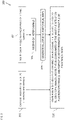
- Fig. 2 is a flow chart 100 including steps in a method of training an artificial neural network 52 for use in the system 10 of Fig. 1 .
- Fig. 3 is a schematic illustration of the artificial neural network 52 being trained in the system 10 of Fig. 1 .
- the fluoroscopic imaging device 37 ( Fig. 1 ) is configured to capture (block 102) multiple sets of 2D fluoroscopic images 54 of respective body parts 56 of respective living subjects 58.
- Each of the multiple sets of 2D fluoroscopic images 54 is associated with 3D coordinates 60 of the images.
- each 2D fluoroscopic image may include coordinates identifying at least two given points in the image (e.g., two corners of the image, or a corner and the center of the image, or any other suitable points) within a coordinate space.
- each 2D fluoroscopic image may include coordinates identifying one given point in the image and an orientation of the image in the coordinate space. The coordinates may be supplied by the controller of the fluoroscopic imaging device 37 with reference to the coordinate space 31 ( Fig.
- any other coordinate space which is registered with the coordinate space 31 If the fluoroscopic imaging device 37 is fixed with respect to the coordinate space 31, the 3D coordinates of the multiple sets of 2D fluoroscopic images 54 do not need to be supplied by the controller of the fluoroscopic imaging device 37 for each of the multiple sets of 2D fluoroscopic images 54 as the 3D coordinates of the multiple sets of 2D fluoroscopic images 54 are known to the medical system 10.
- each of the multiple sets of 2D fluoroscopic images 54 includes only two 2D fluoroscopic images.
- the two 2D fluoroscopic images are generally of orthogonal projections of the body parts 56.
- the multiple sets of 2D fluoroscopic images 54 include respective anterior-posterior projections and respective left anterior-oblique projections of the respective body parts 56 as shown in Fig. 3 .
- At least one catheter (for example, the catheter 14) comprises electrodes (e.g., the electrodes 21) and is configured to be inserted (block 104) into the body parts 56 of the respective living subjects 58.
- the processing circuitry 22 is configured to receive (block 106) signals from the electrodes (e.g., electrodes 21) of the catheter(s) (e.g., catheter 14). The catheter(s) is moved carefully around the body parts 56 to ensure that the maps generated from the catheter(s) movements are accurate.
- the processing circuitry 22 ( Fig.
- the multiple 3D anatomical maps 62 may be defined with reference to respective 3D coordinates 64 that may include mesh vertices of a 3D mesh and/or a 3D point cloud.
- the processing circuitry 22 is configured to train (block 110) the artificial neural network 52 to generate 3D anatomical maps responsively to training data comprising: (a) the multiple sets of 2D fluoroscopic images 54 of respective body parts 56 of respective living subjects 58 (captured by the fluoroscopic imaging device 37); (b) the respective 3D coordinates 60 of the multiple sets of 2D fluoroscopic images 54; and (c) respective 3D coordinates 64 of multiple 3D anatomical maps 62 of the respective body parts 56 of the respective living subjects 58.
- Each set of 2D fluoroscopic images 54 and the respective 3D coordinates 60 of that set of 2D fluoroscopic images 54 has an associated 3D anatomical map 62 (with corresponding 3D coordinates 64) which was captured for a respective body part 56 of a respective living subject 58.
- the training data includes one set of 2D fluoroscopic images 54, respective 3D coordinates 60 of that set of 2D fluoroscopic images 54, and 3D coordinates 64 of one of the multiple 3D anatomical maps 62 for the body part of each of the living subjects 58.
- the body parts 56 may include any suitable body part, for example, a chamber of a heart.
- the body parts 56 used in training the artificial neural network 52 are the same type of body part, e.g., chambers of hearts.
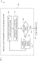
- Fig. 4 is a flow chart including detailed sub-steps in the step of block 110 of Fig. 2 . Reference is also made to Fig. 3 .
- a neural network is a network or circuit of neurons, or in a modern sense, an artificial neural network, composed of artificial neurons or nodes.
- the connections of the biological neuron are modeled as weights.
- a positive weight reflects an excitatory connection, while negative values mean inhibitory connections.
- Inputs are modified by a weight and summed using a linear combination.
- An activation function may control the amplitude of the output. For example, an acceptable range of output is usually between 0 and 1, or it could be -1 and 1.
- These artificial networks may be used for predictive modeling, adaptive control and applications and can be trained via a dataset. Self-learning resulting from experience can occur within networks, which can derive conclusions from a complex and seemingly unrelated set of information.
- a biological neural network is composed of a group or groups of chemically connected or functionally associated neurons.
- a single neuron may be connected to many other neurons and the total number of neurons and connections in a network may be extensive. Connections, called synapses, are usually formed from axons to dendrites, though dendrodendritic synapses and other connections are possible.
- connections called synapses, are usually formed from axons to dendrites, though dendrodendritic synapses and other connections are possible.
- Artificial intelligence, cognitive modeling, and neural networks are information processing paradigms inspired by the way biological neural systems process data. Artificial intelligence and cognitive modeling try to simulate some properties of biological neural networks. In the artificial intelligence field, artificial neural networks have been applied successfully to speech recognition, image analysis and adaptive control, in order to construct software agents (in computer and video games) or autonomous robots.
- a neural network in the case of artificial neurons called artificial neural network (ANN) or simulated neural network (SNN), is an interconnected group of natural or artificial neurons that uses a mathematical or computational model for information processing based on a connectionistic approach to computation.
- an ANN is an adaptive system that changes its structure based on external or internal information that flows through the network.
- neural networks are non-linear statistical data modeling or decision-making tools. They can be used to model complex relationships between inputs and outputs or to find patterns in data.
- the artificial neural network 52 includes a fully connected neural network, e.g., a convolutional neural network.
- the artificial neural network 52 may comprise any suitable ANN.
- the artificial neural network 52 may comprise software executed by the processing circuitry 22 ( Fig. 1 ) and/or hardware modules configured to perform the functions of the artificial neural network 52.
- the artificial neural network 52 includes an input layer 80 into which an input is received, and one or more hidden layers 82 which progressively process the input to an output layer 84 from which the output of the artificial neural network 52 is provided.
- the artificial neural network 52 may include layer weights between the layers 80, 82, 84 of the artificial neural network 52.
- the artificial neural network 52 manipulates the data received at the input layer 80 according to the values of the various layer weights between the layers 80, 82, 84 of the artificial neural network 52.
- the layer weights of the artificial neural network 52 are updated during training of the artificial neural network 52 so that the artificial neural network 52 performs a data manipulation task that the artificial neural network 52 is trained to perform.
- the number of layers in the artificial neural network 52 and the width of the layers may be configurable. As the number of layers and width of the layers increases so does the accuracy to which the artificial neural network 52 can manipulate data according to the task at hand. However, a larger number of layers, and wider layers, generally requires more training data, more training time and the training may not converge.
- the input layer 80 may include 400 neurons (e.g., to compress a batch of 400 samples) and the output layer may also include 400 neurons.
- Training the artificial neural network 52 is generally an iterative process.
- the processing circuitry 22 ( Fig. 1 ) is configured to iteratively adjust (block 112) parameters (e.g., layer weights) of the artificial neural network 52 to reduce a difference between an output of the artificial neural network 52 and a desired output of the artificial neural network 52.
- the desired output includes the respective 3D coordinates 64 of the multiple 3D anatomical maps 62.
- the processing circuitry 22 ( Fig. 1 ) is configured to input (block 114, arrow 70) into the input layer 80 of the artificial neural network 52 the multiple sets of 2D fluoroscopic images 54 of the respective body parts 56 of the respective living subjects 58 and the respective 3D coordinates 60 of the multiple sets of 2D fluoroscopic images 54.
- the processing circuitry 22 is configured to compare (block 116, arrow 72) the output of the artificial neural network 52 with the desired output, i.e., the corresponding respective 3D coordinates 64 of the multiple 3D anatomical maps 62.
- the comparison is generally performed using a suitable loss function, which computes the overall difference between all the outputs of the artificial neural network 52 and all the desired outputs (e.g., the 3D coordinates 64 of all of the respective multiple 3D anatomical maps 62).
- the processing circuitry 22 ( Fig. 1 ) is configured to determine if the difference between the output of the artificial neural network 52 and desired output is small enough. If the difference between the output of the artificial neural network 52 and the desired output is small enough (branch 120), the processing circuitry 22 is configured to save (block 122) the parameters (e.g., weights) of the trained artificial neural network 52 for use in applying the trained artificial neural network 52 as described in more detail with reference to Figs. 5 and 6 .
- the parameters e.g., weights
- the processing circuitry 22 is configured to amend (block 126) parameters (e.g., weights) of the artificial neural network 52 to reduce the difference between the output of the artificial neural network 52 and the desired output of the artificial neural network 52.
- parameters e.g., weights
- the difference being minimized in the above example is the overall difference between all the outputs of the artificial neural network 52 and all the desired outputs (e.g., the 3D coordinates 64 of all of the respective multiple 3D anatomical maps 62).
- the processing circuitry 22 is configured to amend the parameters using any suitable optimization algorithm, for example, a gradient descent algorithm such as Adam Optimization. The steps of blocks 114-118 are then repeated.
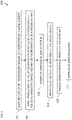
- Fig. 5 is a flow chart 200 including steps in a method of applying the trained artificial neural network 52 in the system 10 of Fig. 1 .
- Fig. 6 is a schematic illustration of the trained artificial network 52 being applied in the system 10 of Fig. 1 .
- the fluoroscopic imaging device 37 ( Fig. 1 ) is configured to capture (block 202) a set of 2D fluoroscopic images 86 of a body part 88 of a living subject 90.
- the set of 2D fluoroscopic images 86 includes only two 2D fluoroscopic images.
- the set of 2D fluoroscopic images 86 includes an anterior-posterior (AP) projection of the body part 88 and a left anterior-oblique projection (LAO) of the body part 86, or any other suitable pair of projections.
- the body part 88 may be any suitable body part, for example, a chamber of a heart.
- the processing circuitry 22 ( Fig. 1 ) is configured to apply (block 204) the trained artificial neural network 52 to: (a) the set of 2D fluoroscopic images 86 of the body part 88 of the living subject 90; and (b) respective 3D coordinates 96 of the set of 2D fluoroscopic images 86, yielding 3D coordinates 92 of a 3D anatomical map 94.
- the 3D coordinates 96 of the set of 2D fluoroscopic images 86 and the 3D coordinates 92 of the 3D anatomical map 94 are in the same coordinate space and registered to the coordinate space of the positioning sub-system described above with reference to Fig. 1 (e.g., the coordinate space 31 of Fig. 1 ).
- the 3D coordinates 92 of the 3D anatomical map 94 may include mesh vertices of a 3D mesh and/or a 3D point cloud.
- the processing circuitry 22 is configured to render (block 206) to the display 29 ( Fig. 1 ) the 3D anatomical map 94 responsively to the 3D coordinates 92.
- the catheter 14 ( Fig. 1 ) is configured to be inserted (block 208) into the body part 88 of the living subject 90 and moved around the body part 88 in order to acquire signals via the electrodes 21 ( Fig. 1 ) to correct and improve the 3D anatomical map 94.
- the processing circuitry 22 is configured to receive (block 210) signals from the electrodes 21 of the catheter 14.
- the processing circuitry 22 is configured to improve (block 212) the 3D anatomical map 94 responsively to signals received from the electrodes 21 of the catheter 14.
- processing circuitry 22 may be combined in a single physical component or, alternatively, implemented using multiple physical components. These physical components may comprise hard-wired or programmable devices, or a combination of the two. In some embodiments, at least some of the functions of the processing circuitry 22 may be carried out by a programmable processor under the control of suitable software. This software may be downloaded to a device in electronic form, over a network, for example. Alternatively, or additionally, the software may be stored in tangible, non-transitory computer-readable storage media, such as optical, magnetic, or electronic memory.
- the terms “about” or “approximately” for any numerical values or ranges indicate a suitable dimensional tolerance that allows the part or collection of components to function for its intended purpose as described herein. More specifically, “about” or “approximately” may refer to the range of values ⁇ 20% of the recited value, e.g. "about 90%” may refer to the range of values from 72% to 108%.
Landscapes
- Engineering & Computer Science (AREA)
- Health & Medical Sciences (AREA)
- Life Sciences & Earth Sciences (AREA)
- Physics & Mathematics (AREA)
- Theoretical Computer Science (AREA)
- General Health & Medical Sciences (AREA)
- Medical Informatics (AREA)
- Biomedical Technology (AREA)
- Molecular Biology (AREA)
- Biophysics (AREA)
- Public Health (AREA)
- Surgery (AREA)
- General Physics & Mathematics (AREA)
- Animal Behavior & Ethology (AREA)
- Veterinary Medicine (AREA)
- Heart & Thoracic Surgery (AREA)
- Pathology (AREA)
- Software Systems (AREA)
- Artificial Intelligence (AREA)
- Evolutionary Computation (AREA)
- Nuclear Medicine, Radiotherapy & Molecular Imaging (AREA)
- Geometry (AREA)
- General Engineering & Computer Science (AREA)
- Mathematical Physics (AREA)
- Radiology & Medical Imaging (AREA)
- Computer Vision & Pattern Recognition (AREA)
- Data Mining & Analysis (AREA)
- Computing Systems (AREA)
- Computer Graphics (AREA)
- Computational Linguistics (AREA)
- Optics & Photonics (AREA)
- High Energy & Nuclear Physics (AREA)
- Cardiology (AREA)
- Physiology (AREA)
- Epidemiology (AREA)
- Primary Health Care (AREA)
- Computer Hardware Design (AREA)
- Fuzzy Systems (AREA)
- Psychiatry (AREA)
- Signal Processing (AREA)
Applications Claiming Priority (2)
| Application Number | Priority Date | Filing Date | Title |
|---|---|---|---|
| US202063118047P | 2020-11-25 | 2020-11-25 | |
| US17/402,922 US11694401B2 (en) | 2020-11-25 | 2021-08-16 | Reconstruction of registered geometry based on constant fluoroscopic snapshot |
Publications (3)
| Publication Number | Publication Date |
|---|---|
| EP4009283A1 true EP4009283A1 (fr) | 2022-06-08 |
| EP4009283B1 EP4009283B1 (fr) | 2025-09-10 |
| EP4009283C0 EP4009283C0 (fr) | 2025-09-10 |
Family
ID=79024463
Family Applications (1)
| Application Number | Title | Priority Date | Filing Date |
|---|---|---|---|
| EP21210115.8A Active EP4009283B1 (fr) | 2020-11-25 | 2021-11-24 | Reconstruction de géométrie enregistrée basée sur un instantané fluoroscopique constant |
Country Status (5)
| Country | Link |
|---|---|
| US (1) | US11694401B2 (fr) |
| EP (1) | EP4009283B1 (fr) |
| JP (1) | JP7772300B2 (fr) |
| CN (1) | CN114532979A (fr) |
| IL (1) | IL288120B2 (fr) |
Citations (20)
| Publication number | Priority date | Publication date | Assignee | Title |
|---|---|---|---|---|
| US5391199A (en) | 1993-07-20 | 1995-02-21 | Biosense, Inc. | Apparatus and method for treating cardiac arrhythmias |
| WO1996005768A1 (fr) | 1994-08-19 | 1996-02-29 | Biosense, Inc. | Systemes medicaux de diagnostic, de traitement et d'imagerie |
| US6226542B1 (en) | 1998-07-24 | 2001-05-01 | Biosense, Inc. | Three-dimensional reconstruction of intrabody organs |
| US6239724B1 (en) | 1997-12-30 | 2001-05-29 | Remon Medical Technologies, Ltd. | System and method for telemetrically providing intrabody spatial position |
| US6301496B1 (en) | 1998-07-24 | 2001-10-09 | Biosense, Inc. | Vector mapping of three-dimensionally reconstructed intrabody organs and method of display |
| US6332089B1 (en) | 1996-02-15 | 2001-12-18 | Biosense, Inc. | Medical procedures and apparatus using intrabody probes |
| US20020065455A1 (en) | 1995-01-24 | 2002-05-30 | Shlomo Ben-Haim | Medical diagnosis, treatment and imaging systems |
| US6484118B1 (en) | 2000-07-20 | 2002-11-19 | Biosense, Inc. | Electromagnetic position single axis system |
| US20030120150A1 (en) | 2001-12-21 | 2003-06-26 | Assaf Govari | Wireless position sensor |
| US6618612B1 (en) | 1996-02-15 | 2003-09-09 | Biosense, Inc. | Independently positionable transducers for location system |
| US20040068178A1 (en) | 2002-09-17 | 2004-04-08 | Assaf Govari | High-gradient recursive locating system |
| US6814733B2 (en) | 2002-01-31 | 2004-11-09 | Biosense, Inc. | Radio frequency pulmonary vein isolation |
| US6892091B1 (en) | 2000-02-18 | 2005-05-10 | Biosense, Inc. | Catheter, method and apparatus for generating an electrical map of a chamber of the heart |
| US6997924B2 (en) | 2002-09-17 | 2006-02-14 | Biosense Inc. | Laser pulmonary vein isolation |
| US7156816B2 (en) | 2002-11-26 | 2007-01-02 | Biosense, Inc. | Ultrasound pulmonary vein isolation |
| US7536218B2 (en) | 2005-07-15 | 2009-05-19 | Biosense Webster, Inc. | Hybrid magnetic-based and impedance-based position sensing |
| US7756576B2 (en) | 2005-08-26 | 2010-07-13 | Biosense Webster, Inc. | Position sensing and detection of skin impedance |
| US20120071751A1 (en) * | 2010-09-20 | 2012-03-22 | Jasbir Sra | 3D Model Creation of Anatomic Structures Using Single-Plane Fluoroscopy |
| US20140005664A1 (en) | 2012-07-02 | 2014-01-02 | Assaf Govari | Real time assessment of ablation from electrocardiogram signals |
| US20150223762A1 (en) * | 2014-02-07 | 2015-08-13 | Biosense Webster (Israel) Ltd. | Synchronizing between image sequences of the heart acquired at different heartbeat rates |
Family Cites Families (15)
| Publication number | Priority date | Publication date | Assignee | Title |
|---|---|---|---|---|
| US5588033A (en) | 1995-06-06 | 1996-12-24 | St. Jude Children's Research Hospital | Method and apparatus for three dimensional image reconstruction from multiple stereotactic or isocentric backprojections |
| DE102004001858A1 (de) * | 2003-10-22 | 2005-05-25 | Schaerer Mayfield Technologies Gmbh | Verfahren zur Fluoroskopie-basierten Neuronavigation |
| JP5269500B2 (ja) * | 2008-07-04 | 2013-08-21 | 株式会社東芝 | 画像処理装置 |
| JP2011234864A (ja) * | 2010-05-10 | 2011-11-24 | Toshiba Corp | X線診断装置及び支援データ生成用制御プログラム |
| US9668699B2 (en) | 2013-10-17 | 2017-06-06 | Siemens Healthcare Gmbh | Method and system for anatomical object detection using marginal space deep neural networks |
| WO2016116946A2 (fr) * | 2015-01-20 | 2016-07-28 | Indian Institute Of Technology, Bombay | Système et procédé permettant d'obtenir des images tridimensionnelles à l'aide d'images radiologiques bidimensionnelles classiques |
| US11399895B2 (en) * | 2016-02-12 | 2022-08-02 | Intuitive Surgical Operations, Inc. | Systems and methods of pose estimation and calibration of perspective imaging system in image guided surgery |
| WO2017223560A1 (fr) | 2016-06-24 | 2017-12-28 | Rensselaer Polytechnic Institute | Reconstruction d'images tomographiques par apprentissage machine |
| AU2019200594B2 (en) * | 2018-02-08 | 2020-05-28 | Covidien Lp | System and method for local three dimensional volume reconstruction using a standard fluoroscope |
| LU101007B1 (en) * | 2018-11-26 | 2020-05-26 | Metamorphosis Gmbh | Artificial-intelligence based reduction support |
| CN113747828B (zh) * | 2019-01-15 | 2024-06-11 | Apn健康有限责任公司 | 心脏导管接触力确定 |
| US11748899B2 (en) * | 2019-02-07 | 2023-09-05 | Siemens Healthcare Gmbh | Dense body marker estimation from camera data for patient positioning in medical imaging |
| US12502218B2 (en) * | 2019-02-08 | 2025-12-23 | Stryker Corporation | Systems and methods for treating a joint |
| US10867436B2 (en) * | 2019-04-18 | 2020-12-15 | Zebra Medical Vision Ltd. | Systems and methods for reconstruction of 3D anatomical images from 2D anatomical images |
| WO2021030536A1 (fr) * | 2019-08-13 | 2021-02-18 | Duluth Medical Technologies Inc. | Procédés et appareils chirurgicaux robotiques |
-
2021
- 2021-08-16 US US17/402,922 patent/US11694401B2/en active Active
- 2021-11-15 IL IL288120A patent/IL288120B2/en unknown
- 2021-11-24 JP JP2021189971A patent/JP7772300B2/ja active Active
- 2021-11-24 EP EP21210115.8A patent/EP4009283B1/fr active Active
- 2021-11-25 CN CN202111413642.0A patent/CN114532979A/zh active Pending
Patent Citations (21)
| Publication number | Priority date | Publication date | Assignee | Title |
|---|---|---|---|---|
| US5391199A (en) | 1993-07-20 | 1995-02-21 | Biosense, Inc. | Apparatus and method for treating cardiac arrhythmias |
| WO1996005768A1 (fr) | 1994-08-19 | 1996-02-29 | Biosense, Inc. | Systemes medicaux de diagnostic, de traitement et d'imagerie |
| US6690963B2 (en) | 1995-01-24 | 2004-02-10 | Biosense, Inc. | System for determining the location and orientation of an invasive medical instrument |
| US20020065455A1 (en) | 1995-01-24 | 2002-05-30 | Shlomo Ben-Haim | Medical diagnosis, treatment and imaging systems |
| US6332089B1 (en) | 1996-02-15 | 2001-12-18 | Biosense, Inc. | Medical procedures and apparatus using intrabody probes |
| US6618612B1 (en) | 1996-02-15 | 2003-09-09 | Biosense, Inc. | Independently positionable transducers for location system |
| US6239724B1 (en) | 1997-12-30 | 2001-05-29 | Remon Medical Technologies, Ltd. | System and method for telemetrically providing intrabody spatial position |
| US6301496B1 (en) | 1998-07-24 | 2001-10-09 | Biosense, Inc. | Vector mapping of three-dimensionally reconstructed intrabody organs and method of display |
| US6226542B1 (en) | 1998-07-24 | 2001-05-01 | Biosense, Inc. | Three-dimensional reconstruction of intrabody organs |
| US6892091B1 (en) | 2000-02-18 | 2005-05-10 | Biosense, Inc. | Catheter, method and apparatus for generating an electrical map of a chamber of the heart |
| US6484118B1 (en) | 2000-07-20 | 2002-11-19 | Biosense, Inc. | Electromagnetic position single axis system |
| US20030120150A1 (en) | 2001-12-21 | 2003-06-26 | Assaf Govari | Wireless position sensor |
| US6814733B2 (en) | 2002-01-31 | 2004-11-09 | Biosense, Inc. | Radio frequency pulmonary vein isolation |
| US20040068178A1 (en) | 2002-09-17 | 2004-04-08 | Assaf Govari | High-gradient recursive locating system |
| US6997924B2 (en) | 2002-09-17 | 2006-02-14 | Biosense Inc. | Laser pulmonary vein isolation |
| US7156816B2 (en) | 2002-11-26 | 2007-01-02 | Biosense, Inc. | Ultrasound pulmonary vein isolation |
| US7536218B2 (en) | 2005-07-15 | 2009-05-19 | Biosense Webster, Inc. | Hybrid magnetic-based and impedance-based position sensing |
| US7756576B2 (en) | 2005-08-26 | 2010-07-13 | Biosense Webster, Inc. | Position sensing and detection of skin impedance |
| US20120071751A1 (en) * | 2010-09-20 | 2012-03-22 | Jasbir Sra | 3D Model Creation of Anatomic Structures Using Single-Plane Fluoroscopy |
| US20140005664A1 (en) | 2012-07-02 | 2014-01-02 | Assaf Govari | Real time assessment of ablation from electrocardiogram signals |
| US20150223762A1 (en) * | 2014-02-07 | 2015-08-13 | Biosense Webster (Israel) Ltd. | Synchronizing between image sequences of the heart acquired at different heartbeat rates |
Also Published As
| Publication number | Publication date |
|---|---|
| JP2022084009A (ja) | 2022-06-06 |
| EP4009283B1 (fr) | 2025-09-10 |
| IL288120B2 (en) | 2024-07-01 |
| US20220165028A1 (en) | 2022-05-26 |
| IL288120A (en) | 2022-06-01 |
| US11694401B2 (en) | 2023-07-04 |
| IL288120B1 (en) | 2024-03-01 |
| CN114532979A (zh) | 2022-05-27 |
| JP7772300B2 (ja) | 2025-11-18 |
| EP4009283C0 (fr) | 2025-09-10 |
Similar Documents
| Publication | Publication Date | Title |
|---|---|---|
| US12502139B2 (en) | Removing noise from cardiac signals | |
| US12484959B2 (en) | Accurate tissue proximity | |
| EP3967218B1 (fr) | Système d'analyse de temps d'activation locale | |
| EP4009283B1 (fr) | Reconstruction de géométrie enregistrée basée sur un instantané fluoroscopique constant | |
| EP3991658B1 (fr) | Réduction de bruit d'appareil d'enregistrement | |
| US12004862B2 (en) | Removing far-field from intracardiac signals | |
| JP2022040077A5 (fr) | ||
| CN115702819A (zh) | 用于减少电生理规程的设置时间的图形用户界面模板 |
Legal Events
| Date | Code | Title | Description |
|---|---|---|---|
| PUAI | Public reference made under article 153(3) epc to a published international application that has entered the european phase |
Free format text: ORIGINAL CODE: 0009012 |
|
| STAA | Information on the status of an ep patent application or granted ep patent |
Free format text: STATUS: THE APPLICATION HAS BEEN PUBLISHED |
|
| AK | Designated contracting states |
Kind code of ref document: A1 Designated state(s): AL AT BE BG CH CY CZ DE DK EE ES FI FR GB GR HR HU IE IS IT LI LT LU LV MC MK MT NL NO PL PT RO RS SE SI SK SM TR |
|
| STAA | Information on the status of an ep patent application or granted ep patent |
Free format text: STATUS: REQUEST FOR EXAMINATION WAS MADE |
|
| 17P | Request for examination filed |
Effective date: 20220812 |
|
| RBV | Designated contracting states (corrected) |
Designated state(s): AL AT BE BG CH CY CZ DE DK EE ES FI FR GB GR HR HU IE IS IT LI LT LU LV MC MK MT NL NO PL PT RO RS SE SI SK SM TR |
|
| GRAP | Despatch of communication of intention to grant a patent |
Free format text: ORIGINAL CODE: EPIDOSNIGR1 |
|
| STAA | Information on the status of an ep patent application or granted ep patent |
Free format text: STATUS: GRANT OF PATENT IS INTENDED |
|
| GRAS | Grant fee paid |
Free format text: ORIGINAL CODE: EPIDOSNIGR3 |
|
| INTG | Intention to grant announced |
Effective date: 20250707 |
|
| GRAA | (expected) grant |
Free format text: ORIGINAL CODE: 0009210 |
|
| STAA | Information on the status of an ep patent application or granted ep patent |
Free format text: STATUS: THE PATENT HAS BEEN GRANTED |
|
| AK | Designated contracting states |
Kind code of ref document: B1 Designated state(s): AL AT BE BG CH CY CZ DE DK EE ES FI FR GB GR HR HU IE IS IT LI LT LU LV MC MK MT NL NO PL PT RO RS SE SI SK SM TR |
|
| REG | Reference to a national code |
Ref country code: GB Ref legal event code: FG4D |
|
| REG | Reference to a national code |
Ref country code: CH Ref legal event code: EP |
|
| REG | Reference to a national code |
Ref country code: DE Ref legal event code: R096 Ref document number: 602021038189 Country of ref document: DE |
|
| REG | Reference to a national code |
Ref country code: IE Ref legal event code: FG4D |
|
| U01 | Request for unitary effect filed |
Effective date: 20251007 |
|
| U07 | Unitary effect registered |
Designated state(s): AT BE BG DE DK EE FI FR IT LT LU LV MT NL PT RO SE SI Effective date: 20251013 |
|
| U20 | Renewal fee for the european patent with unitary effect paid |
Year of fee payment: 5 Effective date: 20251023 |
|
| PGFP | Annual fee paid to national office [announced via postgrant information from national office to epo] |
Ref country code: GB Payment date: 20251016 Year of fee payment: 5 |
|
| PG25 | Lapsed in a contracting state [announced via postgrant information from national office to epo] |
Ref country code: NO Free format text: LAPSE BECAUSE OF FAILURE TO SUBMIT A TRANSLATION OF THE DESCRIPTION OR TO PAY THE FEE WITHIN THE PRESCRIBED TIME-LIMIT Effective date: 20251210 |
|
| PG25 | Lapsed in a contracting state [announced via postgrant information from national office to epo] |
Ref country code: HR Free format text: LAPSE BECAUSE OF FAILURE TO SUBMIT A TRANSLATION OF THE DESCRIPTION OR TO PAY THE FEE WITHIN THE PRESCRIBED TIME-LIMIT Effective date: 20250910 |
|
| PG25 | Lapsed in a contracting state [announced via postgrant information from national office to epo] |
Ref country code: GR Free format text: LAPSE BECAUSE OF FAILURE TO SUBMIT A TRANSLATION OF THE DESCRIPTION OR TO PAY THE FEE WITHIN THE PRESCRIBED TIME-LIMIT Effective date: 20251211 |
|
| PG25 | Lapsed in a contracting state [announced via postgrant information from national office to epo] |
Ref country code: PL Free format text: LAPSE BECAUSE OF FAILURE TO SUBMIT A TRANSLATION OF THE DESCRIPTION OR TO PAY THE FEE WITHIN THE PRESCRIBED TIME-LIMIT Effective date: 20250910 |
|
| PG25 | Lapsed in a contracting state [announced via postgrant information from national office to epo] |
Ref country code: RS Free format text: LAPSE BECAUSE OF FAILURE TO SUBMIT A TRANSLATION OF THE DESCRIPTION OR TO PAY THE FEE WITHIN THE PRESCRIBED TIME-LIMIT Effective date: 20251210 |
|
| PG25 | Lapsed in a contracting state [announced via postgrant information from national office to epo] |
Ref country code: ES Free format text: LAPSE BECAUSE OF FAILURE TO SUBMIT A TRANSLATION OF THE DESCRIPTION OR TO PAY THE FEE WITHIN THE PRESCRIBED TIME-LIMIT Effective date: 20250910 |
|
| PG25 | Lapsed in a contracting state [announced via postgrant information from national office to epo] |
Ref country code: SM Free format text: LAPSE BECAUSE OF FAILURE TO SUBMIT A TRANSLATION OF THE DESCRIPTION OR TO PAY THE FEE WITHIN THE PRESCRIBED TIME-LIMIT Effective date: 20250910 |
|
| PG25 | Lapsed in a contracting state [announced via postgrant information from national office to epo] |
Ref country code: IS Free format text: LAPSE BECAUSE OF FAILURE TO SUBMIT A TRANSLATION OF THE DESCRIPTION OR TO PAY THE FEE WITHIN THE PRESCRIBED TIME-LIMIT Effective date: 20260110 |
|
| PG25 | Lapsed in a contracting state [announced via postgrant information from national office to epo] |
Ref country code: CZ Free format text: LAPSE BECAUSE OF FAILURE TO SUBMIT A TRANSLATION OF THE DESCRIPTION OR TO PAY THE FEE WITHIN THE PRESCRIBED TIME-LIMIT Effective date: 20250910 |
|
| PG25 | Lapsed in a contracting state [announced via postgrant information from national office to epo] |
Ref country code: SK Free format text: LAPSE BECAUSE OF FAILURE TO SUBMIT A TRANSLATION OF THE DESCRIPTION OR TO PAY THE FEE WITHIN THE PRESCRIBED TIME-LIMIT Effective date: 20250910 |