EP4013433B1 - Hohle dreidimensionale einheit aus retinalem gewebe und deren verwendung zur behandlung von retinopathien - Google Patents
Hohle dreidimensionale einheit aus retinalem gewebe und deren verwendung zur behandlung von retinopathien Download PDFInfo
- Publication number
- EP4013433B1 EP4013433B1 EP20761761.4A EP20761761A EP4013433B1 EP 4013433 B1 EP4013433 B1 EP 4013433B1 EP 20761761 A EP20761761 A EP 20761761A EP 4013433 B1 EP4013433 B1 EP 4013433B1
- Authority
- EP
- European Patent Office
- Prior art keywords
- cells
- retinal
- pigment epithelium
- retinal pigment
- tissue
- Prior art date
- Legal status (The legal status is an assumption and is not a legal conclusion. Google has not performed a legal analysis and makes no representation as to the accuracy of the status listed.)
- Active
Links
Images

Classifications
-
- C—CHEMISTRY; METALLURGY
- C12—BIOCHEMISTRY; BEER; SPIRITS; WINE; VINEGAR; MICROBIOLOGY; ENZYMOLOGY; MUTATION OR GENETIC ENGINEERING
- C12N—MICROORGANISMS OR ENZYMES; COMPOSITIONS THEREOF; PROPAGATING, PRESERVING, OR MAINTAINING MICROORGANISMS; MUTATION OR GENETIC ENGINEERING; CULTURE MEDIA
- C12N5/00—Undifferentiated human, animal or plant cells, e.g. cell lines; Tissues; Cultivation or maintenance thereof; Culture media therefor
- C12N5/06—Animal cells or tissues; Human cells or tissues
- C12N5/0602—Vertebrate cells
- C12N5/0618—Cells of the nervous system
- C12N5/0621—Eye cells, e.g. cornea, iris pigmented cells
-
- A—HUMAN NECESSITIES
- A61—MEDICAL OR VETERINARY SCIENCE; HYGIENE
- A61K—PREPARATIONS FOR MEDICAL, DENTAL OR TOILETRY PURPOSES
- A61K35/00—Medicinal preparations containing materials or reaction products thereof with undetermined constitution
- A61K35/12—Materials from mammals; Compositions comprising non-specified tissues or cells; Compositions comprising non-embryonic stem cells; Genetically modified cells
- A61K35/30—Nerves; Brain; Eyes; Corneal cells; Cerebrospinal fluid; Neuronal stem cells; Neuronal precursor cells; Glial cells; Oligodendrocytes; Schwann cells; Astroglia; Astrocytes; Choroid plexus; Spinal cord tissue
-
- A—HUMAN NECESSITIES
- A61—MEDICAL OR VETERINARY SCIENCE; HYGIENE
- A61L—METHODS OR APPARATUS FOR STERILISING MATERIALS OR OBJECTS IN GENERAL; DISINFECTION, STERILISATION OR DEODORISATION OF AIR; CHEMICAL ASPECTS OF BANDAGES, DRESSINGS, ABSORBENT PADS OR SURGICAL ARTICLES; MATERIALS FOR BANDAGES, DRESSINGS, ABSORBENT PADS OR SURGICAL ARTICLES
- A61L27/00—Materials for grafts or prostheses or for coating grafts or prostheses
- A61L27/14—Macromolecular materials
- A61L27/20—Polysaccharides
-
- A—HUMAN NECESSITIES
- A61—MEDICAL OR VETERINARY SCIENCE; HYGIENE
- A61L—METHODS OR APPARATUS FOR STERILISING MATERIALS OR OBJECTS IN GENERAL; DISINFECTION, STERILISATION OR DEODORISATION OF AIR; CHEMICAL ASPECTS OF BANDAGES, DRESSINGS, ABSORBENT PADS OR SURGICAL ARTICLES; MATERIALS FOR BANDAGES, DRESSINGS, ABSORBENT PADS OR SURGICAL ARTICLES
- A61L27/00—Materials for grafts or prostheses or for coating grafts or prostheses
- A61L27/36—Materials for grafts or prostheses or for coating grafts or prostheses containing ingredients of undetermined constitution or reaction products thereof, e.g. transplant tissue, natural bone, extracellular matrix
- A61L27/38—Materials for grafts or prostheses or for coating grafts or prostheses containing ingredients of undetermined constitution or reaction products thereof, e.g. transplant tissue, natural bone, extracellular matrix containing added animal cells
- A61L27/3804—Materials for grafts or prostheses or for coating grafts or prostheses containing ingredients of undetermined constitution or reaction products thereof, e.g. transplant tissue, natural bone, extracellular matrix containing added animal cells characterised by specific cells or progenitors thereof, e.g. fibroblasts, connective tissue cells, kidney cells
- A61L27/3813—Epithelial cells, e.g. keratinocytes, urothelial cells
-
- A—HUMAN NECESSITIES
- A61—MEDICAL OR VETERINARY SCIENCE; HYGIENE
- A61L—METHODS OR APPARATUS FOR STERILISING MATERIALS OR OBJECTS IN GENERAL; DISINFECTION, STERILISATION OR DEODORISATION OF AIR; CHEMICAL ASPECTS OF BANDAGES, DRESSINGS, ABSORBENT PADS OR SURGICAL ARTICLES; MATERIALS FOR BANDAGES, DRESSINGS, ABSORBENT PADS OR SURGICAL ARTICLES
- A61L27/00—Materials for grafts or prostheses or for coating grafts or prostheses
- A61L27/36—Materials for grafts or prostheses or for coating grafts or prostheses containing ingredients of undetermined constitution or reaction products thereof, e.g. transplant tissue, natural bone, extracellular matrix
- A61L27/38—Materials for grafts or prostheses or for coating grafts or prostheses containing ingredients of undetermined constitution or reaction products thereof, e.g. transplant tissue, natural bone, extracellular matrix containing added animal cells
- A61L27/3804—Materials for grafts or prostheses or for coating grafts or prostheses containing ingredients of undetermined constitution or reaction products thereof, e.g. transplant tissue, natural bone, extracellular matrix containing added animal cells characterised by specific cells or progenitors thereof, e.g. fibroblasts, connective tissue cells, kidney cells
- A61L27/3834—Cells able to produce different cell types, e.g. hematopoietic stem cells, mesenchymal stem cells, marrow stromal cells, embryonic stem cells
-
- A—HUMAN NECESSITIES
- A61—MEDICAL OR VETERINARY SCIENCE; HYGIENE
- A61L—METHODS OR APPARATUS FOR STERILISING MATERIALS OR OBJECTS IN GENERAL; DISINFECTION, STERILISATION OR DEODORISATION OF AIR; CHEMICAL ASPECTS OF BANDAGES, DRESSINGS, ABSORBENT PADS OR SURGICAL ARTICLES; MATERIALS FOR BANDAGES, DRESSINGS, ABSORBENT PADS OR SURGICAL ARTICLES
- A61L27/00—Materials for grafts or prostheses or for coating grafts or prostheses
- A61L27/50—Materials characterised by their function or physical properties, e.g. injectable or lubricating compositions, shape-memory materials, surface modified materials
- A61L27/52—Hydrogels or hydrocolloids
-
- A—HUMAN NECESSITIES
- A61—MEDICAL OR VETERINARY SCIENCE; HYGIENE
- A61P—SPECIFIC THERAPEUTIC ACTIVITY OF CHEMICAL COMPOUNDS OR MEDICINAL PREPARATIONS
- A61P27/00—Drugs for disorders of the senses
- A61P27/02—Ophthalmic agents
-
- C—CHEMISTRY; METALLURGY
- C08—ORGANIC MACROMOLECULAR COMPOUNDS; THEIR PREPARATION OR CHEMICAL WORKING-UP; COMPOSITIONS BASED THEREON
- C08L—COMPOSITIONS OF MACROMOLECULAR COMPOUNDS
- C08L5/00—Compositions of polysaccharides or of their derivatives not provided for in groups C08L1/00 or C08L3/00
- C08L5/04—Alginic acid; Derivatives thereof
-
- A—HUMAN NECESSITIES
- A61—MEDICAL OR VETERINARY SCIENCE; HYGIENE
- A61F—FILTERS IMPLANTABLE INTO BLOOD VESSELS; PROSTHESES; DEVICES PROVIDING PATENCY TO, OR PREVENTING COLLAPSING OF, TUBULAR STRUCTURES OF THE BODY, e.g. STENTS; ORTHOPAEDIC, NURSING OR CONTRACEPTIVE DEVICES; FOMENTATION; TREATMENT OR PROTECTION OF EYES OR EARS; BANDAGES, DRESSINGS OR ABSORBENT PADS; FIRST-AID KITS
- A61F2/00—Filters implantable into blood vessels; Prostheses, i.e. artificial substitutes or replacements for parts of the body; Appliances for connecting them with the body; Devices providing patency to, or preventing collapsing of, tubular structures of the body, e.g. stents
- A61F2/02—Prostheses implantable into the body
- A61F2/14—Eye parts, e.g. lenses or corneal implants; Artificial eyes
-
- A—HUMAN NECESSITIES
- A61—MEDICAL OR VETERINARY SCIENCE; HYGIENE
- A61K—PREPARATIONS FOR MEDICAL, DENTAL OR TOILETRY PURPOSES
- A61K35/00—Medicinal preparations containing materials or reaction products thereof with undetermined constitution
- A61K35/12—Materials from mammals; Compositions comprising non-specified tissues or cells; Compositions comprising non-embryonic stem cells; Genetically modified cells
- A61K2035/126—Immunoprotecting barriers, e.g. jackets, diffusion chambers
- A61K2035/128—Immunoprotecting barriers, e.g. jackets, diffusion chambers capsules, e.g. microcapsules
-
- A—HUMAN NECESSITIES
- A61—MEDICAL OR VETERINARY SCIENCE; HYGIENE
- A61L—METHODS OR APPARATUS FOR STERILISING MATERIALS OR OBJECTS IN GENERAL; DISINFECTION, STERILISATION OR DEODORISATION OF AIR; CHEMICAL ASPECTS OF BANDAGES, DRESSINGS, ABSORBENT PADS OR SURGICAL ARTICLES; MATERIALS FOR BANDAGES, DRESSINGS, ABSORBENT PADS OR SURGICAL ARTICLES
- A61L2430/00—Materials or treatment for tissue regeneration
- A61L2430/16—Materials or treatment for tissue regeneration for reconstruction of eye parts, e.g. intraocular lens, cornea
-
- C—CHEMISTRY; METALLURGY
- C12—BIOCHEMISTRY; BEER; SPIRITS; WINE; VINEGAR; MICROBIOLOGY; ENZYMOLOGY; MUTATION OR GENETIC ENGINEERING
- C12N—MICROORGANISMS OR ENZYMES; COMPOSITIONS THEREOF; PROPAGATING, PRESERVING, OR MAINTAINING MICROORGANISMS; MUTATION OR GENETIC ENGINEERING; CULTURE MEDIA
- C12N2513/00—3D culture
-
- C—CHEMISTRY; METALLURGY
- C12—BIOCHEMISTRY; BEER; SPIRITS; WINE; VINEGAR; MICROBIOLOGY; ENZYMOLOGY; MUTATION OR GENETIC ENGINEERING
- C12N—MICROORGANISMS OR ENZYMES; COMPOSITIONS THEREOF; PROPAGATING, PRESERVING, OR MAINTAINING MICROORGANISMS; MUTATION OR GENETIC ENGINEERING; CULTURE MEDIA
- C12N2533/00—Supports or coatings for cell culture, characterised by material
- C12N2533/90—Substrates of biological origin, e.g. extracellular matrix, decellularised tissue
-
- C—CHEMISTRY; METALLURGY
- C12—BIOCHEMISTRY; BEER; SPIRITS; WINE; VINEGAR; MICROBIOLOGY; ENZYMOLOGY; MUTATION OR GENETIC ENGINEERING
- C12N—MICROORGANISMS OR ENZYMES; COMPOSITIONS THEREOF; PROPAGATING, PRESERVING, OR MAINTAINING MICROORGANISMS; MUTATION OR GENETIC ENGINEERING; CULTURE MEDIA
- C12N2535/00—Supports or coatings for cell culture characterised by topography
Definitions
- the present invention relates to the treatment of retinal diseases, in particular by the use of specific tissue units comprising at least one retinal pigment epithelium.
- the invention also relates to a method for preparing these tissue units as well as kits for implanting these tissue units in the eye to perform grafts of all or part of the retina.
- Retinal diseases or retinopathies
- the retina which lines the back of the eye, is made up of pigment epithelial cells and nerve cells that receive light. These nerve cells translate light into electrical signals that travel to the brain via the optic nerve.
- the cells of the retina particularly the retinal pigment epithelium, degenerate or no longer function, blind areas of the visual field appear.
- Retinopathies can have various origins: they can be linked to aging, such as age-related macular degeneration (AMD), be hereditary, such as pigmentary retinopathy or retinal dystrophy, be linked to trauma such as solar retinopathy, or derive from another pathology, such as diabetic retinopathy or hypertensive retinopathy.
- AMD age-related macular degeneration
- hereditary such as pigmentary retinopathy or retinal dystrophy
- trauma such as solar retinopathy
- derive from another pathology such as diabetic retinopathy or hypertensive retinopathy.
- Retinopathy pigmentosa is a genetic disease that can be hereditary. It is characterized by degeneration of the cells of the retina linked to the mutation of one or more genes. The progression of the disease is slow to the point of causing blindness and there is currently no treatment.
- Diabetic retinopathy is a retinal disorder occurring in the context of diabetes. It is linked to excessive sugar concentration in the small blood vessels of the retina, which leads to their degradation. The lack of oxygen supply induces the formation of new, more fragile blood vessels. Their rupture and the microhemorrhages that follow can lead to retinal detachment. As with the wet form of AMD, it is possible to perform injections of anti-angiogenic drugs, but this treatment does not work well. Laser treatment can also be performed to burn the abnormal vessels, but its effectiveness is limited.
- the request EP3211071 also describes retinal tissues in the form of suspended aggregates obtained from pluripotent stem cells. The suspension is then injected into the eye. This solution is also not satisfactory because the injection does not allow for the preservation of a functional structure (in particular a functional polarization): i) in the case of the pigment epithelium, the apical face of the cells must be presented opposite the external segments of the photoreceptors of the graft, limiting the survival and functionality of the graft ii) in the case of the photoreceptors, the external segment must be positioned towards the outside of the eye opposite the cells of the pigment epithelium and the synaptic terminal must face the center of the eye to connect the rest of the neural retina).
- a functional structure in particular a functional polarization
- WO 2017/176810 describes a three-dimensional retinal tissue unit comprising a concentric laminar structure comprising a retinal pigment epithelium cell core, a layer of RGCs, a layer of second-order retinal neurons, a layer of photoreceptors and a layer of retinal pigment epithelium cells as well as a hydrogel.
- the objective of the invention is to overcome these various problems of the prior art, and to propose a solution for the easy, rapid and minimally invasive implantation of correctly polarized retinal cells, and their integration into the host organ, so as to replace in a durable and certain retinal cells that have degenerated in patients with degenerative retinal diseases.
- the effectiveness of the retinal tissue graft depends largely on the good in situ incorporation (in particular correct apico-basal polarization) of the graft via the adhesion of the basal face of the retinal pigment epithelium cells at the Bruch membrane.
- the inventors have developed particular hollow three-dimensional cellular networks comprising at least one layer of differentiated living human retinal pigment epithelium cells, organized around a lumen, said retinal pigment epithelium cells having their basal faces oriented outwards.
- This tissue unit preferably also comprises an outer layer of extracellular matrix at the basal face of the retinal pigment epithelium cells promoting the integration and survival of the cells, once injected into the eye.
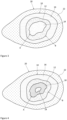
- the tissue unit according to the invention may also contain other retinal cells, organized in the form of one or more layers within the retinal pigment epithelium cell layer, in the inner lumen, i.e. at the apical face of the retinal pigment epithelium cells. These other cells preferentially form all or part of a retinal neural tissue.
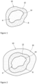
- the invention therefore relates to a hollow three-dimensional tissue unit, comprising, organized around an internal lumen, at least one layer of differentiated living human retinal pigment epithelium cells, the basal face of each retinal pigment epithelium cell of which is positioned towards the outside and the apical face towards the internal lumen.
- the tissue unit according to the invention is preferably in the form of a hollow ovoid, a hollow cylinder, a hollow spheroid or a hollow sphere, or a section of these elements along a plane.
- the invention also relates to such a hollow three-dimensional tissue unit for use in the treatment of retinal diseases, in particular by implantation in the eye, at the level of Bruch's membrane.
- the intervention does not require the precise positioning of a single graft as in the prior art.
- the invention has the advantage of being able to limit the surgical intervention to a single incision of the eye compared to three to four today.
- the polarization of the retinal pigment epithelium layer allows the retinal tissue units to position themselves correctly when grafted into the eye and to ensure the success of the graft.
- the retinal tissue units according to the invention attach to the extracellular matrix of the back of the eye (Brusch's membrane) by emitting cells adherent to the substrate which migrate and form a monolayer.
- alginate means linear polysaccharides formed from ⁇ -D-mannuronate and ⁇ -L-guluronate, salts and derivatives thereof.
- hydrogel capsule means a three-dimensional structure formed from a matrix of polymer chains, swollen by a liquid and preferably water.
- human cells means human cells or immunologically humanized non-human mammalian cells. Even when this is not specified, the cells, stem cells, progenitor cells and tissues according to the invention are constituted or are obtained from human cells or from immunologically humanized non-human mammalian cells.
- progenitor cell means a stem cell already engaged in cellular differentiation into retinal cells but not yet differentiated.
- embryonic stem cell means a pluripotent stem cell derived from the inner cell mass of the blastocyst.
- the pluripotency of embryonic stem cells can be assessed by the presence of markers such as the transcription factors OCT4 and NANOG and surface markers such as SSEA3/4, Tra-1-60 and Tra-1-81.
- the embryonic stem cells according to the invention are obtained without destroying the embryo from which they originate, for example using the technique described in Chang et al. (Cell Stem Cell, 2008, 2(2)): 113-117 ). Eventually, embryonic stem cells from human beings may be excluded.
- pluripotent stem cell or “pluripotent cell” means a cell that has the capacity to form all tissues present in the entire original organism, without being able to form an entire organism as such.
- these may be induced pluripotent stem cells, embryonic stem cells or MUSE cells (for "Multilineage-differentiating Stress Enduring”).
- induced pluripotent stem cell means a pluripotent stem cell induced to pluripotency by genetic reprogramming of differentiated somatic cells. These cells are in particular positive for pluripotency markers, such as alkaline phosphatase staining and expression of the proteins NANOG, SOX2, OCT4 and SSEA3/4.
- differentiated cells are understood to mean cells which exhibit a particular phenotype, as opposed to pluripotent stem cells which are not differentiated.
- Fiber diameter of a unit of fabric is meant the distance “d” between two tangents to said unit of fabric, these two tangents being parallel, such that the entire projection of the tissue unit is included between these two parallel tangents.
- implantation or "graft” in the eye within the meaning of the invention, we mean the action of depositing in the eye at a particular location, at least one unit of tissue according to the invention.
- the implantation can be carried out by any means, in particular by injection.
- the term “largest dimension” of a unit of fabric means the value of the largest Feret diameter of said unit of fabric.
- the term “smallest dimension” of a unit of fabric means the value of the smallest Feret diameter of said unit of fabric.
- tissue unit or “retinal tissue unit” according to the invention is meant a unit comprising at least one retinal tissue.
- the retinal tissue unit may comprise several retinal tissues assembled together with a functional structure.
- the tissue unit according to the invention comprises at least one retinal pigment epithelial tissue and it may also contain another retinal tissue, in particular a retinal neural or retinal vascular tissue and/or at least one other constituent such as, for example, an extracellular matrix.
- the invention therefore relates to a three-dimensional retinal tissue unit.
- the tissue unit according to the invention is hollow. It always comprises an internal light or lumen which constitutes the hollow part of the tissue unit. This light is generated at the time of the formation of the tissue unit by the retinal cells which multiply and develop.
- the lumen contains a liquid, in particular a culture medium (such as a medium based on DMEM or DMEM-F12 and/or Neurobasal and supplemented with B27 or N-2 or NS21) and/or a liquid secreted by the cells of the tissue unit.
- a culture medium such as a medium based on DMEM or DMEM-F12 and/or Neurobasal and supplemented with B27 or N-2 or NS21
- the tissue unit according to the invention comprises at least one layer of retinal pigment epithelium cells. These cells are human cells, living and differentiated into retinal pigment epithelium cells.
- the layer of retinal epithelium cells is organized around the inner lumen. The cells constituting this layer together form a retinal pigment epithelium, and their basal faces are all positioned towards the outside of the cell unit, and their apical faces are all positioned inward, that is, towards the internal lumen.
- the juxtaposed cells are linked together on their lateral faces by tight junctions.
- the tissue unit according to the invention also comprises an external layer of extracellular matrix.
- This external layer of extracellular matrix is located at the basal face of the retinal pigment epithelium cells.
- the layer of cellular matrix may consist of cellular matrix secreted by retinal pigment epithelium cells and/or of extracellular matrix added at the time of preparation of the cell unit.
- the extracellular matrix layer may form a gel. It comprises a mixture of proteins and extracellular compounds necessary for the culture of retinal pigment epithelium cells.
- the extracellular matrix comprises structural proteins, such as collagen, laminins, entactin, vitronectin, as well as growth factors, such as TGF-beta and/or EGF.
- the extracellular matrix layer may consist of or comprise Matrigel ® and/or Geltrex ® and/or a hydrogel-type matrix of plant origin such as modified alginates or of synthetic origin or of a copolymer of poly(N-isopropylacrylamide) and poly(ethylene glycol) (PNIPAAm-PEG) type Mebiol ® .
- the extracellular matrix may optionally contain one or more retinal pigment epithelium cells.
- the extracellular matrix When the extracellular matrix is present, advantageously the retinal cells organized in three dimensions around the internal lumen, already interact with an extracellular matrix, which facilitates their implantation at the level of the retina.
- the tissue unit according to the invention may comprise one or more other layers of retinal cells other than retinal pigment epithelium cells. These cells are human cells, which are alive and differentiated into retinal cells other than retinal pigment epithelium cells.
- the one or more layers of retinal cells other than retinal pigment epithelium cells are arranged inside the layer of retinal epithelium cells, i.e. at the apical face of the retinal pigment epithelium cells, organized around the lumen.
- the retinal cells other than retinal pigment epithelium cells are selected from rods, cones, ganglion cells, amacrine cells, bipolar cells and horizontal cells.
- the cellular unit comprises at least two layers of retinal cells other than retinal pigment epithelium cells, the different layers are organized successively around the inner lumen.
- the tissue unit according to the invention contains between 10 and 100,000 retinal cells.
- the different differentiated cells constituting the tissue unit according to the invention may possibly have been obtained from pluripotent stem cells, in particular human pluripotent stem cells, or possibly have been directly reprogrammed from adult cells such as fibroblasts or peripheral blood mononuclear cells for example.
- the fabric unit according to the invention can be in any three-dimensional form, that is to say it can have the shape of any object in space.
- the fabric unit according to the invention is in the form of a hollow ovoid, a hollow cylinder, a hollow spheroid or a hollow sphere.
- It is the external layer of the tissue unit, i.e. the retinal pigment epithelium layer or the extracellular matrix layer when present, which gives its size and shape to the tissue unit according to the invention.
- the largest dimension of the tissue unit according to the invention is less than 1 cm, even more preferably less than 0.5 cm. According to a suitable and preferred embodiment, the largest dimension of the tissue unit according to the invention is between 100 and 1000 ⁇ m.
- the tissue unit can also be between 200 and 1000 ⁇ m or between 300 and 1000 ⁇ m.
- This dimension ensures easy implantation in the eye, in particular by injection, the largest dimension not having to be too large to be able to be implanted with a prior incision of very small dimension. The larger the largest dimension, the larger the incision made in the eye must be and it is important to limit the size of the incision as much as possible to limit the risks and the impact on the patient treated.
- the smallest dimension of the tissue unit is less than 1000 ⁇ m. According to one embodiment, it is between 10 and 1000 ⁇ m, preferably between 100 and 400 ⁇ m and even more preferably between 200 and 300 ⁇ m. This smaller dimension is important for graft survival in vitro, particularly to promote retinal cell survival within the retinal tissue unit and to optimize reorganization and vascularization of the tissue unit after implantation in the eye.
- the thickness of the retinal pigment epithelium cell layer in the tissue unit is preferably between 5 ⁇ m and 200 ⁇ m.
- the tissue unit according to the invention comprises one or more other layers of retinal cells which are not retinal pigment epithelium cells, this layer or these layers together if there are several, have a thickness preferably between 20 ⁇ m and 500 ⁇ m.
- the thickness of this external layer of extracellular matrix is preferably between 30 nm and 500 nm.
- the light preferably represents between 10% and 90% of the volume of the tissue unit according to the invention.
- the retinal tissue unit according to the invention is particularly useful as a graft implantable in the eye of a human being in particular for the treatment of retinal diseases.
- the shape, size and constitution of the retinal cell unit according to the invention allow the survival of cells within the tissue unit before implantation and the successful implantation, reorganization and vascularization of the graft once implanted in the eye.
- the tissue unit may optionally be encapsulated in a hydrogel capsule, in which it has been preferably prepared.
- the hydrogel capsule is preferably removed before implantation in the eye.
- the tissue units according to the invention can be frozen for storage until implantation.
- the invention also relates to a kit for implanting at least one tissue unit.
- the means for removing the hydrogel capsule must allow the capsule to be removed by hydrolysis, dissolution, piercing and/or rupture by any biocompatible means, i.e. non-toxic to cells.
- the means of removal are chosen from buffer solutions (such as for example phosphate buffered saline also called PBS for “phosphate buffered saline”), a buffer containing a divalent ion chelator (such as for example EDTA), enzymes capable of lysing the hydrogel (to be chosen according to the nature of the hydrogel).
- the surgical implantation device may be a needle or a cannula whose internal diameter allows the passage of the tissue units according to the invention to be grafted, preferably between 100 ⁇ m and 1 mm and whose external diameter is not too traumatic for the structure of the treated eye, preferably less than 2 mm.
- the number of tissue units according to the invention present in the device is between 1 and 10,000, preferably between 10 and 1000. This number varies depending on the retinal disease to be treated and the size of the area of the retina which is no longer functional.
- the tissue units may be outside the implantation device and/or already introduced in whole or in part into the surgical implantation device.
- the tissue units according to the invention present in the kit may be frozen outside the device and/or frozen in the surgical implantation device.
- the tissue units according to the invention must be thawed before use by any suitable means allowing all the properties of the tissue units to be preserved. This may include standard cell biology protocols using DMSO, as antifreeze, or those applied for the freezing of in vitro fertilization embryos using sugars such as sucrose and alcohols such as ethylene glycol.
- tissue units were frozen encapsulated in a hydrogel capsule, the encapsulated tissue units should first be thawed and then the hydrogel capsules should be disposed of.
- the capsule is then preferentially removed to allow cells from the tissue unit to implant in the retina after transplantation into the eye.
- the total or partial encapsulation in the hydrogel and the provision of extracellular matrix combined is a suitable means for allowing the polarization of the cells of the retinal pigment epithelium.
- the polarization of said cells can be obtained by deposition of a layer of matrix on the internal face of the hydrogel capsules which positions the basal face of the cells, the tissue organizes itself around the lumen following this indication of polarity (as illustrated in the Figure 9 which shows that an extracellular matrix layer anchored to the alginate shell induces cell polarization as evidenced by the flattening of the tissues against the alginate due to the high tensile strength of the gel dictating the shape of the tissue).
- the hydrogel used is biocompatible, i.e.
- the capsule comprises alginate. It may consist exclusively of alginate.
- the alginate may in particular be a sodium alginate, composed of 80% ⁇ -L-guluronate and 20% ⁇ -D-mannuronate, with an average molecular mass of 100 to 400 kDa and a total concentration of between 0.5 and 5% by mass.
- the hydrogel capsule makes it possible to protect the cells from the external environment, to limit the uncontrolled proliferation of the cells, and their controlled differentiation into retinal cells, at least into retinal pigment epithelium cells.
- a capsule very preferentially surrounds a single tissue unit according to the invention and each tissue unit is surrounded by a single hydrogel capsule.
- the capsule is removed.
- the removal of the capsule can be carried out at the end of the process or later in time before implantation in the eye.
- the removal of the capsule can be carried out by hydrolysis, dissolution, piercing and/or rupture by any biocompatible means, i.e. non-toxic to the cells.
- the removal can be carried out using a phosphate buffered saline, a divalent ion chelator, an enzyme such as alginate lyase if the hydrogel comprises alginate and/or laser microdissection. Since the elimination of the hydrogel is total, the tissue unit according to the invention is free of hydrogel when it is implanted in the eye.
- the encapsulated cells may be immunocompatible with the person to receive the tissue unit, to avoid any risk of rejection. In one embodiment, the encapsulated cells have been previously collected from the person in whom the tissue unit(s) are to be implanted.
- Differentiation into retinal pigment epithelium cells may be achieved by any suitable method. This may include a method known as one of the methods described in Leach et al. (“Concise Review: Making Stem Cells Retinal: Methods for Deriving Retinal Pigment Epithelium and Implications for Patients With Ocular Disease”, Stem Cells 2015;33:2363-2373 ).
- the basal medium can be DMEM (Dulbecco's modified Eagle's medium) or DMEMF12 which can be supplemented with KSR-XF (KnockOut DMEM medium) or N-2 and/or B27, 1% GlutaMax, and 1% non-essential amino acid solution.
- Differentiation into retinal cells other than retinal pigment epithelium cells may be achieved by any suitable method. This may include a method known as the method described in Barnea-Cramer et al. (“Function of human pluripotent stem cell-derived photoreceptor progenitors in blind mice” Nature, Scientific Reports, published July 13, 2016 ). Differentiation can therefore be achieved with DMEMF12 which can be supplemented with KSR-XF (KnockOut DMEM medium) or N-2 and/or B27, 1% GlutaMax, and 1% non-essential amino acid solution.
- KSR-XF KnockOut DMEM medium
- the method comprises co-encapsulating retinal pigment epithelium cells and the other cells.
- cells from a few days of each differentiation are co-encapsulated to form a structure containing one layer of retinal pigment epithelium and one layer of neural retina.
- the cells are then matured in the capsule for 5 to 50 days, more preferably 10 to 25 days before obtaining a tissue unit according to the invention.
- Light is generated at the time of formation of the three-dimensional tissue unit, by the cells that multiply and develop.
- the light may contain a liquid and in particular the culture medium used for the implementation of the method.
- the method according to the invention may comprise a step of amplifying the retinal pigment epithelium cells in the microcompartment.
- the method according to the invention may comprise a step of verifying the phenotype of the cells contained in the capsule. This verification may be carried out by identifying the expression of RPE65 by the pigment epithelium in the external position of the tissue units, in a certain embodiment and in particular for the case of tissue elements containing elements of the neural retina of recoverin in the internal position of the tissue units, on the light side, expressed by all the photoreceptors and of rhodopsin/PDE6beta for the rods, in a certain embodiment the lumen may contain vitrosin and opticin.
- the method according to the invention may comprise a step of freezing the microcompartments containing the tissue units according to the invention before removing the hydrogel layer or of freezing the tissue units after the step of removing the hydrogel capsule.
- the freezing is preferably carried out at a temperature between -190°C and -80°C.
- the microcompartments containing the tissue units according to the invention before removal of the hydrogel layer or the tissue units after the step of removing the hydrogel capsule, can be stored under the following conditions between +4°C and room temperature.
- the tissue units according to the invention can also be used directly after implementing the method according to the invention, without storage.
- the preparation method according to the invention before or after possible thawing of the microcompartments containing the tissue units before removal of the hydrogel layer or the tissue units, may also comprise an additional step consisting of loading a surgical implantation device with at least one tissue unit according to the invention, preferably between 10 and 1000 units of fabric, even more preferably between 10 and 100 units of fabric.
- the hollow three-dimensional tissue unit comprising, organized around an internal lumen, at least one layer of retinal pigment epithelium cells of which the basal face of each retinal pigment epithelium cell is positioned outward and the apical face toward the internal lumen, can be used in the treatment of a retinal disease, in particular in a patient in need thereof.
- Treatment means preventive, curative or symptomatic treatment, i.e. any act intended to improve a person's eyesight temporarily or permanently, and preferably also to eradicate the disease and/or stop or delay the progression of the disease and/or promote the regression of the disease.
- tissue units according to the invention can be used for the treatment of retinal diseases in humans, in particular degenerative diseases of the retina, and preferably a disease chosen from age-related macular degeneration, diabetic retinopathy, retinopathies linked to eye trauma and hereditary retinopathies.
- tissue units according to the invention are implanted.
- tissue units according to the invention allows patients suffering from retinal diseases, and in particular degenerative diseases of the retina, to at least partially recover their sight.
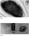
- a mature monostratified pigmented squamous epithelium black arrow
- tight junctions white arrow typical of the tissue structure of pigmented epithelial cells of the retina in vivo.
- the capsules obtained by encapsulation of pigmented epithelial cells of the retina resulting from the differentiation of cells induced to pluripotency show the structure in pigmented epithelial sheet whatever the number of cells in the tissue (of the order of a hundred cells on the left, size: 50 ⁇ m, insert A, and of the order of a thousand cells on the right, size: 250 ⁇ m, insert B) typical of the tissue structure of pigmented epithelial cells of the retina in vivo.
- the cells are organized in a hollow spheroid (insert B) hollow ovoid (insert E) according to an apico-basal polarization characterized in that the apical face is positioned towards the inside of the tissue unit and the basal face towards the outside of the tissue unit located here in contact with the alginate layer.
- the intracellular pigments are located on the apical face of the cells (insert D, transmitted light, white arrow) with a basal organization of the nuclei (insert B, DAPI, white arrow) corresponding to the topology encountered in vivo.
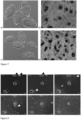
- retinal tissue units according to the invention were placed on a substrate simulating the extracellular matrix of the fundus of the eye (here matrigel simulating Brusch's membrane).
- the retinal tissue units black arrows
- the retinal tissue units are capable, thanks to the positioning of their basal face on the outer side, of attaching to the substrate simulating the extracellular matrix of the fundus of the eye by emitting cells adherent to the substrate (white arrows) which migrate (gray arrows) to cover the substrate and form a monolayer (bottom right insert corresponding to 122 hours of culture).
Landscapes
- Health & Medical Sciences (AREA)
- Life Sciences & Earth Sciences (AREA)
- Engineering & Computer Science (AREA)
- Chemical & Material Sciences (AREA)
- Biomedical Technology (AREA)
- General Health & Medical Sciences (AREA)
- Veterinary Medicine (AREA)
- Medicinal Chemistry (AREA)
- Animal Behavior & Ethology (AREA)
- Public Health (AREA)
- Cell Biology (AREA)
- Epidemiology (AREA)
- Transplantation (AREA)
- Oral & Maxillofacial Surgery (AREA)
- Dermatology (AREA)
- Chemical Kinetics & Catalysis (AREA)
- Zoology (AREA)
- Developmental Biology & Embryology (AREA)
- Ophthalmology & Optometry (AREA)
- Urology & Nephrology (AREA)
- Botany (AREA)
- Pharmacology & Pharmacy (AREA)
- Organic Chemistry (AREA)
- Bioinformatics & Cheminformatics (AREA)
- Biotechnology (AREA)
- Neurology (AREA)
- Neurosurgery (AREA)
- Nuclear Medicine, Radiotherapy & Molecular Imaging (AREA)
- Dispersion Chemistry (AREA)
- Hematology (AREA)
- General Chemical & Material Sciences (AREA)
- Immunology (AREA)
- Virology (AREA)
- Vascular Medicine (AREA)
- Heart & Thoracic Surgery (AREA)
- Polymers & Plastics (AREA)
- Cardiology (AREA)
- Wood Science & Technology (AREA)
- Genetics & Genomics (AREA)
- General Engineering & Computer Science (AREA)
Claims (18)
- Hohle dreidimensionale Netzhautgewebeeinheit, umfassend, um ein inneres Lumen herum organisiert, mindestens eine Schicht differenzierter lebender menschlicher Netzhautpigmentepithelzellen, wobei die basale Fläche jeder Netzhautpigmentepithelzelle nach außen und die apikale Fläche zu dem inneren Lumen positioniert ist.
- Netzhautgewebeeinheit nach Anspruch 1,
dadurch gekennzeichnet, dass sie auch eine äußere Schicht einer extrazellulären Matrix umfasst, die sich an der basalen Fläche der Netzhautpigmentepithelzellen befindet. - Netzhautgewebeeinheit nach einem der vorstehenden Ansprüche, dadurch gekennzeichnet, dass sie in der Form eines hohlen Ovoids, eines hohlen Zylinders, eines hohlen Sphäroids oder einer hohlen Kugel vorliegt.
- Netzhautgewebeeinheit nach dem vorstehenden Anspruch,
dadurch gekennzeichnet, dass ihre größte Abmessung zwischen 100 und 1000 µm liegt. - Netzhautgewebeeinheit nach dem vorstehenden Anspruch,
dadurch gekennzeichnet, dass ihre kleinste Abmessung zwischen 10 und 1000 µm liegt. - Netzhautgewebeeinheit nach einem der vorstehenden Ansprüche, dadurch gekennzeichnet, dass nebeneinanderliegende Netzhautpigmentepithelzellen an ihren seitlichen Flächen durch Tight Junctions miteinander verbunden sind.
- Netzhautgewebeeinheit nach einem der vorstehenden Ansprüche, dadurch gekennzeichnet, dass sie ferner an der apikalen Fläche der Netzhautpigmentepithelzellen um das innere Lumen herum organisiert mindestens eine Schicht differenzierter lebender menschlicher Netzhautzellen außer den Netzhautpigmentepithelzellen umfasst.
- Netzhautgewebeeinheit nach dem vorstehenden Anspruch,
dadurch gekennzeichnet, dass differenzierte lebende menschliche Netzhautzellen außer den Netzhautpigmentepithelzellen aus Stäbchen, Zapfen, Lymphknotenzellen, Amakrinzellen, bipolaren Zellen und horizontalen Zellen ausgewählt sind. - Netzhautgewebeeinheit nach einem der vorstehenden Ansprüche, dadurch gekennzeichnet, dass sie 10 bis 100.000 Netzhautzellen enthält.
- Netzhautgewebeeinheit nach einem der vorstehenden Ansprüche, dadurch gekennzeichnet, dass die Netzhautpigmentepithelzellen und/oder die optionalen anderen Netzhautzellen aus induzierten pluripotenten Stammzellen (IPS) erhalten wurden.
- Netzhautgewebeeinheit nach einem der vorstehenden Ansprüche, dadurch gekennzeichnet, dass sie in einer Hydrogelkapsel eingekapselt ist.
- Verfahren zum Herstellen einer Netzhautgewebeeinheit nach einem der Ansprüche 1 bis 11, umfassend die Schritte bestehend aus:- Erzeugen eines zellulären Mikrokompartiments, umfassend, im Inneren einer Hydrogelkapsel:* optional mindestens Elemente der extrazellulären Matrix, die durch die Zellen abgesondert oder zugesetzt sind,* Zellen, die geeignet sind, sich mindestens in Netzhautpigmentepithelzellen zu differenzieren, oder mindestens differenzierte Netzhautpigmentepithelzellen, wobei die Zellen, die geeignet sind, sich mindestens in Netzhautpigmentepithelzellen zu differenzieren, ausgewählt sind aus:* Stammzellen, die in der Lage sind, sich in Netzhautzellen, mindestens in Netzhautpigmentepithelzellen, zu differenzieren,* Vorläuferzellen, die in der Lage sind, sich in Netzhautzellen, mindestens in Netzhautpigmentepithelzellen, zu differenzieren,*differenzierte Zellen, die in der Lage sind, eine Transdifferenzierung in Netzhautzellen, mindestens in Netzhautpigmentepithelzellen, zu erfahren,* differenzierte Zellen, die in der Lage sind, eine Umprogrammierung zu erfahren, sodass sie zu induzierten pluripotenten Stammzellen werden, die in der Lage sind, sich in Netzhautzellen, mindestens in Netzhautpigmentepithelzellen, zu differenzieren- falls die in das Mikrokompartiment eingebrachten Zellen Zellen sind, die geeignet sind, sich mindestens in Netzhautpigmentepithelzellen zu differenzieren: Induzieren der Zelldifferenzierung innerhalb des zellulären Mikrokompartiments, um mindestens Netzhautpigmentepithelzellen und optional andere Netzhautzellen zu erhalten,- Entfernen der Hydrogelkapseln durch Hydrolyse, Auflösung, Durchbohren und/oder Aufbrechen zum Gewinnen der Netzhautpigmentepithelzellen und der optionalen anderen Netzhautzellen in Form einer hohlen dreidimensionalen Netzhautgewebeeinheit,
- Verfahren nach dem vorstehenden Anspruch,
dadurch gekennzeichnet, dass es einen Schritt einer Amplifikation von Netzhautpigmentepithelzellen umfasst. - Verfahren nach einem der Ansprüche 12 oder 13,
dadurch gekennzeichnet, dass die Zellen, die geeignet sind, sich mindestens in Netzhautpigmentepithelzellen zu differenzieren, pluripotente Stammzellen sind. - Verfahren nach dem vorstehenden Anspruch,
dadurch gekennzeichnet, dass die pluripotenten Stammzellen induzierte pluripotente Stammzellen (IPS) sind. - Verfahren zum Herstellen einer Netzhautgewebeeinheit nach einem der Ansprüche 12 bis 15, dadurch gekennzeichnet, dass es einen zusätzlichen Schritt umfasst, der darin besteht, eine chirurgische Implantationsvorrichtung, die für eine Injektion in das Auge geeignet ist, mit der Gewebeeinheit zu beladen.
- Kit zum Implantieren in das Auge von Gewebeeinheiten nach einem der Ansprüche 1 bis 11, dadurch gekennzeichnet, dass es umfasst:- zwischen 1 und 10.000 Gewebeeinheiten nach einem der Ansprüche 1 bis 11,- eine chirurgische Implantationsvorrichtung, die geeignet ist, die Gewebeeinheit(en) in ein menschliches Auge zu implantieren.
- Kit zum Implantieren von Gewebeeinheiten in das Auge nach Anspruch 17, dadurch gekennzeichnet, dass es umfasst:- zwischen 1 und 10.000 Gewebeeinheiten nach Anspruch 11,- Mittel zum Entfernen von Hydrogelkapseln,- eine chirurgische Implantationsvorrichtung, die geeignet ist, die Gewebeeinheit(en) in ein menschliches Auge zu implantieren.
Applications Claiming Priority (2)
| Application Number | Priority Date | Filing Date | Title |
|---|---|---|---|
| FR1909155A FR3099882A1 (fr) | 2019-08-12 | 2019-08-12 | Unité tridimensionnelle creuse de tissu rétinien et utilisation dans le traitement des rétinopathies |
| PCT/EP2020/072567 WO2021028456A1 (fr) | 2019-08-12 | 2020-08-12 | Unite tridimensionnelle creuse de tissu retinien et utilisation dans le traitement des retinopathies |
Publications (2)
| Publication Number | Publication Date |
|---|---|
| EP4013433A1 EP4013433A1 (de) | 2022-06-22 |
| EP4013433B1 true EP4013433B1 (de) | 2024-11-20 |
Family
ID=70008570
Family Applications (1)
| Application Number | Title | Priority Date | Filing Date |
|---|---|---|---|
| EP20761761.4A Active EP4013433B1 (de) | 2019-08-12 | 2020-08-12 | Hohle dreidimensionale einheit aus retinalem gewebe und deren verwendung zur behandlung von retinopathien |
Country Status (13)
| Country | Link |
|---|---|
| US (1) | US20220273727A1 (de) |
| EP (1) | EP4013433B1 (de) |
| JP (1) | JP7653974B2 (de) |
| KR (1) | KR20220054613A (de) |
| CN (1) | CN114423857A (de) |
| AU (1) | AU2020327612A1 (de) |
| BR (1) | BR112022002624A2 (de) |
| CA (1) | CA3147254A1 (de) |
| ES (1) | ES3013617T3 (de) |
| FR (1) | FR3099882A1 (de) |
| HU (1) | HUE070580T2 (de) |
| IL (1) | IL290532A (de) |
| WO (1) | WO2021028456A1 (de) |
Families Citing this family (3)
| Publication number | Priority date | Publication date | Assignee | Title |
|---|---|---|---|---|
| JP2024521447A (ja) * | 2021-06-16 | 2024-05-31 | ツリーフロッグ テラピューティクス | 複数の嚢胞を含む大型細胞微小区画 |
| FR3124193B3 (fr) * | 2021-06-16 | 2023-09-22 | Treefrog Therapeutics | Microcompartiments cellulaires de grande taille comprenant plusieurs cystes |
| CN117417891A (zh) * | 2023-12-19 | 2024-01-19 | 南京艾尔普再生医学科技有限公司 | 一种视网膜色素上皮细胞球形制剂 |
Family Cites Families (8)
| Publication number | Priority date | Publication date | Assignee | Title |
|---|---|---|---|---|
| WO2003018040A1 (en) * | 2001-08-24 | 2003-03-06 | The Schepens Eye Research Institute, Inc. | Composite graft for treatment of retinal diseases |
| JP5867926B2 (ja) | 2010-05-10 | 2016-02-24 | 公立大学法人名古屋市立大学 | 細胞シート作製方法 |
| US9248013B2 (en) | 2011-12-05 | 2016-02-02 | California Institute Of Technology | 3-Dimensional parylene scaffold cage |
| EP3211071B1 (de) | 2014-10-24 | 2021-05-05 | Sumitomo Dainippon Pharma Co., Ltd. | Herstellungsverfahren für netzhautgewebe |
| WO2017176810A1 (en) * | 2016-04-04 | 2017-10-12 | Biotime, Inc. | Pluripotent stem cell-derived 3d retinal tissue and uses thereof |
| FR3058892B1 (fr) * | 2016-11-23 | 2021-04-09 | Univ Bordeaux | Unite de tissu neural et utilisation d'une telle unite pour l'implantation dans le systeme nerveux d'un mammifere |
| FR3059009B1 (fr) | 2016-11-23 | 2018-12-07 | Universite de Bordeaux | Microcompartiment cellulaire et procedes de preparation |
| CA3074426A1 (en) * | 2017-09-08 | 2019-03-14 | Sumitomo Dainippon Pharma Co., Ltd. | Cell aggregate including retinal tissue and production method therefor |
-
2019
- 2019-08-12 FR FR1909155A patent/FR3099882A1/fr active Pending
-
2020
- 2020-08-12 CN CN202080065333.6A patent/CN114423857A/zh active Pending
- 2020-08-12 CA CA3147254A patent/CA3147254A1/fr active Pending
- 2020-08-12 JP JP2022508984A patent/JP7653974B2/ja active Active
- 2020-08-12 AU AU2020327612A patent/AU2020327612A1/en not_active Abandoned
- 2020-08-12 HU HUE20761761A patent/HUE070580T2/hu unknown
- 2020-08-12 BR BR112022002624A patent/BR112022002624A2/pt unknown
- 2020-08-12 ES ES20761761T patent/ES3013617T3/es active Active
- 2020-08-12 US US17/634,794 patent/US20220273727A1/en active Pending
- 2020-08-12 KR KR1020227008319A patent/KR20220054613A/ko not_active Ceased
- 2020-08-12 WO PCT/EP2020/072567 patent/WO2021028456A1/fr not_active Ceased
- 2020-08-12 EP EP20761761.4A patent/EP4013433B1/de active Active
-
2022
- 2022-02-10 IL IL290532A patent/IL290532A/en unknown
Also Published As
| Publication number | Publication date |
|---|---|
| IL290532A (en) | 2022-04-01 |
| JP7653974B2 (ja) | 2025-03-31 |
| KR20220054613A (ko) | 2022-05-03 |
| CN114423857A (zh) | 2022-04-29 |
| EP4013433A1 (de) | 2022-06-22 |
| HUE070580T2 (hu) | 2025-06-28 |
| ES3013617T3 (en) | 2025-04-14 |
| US20220273727A1 (en) | 2022-09-01 |
| JP2022544941A (ja) | 2022-10-24 |
| FR3099882A1 (fr) | 2021-02-19 |
| AU2020327612A1 (en) | 2022-03-03 |
| WO2021028456A1 (fr) | 2021-02-18 |
| CA3147254A1 (fr) | 2021-02-18 |
| BR112022002624A2 (pt) | 2022-05-03 |
Similar Documents
| Publication | Publication Date | Title |
|---|---|---|
| EP3903795B1 (de) | Nervengewebeeinheit und verwendung solch einer einheit zur implantation im nervensystem eines säugers | |
| Stern et al. | Regenerating eye tissues to preserve and restore vision | |
| EP4013433B1 (de) | Hohle dreidimensionale einheit aus retinalem gewebe und deren verwendung zur behandlung von retinopathien | |
| Lee et al. | Transplantation of human corneal limbal epithelial cell sheet harvested on synthesized carboxymethyl cellulose and dopamine in a limbal stem cell deficiency | |
| FR3141945A1 (fr) | Substitut de matrice extracellulaire dans un microcompartiment cellulaire | |
| CA3222350A1 (fr) | Microcompartiments cellulaires de grande taille comprenant plusieurs cystes | |
| WO2022058617A1 (fr) | Tissu cardiaque particulier et utilisation dans le traitement de pathologies cardiaque | |
| EP4214305A1 (de) | Zellmikrokompartimente mit menschlichen zellen, die herzdifferenziert werden, aus diesen mikrokompartimenten erhaltene gewebe und verwendungen davon | |
| FR3164726A3 (fr) | Unité de tissu neural et utilisation d’une telle unité comme médicament | |
| WO2026022106A1 (fr) | Unité de tissu neural et utilisation d'une telle unité comme médicament |
Legal Events
| Date | Code | Title | Description |
|---|---|---|---|
| STAA | Information on the status of an ep patent application or granted ep patent |
Free format text: STATUS: UNKNOWN |
|
| STAA | Information on the status of an ep patent application or granted ep patent |
Free format text: STATUS: THE INTERNATIONAL PUBLICATION HAS BEEN MADE |
|
| PUAI | Public reference made under article 153(3) epc to a published international application that has entered the european phase |
Free format text: ORIGINAL CODE: 0009012 |
|
| STAA | Information on the status of an ep patent application or granted ep patent |
Free format text: STATUS: REQUEST FOR EXAMINATION WAS MADE |
|
| 17P | Request for examination filed |
Effective date: 20220204 |
|
| AK | Designated contracting states |
Kind code of ref document: A1 Designated state(s): AL AT BE BG CH CY CZ DE DK EE ES FI FR GB GR HR HU IE IS IT LI LT LU LV MC MK MT NL NO PL PT RO RS SE SI SK SM TR |
|
| DAV | Request for validation of the european patent (deleted) | ||
| DAX | Request for extension of the european patent (deleted) | ||
| STAA | Information on the status of an ep patent application or granted ep patent |
Free format text: STATUS: EXAMINATION IS IN PROGRESS |
|
| 17Q | First examination report despatched |
Effective date: 20230309 |
|
| P01 | Opt-out of the competence of the unified patent court (upc) registered |
Effective date: 20230518 |
|
| GRAP | Despatch of communication of intention to grant a patent |
Free format text: ORIGINAL CODE: EPIDOSNIGR1 |
|
| STAA | Information on the status of an ep patent application or granted ep patent |
Free format text: STATUS: GRANT OF PATENT IS INTENDED |
|
| INTG | Intention to grant announced |
Effective date: 20240617 |
|
| GRAS | Grant fee paid |
Free format text: ORIGINAL CODE: EPIDOSNIGR3 |
|
| GRAA | (expected) grant |
Free format text: ORIGINAL CODE: 0009210 |
|
| STAA | Information on the status of an ep patent application or granted ep patent |
Free format text: STATUS: THE PATENT HAS BEEN GRANTED |
|
| AK | Designated contracting states |
Kind code of ref document: B1 Designated state(s): AL AT BE BG CH CY CZ DE DK EE ES FI FR GB GR HR HU IE IS IT LI LT LU LV MC MK MT NL NO PL PT RO RS SE SI SK SM TR |
|
| REG | Reference to a national code |
Ref country code: GB Ref legal event code: FG4D Free format text: NOT ENGLISH |
|
| REG | Reference to a national code |
Ref country code: CH Ref legal event code: EP |
|
| REG | Reference to a national code |
Ref country code: DE Ref legal event code: R096 Ref document number: 602020041706 Country of ref document: DE |
|
| REG | Reference to a national code |
Ref country code: IE Ref legal event code: FG4D Free format text: LANGUAGE OF EP DOCUMENT: FRENCH |
|
| REG | Reference to a national code |
Ref country code: NL Ref legal event code: FP |
|
| REG | Reference to a national code |
Ref country code: SE Ref legal event code: TRGR |
|
| REG | Reference to a national code |
Ref country code: LT Ref legal event code: MG9D |
|
| PG25 | Lapsed in a contracting state [announced via postgrant information from national office to epo] |
Ref country code: HR Free format text: LAPSE BECAUSE OF FAILURE TO SUBMIT A TRANSLATION OF THE DESCRIPTION OR TO PAY THE FEE WITHIN THE PRESCRIBED TIME-LIMIT Effective date: 20241120 Ref country code: IS Free format text: LAPSE BECAUSE OF FAILURE TO SUBMIT A TRANSLATION OF THE DESCRIPTION OR TO PAY THE FEE WITHIN THE PRESCRIBED TIME-LIMIT Effective date: 20250320 Ref country code: PT Free format text: LAPSE BECAUSE OF FAILURE TO SUBMIT A TRANSLATION OF THE DESCRIPTION OR TO PAY THE FEE WITHIN THE PRESCRIBED TIME-LIMIT Effective date: 20250320 |
|
| REG | Reference to a national code |
Ref country code: ES Ref legal event code: FG2A Ref document number: 3013617 Country of ref document: ES Kind code of ref document: T3 Effective date: 20250414 |
|
| PG25 | Lapsed in a contracting state [announced via postgrant information from national office to epo] |
Ref country code: FI Free format text: LAPSE BECAUSE OF FAILURE TO SUBMIT A TRANSLATION OF THE DESCRIPTION OR TO PAY THE FEE WITHIN THE PRESCRIBED TIME-LIMIT Effective date: 20241120 |
|
| REG | Reference to a national code |
Ref country code: AT Ref legal event code: MK05 Ref document number: 1742975 Country of ref document: AT Kind code of ref document: T Effective date: 20241120 |
|
| PG25 | Lapsed in a contracting state [announced via postgrant information from national office to epo] |
Ref country code: BG Free format text: LAPSE BECAUSE OF FAILURE TO SUBMIT A TRANSLATION OF THE DESCRIPTION OR TO PAY THE FEE WITHIN THE PRESCRIBED TIME-LIMIT Effective date: 20241120 |
|
| PG25 | Lapsed in a contracting state [announced via postgrant information from national office to epo] |
Ref country code: NO Free format text: LAPSE BECAUSE OF FAILURE TO SUBMIT A TRANSLATION OF THE DESCRIPTION OR TO PAY THE FEE WITHIN THE PRESCRIBED TIME-LIMIT Effective date: 20250220 |
|
| PG25 | Lapsed in a contracting state [announced via postgrant information from national office to epo] |
Ref country code: AT Free format text: LAPSE BECAUSE OF FAILURE TO SUBMIT A TRANSLATION OF THE DESCRIPTION OR TO PAY THE FEE WITHIN THE PRESCRIBED TIME-LIMIT Effective date: 20241120 Ref country code: LV Free format text: LAPSE BECAUSE OF FAILURE TO SUBMIT A TRANSLATION OF THE DESCRIPTION OR TO PAY THE FEE WITHIN THE PRESCRIBED TIME-LIMIT Effective date: 20241120 Ref country code: GR Free format text: LAPSE BECAUSE OF FAILURE TO SUBMIT A TRANSLATION OF THE DESCRIPTION OR TO PAY THE FEE WITHIN THE PRESCRIBED TIME-LIMIT Effective date: 20250221 |
|
| PG25 | Lapsed in a contracting state [announced via postgrant information from national office to epo] |
Ref country code: PL Free format text: LAPSE BECAUSE OF FAILURE TO SUBMIT A TRANSLATION OF THE DESCRIPTION OR TO PAY THE FEE WITHIN THE PRESCRIBED TIME-LIMIT Effective date: 20241120 |
|
| PG25 | Lapsed in a contracting state [announced via postgrant information from national office to epo] |
Ref country code: RS Free format text: LAPSE BECAUSE OF FAILURE TO SUBMIT A TRANSLATION OF THE DESCRIPTION OR TO PAY THE FEE WITHIN THE PRESCRIBED TIME-LIMIT Effective date: 20250220 |
|
| REG | Reference to a national code |
Ref country code: HU Ref legal event code: AG4A Ref document number: E070580 Country of ref document: HU |
|
| PG25 | Lapsed in a contracting state [announced via postgrant information from national office to epo] |
Ref country code: SM Free format text: LAPSE BECAUSE OF FAILURE TO SUBMIT A TRANSLATION OF THE DESCRIPTION OR TO PAY THE FEE WITHIN THE PRESCRIBED TIME-LIMIT Effective date: 20241120 |
|
| PG25 | Lapsed in a contracting state [announced via postgrant information from national office to epo] |
Ref country code: DK Free format text: LAPSE BECAUSE OF FAILURE TO SUBMIT A TRANSLATION OF THE DESCRIPTION OR TO PAY THE FEE WITHIN THE PRESCRIBED TIME-LIMIT Effective date: 20241120 |
|
| PG25 | Lapsed in a contracting state [announced via postgrant information from national office to epo] |
Ref country code: EE Free format text: LAPSE BECAUSE OF FAILURE TO SUBMIT A TRANSLATION OF THE DESCRIPTION OR TO PAY THE FEE WITHIN THE PRESCRIBED TIME-LIMIT Effective date: 20241120 |
|
| PG25 | Lapsed in a contracting state [announced via postgrant information from national office to epo] |
Ref country code: RO Free format text: LAPSE BECAUSE OF FAILURE TO SUBMIT A TRANSLATION OF THE DESCRIPTION OR TO PAY THE FEE WITHIN THE PRESCRIBED TIME-LIMIT Effective date: 20241120 |
|
| PG25 | Lapsed in a contracting state [announced via postgrant information from national office to epo] |
Ref country code: SK Free format text: LAPSE BECAUSE OF FAILURE TO SUBMIT A TRANSLATION OF THE DESCRIPTION OR TO PAY THE FEE WITHIN THE PRESCRIBED TIME-LIMIT Effective date: 20241120 |
|
| REG | Reference to a national code |
Ref country code: DE Ref legal event code: R097 Ref document number: 602020041706 Country of ref document: DE |
|
| PGFP | Annual fee paid to national office [announced via postgrant information from national office to epo] |
Ref country code: NL Payment date: 20250821 Year of fee payment: 6 |
|
| PGFP | Annual fee paid to national office [announced via postgrant information from national office to epo] |
Ref country code: HU Payment date: 20250807 Year of fee payment: 6 |
|
| PLBE | No opposition filed within time limit |
Free format text: ORIGINAL CODE: 0009261 |
|
| STAA | Information on the status of an ep patent application or granted ep patent |
Free format text: STATUS: NO OPPOSITION FILED WITHIN TIME LIMIT |
|
| PGFP | Annual fee paid to national office [announced via postgrant information from national office to epo] |
Ref country code: ES Payment date: 20250917 Year of fee payment: 6 |
|
| PGFP | Annual fee paid to national office [announced via postgrant information from national office to epo] |
Ref country code: DE Payment date: 20250819 Year of fee payment: 6 |
|
| PGFP | Annual fee paid to national office [announced via postgrant information from national office to epo] |
Ref country code: IT Payment date: 20250829 Year of fee payment: 6 |
|
| PGFP | Annual fee paid to national office [announced via postgrant information from national office to epo] |
Ref country code: GB Payment date: 20250822 Year of fee payment: 6 Ref country code: BE Payment date: 20250820 Year of fee payment: 6 |
|
| PGFP | Annual fee paid to national office [announced via postgrant information from national office to epo] |
Ref country code: FR Payment date: 20250822 Year of fee payment: 6 |
|
| PGFP | Annual fee paid to national office [announced via postgrant information from national office to epo] |
Ref country code: CH Payment date: 20250901 Year of fee payment: 6 Ref country code: SE Payment date: 20250821 Year of fee payment: 6 |
|
| 26N | No opposition filed |
Effective date: 20250821 |
|
| PGFP | Annual fee paid to national office [announced via postgrant information from national office to epo] |
Ref country code: CZ Payment date: 20250801 Year of fee payment: 6 |
|
| PG25 | Lapsed in a contracting state [announced via postgrant information from national office to epo] |
Ref country code: MC Free format text: LAPSE BECAUSE OF FAILURE TO SUBMIT A TRANSLATION OF THE DESCRIPTION OR TO PAY THE FEE WITHIN THE PRESCRIBED TIME-LIMIT Effective date: 20241120 |
|
| PG25 | Lapsed in a contracting state [announced via postgrant information from national office to epo] |
Ref country code: LU Free format text: LAPSE BECAUSE OF NON-PAYMENT OF DUE FEES Effective date: 20250812 |