EP4019552A1 - Anticorps spécifiques de l'anhydrase carbonique ix et leurs utilisations - Google Patents
Anticorps spécifiques de l'anhydrase carbonique ix et leurs utilisations Download PDFInfo
- Publication number
- EP4019552A1 EP4019552A1 EP21214012.3A EP21214012A EP4019552A1 EP 4019552 A1 EP4019552 A1 EP 4019552A1 EP 21214012 A EP21214012 A EP 21214012A EP 4019552 A1 EP4019552 A1 EP 4019552A1
- Authority
- EP
- European Patent Office
- Prior art keywords
- fragment
- antibody
- sequence
- isolated
- seq
- Prior art date
- Legal status (The legal status is an assumption and is not a legal conclusion. Google has not performed a legal analysis and makes no representation as to the accuracy of the status listed.)
- Withdrawn
Links
Images










Classifications
-
- C—CHEMISTRY; METALLURGY
- C07—ORGANIC CHEMISTRY
- C07K—PEPTIDES
- C07K16/00—Immunoglobulins [IG], e.g. monoclonal or polyclonal antibodies
- C07K16/40—Immunoglobulins [IG], e.g. monoclonal or polyclonal antibodies against enzymes
-
- A—HUMAN NECESSITIES
- A61—MEDICAL OR VETERINARY SCIENCE; HYGIENE
- A61K—PREPARATIONS FOR MEDICAL, DENTAL OR TOILETRY PURPOSES
- A61K47/00—Medicinal preparations characterised by the non-active ingredients used, e.g. carriers or inert additives; Targeting or modifying agents chemically bound to the active ingredient
- A61K47/50—Medicinal preparations characterised by the non-active ingredients used, e.g. carriers or inert additives; Targeting or modifying agents chemically bound to the active ingredient the non-active ingredient being chemically bound to the active ingredient, e.g. polymer-drug conjugates
- A61K47/51—Medicinal preparations characterised by the non-active ingredients used, e.g. carriers or inert additives; Targeting or modifying agents chemically bound to the active ingredient the non-active ingredient being chemically bound to the active ingredient, e.g. polymer-drug conjugates the non-active ingredient being a modifying agent
- A61K47/68—Medicinal preparations characterised by the non-active ingredients used, e.g. carriers or inert additives; Targeting or modifying agents chemically bound to the active ingredient the non-active ingredient being chemically bound to the active ingredient, e.g. polymer-drug conjugates the non-active ingredient being a modifying agent the modifying agent being an antibody, an immunoglobulin or a fragment thereof, e.g. an Fc-fragment
- A61K47/6801—Drug-antibody or immunoglobulin conjugates defined by the pharmacologically or therapeutically active agent
- A61K47/6803—Drugs conjugated to an antibody or immunoglobulin, e.g. cisplatin-antibody conjugates
- A61K47/68033—Drugs conjugated to an antibody or immunoglobulin, e.g. cisplatin-antibody conjugates the drug being a maytansine
-
- A—HUMAN NECESSITIES
- A61—MEDICAL OR VETERINARY SCIENCE; HYGIENE
- A61K—PREPARATIONS FOR MEDICAL, DENTAL OR TOILETRY PURPOSES
- A61K47/00—Medicinal preparations characterised by the non-active ingredients used, e.g. carriers or inert additives; Targeting or modifying agents chemically bound to the active ingredient
- A61K47/50—Medicinal preparations characterised by the non-active ingredients used, e.g. carriers or inert additives; Targeting or modifying agents chemically bound to the active ingredient the non-active ingredient being chemically bound to the active ingredient, e.g. polymer-drug conjugates
- A61K47/51—Medicinal preparations characterised by the non-active ingredients used, e.g. carriers or inert additives; Targeting or modifying agents chemically bound to the active ingredient the non-active ingredient being chemically bound to the active ingredient, e.g. polymer-drug conjugates the non-active ingredient being a modifying agent
- A61K47/68—Medicinal preparations characterised by the non-active ingredients used, e.g. carriers or inert additives; Targeting or modifying agents chemically bound to the active ingredient the non-active ingredient being chemically bound to the active ingredient, e.g. polymer-drug conjugates the non-active ingredient being a modifying agent the modifying agent being an antibody, an immunoglobulin or a fragment thereof, e.g. an Fc-fragment
- A61K47/6835—Medicinal preparations characterised by the non-active ingredients used, e.g. carriers or inert additives; Targeting or modifying agents chemically bound to the active ingredient the non-active ingredient being chemically bound to the active ingredient, e.g. polymer-drug conjugates the non-active ingredient being a modifying agent the modifying agent being an antibody, an immunoglobulin or a fragment thereof, e.g. an Fc-fragment the modifying agent being an antibody or an immunoglobulin bearing at least one antigen-binding site
- A61K47/6871—Medicinal preparations characterised by the non-active ingredients used, e.g. carriers or inert additives; Targeting or modifying agents chemically bound to the active ingredient the non-active ingredient being chemically bound to the active ingredient, e.g. polymer-drug conjugates the non-active ingredient being a modifying agent the modifying agent being an antibody, an immunoglobulin or a fragment thereof, e.g. an Fc-fragment the modifying agent being an antibody or an immunoglobulin bearing at least one antigen-binding site the antibody targeting an enzyme
-
- A—HUMAN NECESSITIES
- A61—MEDICAL OR VETERINARY SCIENCE; HYGIENE
- A61K—PREPARATIONS FOR MEDICAL, DENTAL OR TOILETRY PURPOSES
- A61K51/00—Preparations containing radioactive substances for use in therapy or testing in vivo
- A61K51/02—Preparations containing radioactive substances for use in therapy or testing in vivo characterised by the carrier, i.e. characterised by the agent or material covalently linked or complexing the radioactive nucleus
- A61K51/04—Organic compounds
- A61K51/08—Peptides, e.g. proteins, carriers being peptides, polyamino acids, proteins
- A61K51/10—Antibodies or immunoglobulins; Fragments thereof, the carrier being an antibody, an immunoglobulin or a fragment thereof, e.g. a camelised human single domain antibody or the Fc fragment of an antibody
- A61K51/1075—Antibodies or immunoglobulins; Fragments thereof, the carrier being an antibody, an immunoglobulin or a fragment thereof, e.g. a camelised human single domain antibody or the Fc fragment of an antibody the antibody being against an enzyme
-
- G—PHYSICS
- G01—MEASURING; TESTING
- G01N—INVESTIGATING OR ANALYSING MATERIALS BY DETERMINING THEIR CHEMICAL OR PHYSICAL PROPERTIES
- G01N33/00—Investigating or analysing materials by specific methods not covered by groups G01N1/00 - G01N31/00
- G01N33/48—Biological material, e.g. blood, urine; Haemocytometers
- G01N33/50—Chemical analysis of biological material, e.g. blood, urine; Testing involving biospecific ligand binding methods; Immunological testing
- G01N33/53—Immunoassay; Biospecific binding assay; Materials therefor
- G01N33/573—Immunoassay; Biospecific binding assay; Materials therefor for enzymes or isoenzymes
-
- A—HUMAN NECESSITIES
- A61—MEDICAL OR VETERINARY SCIENCE; HYGIENE
- A61K—PREPARATIONS FOR MEDICAL, DENTAL OR TOILETRY PURPOSES
- A61K39/00—Medicinal preparations containing antigens or antibodies
- A61K2039/505—Medicinal preparations containing antigens or antibodies comprising antibodies
-
- C—CHEMISTRY; METALLURGY
- C07—ORGANIC CHEMISTRY
- C07K—PEPTIDES
- C07K2317/00—Immunoglobulins specific features
- C07K2317/20—Immunoglobulins specific features characterized by taxonomic origin
- C07K2317/24—Immunoglobulins specific features characterized by taxonomic origin containing regions, domains or residues from different species, e.g. chimeric, humanized or veneered
-
- C—CHEMISTRY; METALLURGY
- C07—ORGANIC CHEMISTRY
- C07K—PEPTIDES
- C07K2317/00—Immunoglobulins specific features
- C07K2317/30—Immunoglobulins specific features characterized by aspects of specificity or valency
- C07K2317/33—Crossreactivity, e.g. for species or epitope, or lack of said crossreactivity
-
- C—CHEMISTRY; METALLURGY
- C07—ORGANIC CHEMISTRY
- C07K—PEPTIDES
- C07K2317/00—Immunoglobulins specific features
- C07K2317/30—Immunoglobulins specific features characterized by aspects of specificity or valency
- C07K2317/34—Identification of a linear epitope shorter than 20 amino acid residues or of a conformational epitope defined by amino acid residues
-
- C—CHEMISTRY; METALLURGY
- C07—ORGANIC CHEMISTRY
- C07K—PEPTIDES
- C07K2317/00—Immunoglobulins specific features
- C07K2317/50—Immunoglobulins specific features characterized by immunoglobulin fragments
- C07K2317/56—Immunoglobulins specific features characterized by immunoglobulin fragments variable (Fv) region, i.e. VH and/or VL
-
- C—CHEMISTRY; METALLURGY
- C07—ORGANIC CHEMISTRY
- C07K—PEPTIDES
- C07K2317/00—Immunoglobulins specific features
- C07K2317/50—Immunoglobulins specific features characterized by immunoglobulin fragments
- C07K2317/56—Immunoglobulins specific features characterized by immunoglobulin fragments variable (Fv) region, i.e. VH and/or VL
- C07K2317/565—Complementarity determining region [CDR]
-
- C—CHEMISTRY; METALLURGY
- C07—ORGANIC CHEMISTRY
- C07K—PEPTIDES
- C07K2317/00—Immunoglobulins specific features
- C07K2317/50—Immunoglobulins specific features characterized by immunoglobulin fragments
- C07K2317/56—Immunoglobulins specific features characterized by immunoglobulin fragments variable (Fv) region, i.e. VH and/or VL
- C07K2317/567—Framework region [FR]
-
- C—CHEMISTRY; METALLURGY
- C07—ORGANIC CHEMISTRY
- C07K—PEPTIDES
- C07K2317/00—Immunoglobulins specific features
- C07K2317/70—Immunoglobulins specific features characterized by effect upon binding to a cell or to an antigen
- C07K2317/76—Antagonist effect on antigen, e.g. neutralization or inhibition of binding
-
- C—CHEMISTRY; METALLURGY
- C07—ORGANIC CHEMISTRY
- C07K—PEPTIDES
- C07K2317/00—Immunoglobulins specific features
- C07K2317/90—Immunoglobulins specific features characterized by (pharmaco)kinetic aspects or by stability of the immunoglobulin
- C07K2317/92—Affinity (KD), association rate (Ka), dissociation rate (Kd) or EC50 value
-
- G—PHYSICS
- G01—MEASURING; TESTING
- G01N—INVESTIGATING OR ANALYSING MATERIALS BY DETERMINING THEIR CHEMICAL OR PHYSICAL PROPERTIES
- G01N2333/00—Assays involving biological materials from specific organisms or of a specific nature
- G01N2333/90—Enzymes; Proenzymes
- G01N2333/988—Lyases (4.), e.g. aldolases, heparinase, enolases, fumarase
Definitions
- the present invention relates to Carbohydrate Anhydrase IX-specific antibodies, fragments thereof, and uses thereof. More specifically, the present invention relates to high-affinity Carbohydrate Anhydrase IX-specific antibodies and fragments thereof and their use as antibody-drug conjugates.
- CA Carbonic anhydrases
- CA-IX has been shown to be involved in cell adhesion and migration (Svastova et al., 2011) and has been associated with cancer progression, metastasis and poor clinical outcome (Neri et al., 2011).
- CA-IX also known as MN, P54/58N or Renal Cell Carcinoma (RCC)-associated protein G250
- RRC Renal Cell Carcinoma
- CA-IX The C-terminal intracellular portion of CA-IX is involved in the inside-out regulation of the extracellular catalytic domain through the phosphorylation of Thr-443 by protein kinase A (PKA) (Hulikova et al., 2009; Ditte et al., 2011). Expression of CA-IX is tightly controlled by hypoxia-inducible factor 1 alpha (HIF-1 ⁇ ). CA-IX is expressed on the surface of tumor cells located in pre-necrotic areas of tumors (Wykoff et al., 2000) where it is involved in promoting tumor cell survival, the accelerated degradation of the extracellular matrix (ECM) and metastasis.
- ECM extracellular matrix
- CA-IX has a very selective expression pattern in normal tissue.
- the mucosa of the gall bladder and stomach express high levels of CA-IX.
- Low expression levels of CA-IX levels can be found in the intestinal epithelium, and even lower levels in pancreatic duct epithelium, male reproductive organs, and cells that line the body cavity. All other normal tissues do not express CA-IX. Cancerous tissues however, especially those of the cervix, kidney and lung, express high levels of CA-IX thus making CA-IX a very attractive therapeutic tumor target.
- M75 One of the earliest monoclonal antibodies (mAb) raised against CA-IX is M75 (Pastorekova et al., 1992), which binds to CA-IX's PG-like domain. M75 has been predominately used as tool for CA-IX detection in vitro and in vivo (Chrastina et al., 2003a, 2003b; Zatovicova et al., 2010).
- mAb G250 A second anti-CA-IX mAb, mAb G250 (Oosterwijk et al., 1986), was shown to interact with CA-IX's catalytic domain without however inhibiting its enzyme activity.
- a chimeric version of G250 (designated cG250) was developed as a therapeutic antibody (Surfus et al., 1996; Oosterwijk, 2008) with a mechanism of action that was shown to rely predominantly on an Antibody-Dependent Cellular Cytotoxicity (ADCC) response.
- cG250 does however not improve the disease-free survival rate of patients (> 6-year span) compared to a placebo (Bleumer et al., 2004).
- the mAb continues to be developed for the treatment of cancer in combination with IL2 or IFN- ⁇ , as an imaging diagnostic agent and for in vitro diagnostics (IVD) immunohistochemistry (IHC) assays.
- cG250 is also used as a vehicle for the delivery of radionuclides. Specifically, Brouwers et al. (2004) successfully used cG250 to shuttle 177 Lu and 90 Y into tumor cells, causing growth retardation of xenograft tumors. Clinical phase II/III studies with these labeled mAbs are currently underway (Stillebroer et al., 2012). Also in development are antibody-drug conjugates (ADC) based on cG250, however little is known their efficacy. Such antibody-drug conjugates are an attractive option in cancer therapy, as they combine the selective targeting ability of the antibody with the cell-killing capabilities of the cytotoxic drug.
- ADC antibody-drug conjugates
- CA-IX As a therapeutic target has become an active area of research. Although several antibodies have been identified showing enzyme inhibition, only one has been evaluated in vivo (VII/20 mAb; Zatovicova et al., 2010). Similarly, the use of these mAb for the delivery of cytotoxic agents or radionuclides to tumor cells expressing CA-IX has been an area of much investigative research. For example, Petrul et al. (2012) isolated the 3ee9 Fab, which was subsequently engineered into a mAb and further developed as an ADC by conjugation to monomethyl auristatin E. This ADC showed potent antitumor efficacy and a Phase I clinical trial to determine the maximal tolerated dose (MTD) was terminated early due to safety concerns.
- MTD maximal tolerated dose
- CA-IX As a target for ADC, there is currently little certainty surrounding ongoing investigations involving these known antibodies.
- the ability of an antibody to function as an ADC is difficult to predict, and relies on design strategies, target biology and routing behaviour that go beyond its ability to be internalized by its specific target. Therefore, there remains a need in the art to develop further anti-CA-IX antibodies that have potential as ADC candidates. Needless to say such antibodies should display a high target affinity and specific while avoiding off-target effects, toxicity, and therapeutic resistance.
- the present invention relates to Carbohydrate Anhydrase IX-specific antibodies, fragments thereof, and uses thereof. More specifically, the present invention relates to high-affinity Carbohydrate Anhydrase IX-specific antibodies and fragments thereof and their use as antibody-drug conjugates.
- the present invention provides an isolated or purified antibody or fragment thereof, comprising
- the isolated or purified antibody or fragment thereof may be selected from the group consisting of:
- the isolated or purified antibody or fragment thereof may comprise
- the antibody or fragment thereof just defined specifically binds to the extracellular domain of CA-IX.
- the isolated or purified antibody or fragment thereof of the present invention may comprise
- the isolated or purified antibody or fragment thereof as described herein may exhibit a high degree of internalization, thus rendering it suitable as a delivery agent for the intracellular delivery of drugs or toxins.
- the isolated or purified antibody or fragment thereof as described herein may a full-length IgG, Fv, scFv, Fab, or F(ab') 2 ; the antibody or fragment thereof may also comprise framework regions from IgA, IgD, IgE, IgG, or IgM.
- the isolated or purified antibody or fragment thereof of the present invention may be chimeric; for example, and without wishing to be limiting, such a chimeric antibody or fragment thereof may comprise the V L and V H domains from mouse and framework regions (constant domains) from human IgG1, more specifically human kappa 1 light chain and human IgG1 heavy chain
- the isolated or purified antibody or fragment thereof of the present invention may comprise
- the present invention also provides a nucleic acid molecule encoding the isolated or purified antibody or fragment thereof as described herein.
- a vector comprising the nucleic acid molecule as just described is also provided.
- the isolated or purified antibody or fragment thereof as described herein may be immobilized onto a surface, or may be linked to a cargo molecule.
- the cargo molecule may be a detectable agent, a therapeutic agent, a drug, a peptide, an enzyme, a growth factor, a cytokine, a receptor trap, an antibody or fragment thereof (e.g., IgG, scFv, Fab, V H H, etc) a chemical compound, a carbohydrate moiety, DNA-based molecules (anti-sense oligonucleotide, microRNA, siRNA, plasmid), a cytotoxic agent, viral vector (adeno-, lenti-, retro-), one or more liposomes or nanocarriers loaded with any of the previously recited types of cargo molecules, or one or more nanoparticle, nanowire, nanotube, or quantum dots.
- the cargo molecule is a cytotoxic agent.
- the present invention provides a composition
- a composition comprising one or more than one isolated or purified antibody or fragment thereof as described herein and a pharmaceutically-acceptable carrier, diluent, or excipient.
- An in vitro method of detecting CA-IX is also provided, the method comprising
- the method may detect CA-IX in circulating cells and the sample may be a serum sample.
- the step of detecting may be performed using optical imaging, immunohistochemistry, molecular diagnostic imaging, ELISA, or other suitable method.
- the present invention further provides an in vivo method of detecting CA-IX expression in a subject, comprising:
- the step of detecting is performed using PET, SPECT, fluorescence imaging, or any other suitable method.
- the present invention additionally provides a method of transporting a molecule of interest into cells expressing CA-IX.
- the method may comprise administering one or more than one isolated or purified antibody or fragment thereof as described herein linked to the molecule of interest to a subject. Once administered, the one or more than one isolated or purified antibody or fragment thereof delivers the molecule of interest to cells expressing CA-IX in the subject.
- the molecule of interest may be any suitable molecule, for example a molecule selected from the group consisting of a detectable agent, a therapeutic agent, a drug, a peptide, an enzyme, a growth factor, a cytokine, a receptor trap, an antibody or fragment thereof (e.g., IgG, scFv, Fab, V H H, etc) a chemical compound, a carbohydrate moiety, DNA-based molecules (anti-sense oligonucleotide, microRNA, siRNA, plasmid), a cytotoxic agent, viral vector (adeno-, lenti-, retro-), one or more liposomes loaded with any of the previously recited types of cargo molecules, or one or more nanoparticle, nanowire, nanotube, or quantum dots.
- the molecule of interest is a cytotoxic agent.
- the minimal epitopes of the antibodies were determined by epitope mapping using Yeast Surface Display.
- the minimal epitope for c12H8 was determined to be LPRMQEDSP (SEQ ID NO:52; corresponding to aa 40-48 of CA-IX); and that of c11H9 was determined to be EDLPGEED (SEQ ID NO:53; corresponding to aa 81-88 and aa 87-94 of CA-IX.
- mAb 12H8, 11H9 and 2C7 were either equal to or better than the M75 mAb (a known antibody) in reducing cell viability.
- Chimeric (c)11H9, c12H8 and c2C7 antibodies were similarly tested and shown to retain the ADC potential of their respective monoclonal versions. Additionally, ADC assays using non-conjugated chimeric antibodies and chimeric antibodies conjugated to DM1 (c11H9-DM1, c12H8-DM1 and c2C7-DM1) were performed. Results showed the specificity of c11H9-DM1, c12H8-DM1 and c2C7-DM1 in terms of killing the cells, whereas the unconjugated antibodies had no effect.
- the present invention relates to Carbohydrate Anhydrase IX-specific antibodies, fragments thereof, and uses thereof. More specifically, the present invention relates to high-affinity Carbohydrate Anhydrase IX-specific antibodies and fragments thereof and their use as antibody-drug conjugates.
- the present invention provides an isolated or purified antibody or fragment thereof, comprising
- immunoglobulin refers to a protein constructed from paired heavy and light polypeptide chains; various Ig isotypes exist, including IgA, IgD, IgE, IgG, and IgM.
- each chain fold folds into a number of distinct globular domains joined by more linear polypeptide sequences.
- V L variable
- C L constant
- C H constant
- C H constant
- C H constant
- Fv antigen binding region
- the light and heavy chain variable regions are responsible for binding the target antigen and can therefore show significant sequence diversity between antibodies.
- the constant regions show less sequence diversity, and are responsible for binding a number of natural proteins to elicit important biochemical events.
- the variable region of an antibody contains the antigen-binding determinants of the molecule, and thus determines the specificity of an antibody for its target antigen.
- the majority of sequence variability occurs in six hypervariable regions, three each per variable heavy (V H ) and light (V L ) chain; the hypervariable regions combine to form the antigen-binding site, and contribute to binding and recognition of an antigenic determinant.
- the specificity and affinity of an antibody for its antigen is determined by the structure of the hypervariable regions, as well as their size, shape, and chemistry of the surface they present to the antigen.
- Various schemes exist for identification of the regions of hypervariability the two most common being those of Kabat and of Chothia and Lesk.
- Kabat et al (1991) define the "complementarity-determining regions” (CDR) based on sequence variability at the antigen-binding regions of the V H and V L domains.
- CDR complementarity-determining regions
- Chothia and Lesk (1987) define the "hypervariable loops" (H or L) based on the location of the structural loop regions in the V H and V L domains.
- CDR and hypervariable loop regions that are adjacent or overlapping
- those of skill in the antibody art often utilize the terms "CDR” and “hypervariable loop” interchangeably, and they may be so used herein.
- CDR CDR
- hypervariable loop A more recent scheme is the IMGT numbering system (Lefranc et al., 2003), which was developed to facilitate comparison of variable domains. In this system, conserved amino acids (such as Cys23, Trp41, Cys104, Phe/Trp118, and a hydrophobic residue at position 89) always have the same position.
- FR1 positions 1 to 26; FR2: 39 to 55; FR3: 66 to 104; and FR4: 118 to 129) and of the CDR (CDR1: 27 to 38, CDR2: 56 to 65; and CDR3: 105 to 117) is provided.
- the CDR/loops are referred to herein according to the Kabat scheme for all CDR.
- the CDR of the antibodies of the present invention are referred to herein as CDR L1, L2, L3 for CDR in the light chain, and CDR H1, H2, H3 for CDR in the heavy chain.
- an "antibody fragment” as referred to herein may include any suitable antigen-binding antibody fragment known in the art.
- the antibody fragment may be a naturally-occurring antibody fragment, or may be obtained by manipulation of a naturally-occurring antibody or by using recombinant methods.
- an antibody fragment may include, but is not limited to a Fv, single-chain Fv (scFv; a molecule consisting of V L and V H connected with a peptide linker), Fab, F(ab') 2 , and multivalent presentations of any of these.
- Antibody fragments such as those just described may require linker sequences, disulfide bonds, or other type of covalent bond to link different portions of the fragments; those of skill in the art will be familiar with various approaches.
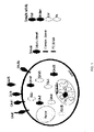
- CA-IX is a metalloenzyme that catalyzes the reversible hydration of carbon dioxide to bicarbonate and protons ( Figure 1 ).
- CA-IX is a transmembrane protein with an extracellular catalytic site and an NH 2 -terminal proteoglycan (PG)-like domain.
- An antibody and a fragment thereof "specifically binds" CA-IX if it binds CA-IX with an equilibrium dissociation constant (K D , i.e., a ratio of K d /K a , K d and K a are the dissociation rate and the association rate, respectively) less than 10 -5 M (e.g., less than 10 -6 M, 10 -7 M, 10 -8 M, 10 -9 M, 10 -10 M, 10 -11 M, 10 -12 M, or 10 -13 M), while not significantly binding other components present in a test sample (e.g., with a K D that is at least 10 times, such as 50 times or 100 times, more than K D for binding CA-IX). Affinities of an antibody and a fragment thereof disclosed herein and CA-IX can be readily determined using the method described in Example 5 of the present disclosure.
- K D equilibrium dissociation constant
- the antibody or fragment thereof as described herein should exhibit a high degree of internalization. Without wishing to be bound by theory, the antibodies or fragments thereof presently described bind to the extracellular domain of CA-IX. The antibodies or fragments thereof are then internalized by the cell and delivered into subcellular organelles, including endosomes and lysosomes. The antibody or fragment thereof as described herein may also reduce cell viability. Antibody internalization may be measured by any appropriate methods known in the art, including antibody internalization assays offered by Life Technologies, Zap Antibody Internalization Kit by Advanced targeting Systems, and/or quantitative assessment described in Liao-Chan et al., 2015.
- antibody and “antibody fragment” (“fragment thereof”) are as defined above.
- the antibody or fragment thereof may be from any source, human, mouse, or other; may be any isotype, including IgA, IgD, IgE, IgG, and IgM; and may be any type of fragment, including but not limited to Fv, scFv, Fab, and F(ab') 2 .
- the present invention provides an isolated or purified antibody or fragment thereof selected from the group consisting of:
- the isolated or purified antibody or fragment thereof may comprise
- the antibody or fragment thereof just defined specifically binds to the extracellular domain of CA-IX.
- the isolated or purified antibody or fragment thereof of the present invention may comprise
- the isolated or purified antibody specific for CA-IX may comprise:
- a substantially identical sequence may comprise one or more conservative amino acid mutations. It is known in the art that one or more conservative amino acid mutations to a reference sequence may yield a mutant peptide with no substantial change in physiological, chemical, physico-chemical or functional properties compared to the reference sequence; in such a case, the reference and mutant sequences would be considered "substantially identical" polypeptides.
- a conservative amino acid substitution is defined herein as the substitution of an amino acid residue for another amino acid residue with similar chemical properties (e.g. size, charge, or polarity). These conservative amino acid mutations may be made to the framework regions of the antibody or fragment thereof while maintaining the CDR sequences listed above and the overall structure of the antibody or fragment; thus the specificity and binding of the antibody are maintained.
- a conservative mutation may be an amino acid substitution.
- Such a conservative amino acid substitution may substitute a basic, neutral, hydrophobic, or acidic amino acid for another of the same group.
- basic amino acid it is meant hydrophilic amino acids having a side chain pK value of greater than 7, which are typically positively charged at physiological pH.
- Basic amino acids include histidine (His or H), arginine (Arg or R), and lysine (Lys or K).
- neutral amino acid also “polar amino acid”
- hydrophilic amino acids having a side chain that is uncharged at physiological pH, but which has at least one bond in which the pair of electrons shared in common by two atoms is held more closely by one of the atoms.
- Polar amino acids include serine (Ser or S), threonine (Thr or T), cysteine (Cys or C), tyrosine (Tyr or Y), asparagine (Asn or N), and glutamine (Gln or Q).
- hydrophobic amino acid (also “non-polar amino acid”) is meant to include amino acids exhibiting a hydrophobicity of greater than zero according to the normalized consensus hydrophobicity scale of Eisenberg (1984). Hydrophobic amino acids include proline (Pro or P), isoleucine (Ile or I), phenylalanine (Phe or F), valine (Val or V), leucine (Leu or L), tryptophan (Trp or W), methionine (Met or M), alanine (Ala or A), and glycine (Gly or G).
- “Acidic amino acid” refers to hydrophilic amino acids having a side chain pK value of less than 7, which are typically negatively charged at physiological pH. Acidic amino acids include glutamate (Glu or E), and aspartate (Asp or D).
- Sequence identity is used to evaluate the similarity of two sequences; it is determined by calculating the percent of residues that are the same when the two sequences are aligned for maximum correspondence between residue positions. Any known method may be used to calculate sequence identity; for example, computer software is available to calculate sequence identity. Without wishing to be limiting, sequence identity can be calculated by software such as NCBI BLAST2 service maintained by the Swiss Institute of Bioinformatics (and as found at ca.expasy.org/tools/blast/), or any other appropriate software that is known in the art.
- the substantially identical sequences of the present invention may be at least 90% identical; in another example, the substantially identical sequences may be at least 90, 91, 92, 93, 94, 95, 96, 97, 98, 99, or 100% identical, or any percentage there between, at the amino acid level to sequences described herein. Importantly, the substantially identical sequences retain the activity and specificity of the reference sequence. In a non-limiting embodiment, the difference in sequence identity may be due to conservative amino acid mutation(s). In a non-limiting example, the present invention may be directed to an antibody or fragment thereof comprising a sequence at least 95%, 98% or 99% identical to that of the antibodies described herein.
- the present invention further encompasses an antibody or fragment thereof that is chimeric (or chimerized), veneered, or humanized.
- the antibody or fragment thereof may be chimeric, in that the antibody or fragment thereof is a combination of protein sequences originating from more than one species.
- a chimeric antibody is produced by combining genetic material from a nonhuman source (for example but not limited to a mouse) with genetic material from a human.
- a nonhuman source for example but not limited to a mouse
- human constant domains can be fused to mouse V H and V L sequences (see Gonzales et al 2005).
- Veneering also referred to in the art as "variable region resurfacing", of antibodies involves replacing solvent-exposed residues in the framework region of the native antibody or fragment thereof with the amino acid residues in their human counterpart (Padlan, 1991; Gonzales et al 2005); thus, buried non-humanized residues, which may be important for CDR conformation, are preserved while the potential for immunological reaction against solvent-exposed regions is minimized.
- Humanization of an antibody or antibody fragment comprises replacing an amino acid in the sequence with its human counterpart, as found in the human consensus sequence, without loss of antigen-binding ability or specificity; this approach reduces immunogenicity of the antibody or fragment thereof when introduced into human subjects.
- one or more than one of the CDR defined herein may be fused or grafted to a human variable region (V H , or V L ), to other human antibody (IgA, IgD, IgE, IgG, and IgM), to human antibody fragment framework regions (Fv, scFv, Fab), or to human proteins of similar size and nature onto which CDR can be grafted (Nicaise et al, 2004).
- V H human variable region
- V L human variable region
- IgA, IgD, IgE, IgG, and IgM human antibody fragment framework regions
- Fv, scFv, Fab human proteins of similar size and nature onto which CDR can be grafted
- the present invention thus provides an isolated or purified antibody or fragment thereof specific for CA-IX may be a chimeric antibody comprising the variable domain as defined above linked to human IgG1 constant domains.
- the human IgG1 constant domains may comprise a human kappa 1 light chain constant domain and human IgG1 heavy chain constant domains.
- the antibody or fragment thereof of the present invention may also comprise additional sequences to aid in expression, detection or purification of a recombinant antibody or fragment thereof. Any such sequences or tags known to those of skill in the art may be used.
- the antibody or fragment thereof may comprise a targeting or signal sequence (for example, but not limited to ompA), a detection/purification tag (for example, but not limited to c-Myc, His 5 , His 6 , or His 8 G), or a combination thereof.
- the signal peptide may be MVLQTQVFISLLLWISGAYG (SEQ ID NO:31) or MDWTWRILFLVAAATGTHA (SEQ ID NO:32).
- the additional sequence may be a biotin recognition site such as that described by Cronan et al in WO 95/04069 or Voges et al in WO/2004/076670 .
- linker sequences may be used in conjunction with the additional sequences or tags, or may serve as a detection/purification tag.
- the antibody or fragment thereof of the present invention may also be in a multivalent display format, also referred to herein as multivalent presentation.
- Multimerization may be achieved by any suitable method known in the art. For example, and without wishing to be limiting in any manner, multimerization may be achieved using self-assembly molecules such as those described in Zhang et al (2004a; 2004b) and WO2003/046560 .
- the described method produces pentabodies by expressing a fusion protein comprising the antibody or fragment thereof of the present invention and the pentamerization domain of the B-subunit of an AB 5 toxin family (Merritt & Hol, 1995); the pentamerization domain assembles into a pentamer.
- a multimer may also be formed using the multimerization domains described by Zhu et al. (2010); this form, referred to herein as a "combody” form, is a fusion of the antibody or fragment of the present invention with a coiled-coil peptide resulting in a multimeric molecule (Zhu et al., 2010).
- Other forms of multivalent display are also encompassed by the present invention.
- the antibody or fragment thereof may be presented as a dimer, a trimer, or any other suitable oligomer.
- Each subunit of the multimers described above may comprise the same or different antibodies or fragments thereof of the present invention, which may have the same or different specificity.
- the multimerization domains may be linked to the antibody or antibody fragment using a linker, as required; such a linker should be of sufficient length and appropriate composition to provide flexible attachment of the two molecules, but should not hamper the antigen-binding properties of the antibody.
- a linker should be of sufficient length and appropriate composition to provide flexible attachment of the two molecules, but should not hamper the antigen-binding properties of the antibody.
- the antibody or fragments thereof may be presented in a bi-specific antibody.
- the invention also encompasses the antibody or fragment thereof as described above linked to a cargo molecule.
- the cargo molecule may be any suitable molecule.
- the cargo molecule may be a detectable agent, a therapeutic agent, a drug, a peptide, an enzyme, a growth factor, a cytokine, a receptor trap, an antibody or fragment thereof (e.g., IgG, scFv, Fab, V H H, V H , V L , etc) a chemical compound, a carbohydrate moiety, DNA-based molecules (anti-sense oligonucleotide, microRNA, siRNA, plasmid), a cytotoxic agent, viral vector (adeno-, lenti-, retro-), one or more liposomes or nanocarriers loaded with any of the previously recited types of cargo molecules, or one or more nanoparticle, nanowire, nanotube, or quantum dots.
- the antibody or fragment thereof may be linked to the cargo molecule
- the cargo molecule may be a detectable label, a radioisotope, a paramagnetic label such as gadolinium or iron oxide, a fluorophore, a fluorescent agent, Near Infra-Red (NIR) fluorochrome or dye such as Cy5.5, an echogenic microbubble, an affinity label (for example biotin, avidin, etc), a detectable protein-based molecule, nucleotide, quantum dot, nanoparticle, nanowire, or nanotube or any other suitable agent that may be detected by imaging methods.
- a detectable label such as gadolinium or iron oxide
- a fluorophore such as gadolinium or iron oxide
- a fluorophore such as gadolinium or iron oxide
- a fluorophore such as gadolinium or iron oxide
- a fluorophore such as gadolinium or iron oxide
- a fluorophore such as gadolinium or iron oxide
- a fluorophore such
- the anti-CA-IX or fragment thereof may be linked to a near infrared fluorescence (NIRF) imaging dye, for example and not wishing to be limiting Cy5.5, Alexa680, Dylight680, or Dylight800.
- NIRF near infrared fluorescence
- the antibody or fragment thereof as described herein is linked to a drug, thus providing an antibody-drug conjugate (ADC).
- the drug may be any type of drug, for example but not limited to a cytotoxic agent.
- the cytotoxic agent may include, but is not limited to anti-microtubule agents (such as taxanes, maytansines and auristatins), DNA damaging agents (such as calicheamicin and duocarmydin), RNA polymerase inhibitors (such as alpha-amantin), and other potent cytotoxic drugs (such as anthracyclines).
- the antibody-drug conjugate allows for targeted delivery of a drug, thus limiting systemic exposure.
- the antibody or fragment thereof as described herein binds to the extracellular domain of CA-IX; the drug linked to the antibody or fragment thereof is thus internalized.
- the cytotoxic agent Upon internalization the cytotoxic agent is released within the target cells upon degradation of the human CA-IX antibody-DM1 complex in lysosomes. Depending on the intracellular concentration of the cytotoxic agent accumulated in cancer cells, rapid apoptosis occurs.
- the cargo molecule as described herein may be linked, also referred to herein as "conjugated", to the antibody or fragment thereof by any suitable method known in the art.
- the cargo molecule may be linked to the peptide by a covalent bond or ionic interaction.
- the linkage may be achieved through a chemical cross-linking reaction, or through fusion using recombinant DNA methodology combined with any peptide expression system, such as bacteria, yeast or mammalian cell-based systems.
- a suitable linker may be used. Methods for linking an antibody or fragment thereof to a cargo molecule such as a therapeutic or detectable agent would be well-known to a person of skill in the art.
- the present invention also encompasses nucleic acid sequences encoding the molecules as described herein. Given the degeneracy of the genetic code, a number of nucleotide sequences would have the effect of encoding the desired polypeptide, as would be readily understood by a skilled artisan.
- the nucleic acid sequence may be codon-optimized for expression in various micro-organisms.
- the present invention also encompasses vectors comprising the nucleic acids as just described. Furthermore, the invention encompasses cells comprising the nucleic acid and/or vector as described.
- the present invention further encompasses the isolated or purified antibody or fragments thereof immobilized onto a surface using various methodologies; for example, and without wishing to be limiting, the antibody or fragment may be linked or coupled to the surface via His-tag coupling, biotin binding, covalent binding, adsorption, and the like. Immobilization of the antibody or fragment thereof of the present invention may be useful in various applications for capturing, purifying or isolating proteins.
- the solid surface may be any suitable surface, for example, but not limited to the well surface of a microtiter plate, channels of surface plasmon resonance (SPR) sensorchips, membranes, beads (such as magnetic-based or sepharose-based beads or other chromatography resin), glass, plastic, stainless steel, a film, or any other useful surface such as nanoparticles, nanowires and cantilever surfaces.
- SPR surface plasmon resonance
- a purified antibody or fragment thereof immobilized onto a surface may be used in a variety of methods, including diagnostic methods.
- the present invention further provides an in vitro method of detecting CA-IX, comprising contacting a tissue sample with one or more than one isolated or purified antibody or fragment thereof of the present invention linked to a detectable agent.
- the CA-IX-antibody complex can then be detected using detection and/or imaging technologies known in the art.
- the tissue sample in the method as just described may be any suitable tissue sample, for example but not limited to a serum sample, a vascular tissue sample, or a tumour tissue sample; the tissue sample may be from a human or animal subject.
- the step of contacting is done under suitable conditions, known to those skilled in the art, for formation of a complex between the antibody or fragment thereof and CA-IX.
- the step of detecting may be accomplished by any suitable method known in the art, for example, but not limited to optical imaging, immunohistochemistry, molecular diagnostic imaging, ELISA, or other suitable method.
- the isolated or purified antibody or fragment thereof linked to a detectable agent may be used in immunoassays (IA) including, but not limited to enzyme IA (EIA), ELISA, "rapid antigen capture", “rapid chromatographic IA", and "rapid EIA”.
- the in vitro method is for detection of CA-IX in circulating cells and the tissue sample is a serum sample.
- the present invention also provides an in vivo method of detecting CA-IX expression in a subject.
- the method comprises administering one or more than one isolated or purified antibody or fragment thereof as described herein linked to a detectable agent to the subject, then detecting the labelled antibody or fragment thereof bound to CA-IX.
- the step of detecting may comprise any suitable method known in the art, for example, but not limited to PET, SPECT, or fluorescence imaging, or any other suitable method.
- the method as just described may be useful in detecting the expression of CA-IX in tissues, for example but not limited to tumor tissues.
- the in vivo detection step in the methods described above may be whole body imaging for diagnostic purposes or local imaging at specific sites, such as but not limited to sites of solid tumor growth, in a quantitative manner to assess the progression of disease or host response to a treatment regimen.
- the detection step in the methods as described above may be immunohistochemistry, or a non-invasive (molecular) diagnostic imaging technology including, but not limited to:
- the present invention further provides a method of transporting a molecule of interest into cells expressing CA-IX.
- the method comprises administering the molecule linked to an antibody or fragment thereof as described herein to a subject.
- the molecule may be any desired molecule, including the cargo molecules, as previously described; the molecule may be "linked" to the antibody or fragment thereof using any suitable method, including, but not limited to conjugation or expression as a fusion protein.
- the administration may be by any suitable method, for example parenteral administration, including but not limited to intravenous (iv), subcutaneous (sc), and intramuscular (im) administration.
- the antibody or fragment thereof of the present invention delivers the desired molecule to cells in a targeted fashion.
- the present invention also encompasses a composition comprising one or more than one isolated or purified antibody or fragment thereof as described herein.
- the composition may comprise a single antibody or fragment as described above, or may be a mixture of antibodies or fragments.
- the antibodies may have the same specificity, or may differ in their specificities; for example, and without wishing to be limiting in any manner, the composition may comprise antibodies or fragments thereof specific to CA-IX (same or different epitope).
- the composition may also comprise one or more than one antibody or fragments of the present invention linked to one or more than one cargo molecule.
- the composition may comprise one or more than one ADC in accordance with the present invention.
- the composition may also comprise a pharmaceutically acceptable diluent, excipient, or carrier.
- the diluent, excipient, or carrier may be any suitable diluent, excipient, or carrier known in the art, and must be compatible with other ingredients in the composition, with the method of delivery of the composition, and is not deleterious to the recipient of the composition.
- the composition may be in any suitable form; for example, the composition may be provided in suspension form, powder form (for example, but limited to lyophilised or encapsulated), capsule or tablet form.
- the carrier when the composition is provided in suspension form, may comprise water, saline, a suitable buffer, or additives to improve solubility and/or stability; reconstitution to produce the suspension is effected in a buffer at a suitable pH to ensure the viability of the antibody or fragment thereof.
- Dry powders may also include additives to improve stability and/or carriers to increase bulk/volume; for example, and without wishing to be limiting, the dry powder composition may comprise sucrose or trehalose.
- the composition may be so formulated as to deliver the antibody or fragment thereof to the gastrointestinal tract of the subject.
- the composition may comprise encapsulation, time-release, or other suitable technologies for delivery of the antibody or fragment thereof. It would be within the competency of a person of skill in the art to prepare suitable compositions comprising the present compounds.
- a 416 amino acid long recombinant fragment of the extracellular domain of human (h)CA-IX with a C-terminal His-tag was prepared.
- the sequence of the recombinant fragment is shown in SEQ ID NO:33.
- a PTT5 construct containing a synthetic recombinant fragment of the extracellular domain (ECD) of hCA-IX linked to a C-terminal His-tag was synthesized by GeneART.
- a schematic of the plasmid construct is shown in Figure 2 .
- Transient transfection 2 and 50 mL transfections of the plasmid DNA into CHO cells were prepared. The day before the transfection, CHO-3E7 cells were diluted to 0.7 ⁇ 10 6 cells/ml in complete growth medium (Freestyle TM F17 media (Invitrogen) supplemented with L-glutamine (Hyclone) 4 mM final and Kolliphor ® P188 0.1% final) and grown in shaker flasks (agitation rate of 100 rpm) at 37°C, 5% CO 2 in a humidified incubator. At the time of transfection, the cell density was between 1.7 ⁇ 10 6 and 2.2 ⁇ 10 6 cells/mL and viability was greater than 97%.
- complete growth medium Freestyle TM F17 media (Invitrogen) supplemented with L-glutamine (Hyclone) 4 mM final and Kolliphor ® P188 0.1% final
- shaker flasks agitation rate of 100 rpm
- the DNA-PEI mixture was prepared by a multi-step process that is outlined briefly: a master mix of AKTdd pTT22 (constitutively active mutant of AKT) and ssDNA (0.33 ⁇ g AKTdd pTT22 + 0.77 ⁇ g ssDNA/ 2 ml transfection) in complete cell culture media was prepared and added to labelled 1.5 ml tubes.
- plasmid DNA pTT5_CAIX coopt_H10G
- pTT5_CAIX coopt_H10G plasmid DNA
- PEI pro TM polyethtleneimine from Polyplus-transfection #115-375
- the DNA mixture was vortexed and allowed to incubate for 3-10 minutes, after which the DNA-PEI mixture was added to the cells in the six-well plates. The contents of the plates were mixed with gentle swirling and incubated at 37°C, 5% CO 2 humidified incubator.
- the transfected cell population was transferred to a 1.5 ml tube. The cells were removed by centrifugation at 1500g for 10 minutes and the supernatant was transferred to a new tube.
- the transfected cell population was transferred to a 50 ml tube and the cells were removed by centrifugation at 250 g for 10 minutes. The supernatant was filtered using a Millipore steriflip unit (Millipore).
- the filtered culture supernatant was passed through a 1 ml protein A MabSelect SuRe resin (GE Healthcare) column. Five (5) bed volumes PBS D-PBS pH 7.4 w/o Ca, w/o Mg (Hyclone) were added to remove unbound material.
- the protein was eluted with 1 ml of 100 mM Na Citrate pH 3.0 and the eluate was neutralized with 1 M Tris, pH 9. Buffer exchange into PBS was carried out using dilution and filtration using Ultracel 10K centrifuge filters.
- the rhCA-IX ECD was expressed in CHO cells, purified by Ni-agarose and verified by Sodium Dodecyl Sulfate PolyAcrylamide Gel Electrophoresis (SDS-PAGE) under non-reducing and reducing conditions ( Figure 3 ).
- the protein was shown to be a 50/50 mixture of rhCA-IX ECD monomer ( ⁇ 48 kDa) and dimer ( ⁇ 110 kDa).
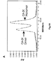
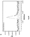
- the rhCA-IX ECD monomer/dimer was further purified by size exclusion chromatography (SEC) of the protein mixture ( Figure 4A ). Fractions with the highest protein concentration were selected and kept at 4°C for several weeks. Prior to further biophysical characterization measurements, samples were reanalyzed by SEC ( Figures 4B-D ). The results of this evaluation showed that both the monomer and dimer are stable upon storage.
- mice were immunized with the rhCA-IX ECD obtained in Example 1. Hybridomas were generated and conditioned medium was evaluated for mAb binding to the rhCA-IX ECD protein.
- mice 6-week old A/J mice were bled (pre-immune serum) and immunized i.p. and s.c. with 33 ⁇ g of rhCA-IX monomer/dimer mixture in incomplete Freund adjuvant, as part of a 3-protein multiplexed immunization mixture.
- a second injection of 33 ⁇ g of rhCA-IX protein in PBS was done and mice were bled 7-10 days later.
- the serum titer was measured by ELISA.
- a final i.p. booster injection using 33 ⁇ g of rhCA-IX protein in PBS was done 4 days prior to fusion experiment.
- Spleen cells were harvested from immunized mice in IMDM (Hy-Clone) and fused to NS0 myeloma cell line using PEG fusion protocol. To this end, spleen cells and myeloma cells were washed in IMDM, counted in RBC lysing buffer (Sigma) and mixed together at a 5:1 ratio. Pelleted cells were fused together by adding 1 ml of a 50% solution of PEG 4000 (EMD-Millipore) in PBS preheated at 37°C drop-wise over one minute, and incubated at 37°C for an additional 90 sec.
- IMDM Hy-Clone
- PEG 4000 EMD-Millipore
- IMDM IMDM supplemented with 10% heat inactivated FBS (Sigma), and suspended at a concentration of 2 ⁇ 10 5 input myeloma cells per ml in HAT selection medium (IMDM containing 20% heat inactivated FBS, penicillin-streptomycin (Sigma), 1 ng/ml mouse IL-6 (Biosource), HAT media supplement (Sigma) and L-glutamine) and incubated at 37°C, 5% CO 2 .
- HAT selection medium IMDM containing 20% heat inactivated FBS, penicillin-streptomycin (Sigma), 1 ng/ml mouse IL-6 (Biosource), HAT media supplement (Sigma) and L-glutamine
- hybridoma cells were washed and suspended at a concentration of 2 ⁇ 10 5 input myeloma cells per ml in semi-solid medium D (StemCell) supplemented with 5% heat inactivated FBS, 1 ng/ml mouse IL-6 and 10 ⁇ g/ml FITC- Fab'2 Goat anti-mouse IgG (H+L) (Jackson).
- the cell mixture was plated in Petri dish (Genetix) and further incubated for 6-7 days at 37°C, 5% CO 2 .
- Secretor clones were then transferred using a mammalian cell clone picker (ClonepixFL, Molecular Devices) into sterile 96-well plates (Costar) containing 200 ⁇ l of IMDM supplemented with 20% heat inactivated FBS, penicillin-streptomycin (Sigma), 1 ng/ml mouse IL-6 (Biosource), HT media supplement (Sigma) and L-glutamine and incubated for 2-3 days at 37°C, 5% CO 2 .
- mammalian cell clone picker (ClonepixFL, Molecular Devices) into sterile 96-well plates (Costar) containing 200 ⁇ l of IMDM supplemented with 20% heat inactivated FBS, penicillin-streptomycin (Sigma), 1 ng/ml mouse IL-6 (Biosource), HT media supplement (Sigma) and L-glutamine and incubated for 2-3 days at 37°C, 5% CO 2 .
- Hybridoma selection Hybridoma supernatant were screened by ELISA to detect specific binders. To this end, 96-well half-area plates (Costar) were coated with 25 ⁇ l of CA-IX at 5 ⁇ g/ml in PBS and incubated overnight at 4°C. Microplates were washed 3 times with PBS, blocked with PBS-BSA 1%, and 25 ⁇ l of hybridoma supernatant were added and incubated at 37°C, 5% CO 2 for 2 hours.
- the anti-CA-IX monoclonal antibodies obtained in Example 2 were characterized using Western blot and Reverse Phase Protein Array (RPPA).
- RPPA Reverse Phase Protein Array
- RPPA Reverse Phase Protein Array
- Each array was simultaneously probed with a hybridoma supernatant (secreted mAb, Example 2) and an anti-GFP antibody to normalize for the spotted protein volume.
- Slides were assembled in the 16-well incubation chambers (Whatman) and in the multi-slide frame (Whatman) that is a reusable holder for up to four slides. In this assembly, a liquid-tight well was created on top of each individual array pad.
- 100 ⁇ l of blocking solution (1x PBS / 3% BSA / 0.1% Tween20) was added to each well.
- Each chamber was sealed with aluminum tape to prevent evaporation and incubated (1h, RT) with gentle agitation. The nitrocellulose pads were never allowed to dry from this step on.
- mAb were prepared by mixing 30 ⁇ l of each crude hybridoma supernatant with 30 ⁇ l of incubation solution (1x PBS / 2% BSA / 0.05% Tween20 / Rabbit anti-GFP-AlexaFluor555 (0.9 ⁇ g/ml)). Blocking solution was aspired and replaced by the diluted mAb solution, incubation chambers were sealed with aluminum tape, and incubated while protected from light (2h RT, 130 rpm shaking). mAb solutions were removed, wells were washed three times with 100 ⁇ l washing solution (1x PBS / 0.1% Tween20), and the chambers were dismantled.
- Table 3 RPPA results showing the rhCA-IX ECD binding specificity of mAbs 11H9, 12H8 and 2C7 Purified Antigen Clone Native (A) Denatured (B) Ratio (A)/(B) (Possibly epitope related) 11H9 119.58 85.82 1.39 12H8 158.01 142.32 1.11 2C7 178.94 208.4 0.86 Antigen in protein mixture Clone Native (C) Denatured (D) mAb binding specificity mAb concentration in hybridoma CM ( ⁇ g/ml) 11H9 57.83 8.33 rhCA-IX ECD 4.64 12H8 50.26 4.15 rhCA-IX ECD 2.52 2C7 44.18 3.68 rhCA-IX ECD 20.6
- mAb were recombinantly expressed in CHO cells.
- V H and V L mAb 11H9, 12H8 and 2C7 were sequenced and found to be as follows:
- V L and V H sequences were cloned into a human kappa1 light chain and human IgG1 heavy chain frameworks (constant domains), respectively, in the pTT5 vector by Genscript (Piscataway NJ, USA), thereby generating chimeric (c) mAb.
- Genscript Procataway NJ, USA
- the sequences for the chimeric antibodies were as follows:
- all light chain sequences comprised a signal sequence MVLQTQVFISLLLWISGAYG (SEQ ID NO:31) at the N-terminus
- heavy chain sequences comprised the signal sequence MDWTWRILFLVAAATGTHA (SEQ ID NO:32) at the N-terminus.
- CDR regions (Table 4) were also analyzed for a consensus binding sequence by reconstructing a phylogenetic tree ( Figure 6 ) of the CDR 1-3 regions of the V H and V L chains using web-based software (Dereeper A et al., 2008; phylogeny.lirmm.fr/phylo_cgi/index.cgi). The results of this analysis indicated that the CDR regions of both the V L and V H regions of mAb 11H9 and 12H8 are more similar to each other than to that of mAb 2C7. Table 4. CDR region sequences for mAb 11H9, 12H8 and 2C7.
- Chimeric mAb expression was validated through a 2 mL expression scout, where CHO cells were transiently transfected with V L - and V H -containing constructs (1:1 ratio); conditioned medium (CM) was harvested on day 7, and mAb expression levels were evaluated by SDS-PAGE (data not shown).
- CM conditioned medium
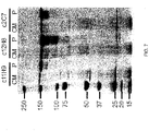
- Conditioned medium (CM) was harvested on day 7, chimeric mAb were purified (ProtA), quantitated, and evaluated by SDS-PAGE.
- Results are shown in Table 5 and Figure 7 .
- the data show that all three chimeric mAbs are well expressed by the transiently transfected CHO cells.
- a 'c' for 'chimeric' is added to the ID of these mAbs.
- Table 5 Recombinant c11H9, c12H8 and c2C7 concentration from 50 mL CHO scout culture by nanodrop after Protein A purification.
- the anti-CA-IX antibodies obtained in Examples 2 and 4 were characterized using Surface Plasmon Resonance (SPR).
- SPR Surface Plasmon Resonance
- the chimeric antibodies of Example 4 were also analyzed for CA cross-reactivity and by Differential Scanning Calorimetry (DSC).
- SPR experiments were carried out by capturing the CA-IX mAb (Example 2) from the CM with an anti-mouse Fc antibody immobilized on the chip surface via amine coupling, after which 60 nM rhCA-IX EDC mixture, rhCA-IX EDC monomer, or dimer (Example 1) were flowed over the mAb surface to measure association (binding) followed by flowing running buffer to measure dissociation of any complex formed. All SPR assays were carried out using a BioRad ProteOn XPR36 instrument (Bio-Rad Laboratories (Canada) Ltd.
- the screening of the chimeric antibody variants (Example 4) for binding to the rhCA-IX antigen targets occurred in two steps: an indirect capture of the antibody variants onto the anti-human Fc antibody surface in the ligand direction followed by the simultaneous injection of 5 concentrations of purified rhCA-IX antigen (Example 1) and one buffer blank for double referencing in the analyte direction.
- the change in response due to binding was measured during this antigen injection phase followed by injection of running buffer to measure dissociation of the complex formed.
- one buffer injection for 30 s at 100 ⁇ L/min in the ligand direction was used to stabilize the baseline.
- non-purified variants in cell-culture media were diluted to 4% in PBST.
- One to five variants or controls were simultaneously injected in individual ligand channels for 240 s at flow 25 ⁇ L/min . This resulted in a capture of approximately 400 to 600 RUs onto the anti-human Fc surface.
- the first ligand channel was left empty to use as a blank control if required. This capture step was immediately followed by two buffer injections in the analyte direction to stabilize the baseline. Then 60nM, 20nM, 6.7nM, 2.2nM and 0.74nM rhCA-IX antigen along with a buffer blank was simultaneously injected at 50 ⁇ L/min for 120 s with a 300 s dissociation phase.
- the captured antibody surfaces were regenerated by an 18 s pulse of 0.85% phosphoric acid for 18 s at 100 ⁇ L/min to prepare for the next injection cycle.
- Sensorgrams were aligned and double-referenced using the buffer blank injection and interspots, and the resulting sensorgrams were analyzed using ProteOn Manager software v3.1.
- the double-referenced sensorgrams were fit to the 1:1 binding model to determine the rate constants for association and dissociation (k a and k d ) and the corresponding affinity (K D ).
- Results are shown in Table 6.
- mAb 11H9 and 2C7 have a slight preference for the rhCA-IX dimer over the monomer. No data could be obtained for mAb 12H8 when using the monomer, however this mAb binds to the rhCA-IX ECD dimer in a similar manner as mAb 11H9 and 2C7.
- Table 6 Overview SPR results of purified hybridoma-derived mAb 11H9, 12H8, and 2C7 and the CHO expressed recombinant chimeric Ab c11H9, c12H8 and c2C7 using hCA-IX ECD monomer and/or dimer preps.
- Cross-reactivity determination by SPR To evaluate the cross-reactivity of c11H9, c12H8, and c2C7 to other hCA, binding of the chimeric antibodies to rhCA-IV, rhCA-XII and rhCA-XIV as well as to recombinant murine (rm)CA-IX was measured by SPR by indirect capture (as described previously).
- CA proteins for the analyte injection were purchased from Sino Biologics Inc.; the proteins were reconstituted as recommended by the manufacturer and diluted to 100nM in SPR running buffer for binding analysis.
- DSC Differential Scanning Calorimetry
- Example 2 The monoclonal antibodies of Example 2 and/or the chimeric antibodies of Example 4 were used to perform various methods of epitope mapping.
- Pepscan Epitope mapping services were provided by Pepscan (Lelystad, The Netherlands; pepscan.com). CLIPS TM technology (Chemical Linkage of Peptides onto Scaffolds) was used to gain insight into the binding epitope. The method is based on the chemical linkage of homocysteines that flank synthetic peptides (15 aa long) covering the entire hCA-IX protein, in addition to a synthetic scaffold containing a benzyl-bromide group (Timmerman et al., 2009). The purified mAb of Example 2 were screened against peptides in a 'linear' and single 'cycle' format.
- Creating antibodies array - Antibodies were immobilized on the GLM chip surface at 30 ⁇ l/min.
- the activation reagents (at stock concentration of 0.4M EDC and 0.1M SNHS in water) were diluted 20-fold each in water.
- the top (A1) horizontal channel (the Analyte channel in the Proteon control software) was activated for 3 min with the diluted activation reagents.
- c11H9, c12H8, and c2C7 were each diluted to 20 ⁇ g/ml in 10 mM acetate pH4.5 and injected for 3 min in separate vertical (L1 to L6) channels (the Ligand channel in the Proteon control software), followed with a 3 min injection of ethanolamine to block the reactive spots.
- the multi-channel module (MCM) was then rotated and another 3 min injection of ethanolamine was done on the activated horizontal channel (A1). This four-step 'activation-binding-and-2x deactivation' procedure was then repeated on each of the horizontal channels (A2 to A6).
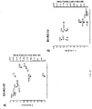
- Sandwich epitope binning The two-step sandwich epitope binning was done at 30 ⁇ l/min in the analyte orientation. 100nM CA-IX antigen was injected for 3 min, immediately followed by a 3 min injection of Mab2 at 100nM. Immobilized mAb (mAb1) surfaces were regenerated by a 18s injection of 0.85% phosphoric acid at 100 ⁇ l/min. This two-step injection (CA-IX-mAb2) was repeated for each individual Mab2 with monomer and dimer CA-IX. Each mAb were also tested simultaneously and used as Mab1 (immobilized on the chip) and mAb2 (in solution). To monitor the CA-IX dissociation from the immobilized Mab1 PBST was injected instead of mAb2.
- Results are shown in Figures 12B .
- the data shows that none of the antibodies compete for binding to either the rhCA-IX ECD monomer or dimer, indicating that these three mAb bind to distinct, non-overlapping CA-IX epitopes.
- YSD yeast surface display
- the hCA-IX ectodomain (ECD) and fragments thereof were expressed and covalently displayed on the surface of yeast cells (Feldhaus et al., 2003).
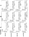
- the hCA-IX fragments covering the entire hCA-IX ECD ( Figure 13A ) or the PG domain alone ( Figure 13B ) were expressed as fusion proteins (Aga2-HA-(CA-IX)-MYC (pPNL6 vector) or (CA-IX)-Aga2-MYC (pPNL200 vector)) on the yeast cell surface.
- the YSD vectors (pPNL6 and pPNL200) were from The Pacific Northwest National Laboratory, USA.
- the displayed CA-IX fragments were used to map the amino acid sequences to which 11H9, 12H8, and 2C7 bind.
- the binding of the mAb to yeast cells was performed using a whole yeast cell ELISA.
- the amount of properly displayed fusion protein was measured by probing with an anti-MYC antibody, followed by an HRP-conjugated secondary antibody.
- the anti-MYC signal was then used to normalize the binding signal for c11H9, c12H8, and c2C7.
- yeast cells with displayed CA-IX fragments were heated at 80°C for 30 min, then chilled on ice for 20 min prior to labeling with antibodies.
- the binding of mAb to denatured yeast cells was normalized to the anti-MYC signal.
- the ratio of normalized anti-CA-IX signals of each mAb on native versus denatured hCA-IX peptide was thus indicative of the conformational nature of the epitope.
- the commercial M75 mAb was used as a positive control; the epitope of this mAb is known (Zavada et al., 2000) and is located in the hCA-IX PG domain.
- Tables 7 (native hCA-IX ECD) and 8 (denatured hCA-IX ECD) show that the binding epitopes for c11H9 and c12H8 are located in hCA-IX's PG domain (as is that for M75), whereas c2C7 binds to an epitope in the catalytic domain, thereby confirming the PepScan observations.
- This data set also indicates that the c11H9 and c12H8 epitopes are likely to be unstructured, whereas that of the c2C7 mAb is probably structured, given the observation that binding is lost to the denatured hCA-IX protein fragments.
- tiling peptides of 15 amino acid (aa) residues (with a 5 aa overlap) encompassing aa 37-140 of the native hCA-IX PG domain (Tables 9 and 10) were prepared.
- Corresponding DNA sequences were PCR-amplified, cloned into pPNL6 using GAP repair (Gietz et al., 1992), and displayed on the yeast surface.
- Table 9 Amino acid sequence of 15 amino acid tiling peptides encompassing amino acids 37-140 of the entire hCA-IX (see Figure 13B).
- Yeast cell ELISA with these YSD tiling peptides showed that c11H9 and M75 share similar, although not identical binding epitopes; the c11H9 epitope contained the repeat GEEDLP (SEQ ID NO:37) sequence, which appeared four times in the PG domain.
- c12H8 bound to an epitope that was distinguishable in the PG domain from that of c11H9 and M75; specifically, c12H8 bound to an epitope containing the unique sequence PQRLPRMQEDSPLGG (SEQ ID NO:38) located at the N-terminus.
- EEDLPGEE SEQ ID NO:34
- PQRLPRMQEDSPLGG SEQ ID NO:38
- PSDFSRYFQYEGSL SEQ ID NO: 36
- Example 2 The ability of the anti-CA-IX mAb obtained in Example 2 to inhibit in vitro enzyme activity was evaluated, the cell line was verified and an ADC assay was performed.
- the enzyme was pre-incubated for 30 minutes at room temperature with increasing concentration of mAb (0.0625 to 4 ⁇ M) diluted in DPBS (Hyclone). Reactions were initiated by the addition of 100 ⁇ M 4-Methylumbelliferylacetate. The rate of substrate hydrolysis was determined by monitoring 4-Methylumbelliferone-released fluorescence (excitation ⁇ , 380 nm; emission ⁇ , 440nm) as a function of time using the EnVision 2104 plate reader (Perkin Elmer). The substrate autohydrolysis control values were substracted from the observed total reaction velocities.
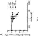
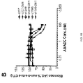
- Acetazolamide (Sigma Aldrich) at a concentration of 10 ⁇ M fully inhibited the activity of the rhCA-IX enzyme. Rates were determined in the initial and 'end portion' of each of the curves. Several concentrations of the rhCA-IX ECD mixture of Example 1 were tested and bench-marked against rhCA-IX purchased from R&D (data not shown). Using 1 ⁇ M rhCA-IX ECD, enzyme activity of the mAb was measured using a 4:1 (2C7) or 1:1 (11H9, 12H8) molar mAb:rhCA-IX ECD ratio. Results are shown in Figure 14 and Table 11. None of the tested mAb showed any enzyme inhibition activity.
- Table 11 Enzyme inhibition data for hybridoma-derived 11H9, 12H8 and 2C7 using the rhCA-IX ECD mixture.
- a ratio of (mAb:rhCA-IX ECD mixture) of 4:1 was used.
- 11H9 and 12H8 a ratio of (mAb:rhCA-IX ECD mixture) of 1:1 was used.
- Non-transfected human renal tumor sk-rc-59 and sk-rc-52 cell lines were obtained to evaluate binding of the anti-CA-IX mAb to cell surface CA-IX.
- Western blot analysis of these cell lines under non-reducing and reducing conditions confirmed the hCA-IX expression levels of the sk-rc-52 cell line (arrow), whereas no CA-IX could be detected in the sk-rc-59 cell line.
- Equal protein quantities were loaded (BCA protein assay) and confirmed by actin blot.
- mAb were evaluated in a flow cytometry experiment using the sk-rc-59 (low CA-IX expression) and sk-rc-52 (high CA-IX expression) cell lines.
- Cells were grown to 80% confluency in T75 flasks using MEM/10% FBS medium supplemented with non-essential amino acids (NEAA), washed with dPBS (37°C) and harvested using non-enzymatic cell dissociation buffer (37°C). Cells were then transferred to 50 mL tubes, spun down (1100 rpm, RT, 5 min) and re-suspended in cell growth medium.
- NEAA non-essential amino acids
- Cells were counted and divided in 10 6 cell containing aliquots, spun down (1100 rpm, RT, 2 min), re-suspended in ice-cold dPBS/10% FBS containing anti-CA-IX mAb (10 ⁇ g/mL final concentration) and incubated (50 min, 4°C). Cells were then washed with ice-cold dPBS/10% FBS, spun down (1100 rpm, 4°C, 3 min), and re-suspended in 500 ⁇ L ice-cold dPBS/10% FBS containing Alexa-488 Goat anti-mouse IgG (10 ⁇ g/mL final concentration).
- mAb were evaluated in a surrogate ADC assay using the high hCA-IX-expressing sk-rc-52 cells.
- the ADC assay was carried out using a commercially available ADC kit (atsbio.com) according to the manufacturer's instructions with some modifications. Briefly, sk-rc-52 (expressing high levels of hCA-IX) and sk-rc-59 (expressing low levels of hCA-IX) cells were seeded in RPMI/5% FBS in 96-well plates at a density of 5000 and 3000 cells/well, respectively.
- Results show that mAb 12H8, 11H9 and 2C7 are either equal to or better than the M75 mAb in reducing cell viability.
- Next serial dilutions (0-100 nM) of recombinantly expressed c11H9, c12H8 and c2C7 were tested in the surrogate ADC assay using sk-rc-52 cells (as described above).
- the results show that the recombinantly expressed chimeric antibodies have retained their ADC potential.
- ADC Assay Recombinantly expressed chimeric c11H9, c12H8 and c2C7 were conjugated to DM1 (mertansine). Briefly, a 10 mM stock solution of SMCC-DM1 was prepared in dimethylacetamide (DMA) and stored at -20°C. Just prior to use, stock solutions were quickly brought to room temperature and two working stocks prepared (2000 ⁇ M and 2660 ⁇ M) by diluting concentrated stock into DMA. For small-scale optimization experiments, two aliquots (100 ⁇ L) of antibody (2 mg/mL) was mixed 10 ⁇ L each of 10-fold conjugation buffer (1000 mM sodium phosphate, 200 mM NaCl, 30 mM EDTA, pH 7.2).
- 10-fold conjugation buffer 1000 mM sodium phosphate, 200 mM NaCl, 30 mM EDTA, pH 7.2.
- DAR Drug antibody ratio
- IC 50 values for non-conjugated and DM1 conjugated chimeric anti-CA-IX antibodies Construct IC 50 (nM) c11H9 6.32 c11H9-DM1 5.84 c12H8 23.55 c12H8-DM1 17.88 c2C7 2.20 c2C7-DM1 10.11
- Both on-target and off-target toxicity relating to the drug payload carried by an ADC is critical information that needs to be evaluated early in an ADC candidate selection.
- Prior clinical data reported for an anti-CA-IX ADC candidate in Phase I described unresolved issues concerning cardiotoxicity.
- Human stem-cell derived cardiomyocytes (Cellular Dynamics; Cohen et al, 2011) from Cellular Dynamics were used to evaluate cardiotoxicity of the anti-CA-IX mAb obtained in Example 4, and their derived ADC in an in vitro setting.
- iPS cardiomyocytes Human-induced pluripotent (iPS) cardiomyocytes were obtained from Cellular Dynamics Inc. iPS cardiomyocytes (iCells) were thawed and cultured according to the manufacturers instructions. Briefly, cells were thawed, resuspended in iCell plating media, seeded in 0.1% gelatin-coated 96-well plates (15,000 cells/well) and maintained in a humidified incubator with 5% CO 2 at 37°C for 4 hours. Cells were then washed to remove debris and the plating media was replaced with iCell Maintenance Media which was then changed every other day during the course of the experiment.
- Cells were treated 4 days post-thaw with either the unconjugated antibodies c2C7 and c12H8 or the corresponding DM1-conjugated ADCs at a concentration of 10 nM, and incubated for an additional 5 days.
- a non-specific negative control human IgG1 and the corresponding DM1-conjugated ADC were also used for treated the cells using the same protocol.
- Cell viability was measured 3 and 5 days post-treatment using the CellTiter-Glo Luminescent cell viability Assay (Promega, Madison, WI). Briefly, the plates and reagents were equilibrated to room temperature for 30 minutes. A volume of CellTiter-Glo reagent was added to the volume of cell culture medium present in each well and mixed on an orbital shaker for 45 minutes. The luminescence was then recorded using an EnVision plate reader (PerkinElmer Inc.)
- a ProteON GLC chip was activated with sEDS and sNHS prior to immobilzing approximately 4000 RUs of Goat anti-human Fc (Jackson InnumoResearch) via amine (lysine) coupling in 10 mM acetate buffer pH 4.5. This surface was then used to capture c2C7 and c12H8 on different flow cells.
- results are shown in Table 13 and Figure 20 .
- cynomolgus monkey was the only one which met the affinity criteria (Table 13). Therefore, cynomolgus monkey will be the clinically-relevant non-human primate model for testing toxicology for c2C7, c12H8 and the corresponding DM1-conjugated ADC. Since CA-XII is often co-expressed with CA-IX in the same tumors, there should be no cross-reactivity between the CA-IX antibodies and CA-XII. Using SPR, there was no observable binding of any of the anti-CA-IX antibodies to CA-XII ( Figure 20 ).
Landscapes
- Health & Medical Sciences (AREA)
- Life Sciences & Earth Sciences (AREA)
- Engineering & Computer Science (AREA)
- Chemical & Material Sciences (AREA)
- Immunology (AREA)
- Bioinformatics & Cheminformatics (AREA)
- General Health & Medical Sciences (AREA)
- Medicinal Chemistry (AREA)
- Molecular Biology (AREA)
- Animal Behavior & Ethology (AREA)
- Pharmacology & Pharmacy (AREA)
- Epidemiology (AREA)
- Public Health (AREA)
- Veterinary Medicine (AREA)
- Proteomics, Peptides & Aminoacids (AREA)
- Hematology (AREA)
- Organic Chemistry (AREA)
- Urology & Nephrology (AREA)
- Biochemistry (AREA)
- Biomedical Technology (AREA)
- Physics & Mathematics (AREA)
- Biotechnology (AREA)
- Cell Biology (AREA)
- Microbiology (AREA)
- Biophysics (AREA)
- Food Science & Technology (AREA)
- Genetics & Genomics (AREA)
- Analytical Chemistry (AREA)
- General Physics & Mathematics (AREA)
- Pathology (AREA)
- Optics & Photonics (AREA)
- Peptides Or Proteins (AREA)
- Medicines Containing Antibodies Or Antigens For Use As Internal Diagnostic Agents (AREA)
Applications Claiming Priority (3)
| Application Number | Priority Date | Filing Date | Title |
|---|---|---|---|
| US201562173405P | 2015-06-10 | 2015-06-10 | |
| PCT/IB2016/053448 WO2016199097A1 (fr) | 2015-06-10 | 2016-06-10 | Anticorps spécifiques de l'anhydrase carbonique ix et leurs utilisations |
| EP16807004.3A EP3307784B1 (fr) | 2015-06-10 | 2016-06-10 | Anticorps spécifiques de l'anhydrase carbonique ix et leurs utilisations |
Related Parent Applications (1)
| Application Number | Title | Priority Date | Filing Date |
|---|---|---|---|
| EP16807004.3A Division EP3307784B1 (fr) | 2015-06-10 | 2016-06-10 | Anticorps spécifiques de l'anhydrase carbonique ix et leurs utilisations |
Publications (1)
| Publication Number | Publication Date |
|---|---|
| EP4019552A1 true EP4019552A1 (fr) | 2022-06-29 |
Family
ID=57503137
Family Applications (2)
| Application Number | Title | Priority Date | Filing Date |
|---|---|---|---|
| EP21214012.3A Withdrawn EP4019552A1 (fr) | 2015-06-10 | 2016-06-10 | Anticorps spécifiques de l'anhydrase carbonique ix et leurs utilisations |
| EP16807004.3A Active EP3307784B1 (fr) | 2015-06-10 | 2016-06-10 | Anticorps spécifiques de l'anhydrase carbonique ix et leurs utilisations |
Family Applications After (1)
| Application Number | Title | Priority Date | Filing Date |
|---|---|---|---|
| EP16807004.3A Active EP3307784B1 (fr) | 2015-06-10 | 2016-06-10 | Anticorps spécifiques de l'anhydrase carbonique ix et leurs utilisations |
Country Status (4)
| Country | Link |
|---|---|
| US (1) | US10487153B2 (fr) |
| EP (2) | EP4019552A1 (fr) |
| CA (1) | CA2988912C (fr) |
| WO (1) | WO2016199097A1 (fr) |
Families Citing this family (8)
| Publication number | Priority date | Publication date | Assignee | Title |
|---|---|---|---|---|
| WO2019204939A1 (fr) * | 2018-04-27 | 2019-10-31 | National Research Council Of Canada | Anticorps monoclonaux à haute affinité (mab) dirigés contre l'anhydrase carbonique ix humaine exprimée en surface cellulaire (hca-ix), et utilisations associées |
| AU2019342131A1 (en) * | 2018-09-19 | 2021-04-29 | Totient, Inc. | Cancer associated antibody compositions and methods of use |
| US12351645B2 (en) | 2019-05-06 | 2025-07-08 | Navi Bio-Therapeuticis, Inc. | Anti-carbonic anhydrase IX antibody |
| KR20220054600A (ko) * | 2019-07-30 | 2022-05-03 | 이뮤놈 인코포레이티드 | Il-38-특이적 항체 |
| CN116217728A (zh) | 2019-09-10 | 2023-06-06 | 普米斯生物技术(珠海)有限公司 | 靶向caix抗原的纳米抗体及其应用 |
| EP4281485A4 (fr) * | 2021-01-25 | 2025-07-16 | Nat Res Council Canada | Anticorps à domaine unique ciblant ca-ix ainsi que compositions les comprenant |
| WO2024123822A1 (fr) * | 2022-12-05 | 2024-06-13 | Biogate Precision Medicine Corp. | Anticorps recombinant, composition pharmaceutique le comprenant et utilisations associées dans le traitement de cancers |
| EP4724490A2 (fr) | 2023-06-12 | 2026-04-15 | Amgen Inc. | Protéines de liaison à un agoniste du récepteur bêta de la lymphotoxine |
Citations (4)
| Publication number | Priority date | Publication date | Assignee | Title |
|---|---|---|---|---|
| EP0519596A1 (fr) | 1991-05-17 | 1992-12-23 | Merck & Co. Inc. | Procédé pour réduire l'immunogénécité des domaines variables d'anticorps |
| WO1995004069A1 (fr) | 1993-07-30 | 1995-02-09 | Affymax Technologies N.V. | Biotinylation de proteines |
| WO2003046560A2 (fr) | 2001-11-30 | 2003-06-05 | National Research Council Of Canada | Nouvelles molécules d'auto-assemblage |
| WO2004076670A1 (fr) | 2003-02-28 | 2004-09-10 | Roche Diagnostics Gmbh | Expression accrue de polypeptides de fusion comportant un marqueur de biotinylation |
Family Cites Families (19)
| Publication number | Priority date | Publication date | Assignee | Title |
|---|---|---|---|---|
| EP0366707A1 (fr) | 1987-05-06 | 1990-05-09 | OOSTERWIJK, Egbert | Anticorps monoclonaux contre le carcinome des cellules renales |
| US6297041B1 (en) | 1992-03-11 | 2001-10-02 | Institute Of Virology, Slovak Academy Of Sciences | MN gene and protein |
| US6297051B1 (en) | 1997-01-24 | 2001-10-02 | Institute Of Virology, Slovak Academy Of Sciences | MN gene and protein |
| US6027887A (en) | 1992-03-11 | 2000-02-22 | Institute Of Virology, Solvak Academy Of Sciences | MN gene and protein |
| AU2002351208A1 (en) | 2001-12-03 | 2003-06-17 | Abgenix, Inc. | Antibodies against carbonic anhydrase IX (CA IX) tumor antigen |
| US7910549B2 (en) | 2001-12-13 | 2011-03-22 | Institute Of Virology Of The Slovak Academy Of Sciences | MN gene and protein |
| US20090162382A1 (en) | 2002-03-01 | 2009-06-25 | Bernett Matthew J | Optimized ca9 antibodies and methods of using the same |
| WO2006037604A1 (fr) * | 2004-10-01 | 2006-04-13 | MAX-PLANCK-Gesellschaft zur Förderung der Wissenschaften e.V. | Nouveaux anticorps diriges contre la proteine de canal ionique eag1 mammifere |
| US8466263B2 (en) | 2005-12-02 | 2013-06-18 | Dana-Farber Cancer Institute, Inc. | Carbonic anhydrase IX (G250) anitbodies |
| US20100056611A1 (en) | 2006-10-12 | 2010-03-04 | Jaromir Pastorek | MN/CA9 Splice Variants |
| WO2008091798A2 (fr) | 2007-01-22 | 2008-07-31 | Xencor, Inc. | Anticorps de ca9 optimises et methodes d'utilisation associees |
| WO2009056342A1 (fr) | 2007-11-02 | 2009-05-07 | Wilex Ag | Épitopes de liaison avec un anticorps g250 |
| DK2237803T3 (en) * | 2007-12-28 | 2015-10-05 | Prothena Biosciences Ltd | Treatment and prophylaxis of amyloidosis |
| GB0807018D0 (en) * | 2008-04-17 | 2008-05-21 | Fusion Antibodies Ltd | Antibodies and treatment |
| WO2011016238A1 (fr) * | 2009-08-06 | 2011-02-10 | Immunas Pharma, Inc. | Anticorps qui se lient spécifiquement à des bêta-oligomères et utilisation de ceux-ci |
| WO2011139375A1 (fr) | 2010-05-06 | 2011-11-10 | Ludwig Institute For Cancer Research Ltd | Anticorps dirigés contre l'anhydrase carbonique ix et méthodes et utilisations associées |
| WO2012144208A1 (fr) | 2011-04-18 | 2012-10-26 | 国立大学法人東京大学 | Diagnostic et traitement du cancer à l'aide d'un anticorps anti-itm2a |
| MX369148B (es) | 2012-09-19 | 2019-10-30 | Innate Pharma | Agentes de unión kir3dl2. |
| ES2878749T3 (es) * | 2013-02-20 | 2021-11-19 | Innate Pharma | Un compuesto que se une específicamente a KIR3DL2 para el uso en el tratamiento de linfoma de células T periférico |
-
2016
- 2016-06-10 EP EP21214012.3A patent/EP4019552A1/fr not_active Withdrawn
- 2016-06-10 US US15/580,713 patent/US10487153B2/en active Active
- 2016-06-10 WO PCT/IB2016/053448 patent/WO2016199097A1/fr not_active Ceased
- 2016-06-10 CA CA2988912A patent/CA2988912C/fr active Active
- 2016-06-10 EP EP16807004.3A patent/EP3307784B1/fr active Active
Patent Citations (4)
| Publication number | Priority date | Publication date | Assignee | Title |
|---|---|---|---|---|
| EP0519596A1 (fr) | 1991-05-17 | 1992-12-23 | Merck & Co. Inc. | Procédé pour réduire l'immunogénécité des domaines variables d'anticorps |
| WO1995004069A1 (fr) | 1993-07-30 | 1995-02-09 | Affymax Technologies N.V. | Biotinylation de proteines |
| WO2003046560A2 (fr) | 2001-11-30 | 2003-06-05 | National Research Council Of Canada | Nouvelles molécules d'auto-assemblage |
| WO2004076670A1 (fr) | 2003-02-28 | 2004-09-10 | Roche Diagnostics Gmbh | Expression accrue de polypeptides de fusion comportant un marqueur de biotinylation |
Non-Patent Citations (56)
| Title |
|---|
| ABDICHE YNLINDQUIST KCPINKERTON APONS JRAJPAL A: "Expanding the ProteOn XPR36 biosensor into a 36-ligand array expedites protein interaction analysis", ANAL BIOCHEM., vol. 411, no. 1, 1 April 2011 (2011-04-01), pages 139 - 51, XP028361711, DOI: 10.1016/j.ab.2010.12.020 |
| BAGNA BAO ET AL: "In Vivo Imaging and Quantification of Carbonic Anhydrase IX Expression as an Endogenous Biomarker of Tumor Hypoxia", PLOS ONE, vol. 7, no. 11, 30 November 2012 (2012-11-30), pages e50860, XP055086820, DOI: 10.1371/journal.pone.0050860 * |
| BLEUMER IKNUTH AOOSTERWIJK EHOFMANN RVARGA ZLAMERS CKRUIT WMELCHIOR SMALA CULLRICH S: "A phase II trial of chimeric monoclonal antibody G250 for advanced renal cell carcinoma patients", BR J CANCER., vol. 90, no. 5, 8 March 2004 (2004-03-08), pages 985 - 90, XP055712916, DOI: 10.1038/sj.bjc.6601617 |
| BROUWERS AHVAN EERD JEFRIELINK COOSTERWIJK EOYEN WJCORSTENS FHBOERMAN OC: "Optimization of radioimmunotherapy of renal cell carcinoma: labeling of monoclonal antibody cG250 with 1311, 90Y, 177Lu, or 186Re", J NUCL MED., vol. 45, no. 2, February 2004 (2004-02-01), pages 327 - 37, XP008051713 |
| CANCER RES., vol. 71, no. 12, 15 June 2011 (2011-06-15), pages 4325 |
| CANCER RES., vol. 71, no. 13, 1 July 2011 (2011-07-01), pages 4733 |
| CHOTHIA CLESK AM: "Canonical structures for the hypervariable regions of immunoglobulins", J MOL BIOL., vol. 196, no. 4, 20 August 1987 (1987-08-20), pages 901 - 17, XP024010426, DOI: 10.1016/0022-2836(87)90412-8 |
| CHRASTINA APASTOREKOVA SPASTOREK J.: "Immunotargeting of human cervical carcinoma xenograft expressing CA IX tumor-associated antigen by 1251-labeled M75 monoclonal antibody", NEOPLASMA, vol. 50, no. 1, 2003, pages 13 - 21 |
| CHRASTINA AZAVADA JPARKKILA SKALUZ SKALUZOVA MRAJCANI JPASTOREK JPASTOREKOVA S.: "Biodistribution and pharmacokinetics of 1251-labeled monoclonal antibody M75 specific for carbonic anhydrase IX, an intrinsic marker of hypoxia, in nude mice xenografted with human colorectal carcinoma", INT J CANCER, vol. 105, no. 6, 2003, pages 873 - 81, XP002968995, DOI: 10.1002/ijc.11142 |
| DE KRUIF JLOGTENBERG T: "Leucine zipper dimerized bivalent and bispecific scFv antibodies from a semi-synthetic antibody phage display library", J BIOL CHEM., vol. 271, no. 13, 29 March 1996 (1996-03-29), pages 7630 - 4, XP002337214, DOI: 10.1074/jbc.271.13.7630 |
| DEREEPER AAUDIC SCLAVERIE JMBLANC G: "BLAST-EXPLORER helps you building datasets for phylogenetic analysis", BMC EVOL BIOL., vol. 10, 12 January 2010 (2010-01-12), pages 8, XP021065913 |
| DEREEPER AGUIGNON VBLANC GAUDIC SBUFFET SCHEVENET FDUFAYARD JFGUINDON SLEFORT VLESCOT M: "Phylogeny.fr: robust phylogenetic analysis for the non-specialist", NUCLEIC ACIDS RES., vol. 36, 1 July 2008 (2008-07-01) |
| DITTE PDEQUIEDT FSVASTOVA EHULIKOVA AOHRADANOVA-REPIC AZATOVICOVA MCSADEROVA LKOPACEK JSUPURAN CTPASTOREKOVA S: "Phosphorylation of carbonic anhydrase IX controls its ability to mediate extracellular acidification in hypoxic tumors", CANCER RES., vol. 71, no. 24, 15 December 2011 (2011-12-15), pages 7558 - 67 |
| EDGAR RC: "Local homology recognition and distance measures in linear time using compressed amino acid alphabets", NUCLEIC ACIDS RES., vol. 32, no. 1, 16 January 2004 (2004-01-16), pages 380 - 5 |
| EISENBERG, D.SCHWARZ, E.KOMAROMY, M.WALL, R: "Analysis of membrane and surface protein sequences with the hydrophobic moment plot", J. MOL. BIOL., vol. 179, 1984, pages 125 - 142, XP024018280, DOI: 10.1016/0022-2836(84)90309-7 |
| FELDHAUS MJSIEGEL RWOPRESKO LKCOLEMAN JRFELDHAUS JMYEUNG YACOCHRAN JRHEINZELMAN PCOLBY DSWERS J: "Flow-cytometric isolation of human antibodies from a nonimmune Saccharomyces cerevisiae surface display library", NAT BIOTECHNOL., vol. 21, no. 2, February 2003 (2003-02-01), pages 163 - 70, XP002451748 |
| FENNER, L.WIDMER, A.F.GOY, G.RUDIN, S.FREI, R: "Rapid and reliable diagnostic algorithm for detection of Clostridium difficile", J. CLIN. MICROBIOL., vol. 46, 2008, pages 328 - 330, XP055216114, DOI: 10.1128/JCM.01503-07 |
| GIETZ DST JEAN AWOODS RASCHIESTL RH: "Improved method for high efficiency transformation of intact yeast cells", NUCLEIC ACIDS RES., vol. 20, no. 6, 25 March 1992 (1992-03-25), pages 1425 |
| GONZALES NRDEPASCALIS RSCHLOM JKASHMIRI SVS, TUMOR BIOL, vol. 26, 2005, pages 31 - 43 |
| HULIKOVA AZATOVICOVA MSVASTOVA EDITTE PBRASSEUR RKETTMANN RSUPURAN CTKOPACEK JPASTOREK JPASTOREKOVA S: "Intact intracellular tail is critical for proper functioning of the tumor-associated, hypoxia-regulated carbonic anhydrase IX", FEBS LETT., vol. 583, no. 22, 19 November 2009 (2009-11-19), pages 3563 - 8, XP026782208, DOI: 10.1016/j.febslet.2009.10.060 |
| JONES PTDEAR PHFOOTE JNEUBERGER MSWINTER G, NATURE, vol. 321, 1986, pages 522 - 525 |
| KABAT EAWU TT: "Identical V region amino acid sequences and segments of sequences in antibodies of different specificities. Relative contributions of VH and VL genes, minigenes, and complementarity-determining regions to binding of antibody-combining sites", J IMMUNOL., vol. 147, 1991, pages 1709 - 19, XP055204516 |
| LEFRANC MPPOMMIE CRUIZ MGIUDICELLI VFOULQUIER ETRUONG LTHOUVENIN-CONTET VLEFRANC G: "IMGT unique numbering for immunoglobulin and T cell receptor variable domains and Ig superfamily V-like domains", DEV COMP IMMUNOL., vol. 27, no. 1, January 2003 (2003-01-01), pages 55 - 77, XP055585227, DOI: 10.1016/S0145-305X(02)00039-3 |
| LIAO-CHAN SDAINE-MATSUOKA BHEALD NWONG T.LIN TCAI AGLAI MD'ALESSIO JATHEUNISSEN J-W: "Quantitative assessment of antibody internalization with novel nomoclonal antibodies against Alexa fluorophores", PLOS ONE, vol. 10, no. 4, 2015, pages e0124708 |
| LOU YMCDONALD PCOLOUMI ACHIA SOSTLUND CAHMADI AKYLE AAUF DEM KELLER ULEUNG SHUNTSMAN D: "Targeting tumor hypoxia: suppression of breast tumor growth and metastasis by novel carbonic anhydrase IX inhibitors", CANCER RES., vol. 71, no. 9, 1 May 2011 (2011-05-01), pages 3364 - 76, XP055050540, DOI: 10.1158/0008-5472.CAN-10-4261 |
| MUSHER, D.M.MANHAS, A.JAIN, P.NUILA, F.WAQAR, A.LOGAN, N.MARINO, B.GRAVISS, E.A: "Detection of Clostridium difficile toxin: comparison of enzyme immunoassay results with results obtained by cytotoxicity assay", J. CLIN. MICROBIOL., vol. 45, 2007, pages 2737 - 2739 |
| NERI DSUPURAN CT: "Interfering with pH regulation in tumours as a therapeutic strategy", NAT REV DRUG DISCOV., vol. 10, no. 10, 16 September 2011 (2011-09-16), pages 767 - 77, XP055259100, DOI: 10.1038/nrd3554 |
| NICAISE MVALEIO-LEPINIEC MMINARD PDESMADRIL M.: "Affinity transfer by CDR grafting on a nonimmunoglobulin scaffold", PROTEIN SCI., vol. 13, no. 7, 2004, pages 1882 - 1891, XP009098876 |
| NIELSEN UBADAMS GPWEINER LMMARKS JD: "Targeting of bivalent anti-ErbB2 diabody antibody fragments to tumor cells is independent of the intrinsic antibody affinity", CANCER RES., vol. 60, no. 22, 15 November 2000 (2000-11-15), pages 6434 - 40, XP003014072 |
| OOSTERWIJK EBOERMAN OCOYEN WJOLD LJMULDERS PF: "Antibody therapy in renal cell carcinoma", WORLD J UROL., vol. 26, no. 2, April 2008 (2008-04-01), pages 141 - 6, XP019627166 |
| OOSTERWIJK ERUITER DJWAKKA JCHUISKENS-VAN DER MEIJ JWJONAS UFLEUREN GJZWARTENDIJK JHOEDEMAEKER PWARNAAR SO: "Immunohistochemical analysis of monoclonal antibodies to renal antigens. Application in the diagnosis of renal cell carcinoma", AM J PATHOL., vol. 123, no. 2, May 1986 (1986-05-01), pages 301 - 9 |
| PACCHIANO FCARTA FVULLO DSCOZZAFAVA ASUPURAN CT: "Inhibition of p-carbonic anhydrases with ureido-substituted benzenesulfonamides", BIOORG MED CHEM LETT., vol. 21, no. 1, 1 January 2011 (2011-01-01), pages 102 - 5, XP027566603 |
| PADLAN EA: "A possible procedure for reducing the immunogenicity of antibody variable domains while preserving their ligand-binding properties", MOL IMMUNOL, vol. 28, 1991, pages 489 - 498, XP023795271, DOI: 10.1016/0161-5890(91)90163-E |
| PASTOREKOVA SPARKKILA SPASTOREK JSUPURAN CT: "Carbonic anhydrases: current state of the art, therapeutic applications and future prospects", J ENZYME INHIB MED CHEM., vol. 19, no. 3, June 2004 (2004-06-01), pages 199 - 229, XP009084447, DOI: 10.1080/14756360410001689540 |
| PASTOREKOVA SPASTOREKOVA SZAVADOVA ZKOSTAL MBABUSIKOVA OZAVADA J: "A novel quasi-viral agent, MaTu, is a two-component system", VIROLOGY, vol. 187, no. 2, April 1992 (1992-04-01), pages 620 - 6, XP026462282, DOI: 10.1016/0042-6822(92)90464-Z |
| PETRUL HMSCHATZ CAKOPITZ CCADNANE LMCCABE TJTRAIL PHA SCHANG YSVOZNESENSKY ARANGES G: "Therapeutic mechanism and efficacy of the antibody-drug conjugate BAY 79-4620 targeting human carbonic anhydrase 9", MOL CANCER THER., vol. 11, no. 2, February 2012 (2012-02-01), pages 340 - 9, XP055038331, DOI: 10.1158/1535-7163.MCT-11-0523 |
| PLANCHE, T.AGHAIZU, A.HOLLIMAN, R.RILEY, P.POLONIECKI, J.BREATHNACH, A.KRISHNA, S.: "Diagnosis of Clostridium difficile infection by toxin detection kits: a systematic review", LANCET INFECT. DIS., vol. 8, 2008, pages 777 - 784, XP025674183, DOI: 10.1016/S1473-3099(08)70233-0 |
| QUEEN CSCHNEIDER WPSELICK HEPAYNE PWLANDOLFI NFDUNCAN JFAVDALOVIC NMLEVITT MJUNGHANS RPWALDMANN TA, PROC NATL ACAD SCI USA, vol. 86, 1989, pages 10029 - 10033 |
| RIDGWAY, J.B.PRESTA, L.G.CARTER, P.: "Knobs-into-holes' engineering of antibody CH3 domains for heavy chain heterodimerization", PROTEIN ENG., vol. 9, 1996, pages 617 - 621, XP002610995, DOI: 10.1093/protein/9.7.617 |
| RIECHMANN LCLARK MWALDMANN HWINTER G, NATURE, vol. 332, 1988, pages 323 - 327 |
| RUSSMANN, H.PANTHEL, K.BADER, R.C.SCHMITT, C.SCHAUMANN, R.: "Evaluation of three rapid assays for detection of Clostridium difficile toxin A and toxin B in stool specimens", EUR. J. CLIN. MICROBIOL. INFECT. DIS., vol. 26, 2007, pages 115 - 119, XP019475639, DOI: 10.1007/s10096-006-0251-7 |
| SLOAN, L.M.DURESKO, B.J.GUSTAFSON, D.R.ROSENBLATT, J.E.: "Comparison of real-time PCR for detection of the tcdC gene with four toxin immunoassays and culture in diagnosis of Clostridium difficile infection", J. CLIN. MICROBIOL., vol. 46, 2008, pages 1996 - 2001, XP002585932, DOI: 10.1128/JCM.00032-08 |
| STILLEBROER ABBOERMAN OCDESAR IMBOERS-SONDEREN MJVAN HERPEN CMLANGENHUIJSEN JFSMITH-JONES PMOOSTERWIJK EOYEN WJMULDERS PF: "Phase 1 radioimmunotherapy study with lutetium 177-labeled anti-carbonic anhydrase IX monoclonal antibody girentuximab in patients with advanced renal cell carcinoma", EUR UROL., vol. 64, no. 3, September 2013 (2013-09-01), pages 478 - 85 |
| SUPURAN CT: "Diuretics: from classical carbonic anhydrase inhibitors to novel applications of the sulfonamides", CURR PHARM DES., vol. 14, no. 7, 2008, pages 641 - 8, XP055194562, DOI: 10.2174/138161208783877947 |
| SURFUS JEHANK JAOOSTERWIJK EWELT SLINDSTROM MJALBERTINI MRSCHILLER JHSONDEL PM: "Anti-renal-cell carcinoma chimeric antibody G250 facilitates antibody-dependent cellular cytotoxicity with in vitro and in vivo interleukin-2-activated effectors", J IMMUNOTHER EMPHASIS TUMOR IMMUNOL., vol. 19, no. 3, May 1996 (1996-05-01), pages 184 - 91, XP009044915 |
| SVASTOVA EWITARSKI WCSADEROVA LKOSIK ISKVARKOVA LHULIKOVA AZATOVICOVA MBARATHOVA MKOPACEK JPASTOREK J: "Carbonic anhydrase IX interacts with bicarbonate transporters in lamellipodia and increases cell migration via its catalytic domain", J BIOL CHEM., vol. 287, no. 5, 27 January 2012 (2012-01-27), pages 3392 - 402 |
| TEMPEST PRBREMMER PLAMBERT MTAYLOR GFURZE JMCARR FJHARRIS WJ, BIOTECHNOLOGY, vol. 9, 1991, pages 266 - 271 |
| THIRY ADOGNE JMMASEREEL BSUPURAN CT: "Targeting tumor-associated carbonic anhydrase IX in cancer therapy", TRENDS PHARMACOL SCI., vol. 27, no. 11, November 2006 (2006-11-01), pages 566 - 73, XP025029494, DOI: 10.1016/j.tips.2006.09.002 |
| TSURUSHITA NHINTON, RPKUMAR S: "Design of humanized antibodies: From anti-Tac to Zenapax", METHODS, vol. 36, 2005, pages 69 - 83, XP004852554, DOI: 10.1016/j.ymeth.2005.01.007 |
| TURGEON, D.K.NOVICKI, T.J.QUICK, J.CARLSON, L.MILLER, P.ULNESS, B.CENT, A.ASHLEY, R.LARSON, A.COYLE, M.: "Six rapid tests for direct detection of Clostridium difficile and its toxins in fecal samples compared with the fibroblast cytotoxicity assay", J. CLIN. MICROBIOL., vol. 41, 2003, pages 667 - 670 |
| WYKOFF CCBEASLEY NJWATSON PHTURNER KJPASTOREK JSIBTAIN AWILSON GDTURLEY HTALKS KLMAXWELL PH: "Hypoxia-inducible expression of tumor-associated carbonic anhydrases", CANCER RES., vol. 60, no. 24, 15 December 2000 (2000-12-15), pages 7075 - 83, XP002968993 |
| ZATOVICOVA MJELENSKA LHULIKOVA ACSADEROVA LDITTE ZDITTE PGOLIASOVA TPASTOREK JPASTOREKOVA S: "Carbonic anhydrase IX as an anticancer therapy target: preclinical evaluation of internalizing monoclonal antibody directed to catalytic domain", CURR PHARM DES., vol. 16, no. 29, 2010, pages 3255 - 63, XP055650435, DOI: 10.2174/138161210793429832 |
| ZATOVICOVA MSEDLAKOVA OSVASTOVA EOHRADANOVA ACIAMPOR FARRIBAS JPASTOREK JPASTOREKOVA S: "Ectodomain shedding of the hypoxia-induced carbonic anhydrase IX is a metalloprotease-dependent process regulated by TACE/ADAM17", BR J CANCER., vol. 93, no. 11, 28 November 2005 (2005-11-28), pages 1267 - 76 |
| ZAVADA JZAVADOVA ZPASTOREK JBIESOVA ZJEZEK JVELEK J: "Human tumour-associated cell adhesion protein MN/CA IX: identification of M75 epitope and of the region mediating cell adhesion", BR J CANCER., vol. 82, no. 11, June 2000 (2000-06-01), pages 1808 - 13, XP002462417, DOI: 10.1054/bjoc.2000.1111 |
| ZHANG, J.TANHA, J.HIRAMA, T.KHIEW, N.H.TO, R.TONG-SEVINC, H.STONE, E.BRISSON, J.R.MACKENZIE, C.R.: "A pentavalent single-domain antibody approach to tumor antigen discovery and the development of novel proteomics reagents", J. MOL. BIOL., vol. 335, 2004, pages 49 - 56 |
| ZHU XWANG LLIU RFLUTTER BLI SDING JTAO HLIU CSUN MGAO B: "COMBODY: one-domain antibody multimer with improved avidity", IMMUNOL CELL BIOL., vol. 88, no. 6, August 2010 (2010-08-01), pages 667 - 75, XP055129810, DOI: 10.1038/icb.2010.21 |
Also Published As
| Publication number | Publication date |
|---|---|
| US10487153B2 (en) | 2019-11-26 |
| EP3307784A1 (fr) | 2018-04-18 |
| WO2016199097A1 (fr) | 2016-12-15 |
| EP3307784A4 (fr) | 2019-03-06 |
| US20180186893A1 (en) | 2018-07-05 |
| EP3307784B1 (fr) | 2021-12-22 |
| CA2988912A1 (fr) | 2016-12-15 |
| CA2988912C (fr) | 2023-09-12 |
Similar Documents
| Publication | Publication Date | Title |
|---|---|---|
| EP3307784B1 (fr) | Anticorps spécifiques de l'anhydrase carbonique ix et leurs utilisations | |
| US20230263916A1 (en) | Anti-her2 nanobody and coding sequence and use thereof | |
| JP7366886B2 (ja) | Axl特異的抗体及びその使用 | |
| CN110452297B (zh) | 抗vegf单域抗体及其应用 | |
| CA2813133C (fr) | Anticorps anti-ceacam6 et leurs utilisations | |
| JP7800946B2 (ja) | 抗tslpナノ抗体およびその使用 | |
| JP7691720B2 (ja) | 抗ヒトcd73抗体およびその適用 | |
| CN109485726B (zh) | 放射性标记抗纳米抗体在癌症的预后、诊断中的应用 | |
| KR20230061433A (ko) | 항-vegf-항-pd-l1 이중특이적 항체, 이의 약제학적 조성물, 및 이의 용도 | |
| US12565534B2 (en) | Nano-antibody targeting CAIX antigen and application thereof | |
| US20240076406A1 (en) | Single domain antibodies targeting ca-ix as well as compositions comprising same | |
| US20210238302A1 (en) | High affinity monoclonal antibodies (mabs) against cell surface expressed human carbonic anhydrase ix (hca-ix), and uses thereof | |
| CN113045659B (zh) | 抗cd73人源化抗体 | |
| JP2024546939A (ja) | ヒトcd73と結合する抗体、その製造方法と使用 | |
| TW202613181A (zh) | 抗人cdh17抗體 | |
| WO2025252211A1 (fr) | Anticorps anti-cdh17 humain | |
| HK40127994A (zh) | 抗cdh17抗体、其抗体药物偶联物及其用途 | |
| KR20220160670A (ko) | 항 pd-l1 및 pd-l2 항체 및 이의 유도체 및 용도 | |
| HK40069791B (zh) | 靶向caix抗原的纳米抗体及其应用 |
Legal Events
| Date | Code | Title | Description |
|---|---|---|---|
| PUAI | Public reference made under article 153(3) epc to a published international application that has entered the european phase |
Free format text: ORIGINAL CODE: 0009012 |
|
| STAA | Information on the status of an ep patent application or granted ep patent |
Free format text: STATUS: THE APPLICATION HAS BEEN PUBLISHED |
|
| AC | Divisional application: reference to earlier application |
Ref document number: 3307784 Country of ref document: EP Kind code of ref document: P |
|
| AK | Designated contracting states |
Kind code of ref document: A1 Designated state(s): AL AT BE BG CH CY CZ DE DK EE ES FI FR GB GR HR HU IE IS IT LI LT LU LV MC MK MT NL NO PL PT RO RS SE SI SK SM TR |
|
| STAA | Information on the status of an ep patent application or granted ep patent |
Free format text: STATUS: REQUEST FOR EXAMINATION WAS MADE |
|
| 17P | Request for examination filed |
Effective date: 20221214 |
|
| RBV | Designated contracting states (corrected) |
Designated state(s): AL AT BE BG CH CY CZ DE DK EE ES FI FR GB GR HR HU IE IS IT LI LT LU LV MC MK MT NL NO PL PT RO RS SE SI SK SM TR |
|
| STAA | Information on the status of an ep patent application or granted ep patent |
Free format text: STATUS: THE APPLICATION HAS BEEN WITHDRAWN |
|
| 18W | Application withdrawn |
Effective date: 20230719 |