EP4020051A1 - Lichtmikroskopieverfahren und lichtmikroskopievorrichtung - Google Patents
Lichtmikroskopieverfahren und lichtmikroskopievorrichtung Download PDFInfo
- Publication number
- EP4020051A1 EP4020051A1 EP20216708.6A EP20216708A EP4020051A1 EP 4020051 A1 EP4020051 A1 EP 4020051A1 EP 20216708 A EP20216708 A EP 20216708A EP 4020051 A1 EP4020051 A1 EP 4020051A1
- Authority
- EP
- European Patent Office
- Prior art keywords
- light
- impeding
- additional
- intensity
- impeding light
- Prior art date
- Legal status (The legal status is an assumption and is not a legal conclusion. Google has not performed a legal analysis and makes no representation as to the accuracy of the status listed.)
- Withdrawn
Links
- 238000000034 method Methods 0.000 title claims abstract description 78
- 238000000386 microscopy Methods 0.000 title claims abstract description 63
- 238000009826 distribution Methods 0.000 claims abstract description 170
- 230000005284 excitation Effects 0.000 claims abstract description 60
- 239000007850 fluorescent dye Substances 0.000 claims abstract description 45
- 230000010287 polarization Effects 0.000 claims description 132
- 230000003287 optical effect Effects 0.000 claims description 85
- 210000001747 pupil Anatomy 0.000 claims description 23
- 230000001965 increasing effect Effects 0.000 claims description 18
- 230000001939 inductive effect Effects 0.000 claims description 5
- 235000012489 doughnuts Nutrition 0.000 description 23
- 238000003705 background correction Methods 0.000 description 19
- 238000010870 STED microscopy Methods 0.000 description 12
- 230000036961 partial effect Effects 0.000 description 11
- 230000008901 benefit Effects 0.000 description 10
- 238000005259 measurement Methods 0.000 description 10
- 230000001066 destructive effect Effects 0.000 description 8
- 230000005283 ground state Effects 0.000 description 8
- 238000000799 fluorescence microscopy Methods 0.000 description 6
- 230000002123 temporal effect Effects 0.000 description 6
- 230000000694 effects Effects 0.000 description 5
- 230000005281 excited state Effects 0.000 description 5
- 239000013307 optical fiber Substances 0.000 description 5
- 230000007704 transition Effects 0.000 description 4
- 230000004075 alteration Effects 0.000 description 3
- 238000001514 detection method Methods 0.000 description 3
- 238000003384 imaging method Methods 0.000 description 3
- 230000007246 mechanism Effects 0.000 description 3
- 230000002829 reductive effect Effects 0.000 description 3
- 239000000758 substrate Substances 0.000 description 3
- 238000009825 accumulation Methods 0.000 description 2
- 238000010226 confocal imaging Methods 0.000 description 2
- 230000008878 coupling Effects 0.000 description 2
- 238000010168 coupling process Methods 0.000 description 2
- 238000005859 coupling reaction Methods 0.000 description 2
- 238000002073 fluorescence micrograph Methods 0.000 description 2
- 230000004048 modification Effects 0.000 description 2
- 238000012986 modification Methods 0.000 description 2
- 230000002441 reversible effect Effects 0.000 description 2
- 241000219793 Trifolium Species 0.000 description 1
- 238000004061 bleaching Methods 0.000 description 1
- 238000004624 confocal microscopy Methods 0.000 description 1
- 238000012937 correction Methods 0.000 description 1
- 230000003247 decreasing effect Effects 0.000 description 1
- 230000003111 delayed effect Effects 0.000 description 1
- 238000001917 fluorescence detection Methods 0.000 description 1
- 238000005286 illumination Methods 0.000 description 1
- CNQCVBJFEGMYDW-UHFFFAOYSA-N lawrencium atom Chemical compound [Lr] CNQCVBJFEGMYDW-UHFFFAOYSA-N 0.000 description 1
- 239000004973 liquid crystal related substance Substances 0.000 description 1
- 230000001151 other effect Effects 0.000 description 1
- 230000008832 photodamage Effects 0.000 description 1
- 230000008569 process Effects 0.000 description 1
- 239000002096 quantum dot Substances 0.000 description 1
- 230000004044 response Effects 0.000 description 1
- 230000000979 retarding effect Effects 0.000 description 1
- 238000012552 review Methods 0.000 description 1
- 238000010869 super-resolution microscopy Methods 0.000 description 1
- 230000001629 suppression Effects 0.000 description 1
- 238000012546 transfer Methods 0.000 description 1
Images




Classifications
-
- G—PHYSICS
- G02—OPTICS
- G02B—OPTICAL ELEMENTS, SYSTEMS OR APPARATUS
- G02B21/00—Microscopes
- G02B21/0004—Microscopes specially adapted for specific applications
- G02B21/002—Scanning microscopes
- G02B21/0024—Confocal scanning microscopes (CSOMs) or confocal "macroscopes"; Accessories which are not restricted to use with CSOMs, e.g. sample holders
- G02B21/0052—Optical details of the image generation
- G02B21/0076—Optical details of the image generation arrangements using fluorescence or luminescence
-
- G—PHYSICS
- G01—MEASURING; TESTING
- G01N—INVESTIGATING OR ANALYSING MATERIALS BY DETERMINING THEIR CHEMICAL OR PHYSICAL PROPERTIES
- G01N21/00—Investigating or analysing materials by the use of optical means, i.e. using sub-millimetre waves, infrared, visible or ultraviolet light
- G01N21/62—Systems in which the material investigated is excited whereby it emits light or causes a change in wavelength of the incident light
- G01N21/63—Systems in which the material investigated is excited whereby it emits light or causes a change in wavelength of the incident light optically excited
- G01N21/64—Fluorescence; Phosphorescence
- G01N21/645—Specially adapted constructive features of fluorimeters
- G01N21/6456—Spatial resolved fluorescence measurements; Imaging
- G01N21/6458—Fluorescence microscopy
-
- G—PHYSICS
- G02—OPTICS
- G02B—OPTICAL ELEMENTS, SYSTEMS OR APPARATUS
- G02B21/00—Microscopes
- G02B21/0004—Microscopes specially adapted for specific applications
- G02B21/002—Scanning microscopes
- G02B21/0024—Confocal scanning microscopes (CSOMs) or confocal "macroscopes"; Accessories which are not restricted to use with CSOMs, e.g. sample holders
- G02B21/0032—Optical details of illumination, e.g. light-sources, pinholes, beam splitters, slits, fibers
-
- G—PHYSICS
- G02—OPTICS
- G02B—OPTICAL ELEMENTS, SYSTEMS OR APPARATUS
- G02B21/00—Microscopes
- G02B21/0004—Microscopes specially adapted for specific applications
- G02B21/002—Scanning microscopes
- G02B21/0024—Confocal scanning microscopes (CSOMs) or confocal "macroscopes"; Accessories which are not restricted to use with CSOMs, e.g. sample holders
- G02B21/008—Details of detection or image processing, including general computer control
- G02B21/0084—Details of detection or image processing, including general computer control time-scale detection, e.g. strobed, ultra-fast, heterodyne detection
-
- G—PHYSICS
- G02—OPTICS
- G02B—OPTICAL ELEMENTS, SYSTEMS OR APPARATUS
- G02B27/00—Optical systems or apparatus not provided for by any of the groups G02B1/00 - G02B26/00, G02B30/00
- G02B27/09—Beam shaping, e.g. changing the cross-sectional area, not otherwise provided for
- G02B27/0927—Systems for changing the beam intensity distribution, e.g. Gaussian to top-hat
-
- G—PHYSICS
- G02—OPTICS
- G02B—OPTICAL ELEMENTS, SYSTEMS OR APPARATUS
- G02B27/00—Optical systems or apparatus not provided for by any of the groups G02B1/00 - G02B26/00, G02B30/00
- G02B27/58—Optics for apodization or superresolution; Optical synthetic aperture systems
Definitions
- the invention relates to a light microscopy method, particularly a scanning fluorescence microscopy method, and a light microscopy device, particularly for performing the method according to the invention.
- the method involves exciting fluorophores in a sample by illuminating the sample with excitation light and illuminating a partial area of the sample with impeding light to impede fluorescence around a local minimum to improve the resolution of microscopic imaging beyond the diffraction limit.
- fluorescent dye molecules in the sample are excited by an excitation light beam, and an impeding or depletion light beam having a local minimum is overlaid with the excitation light beam.
- the first impeding light beam impedes fluorescence by disrupting the excited state of the fluorophores by stimulated emission except in a small area around the local minimum. This results in a fluorescence signal which in theory stems only from the local minimum, thereby leading to an improved effective resolution.
- impeding light beams with a very small local minimum and a steep intensity increase around the minimum effective resolutions on the order of 10 nm or less have been achieved.
- a first source of background signal is direct excitation of fluorophores by the STED beam which may occur at the high light intensities required for stimulated emission depletion.
- the STED beam may not completely deplete the fluorescence excited by the excitation light in certain fluorescent dye molecules or other molecules in the sample due to different mechanisms, resulting in background fluorescence.
- the problem of background fluorescence is even exacerbated when two incoherent STED beams are combined to deplete fluorescence both around a central spot in the focal plane (x-y plane) and above and below the focal plane along the optical axis (z-direction).
- the total light intensity is even higher than in case of only one STED beam, leading to increased direct excitation.
- residual STED intensity at the minimum is often observed due to the combination of the two STED beams, which reduces the signal of interest and therefore leads to a lower signal-to-noise ratio.
- sub-STED an image of the sample illuminated only by the STED beam, but not by the excitation beam, is recorded and subsequently subtracted from the regular STED image ( Hanne J, Falk, HJ, Gorlitz F, Hoyer P, Engelhardt J, Sahl SJ, Hell SW; STED Nanoscopy with Fluorescent Quantum Dots. Nat. Comm. 2015 6 (May), 7127 ; Vicidomini G, Moneron G, Eggeling C, Rittweger E, Hell SW; STED with Wavelengths Closer to the Emission Maximum. Opt. Express 2012, 20 (5), 5225 ).
- a second STED beam without the local minimum is provided to reduce background fluorescence from incomplete depletion ( Gao P, Prunsche B, Zhou L, Nienhaus K, Nienhaus GU; Background Subtraction in Stimulated Emission Double Depletion Nanoscopy. Opt. Lett. 2017, 42 (4), 831 , Gao P, Prunsche B, Zhou L, Nienhaus K, Nienhaus GU. Background Suppression in Fluorescence Nanoscopy with Stimulated Emission Double Depletion. Nat. Photonics 2017, 11 (3), 167-169 ).
- Another strategy described in the prior art involves gating the fluorescence detection time to reduce background fluorescence, wherein early photons which suffer from incomplete depletion are selectively rejected ( Vicidomini G, Moneron G, Han KY, Westphal V, Ta H, Reuss M, Engelhardt J, Eggeling C, Hell SW. Sharper Low-Power STED Nanoscopy by Time Gating. Nat. Methods 2011, 8 (7), 571-573 ).
- the above-described method serves to provide additional depletion light at the intensity minimum, but also results in significant changes in the shape of the intensity distribution around the minimum. Since two signals resulting from differently shaped intensity distributions of the depletion light are subtracted from each other, background correction is not optimal and may introduce artefacts to the corrected signal.
- the problem underlying the present invention is to provide a method for background correction of a signal from a fluorescence microscope with a resolution lower than the diffraction limit with an improved accuracy.
- a first aspect of the invention relates to a light microscopy method comprising illuminating a sample comprising fluorescent dye molecules by an excitation light beam comprising excitation light capable of exciting the fluorescent dye molecules, illuminating the sample by a first impeding light beam comprising impeding light capable of impeding the fluorescence of the fluorescent dye molecules, generating an intensity distribution of the impeding light with at least one local minimum, providing additional impeding light in an area overlapping with the local minimum, obtaining a first fluorescence signal from the area overlapping with the local minimum in the absence of the additional impeding light, and obtaining a second fluorescence signal from the area overlapping with the local minimum in the presence of the additional impeding light, and correcting the first fluorescence signal by the second fluorescence signal.
- the excitation light is capable of exciting the fluorescent dye molecules, in other words the excitation light comprises light of a wavelength capable of transferring electrons of the fluorescent dye molecules from a ground state to an excited state, which decays to the ground state upon emitting fluorescence light.
- the sample is illuminated by the excitation light by passing the excitation light beam from a light source, e.g., a laser, through an objective onto the sample, more particularly such that a focal plane of the objective coincides with a location within or at a surface of the sample.
- a light source e.g., a laser
- the sample is particularly illuminated by the impeding light by passing the first impeding light beam from a light source, e.g., through an objective onto the sample, more particularly such that a focal plane of the objective coincides with a location of within or at a surface of the sample.
- the same objective may be used to illuminate the sample with the excitation light and the impeding light.
- the impeding light comprises a wavelength capable to inducing stimulated emission of the fluorescent dye molecules, in other words causing a transition of electrons of the fluorescent dye molecule from the excited state to a vibrational energy level above the ground state, thereby preventing fluorescence emission.
- the associated microscopy method is termed STED microscopy.
- the impeding light may impede the fluorescence of the fluorescent dye molecules by other mechanisms: e.g., the fluorescent dye molecules are selectively switched off by the impeding light, such as known from the method termed Reversible Saturable Optical Fluorescence Transitions (RESOLFT) fluorescence microscopy.
- the impeding light may be applied to transfer the fluorescent dye molecules from their ground state to a dark state, in which the fluorophores are not capable of being excited by the excitation light.
- the associated microscopy method is known as Ground State Depletion (GSD) fluorescence microscopy.
- intensity distribution particularly describes a spatial distribution of the light intensity in and around the focal plane of an objective through which the sample is illuminated by the excitation light and/or impeding light, such as described by a point spread function (PSF).
- PSF point spread function
- the light intensity of the intensity distribution at the local minimum is as small as possible (particularly close to zero) to ensure that as little impeding light as possible is present at the local minimum when the first fluorescence signal is obtained.
- the light intensity preferably increases as steeply as possible with increasing distance from the local minimum (particularly both in the focal plane, i.e., in the x-y plane, and perpendicular to the focal plane, i.e., in the z-direction). This allows to obtain the first fluorescence signal from a spot which is as small as possible, whereas the fluorescence around the local minimum is preferably completely impeded by the impeding light, increasing the resolution of fluorescence microscopic imaging beyond the diffraction limit.
- the intensity distribution comprises at least one local minimum, i.e., either a single minimum, such as, e.g., for a donut shaped PSF which is commonly applied in STED microscopy, or a plurality of local minima separated by local maxima, e.g., in case of a standing intensity wave in the focal plane.
- a single minimum such as, e.g., for a donut shaped PSF which is commonly applied in STED microscopy
- a plurality of local minima separated by local maxima e.g., in case of a standing intensity wave in the focal plane.
- the term "local minimum” is to be understood in the sense of a point having the lowest light intensity in its vicinity, whereas other minima with the same or lower intensity may exist at other points in the intensity distribution.
- this minimum can be regarded both as a local minimum and as a global minimum (i.e., the point or points in space having the lowest intensity of the entire intensity distribution).
- the local minimum may correspond to a spot in the focal plane or to a line, particularly extending in the focal plane (i.e., in a direction parallel to the focal plane).
- the local minimum may be positioned at a center of the intensity distribution.
- the intensity distribution (and the underlying point spread function describing the intensity distribution) is symmetric in the focal plane (i.e., in the x-y-plane, such as a donut shape) and/or symmetric perpendicular to the focal plane (i.e., in the z-direction, such as a bottle beam).
- An intensity distribution in or around the focal plane with at least one local minimum can be generated, e.g., by imposing certain phase patterns or amplitude patterns onto the first impeding light beam at the location of the back aperture (exit pupil) of the objective.
- This may be achieved, e.g., by placing a transmissive or reflective wave front modulator at a location in the beam path of the first impeding light beam which is conjugated to the back aperture of the objective (so-called pupil plane), or near such a location.
- an active area of such a wave front modulator may be placed at a pupil plane or at an angle to the pupil plane.
- the active area of the wave front modulator may be placed near such a location, wherein the wave front imposed on the first impeding light beam by the wave front modulator is adjusted to correct for the location mismatch, thereby ensuring that the desired amplitude or phase pattern is obtained at the location of the back aperture.
- suitable wave front modulators resulting in a phase pattern in the first impeding light beam are structured phase plates and phase modulating spatial light modulators, on an active area of which a phase pattern may be generated by an arrangement of phase retarding liquid crystals.
- a vortex shaped phase pattern increasing from zero to 2 ⁇ in a circumferential direction with respect to the optical axis of the objective can be imposed on the first impeding light beam at the back aperture (by placing an active surface of the wavefront modulator at or near a pupil plane), and the first impeding light beam can be subsequently circularly polarized (e.g., by an appropriately oriented quarter wave plate).
- the described vortex-shaped phase pattern can be generated by appropriately patterned fixed phase plates or by phase-modulating spatial light modulators, for example.
- a bottle-beam shaped intensity distribution may be obtained by generating a phase pattern characterized by a phase jump of ⁇ between two concentrically oriented areas (i.e., a central circle and a ring around the circle) at the location of the back aperture.
- a bottle beam is an intensity distribution with a minimum at the focal plane and a relatively steep intensity increase in the z-direction, perpendicular to the focal plane.
- a donut shaped intensity distribution is overlaid with a bottle beam shaped intensity distribution, wherein the two different distributions are obtained by two incoherent light beams, such that they do not interfere at the focal plane.
- a three-dimensional intensity distribution with a local minimum with steeply increasing intensity both in the focal plane (x-y plane) and along the optical axis (z-direction) is obtained, which is advantageous for three-dimensional imaging of fluorescently labeled samples.
- Another type of intensity distribution which is in the simplest form characterized by a line-shaped minimum extending in the focal plane can be generated by modulating linearly polarized light by a phase modulating element having a phase jump of ⁇ along a line oriented perpendicular to the polarization direction of the light.
- An amplitude pattern can be imposed on the first impeding light beam, e.g., by an array of tiltable micromirrors.
- the micromirrors can be individually switched between a first orientation configured to reflect incoming light into the beam path and a second orientation reflecting light outside of the beam path resulting in a decreased amplitude.
- additional impeding light is provided in an area overlapping with the local minimum.
- additional relates to the position in space at or near the local minimum, but not necessarily to the remaining intensity distribution. That is, the “additional impeding light” may or may not result in reduced impeding light further away from the local minimum.
- the additional impeding light may be generated by altering the phase or amplitude pattern used to generate the intensity distribution of the impeding light. In this case the total power of the impeding light remains constant, but the intensity distribution changes, at least slightly. In this situation, the additional impeding light would be additional in respect of a small area around the local minimum, but not additional relative to the total intensity of the impeding light.
- the additional impeding light may be generated, e.g., by a second impeding light beam from a dedicated light source separate from the light source used to generate the impeding light during obtaining the first fluorescence signal.
- the "additional impeding light” is additional both in respect of the intensity in the area around the local minimum and the total intensity of the impeding light.
- the additional impeding light is generated to selectively impede the fluorescence signal from the small area of the local minimum, which is the area of interest used for high resolution image acquisition. This results in a second fluorescence signal which ideally consists only of (normally unwanted) background fluorescence. Using this second fluorescence signal, the first fluorescence signal may be background corrected resulting in increased contrast of the microscopic image.
- Providing the additional impeding light in an area overlapping with the local minimum particularly means providing the additional impeding light at the local minimum and in an area around the local minimum. More particularly the additional impeding light is provided at the local minimum at the center of the intensity distribution of the impeding light.
- the light intensity of the impeding light differs by 20 % or less, particularly 10 % or less, more particularly 5 % or less, even more particularly 2,5 % or less between the intensity distribution in the presence of the additional impeding light and the intensity distribution in the absence of the additional impeding light.
- the term "saturation intensity" designates the light intensity of the impeding light, at which 50 % of the fluorescent dye molecules are impeded from emitting fluorescence.
- the saturation intensity refers to the light intensity of the impeding light at which 50 % of the fluorescent dye molecules are transferred from the excited state to the vibrational energy state above the ground state by stimulated emission depletion. It can be shown that in STED microscopy, when the intensity of the impeding light is equal to I sat , the resolution is improved by a factor of ⁇ 2 compared to confocal microscopy.
- the intensity preferably increases steeply with increasing distance from the local minimum (particularly from the center of the intensity distribution).
- the positions in the intensity distribution where the intensity is equal to I sat are relatively close to the local minimum.
- the intensity distribution changes only by a small amount radially outside from these positions when the additional depletion light is provided at the local minimum.
- the light intensity of the impeding light differs by 90 % or less, particularly 89 % or less, more particularly 88 % or less, even more particularly 87 % or less, even more particularly 86 % or less, even more particularly 85 % or less, even more particularly 84 % or less, even more particularly 83 % or less, even more particularly 82 % or less, even more particularly 81 % or less, even more particularly 80 % or less, even more particularly 79 % or less, even more particularly 78 % or less, even more particularly 77 % or less, even more particularly 76 % or less, even more particularly 75 % or less, even more particularly 74 % or less, even more particularly 73 % or less, even more particularly 72 % or less, even more particularly 71 % or less, even more particularly 70 % or less, even more particularly 69 % or
- the light intensity of the impeding light differs by 50 % or less, particularly 49 % or less, more particularly 48 % or less, even more particularly 47 % or less, even more particularly 46 % or less, even more particularly 45 % or less, even more particularly 44 % or less, even more particularly 43 % or less, even more particularly 42 % or less, even more particularly 41 % or less, even more particularly 40 % or less, even more particularly 39 % or less, even more particularly 38 % or less, even more particularly 37 % or less, even more particularly 36 % or less, even more particularly 35 % or less, even more particularly 34 % or less, even more particularly 33 % or less, even more particularly 32 % or less, even more particularly 31 % or less, even more particularly 30 % or less, even more particularly 29 % or less, even more particularly 28 % or less, even more particularly 27
- the light intensity of the impeding light differs by 20 % or less, particularly 19 % or less, more particularly 18 % or less, even more particularly 17 % or less, even more particularly 16 % or less, even more particularly 15 % or less, even more particularly 14 % or less, even more particularly 13 % or less, even more particularly 12 % or less, even more particularly 11 % or less, even more particularly 10 % or less, even more particularly 9 % or less, even more particularly 8 % or less, even more particularly 7 % or less, even more particularly 6 % or less, even more particularly 5 % or less, even more particularly 4 % or less, even more particularly 3 % or less, even more particularly 2 % or less, even more particularly 1 % or less, between the intensity distribution in the presence of the additional impeding light at the neighboring local minimum and the intensity distribution in the absence of the additional imped
- a light intensity of the depletion light differs by 10 % or less, particularly 5 % or less, more particularly 2,5 % or less, between the intensity distribution in the presence of the additional depletion light in the area overlapping the local minimum and the intensity distribution in the absence of the additional depletion light, wherein particularly the points have a distance of 300 nm or less from the local minimum.
- a light intensity of the depletion light differs by 10 % or less, particularly 5 % or less, more particularly 2,5 % or less, between the intensity distribution in the presence of the additional depletion light and the intensity distribution in the absence of the additional depletion light, wherein particularly the points have a distance of 300 nm or less from the neighboring local minimum.
- the method further comprises adjusting the light intensity of the additional impeding light at the local minimum to a value of 20 % or less, more particularly 10 % or less, even more particularly 5 % or less, even more particularly 2 % or less, most particularly 1 % or less, of a maximum light intensity of the impeding light, wherein the maximum light intensity refers to the impeding light in the absence of the additional impeding light at the local minimum.
- the light intensity of the additional impeding light is set to a small percentage of the light intensity at one or several locations of the intensity distribution having the highest light intensity of the entire intensity distribution.
- the method comprises adjusting a light intensity of the additional impeding light by a variable amount independently of adjusting a power of the first impeding light beam.
- the inventors have realized that by variably adjusting the light intensity of the additional impeding light at or near the local minimum without changing the power of the first impeding light beam, background correction can be significantly improved. This is due to the fact that the signal of interest (first fluorescence signal) and the background signal (second fluorescent signal) can be made essentially identical except at the local minimum. In contrast, disrupting the total intensity distribution by the methods known from the prior art results in additional light intensity at the minimum, but severely affects the overall shape of the intensity distribution, resulting in artefacts when the two resulting signals are subtracted during background correction.
- An additional advantage of providing a variable amount of additional impeding light at the local minimum independently of the power of the first impeding light beam is that an optimal amount of additional impeding light required for background correction can be selected for any measurement condition, e.g., illumination source, optical setup, type of fluorophore, and type of sample.
- variable amount of impeding light can be adjusted at any time, both before (e.g., during setup of the optical system or between measurements) and during measurements to optimize imaging conditions.
- the adjusting the light intensity of the additional impeding light by the variable amount comprises selecting a light intensity value from at least two different discrete light intensity values above zero or from a continuous range of light intensity values (above zero) and setting the light intensity of the additional impeding light to the selected value, particularly gradually or in a stepwise manner.
- Variably adjusting the amount of the additional impeding light may be performed in several different ways as described below.
- the method further comprises modifying an amplitude distribution, a polarization or a phase distribution of the impeding light by a variable amount to provide the additional impeding light and/or to adjust the light intensity of the additional impeding light by the variable amount.
- the amplitude distribution, the polarization or the phase distribution is modified at or near a pupil plane of the objective. In this manner, the intensity distribution at and around the focal plane is altered by a desired amount, whereby preferably only a part of the intensity distribution around the local minimum is substantially changed.
- Modifying the phase distribution or amplitude distribution may be achieved by adjusting a wavefront modulator, e.g., a spatial light modulator, in the beam path of the impeding light, particularly the same wavefront modulator that is used to generate the phase or amplitude distribution resulting in the intensity distribution of the impeding light with the local minimum.
- a wavefront modulator e.g., a spatial light modulator
- this has the advantage that no additional optical elements are required to realize the method of the invention.
- modifying the phase or amplitude distribution may also be performed by a separate additional wavefront modulator within the scope of the present invention.
- the method further comprises modulating a wavefront of the impeding light, such that a phase pattern and/or an amplitude pattern of the impeding light is generated in a back aperture (also termed exit pupil) of an objective through which the sample is illuminated by the impeding light.
- a back aperture also termed exit pupil
- the phase pattern or the amplitude pattern is generated by a wave front modulator, more particularly a spatial light modulator.
- the wave front modulator comprises an active area arranged in a pupil plane (that is, a plane conjugated to the back aperture) or intersecting a pupil plane of the objective.
- the method further comprises modifying the phase distribution or amplitude distribution by adjusting the wave front modulator to provide the additional impeding light in the area overlapping with the local minimum.
- the method comprises modifying the phase distribution or amplitude distribution by a variable amount by adjusting the wave front modulator to adjust the light intensity of the additional impeding light by the variable amount.
- Wavefront modulation of the first impeding light beam may be used to generate the intensity distribution with the local minimum, e.g., by passing the first impeding light beam through a vortex phase plate or reflecting the first impeding light beam on a phase modulating spatial light modulator comprising a vortex-shaped phase pattern and subsequently circularly polarizing the first impeding light beam to generate a donut shaped intensity distribution at the focus or by other methods described in the present specification.
- the wavefront modulation may be modified to generate the additional impeding light in the area overlapping the local minimum. This modification may be adjusted to a desired amount to control the intensity of the additional impeding light at the local minimum. This has the advantage that no additional optical elements are required to realize the method according to the invention.
- the method comprises modulating the wavefront of the impeding light by an array of tiltable micromirrors, such that an amplitude pattern of the impeding light is generated in the back aperture of the objective and modifying the amplitude pattern by tilting a subset of the micromirrors to provide the additional impeding light in the area overlapping with the local minimum.
- the method comprises modifying the amplitude pattern by a variable amount by tilting a subset of the micromirrors to adjust the light intensity of the additional impeding light by the variable amount.
- the wave front modulator is the array of tiltable micromirrors according to this embodiment.
- This embodiment has the advantage that the light intensity of the additional impeding light can be controlled very accurately and incremented in very small amounts.
- switching the additional impeding light at the local minimum on and off is extremely fast, e.g., up to about 100 kHz.
- Such fast switching offers the possibility to measure the first fluorescence signal (fluorescence signal of the sample) and the second fluorescent signal (background fluorescence signal) pixel by pixel during scanning the sample with the detector to acquire a scanning fluorescence microscopy image.
- the wavefront of the impeding light is modulated such that a vortex-shaped phase pattern of the impeding light is obtained at the back aperture of the objective, wherein the vortex-shaped phase pattern comprises a phase increasing from zero to a maximum value, particularly 2 ⁇ , in a circumferential direction in respect of an optical axis of the first impeding light beam, wherein the method further comprises circularly polarizing the first impeding light beam after obtaining the vortex-shaped phase pattern, and wherein the method further comprises modifying the phase pattern to generate the additional impeding light in the area overlapping with the local minimum.
- the method comprises modifying the phase pattern by a variable amount to adjust the light intensity of the additional impeding light.
- This type of wavefront modulation of the first impeding light beam results in a donut shaped intensity distribution with a central local minimum at the focus.
- the vortex-shaped phase pattern is modified by overlaying a ring or circular shaped phase pattern having a constant phase with the vortex-shaped phase pattern in the back aperture of the objective to generate the additional impeding light in the area overlapping with the local minimum.
- the method further comprises controlling an area of the ring or circular shaped pattern to adjust the light intensity of the additional impeding light by the variable amount.
- This modification generates additional impeding light essentially only at the center of the donut shaped intensity distribution.
- increasing the inner radius of the ring-shaped pattern of a constant phase value in a plane perpendicular to the optical axis leads to a smaller spot of additional impeding light at the center of the intensity distribution at the focus.
- the intensity of the additional impeding light is increased.
- the ring- or circular shaped pattern can be generated by a phase-modulating spatial light modulator. This has the advantage that the location at which the additional impeding light is generated, and its intensity can be easily tuned and switched relatively quickly by controlling the active area of the spatial light modulator.
- the vortex-shaped phase pattern is modified by modifying the maximum value of the phase pattern to adjust the light intensity of the additional impeding light by the variable amount.
- a common phase pattern used to generate a donut shaped intensity distribution in STED microscopy is a vortex shaped phase pattern increasing from zero to a maximum value of 2 ⁇ and circularly polarizing the patterned light beam leading to destructive interference at the center of the intensity distribution.
- slightly modifying the maximum value e.g., reducing the maximum value from 2 ⁇ to 1,9 ⁇
- a slight mismatch with the circular polarization is obtained which generates a small amount of additional impeding light at the center (local minimum) of the donut shaped intensity distribution.
- the amount of additional impeding light can then easily be tuned by adjusting the maximum value.
- This adjustment can be realized, e.g., by reflecting the first impeding light beam on a phase-modulating spatial light modulator, which offers easy tuning and relatively fast switching of the phase pattern.
- the wavefront of the impeding light is modulated such that a phase pattern comprising a first area comprising a constant first phase and a second area comprising a constant second phase, which differs from the first phase, particularly by a difference of ⁇ , is generated, wherein the first area and the second area each extend perpendicular to an optical axis of the first impeding light beam, wherein particularly the method further comprises adjusting the first phase and/or the second phase or adjusting the size of the first area and/or the second area to adjust the light intensity of the additional impeding light by the variable amount.
- Such patterns can be realized easily by structured phase plates.
- the first area and the second area are separated along a line which extends perpendicular to the optical axis, wherein particularly the impeding light beam is linearly polarized in a plane parallel to the line.
- phase pattern comprising a linear phase jump results in an intensity distribution with a line-shaped local minimum (see Klar, TA, Engel E, Hell SW: Breaking Abbe's diffraction resolution limit in fluorescence microscopy with stimulated emission depletion beams of various shapes; Physical Review E, vol. 64, 2001, 066613-1 - 066613-9 ).
- a line-shaped local minimum see Klar, TA, Engel E, Hell SW: Breaking Abbe's diffraction resolution limit in fluorescence microscopy with stimulated emission depletion beams of various shapes; Physical Review E, vol. 64, 2001, 066613-1 - 066613-9 .
- the first area is circular
- the second area is shaped as a circular ring around the first area.
- Such a pattern can generate a so-called bottle beam intensity distribution with a central minimum at the focus.
- the bottle beam distribution has the advantage of a steep intensity increase (therefore a small central minimum) in the z-direction (along the optical axis) and is therefore commonly combined with a donut shaped intensity distribution in 3D STED microscopy.
- the additional impeding light at the local minimum may be produced by adjusting the amount of the phase jump between the first and the second area or by adjusting the relative size of the first area and the second area.
- the method further comprises modifying a phase pattern of the impeding light by a first polarization modifying element and rotating the first polarization modifying element around an optical axis of the first impeding light beam by an angle smaller than 45° to provide the additional impeding light in the area overlapping with the local minimum.
- the first polarization modifying element is rotated around the optical axis by a variable angle smaller than 45° to adjust the light intensity of the additional impeding light by the variable amount.
- the first polarization modifying element is a phase plate or an electro optical modulator.
- the phase pattern may be a vortex shaped phase pattern, particularly increasing from 0 to 2 ⁇ , in a circumferential direction with respect to the optical axis.
- the first polarization modifying element is rotated around the optical axis by a variable angle smaller than 40°, particularly smaller than 35°, even more particularly smaller than 30°, even more particularly smaller than 25°, even more particularly smaller than 20°, even more particularly smaller than 15°, even more particularly smaller than 10°, most particularly smaller than 5°.
- the method further comprises generating a phase pattern of the first impeding light beam by a first active area of a wave front modulator, subsequently rotating the polarization direction of the impeding light by an angle by means of a second polarization modifying element, particularly an electro optical modulator, subsequently further modulating the first impeding light beam by a second active area of a polarization-sensitive wave front modulator and adjusting the angle to provide the additional impeding light in the area overlapping with the local minimum.
- the angle is adjusted by a variable amount to adjust the light intensity of the additional impeding light by the variable amount.
- the second polarization modifying element is an electro optical modulator.
- the method further comprises generating the phase pattern in the impeding light by the first active area in a first polarization direction and generating a further phase pattern in the impeding light by the second active area in a second polarization direction perpendicular to the first polarization direction.
- the first impeding light beam is linearly polarized and then sequentially modulated by a first and a second active area of a phase-modulating spatial light modulator (SLM, serving as first and second wave front modulator according to the terminology used above), wherein the polarization direction of the first impeding light beam is turned between the first active area and the second active area by passing a quarter wave plate twice or passing a half wave plate once (equivalent to the second polarization modifying element).
- SLM phase-modulating spatial light modulator
- the first active area of the SLM is oriented such that only a first polarization direction of the first impeding light beam is wavefront modulated, wherein a second polarization direction of the first impeding light beam which is perpendicular to the first polarization direction is unaffected.
- the second active area of the SLM affects only the phase pattern in the second polarization direction, but not in the first polarization direction.
- two independent phase patterns can be imprinted on a single light beam, for example to generate an overlay of a donut-shaped intensity distribution and a bottle beam shaped intensity distribution at the focus.
- the spatial light modulator with its two active areas is replaced by two fixed phase plates between which a half wave plate is oriented.
- the first phase plate modulates only the component of the light beam having the first polarization direction
- the second phase plate modulates only the component of the light beam having the second polarization direction
- the polarization of the light beam is turned by 90° by the half wave plate.
- additional impeding light at the local minimum of the intensity distribution may either be generated according to the present invention by adjusting the phase pattern introduced by the first wave front modulator or the second wave front modulator, particularly as described above.
- the polarization of the light beam can be rotated between the first and the second wave front modulator by an angle less than 90°, such that the polarization sensitive second wave front modulator modulates also the component of the light beam having the first polarization direction (at least by a small amount). In this manner, additional impeding light at the local minimum may be generated.
- phase plates may be replaced by electro optical modulators according to the present invention.
- a separate second polarization modifying element particularly an electro optical modulator (EOM) may be provided between the first wave front modulator and the second wave front modulators.
- EOM electro optical modulator
- Fast switching of the additional impeding light may be realized by such an EOM serving as the second polarization modifying element, particularly at a speed sufficient for detecting the first fluorescence signal (fluorescence of interest) and the second fluorescence signal (background fluorescence) pixel by pixel.
- the angle of the second polarization modifying element (e.g., EOM) with respect to the first and second polarization direction may be adjusted before or after switching the EOM on.
- the method further comprises illuminating the sample in the area overlapping with the local minimum by a second impeding light beam capable of impeding the fluorescence of the fluorescent dye molecules, particularly by inducing stimulated emission depletion, in the fluorescent dye molecules, and particularly controlling the intensity of the additional light beam to adjust the light intensity of the additional impeding light by the variable amount.
- Providing an additional light beam of impeding light offers the possibility to provide a controlled amount of additional impeding light independently of the first impeding light beam without modifying phase or amplitude patterns.
- the second impeding light beam comprises a power or a maximum intensity which is smaller than a power of the first impeding light beam.
- the power or the maximum intensity of the second impeding light beam is 20 % or less, particularly 15 % or less, more particularly 10 % or less, even more particularly 5 % or less, even more particularly 2 % or less, most particularly 1 % or less, of the power or the maximum intensity of the first impeding light beam.
- This provides a controlled amount of additional impeding light at the local minimum while affecting the overall shape of the intensity distribution of the impeding light by a minimal amount.
- the method further comprises generating the second impeding light beam by splitting impeding light from the first impeding light beam, particularly by a beam splitter, prior to generating the intensity distribution comprising the at least one local minimum.
- the second impeding light beam is then coupled into the beam path of the first impeding light beam, particularly bypassing wavefront modulators used to generate the intensity distribution.
- the second impeding light beam is split from the first impeding light beam by a polarizing beam splitter, wherein an intensity ratio between the first impeding light beam and the second impeding light beam is set by adjusting an angle of a third polarization modifying element, particularly a half wave plate or an electro optical modulator (EOM), positioned in a beam path of the impeding light between a light source for generating the first impeding light beam and the polarizing beam splitter.
- a third polarization modifying element particularly a half wave plate or an electro optical modulator (EOM)
- the above-described embodiments allow to generate two impeding light beams of selectable intensity by a single light source.
- the method further comprises generating the first impeding light beam by a first impeding light source and generating the second impeding light beam by a second impeding light source.
- the method further comprises scanning the sample to obtain the first fluorescence signal at a plurality of pixels of a detector constituting a plurality of lines along a scanning direction and correcting the first fluorescence signal by the second fluorescence signal for each pixel or for each line.
- the first fluorescence signal and the second fluorescence signal from a first pixel may be detected sequentially (at a switching speed of the additional impeding light which is significantly faster than the scanning speed), and subsequently after moving the detector with respect to the sample, the first fluorescence signal and the second fluorescence signal may be obtained from a second pixel. This process is then repeated to scan the entire sample.
- This embodiment is preferred in case the speed of the applied switching mechanism of the additional impeding light is sufficient, since the background correction is virtually unaffected by effects such as sample drift, photobleaching of the fluorescent dye molecules and photo damage to the sample.
- the method comprises generating a temporal series of first pulses and a temporal series of second pulses of the impeding light, wherein the first pulses are interleaved with the second pulses, and wherein the additional impeding light is provided in the area overlapping with the local minimum during the second pulses.
- such pulses can be obtained by a pulsed laser source or pulsed laser sources capable of generating the first impeding light beam.
- the first pulses are interleaved with the second pulses, meaning that each second pulse occurs between two first pulses, and each first pulse occurs between two second pulses.
- a total background corrected fluorescence signal is generated by accumulating detected photons occurring after each first pulse and detected photons occurring after each second pulse. More particularly, the detected photons are accumulated by a single counter which alternately counts up after each first pulse and down after each second pulse.
- the second pulses are generated by splitting a partial light beam from the first impeding light beam and delaying the pulses of the partial light beam.
- the first pulses are generated by a first pulsed laser source and the second pulses are generated by a second pulsed laser source.
- the second pulses are coupled into the beam path after the optical modulation system, such that only the first pulses, but not the second pulses are modulated by the optical modulation system.
- the first fluorescence signal is detected during scanning along a line on the sample, the additional impeding light is subsequently switched on, and the second fluorescence signal is detected during a further scan along the line.
- the first scan may occur in a first direction and the second scan may be performed in the opposite direction.
- a line scan provides background correction which is affected by sample drift, photobleaching and similar effects by a relatively small amount (compared to acquiring an entire image before switching on the additional impeding light), while being possible at moderate switching speed of the additional impeding light.
- an entire first image may be obtained by scanning the sample in the absence of the additional impeding light at the local minimum, and subsequently a second image may be obtained by scanning the sample in the presence of the additional impeding light at the local minimum, wherein the second image is subtracted from the first image.
- a second aspect of the invention relates to a light microscopy device, particularly for executing the method according to the first aspect, wherein the light microscopy device comprises at least one light source for generating an excitation light beam and a first impeding light beam for illuminating a sample comprising fluorescent dye molecules, wherein the excitation light beam comprises excitation light capable of exciting the fluorescent dye molecules and the first impeding light beam comprises impeding light capable of impeding the fluorescence of the fluorescent dye molecules, particularly by inducing stimulated emission depletion of the fluorescent dye molecules.
- the light microscopy device further comprises an optical modulation system for generating an intensity distribution of the impeding light with at least one local minimum, a control device configured to control the light microscopy device, such that additional impeding light is generated in an area overlapping with the local minimum, and a detector for obtaining a fluorescence signal from the illuminated sample.
- the light microscopy device comprises a computation device configured to correct a first fluorescence signal obtained by the detector from the area overlapping with the local minimum in the absence of the additional impeding light by a second fluorescence signal obtained by the detector from the area overlapping with the local minimum in the presence of the additional impeding light.
- the optical modulation system and the control device are configured such that at each point of the intensity distribution at which the impeding light has a light intensity greater than a saturation intensity of the impeding light, the light intensity of the impeding light differs by 20 % or less, particularly 10 % or less, more particularly 5 % or less, even more particularly 2,5 % or less between the intensity distribution in the presence of the additional impeding light and the intensity distribution in the absence of the additional impeding light.
- the light microscopy device comprises an adjusting means configured to adjust a light intensity of the additional impeding light by a variable amount independently of adjusting a power of the first impeding light beam.
- the light microscopy device comprises an objective configured to illuminate the sample with the impeding light
- the optical modulation system comprises a wave front modulator configured to modulate a wavefront of the impeding light, such that a phase pattern and/or amplitude pattern of the impeding light is generated in a back aperture of the objective.
- the objective is also used to collect fluorescence light emitted by the fluorescent dye molecules, which is then detected by the detector, particularly in a confocal plane of the objective.
- the adjusting means is configured to adjust the wave front modulator to modify the phase pattern and/or the amplitude pattern by a variable amount to adjust the light intensity of the additional impeding light by the variable amount.
- the adjusting means is configured to adjust the light intensity of the additional impeding light by the variable amount by receiving an input comprising a light intensity value selected from at least two different discrete light intensity values above zero or from a continuous range of light intensity values and setting the light intensity of the additional impeding light to the selected value, particularly gradually or in a stepwise manner.
- the adjusting means is configured to modify an amplitude distribution, a polarization or a phase distribution of the impeding light by a variable amount to adjust the light intensity of the additional impeding light by the variable amount.
- the adjusting means is configured to adjust the light intensity of the additional impeding light at the local minimum to a value of 20 % or less, more particularly 10 % or less, even more particularly 5 % or less, of a maximum light intensity of the impeding light, wherein the maximum light intensity refers to the impeding light in the absence of the additional impeding light at the local minimum.
- control device is configured to modify an amplitude distribution, a polarization or a phase distribution of the impeding light to provide the additional impeding light in the area overlapping with the local minimum of the intensity distribution.
- the optical modulation system is configured to modulate a wavefront of the impeding light, such that a phase pattern and/or amplitude pattern of the impeding light is generated in a back aperture of an objective through which the sample is illuminated by the impeding light.
- the optical modulation system comprises a wave front modulator comprising an active area arranged in a pupil plane or intersecting a pupil plane of the objective.
- the adjusting means is configured to modify the phase distribution or amplitude distribution by a variable amount by adjusting the wave front modulator to adjust the light intensity of the additional impeding light by the variable amount.
- the optical modulation system comprises an array of tiltable micromirrors for modulating the wavefront of the impeding light, such that an amplitude distribution of the impeding light is generated in the back aperture of the objective, wherein the control device is configured to modify the amplitude pattern by tilting a subset of the micromirrors to provide the additional impeding light in the area overlapping with the local minimum.
- the adjusting means is configured to modify the amplitude pattern by a variable amount by tilting a subset of the micromirrors to adjust the light intensity of the additional impeding light by the variable amount.
- the optical modulation system comprises a wave front modulator, particularly a phase-modulating spatial light modulator or a phase plate comprising a vortex-shaped phase pattern comprising a phase increasing from zero to a maximum value, particularly 2 ⁇ , in a circumferential direction in respect of an optical axis of the first impeding light beam or configured to generate a vortex-shaped phase pattern comprising a phase increasing from zero to a maximum value, particularly 2 ⁇ , in a circumferential direction in respect of the optical axis of the first impeding light beam, and a polarization modifying element configured to circularly polarize the first impeding light beam, wherein the control device is configured to modify the wave front modulator to provide the additional impeding light in the area overlapping with the local minimum.
- a wave front modulator particularly a phase-modulating spatial light modulator or a phase plate comprising a vortex-shaped phase pattern comprising a phase increasing from zero to a maximum value, particularly 2 ⁇ , in a circumferential direction in respect of the optical
- the adjusting means is configured to modify the wave front modulator by a variable amount to adjust the light intensity of the additional impeding light.
- an active area of the wave front modulator is positioned at a pupil plane, near a pupil plane or at an angle to a pupil plane of the objective, such that a vortex-shaped phase pattern of the impeding light is obtained at the back aperture of the objective.
- This setup generates a donut shaped intensity distribution at and around the focus, which can be adjusted to include additional impeding light at the local minimum by the adjusting means.
- control device is configured to control the optical modulation system, such that a ring or circular shaped phase pattern having a constant phase is overlaid with the vortex-shaped phase pattern to generate the additional impeding light in the area overlapping with the local minimum.
- the adjusting means is configured to control an area of the ring or circular shaped pattern to adjust the light intensity of the additional impeding light by the variable amount, or the adjusting means is configured to modify the maximum value of the phase pattern to adjust the light intensity of the additional impeding light by the variable amount.
- the optical modulation system comprises a wave front modulator, particularly a phase-modulating spatial light modulator, configured to modulate the wavefront of the impeding light, such that a phase pattern comprising a first area comprising a constant first phase and a second area comprising a constant second phase, which differs from the first phase, particularly by a difference of ⁇ , is generated, wherein the first area and the second area each extend perpendicular to an optical axis of the first impeding light beam, and wherein the control device is configured to adjust the first phase and/or the second phase or the control device is configured to adjust the size of the first area and/or the second area to provide the additional impeding light in the area overlapping with the local minimum.
- the adjusting means is configured to adjust the first phase and/or the second phase or the adjusting means is configured to adjust the size of the first area and/or the second area to adjust the light intensity of the additional impeding light by the variable amount.
- the first area and the second area are separated along a line which extends perpendicular to the optical axis, particularly a line which is parallel to a linear polarization direction of the first impeding light beam.
- a phase pattern can be used to generate a line-shaped intensity minimum at the focus.
- the first area is circular
- the second area is shaped as a circular ring around the first area. This phase pattern can be used to generate a bottle beam shaped intensity distribution at the focus.
- the optical modulation system comprises a first polarization modifying element
- the control device is configured to rotate the first polarization modifying element around an optical axis of the first impeding light beam by an angle smaller than 45° to provide the additional impeding light in the area overlapping the local minimum.
- the adjusting means is configured to rotate the first polarization modifying element around the optical axis of the first impeding light beam by a variable angle smaller than 45° to adjust the light intensity of the additional impeding light by the variable amount.
- the optical modulation system comprises a first wave front modulator configured to generate a phase pattern of the first impeding light beam, a second polarization-sensitive wave front modulator, and a second polarization modifying element arranged in a beam path of the first impeding light beam between the first wave front modulator and the second wave front modulator, wherein the second polarization modifying element is configured to rotate the polarization direction of the impeding light by an angle, wherein the control device is configured to adjust the angle to provide the additional impeding light in the area overlapping the local minimum.
- the adjusting means is configured to adjust the angle by a variable amount to adjust the light intensity of the additional impeding light by the variable amount.
- the first wave front modulator is configured to generate the phase pattern in a first polarization direction of the impeding light and the second wave front modulator is configured to generate a further phase pattern in a second polarization direction of the impeding light, wherein the second polarization direction is perpendicular to the first polarization direction.
- the light microscopy device comprises a beam splitter configured to split impeding light from the first impeding light beam to generate a second impeding light beam before the intensity distribution is generated by the optical modulation system
- the control device is configured to control the light microscopy device, such that the second impeding light beam illuminates the sample to provide the additional impeding light in the area overlapping the local minimum.
- the adjusting means is configured to adjust the amount of light split from the first impeding light beam by the beam splitter for controlling the intensity of the second impeding light beam to adjust the light intensity of the additional impeding light by the variable amount.
- the beam splitter is a polarizing beam splitter
- the light microscopy device comprises a third polarization modifying element, particularly a half wave plate or an electro optical modulator, positioned in a beam path of the impeding light between the light source and the polarizing beam splitter
- the adjusting means is configured to adjust an angle of the third polarization modifying element to set an intensity ratio between the first impeding light beam and the second impeding light beam to adjust the light intensity of the additional impeding light by the variable amount.
- the light microscopy device comprises a first impeding light source configured to generate the first impeding light beam and a second impeding light source configured to generate a second impeding light beam for generating the additional impeding light
- the control device is configured to adjust the light microscopy device, such that the second impeding light beam illuminates the sample to provide the additional impeding light in the area overlapping the local minimum.
- the light microscopy device comprises an intensity modulator configured to control a light intensity of the second impeding light beam, wherein the adjusting means is configured to control the intensity modulator to control the light intensity of the second impeding light beam by the variable amount.
- the impeding light source is a pulsed laser source configured to generate the first impeding light beam
- the light microscopy device comprises a beam splitter configured to split the first impeding light beam into a first branch and a second branch, wherein the light microscopy device further comprises a pulse delaying element configured to generate a temporal delay of second pulses traveling on the second branch with respect to first pulses traveling on the first branch.
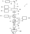
- Fig. 1 schematically depicts general aspects of the light microscopy device 1 according to the present invention.
- the light microscopy device 1 comprises an excitation light source 10 for generating an excitation light beam 14 comprising excitation light capable of exciting fluorescent dye molecules F in a sample S.
- the excitation light source 10 is particularly a laser source which creates pulses or a continuous wave of excitation light.
- the excitation light beam 14 is coupled into a beam path 2 of the light microscopy device 1 by a beam splitter 12. Additional optical components may be provided, e. g. to adjust the pulse length of the laser pulses of the excitation light in case of a pulsed laser source as known in the art (not shown).
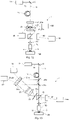
- the excitation light source 10 and the excitation light beam are not explicitly shown in Fig. 7-18 and Fig. 20 described below, but may be set up, e.g., as shown in Fig. 1 .
- the light microscopy device 1 further comprises a first impeding light source 11 for generating a first impeding light beam 15 comprising impeding light I capable of impeding the fluorescence of the fluorescent dye molecules F, e.g., by inducing stimulated emission depletion (STED) of the fluorescent dye molecules F, in other words to induce a transition of the fluorescent dye molecules F from the excited state to a vibrational energy level above the ground state.
- the wavelength of the impeding light I should be larger than the wavelength of the excitation light.
- the first impeding light beam 15 is coupled into the beam path 2 by a mirror 13.
- the excitation light and the impeding light I can also be generated from a single light source in the scope of the present invention.
- additional optical components such as further beam splitters and filters may be used to obtain the excitation light and the impeding light I at the desired wavelengths (not shown).
- the combined excitation light beam 14 and first impeding light beam 15 pass through a tube lens 71 and an objective 70 and are thereby focused on a focal plane in a sample S containing fluorescent dye molecules F provided on a sample stage 100.
- the excitation light thereby excites the fluorescent dye molecules F in the vicinity of the focal plane causing them to emit fluorescence light.
- This light is collected by the objective 70 and reflected onto a detector 40 by a dichroic mirror 41 which is reflective for the fluorescence light, but transmissive for the excitation light and the impeding light I.
- a pinhole or an optical fiber may be used (not shown).
- detector 40 may be an area detector.
- the beam splitter 41 may also be used to couple the excitation light beam 14 into the beam path 2.
- an optical modulation system 20 is provided in the beam path 2.
- a phase pattern or an amplitude pattern is generated in the first impeding light beam 15 by the optical modulation system 20 resulting in the intensity distribution.
- the optical modulation system 20 may comprise a wavefront modulator 21 with an active area which is located in a pupil plane, intersecting a pupil plane or close to a pupil plane of the objective 70. Therein, the pupil plane is conjugated to the back aperture of the objective 70.
- the intensity distribution D is particularly chosen such that the light intensity at the local minimum M is as small as possible (ideally zero), and that the light intensity steeply increases around the local minimum M at least in the focal plane (x- and y-direction, perpendicular to the optical axis of the objective 70), and preferably also parallel to the optical axis (z-direction, perpendicular to the focal plane).
- the excitation light beam 14 is coupled into the beam path 2 between the optical modulation system 20 and the objective 70, such that the phase or amplitude pattern is not generated in the excitation light beam 14.
- the excitation light beam 14 does run through the optical modulation system 20, and it may particularly be ensured by additional means that the phase pattern or amplitude pattern is not generated in the excitation light beam 14.
- an appropriately oriented first polarization modifying element 90 (e.g., a quarter wave plate), is required in the beam path 2 as explained below.
- the polarization modifying element 90 is shown between the optical modulation system 20 and the beam splitter 12 at which the excitation light beam 14 is coupled into the beam path 2.
- the first polarization modifying element 90 may be arranged between the beam splitter 12 and the tube lens 71. In this case, the polarization of the excitation light beam 14 is also affected by the polarization modifying element 90.
- background fluorescence which is a result, e.g., of direct excitation and incomplete depletion caused by the impeding light I, is removed from the fluorescence signal by a control measurement of the background fluorescence (but not the signal of interest).
- additional impeding light I is provided in an area overlapping with the local minimum M.
- the optical modulation system 20 is controlled by a control device 30.
- the resulting light intensity of the additional impeding light I is optionally adjusted by a variable amount by an adjusting means 60.
- the control device 30 and the adjusting means 60 may control other components of the light microscopy device 1 as explained below.
- the control device 30 and the adjusting means 60 ensure that the intensity distribution D is only minimally changed outside of the local minimum M, in particular to reduce artifacts and further improve background correction.
- control device 30 and the adjusting means 60 may be separate components as illustrated for simplicity in Fig. 1 but can also be modules or functionalities provided in the same component or device.
- Background correction in other words obtaining a first fluorescence signal without the additional impeding light I, obtaining a second fluorescence signal with the additional impeding light I, and subtracting the second fluorescence signal from the first fluorescence signal, is performed by a computation device 50.
- the sample S is scanned by the excitation light beam 14 and the first impeding light beam 15 using a scanner (not shown).
- background correction may be performed pixel by pixel, line by line or for an entire image depending on the speed of the optical modulation system 20.
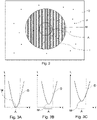
- Fig. 2 schematically shows a part of the sample S with the fluorescent dye molecules F which are distributed through the sample S.
- the sample S is shown in the focal plane (x-y-direction perpendicular to the optical axis of the objective 70).
- the local minimum M of the light intensity of the impeding light I is indicated by an asterisk.
- a cross-hatched circular region around the local minimum M is illuminated by impeding light I, wherein the intensity of the impeding light I is not indicated in Fig. 2 .
- the local minimum M is located at the center of the intensity distribution D.
- a circular area A around the local minimum M is further indicated. In this area A, the additional impeding light I is provided for the background measurement.
- the shape of the impeding light I illustrated in Fig. 2 is only an example, and other shapes and distributions (including distributions with more than one local minimum M) are within the scope of the invention.
- Fig. 3 depicts a part of the intensity distribution D of the impeding light I around the local minimum M in a simplified manner. Simplified plots of the light intensity I l as a function of the x-coordinate (in the focal plane) are shown and the area A around the local minimum M where the additional impeding light I is provided is indicated. Fig. 3A shows the situation in the absence of the additional impeding light I, where the light intensity is zero at the local minimum M in this example. Fig. 3B and 3C illustrate intensity distributions D with different amounts of additional impeding light I provided in the area A. By means of the adjusting means 60 according to the invention, the desired amount of additional impeding light I may be selected or gradually adjusted (before or during a measurement), e.g., between the examples shown in Fig. 3B and 3C .
- Fig. 4-6 show examples of phase patterns which can be introduced to the first impeding light beam 15 at a pupil plane, intersecting a pupil plane or near a pupil plane to obtain a corresponding intensity distribution D with a local minimum M at the focus.
- Fig. 4A shows a vortex-shaped phase pattern, where the phase increases, particularly gradually, from 0 to 2 ⁇ in a circumferential direction with respect to the optical axis.
- This phase pattern results in a donut shaped intensity distribution at the focus with a narrow minimum intensity spot in the focal plane if the first impeding light beam 15 is circularly polarized prior to entering the objective 70.
- This pattern can be modified to obtain additional impeding light I at the central minimum M of the donut shaped distribution, e.g., as shown in Fig. 4B and 4C .
- a ring having a constant phase value C is overlaid with the vortex-shaped pattern shown in Fig. 4A .
- the radial distance of the ring to the center of the phase pattern influences the size of the area A in which the additional impeding light I is provided: If the ring is moved further outside from the center, the resulting additional impeding light I is more concentrated at the center of the distribution.
- the area of the ring influences the light intensity I l of the additional impeding light I, wherein a larger area leads to an increased intensity I l . Therefore, by adapting the area of the ring, the variable amount of impeding light may be tuned.
- Fig. 4C Another possibility of changing the vortex-shaped phase pattern to generate additional impeding light I at the center is depicted in Fig. 4C :
- the maximum phase value of the pattern is altered by a small amount, in this case from 2 ⁇ to 1,9 ⁇ and the function of phase increase in the circumferential direction is adapted accordingly.
- the light intensity of the additional impeding light I can be tuned.
- Fig. 5A-C illustrate phase patterns which lead to a line shaped minimum in the focal plane.
- the initial pattern ( Fig. 5A ) for generating the intensity distribution D with the local minimum M consists of a first area A 1 of phase ⁇ and a second area A 2 having the phase 0, separated by a line, in other words a linear phase jump.
- additional impeding light I is generated by imperfect destructive interference at the center of the distribution D.
- the intensity of the additional impeding light I can be tuned by adjusting the relative area.
- the phase value of the first area A 1 can be adjusted ( Fig. 5C ) to generate a slight mismatch and therefore imperfect destructive interference at the center.
- the phase of a part of the first area A 1 could be switched from ⁇ to another value, e.g., 0.
- Fig. 6A depicts a phase pattern with a circular first area A 1 of phase 0 and a ring shaped second area A 2 around the first area A 1 comprising a phase value of ⁇ .
- This pattern results in an intensity distribution around the focus which is called "bottle beam", and which is characterized by a steep intensity increase in the z-direction (parallel to the optical axis of the objective 70), and is thus commonly combined, e.g., with a donut shaped distribution to obtain a three-dimensional intensity distribution with a spot of relatively small dimensions, both in the x-y plane and in the z-direction.
- the pattern of Fig. 6A may be altered by adjusting the relative surface area of the first area A 1 and the second area A 2 ( Fig. 6B ) or by adjusting the phase value of one of the areas A 1 , A 2 ( Fig. 6C ) to obtain additional impeding light I in the area around the local minimum M.
- Fig. 7-18 and 20 show different embodiments of the light microscopy device 1 which may be used to implement the method according to the invention.
- the excitation light source 10 and the excitation light beam 14 are not explicitly shown for simplicity (see Fig. 1 ) but are present in the light microscopy devices 1 according to the examples.
- an additional first polarization modifying element 90 which may be required to obtain certain intensity distributions (see Fig. 1 ) is not shown except in Fig. 16 .
- Fig. 7A and 7B illustrate a light microcopy device 1 where the optical modulation system 20 comprises a wave front modulator 21 which can generate an amplitude pattern across the beam cross-section (perpendicular to the optical axis): an array 210 of tiltable micromirrors 211, which is sometimes referred to as a digital light projector (DLP).
- the array 210 comprises a substrate 213, to which a plurality of micromirrors 211 are coupled by respective hinges 212.
- the micromirrors 211 are positioned on the substrate 213 in a two-dimensional arrangement. Three micromirrors 211 are depicted in Fig.
- the array 210 may comprise many more (e.g., hundreds to thousands) micromirrors 211.
- the micromirrors 211 can individually be tilted around the hinge 212 between a first configuration and a second configuration.
- Fig. 7A the illustrated part of the first impeding light beam 15 is reflected by a micromirror 211 into the beam path 2 of the light microscopy device 1.
- said micromirror 211 is tilted such that the corresponding part of the first impeding light beam 15 hits a beam dump 214 outside of the beam path 2.
- the amplitude of the first impeding light beam 15 is modulated by the array 210 to generate the desired intensity distribution D at the focus.
- the amplitude pattern is modified by the signals of the control device 30 and optionally the adjusting means 60 which are coupled to the array 210 according to this embodiment.
- the excitation light source 10 and the corresponding excitation light beam 14 is not shown for simplicity, but the excitation light source 10 is particularly set up in an analogous manner as in the embodiment of Fig. 1 .
- Fig. 8 depicts a further embodiment of the light microscopy device 1 according to the invention which comprises an optical modulation system 20 based on a phase-modulating wavefront modulator 21, namely a spatial light modulator 22 with an active area 21a.
- the first impeding light beam 15 is linearly polarized (using a polarization filter not shown in Fig. 8 ), and the polarization direction is particularly adjusted to match the arrangement of the spatial light modulator 22.
- a phase pattern is introduced to the first impeding light beam 15, which results in an intensity distribution D with a local minimum M around the focus.
- the phase pattern is then altered by adjusting the spatial light modulator 22 by the control device 30 to provide additional impeding light I in the area A around the local minimum M, and the light intensity I l of the additional impeding light I is optionally tuned by the adjusting means 60, which is operationally coupled to the spatial light modulator 22.
- Fig. 9 shows a setup which can be used to generate an intensity distribution D with a small local minimum in three dimensions, e.g., in so-called 3D-STED microscopy.
- a double-diffractive optical fiber 19 is provided in a beam path 2 of the light microscopy device 1 to retard a first component of the impeding light I with a first polarization direction with respect to a second component of the impeding light I with a second polarization direction which is perpendicular to the first polarization direction by a distance which is greater than the coherence length of the impeding light I. Therefore, two overlayed incoherent light beams of transversal polarization directions are generated.
- a first phase pattern is introduced only to the first component by diffracting the first impeding light beam 15 on a first active area 21 a of a spatial light modulator 22.
- the diffracted light beam is then passed through a second polarization modifying element 23, particularly a quarter wave plate or an electro optical modulator, which is arranged adjacent to a mirror surface 24.
- the first impeding light beam 15 is reflected on the mirror surface 24, and therefore passes the second polarization modifying element 23 twice, such that the polarization of the first impeding light beam is turned by 90°.
- the reflected light beam is diffracted on a second active area 21b of the spatial light modulator 22, which generates a further phase pattern in the first impeding light beam 15.
- the second active area 21b is oriented such that only the second component, but not the first component of the first impeding light beam 15 is phase modulated by the second active area 21b.
- the resulting first impeding light beam 15 carries two phase patterns on two incoherent and orthogonally polarized components. This leads to a desired three-dimensional intensity distribution D of the impeding light I at and around the focal plane. For example, a donut shape and a bottle beam shape may be combined in this manner to obtain an intensity distribution with a narrow minimum in all three dimensions.
- two separate spatial light modulators 22 instead of one spatial light modulator 22 with two active areas 21a, 21b.
- control device 30 and the adjusting means 60 are coupled to the spatial light modulator 22 to provide additional impeding light I at the local minimum M at the desired variable intensity I l .
- Fig. 10 shows the same optical setup as Fig. 9 , except that the second polarization modifying element 23 and the mirror surface 24 are coupled to a rotary actuator 25 which is configured to rotate the second polarization modifying element 23 around a rotation axis R to misalign the polarization direction of the first impeding light beam 15 in respect of the second active surface 21b, such that the phase pattern is introduced not only to the second component, but also to the first component by the second active surface 21b. Due to this mismatch, additional impeding light I is generated at the local minimum M.
- the control device 30 is coupled to the second polarization modifying element 23, which is particularly a switchable element, such as an electro optical modulator.
- the adjusting means 60 is optionally coupled to the rotary actuator 25 to adjust the angle of rotation, and therefore the intensity of the additional impeding light I.
- the spatial light modulator 22 is replaced by transmissive wave front modulators 21 (e.g., phase plates) in the beam path 2, between which a second polarization modifying element 23 is arranged.
- transmissive wave front modulators 21 e.g., phase plates
- a first phase pattern is generated in the first component of the first impeding light beam 15 by the first wave front modulator 21, the polarization is turned, and a second phase pattern is generated in the second component of the first impeding light beam 15 by the second wave front modulator 21.
- the first wave front modulator 21 may be coupled to a rotary actuator 25, the rotation of which is controlled by the control device 30, the angle optionally being controlled by the adjusting means 60.
- two polarization modifying elements 23, 27 are provided between the first and the second wavefront modulator 21.
- the polarization modifying element 27 is switchable (the polarization modifying element 27 is particularly an electro optical modulator) and coupled to a rotary actuator 25.
- the polarization modifying element 27 is inactive and therefore does not affect the polarization of the first impeding light beam 15.
- the polarization modifying element 23 turns the polarization direction by 90°. To obtain additional impeding light I for background correction, the polarization modifying element 27 can be switched on by the control device 30.
- the polarization modifying element 27 can be rotated by a desired angle to obtain a desired orientation with respect to the second wave front modulator 21, and the intensity of the additional impeding light I can be tuned.
- the advantage of this setup is the possibility to quickly switch the additional impeding light on an off.
- Fig. 13 and 14 illustrate an alternative setup, by which two phase patterns may be introduced to the first impeding light beam 15, e.g., in 3D-STED microscopy.
- the first impeding light beam 15 is reflected by mirror 28a onto a first active area 21a of a spatial light modulator 22, and then passes a second polarization modifying element 23, e.g., a quarter wave plate, is focused by a lens 29 onto a further mirror 28c, which reflects the light beam back through the lens 29, the second polarization modifying element 23, and onto a second active area 21b of the spatial light modulator 22.
- a second polarization modifying element 23 e.g., a quarter wave plate
- the first active area 21a generates the first phase pattern in the first component of the first impeding light beam 15, and the second active area 21b generates the second phase pattern in the second component of the first impeding light beam 15.
- the first active area 21a and the second active area 21b are arranged in a holographic setup, such that the first active area 21a is imaged onto the second active area 21b.
- the resulting first impeding light beam 15 carrying both patterns is then coupled back into the beam path 2 by the mirror 28b.
- the spatial light modulator 22 is controlled by the control device 30 and optionally the adjusting means 60 to modify the phase patterns and obtain additional impeding light I at the local minimum M, particularly at a variable intensity.
- a rotary actuator 25 is coupled to the second polarization modifying element 23. Switching of the rotary actuator 25 by the control device 30 and particularly controlling the angle of rotation by the adjusting means 60 functions as described above for the embodiment of Fig. 10 .
- Fig. 15 shows a further embodiment of the light microscopy device 1 in a further configuration for generating two phase patterns to obtain a desired three-dimensional intensity distribution of the first impeding light beam 15 at and around the focus.
- the first impeding light beam 15 is passed through a double-diffractive optical fiber 19 as described above and is then split into a first partial impeding light beam 15a and a second partial impeding light beam 15b at a beam splitter 201.
- a first phase pattern is generated in the first partial impeding light beam 15a by a first spatial light modulator 22 having a first active area 21a and is then coupled into the beam path by mirrors 202 and 203.
- a second phase pattern is generated in the second partial impeding light beam 15b, and the second partial impeding light beam is coupled into the beam path by mirrors 205 and 206.
- the control device 30 and the adjusting means 60 are coupled to the first spatial light modulator 22 to alter the first phase pattern and generate additional impeding light I of a desired intensity, e.g., as described above.
- the second spatial light modulator 22 may be adjusted to alter the second phase pattern or both patterns could be altered to generate the additional impeding light I at the desired intensity.
- Fig. 16 shows a light microscopy device 1 according to the invention, which is similar to the one depicted in Fig. 1 , wherein the optical modulation system 20 is a wavefront modulator 21 having an active area 21a configured to generate a phase pattern in the first impeding light beam 15. Further, a first polarization modifying element 90 is coupled to a rotary actuator 25, which in turn is controlled by the control device 30 and can be rotated by a variable small angle controlled by the adjusting means 60 to generate a mismatch between the phase pattern and the polarization introduced by the first polarization modifying element 90 and therefore generate additional impeding light I of a desired intensity.
- the optical modulation system 20 is a wavefront modulator 21 having an active area 21a configured to generate a phase pattern in the first impeding light beam 15.
- a first polarization modifying element 90 is coupled to a rotary actuator 25, which in turn is controlled by the control device 30 and can be rotated by a variable small angle controlled by the adjusting means 60 to generate a mis
- the active area 21a particularly generates a vortex-shaped phase-pattern
- the first polarization modifying element 90 particularly circularly polarizes the first impeding light beam 15, such that a donut shaped intensity distribution D is generated at and around the focus.
- Fig. 17 shows a light microscopy device 1 according to the invention comprising a fourth polarization modifying element 81 and a polarizing beam splitter 80 configured to split a selected fraction from the first impeding light beam 15 to generate a second impeding light beam 16, which is coupled into the beam path 2 of the objective 70 via mirrors 82 and 83 bypassing the optical modulation system 20, such that only the first impeding light beam 15, but not the second impeding light beam 16 is phase modulated. Therefore, in particular the second impeding light beam 16 generates an approximately Gaussian-shaped intensity distribution of impeding light I which is overlaid with the intensity distribution D comprising the local minimum M from the first impeding light beam 15 at the focus.
- additional impeding light I may be generated at the local minimum M.
- the intensity of the additional impeding light I can be adjusted controlled by the adjusting means 60.
- Fig. 18 shows a light microscopy device 1 according to the invention comprising a first impeding light source 11 for generating a first impeding light beam 15, and a second impeding light source 17 for generating a second impeding light beam 16 to generate additional impeding light I at the local minimum M of the intensity distribution.
- the optical modulation system 20 is bypassed by the second impeding light beam 16.
- the second impeding light source 17 can be switched on an off by the control device 30, and the intensity of the second impeding light beam 16 can optionally be adjusted by an intensity modulator 18 which is coupled to the adjusting means 60.
- Fig. 19 depicts a time series of interleaved first pulses P 1 and second pulses P 2 from a pulsed laser source capable of generating the impeding light I.
- Such pulses P 1 , P 2 may be generated, e.g., by a first impeding light source 11 (a pulsed laser source) as depicted in Fig. 20 (explained below).
- the first pulses P 1 and the second pulses P 2 are generated at equal time intervals (e.g., in the order of about 10 ns between a first pulse P 1 and a second pulse P 2 ).
- photons hv emitted by a fluorescent dye molecule F in the sample S are detected by the detector 40.
- the depicted series of pulses P 1 , P 2 occurs during pixel wise scanning and detection of the fluorescent signal, that is, the entire pulse series occurs during detection of the fluorescent signal as a single pixel at the detector 40.
- the time intervals between the pulses P 1 , P 2 are therefore much smaller than the scanning speed of the sample S.
- the photons hv detected by the detector 40 during the pulse series are counted by a counting logic of the light microscopy device 1 and the number of counted photons hv relates to the detected signal intensity.
- the additional impeding light I is generated at and around the local minimum M of the intensity distribution D, but during the first pulses P 1 , the additional impeding light I is not present at and around the local minimum M.
- This advantageously allows to perform background correction in an interleaved manner, resulting in a high accuracy of the correction, since temporal effects (e.g., photobleaching, sample drift) are even less relevant than in pixel-for-pixel measurement of the signal of interest (first fluorescence signal) and the background (second fluorescence signal).
- two separate counters which are appropriately coordinated with the system clock can be used to count the photons detected after a first pulse P 1 and the photons detected after a second pulse P 2 . After accumulation, the two signals can be subtracted from each other to obtain the background corrected signal.
- a single counter may be used which alternately counts up and down.
- This counter may count up in the time intervals after a first pulse P 1 and down in the time intervals after a second pulse P 2 , such that background correction is already performed during the accumulation of the fluorescence signal. This is due to the fact that the sum of all photons detected after a second pulse P 2 subtracted from the sum of all photons detected after a first pulse P 1 is the same as the sum obtained by alternately adding and subtracting the detected photons.
- Fig. 20 shows an optical setup that can be used to generate alternating first pulses P 1 and second pulses P 2 .
- the shown light microscopy device 1 is set up analogously as the light microscopy device 1 shown in Fig. 1 , wherein the first impeding light source 11 is a pulsed laser source, and the setup comprises an additional beam splitter 101 that splits the first impeding light beam 15 generated by the pulsed impeding light source 11 into a first branch 15' and a second branch 15".
- the first branch 15' generates the first pulses P 1 (see Fig. 19 ), which are coupled into the beam path 2 via the mirror 13.
- the second branch 15" contains a mirror 102 and a pulse delaying element 103, which generates a temporal delay of the pulses traveling on the second branch 15" with respect to the pulses traveling on the first branch 15'. Therefore, the second branch 15" generates the second pulses P 2 which are interleaved with the first pulses P 1 , and the second pulses P 2 are coupled into the beam path 2 by the mirror 104.
- two separate laser sources which are appropriately timed may be used instead of the setup shown in Fig. 20 to generate the interleaved first pulses P 1 and second pulses P 2 .
- the additional impeding light I at and around the local minimum M of the intensity distribution D is generated during the second pulses P 2 , but not during the first pulses P 1 .
- This may be achieved by the setup shown in Fig. 20 , e.g., by appropriate switching of the optical modulation system 20, such that, e.g., the phase pattern or amplitude pattern introduced to the first impeding light beam 15 is changed during the second pulses P 2 compared to the first pulses P 1 .
- the optical modulation system 20 or the light generated by an additional pulsed laser source may be coupled into the beam path 2 between the optical modulation system 20 and the objective 70, such as, e.g., depicted for the second impeding light beam 16 in Fig. 17 and 18 , such that the optical modulation system 20 modulates only the first pulses P 1 , but not the second pulses P 2 .
Landscapes
- Physics & Mathematics (AREA)
- General Physics & Mathematics (AREA)
- Optics & Photonics (AREA)
- Chemical & Material Sciences (AREA)
- Analytical Chemistry (AREA)
- Health & Medical Sciences (AREA)
- Biochemistry (AREA)
- Life Sciences & Earth Sciences (AREA)
- Nuclear Medicine, Radiotherapy & Molecular Imaging (AREA)
- General Health & Medical Sciences (AREA)
- Immunology (AREA)
- Pathology (AREA)
- Engineering & Computer Science (AREA)
- Computer Vision & Pattern Recognition (AREA)
- General Engineering & Computer Science (AREA)
- Microscoopes, Condenser (AREA)
- Investigating, Analyzing Materials By Fluorescence Or Luminescence (AREA)
Priority Applications (4)
| Application Number | Priority Date | Filing Date | Title |
|---|---|---|---|
| EP20216708.6A EP4020051A1 (de) | 2020-12-22 | 2020-12-22 | Lichtmikroskopieverfahren und lichtmikroskopievorrichtung |
| EP21843709.3A EP4268003B1 (de) | 2020-12-22 | 2021-12-21 | Lichtmikroskopieverfahren und lichtmikroskopievorrichtung |
| PCT/EP2021/087000 WO2022136375A1 (en) | 2020-12-22 | 2021-12-21 | Light microscopy method and light microscopy device |
| US18/268,273 US20240302637A1 (en) | 2020-12-22 | 2021-12-21 | Light microscopy method and light microscopy device |
Applications Claiming Priority (1)
| Application Number | Priority Date | Filing Date | Title |
|---|---|---|---|
| EP20216708.6A EP4020051A1 (de) | 2020-12-22 | 2020-12-22 | Lichtmikroskopieverfahren und lichtmikroskopievorrichtung |
Publications (1)
| Publication Number | Publication Date |
|---|---|
| EP4020051A1 true EP4020051A1 (de) | 2022-06-29 |
Family
ID=73856922
Family Applications (2)
| Application Number | Title | Priority Date | Filing Date |
|---|---|---|---|
| EP20216708.6A Withdrawn EP4020051A1 (de) | 2020-12-22 | 2020-12-22 | Lichtmikroskopieverfahren und lichtmikroskopievorrichtung |
| EP21843709.3A Active EP4268003B1 (de) | 2020-12-22 | 2021-12-21 | Lichtmikroskopieverfahren und lichtmikroskopievorrichtung |
Family Applications After (1)
| Application Number | Title | Priority Date | Filing Date |
|---|---|---|---|
| EP21843709.3A Active EP4268003B1 (de) | 2020-12-22 | 2021-12-21 | Lichtmikroskopieverfahren und lichtmikroskopievorrichtung |
Country Status (3)
| Country | Link |
|---|---|
| US (1) | US20240302637A1 (de) |
| EP (2) | EP4020051A1 (de) |
| WO (1) | WO2022136375A1 (de) |
Citations (3)
| Publication number | Priority date | Publication date | Assignee | Title |
|---|---|---|---|---|
| DE102014113716A1 (de) | 2014-09-23 | 2016-03-24 | Abberior Instruments Gmbh | Vorrichtung zum getrennten Modulieren der Wellenfronten von zwei Komponenten eines Lichtstrahls |
| EP3686643A1 (de) * | 2019-01-25 | 2020-07-29 | Hochschule Für Angewandte Wissenschaften München | Impulsformung für die mikroskopie mit stimulierter emissionsminderung |
| WO2020198487A1 (en) | 2019-03-26 | 2020-10-01 | The Johns Hopkins University | Background-suppressed sted nanoscope |
-
2020
- 2020-12-22 EP EP20216708.6A patent/EP4020051A1/de not_active Withdrawn
-
2021
- 2021-12-21 WO PCT/EP2021/087000 patent/WO2022136375A1/en not_active Ceased
- 2021-12-21 US US18/268,273 patent/US20240302637A1/en active Pending
- 2021-12-21 EP EP21843709.3A patent/EP4268003B1/de active Active
Patent Citations (3)
| Publication number | Priority date | Publication date | Assignee | Title |
|---|---|---|---|---|
| DE102014113716A1 (de) | 2014-09-23 | 2016-03-24 | Abberior Instruments Gmbh | Vorrichtung zum getrennten Modulieren der Wellenfronten von zwei Komponenten eines Lichtstrahls |
| EP3686643A1 (de) * | 2019-01-25 | 2020-07-29 | Hochschule Für Angewandte Wissenschaften München | Impulsformung für die mikroskopie mit stimulierter emissionsminderung |
| WO2020198487A1 (en) | 2019-03-26 | 2020-10-01 | The Johns Hopkins University | Background-suppressed sted nanoscope |
Non-Patent Citations (9)
| Title |
|---|
| GAO PENG ET AL: "Precise background subtraction in stimulated emission double depletion nanoscopy", OPTICS LETTERS, vol. 42, no. 4, 15 February 2017 (2017-02-15), US, pages 831, XP055813521, ISSN: 0146-9592, DOI: 10.1364/OL.42.000831 * |
| GAO PPRUNSCHE BZHOU LNIENHAUS KNIENHAUS GU: "Background Subtraction in Stimulated Emission Double Depletion Nanoscopy", OPT. LETT., vol. 42, no. 4, 2017, pages 831 |
| GAO PPRUNSCHE BZHOU LNIENHAUS KNIENHAUS GU: "Background Suppression in Fluorescence Nanoscopy with Stimulated Emission Double Depletion", NAT. PHOTONICS, vol. 11, no. 3, 2017, pages 167 - 169 |
| HANNE JFALK, HJGORLITZ FHOYER PENGELHARDT JSAHL SJHELL SW, STED NANOSCOPY WITH FLUORESCENT QUANTUM DOTS. NAT. COMM., 6 May 2015 (2015-05-06), pages 7127 |
| KLAR, TAENGEL EHELL SW: "Breaking Abbe's diffraction resolution limit in fluorescence microscopy with stimulated emission depletion beams of various shapes", PHYSICAL REVIEW E, vol. 64, 2001, pages 066613 - 1,066613-9, XP055149722, DOI: 10.1103/PhysRevE.64.066613 |
| MÖHL ANNA ET AL: "Improving quantitative fluorescence imaging with flat field illumination", PROGRESS IN BIOMEDICAL OPTICS AND IMAGING, SPIE - INTERNATIONAL SOCIETY FOR OPTICAL ENGINEERING, BELLINGHAM, WA, US, vol. 10881, 4 March 2019 (2019-03-04), pages 1088103 - 1088103, XP060117527, ISSN: 1605-7422, ISBN: 978-1-5106-0027-0, DOI: 10.1117/12.2507491 * |
| VICIDOMINI GMONERON GEGGELING CRITTWEGER EHELL SW: "STED with Wavelengths Closer to the Emission Maximum", OPT. EXPRESS, vol. 20, no. 5, 2012, pages 5225, XP055115467, DOI: 10.1364/OE.20.005225 |
| VICIDOMINI GMONERON GHAN KYWESTPHAL VTA HREUSS MENGELHARDT JEGGELING CHELL SW: "Sharper Low-Power STED Nanoscopy by Time Gating", NAT. METHODS, vol. 8, no. 7, 2011, pages 571 - 573, XP055115226, DOI: 10.1038/nmeth.1624 |
| YE MA ET AL: "Fight against background noise in stimulated emission depletion nanoscopy", PHYSICAL BIOLOGY, INSTITUTE OF PHYSICS PUBLISHING, BRISTOL, GB, vol. 16, no. 5, 5 July 2019 (2019-07-05), pages 51002, XP020342809, ISSN: 1478-3975, DOI: 10.1088/1478-3975/AB255C * |
Also Published As
| Publication number | Publication date |
|---|---|
| WO2022136375A1 (en) | 2022-06-30 |
| EP4268003B1 (de) | 2025-04-23 |
| EP4268003A1 (de) | 2023-11-01 |
| US20240302637A1 (en) | 2024-09-12 |
Similar Documents
| Publication | Publication Date | Title |
|---|---|---|
| JP5554965B2 (ja) | 位相変調型空間光変調器を用いたレーザ顕微鏡 | |
| JP4723806B2 (ja) | 共焦点顕微鏡 | |
| US8207510B2 (en) | Method and arrangement for collimated microscopic imaging | |
| US8362448B2 (en) | Apparatus and method for high spatial resolution imaging of a structure of a sample | |
| US7679741B2 (en) | Method and microscope for high spatial resolution examination of samples | |
| US9864182B2 (en) | High-resolution scanning microscopy | |
| US10942346B2 (en) | Methods and systems for generating non-diffracting light sheets for multicolor fluorescence microscopy | |
| US20240248291A1 (en) | Light modulation device for a microscope, microscope and method for modulating a light beam | |
| US20090161203A1 (en) | Method and Configuration for the Optical Detection of an Illuminated Specimen | |
| US20240264420A1 (en) | Focusing device, focusing system, microscope and method for imaging and/or localizing molecules or particles in a sample | |
| JP6023147B2 (ja) | 照明された試料を光学的に捕捉するための方法および装置 | |
| JP2001290081A (ja) | 試料照射装置 | |
| JP2007233370A (ja) | 試料を高い空間分解能で検査するための方法および顕微鏡 | |
| US11022788B2 (en) | Illumination apparatus for a microscope | |
| US10795138B2 (en) | Fluorescence microscope instrument comprising an actively switched beam path separator | |
| US20220066187A1 (en) | Two-color confocal colocalization microscopy | |
| JP2002303798A (ja) | 二重共焦点走査顕微鏡 | |
| EP4268003B1 (de) | Lichtmikroskopieverfahren und lichtmikroskopievorrichtung | |
| US12468136B2 (en) | Random access projection microscopy | |
| US12631863B2 (en) | Luminescence microscope for imaging a sample or for localizing or tracking emitters in a sample | |
| US20240134176A1 (en) | Luminescence microscope for imaging a sample or for localizing or tracking emitters in a sample | |
| US11933956B2 (en) | Method and device for illuminating a sample plane |
Legal Events
| Date | Code | Title | Description |
|---|---|---|---|
| PUAI | Public reference made under article 153(3) epc to a published international application that has entered the european phase |
Free format text: ORIGINAL CODE: 0009012 |
|
| STAA | Information on the status of an ep patent application or granted ep patent |
Free format text: STATUS: THE APPLICATION HAS BEEN PUBLISHED |
|
| AK | Designated contracting states |
Kind code of ref document: A1 Designated state(s): AL AT BE BG CH CY CZ DE DK EE ES FI FR GB GR HR HU IE IS IT LI LT LU LV MC MK MT NL NO PL PT RO RS SE SI SK SM TR |
|
| STAA | Information on the status of an ep patent application or granted ep patent |
Free format text: STATUS: THE APPLICATION IS DEEMED TO BE WITHDRAWN |
|
| 18D | Application deemed to be withdrawn |
Effective date: 20230103 |