EP4023231A1 - Vésicules extracellulaires dérivées de cellules stromales mésenchymateuses génétiquement modifiées pour la surexpression hif-1a et htert - Google Patents
Vésicules extracellulaires dérivées de cellules stromales mésenchymateuses génétiquement modifiées pour la surexpression hif-1a et htert Download PDFInfo
- Publication number
- EP4023231A1 EP4023231A1 EP20383170.6A EP20383170A EP4023231A1 EP 4023231 A1 EP4023231 A1 EP 4023231A1 EP 20383170 A EP20383170 A EP 20383170A EP 4023231 A1 EP4023231 A1 EP 4023231A1
- Authority
- EP
- European Patent Office
- Prior art keywords
- hsa
- mir
- evs
- msc
- hif
- Prior art date
- Legal status (The legal status is an assumption and is not a legal conclusion. Google has not performed a legal analysis and makes no representation as to the accuracy of the status listed.)
- Pending
Links
Images


Classifications
-
- C—CHEMISTRY; METALLURGY
- C07—ORGANIC CHEMISTRY
- C07K—PEPTIDES
- C07K14/00—Peptides having more than 20 amino acids; Gastrins; Somatostatins; Melanotropins; Derivatives thereof
- C07K14/435—Peptides having more than 20 amino acids; Gastrins; Somatostatins; Melanotropins; Derivatives thereof from animals; from humans
- C07K14/46—Peptides having more than 20 amino acids; Gastrins; Somatostatins; Melanotropins; Derivatives thereof from animals; from humans from vertebrates
- C07K14/47—Peptides having more than 20 amino acids; Gastrins; Somatostatins; Melanotropins; Derivatives thereof from animals; from humans from vertebrates from mammals
- C07K14/4701—Peptides having more than 20 amino acids; Gastrins; Somatostatins; Melanotropins; Derivatives thereof from animals; from humans from vertebrates from mammals not used
- C07K14/4702—Regulators; Modulating activity
-
- A—HUMAN NECESSITIES
- A61—MEDICAL OR VETERINARY SCIENCE; HYGIENE
- A61K—PREPARATIONS FOR MEDICAL, DENTAL OR TOILETRY PURPOSES
- A61K35/00—Medicinal preparations containing materials or reaction products thereof with undetermined constitution
- A61K35/12—Materials from mammals; Compositions comprising non-specified tissues or cells; Compositions comprising non-embryonic stem cells; Genetically modified cells
- A61K35/28—Bone marrow; Haematopoietic stem cells; Mesenchymal stem cells of any origin, e.g. adipose-derived stem cells
-
- C—CHEMISTRY; METALLURGY
- C12—BIOCHEMISTRY; BEER; SPIRITS; WINE; VINEGAR; MICROBIOLOGY; ENZYMOLOGY; MUTATION OR GENETIC ENGINEERING
- C12N—MICROORGANISMS OR ENZYMES; COMPOSITIONS THEREOF; PROPAGATING, PRESERVING, OR MAINTAINING MICROORGANISMS; MUTATION OR GENETIC ENGINEERING; CULTURE MEDIA
- C12N5/00—Undifferentiated human, animal or plant cells, e.g. cell lines; Tissues; Cultivation or maintenance thereof; Culture media therefor
- C12N5/06—Animal cells or tissues; Human cells or tissues
- C12N5/0602—Vertebrate cells
- C12N5/0652—Cells of skeletal and connective tissues; Mesenchyme
- C12N5/0662—Stem cells
- C12N5/0664—Dental pulp stem cells, Dental follicle stem cells
-
- C—CHEMISTRY; METALLURGY
- C12—BIOCHEMISTRY; BEER; SPIRITS; WINE; VINEGAR; MICROBIOLOGY; ENZYMOLOGY; MUTATION OR GENETIC ENGINEERING
- C12N—MICROORGANISMS OR ENZYMES; COMPOSITIONS THEREOF; PROPAGATING, PRESERVING, OR MAINTAINING MICROORGANISMS; MUTATION OR GENETIC ENGINEERING; CULTURE MEDIA
- C12N9/00—Enzymes; Proenzymes; Compositions thereof; Processes for preparing, activating, inhibiting, separating or purifying enzymes
- C12N9/10—Transferases (2.)
- C12N9/12—Transferases (2.) transferring phosphorus containing groups, e.g. kinases (2.7)
- C12N9/1241—Nucleotidyltransferases (2.7.7)
- C12N9/1276—RNA-directed DNA polymerase (2.7.7.49), i.e. reverse transcriptase or telomerase
-
- C—CHEMISTRY; METALLURGY
- C12—BIOCHEMISTRY; BEER; SPIRITS; WINE; VINEGAR; MICROBIOLOGY; ENZYMOLOGY; MUTATION OR GENETIC ENGINEERING
- C12Y—ENZYMES
- C12Y207/00—Transferases transferring phosphorus-containing groups (2.7)
- C12Y207/07—Nucleotidyltransferases (2.7.7)
- C12Y207/07049—RNA-directed DNA polymerase (2.7.7.49), i.e. telomerase or reverse-transcriptase
-
- C—CHEMISTRY; METALLURGY
- C12—BIOCHEMISTRY; BEER; SPIRITS; WINE; VINEGAR; MICROBIOLOGY; ENZYMOLOGY; MUTATION OR GENETIC ENGINEERING
- C12N—MICROORGANISMS OR ENZYMES; COMPOSITIONS THEREOF; PROPAGATING, PRESERVING, OR MAINTAINING MICROORGANISMS; MUTATION OR GENETIC ENGINEERING; CULTURE MEDIA
- C12N2501/00—Active agents used in cell culture processes, e.g. differentation
- C12N2501/20—Cytokines; Chemokines
- C12N2501/23—Interleukins [IL]
-
- C—CHEMISTRY; METALLURGY
- C12—BIOCHEMISTRY; BEER; SPIRITS; WINE; VINEGAR; MICROBIOLOGY; ENZYMOLOGY; MUTATION OR GENETIC ENGINEERING
- C12N—MICROORGANISMS OR ENZYMES; COMPOSITIONS THEREOF; PROPAGATING, PRESERVING, OR MAINTAINING MICROORGANISMS; MUTATION OR GENETIC ENGINEERING; CULTURE MEDIA
- C12N2501/00—Active agents used in cell culture processes, e.g. differentation
- C12N2501/20—Cytokines; Chemokines
- C12N2501/24—Interferons [IFN]
-
- C—CHEMISTRY; METALLURGY
- C12—BIOCHEMISTRY; BEER; SPIRITS; WINE; VINEGAR; MICROBIOLOGY; ENZYMOLOGY; MUTATION OR GENETIC ENGINEERING
- C12N—MICROORGANISMS OR ENZYMES; COMPOSITIONS THEREOF; PROPAGATING, PRESERVING, OR MAINTAINING MICROORGANISMS; MUTATION OR GENETIC ENGINEERING; CULTURE MEDIA
- C12N2501/00—Active agents used in cell culture processes, e.g. differentation
- C12N2501/20—Cytokines; Chemokines
- C12N2501/25—Tumour necrosing factors [TNF]
-
- C—CHEMISTRY; METALLURGY
- C12—BIOCHEMISTRY; BEER; SPIRITS; WINE; VINEGAR; MICROBIOLOGY; ENZYMOLOGY; MUTATION OR GENETIC ENGINEERING
- C12N—MICROORGANISMS OR ENZYMES; COMPOSITIONS THEREOF; PROPAGATING, PRESERVING, OR MAINTAINING MICROORGANISMS; MUTATION OR GENETIC ENGINEERING; CULTURE MEDIA
- C12N2510/00—Genetically modified cells
- C12N2510/04—Immortalised cells
Definitions
- the present invention relates generally to the Health Sector, in particular the field of Immunology and Tissue Regeneration. Particularly, the invention relates to Extracellular Vesicles (EVs) derived from mesenchymal stromal cells genetically modified to overexpress HIF-1 ⁇ and hTERT (MSCs-T-HIF). The invention also contemplates the method for obtaining said EVs, pharmaceuticals compositions comprising said EVs, and clinical applications thereof.
- EVs Extracellular Vesicles
- MSCs-T-HIF mesenchymal stromal cells genetically modified to overexpress HIF-1 ⁇ and hTERT
- the invention also contemplates the method for obtaining said EVs, pharmaceuticals compositions comprising said EVs, and clinical applications thereof.
- MSCs Mesenchymal stromal cells
- plastic adherent in standard conditions with a specific surface antigen expression and capable to differentiate into the chondrocyte, osteoblast or adipocyte lineages
- Dominici, M. et al Minimum criteria for defining multipotent mesenchymal stromal cells. The International Society for Cellular Therapy position statement" Cytotherapt (2006) Vol. 8, n° 4, 315-317 ).
- MSCs emerged as a potential therapeutic element to treat multiple diseases based on both their tissue regeneration and immunosuppressive capacity ( Dorronsoro, A. et al. Human mesenchymal stromal cell-mediated immunoregulation: mechanisms of action and clinical applications.
- Human bone marrow stromal cells inhibit allogeneic T-cell responses by indoleamine 2,3-dioxygenase-mediated tryptophan degradation.
- MSC favour the appearance of regulatory T cells by secreting TGF- ⁇ ( English, K. et al. Cell contact, prostaglandin E2 and transforming growth factor beta 1 play non-redundant roles in human mesenchymal stem cell induction of CD4+CD25Highforkhead box P3+ regulatory T cells. Clin. Exp. Immunol. 156, 149-160 (2009 ).
- MSC are also able to inhibit the activation of M1 macrophages and promote M1 to M2 polarization ( Németh, K. et al. Bone marrow stromal cells attenuate sepsis via prostaglandin E 2-dependent reprogramming of host macrophages to increase their interleukin-10 production.
- NK cells reduce the proliferation and cytotoxic activity of these cells ( Aggarwal, S. et al 2005; Rasmusson, I., Ringdén, O., Sundberg, B. & Le Blanc, K. Mesenchymal stem cells inhibit the formation of cytotoxic T lymphocytes, but not activated cytotoxic T lymphocytes or natural killer cells. Transplantation 76, 1208-13 (2003 ); Spaggiari, G. M., Capobianco, A., Becchetti, S., Mingari, M. C. & Moretta, L. Mesenchymal stem cell-natural killer cell interactions: Evidence that activated NK cells are capable of killing MSCs, whereas MSCs can inhibit IL-2-induced NK-cell proliferation.
- Small extracellular vesicles are small particles 30 to 200 nm in diameter and delimited by a lipidic bilayer membrane. EVs contain a diverse cargo composed by different cytokines, membrane trafficking molecules, chemokines, heat shock proteins and even mRNA and microRNAs ( Théry, C. et al. Minimal information for studies of extracellular vesicles 2018 (MISEV2018): a position statement of the International Society for Extracellular Vesicles and update of the MISEV2014 guidelines. J. Extracell. Vesicles 7, 1535750 (2018 ); Théry, C., Zitvogel, L. & Amigorena, S. Exosomes: Composition, biogenesis and function. Nature Reviews Immunology 2, 569-579 (2002 ).
- Vesicle-based therapeutic strategies have great advantages in terms of biosafety and production under clinical-grade conditions. Since they are not living elements and do not carry nuclear DNA, EVs transplantation involves a lower risk of causing the growth of foreign bodies or malignant tumors. Similarly, the use of EVs facilitates pharmaceutical production and clinical logistics, significantly reducing the cost of a potential therapy.
- HIF-1 ⁇ is a master regulator of adaptation to hypoxia ( Ratcliffe, P. J. From erythropoietin to oxygen: Hypoxia-inducible factor hydroxylases and the hypoxia signal pathway. in Blood Purification 20, 445-450 (2002 ) that modulates different cellular processes in MSCs such as survival, proliferation, migration and differentiation ( Stubbs, S. L. et al. Hypoxic preconditioning enhances survival of human adipose-derived stem cells and conditions endothelial cells in vitro. Stem Cells Dev. 21, 1887-1896 (2012 )).
- hypoxia preconditioning Zou, D. et al.
- HIF-1 ⁇ potentiates immunosuppressive capacity of MSCs over immune cell populations such as dendritic cells, NK cells and monocytes
- immune cell populations such as dendritic cells, NK cells and monocytes
- overexpression of hypoxia-inducible factor 1 alpha improves immunomodulation by dental mesenchymal stem cells.
- the immunosuppressive ability of MSCs is not constitutive but rather induced by crosstalk with different cells of the immune system. This extent is relevant for any EVs based therapy since their healing features depend on physiological state of parental cells at the moment of secretion.
- immunosuppressive properties of MSC must be activated by external stimuli in order to transfer that capacity to EVs before they are collected ( Joo, Suh, Lee, Bang & Lee. Current Knowledge and Future Perspectives on Mesenchymal Stem Cell-Derived Exosomes as a New Therapeutic Agent. Int. J. Mol. Sci. 21, 727 (2020 ).
- the authors of the present invention after a significant research effort, have developed a new strategy to improve therapeutic capacity of EVs combining genetic modifications and proinflammatory stimulation on MSCs to obtain a constant source of boosted immunosuppressive EVs.
- they have generated a genetically modified MSC cell line, by overexpressing Hypoxia inducible factor 1 ⁇ (HIF-1 ⁇ ) and human telomerase (hTERT), to be used as EVs source.
- Secreted EVs have an improved therapeutic capacity which is further increased by a specific cytokine based preconditioning culture medium.
- the present invention provides extracellular vesicles with improved therapeutic potency through genetic modification of parental MSCs.
- the extracellular vesicles of the invention are secreted and isolated vesicles by genetically modified MSCs (MSCs-T-HIF), in which the overexpression of HIF-1 ⁇ gives rise to a greater therapeutic potential and the overexpression of hTERT generates the unlimited and constant division of the cells, giving rise to a stable and homogeneous source of vesicles with improved therapeutic functions.
- MSCs-T-HIF genetically modified MSCs
- the present invention refers to Extracellular Vesicles (EVs) isolated from mesenchymal stromal cells genetically modified to overexpress HIF-1 ⁇ and hTERT (described herein as EV MSC-T-HIF c or EVs of the invention).
- EVs Extracellular Vesicles isolated from mesenchymal stromal cells genetically modified to overexpress HIF-1 ⁇ and hTERT (described herein as EV MSC-T-HIF c or EVs of the invention).
- Parental MSCs can be isolated from different sources such as bone marrow, fat tissue, umbilical cord, peripheral blood or Warton gelly.
- EVs of the present invention are isolated from human mesenchymal stromal cells derived from dental pulp.
- the invention refers to a method for obtaining extracellular vesicles (EVs) isolated from mesenchymal stromal cells genetically modified to overexpress HIF-1 ⁇ and hTERT (MSCs-T-HIF) (method of the invention).
- the method comprises the steps of:
- the present invention refers to those extracellular vesicles obtained according to the method of the invention.
- the EVs of the invention which can be obtained by the method provided herein, show particular characteristics respect to extracellular vesicles isolated from unmodified MSCs, as described below.
- the miRNA profile of the EVs of the invention are different compared to EVs secreted by unmodified MSCs.
- the EVs show a miRNA profile with at least one of the following miRNAs upregulated in comparison to EVs isolated from unmodified MSCs: hsa-miR-92b-3p, hsa-miR-186-5p, hsa-miR-4254, hsa-miR-6720-3p, hsa-miR-938, hsa-miR-138-1-3p, hsa-miR-222-3p, hsa-miR-5089-3p, hsa-miR-654-3p, hsa-miR-143-3p, hsa-miR-30a-5p, hsa-miR-7157-3p, hsa-miR-5585-5p, hsa-miR-590-3p, hsa-miR-12132, hsa-miR-16-5p, hsa-miR
- the EVs of the present invention release PD-L1 in a higher concentration than EVs from unmodified MSCs.
- PD-1 is a protein on the surface of cells that has a role in regulating the immune system's response to the cells of the human body by down-regulating the immune system and promoting self-tolerance by suppressing T cell inflammatory activity. This prevents autoimmune diseases, but it can also prevent the immune system from killing cancer cells.
- the EVs of the present invention release PD-L1 in an amount equal or higher than 1 pg/10 11 EVs.
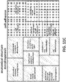
- protein profile of EVs MSC-T-HIF is different compared to protein profile of EVs from unmodified MSCs (Table 1). Protein cargo involved in immune effector processes, immune system processes, secretion by cell, and response to stimulus are over-represented in primed EVs MSC-T-HIF ( Figure 11 ).
- the EVs of the present invention unexpectedly show an increased immunosuppressive capacity, higher than EVs from wildtype parental cells.
- the EVs of the invention can be used in the treatment of autoimmune diseases. Further, the EVs of the invention can be used in the treatment of ischemic diseases.
- the present invention refers to a pharmaceutical composition comprising the EVs of the present invention.
- the pharmaceutical composition is useful in cell therapy, in particular for the treatment of autoimmune diseases, such as graft versus host disease, COVID-19, sepsis, acute solid organ rejection and colitis and for use in the treatment of ischemic diseases, such as myocardial infarction, stroke and hind limb ischemia
- autoimmune diseases such as graft versus host disease, COVID-19, sepsis, acute solid organ rejection and colitis
- ischemic diseases such as myocardial infarction, stroke and hind limb ischemia
- the pharmaceutical compositions can be administered to the subject at a suitable dose.
- the dosage regimen depends upon many factors, including the patient's size, body surface area, age, sex, general health, other drugs being administered concurrently, time and route of administration.
- the composition of the invention is preferably administered to the subject intravenously or by local injection.
- Human dental pulp MSCs were cultured as previously described ( Gandia, C. et al. Human Dental Pulp Stem Cells Improve Left Ventricular Function, Induce Angiogenesis, and Reduce Infarct Size in Rats with Acute Myocardial Infarction. Stem Cells 26, 638-645 (2008 ). Briefly, cells were cultured in Dulbecco's modified Eagle's medium (DMEM) low glucose (Sigma-Aldrich, Spain) supplemented with 10% heat-inactivated fetal bovine serum (FBS; Corning, Manassas, EEUU), 100 U/ml penicillin and 100 ⁇ g/ml streptomycin (P/S, Millipore).
- DMEM Dulbecco's modified Eagle's medium
- FBS heat-inactivated fetal bovine serum
- penicillin 100 ⁇ g/ml streptomycin
- MSCs were cultured in extraction medium (EM) which was prepared by supplementing DMEM with 10% EVs depleted FBS and antibiotics.
- EM extraction medium
- Depleted FBS was generated by ultracentrifugation of regular FBS and DMEM mixed 1:1 ratio and ultracentrifugated at 100,000 g for 16h.
- Buffy coat of healthy donors were obtained from Centro de Transfusion de la Considad Valencia, Spain after informed consent and peripheral blood mononuclear cells (PBMCs) were isolated by density gradient centrifugation with Histopaque (Sigma-Aldrich, St. Loius, MO).
- Isolated PBMCs were cultured in RPMI (Thermo Fisher Scientific) supplemented with 10% FBS, 2 mM glutamine, 100 U/ml penicillin and 100 ⁇ g/ml streptomycin (P/S, Millipore).
- Priming medium consisted of EM supplemented with IFN- ⁇ (50ng/ml), TNF- ⁇ (10ng/ml) and IL-1 ⁇ (10ng/ml).
- osteogenic differentiation cells were seeded at a density of 15.000 cells/cm 2 and cultured in osteogenic medium (OM) for 21 days renewing OM every 3 days.
- the osteogenic medium is composed by complete DMEM supplemented with 100 nM of Dexamethasone, 50 ⁇ M of Ascorbic Acid and 10 mM of Glycerol 2-phosphate (Sigma-Aldrich). Terminal differentiation was evaluated by Alizarin Red staining.
- MSCs were seeded at a density of 25,000 cells/cm 2 and cultured in adipogenic medium for 21 days renewing AM every 3 days.
- AM was composed of complete DMEM supplemented with 1 ⁇ M dexamethasone, 200 ⁇ M indomethacin and 500 ⁇ M 3-isobutyl-1-methylxantine (Sigma-Aldrich). Terminal differentiation was monitored by observing lipid droplets inside cells.
- Viral particles were produced in human embryonic kidney 293T cells (ATCC, www.atcc.org). Briefly, 293T cells were seeded in high-glucose DMEM containing 10% FBS. pMD2.G (Addgene, 12259), psPAX2 (Addgene, 12260), and the lentiviral vector carrying the transgene of interest were transfected into the packaging cell line by calcium phosphate precipitation. MSC transduction was carried out at a multiplicity of infection of 10 in order to achieve >95% infection. pLV-hTERT-IRES-hygro was obtained from Addgene (Addgene, 85140). The efficiency of transduction was evaluated by hygromycin selection test.
- pWPI-HIF-1 ⁇ -GFP vector was previously described ( Cerrada, I. et al. Hypoxia-Inducible Factor 1 Alpha Contributes to Cardiac Healing in Mesenchymal Stem Cells-Mediated Cardiac Repair. Stem Cells Dev. 22, 501-511 (2013 ). Transduction efficiency was evaluated quantifying GFP+ cells by flow cytometry. To assess the proliferative capacity of MSC transduced with hTERT or both hTERT and HIF-1 ⁇ -GFP, MSC-T and MSC-T-HIF, respectively, parallel cultures from three different donors were submitted to 14 serial passages.
- PD Population doublings
- NH is the cell harvest number and N1 the inoculum cell number
- Cumulative population doublings (CPD) were calculated adding to each passage the PD of the previous passage).
- MSC cell lines were seeded at 2.5x10 4 cells/cm 2 density and incubated with 100 ⁇ g/ml of hygromycin for 48h. Cell viability was measured using MTT assay. Briefly, after incubation, cells washed and 0.5 mg/ml MTT solution added for 3 hours. Intracellular formazan was released with DMSO and absorbance measured by absorbance at 550 nm in a Halo Led 96 spectrophotometer (Dynamica Scientific).
- Telomerase activity was determined using a protocol described in Herbert et. Al (2006). Briefly, cell lysate was incubated with TS primer (SEQ ID NO 1, Sigma-Aldrich), ACX reverse primer (SEQ ID NO 2, Sigma-Aldrich), SYBR Green PCR Master Mix (Applied Biosystems) and EGTA (ethylene glycol bis ( ⁇ -aminoethyl ether) -N, N, N', N'-N-tetraacetic acid) at 25°C for 20 minutes. Reaction product was quantified in a ViiA 7 Real-time PCR system. Relative telomerase activity (RTA) was extrapolated from a standard curve performed with HEK-293T cells.
- TS primer SEQ ID NO 1
- ACX reverse primer SEQ ID NO 2
- SYBR Green PCR Master Mix Applied Biosystems
- EGTA ethylene glycol bis ( ⁇ -aminoethyl ether) -N, N, N', N'-N
- Senescence associated ⁇ -galactosidase assay was performed on MSCs seeded at low MSCs using ⁇ -Galactosidase Staining Kit (#9860, CST) following the manufacturer's instructions.
- EVs were concentrated by two rounds of ultracentrifugation at 110,000 g for 120 min (Hitachi CP100NX centrifuge, Beckman Coulter 50.2 Ti rotor) with a washing step in between. When required, sample were passed through a 100 KDa AmiconTM filter (Merck Millipore) to remove the added cytokines and filtered through a 0.22 ⁇ m filter to maintain sterility. EVs protein concentration was determined with the Pierce BCA Protein Assay Kit (ThermoFisher Scientific) to ensure equal amounts of protein were added to the subsequent experiments.
- EVs were suspended in RIPA buffer [1% NP40, 0.5% deoxycholate, 0.1% sodium dodecyl sulfate in Tris-buffered saline (TBS), (Sigma-Aldrich) for western blotting and in phosphate buffered saline solution (PBS) for characterization and functional experiments.
- EVs size distribution and quantification of vesicles was analyzed by Nanoparticle tracking analysis (NTA) using a NanoSight NS3000 System (Malvern Instruments, UK). Electron microscopy was performed as previously described 23.
- isolated EVs were diluted in PBS, loaded onto Formwar carbon-coated grids, contrasted with 2% uranyl acetate and finally examined with a FEI Tecnai G2 Spirit transmission electron microscope. Images were acquired using a Morada CCD Camera (Olympus Soft Image Solutions GmbH). Acetylcholinesterase assay was developed as previously described 24. Thirty microliters of each EVs preparation was added to individual wells of a 96-well flat-bottomed microplate.
- T cell proliferation assay was performed as previously described ( Dorronsoro, A. et al. Human mesenchymal stromal cells modulate T-cell responses through TNF-alpha-mediated activation of NF-kappaB. Eur J Immunol 44, 480-488 (2014 ). Prior culturing, PBMCs were labeled with 5 ⁇ M carboxyfluoroscein succinimidyl ester (CFSE; ThermoFisher Scientific) following manufacturer instructions and activated with Dynabeads TM Human T-Activator CD3/CD28 (ThermoFisher Scientific).
- CFSE carboxyfluoroscein succinimidyl ester
- Percentage of immunosuppression was calculated normalizing data to a 0-100% scale by stablishing 0% immunosuppression for the expansion index of activated PBMCs not treated with EVs and 100% immunosuppression for none activated PBMCs' expansion index.
- CD3+ T cells and CD4+ and CD8+ positive subpopulations were identified by cells surface antigen staining.
- RT-qPCR was performed with SYBR TM Green PCR Master Mix (Applied Biosystems) and the following human-specific sense and antisense primers designed using: GAPDH SEQ ID NO 3 (F) and SEQ ID NO 4 (R); TERT SEQ ID NO 5 (F) and SEQ ID NO 6 (R); IDO SEQ ID NO 7 (F) and SEQ ID NO 8; COX2 SEQ ID NO 9 (F) and SEQ ID NO 10 (R); PD-L1 SEQ ID NO 11 (F) and SEQ ID NO 12 (R); IL6 SEQ ID NO 13 (F) and SEQ ID NO 14 (R); HIF-1 ⁇ SEQ ID NO 15 (F) and SEQ ID NO 16 (R).
- the reaction was performed using a Viia 7 PCR System (Applied Biosystems).
- EVs or cells were lysed in 100 ⁇ L of RIPA buffer containing protease (Complete, Sigma-Aldrich) and phosphatase (PhosSTOP, Sigma-Aldrich) inhibitors. Equal amounts of protein samples were mixed with non-reducing Laemmli sample buffer (BioRad) and denatured at 96°C for 5 min. Proteins were separated on 10% SDS-polyacrylamide gels and transferred to polyvinylidene difluoride membranes (Immobilon-P; Millipore). Membranes were blocked with TBS containing 5% (w/v) non-fat dry milk powder with 0.1% Tween-20.
- Human primary antibodies used for WB anti-Tubulin (dilution; Sigma-Aldrich; T5168), anti-hTERT (dilution; Rockland Immunochemicals; 600-401-252S), anti-HIF-1 ⁇ (dilution; BD Biosciences; 610958), anti-IDO (dilution; Cell signal; #12006), anti-COX2 (dilution; Santa Cruz; H-3), anti-IL-6 (dilution; Santa Cruz; H-183), anti-HSP70 (dilution; Cell Signalling Technology; D69), anti-CD9 (dilution; Santa Cruz; C-4), anti-Tsg101 (dilution; Santa Cruz; C-2), anti-Calnexin (dilution; Santa Cruz, H-70) and anti-PD-L1 (dilution; AB Clonal; A11273).
- EVs or cells were previously incubated with a blocking solution (PBS containing 1% NMS) for 10 min and then incubated with saturating amounts of fluorochrome-conjugated antibodies for 30 min at 4°C.
- a blocking solution PBS containing 1% NMS
- Human antibodies used for Flow citometry anti-CD14 (RPE, Dako; TUK4), anti-CD34 (APC, Dako; C7238), anti-CD45 (PECy5, BD Biosciences; HI-30), anti-CD73 (BV421, BD Biosciences; AD2), anti-CD90 (PE, Immunotech; F15-42-1-5), anti-CD105 (PE, Abcam; SN6), anti-CD3 (PerCP-Cy, BD Biosciences; SK7), anti-CD4(BV510, BD Biosciences; L200), anti-CD8 (PE-Cy7, BD Biosciences; RPA-T8) and anti-CD274 (APC, BD Biosciences; MIH1) at concentrations recommended by manufacturers and analyzed using a BD FACSCANTO II flow cytometer equipped with a Flowjo ® software.
- PD-L1 in EVs were assayed by enzyme-linked immunosorbent assay (ELISA; R&D Systems) following the manufacturer's procedures.
- mice (6 weeks old, 22-26 g) were purchased from Charles River Laboratories Inc., MA, USA, and housed in the IIS La Fe Animal housing facility under standard conditions. All animal procedures were approved by IIS La Fe ethical and animal care committees.
- mice were shaved and sensitized through abdominal topical application of 150 ⁇ l of 3% oxazolone (Santa Cruz) dissolved in acetone 5 days before the experiment. On day 5 post-sensitization, the mice were challenged by topical application of 20 ⁇ l of 1% of oxazolone dissolved in acetone on the inner and outer right ear surfaces. After 6h challenging, treatment of 15 ⁇ g of EVs were administered by a subcutaneous injection in the back of the ear. 50 ⁇ l of sterilized and filtered PBS was used as a vehicle. Control group was only punctured with vehicle. Ear thickness was measured pre-sensitization and 6h, 24, 48 and 72 h after challenge by digital micrometre. The degree of swelling was calculated as the thickness of the right ear (challenged ear) minus the baseline thickness of the left ear (unchallenged ear). The mice were sacrificed, and the ears were collected for ulterior morphometric analysis.
- Ear samples were fixed with ethanol, embedded in paraffin, cut into 5 ⁇ m thick sections and stained with hematoxylin-eosin (Sigma-Aldrich). Slides were visualized using a Leica DMD108 Digital Microscope (Leica Microsystems). For immunofluorescence analysis, slides were blocked with 5% neonatal goat serum (NGS) and 0.1% Triton X-100 in PBS for 1h. Slides were incubated with anti-CD45 (dilution; BD Biosciences) overnight in a humidified chamber at 4°C and then slides were washed three times for 5 min each in PBS.
- NGS neonatal goat serum
- Triton X-100 Triton X-100
- a dental pulp derived MSC line was immortalized by overexpressing human telomerase enzyme (MSC-T) using a lentiviral vector ( FIG. 1A ).
- MSC-T human telomerase enzyme
- Fig. 1A transgene integration
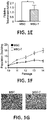
- Fig. 1E telomerase activity
- Fig. 1F telomerase activity
- Fig. 1G ⁇ -Galactosidase activity
- MSC-Tc cytokine-activated MSC-T induced expression of several genes related to the immunosuppressive capacity of MSC, such as IDO, COX2 and PD-L1 ( Fig. 2 A) .
- the results were corroborated by western blot analysis ( Fig. 2B ) and flow cytometry ( Fig.2C ) compared to non-conditioned MSC-T.
- MSC-Tc released a greater amount of EVs measured by bicinchoninic acid, acetylcholinesterase assays and NTA ( Fig. 2D, E, F , respectively) without altering other phenotypic features such as morphology, size and presence of EVs markers ( Figure 7 ).
- Conditioning MSC-T increases the immunosuppressive capacity of EVs.
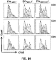
- MSC-Tc Based on the higher expression of immunomodulatory molecules observed in MSC-Tc, the capacity of these cells to secrete more suppressive vesicles compare to non-conditioned cells was evaluated. Thus, the immunomodulation capacity of EVs secreted by MSC-T and MSC-Tc in a T cell proliferation assay was evaluated by measuring CFSE dilution in T cells stimulated with CD3/CD28 beads. As show in figure 3 , MSC-Tc derived EVs (EV MSC-T c ) show improved capacity to regulate T cell proliferation affecting both CD4 and CD8 cells ( Fig. 3A ). T cell proliferation is directly linked to the activation of T cells.
- HIF-1 ⁇ overexpression in MSC-T increases immunomodulatory capacity of secreted EVs.
- HIF-1 ⁇ were transduced in the MSC-T line using lentivirus (MSC-T-HIF). Transduction efficiency was measured by flow cytometry detecting GFP in MSC-T cells and overexpression of the transgene quantified by WB and qPCR. MSC-T-HIF showed similar features to MSC-T in terms of immortalization and other MSC characteristic markers ( figures 8 , 9 ).
- NF-kB pathway is a key regulator of immunosuppressive capacity of MSC. It has been described that activating NF- ⁇ B in MSC is an essential step to trigger the capacity of these cells to avoid T cell proliferation (Dorronsoro et al, 2014). Thus, NF- ⁇ B pathway activity was measured by immunolocalization of nuclear translocation of p65 and by measuring expression of IL-6 after conditioning with cytokine cocktail both MSC-T and MSC-T-HIF. In this manner, overexpressing HIF-1 ⁇ triggers stronger response of MSCs to the conditioning cocktail were observed ( Fig. 4 A) . Accordingly, conditioned MSC-T-HIF (MSC-T-HIFc) express higher levels of immunosuppressive molecules, such IDO, COX2 and PD-L1 ( Fig. 4B , C ).
- immunosuppressive molecules such IDO, COX2 and PD-L1
- EV MSC-T-HIF c show higher level of PD-L1 and IDO and ( Fig. 5E ).
- PD-1 is a protein on the urface of cells that has a role in regulating the immune system's response to the cells of the human body by down-regulating the immune system and promoting self-tolerance by suppressing T cell inflammatory activity. This prevents autoimmune diseases, but it can also prevent the immune system from killing cancer cells.
- the expression of PDL1 in the surface of EVs was further corroborated by flow cytometry and ELISA ( Fig 5F, G ).
- HIF overexpression and priming medium conditions causes alterations in the cargo of proteins and miRNAs of EVs
- LC-MS/MS was used (table 1) . Protein contaminants from the cell culture medium (serum albumin and keratin) were removed from the analysis. An equivalent amount of total protein was used (30 ⁇ g).
- serum albumin and keratin proteins contaminants from the cell culture medium
- An equivalent amount of total protein was used (30 ⁇ g).
- Non-primed EVs MSC contained protein cargo involved in ECM organization, vesicle-mediated transport, wound healing and cell adhesion, whereas additional biological processes were over-represented in primed EVs MSC-T-HIF , such as immune effector process, immune system process, secretion by cell, and response to stimulus.
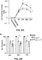
- DTH Delayed-Type Hypersensitivity mouse model
- Severity of the reaction is evaluated by measuring ear thickness and analysing tissue integrity and infiltration by H&E staining and immunofluorescence. Using this model, it was observed that both EV MSC-T c and EV MSC-T-HIF c reduced the inflammatory reaction in the ear ( Fig. 6 A) . Moreover, it was observed that EV MSC-T-HFc are significantly more effective. As show in figure 6B , EV MSC-T-HIFc reduce swelling to 38.64 ⁇ 2.88% 48 hours after challenging and to 40.76 ⁇ 7.18% 72 hours after. Histological analysis of challenged ear showed that DTH reaction was associated with vascular enlargement and orthokeratotic hyperkeratosis ( Fig. 6 C) .
Landscapes
- Health & Medical Sciences (AREA)
- Life Sciences & Earth Sciences (AREA)
- Chemical & Material Sciences (AREA)
- Organic Chemistry (AREA)
- Engineering & Computer Science (AREA)
- Zoology (AREA)
- Genetics & Genomics (AREA)
- General Health & Medical Sciences (AREA)
- Bioinformatics & Cheminformatics (AREA)
- Wood Science & Technology (AREA)
- Biomedical Technology (AREA)
- Biotechnology (AREA)
- Biochemistry (AREA)
- Developmental Biology & Embryology (AREA)
- Cell Biology (AREA)
- General Engineering & Computer Science (AREA)
- Medicinal Chemistry (AREA)
- Molecular Biology (AREA)
- Microbiology (AREA)
- Immunology (AREA)
- Hematology (AREA)
- Rheumatology (AREA)
- Virology (AREA)
- Pharmacology & Pharmacy (AREA)
- Epidemiology (AREA)
- Animal Behavior & Ethology (AREA)
- Public Health (AREA)
- Veterinary Medicine (AREA)
- Toxicology (AREA)
- Gastroenterology & Hepatology (AREA)
- Biophysics (AREA)
- Proteomics, Peptides & Aminoacids (AREA)
- Medicines Containing Material From Animals Or Micro-Organisms (AREA)
Priority Applications (2)
| Application Number | Priority Date | Filing Date | Title |
|---|---|---|---|
| EP20383170.6A EP4023231A1 (fr) | 2020-12-29 | 2020-12-29 | Vésicules extracellulaires dérivées de cellules stromales mésenchymateuses génétiquement modifiées pour la surexpression hif-1a et htert |
| PCT/EP2021/087673 WO2022144333A1 (fr) | 2020-12-29 | 2021-12-27 | Vésicules extracellulaires issues de cellules stromales mésenchymateuses génétiquement modifiées pour surexprimer hif-1a et htert |
Applications Claiming Priority (1)
| Application Number | Priority Date | Filing Date | Title |
|---|---|---|---|
| EP20383170.6A EP4023231A1 (fr) | 2020-12-29 | 2020-12-29 | Vésicules extracellulaires dérivées de cellules stromales mésenchymateuses génétiquement modifiées pour la surexpression hif-1a et htert |
Publications (1)
| Publication Number | Publication Date |
|---|---|
| EP4023231A1 true EP4023231A1 (fr) | 2022-07-06 |
Family
ID=74205591
Family Applications (1)
| Application Number | Title | Priority Date | Filing Date |
|---|---|---|---|
| EP20383170.6A Pending EP4023231A1 (fr) | 2020-12-29 | 2020-12-29 | Vésicules extracellulaires dérivées de cellules stromales mésenchymateuses génétiquement modifiées pour la surexpression hif-1a et htert |
Country Status (2)
| Country | Link |
|---|---|
| EP (1) | EP4023231A1 (fr) |
| WO (1) | WO2022144333A1 (fr) |
Cited By (1)
| Publication number | Priority date | Publication date | Assignee | Title |
|---|---|---|---|---|
| CN116898868A (zh) * | 2023-05-19 | 2023-10-20 | 青岛大学附属医院 | MiR-1909-5p在制备治疗血管内皮细胞铁死亡和/或主动脉夹层产品中的应用 |
Families Citing this family (4)
| Publication number | Priority date | Publication date | Assignee | Title |
|---|---|---|---|---|
| EP4393499A1 (fr) * | 2022-12-27 | 2024-07-03 | Fundación Para la Investigación del Hospital Universitario y Politécnico La Fe de la Comunidad Valenciana | Petites vésicules extracellulaires présentant des propriétés antifibrotiques |
| CN116492464B (zh) * | 2023-04-12 | 2025-08-19 | 天津医科大学总医院 | TNFRSF1A抑制剂miR-3059-5p在治疗脑卒中疾病中的用途 |
| CN118370766B (zh) * | 2024-05-04 | 2025-01-28 | 山东省泉溪生物技术有限公司 | 一种间充质干细胞在预防或治疗肝病中的用途 |
| CN118685365B (zh) * | 2024-08-23 | 2024-11-15 | 昆明医科大学附属口腔医院(云南省口腔医院) | miR-423-5p高丰度工程化囊泡的构建及应用 |
Citations (2)
| Publication number | Priority date | Publication date | Assignee | Title |
|---|---|---|---|---|
| WO2019238693A1 (fr) * | 2018-06-11 | 2019-12-19 | Health And Biotech France (H & B France) | Vésicules extracellulaires dérivées de cellules souches mésenchymateuses |
| WO2020257710A1 (fr) * | 2019-06-21 | 2020-12-24 | Entelexo Biotherapeutics Inc. | Plateformes, compositions et méthodes d'administration de composés thérapeutiques |
-
2020
- 2020-12-29 EP EP20383170.6A patent/EP4023231A1/fr active Pending
-
2021
- 2021-12-27 WO PCT/EP2021/087673 patent/WO2022144333A1/fr not_active Ceased
Patent Citations (2)
| Publication number | Priority date | Publication date | Assignee | Title |
|---|---|---|---|---|
| WO2019238693A1 (fr) * | 2018-06-11 | 2019-12-19 | Health And Biotech France (H & B France) | Vésicules extracellulaires dérivées de cellules souches mésenchymateuses |
| WO2020257710A1 (fr) * | 2019-06-21 | 2020-12-24 | Entelexo Biotherapeutics Inc. | Plateformes, compositions et méthodes d'administration de composés thérapeutiques |
Non-Patent Citations (26)
Cited By (2)
| Publication number | Priority date | Publication date | Assignee | Title |
|---|---|---|---|---|
| CN116898868A (zh) * | 2023-05-19 | 2023-10-20 | 青岛大学附属医院 | MiR-1909-5p在制备治疗血管内皮细胞铁死亡和/或主动脉夹层产品中的应用 |
| CN116898868B (zh) * | 2023-05-19 | 2024-02-06 | 青岛大学附属医院 | MiR-1909-5p在制备治疗血管内皮细胞铁死亡和/或主动脉夹层产品中的应用 |
Also Published As
| Publication number | Publication date |
|---|---|
| WO2022144333A1 (fr) | 2022-07-07 |
Similar Documents
| Publication | Publication Date | Title |
|---|---|---|
| EP4023231A1 (fr) | Vésicules extracellulaires dérivées de cellules stromales mésenchymateuses génétiquement modifiées pour la surexpression hif-1a et htert | |
| Blazar et al. | Immune regulatory cell infusion for graft-versus-host disease prevention and therapy | |
| Liu et al. | Mesenchymal stem cell exosome-derived miR-223 alleviates acute graft-versus-host disease via reducing the migration of donor T cells | |
| Henao Agudelo et al. | Mesenchymal stromal cell-derived microvesicles regulate an internal pro-inflammatory program in activated macrophages | |
| CN110088623B (zh) | 选择用于治疗免疫病症的高效干细胞的方法 | |
| US20230407256A1 (en) | Generation of therapeutic cells using extracellular components of target organs | |
| Guo et al. | Extracellular vesicles from mesenchymal stem cells prevent contact hypersensitivity through the suppression of Tc1 and Th1 cells and expansion of regulatory T cells | |
| AU2013225721B2 (en) | Expansion of alloantigen-reactive regulatory T cells | |
| CN102471753B (zh) | 干细胞免疫调节应用方法和设备 | |
| Haghighitalab et al. | Investigating the effects of IDO1, PTGS2, and TGF-β1 overexpression on immunomodulatory properties of hTERT-MSCs and their extracellular vesicles | |
| Zhao et al. | Mesenchymal stem cells overexpressing IL-35 effectively inhibit CD4+ T cell function | |
| US10590391B2 (en) | Selective cell therapy for the treatment of renal failure | |
| Jin et al. | Effects of age on biological and functional characterization of adipose‑derived stem cells from patients with end‑stage liver disease | |
| Jie et al. | Nrf2 modulates immunosuppressive ability and cellular senescence of human umbilical cord mesenchymal stem cells | |
| Zhang et al. | Syngeneic bone marrow transplantation in combination with PI3K inhibitor reversed hyperglycemia in later-stage streptozotocin-induced diabetes | |
| Gozel et al. | Effects of primed adipose mesenchymal stem cell-derived exosomes on immunomodulation in Behcet uveitis | |
| Feng et al. | Mesenchymal stem cell–derived exosomes derived from induced pluripotent stem cells ameliorate inflammation and promote mucosal healing via miR-34a-5p in Crohn disease | |
| Gonzalez et al. | Aging Adipose‐Derived Mesenchymal Stem Cells, Cultured on a Native Young Extracellular Matrix, Are Protected From Senescence and Apoptosis Along With Increased Expression of HLA‐DR and CD74 Associated With PI3K Signaling | |
| Tung | Dissecting the mechanisms of Regulatory T cell-derived Extracellular Vesicle (EV)-mediated suppression to facilitate the optimisation of these cells and EVs in the clinic | |
| Plaza Rojas | Suffocated CD8 T Cells in the Hypoxic Tumor Microenvironment | |
| Smith | Mechanisms of Thymic Involution and Therapies to Prevent or Treat the loss of Thymic Epithelial Cells | |
| Romano | In vitro characterisation and expansion of human regulatory T cells for their in vivo application in the induction of tolerance in haematopoietic stem cell and solid organ transplantation | |
| Jui et al. | of June 25, 2014. | |
| Jui et al. | Wan-Tseng Hsu, Cheng-Hsin Lin, Bor-Luen Chiang |
Legal Events
| Date | Code | Title | Description |
|---|---|---|---|
| PUAI | Public reference made under article 153(3) epc to a published international application that has entered the european phase |
Free format text: ORIGINAL CODE: 0009012 |
|
| STAA | Information on the status of an ep patent application or granted ep patent |
Free format text: STATUS: THE APPLICATION HAS BEEN PUBLISHED |
|
| AK | Designated contracting states |
Kind code of ref document: A1 Designated state(s): AL AT BE BG CH CY CZ DE DK EE ES FI FR GB GR HR HU IE IS IT LI LT LU LV MC MK MT NL NO PL PT RO RS SE SI SK SM TR |
|
| STAA | Information on the status of an ep patent application or granted ep patent |
Free format text: STATUS: REQUEST FOR EXAMINATION WAS MADE |
|
| 17P | Request for examination filed |
Effective date: 20230102 |
|
| RBV | Designated contracting states (corrected) |
Designated state(s): AL AT BE BG CH CY CZ DE DK EE ES FI FR GB GR HR HU IE IS IT LI LT LU LV MC MK MT NL NO PL PT RO RS SE SI SK SM TR |