EP4026558A1 - Zusammengesetztes proteinmonomer mit nicht-strukturellem virusprotein darauf, aggregat des zusammengesetzten proteinmonomers und kombinationsimpfstoff mit aggregat als wirkstoff - Google Patents
Zusammengesetztes proteinmonomer mit nicht-strukturellem virusprotein darauf, aggregat des zusammengesetzten proteinmonomers und kombinationsimpfstoff mit aggregat als wirkstoff Download PDFInfo
- Publication number
- EP4026558A1 EP4026558A1 EP20856780.0A EP20856780A EP4026558A1 EP 4026558 A1 EP4026558 A1 EP 4026558A1 EP 20856780 A EP20856780 A EP 20856780A EP 4026558 A1 EP4026558 A1 EP 4026558A1
- Authority
- EP
- European Patent Office
- Prior art keywords
- protein
- amino acid
- acid sequence
- virus
- composite
- Prior art date
- Legal status (The legal status is an assumption and is not a legal conclusion. Google has not performed a legal analysis and makes no representation as to the accuracy of the status listed.)
- Pending
Links
Images



Classifications
-
- C—CHEMISTRY; METALLURGY
- C12—BIOCHEMISTRY; BEER; SPIRITS; WINE; VINEGAR; MICROBIOLOGY; ENZYMOLOGY; MUTATION OR GENETIC ENGINEERING
- C12N—MICROORGANISMS OR ENZYMES; COMPOSITIONS THEREOF; PROPAGATING, PRESERVING, OR MAINTAINING MICROORGANISMS; MUTATION OR GENETIC ENGINEERING; CULTURE MEDIA
- C12N15/00—Mutation or genetic engineering; DNA or RNA concerning genetic engineering, vectors, e.g. plasmids, or their isolation, preparation or purification; Use of hosts therefor
- C12N15/09—Recombinant DNA-technology
- C12N15/63—Introduction of foreign genetic material using vectors; Vectors; Use of hosts therefor; Regulation of expression
- C12N15/70—Vectors or expression systems specially adapted for E. coli
-
- A—HUMAN NECESSITIES
- A61—MEDICAL OR VETERINARY SCIENCE; HYGIENE
- A61K—PREPARATIONS FOR MEDICAL, DENTAL OR TOILETRY PURPOSES
- A61K39/00—Medicinal preparations containing antigens or antibodies
- A61K39/12—Viral antigens
-
- A—HUMAN NECESSITIES
- A61—MEDICAL OR VETERINARY SCIENCE; HYGIENE
- A61P—SPECIFIC THERAPEUTIC ACTIVITY OF CHEMICAL COMPOUNDS OR MEDICINAL PREPARATIONS
- A61P31/00—Antiinfectives, i.e. antibiotics, antiseptics, chemotherapeutics
- A61P31/12—Antivirals
- A61P31/14—Antivirals for RNA viruses
-
- C—CHEMISTRY; METALLURGY
- C07—ORGANIC CHEMISTRY
- C07K—PEPTIDES
- C07K14/00—Peptides having more than 20 amino acids; Gastrins; Somatostatins; Melanotropins; Derivatives thereof
- C07K14/005—Peptides having more than 20 amino acids; Gastrins; Somatostatins; Melanotropins; Derivatives thereof from viruses
-
- C—CHEMISTRY; METALLURGY
- C12—BIOCHEMISTRY; BEER; SPIRITS; WINE; VINEGAR; MICROBIOLOGY; ENZYMOLOGY; MUTATION OR GENETIC ENGINEERING
- C12N—MICROORGANISMS OR ENZYMES; COMPOSITIONS THEREOF; PROPAGATING, PRESERVING, OR MAINTAINING MICROORGANISMS; MUTATION OR GENETIC ENGINEERING; CULTURE MEDIA
- C12N2760/00—MICROORGANISMS OR ENZYMES; COMPOSITIONS THEREOF; PROPAGATING, PRESERVING, OR MAINTAINING MICROORGANISMS; MUTATION OR GENETIC ENGINEERING; CULTURE MEDIA ssRNA viruses negative-sense
- C12N2760/00011—Details
- C12N2760/18011—Paramyxoviridae
- C12N2760/18511—Pneumovirus, e.g. human respiratory syncytial virus
- C12N2760/18522—New viral proteins or individual genes, new structural or functional aspects of known viral proteins or genes
-
- C—CHEMISTRY; METALLURGY
- C12—BIOCHEMISTRY; BEER; SPIRITS; WINE; VINEGAR; MICROBIOLOGY; ENZYMOLOGY; MUTATION OR GENETIC ENGINEERING
- C12N—MICROORGANISMS OR ENZYMES; COMPOSITIONS THEREOF; PROPAGATING, PRESERVING, OR MAINTAINING MICROORGANISMS; MUTATION OR GENETIC ENGINEERING; CULTURE MEDIA
- C12N2760/00—MICROORGANISMS OR ENZYMES; COMPOSITIONS THEREOF; PROPAGATING, PRESERVING, OR MAINTAINING MICROORGANISMS; MUTATION OR GENETIC ENGINEERING; CULTURE MEDIA ssRNA viruses negative-sense
- C12N2760/00011—Details
- C12N2760/18011—Paramyxoviridae
- C12N2760/18511—Pneumovirus, e.g. human respiratory syncytial virus
- C12N2760/18534—Use of virus or viral component as vaccine, e.g. live-attenuated or inactivated virus, VLP, viral protein
-
- C—CHEMISTRY; METALLURGY
- C12—BIOCHEMISTRY; BEER; SPIRITS; WINE; VINEGAR; MICROBIOLOGY; ENZYMOLOGY; MUTATION OR GENETIC ENGINEERING
- C12N—MICROORGANISMS OR ENZYMES; COMPOSITIONS THEREOF; PROPAGATING, PRESERVING, OR MAINTAINING MICROORGANISMS; MUTATION OR GENETIC ENGINEERING; CULTURE MEDIA
- C12N2795/00—Bacteriophages
- C12N2795/00011—Details
- C12N2795/10011—Details dsDNA Bacteriophages
- C12N2795/10111—Myoviridae
- C12N2795/10122—New viral proteins or individual genes, new structural or functional aspects of known viral proteins or genes
Definitions
- the present invention relates to a functional protein and to a vaccine employing the protein. More particularly, the invention relates to a composite protein (monomer) formed of a viral non-structural protein (immunogen) with a molecular needle, to an associated product of the monomer, and to a component vaccine containing the associated product as an active ingredient (infection-protective antigen, hereinafter referred to simply as "protective antigen").
- a composite protein formed of a viral non-structural protein (immunogen) with a molecular needle
- vaccines are divided into four types: a live vaccine, an inactivated vaccine, a component vaccine, and a toxoid.
- the live vaccine employs a pathogenic microorganism (e.g., an attenuated virus) as a protective antigen.
- the inactivated vaccine employs the entirety of a killed pathogenic microorganism as a protective antigen.
- the component vaccine employs, as a protective antigen, a protein or a sugar chain which is a part (i.e., a component) of an infectious pathogenic microorganism.
- the toxoid employs a denatured toxin produced by a pathogen as a protective antigen.
- a structural protein of norovirus is attached to a molecular needle.
- the structural protein is introduced into cells, to thereby acquire immunity to the structural protein.
- component vaccines work on the basis of humoral immunity to a structural component (i.e., structure) of a pathogen as acquired immunity.
- the component vaccines fail to elicit acquired immunity with respect to a group of microbial enzymes (i.e., non-structural protein), which are generated during self-replication of a pathogen in a host.
- the live vaccine per se is an attenuated microorganism which is designed to exert no pathogenicity, and forms a non-structural protein for the duplication of the microorganism. Therefore, the live vaccine can induce global acquired immunity (cell-mediated immunity and humoral immunity), targeting even the duplication mechanism of a pathogen, in the vaccinated subject.
- the live vaccine is provided from a live pathogenic virus, and a certain risk is intrinsically involved therein.
- an attenuated pathogenic virus is reverted to a highly toxic pathogenic virus via mutation.
- an attenuated pathogenic virus may elicit toxicity in a human subject having reduced immunity.
- vaccination with a live vaccine must be performed more cautiously as compared with the case of a component vaccine.
- non-structural protein of a pathogenic virus (hereinafter may also be referred to simply as “non-structural protein”) is caused to be attached to the aforementioned molecular needle to obtain a composite protein, and the composite protein is used as an active ingredient of a component vaccine, whereby acquired immunity targeting the duplication mechanism of the microorganism can be induced although it is a component vaccine.
- the non-structural protein is a protein related to a function of a pathogenic virus in a host (e.g., proliferation).
- the non-structural protein is resistive to amino acid variation so as to maintain the above function, whereby gene mutation of the non-structural protein is drastically inhibited as compared to the case of a structural protein.
- achievement of common immunogenicity of different virus variants is highly possible, so long as the variants belong to the same pathogenic virus species. Even in the case of a pathogenic virus species which undergoes mutation in structural protein with considerable frequency, target immunity can be effectively acquired.
- the present invention provides the following aspects:
- the “component vaccine” essentially contains a protective antigen as an active ingredient.
- the concept “component vaccine” excludes the aforementioned live vaccine, inactivated vaccine, and toxoid.
- the active ingredient employed in the vaccine of the present invention contains the associated product (containing trimer and/or hexamer) of the present invention serving as a protective antigen.
- the composite protein of the present invention is a composite protein represented by the amino acid sequence of the following formula (1) and serves as a precursor of the associated product of the present invention.
- the symbol "-" connecting the amino acid sequence units denotes a simple molecular bond (substantially a peptide bond) which is used to clearly isolate from one another the amino acid sequences each having a specific meaning (e.g., W, L 1 , X n , and Y).
- the composite protein is represented by W-L 1 -X n -Y ⁇ (1)
- W represents an amino acid sequence including one or more amino acid sequences of a part or the entirety of a pathogenic virus non-structural protein serving as an immunogen
- L 1 represents a first linker sequence having 0 to 100 amino acids
- X represents an amino acid sequence represented by SEQ ID NO: 1
- Y represents an amino acid sequence of a cell introduction domain
- repetition number n of X is an integer of 1 to 3
- the amino acid sequence of the cell introduction domain Y is represented by the following formula (2) : Y 1 -L 2 -Y 2 -Y 3 ⁇ (2)
- Y 1 represents any one amino acid sequence selected from the group consisting of SEQ ID NOs: 2 to 5
- Y 2 represents any one amino acid sequence selected from the group consisting of SEQ ID NOs: 6 to 9
- L 2 represents a second linker sequence having 0 to 30 amino acids
- Y 3 represents an amino acid sequence for modification; and either of
- the number (n) of repetition of amino acid sequence X in X n is preferably 1, but may be 2 or 3.
- the amino acid sequence represented by X n , Y 1 , or Y 2 includes a modified amino acid sequence thereof obtained by deleting, substituting, or adding one or more amino acid residues from, in, or to the original amino acid sequence.
- the term "deleting” refers to deletion of any amino acid residue in the amino acid sequence included in formula (1) and represented by any of the SEQ ID NOs.
- the amino acid residue at the N-terminal side of the deleted amino acid residue and that at the C-terminal side of the deleted amino acid residue are linked via a peptide bond. In the case of deletion of the N-terminal amino acid residue or the C-terminal amino acid residue, no linkage is present.
- the number of deleted residues is counted as "the number of amino acid deletions.”
- substituted refers to substitution of any amino acid residue in the amino acid sequence included in formula (1) and represented by any of the SEQ ID NOs, "with another amino acid residue.”
- the new amino acid residue is linked to the amino acid residue at the N-terminal side thereof and at the C-terminal side thereof via a peptide bond.
- substitution of the N-terminal side or C-terminal side amino acid residue the amino acid residue is linked via a peptide bond to another C-terminal side or N-terminal side amino acid residue.
- the number of substituted residues is counted as "the number of amino acid substitutions.”
- the term “adding” refers to addition of one or more new amino acid residues to one or more peptide bond sites in the amino acid sequence included in formula (1) and represented by any of the SEQ ID NOs, to thereby form a new peptide bond(s).
- the type and number of the above modifications of amino acid residue may be elucidated by comparison in alignment of the amino acid sequence represented by formula (1) with the resultant amino acid sequence through manpower or employment of appropriate analysis software.
- the linker sequence L 1 or L 2 , or the modification amino acid sequence Y 3 may have any desired sequence within the range of the number of amino acid residues as defined in formula (1).
- the trimer or hexamer (as described below) of the modified composite protein having the modified amino acid sequence preferably has substantially the same immunostimulation activity as that of the trimer or hexamer of the composite protein of formula (1).
- the expression “substantially the same” refers to such a similarity that no significant difference in immunostimulation activity between the trimer or the hexamer of a composite protein having the non-modified amino acid sequence and that of a composite protein having the modified amino acid sequence is confirmed within a significance level range of 5%, when the immunostimulation activity is determined through an established technique such as the "neutralization test.”
- the number of modifications of amino acid residues in each amino acid sequence is preferably ⁇ 8n, more preferably ⁇ 4n, still more preferably ⁇ 2n in the case of X n ; preferably ⁇ 30, more preferably ⁇ 20, still more preferably ⁇ 10 in the case of Y 1 ; and preferably ⁇ 15, more preferably ⁇ 10, still more preferably ⁇ 5 in the case of Y 2 .
- W is an amino acid sequence including one or more amino acid sequences of a part or the entirety of a non-structural protein of a pathogenic virus.
- Proteins encoded by a gene of a pathogenic virus are generally divided into a structural protein and a non-structural protein.
- the structural protein directly forms the structure of the virus, and examples include capsid protein, envelope protein, and core protein.
- the non-structural protein is a protein encoded by a gene other than a gene or genes encoding the structural protein(s) of the virus.
- the non-structural protein is an enzyme protein involved in survival, proliferation, etc. of a virus in a host, and is expressed after entry of the pathogenic virus into the host.
- the entirety specifically refers to "the entirety of each individual non-structural protein of a virus.”
- the non-structural protein is P protein of an RS virus as mentioned in the Examples
- the aforementioned “the entirety” refers to the entire amino acid sequence of the P protein
- the aforementioned "a part” refers to a part of the amino acid sequence of the P protein.
- no particular limitation is imposed on the constitution of the sequence, so long as "a part of each individual non-structural protein of a virus" exhibits antigenicity of the relevant virus.
- the pathogenic virus may be a DNA virus, an RNA virus, or a retrovirus.
- Examples of the pathogenic virus includes not only a pathogenic virus which causes an infection for which a certain vaccine has already been provided, but also a pathogenic virus which causes an infection for which no vaccine has been provided.
- Examples of the pathogenic virus which causes an infection for which a certain vaccine has already been provided include poliovirus, measles virus, rubella virus, mumps virus, varicella-zoster virus, yellow fever virus, rotavirus, herpes virus, influenza virus, norovirus, rabies virus, Japanese encephalitis virus, hepatitis viruses (e.g., HAV, HBV, and HCV), human papilloma virus, HIV, and ebolavirus.
- Examples of the pathogenic virus which causes an infection for which no vaccine has been provided include RS virus (RSV).
- RSV RS virus
- One characteristic feature of the vaccine of the present invention resides in that the vaccine is effective for a virus which causes an infection for which no vaccine has been provided.
- animal which may be infected with the pathogenic virus
- examples of the animal include human and vaccine-needed animals. More specific examples of the animal include farm animals (e.g., cow, horse, sheep, pig, goat, or chicken) and pet animals (e.g., dog and cat).
- the non-structural protein is individually and specifically selected according to the type of virus targeted. On the basis of information obtained with respect to known virus non-structural proteins, RNA polymerase, DNA polymerase, reverse transcriptase, protease, VPg, NTPase, DNA binding protein, RNA binding protein, transcription factor, phosphorylated protein, L protein, N protein, or the like may be identified and selected.
- the concept "non-structural protein” also encompasses a protein whose function has not precisely been revealed and which will potentially serve as a non-structural protein, and a protein which functions as both a structural protein and a non-structural protein.
- non-structural protein of RS virus examples include NS-1 protein, NS-2 protein, N protein, P protein, M2-1 protein, and L protein.
- N protein, P protein, M2-1 protein, and L protein are linked to the genome RNA in a virus particle.
- RNP i.e., called nucleocapsid, which is a complex of RNA with another viral protein or a ribonucleoprotein complex.
- RNP i.e., called nucleocapsid, which is a complex of RNA with another viral protein or a ribonucleoprotein complex.
- RNP i.e., called nucleocapsid, which is a complex of RNA with another viral protein or a ribonucleoprotein complex.
- These non-structural proteins forming RNP are also present in RS virus particles. Since the non-structural proteins belong to the enzyme group involved in transcription of messenger RNA and replication of the genome, the proteins are synthesized in a large amount in cells after infection.
- the associated product of the present invention is formed of the composite protein of the present invention as a monomer. More specifically, the associated product includes a trimer or a hexamer of the composite protein, or a mixture of the trimer and the hexamer.
- the trimer or the hexamer of the associated products of the present invention and the mixture of the trimer and the hexamer may be collectively referred to simply as a "trimer and/or hexamer.”
- the associated product of the present invention contains a trimer or hexamer of the composite protein of the present invention as a monomer.
- the associated product may be defined as an associated product formed through association of molecules of the composite protein of the present invention in an aqueous liquid.
- the associated product of the present invention may fail to be formed depending on the compatibility between the protein to be attached and the relevant molecular needle.
- the target composite protein molecules are brought into contact with one another in an aqueous liquid, and the resultant product is confirmed through SDS-PAGE or the like, so as to easily evaluate whether the target composite protein forms a trimer or a hexamer thereof, and whether or not the product can be used as an active ingredient of the vaccine of the present invention.
- the thus-formed associated product of the present invention per se, can exert an effect to penetrate a target cells.
- the aforementioned trimer is a trimeric protein formed of the same or different composite proteins of the present invention, as a protein monomer, and the aforementioned hexamer is a hexameric protein formed through association of two molecules of the trimeric protein.
- the vaccine of the present invention contains the associated product of the present invention as an active ingredient (protective antigen) and serves as a component vaccine against a target pathogenic virus to be administered in a subcutaneous, intradermal, percutaneous, transmucosal, or intramuscular manner. More specifically, the vaccine of the present invention contains, as an active ingredient, the aforementioned trimer and/or hexamer in which one or more peptides or proteins are included as W, the peptides or proteins including one or more of a part or the entirety of a pathogenic virus non-structural protein, and serves as a component vaccine to be administered in a subcutaneous, intradermal, percutaneous, transmucosal, or intramuscular manner.
- the vaccine of the present invention may contain, as an active ingredient, two or more trimers and/or hexamers in which the entirety or a part of different non-structural proteins from pathogenic virus strains of the same genotype are included as W.
- the vaccine of the present invention may contain, as active ingredients, two or more trimers and/or hexamers in which the entirety or a part of different non-structural proteins from pathogenic viruses having different genotypes are included as W.
- the vaccine of the present invention may include the associated product of the present invention and, as an additional active ingredient, an associated product (including a trimer and/or hexamer) in which a "structural protein" has been attached to a molecular needle (see Patent Document 2), instead of W (non-structural protein) of the trimer and/or hexamer of the present invention.
- the vaccine of the present invention may include, as additional active ingredients, associated products in which structural proteins of pathogenic viruses having different genotypes are included as W, in combination.
- the structural protein may have a domain to which a neutralizing antibody is bound, the antibody having viral infection inhibiting ability.
- the characteristic of acquired immunity becomes similar to that of a live vaccine. Also, when antigenic proteins originating from different pathogenic microorganisms are employed, immunity similar to that of a mixed vaccine can be attained.
- the vaccine of the present invention may contain an adjuvant, which is, for example, a B subunit of cholera toxin (corresponding to W) attached to a molecular needle.
- the vaccine of the present invention may be an adjuvant-free (i.e., adjuvant-removed) vaccine.
- the adjuvant-free vaccine is rather preferred, from the viewpoint of attaining an advantageous feature of the vaccine of the present invention.
- trimer and/or hexamer species in the vaccine of the present invention no particular limitation is imposed on the ratio of the trimer and/or hexamer species in the vaccine of the present invention, and the ratio may be freely selected in accordance with the purpose of the vaccine to be used.
- the associated product of the present invention is produced through the following procedure. Specifically, 3 or more molecules of the composite protein of the present invention are brought into contact with one another by the mediation of an aqueous liquid, to thereby associate the composite protein molecules as monomers and to form a mixture of a trimer and a hexamer. If needed, the trimer or the hexamer may be selectively isolated and recovered.
- the composite protein of the present invention per se is formed by expressing a nucleic acid fragment encoding the target composite protein through a genetic engineering procedure.
- the composite protein is synthesized through a technique of peptide synthesis.
- a trimer and a hexamer of the composite protein are formed in a spontaneous manner.
- a mixture containing a trimer and a hexamer of the composite protein is provided.
- the composite protein of the present invention is produced through a genetic engineering procedure
- the composite protein is biologically formed through gene expression.
- cells in which the target composite protein has been formed are collected, and broken or lysed to extract the composite protein, and the target composite protein is isolated through a known isolation method.
- aqueous liquid such as water or a buffer.
- association proceeds spontaneously, to thereby yield a mixture of the trimer and the hexamer, which is the associated product of the present invention.
- the composite protein of the present invention is produced through total chemical synthesis, or produced by individually synthesizing each part and linking the parts through chemical modification, the product is suspended in an aqueous liquid such as water or a buffer. Even in this case, association proceeds spontaneously, to thereby yield a mixture containing the trimer and the hexamer, which is the associated product of the present invention.
- one most preferred mode of the associated product production method of the present invention is a production method including culturing, in a liquid culture medium, a transformant into which a nucleic acid fragment encoding the composite protein of the present invention has been incorporated, to thereby form the corresponding composite protein through gene expression; and obtaining a mixture containing a trimer and a hexamer of the composite protein (as a monomer) via a spontaneous association process; or further selectively isolating and recovering the trimer and the hexamer from the resulting mixture.
- the thus-produced associated product of the present invention may be used as the active ingredient of the vaccine of the present invention.
- the present invention can provide the following (1) to (3) :
- the composite protein of the present invention is represented by the amino acid sequence of the following formula (1): W-L 1 -X n -Y ⁇ (1) [wherein W represents an amino acid sequence including one or more amino acid sequences of a part or the entirety of a pathogenic virus non-structural protein serving as an immunogen; L 1 represents a first linker sequence having 0 to 100 amino acids; X represents an amino acid sequence represented by SEQ ID NO: 1; Y represents an amino acid sequence of a cell introduction domain; repetition number n of X is an integer of 1 to 3].
- a specific example of the immunogen W is an amino acid sequence based on a part or the entirety of the peptide structure of the virus non-structural protein.
- the wording "based on” refers to the immunogen W including the original peptide structure and also a modified amino acid sequence thereof.
- the first linker sequence L 1 is required for appropriately maintaining the distance between the immunogen W and the molecular needle part Y, to thereby suppress steric hindrance.
- the number of amino acid residues of L 1 is 0 to 100, preferably 4 to 40.
- No particular limitation is imposed on the specific amino acid sequence, and examples thereof include (GGGGS) m and (PAPAP) m [m represents the number of repetitions and is preferably an integer of 1 to 5, particularly preferably 1 to 3]. Needless to say, the above examples are non-limitative ones.
- X is an amino acid residue represented by SEQ ID NO: 1
- the amino acid sequence X n is a sequence of repeating unit Xs with the number of repetitions n (n is an integer).
- the mode of repetition is linear repetition.
- the case of X 2 means "X-X" (wherein symbol "-” denotes a peptide bond).
- the amino acid sequence may be modified in the aforementioned manner.
- the number n is an integer of 1 to 3.
- the number n is preferably 1 but may be 2 or 3.
- the cell introduction domain Y is a basement of the molecular needle and is based on the tail needle of a bacteriophage (i.e., cell-penetrating part).
- the domain Y is a protein represented by the following formula (2): Y 1 -L 2 -Y 2 -Y 3 ⁇ (2) [wherein Y 1 represents an amino acid sequence selected from the group consisting of SEQ ID NOs: 2 to 5; Y 2 represents an amino acid sequence selected from the group consisting of SEQ ID NOs: 6 to 9; L 2 represents a second linker sequence having 0 to 30 amino acids; Y 3 represents an amino acid sequence for modification of interest; and Y 2 or Y 3 may be absent].
- the amino acid sequence from the end of the N-terminal side to the 32 amino acid residue (32 Leu) corresponds to the amino acid sequence of a triple helix ⁇ -sheet structure of the bacteriophage T4.
- the N-terminal amino acid of valine (1 Val) may also be leucine (1 Leu).
- the remaining C-terminal amino acid sequence is an amino acid sequence of the needle protein of the bacteriophage on the C-terminal side.
- amino acid sequence which can be used on the C-terminal side of Y 1 examples include an amino acid sequence of gp5 of bacteriophage T4, an amino acid sequence of gpV of bacteriophage P2, an amino acid sequence of gp45 of bacteriophage Mu, and an amino acid sequence of gp138 of bacteriophage ⁇ 92.
- Y 1 More specific examples of Y 1 include the amino acid sequence (SEQ ID NO: 2) which has the amino acid sequence of gp5 of bacteriophage T4 on the C-terminal side, the amino acid sequence (SEQ ID NO: 3) which has the amino acid sequence of gpV of bacteriophage P2 on the C-terminal side, the amino acid sequence (SEQ ID NO: 4) which has the amino acid sequence of gp45 of bacteriophage Mu on the C-terminal side, and the amino acid sequence (SEQ ID NO: 5) which has the amino acid sequence of gp138 of bacteriophage ⁇ 92 on the C-terminal side.
- the nucleotide sequence encoding the amino acid sequence of Y 1 may be selected in accordance with a generally known amino acid-nucleobase relationship.
- Y 2 in formula (2) is an amino acid sequence of a domain "foldon" of bacteriophage T4 or an amino acid sequence of a domain "tip" of bacteriophage P2, bacteriophage Mu, or bacteriophage ⁇ 92.
- the foldon or tip is a domain forming the tip portion of the molecular needle structure (i.e., fibritin) of bacteriophage.
- Y 2 is not necessarily present. However, when the amino acid sequence of the foldon or the tip is included, the efficiency of incorporation of the molecular needle to the cell membrane can be enhanced. Thus, the presence of Y 2 is preferred.
- the amino acid sequence of the foldon of bacteriophage T4 is represented by SEQ ID NO: 6.
- the nucleotide sequence encoding the amino acid sequence may be selected in accordance with a generally known amino acid-nucleobase relationship.
- the amino acid sequence of the tip of bacteriophage P2 is represented by SEQ ID NO: 7.
- the nucleotide sequence encoding the amino acid sequence may be selected in accordance with a generally known amino acid-nucleobase relationship.
- the amino acid sequence of the tip of bacteriophage Mu is represented by SEQ ID NO: 8.
- the nucleotide sequence encoding the amino acid sequence may be selected in accordance with a generally known amino acid-nucleobase relationship.
- the amino acid sequence of the tip of bacteriophage ⁇ 92 is represented by SEQ ID NO: 9.
- the nucleotide sequence encoding the amino acid sequence may be selected in accordance with a generally known amino acid-nucleobase relationship.
- L 2 serves as a second linker intervening between Y 1 and Y 2 .
- the number of amino acid residues in linker L 2 is 0 to 30, preferably 0 to 5.
- the number of amino acid residue in the linker being 0 means the absence of second linker L 2 .
- Y 3 is an amino acid sequence for modification and may be selectively incorporated into Y.
- the modification amino acid sequence is added in order to purify or protect protein (or for another reason), and examples thereof include tag peptides such as histidine tag, GST tag, and FLAG tag.
- An appropriate linker sequence may be incorporated into Y 3 .
- Such an additional linker sequence may also be a component of the amino acid sequence of Y 3 .
- the composite protein of the present invention may be produced through a known method; specifically, a genetic engineering process or chemical synthesis.
- the entirety of the composite protein of the present invention may be produced in a single process.
- segments of the composite protein are individually produced, and the produced segments are linked through a chemical modification method.
- a linker e.g., L 1 or L 2
- a lysine residue or a cysteine residue of one polypeptide may be linked to that of another polypeptide by the mediation of a linker having a succinimido group or a maleimido group.
- a nucleic acid fragment encoding the entirety or a part of the target composite protein of the present invention may be expressed in host cells (e.g., Escherichia coli, yeast, insect cells, and animal cells) as a transformant, or in a cell-free expression system such as an E. coli extract, a rabbit reticulocyte extract, or a wheat germ extract.
- host cells e.g., Escherichia coli, yeast, insect cells, and animal cells
- a cell-free expression system such as an E. coli extract, a rabbit reticulocyte extract, or a wheat germ extract.
- an expression vector in which the nucleic acid fragment is incorporated a vector corresponding to each expression system can be used. Examples of the expression vector include pET (for expression in E. coli), pAUR (for expression in yeast), pIEx-1 (for expression in insect cells), pBApo-CMV (for expression in animal cells), and pF3A (for expression in wheat germ extract).
- any known method for chemically synthesizing peptides may be employed. More specifically, the entirety or a part of the composite protein of the present invention may be produced through any established customary method (e.g., liquid phase peptide synthesis or solid phase peptide synthesis). Generally known suitable solid phase peptide synthesis techniques (i.e., chemical synthesis) such as a Boc solid phase method or an Fmoc solid phase method may be employed. Also, as described above, a ligation technique may be employed in accordance with need. Needless to say, each amino acid may be produced through a known method, and a commercial product thereof may be used.
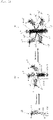
- Fig. 1 is a scheme of forming the associated products of the present invention (trimer and hexamer) from the composite protein of the present invention.
- reference numeral 10 denotes the composite protein of the present invention in the form of monomer.
- Reference numeral 20 denotes the trimer of the present invention, and reference numeral 30 denotes the hexamer of the present invention.
- the composite protein 10 of the present invention is formed of a "molecular needle domain 13 corresponding to Y in formula (1)" in which a "base part 131 corresponding to X n and Y 1 in formula (2)" is linked to a "foldon 132 corresponding Y 2 in formula (2)," and an "immunogen 11 corresponding to W in formula (1),” wherein two components are linked together via a "linker 12 corresponding to L 1 in formula (1).
- a linker or linkers other than linker 12, and the modification sequence corresponding to Y 3 in formula (2) are not illustrated.
- the composite protein 10 of the present invention per se substantially exhibits no function of penetrating the cell membrane of the target cells.
- the trimer 30 is an associated product of three molecules of the aforementioned composite protein 10 serving as a monomer via a spontaneous association process.
- 3 units of the molecular needle domain 13 are combined with association via formation of C-terminal-C-terminal bonds, to thereby provide a trimer parallel ⁇ -sheet structure, and a helix structure of the ⁇ -sheet structure per se (i.e., triple helix ⁇ -sheet structure), which corresponds to a needle structure.
- a molecular needle 13 ⁇ 3 is formed.
- the molecular needle 13 ⁇ 3 is composed of a basic part 131 ⁇ 3 and a foldon aggregate 132 ⁇ 3.
- a "molecular needle” which has a target tissue cell membrane penetration function is formed through trimerization and a self-assembly process.
- the hexamer 60 is formed through linking 2 units of the trimer 30, with formation of a bond between the N-terminals of the molecular needle basic parts (13 ⁇ 3) 1 and (13 ⁇ 3) 2 .
- the hexamer 60 also exhibits a target tissue cell membrane penetration function.
- Six linkers (12 1 , 12 2 , 12 3 , 12 5 , 126, 12 4 (12 4 : not illustrated)) originating from the respective trimers, and six immunogen portions (11 1 , 11 2 , 11 3 , 11 5 , 11 6 , 11 4 (11 4 : not illustrated) linked to the respective linkers exist in the outside area of two molecular needles (13 ⁇ 3) 1 and (13 ⁇ 3) 2 .
- the formed trimer or hexamer exists in a stable state.
- the stability of the trimer or hexamer is remarkably high.
- the trimer or hexamer is stable in an aqueous liquid at 100°C, in an aqueous liquid at a pH of 2 to 11, or in an aqueous liquid containing 50 to 70 vol.% of organic solvent.
- the trimer or hexamer is excellent in safety. When being isolated from the aqueous liquid and dried, the trimer or hexamer is highly stable and retains an excellent cell membrane penetration function.
- the vaccine of the present invention contains, as an active ingredient, the associated product of the present invention which exhibits an excellent cell penetration function and immunogenicity.
- the associated product of the present invention which exhibits an excellent cell penetration function and immunogenicity.
- a part or the entirety of the virus non-structural protein serving as the immunogen can be efficiently transferred to the target tissue cells, to thereby attain immunization.
- the efficacy and safety of a virus component vaccine via subcutaneous administration, intradermal administration, percutaneous administration, mucosal administration, or intramuscular administration can be enhanced.
- One demonstrated characteristic feature of the component vaccine of the invention resides in that the vaccine may be used as an adjuvant-free vaccine.
- the target mucosal tissue to which the vaccine is administered may be freely selected in accordance with the characters of the target virus (for example, the site affected by the virus).
- the target mucosal tissue include the nasal mucous membrane, throat mucous membrane, oral mucous membrane, mucous membrane of bronchus, alimentary canal mucous membrane, and vaginal mucous membrane.
- suitable targets include the nasal mucous membrane, throat mucous membrane, oral mucous membrane, mucous membrane of bronchus, sublingual mucous membrane, and lung mucous membrane.
- the vaccine for mucosal administration there will be disclosed a component vaccine containing only a molecular needle to which the aforementioned pathogenic virus structural protein has been attached as an active ingredient.
- the vaccine of the present invention is provided as a pharmaceutical composition for subcutaneous administration, intradermal administration, percutaneous administration, mucosal administration, or intramuscular administration, the composition containing the aforementioned associated product of the present invention as an active ingredient (protective antigen).
- the associated product is mixed with and suspended in a buffer or the like upon use, to provide a liquid agent to be subcutaneously, intradermally, percutaneously, mucosally, or intramuscularly administered.
- the pharmaceutical composition also encompasses the form of the associated product itself.
- suitable agent forms include spray (e.g., aerosol or spray), capsule, and coating.
- the vaccine of the present invention may be prepared in the form of a pharmaceutical composition by blending the associated product of the present invention serving as an essential active ingredient (protective antigen) with an optional molecular needle to which a virus structural protein has been attached and a further optional adjuvant (including the form of the aforementioned molecular needle) and an appropriate pharmaceutical carrier.
- the pharmaceutical carrier may be selected in accordance with the form of use. Examples of the pharmaceutical carrier which may be used in the invention include a filler, an extender, a binder, a humectant, a disintegrant, a surfactant, an excipient, and a diluent.
- the form of the composition is generally liquid, but may be a dry product, powder, granule, and the like which are diluted with liquid upon use.
- the amount of the associated product of the present invention (in the case where an associated product of a molecular needle to which a structural protein is attached, is also included, the total amount) is not necessarily fixed and may be appropriately chosen.
- the associated product of the present invention is suitably used as a liquid formulation containing the product in a dose of 1 to 10 mass% upon administration.
- the appropriate single dose of administration (inoculation) is about 0.01 ⁇ g to about 10 mg for an adult.
- the initial inoculation may be appropriately combined with the booster inoculation.
- the administration (inoculation) may be carried out one or more times.
- RS virus also called orthopneumovirus
- RS virus is widely identified in the world and can cause lifelong apparent infection of a human subject regardless of age. Particularly in infancy, RS virus is a serious pathogen. Although babies and infants have an antibody to RS virus, which antibody has been transferred from the mother's body, most severe symptoms may be induced in a period of several weeks to several months after birth. In the case of low birth-weight infants, or human subjects having an underlying cardiopulmonary disease or immunodeficiency, the symptoms tend to become more grave. Thus, RS virus has a large impact in clinical settings and on public health.
- Example 1 a composite protein falling within the scope of the present invention was prepared through a genetic engineering technique.
- the composite protein employed, as an attached immunogen, "VPg (viral protein genome-linked)," which serves as a non-structural protein of human norovirus GII.4 (i.e., LM14-2 variant).
- VPg is a non-structural protein included in open reading frame 1 (ORF1) of the norovirus genome.
- ORF1 encodes a series of non-structural proteins of nororvirus including N-terminal protein, NTPase (p48), p22 (3A-like), VPg, protease, and RNA-dependent RNA polymerase (RdRp).
- VPg has been found to play an essential role in replication of the norovirus genome via translation from the genomic RNA and the subgenomic RNA. Actually, VPg functions as a cap substitute in mobilization of ribosomes.
- VPg of LM14-2 variant employed as an immunogen in Example 1 has an amino acid sequence represented by SEQ ID NO: 10 (Notably, Met on the N-terminal originates from start codon ATG). The nucleotide sequence encoding the amino acid sequence may be selected in accordance with a generally known amino acid-nucleobase relationship.
- HNV-VPg As a gene fragment of HNV-VPg, there was used a gene fragment included in a cDNA fragment (7,640 bases: SEQ ID NO: 12) of human norovirus LM14-2 variant incorporated into plasmid pHuNoV-LM14-2F (1 to 12,550 bases: SEQ ID NO: 11) provided by Katayama, Omura Satoshi Memorial Institute, Kitasato University.
- VPg is represented by a sequence formed of 372 bases (SEQ ID NO: 13) corresponding to the 2,630th base to the 3,028th base of a cDNA fragment (7,640 bases) of the LM14-2 variant. Start codon ATG was attached to the 5'-terminal of the above sequence, and the product was used in gene expression.
- UV-vis spectra were measured by means of SHIMADZU UV-2400PC UV-vis spectrometer.
- MALDI-TOF mass spectra were measured by means of Bruker ultrafleXtrme.
- MALDI-TOF-MS measurement each sample was mixed with an equivolume of 70% (v/v) acetonitrile/water containing 0.03% (w/v) sinapic acid and 0.1% (v/v) trifluoroacetic acid.
- GPC Gel permeation chromatography
- PN-VPg is a composite protein represented by the aforementioned formula (1): W-L 1 -X n -Y ⁇ (1), wherein Y represents a cell introduction domain represented by formula (2): Y 1 -L 2 -Y 2 -Y 3 ⁇ (2).
- the immunogen W is "LM14-2 variant-VPg" represented by amino acid sequence SEQ ID NO: 10; the first linker L 1 is amino acid sequences SEQ ID NO: 14 (SNSSSVPGG), 15 (GGGGS), or 16 (PAPAP); the repeating unit of the repeating sequence X n is amino acid sequence SEQ ID NO: 1; the number n is 1; the amino acid sequence of the molecular needle base Y 1 is amino acid sequence SEQ ID NO: 2; the second linker L 2 is "SVE"; the amino acid sequence of the foldon Y 2 is amino acid sequence SEQ ID NO: 6; and the amino acid sequence of the modification sequence Y 3 is amino acid sequence SEQ ID NO: 17 (VEHHHHHH).
- the plasmid forming PN-VPg via gene expression was constructed from a flexible linker (FL: SNSSSVPGG (SEQ ID NO: 14)) as a template, and based on the plasmid, plasmids were constructed from two shorter linkers; a flexible linker (sFL: GGGGS (SEQ ID NO: 15)) and a rigid linker (sRL: PAPAP (SEQ ID NO: 16)).
- FL SNSSSVPGG
- plasmids were constructed from two shorter linkers; a flexible linker (sFL: GGGGS (SEQ ID NO: 15)) and a rigid linker (sRL: PAPAP (SEQ ID NO: 16)).
- sFL GGGGS (SEQ ID NO: 15)
- sRL PAPAP
- a VPg segment obtained from LM14-2 plasmid was amplified through polymerase chain reaction (PCR) by use of a gene amplification primer VPg_F (with an Nde I restriction enzyme site: ACGCCATATGGGCAAGAAAGGGAAGAACAAGTCC (SEQ ID NO: 18)) and a gene amplification primer VPg_R (with an EcoRI restriction enzyme site: GCTCGAATTCGACTCAAAGTTGAGTTTCTCATTGTAGTCAACAC (SEQ ID NO: 19)). Thereafter, the PCR product was cloned into plasmid pKN1-1 (plasmid for GFP-gp5f expression) (Patent Document 2) digested by NdeI-EcoRI.
- the plasmid pKN1-1 was yielded according to the disclosure of Patent Document 2.
- the gene fragment corresponding to the 461th to the 484th amino acid residues of the wac protein of T4 phage was amplified through PCR from the T4 phage genome, followed by cloning to pUC18, to thereby yield a gene encoding the foldon.
- the plasmid was cut by restriction enzymes Eco RI and SalI, and the product was inserted into a plasmid pET29b (Novagen) treated by EcoRI and XhoI, to thereby yield plasmid pMTf1-3.
- the gene fragment corresponding to the 474th to the 575th amino acid residues of gp5 of the T4 phage was amplified through PCR from the T4 phage genome, followed by cloning to pUC18, to thereby yield a gene encoding gp5. Thereafter, the resultant plasmid was cut by restriction enzymes EcoRI and SalI, and the product was inserted into the aforementioned plasmid pMTf1-3 treated by EcoRI and XhoI, to thereby yield plasmid pKA176.
- the GFP expression vector provided by Takahasi of Gunma University was cut by restriction enzymes Nde I and EcoRI, to thereby yield a gene encoding the GFP. The product was inserted into the aforementioned plasmid pKA176 treated by Nde I and EcoRI.
- the thus-cloned gene fragment was introduced into competent cells of E. coli BL21 (DE3), and the presence of the gene fragment was identified through DNA sequencing.
- formation of a plasmid structure including PN and VPg with the interposition of the flexible linker (SNSSSVPGG: SEQ ID NO: 15) "PN-FL-VPg" was confirmed.
- Primer combinations for gene amplification were individually used in inverted PCR employing the "PN-FL-VPg" as a template.
- One primer combination was a gene amplification primer VPgPA-F (with XhoI restriction site: CCGGCTCCGGCCCCACTCGAGGGAAGCAATACAATATTTGTACG (SEQ ID NO: 20)) and a gene amplification primer VPgPA-R (CTCAAAGTTGAGTTTCTCATTGTAGTCAACAC (SEQ ID NO: 21)), for interposing a "shorter rigid linker (sRL: PAPAP)" represented by SEQ ID NO: 16 as linker L 1 .
- sRL shorter rigid linker
- the other primer combination was a gene amplification primer VPgGS-F (with an XhoI restriction site: GGAGGCGGGGGTTCACTCGAGGGAAGCAATACAATATTTGTACG (SEQ ID NO: 22) and a gene amplification primer VPgGS-R (the same as that of the aforementioned VPgPA-R (SEQ ID NO: 21)), for interposing a "shorter flexible linker (sFL: GGGGS)" represented by SEQ ID NO: 15 as linker L 1 .
- sFL shorter flexible linker
- plasmid structure "PN-sRL-VPg” (wherein L 1 is a shorter rigid linker represented by SEQ ID NO: 16) and a plasmid structure "PN-sFL-VPg” (wherein L 1 is a shorter flexible linker represented by SEQ ID NO: 15) were constructed.
- each of the two plasmids was introduced into DH5 ⁇ competent cells.
- the thus-obtained vectors were identified through DNA nucleotide sequencing.
- PN-sRL-VPg and PN-sFL-VPg were formed via gene expression.
- the SDS PAGE analysis showed a band attributable to a monomer having a calculated molecular weight of about 31 kDa, which corresponds to PN-VPg (sFL/sRL linker) (not illustrated).
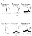
- the MALDI-TOF mass spectra regarding PN-sRL-VPg showed a monomer signal (in Fig. 2(a) , m/z (observed): 31,955, m/z (calculated): 31,514), a trimer signal (in Fig. 2(b) , m/z (observed): 95,257, m/z (calculated): 94,542), and a hexamer (trimer-dimer) signal (in Fig.
- PN-sFL-VPg PN-sFL-VPg monomer signal
- a trimer signal in Fig. 3(b) , m/z (observed): 93,857, m/z (calculated): 94,188
- a hexamer trimer-dimer signal
- the associated product of the present invention was found to include a trimer and a hexamer.
- Example 2 a composite protein falling within the scope of the present invention was prepared through a genetic engineering technique.
- the composite protein had an immunogen, which is a non-structural protein of a human RS virus Long variant; specifically, "P protein (phosphorylated protein: protein involved in phosphorylation)."
- P protein is encoded by a template sequence of messenger RNA of P protein in the RS virus genome.
- Each particle of RS virus has a minus-strand as the genome.
- a complementary chain of messenger RNA of each protein is present at each position.
- Each messenger RNA includes an initiation codon and a termination codon.
- the virus proteins formed via translation undergo a maturation step and exhibit a virus protein function.
- P protein forms a tetramer, which binds to L protein and RNP on its the C-terminal side, whereby L protein and RNP are linked by the mediation of the tetramer.
- P protein serves as a chaperone that can bind free N protein to RNA during synthesis.
- RNP is surrounded by an envelope originating from a host cell membrane. On the surface of the envelope, two virus glycoproteins; i.e., a receptor binding protein and a fusion protein (F protein), form spikes.
- Example 2 the aforementioned P protein was selected as a non-structural protein component of W.
- the actually used P protein of RSV Long variant (RSV-P) serving as an immunogen has an amino acid sequence of SEQ ID NO: 23
- the nucleotide sequence encoding the amino acid sequence may be selected in accordance with a generally known amino acid-nucleobase relationship.
- SEQ ID NO: 24 was employed.
- the RSV-P gene fragment was obtained from the genome RNA of RSV-Long variant provided by Sawada, Virus Infection I , Omura Satoshi Memorial Institute, Kitasato University. Specifically, a terminal stop codon (TGA in parenthesis mentioned above) was removed from the P protein gene sequence, and the resultant part was amplified.
- Example 2 a plasmid structure "PN-FL-P" of interest was constructed through the following procedure. Specifically, the aforementioned RSV-Long variant was subjected to PCR amplification, to amplify the nucleotide sequence (SEQ ID NO: 24) encoding P protein. From the thus-amplified nucleotide sequence (SEQ ID NO: 24) encoding P protein, the terminal stop codon (TGA) was removed, to thereby yield a gene fragment.
- TGA terminal stop codon
- the P domain was amplified by the mediation of In-Fusion RS-P sense 5'- GGAGATATACATATGGAAAAGTTTGCTCCTGA-3' (SEQ ID NO: 25) and In-Fusion RS-P antisense 5'- TGAACCCCCGCCTCCGAAATCATCAAGTGATAGAT-3' (SEQ ID NO: 26), to thereby yield a P domain amplification product into which the underlined portions had been added.
- inverted PCR was performed by use of 5'-GGAGGCGGGGGTTCA-3' (SEQ ID NO: 27) and 5'-ATGTATATCTCCTTCTTAAAG-3' (SEQ ID NO: 28) as primers with the template plasmid structure "PN-FL-VPg" as a template, to thereby amplify the entirety of the vector portion excepting the sequence of VPg.
- a vector body was provided. These two fragments were linked through In-Fusion cloning, to thereby yield a plasmid structure "PN-FL-P" of interest.
- PN-sFL-P was formed through gene expression.
- E. Coli BL21 (DE3) having the "PN-sFL-P" plasmid was incubated overnight in an LB medium containing 30 ⁇ g/mL Kanamicyn at 37°C. After the OD 600 of the incubated solution (37°C) had reached 0.8, 1mM isopropyl- ⁇ -D-1-thiogalactopyranoside (IPTG) and arabinose were added to the solution. Sixteen (16) to seventeen (17) hours after addition of IPTG and arabinose, the solution was centrifuged at 8,000 rpm for 5 minutes, to thereby recover microorganism pellets, and the recovered matter was incubated at 20°C and a rotation rate of 180 rpm.
- IPTG isopropyl- ⁇ -D-1-thiogalactopyranoside
- arabinose arabinose
- the cell pellets were suspended in 100mM Tris-HCl (pH: 8.0) buffer containing 5mM imidazole with one tablet of "cOmplete, EDTA-free," and the cells were lysed through ultrasonication. Cell broken pieces were removed through centrifugation (17,500 rpm for 50 minutes). The supernatant was filtered through a 0.8- ⁇ m. filter, and the filtrate was added to an Ni affinity column. The added matter was eluted at 4°C with the same buffer under a linear imidazole concentration gradient of 5mM to 250mM.
- PN-FL-P associated product was dialyzed against 20mM Tris-HCl (pH: 8.0) and 0.2M NaCl and further against PBS, and the dialysis product was concentrated through ultrafiltration. Through this process, PN-sFL-P spontaneously yielded an associated product containing a trimer and/or a hexamer thereof.
- P protein a recombinant P protein (hereinafter abbreviated as P protein) was obtained based on the aforementioned nucleotide sequence encoding the P protein through a customary procedure according to a series of processes from expression to recovery of "PN-sFL-P.”
- the immunoassay was conducted through nasal immunization by use of 10 ⁇ g/mL PBS solution (purified antigen).
- PN-sFL-P associated product PN(+)
- P protein PN(-)
- Blood collection was performed before the initial inoculation, 1 week after the initial inoculation (before the first booster), 2 weeks after the initial inoculation (before the second booster), 3 weeks after the initial inoculation (before the third booster), and 7 weeks after the initial inoculation.
- Each blood sample was caused to react with RSVP protein, and the IgG titer and the IgA titer to RSVP protein were determined through ELISA.
- the antigen (RSVP protein) was diluted with PBS(-) to 2 ⁇ g/mL.
- the diluted antigen was added to a 96-well ELISA plate at 50 ⁇ L/well and incubated overnight at 4°C.
- the plate was washed thrice with PBST (0.1% Tween 20/PBS) and then, PBSB (1% BSA/PBS) was added to the plate at 80 ⁇ L/well. Incubation was further conducted at room temperature for 2 hours for blocking.
- test serum sample was diluted with PBSB, to thereby prepare test samples (for IgG detection: 5-fold dilution series (10-fold to 31,250-fold), and for IgA detection: 3-fold dilution series (10-fold to 2,430-fold).
- PBSB in the plate was removed, and each test sample was added to the plate at 50 ⁇ L/well. Incubation was performed at room temperature for 2 hours, and the plate was washed 5 times with PBST. Then, an HRP substrate solution was added to the plate at 50 ⁇ L/well. Incubation was performed at room temperature under light shielding conditions until color development was confirmed. The reaction was stopped by adding 2M sulfuric acid to the plate at 25 ⁇ L/well, and absorbance at 490 nm was measured.
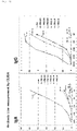
- Fig. 5 shows the results of ELISA (IgG)
- Fig. 6 shows the results of ELISA (IgA), obtained 3 weeks after the initial inoculation (3W) and 7 weeks after the initial inoculation (7W), respectively.
- the vertical axis represents absorbance
- the horizontal axis represents dilution ratio.
- the graphs show the results of the cotton rats tested under different conditions. As shown in the graphs, when the rats were immunized directly with P protein, no rise in absorbance in response to antibody titer was observed in both cases of IgG and IgA.
- the associated product of the present invention can possibly be used as an adjuvant-free vaccine.
- An RS virus infection is caused by inhaling the RS virus via droplet infection. That is, the infection mainly is caused via the oral cavity or the nasal cavity.
- the associated product was nasally inoculated, whereby the associated product directly penetrated the mucosal cells of the nasal cavity through the cell membranes, resulting in the injection of P protein into the nasal mucosal cells.
- cell-mediated immunity and humoral immunity were induced by the injected P protein, to thereby elicit protective immunity to RS virus.
- RS virus-susceptible cotton rats were immunized with a molecular needle to which an RS virus non-structural protein had been attached, and the rats were tested. Through employment of the rats, the effect of inhibiting lung inflammation caused by RS virus infection was directly investigated.
- PN-sFL-P associated product PN(+)
- P protein PN(-)
- nasal inoculation was performed. Specifically, initial inoculation was performed, and then booster inoculations were conducted three times in total at intervals of 7 days, and the final booster was performed eight weeks after the initial inoculation. Further, one week thereafter, the test rats were infected with RS virus. The dose of RS virus infection was 2 ⁇ 10 5 PFU/mL, and the infection was conducted in a manner similar to that of the vaccine inoculation (i.e., nasal inoculation).
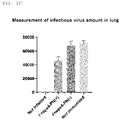
- the lung tissue was recovered from each tested rat, and the infection amount of RS virus in the lung was determined. Specifically, the lung (i.e., an infection target tissue) was removed from each rat, and suspended in a medium by a homogenizer, to thereby release the RS virus. The amount of infectious virus contained in the lung tissue (50 mg) was determined through plaque assay.
- Fig. 7 The results are shown in Fig. 7 .
- the vertical axis represents the RS virus titer (PFU/50 mg in lung tissue) of the recovered lung tissue.
- RS virus infection of rats to which the PN-sFL-P associated product had been administered was found to be inhibited at a ratio of 1/2 or more with a significance of (P ⁇ 0.0001), as compared with the case of RS virus-infected rats (the positive control).
- the vaccine of the present invention was found to exhibit substantial virus infection inhibitory effect through, in particular, topical immunity induction such as mucosal membrane immunity induction or cell-mediated immunity induction.
- the RNP of RS virus is surrounded by an envelope originating from a host cell membrane.
- two virus glycoproteins structural proteins
- a receptor binding protein and a fusion protein F protein
- F protein a fusion protein
- SEQ ID NO: 29: DMPITNDQKKLMS four units of a peptide formed of 13 amino acids (SEQ ID NO: 29: DMPITNDQKKLMS) forming the neutralization epitope domain of the F protein (as a structural protein) were linked in line, to thereby form a (quadruple) fusion protein.
- the resultant protein was attached as an immunogen W to a molecular needle, to thereby yield an associated product.
- the associated product was tested in a manner similar to that of the immunoassay performed in Example 2.
- the F protein of RS virus is formed of 574 amino acids (SEQ ID NO: 30:
- the domain ranging from the 263th amino acid to the 275th amino acid serves as an epitope domain recognized by a neutralizing antibody.
- a gene encoding a (quadruple) fusion protein in which four units of the epitope domain of F protein were linked in line by the mediation of (GGGG) was created.
- a plasmid (pET29b(+)/F-PN) which was designed to fuse with PN by the mediation of a linker (GGGGS: SEQ ID NO: 15) was created.
- E. coli (BL21 DE3) cells were transformed with the plasmid ( Fig 8 ), and expression was induced by IPTG. The expression product was recovered as a soluble protein.
- the aforementioned associated product of the quadruple fusion protein (Fnep ⁇ 4-PN associated product (PN(+)) was purified with TAIRON, and the immunoassay of Example 3 was conducted.
- Each of the Fnep ⁇ 4-PN associated product (PN(+)) and the quadruple fusion protein of the neutralization epitope of F protein (PN(-)) was nasally inoculated to RS virus-susceptible cotton rats.
- a vaccine liquid was added dropwise to the nasal cavity, and the vaccine was caused to be inhaled.
- a purified antigen was caused to be inhaled at 1 mL (20 ⁇ g) per rat through the nasal cavity under anesthesia.
- the nasal inoculation included initial inoculation, and then booster inoculations conducted three times in total at intervals of 7 days.
- Blood collection was performed before the initial inoculation, 1 week after the initial inoculation (before the first booster), 2 weeks after the initial inoculation (before the second booster), 3 weeks after the initial inoculation (before the third booster), and 7 weeks after the initial inoculation.
- Each blood sample was caused to react with F protein, and the IgG titer and the IgA titer to F protein were determined through ELISA.
- ELISA was performed in a manner similar to that of Example 2. However, as described above, F protein was used as an antigen.
- Fig. 9 The results (3 weeks after) are shown in Fig. 9 (in terms of IgA and IgG).
- the vertical axis represents absorbance
- the horizontal axis represents dilution ratio.
- the graphs show the results of the cotton rats tested under different conditions.
- the rats were immunized directly with the quadruple fusion protein of the neutralization epitope of F protein, no rise in absorbance in response to antibody titer was observed in both cases of IgG and IgA.
- the antibody titers i.e., absorbance values
- the rise in antibody titer was remarkable for IgG, but was observed with variation for IgA.
- RS virus-susceptible cotton rats were immunized with a molecular needle to which the quadruple fusion protein of the neutralization epitope of F protein (serving as an RS virus non-structural protein) was attached, and the rats were tested. Through employment of the rats, the effect of inhibiting lung inflammation caused by RS virus infection was directly investigated.
- Each of the Fnep ⁇ 4-PN associated product (PN(+)) and the quadruple fusion protein of the neutralization epitope of F protein (PN(-)) was nasally inoculated to RS virus-susceptible cotton rats.
- a purified antigen was caused to be inhaled at 1 mL (20 ⁇ g) per rat through the nasal cavity under anesthesia.
- the nasal inoculation was conducted in a manner similar to that of "Immunoassay (1).”
- the nasal inoculation included initial inoculation and then booster inoculations three times in total at intervals of 7 days. Seven weeks after the initial inoculation, the final booster was conducted.
- test rats were infected with RS virus.
- the infection dose of the RS virus was 2 ⁇ 10 5 PFU/mL, and the infection was conducted in a manner similar to that of the vaccine inoculation (i.e., nasal inoculation).
- the lung tissue was recovered from each tested rat, and the infection amount of RS virus in the lung was determined. Specifically, the lung (i.e., an infection target tissue) was removed from each rat and suspended in a medium by means of a homogenizer, to thereby release the RS virus. The amount of the infectious virus in the lung tissue (50 mg) was determined through plaque assay.
- test rats there were also employed a positive control group (the aforementioned cotton rats infected with RS virus without nasal inoculation in advance) and a negative control group (the aforementioned cotton rats not infected with RS virus and not subjected to nasal inoculation in advance).
- Fig. 10 The results are shown in Fig. 10 .
- the vertical axis represents the RS virus titer (PFU/50 mg of lung tissue) of the recovered lung tissue.
- the rats to which the Fnep ⁇ 4PN associated product had been administrated exhibited a reduced RS virus infection of about 36% with a significance of P ⁇ 0.0001, as compared with RS virus-infected rats (the positive control).
- the needle vaccine containing the associated product having the quadruple fusion protein of the neutralization epitope of F protein (i.e., the structural protein of RS virus) of Example 3 was found to induce topical immunity such as mucosal immunity or cell-mediated immunity.
- topical immunity such as mucosal immunity or cell-mediated immunity.
- the thus-attained effect was found to be weaker than that of the vaccine of the present invention employing a non-structural protein.
- the virus-neutralizing effect of the secretory IgA i.e., an antibody which was induced through immunization by the associated product having the quadruple fusion protein of the neutralization epitope of F protein, and secreted to the mucin layer of the mucosal epithelium
- the vaccine of the present invention i.e., the molecular needle employing P protein (a non-structural protein)
- P protein a non-structural protein
- a component vaccine containing, for example, the associated product of the present invention of Example 2 which has P protein (i.e., a non-structural protein of RS virus), and an associated product of a molecular needle having a structural protein, such as the associated product of Example 3 which has the quadruple fusion protein of the neutralization epitope of F protein (i.e., a structural protein of RS virus), in combination as active ingredients is expected to have a synergistic effect due to the combination.
Landscapes
- Health & Medical Sciences (AREA)
- Life Sciences & Earth Sciences (AREA)
- Chemical & Material Sciences (AREA)
- Virology (AREA)
- Organic Chemistry (AREA)
- General Health & Medical Sciences (AREA)
- Medicinal Chemistry (AREA)
- Genetics & Genomics (AREA)
- Molecular Biology (AREA)
- Pharmacology & Pharmacy (AREA)
- Animal Behavior & Ethology (AREA)
- Veterinary Medicine (AREA)
- Public Health (AREA)
- Biochemistry (AREA)
- Microbiology (AREA)
- Biophysics (AREA)
- Engineering & Computer Science (AREA)
- Immunology (AREA)
- Proteomics, Peptides & Aminoacids (AREA)
- Nuclear Medicine, Radiotherapy & Molecular Imaging (AREA)
- General Chemical & Material Sciences (AREA)
- Chemical Kinetics & Catalysis (AREA)
- Oncology (AREA)
- Communicable Diseases (AREA)
- Mycology (AREA)
- Epidemiology (AREA)
- Gastroenterology & Hepatology (AREA)
- General Engineering & Computer Science (AREA)
- Biomedical Technology (AREA)
- Biotechnology (AREA)
- Bioinformatics & Cheminformatics (AREA)
- Wood Science & Technology (AREA)
- Zoology (AREA)
- Physics & Mathematics (AREA)
- Plant Pathology (AREA)
- Medicines Containing Antibodies Or Antigens For Use As Internal Diagnostic Agents (AREA)
- Peptides Or Proteins (AREA)
Applications Claiming Priority (3)
| Application Number | Priority Date | Filing Date | Title |
|---|---|---|---|
| JP2019154360 | 2019-08-27 | ||
| JP2020089252 | 2020-05-21 | ||
| PCT/JP2020/032260 WO2021039873A1 (ja) | 2019-08-27 | 2020-08-26 | ウイルスの非構造タンパク質が担持された複合タンパク質単量体、当該単量体の会合体、及び当該会合体を有効成分とするコンポーネントワクチン |
Publications (2)
| Publication Number | Publication Date |
|---|---|
| EP4026558A1 true EP4026558A1 (de) | 2022-07-13 |
| EP4026558A4 EP4026558A4 (de) | 2022-10-19 |
Family
ID=74685133
Family Applications (1)
| Application Number | Title | Priority Date | Filing Date |
|---|---|---|---|
| EP20856780.0A Pending EP4026558A4 (de) | 2019-08-27 | 2020-08-26 | Zusammengesetztes proteinmonomer mit nicht-strukturellem virusprotein darauf, aggregat des zusammengesetzten proteinmonomers und kombinationsimpfstoff mit aggregat als wirkstoff |
Country Status (4)
| Country | Link |
|---|---|
| US (1) | US20220362370A1 (de) |
| EP (1) | EP4026558A4 (de) |
| JP (1) | JP7651129B2 (de) |
| WO (1) | WO2021039873A1 (de) |
Families Citing this family (1)
| Publication number | Priority date | Publication date | Assignee | Title |
|---|---|---|---|---|
| US20240316181A1 (en) * | 2021-01-07 | 2024-09-26 | Tokyo Institute Of Technology | Conjugated protein monomer carrying peptide derived from pathogenic microorganism compatible with mhc molecule, aggregate of said monomers, component vaccine containing said aggregate as active ingredient, and method for acquiring information on secretion of physiologically active substance after immunization |
Family Cites Families (5)
| Publication number | Priority date | Publication date | Assignee | Title |
|---|---|---|---|---|
| WO2004071493A1 (en) * | 2003-02-17 | 2004-08-26 | Peter Burkhard | Peptidic nanoparticles as drug delivery and antigen display systems |
| WO2012140676A2 (en) * | 2011-04-12 | 2012-10-18 | Appaiah C B | Chimeric antibacterial polypeptides |
| CA2915728A1 (en) | 2013-08-21 | 2015-02-26 | Thomas Kramps | Respiratory syncytial virus (rsv) vaccine |
| JP6304683B2 (ja) | 2014-02-28 | 2018-04-04 | 国立大学法人東京工業大学 | 標的タンパク質の細胞内導入剤及び標的タンパク質の細胞内導入方法 |
| JP7062595B2 (ja) * | 2016-10-23 | 2022-05-06 | デンカ株式会社 | 複合ポリペプチド単量体、細胞浸透機能を有する当該複合ポリペプチドの単量体の会合体、及び、当該会合体を有効成分とする皮下、皮内、経皮又は筋肉内投与用ノロウイルスコンポーネントワクチン |
-
2020
- 2020-08-26 EP EP20856780.0A patent/EP4026558A4/de active Pending
- 2020-08-26 WO PCT/JP2020/032260 patent/WO2021039873A1/ja not_active Ceased
- 2020-08-26 US US17/638,085 patent/US20220362370A1/en active Pending
- 2020-08-26 JP JP2021542980A patent/JP7651129B2/ja active Active
Also Published As
| Publication number | Publication date |
|---|---|
| US20220362370A1 (en) | 2022-11-17 |
| EP4026558A4 (de) | 2022-10-19 |
| JPWO2021039873A1 (de) | 2021-03-04 |
| JP7651129B2 (ja) | 2025-03-26 |
| WO2021039873A1 (ja) | 2021-03-04 |
Similar Documents
| Publication | Publication Date | Title |
|---|---|---|
| ES2858315T3 (es) | Proteína F de prefusión de VRS soluble estabilizada para uso en la profilaxis de la infección por VRS | |
| US7413889B2 (en) | Production of a parvovirus vaccine in plants as viral coat protein fusions | |
| CN114213548B (zh) | 同时诱导抗多种病毒的免疫应答的方法 | |
| WO2022110099A1 (en) | Coronavirus vaccines and uses thereof | |
| US20250026793A1 (en) | Chimeric betacoronavirus spike polypeptides | |
| WO2023236822A1 (zh) | H5n6禽流感广谱性疫苗的开发及其应用 | |
| US20220362370A1 (en) | Composite protein monomer having non-structural protein of virus supported thereon, aggregate of composite protein monomer, and component vaccine comprising aggregate as active ingredient | |
| WO2021235503A1 (ja) | コロナウイルスの蛋白質が担持された複合蛋白質単量体、当該単量体の会合体、及び当該会合体を有効成分とするコンポーネントワクチン | |
| CN117003885B (zh) | H5n8禽流感广谱性疫苗的开发及其应用 | |
| KR101366702B1 (ko) | 호흡기 신시치아 바이러스 백신 조성물 및 그의 제조방법 | |
| KR102102065B1 (ko) | 호흡기 세포융합 바이러스 백신 | |
| US20240316181A1 (en) | Conjugated protein monomer carrying peptide derived from pathogenic microorganism compatible with mhc molecule, aggregate of said monomers, component vaccine containing said aggregate as active ingredient, and method for acquiring information on secretion of physiologically active substance after immunization | |
| CN114634579A (zh) | 一种抗新冠病毒基因工程疫苗 | |
| WO1994006468A1 (en) | Recombinant influenza virus vaccine compositions | |
| EP4633675A1 (de) | Modifizierte masernviren zur behandlung von coronavirusinfektionen | |
| CN119497628A (zh) | 一种抵抗呼吸道合胞病毒的mRNA疫苗及其制备方法 | |
| CN106362144B (zh) | 呼吸道合胞病毒疫苗 | |
| CN121620537A (zh) | Rsv和hmpv融合f蛋白及其用途 | |
| CN118185961A (zh) | 针对猫fipv的多表位抗原构建环状rna疫苗的方法 | |
| HK40051597A (en) | Stabilized soluble pre-fusion rsv f proteins for use in the prophylaxis of rsv infection | |
| CN121991245A (zh) | 一种PEDV mRNA疫苗及其应用 | |
| CN119161495A (zh) | 呼吸道合胞病毒重构多肽及其应用 | |
| JPH03130082A (ja) | ワクチン用ポリペプチド | |
| WO2020014013A1 (en) | Viral-vectored vaccine for malaria | |
| HK1261073B (en) | Stabilized soluble pre-fusion rsv f protein for use in the prophylaxis of rsv infection |
Legal Events
| Date | Code | Title | Description |
|---|---|---|---|
| STAA | Information on the status of an ep patent application or granted ep patent |
Free format text: STATUS: THE INTERNATIONAL PUBLICATION HAS BEEN MADE |
|
| PUAI | Public reference made under article 153(3) epc to a published international application that has entered the european phase |
Free format text: ORIGINAL CODE: 0009012 |
|
| STAA | Information on the status of an ep patent application or granted ep patent |
Free format text: STATUS: REQUEST FOR EXAMINATION WAS MADE |
|
| 17P | Request for examination filed |
Effective date: 20220325 |
|
| AK | Designated contracting states |
Kind code of ref document: A1 Designated state(s): AL AT BE BG CH CY CZ DE DK EE ES FI FR GB GR HR HU IE IS IT LI LT LU LV MC MK MT NL NO PL PT RO RS SE SI SK SM TR |
|
| A4 | Supplementary search report drawn up and despatched |
Effective date: 20220916 |
|
| RIC1 | Information provided on ipc code assigned before grant |
Ipc: C12N 15/63 20060101ALI20220912BHEP Ipc: C12N 15/33 20060101ALI20220912BHEP Ipc: C12N 1/21 20060101ALI20220912BHEP Ipc: C12N 1/19 20060101ALI20220912BHEP Ipc: C12N 1/15 20060101ALI20220912BHEP Ipc: C12N 5/10 20060101ALI20220912BHEP Ipc: C07K 19/00 20060101ALI20220912BHEP Ipc: C07K 14/005 20060101ALI20220912BHEP Ipc: A61P 31/12 20060101ALI20220912BHEP Ipc: A61K 39/12 20060101AFI20220912BHEP |
|
| DAV | Request for validation of the european patent (deleted) | ||
| DAX | Request for extension of the european patent (deleted) | ||
| STAA | Information on the status of an ep patent application or granted ep patent |
Free format text: STATUS: EXAMINATION IS IN PROGRESS |
|
| 17Q | First examination report despatched |
Effective date: 20240514 |