EP4040445A1 - Verfahren, programm und vorrichtung zum bewerten des zustandes einer motorfunktion - Google Patents
Verfahren, programm und vorrichtung zum bewerten des zustandes einer motorfunktion Download PDFInfo
- Publication number
- EP4040445A1 EP4040445A1 EP20871340.4A EP20871340A EP4040445A1 EP 4040445 A1 EP4040445 A1 EP 4040445A1 EP 20871340 A EP20871340 A EP 20871340A EP 4040445 A1 EP4040445 A1 EP 4040445A1
- Authority
- EP
- European Patent Office
- Prior art keywords
- patient
- motor function
- brain
- indicator value
- physiological indicator
- Prior art date
- Legal status (The legal status is an assumption and is not a legal conclusion. Google has not performed a legal analysis and makes no representation as to the accuracy of the status listed.)
- Pending
Links
Images






Classifications
-
- G—PHYSICS
- G16—INFORMATION AND COMMUNICATION TECHNOLOGY [ICT] SPECIALLY ADAPTED FOR SPECIFIC APPLICATION FIELDS
- G16H—HEALTHCARE INFORMATICS, i.e. INFORMATION AND COMMUNICATION TECHNOLOGY [ICT] SPECIALLY ADAPTED FOR THE HANDLING OR PROCESSING OF MEDICAL OR HEALTHCARE DATA
- G16H30/00—ICT specially adapted for the handling or processing of medical images
- G16H30/40—ICT specially adapted for the handling or processing of medical images for processing medical images, e.g. editing
-
- A—HUMAN NECESSITIES
- A61—MEDICAL OR VETERINARY SCIENCE; HYGIENE
- A61B—DIAGNOSIS; SURGERY; IDENTIFICATION
- A61B5/00—Measuring for diagnostic purposes; Identification of persons
- A61B5/0033—Features or image-related aspects of imaging apparatus, e.g. for MRI, optical tomography or impedance tomography apparatus; Arrangements of imaging apparatus in a room
- A61B5/004—Features or image-related aspects of imaging apparatus, e.g. for MRI, optical tomography or impedance tomography apparatus; Arrangements of imaging apparatus in a room adapted for image acquisition of a particular organ or body part
- A61B5/0042—Features or image-related aspects of imaging apparatus, e.g. for MRI, optical tomography or impedance tomography apparatus; Arrangements of imaging apparatus in a room adapted for image acquisition of a particular organ or body part for the brain
-
- A—HUMAN NECESSITIES
- A61—MEDICAL OR VETERINARY SCIENCE; HYGIENE
- A61B—DIAGNOSIS; SURGERY; IDENTIFICATION
- A61B5/00—Measuring for diagnostic purposes; Identification of persons
- A61B5/05—Detecting, measuring or recording for diagnosis by means of electric currents or magnetic fields; Measuring using microwaves or radio waves
- A61B5/055—Detecting, measuring or recording for diagnosis by means of electric currents or magnetic fields; Measuring using microwaves or radio waves involving electronic [EMR] or nuclear [NMR] magnetic resonance, e.g. magnetic resonance imaging
-
- A—HUMAN NECESSITIES
- A61—MEDICAL OR VETERINARY SCIENCE; HYGIENE
- A61B—DIAGNOSIS; SURGERY; IDENTIFICATION
- A61B5/00—Measuring for diagnostic purposes; Identification of persons
- A61B5/40—Detecting, measuring or recording for evaluating the nervous system
- A61B5/4058—Detecting, measuring or recording for evaluating the nervous system for evaluating the central nervous system
- A61B5/4064—Evaluating the brain
-
- A—HUMAN NECESSITIES
- A61—MEDICAL OR VETERINARY SCIENCE; HYGIENE
- A61B—DIAGNOSIS; SURGERY; IDENTIFICATION
- A61B5/00—Measuring for diagnostic purposes; Identification of persons
- A61B5/40—Detecting, measuring or recording for evaluating the nervous system
- A61B5/4076—Diagnosing or monitoring particular conditions of the nervous system
- A61B5/4082—Diagnosing or monitoring movement diseases, e.g. Parkinson, Huntington or Tourette
-
- A—HUMAN NECESSITIES
- A61—MEDICAL OR VETERINARY SCIENCE; HYGIENE
- A61B—DIAGNOSIS; SURGERY; IDENTIFICATION
- A61B5/00—Measuring for diagnostic purposes; Identification of persons
- A61B5/48—Other medical applications
- A61B5/4848—Monitoring or testing the effects of treatment, e.g. of medication
-
- G—PHYSICS
- G01—MEASURING; TESTING
- G01R—MEASURING ELECTRIC VARIABLES; MEASURING MAGNETIC VARIABLES
- G01R33/00—Arrangements or instruments for measuring magnetic variables
- G01R33/20—Arrangements or instruments for measuring magnetic variables involving magnetic resonance
- G01R33/44—Arrangements or instruments for measuring magnetic variables involving magnetic resonance using nuclear magnetic resonance [NMR]
- G01R33/48—NMR imaging systems
- G01R33/54—Signal processing systems, e.g. using pulse sequences ; Generation or control of pulse sequences; Operator console
- G01R33/56—Image enhancement or correction, e.g. subtraction or averaging techniques, e.g. improvement of signal-to-noise ratio and resolution
- G01R33/563—Image enhancement or correction, e.g. subtraction or averaging techniques, e.g. improvement of signal-to-noise ratio and resolution of moving material, e.g. flow contrast angiography
- G01R33/56341—Diffusion imaging
-
- G—PHYSICS
- G16—INFORMATION AND COMMUNICATION TECHNOLOGY [ICT] SPECIALLY ADAPTED FOR SPECIFIC APPLICATION FIELDS
- G16H—HEALTHCARE INFORMATICS, i.e. INFORMATION AND COMMUNICATION TECHNOLOGY [ICT] SPECIALLY ADAPTED FOR THE HANDLING OR PROCESSING OF MEDICAL OR HEALTHCARE DATA
- G16H50/00—ICT specially adapted for medical diagnosis, medical simulation or medical data mining; ICT specially adapted for detecting, monitoring or modelling epidemics or pandemics
- G16H50/30—ICT specially adapted for medical diagnosis, medical simulation or medical data mining; ICT specially adapted for detecting, monitoring or modelling epidemics or pandemics for calculating health indices; for individual health risk assessment
Definitions
- the present disclosure relates to a method for evaluating a state of a motor function of a patient who has or is suspected of having a brain injury, and a program, an image analysis apparatus, and an MRI apparatus that can be used in the implementation of the method.
- the present disclosure relates to a method for predicting motor function recovery after regeneration therapy of a patient having a brain focal injury.
- Non Patent Literature 1 In cell therapy, it is important to determine which patient is suitable for cell therapy prior to start of the therapy because the recovery degree of a patient actually administered with a cell varies (Non Patent Literature 1).
- the present inventors found that a state of a motor function of a patient having a brain focal injury is correlated with a parameter obtained from a diffusion weighted image of the brain of the patient.
- a method for evaluating a state of a motor function of a patient who has or is suspected of having a brain injury comprising:
- the method of item 1, wherein the physiological indicator value is obtained from a diffusion weighted image of the brain of the patient which is obtained by diffusion tensor imaging (DTI) method or diffusion kurtosis imaging (DKI) method.
- DTI diffusion tensor imaging
- DKI diffusion kurtosis imaging
- the performing computation evaluates the state of the motor function of the patient by substituting the calculated value indicating the state of the motor function of the patient in a regression line which is prepared in advance and has a value indicating a state of a motor function of a patient and a motor function recovery degree as variables.
- the performing computation calculates a level of a likelihood that the patient reaches a desired motor function recovery degree after regeneration therapy by comparing the calculated value indicating the state of the motor function of the patient with a reference value which is prepared in advance.
- a system for evaluating a state of a motor function of a brain of a patient who has or is suspected of having a brain injury comprising:
- An image analysis apparatus comprising:
- An MRI apparatus comprising:
- the present disclosure also provides the following.
- the present disclosure it is possible to evaluate a state of a motor function of a patient who has or is suspected of having a brain injury such as a brain focal injury, it is thereby possible to predict motor function recovery after regeneration therapy of the patient, and it is also possible to determine whether the patient is suitable for regeneration therapy prior to start of the therapy.
- outer surface region refers to the region on the outer side of the cerebrum, and particularly refers to the gyrus.
- the outer surface region includes fornicate gyrus, superior frontal gyrus, middle frontal gyrus, inferior frontal gyrus, superior temporal gyrus, middle temporal gyrus, inferior temporal gyrus, fusiform gyrus, parahippocampal gyrus, transverse temporal gyrus, precentral gyrus, postcentral gyrus, supramarginal gyrus, angular gyrus, cingulate gyrus, dentate gyrus, paracentral lobule, cuneus, precuneus, lingual gyrus, and the like.
- physiological indicator value refers to an indicator value that is obtained from a diffusion weighted image of the brain of diffusion tensor imaging (DTI), diffusion kurtosis imaging (DKI), or the like, and includes MK (mean kurtosis), AK (axial kurtosis), RK (radial kurtosis), FA (fractional anisotropy), KFA (kurtosis fractional anisotropy), MD (mean diffusivity), AD (axial diffusivity), and RD (radial diffusivity).
- DTI diffusion tensor imaging
- DKI diffusion kurtosis imaging
- MK mean kurtosis
- AK axial kurtosis
- RK radial kurtosis
- FA fractional anisotropy
- KFA kurtosis fractional anisotropy
- MD mean diffusivity
- AD axial diffusivity
- RD radial diffusivity
- state of a motor function refers to a state of a motor function that is comprehensively controlled by the function of the brain
- evaluation of a state of a motor function includes evaluating or predicting whether the motor function is normal or the motor function is partially or completely paralyzed or the like at the present time or after therapy from the state of the brain of a subject.
- evaluation of a state of a motor function includes prediction or the like of recovery of the function of the brain controlling movement by regeneration therapy or the like.
- a method for evaluating a state of a motor function of a patient who has or is suspected of having a brain injury comprising: obtaining a physiological indicator value with an outer surface region on the side of an injured hemisphere of a brain of the patient as a region of interest; and performing computation to calculate a value indicating the state of the motor function of the patient by comparing the physiological indicator value with a reference physiological indicator value, is provided.
- a physiological indicator value can be obtained from an image of the brain of a patient obtained by diffusion tensor imaging (DTI) method or diffusion kurtosis imaging (DKI) method.
- DTI diffusion tensor imaging
- DKI diffusion kurtosis imaging
- DTI diffusion tensor imaging
- ROI region of interest
- a physiological indicator value such as the FA value or MD value in the region of interest is measured, whereby the corticospinal tract can be tracked.
- DTI method In general, ROI setting in diffusion tensor imaging (DTI) method is manually performed, but tracking of the corticospinal tract in this technique relies on the experience of the measurer.
- DKI method can be used without relying on the measurer and as a quantitative image index.
- DTI method assumes that diffusion of water molecules is a normal distribution, it is known that diffusion of water molecules in a living body does not show a normal distribution.
- a diffusion imaging method that does not assume a normal distribution as in DKI method, diffusion of water molecules in a living body can be more precisely reflected. Therefore, in one embodiment of the present disclosure, a diffusion image index capable of predicting the therapeutic effect of cell administration to a patient with cerebral infarction precisely and without relying on the experience of the measurer or the like can be obtained with DKI method.
- a physiological indicator value obtained from a diffusion weighted image of the brain of a patient obtained by diffusion tensor imaging (DTI) method or diffusion kurtosis imaging (DKI) method as described above is not particularly limited as long as the physiological indicator value is an index obtained by a diffusion weighted image that is formed as an image with the direction and the rate of diffusion of water molecules in tissue as parameters.
- a diffusion index such as MK (mean kurtosis), AK (axial kurtosis), RK (radial kurtosis), FA (fractional anisotropy), KFA (kurtosis fractional anisotropy), MD (mean diffusivity), AD (axial diffusivity), and RD (radial diffusivity) can be used.
- MK mean kurtosis
- AK axial kurtosis
- RK radial kurtosis
- FA fractional anisotropy
- KFA kurtosis fractional anisotropy
- MD mean diffusivity
- AD axial diffusivity
- RD radial diffusivity
- the ROI in DTI and DKI may be set anywhere on the outer surface region of the brain and is not particularly limited. In one embodiment, the ROI is preferably set to the precentral gyrus.
- a state of a motor function of a subject can be evaluated from a physiological indicator value obtained from an image of the brain of a patient obtained by diffusion tensor imaging (DTI) method or diffusion kurtosis imaging (DKI) method as described above, wherein this motor function may be a function at the present time or after therapy, and it is also possible to predict a recovery state of the motor function after regeneration therapy.
- DTI diffusion tensor imaging
- DKI diffusion kurtosis imaging
- a state of a motor function can be evaluated by comparing a physiological indicator value obtained when the ROI is placed on the side of an injured hemisphere of the brain and a physiological indicator value obtained when the ROI is placed on the side of an uninjured hemisphere of the brain (called reference physiological indicator value), and preferably by calculating a value represented by the physiological indicator value/the reference physiological indicator value.
- a state of a motor function also can be evaluated by substituting a value represented by a physiological indicator value/a reference physiological indicator value in a regression line which has a value indicating a state of a motor function of a patient and a motor function recovery degree as variables.
- the possibility that a patient reaches a desired motor function recovery degree after regeneration therapy also can be indicated by comparing a value represented by a physiological indicator value/a reference physiological indicator value with a reference value indicating the likelihood that the patient reaches the desired motor function recovery degree after regeneration therapy.
- the method of the present disclosure it is possible to evaluate a state of a motor function of a subject and it is also possible to evaluate the state of the motor function after regeneration therapy, in other words, predict whether the motor function recovers by regeneration therapy, so that it is possible to determine which patient is suitable for cell therapy prior to start of the therapy, and it is also possible to select a patient for which the therapy is highly effective.
- the present disclosure also provides a computer program for causing a computer to execute the above-described method, a storage medium for storing said program, and a system for executing the above-described method.
- a computer program for causing a computer to execute processing of a method for evaluating a state of a motor function of a brain of a patient who has or is suspected of having a brain injury, wherein the method comprises the following processes:
- a storage medium for storing a computer program for causing a computer to execute processing of a method for evaluating a state of a motor function of a brain of a patient who has or is suspected of having a brain injury, wherein the method comprises the following processes:
- an image analysis apparatus comprising:
- an MRI apparatus comprising:
- the detailed configuration of the image analysis apparatus and the MRI apparatus of the present disclosure can comprise the same configuration as that of the image analysis apparatus and the MRI apparatus described in detail in other portions of the present specification.
- Another aspect of the present disclosure relates to a method for predicting motor function recovery after regeneration therapy of a patient having a brain focal injury, and a program, an image analysis apparatus, and an MRI apparatus that can be used in the implementation of the method.
- FIG. 1 shows a block diagram showing the overall configuration of an MRI apparatus that is one embodiment of the present disclosure.
- An MRI apparatus 1 has a nuclear magnetic resonance imaging unit 10 and a computer 20.
- the nuclear magnetic resonance imaging unit 10 is an imaging unit that is used in any known MRI apparatus which can acquire echo data necessary for a DTI image.
- the nuclear magnetic resonance imaging unit 10 has: a bed; a magnet stand comprising a static magnetic field magnet, a gradient magnetic field coil, and an RF coil; and a sequence controlling unit.
- the bed on which a patient is lying is inserted into an opening part of the magnet stand.
- the static magnetic field magnet of the magnet stand generates a static magnetic field, and the gradient magnetic field coil applies a gradient magnetic field accordingly.
- an RF coil for transmission generates a high frequency magnetic field
- an RF coil for reception receives an echo signal emitted from the patient by generation of the high frequency magnetic field.
- the received echo signal is digitized and transmitted to the sequence controlling unit as echo data.
- the sequence controlling unit controls imaging based on sequence information transmitted from the computer 20, and also transfers the received echo data to the computer 20.
- the computer 20 is an apparatus that controls the nuclear magnetic resonance imaging unit 10, collects data, reconstructs an image, and analyzes an image, and has an interface unit 21, an inputting unit 22, a storage unit 23, a computing unit 24, an outputting unit 25, and a controlling unit 26.
- the interface unit 21 controls an input and an output of various data such as echo data that is exchanged with the sequence controlling unit of the nuclear magnetic resonance imaging unit 10.
- the interface unit 21 transmits sequence information for controlling the nuclear magnetic resonance imaging unit 10 to the sequence controlling unit.
- the interface unit 21 also receives echo data from the sequence controlling unit and stores the data in the storage unit 23.
- the inputting unit 22 is a device such as a keyboard, a mouse, a button, or a switch, and inputs a signal in accordance with an operator's operation to said device.
- the storage unit 23 comprises, in its configuration, a storage medium such as a hard disc, a flash memory, a RAM, or a ROM and a reading apparatus for reading information stored in the storage medium.
- the storage unit 23 stores echo data transmitted from the sequence controlling unit, various MRI image data generated from the echo data, reference information for predicting motor function recovery of the patient, a program for image generation and analysis, a program for controlling each function unit of the MRI apparatus 1 which is executed by the controlling unit 26, and various setting information or the like.
- the computing unit 24 consists of hardware such as a CPU.
- the computing unit 24 reads a program stored in the storage unit 23 to reconstruct an image from echo data stored in the storage unit 23, and generates a DTI image from the reconstructed image.
- the computing unit 24 also analyzes a DTI image of the brain of the patient stored in the storage unit 23 and performs computation to predict motor function recovery of the patient.
- the outputting unit 25 outputs various information such as an MRI image generated by the computing unit 24 or a predicted value of a motor function recovery degree obtained by performing computation to the outside of the computer 20, typically, a display connected to the outputting unit 25.
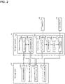
- FIG. 2 is a functional block diagram showing the configuration of a storage unit, a computing unit, an inputting unit, and an outputting unit in one embodiment of the present disclosure.
- the storage unit 23 has an echo data storage unit 231, a reconstructed image storage unit 232, a DTI image storage unit 233, and a reference information storage unit 234.
- the computing unit 24 has an image generating unit 241 and an image analyzing unit 242.
- the image generating unit 241 has an image reconstructing unit 241a and a DTI image generating unit 241b
- the image analyzing unit 242 has a region of interest setting unit 242a, an FA calculating unit 242b, an rFA calculating unit 242c, and a recovery prediction computing unit 242d.
- D xx , D yy , and D zz are diffusion coefficients of when a gradient magnetic field is applied in the x axis direction, the y axis direction, and the z axis direction, respectively, of the MRI apparatus coordinate system.
- D D xx D xy D xz D yx D yy D yz D zx D zy D zz
- D ⁇ 1 0 0 0 ⁇ 2 0 0 0 ⁇ 3
- ADC apparatus diffusion coefficient
- FA fractional anisotropy
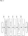
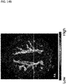
- the region of interest setting unit 242a sets a first region of interest (ROI-1) in a position corresponding to the precentral gyrus on the side of an injured hemisphere and sets a second region of interest (ROI-2) in a position corresponding to the precentral gyrus on the side of an uninjured hemisphere on the DTI image stored in the DTI image storage unit 233, typically on an FA map (step S10).
- the ROI may be manually set based on an anatomical position, may be set while being superposed with another medical image, or may be automatically set based on the function of known DTI image analysis software. When the ROI is manually set, an operation to set the ROI by an operator is accepted via the inputting unit 22.
- the FA calculating unit 242b calculates an injured hemisphere-side FA and an uninjured hemisphere-side FA that are the FA in the ROI-1 and the ROI-2, respectively (step S11).
- the injured hemisphere-side FA and the uninjured hemisphere-side FA are the mean value of the FA of all voxels contained in the ROI-1 and the ROI-2, respectively.
- the rFA calculating unit 242c calculates an rFA represented by the injured hemisphere-side FA/the uninjured hemisphere-side FA (step S12).
- the recovery prediction computing unit 242d performs computation for predicting motor function recovery of the patient after regeneration therapy based on the rFA caculated by the rFA calculating unit 242c (step S13) and outputs the computed result to the outputting unit 25 (step S14).
- the computation for predicting motor function recovery is performed based on the reference information stored in the reference information storage unit 234 in addition to the calculated rFA.
- reference information is a regression line which is prepared in advance and has an rFA of a patient having a brain focal injury prior to regeneration therapy and a motor function recovery degree of the same patient after the therapy as variables.
- the motor function recovery degree is an extent of recovery of a motor function by therapy which is evaluated using a known evaluation method capable of evaluating a motor function (e.g., Barthel Index (BI), Fugl-Meyer Assessment (FMA), or the like).
- the motor function recovery degree can be represented as a difference between the score prior to the therapy and the score after the therapy in the same patient.
- a motor function recovery degree after regeneration therapy is correlated with an rFA prior to the therapy in a patient having a brain focal injury.
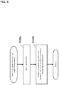
- an rFA prior to therapy corresponding to a desired motor function recovery degree is determined as a reference rFA in advance based on a scatter diagram or a regression line having an rFA calculated from a brain DTI image prior to regeneration therapy and a motor function recovery degree after the therapy of a patient who already received the therapy as variables, and it is determined whether an rFA calculated from a brain DTI image of a subject patient who is going to receive regeneration therapy from now exceeds the reference rFA, whereby the level of a likelihood that the subject patient reaches the desired motor function recovery degree after regeneration therapy can be calculated (steps S130b to S133b).
- the reference rFA can be appropriately set depending on the motor function recovery degree that is a target.
- Reference information such as a regression line or a reference rFA is preferably obtained from a patient having the same type of disease as a patient who is the subject of prediction of motor function recovery after regeneration therapy.
- reference information is preferably prepared by using an rFA calculated from a brain DTI image prior to regeneration therapy and a motor function recovery degree after the therapy of a patient with cerebral infarction who received the therapy.
- Reference information is also preferably obtained from a patient who received the same type of therapy as the therapy to which a patient attempts to be adapted.
- the therapy is administration of a bone marrow mesenchymal stem cell (BMSC)
- reference information is preferably prepared by using an rFA calculated from a brain DTI image prior to therapy and a motor function recovery degree after the therapy of a patient administered with a BMSC.
- BMSC bone marrow mesenchymal stem cell
- brain focal injury refers to a state in which an injury is focally caused to the brain.
- Patient having a brain focal injury refers to a patient in which an injury is focally caused to the brain. Examples of a brain focal injury can include cerebral infarction, head trauma, and cerebral hemorrhage.
- a patient having a brain focal injury is not restricted as long as the patient is a patient in which an injury is focally caused to the brain and whose motor function is damaged, and said patient may be a patient in any stage of the acute stage, the sub-acute stage, and the chronic stage.
- regeneration therapy of a patient having a brain focal injury means therapy for regenerating an injured nervous system cell of a patient and recovering various function disorders caused by the injury by using a cell of the patient himself/herself (autologous) or others (allogeneic) or a secretion thereof.
- a cell that can be used for regeneration therapy of a patient having a brain focal injury may be any cell having differentiation potency into a nervous system cell.
- Examples can include a stem cell having pluripotency such as a mesenchymal stem cell and a neural stem cell and a pluripotent stem cell such as an induced pluripotent stem cell (iPS cell), an embryonic stem cell (ES cell), an embryonic carcinoma cell (EC cell), and an embryonic germ cell (EG cell).
- a cell that is preferably used for therapy is a mesenchymal stem cell, particularly a bone marrow mesenchymal stem cell (BMSC; also called bone marrow stem cell).
- BMSC bone marrow mesenchymal stem cell
- a stem cell having pluripotency may be a cell obtained by inducing differentiation of a pluripotent stem cell.
- the dosage and the route of administration of the above-described cell and the secretion thereof are appropriately set by those skilled in the art while referring to a known dosing regimen relating to those dosage and route of administration.
- 10 4 to 10 9 cells, preferably 10 5 to 10 8 cells, per kg of a patient's body weight can be administered to the patient by systemic administration such as intravenous administration or intra-arterial administration, or 10 2 to 10 9 cells, preferably 10 4 to 10 6 cells, per kg of the patient's body weight can be administered to the patient by local administration such as intracerebral direct administration or intrathecal administration one or multiple times.
- NIHSS Motor Leg
- the bone marrow of the patients was promptly collected, and cell culture and bone marrow stem cell preparation were carried out in the Cell Processing Center of the Clinical Research and Medical Innovation Center of Hokkaido University Hospital.
- the prepared bone marrow stem cells (20,000,000 or 50,000,000 cells/patient) were directly administered to the brain of the patients after 3 to 5 weeks from the collection of the bone marrow.
- the motor function of the patients was evaluated with Barthel Index (BI) and Fugl-Meyer Assessment (FMA).
- the section of stroke rehabilitation evaluation in the Japanese Guideline for the Management of Stroke 2015 issued by the Japan Stroke Society recommends using these indicators.
- the motor function was evaluated as of the case registration (after 14 days from cerebral infarction), 7 days before the cell administration (after 50 days in average from cerebral infarction), after 1 month from the cell administration, after 3 months from the cell administration, after 6 months from the cell administration, and after 12 months from the cell administration.
- Image analysis software (MRFiber Trak, Extended MR Work Space version 2.6.3.4) accompanying the MRI apparatus was used to analyze the DTI image using default setting except for changing minimal FA from 0.15 to 0.18 and changing maximum angle change to 25°.
- ROI was set to one portion, two portions, or all of the precentral gyrus, the posterior limb of the internal capsule, and the pons for each of the left hemisphere and the right hemisphere to calculate an FA, and the FA on the infarction hemisphere-side ROI was divided by the FA on the non-infarction hemisphere-side ROI to calculate an rFA.
- the ROI was set to the precentral gyrus based on an anatomical position.
- Table 1 shows the result of evaluation of the motor function including the background of the patients and the DTI imaging period.
- Tables 2 to 4 show the calculated FA and rFA
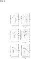
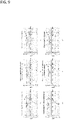
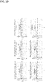
- Figures 6 to 8 show a scatter diagram plotting the rFA and the motor function recovery degree ⁇ BI
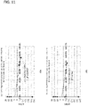
- Figures 9 to 11 show a scatter diagram plotting the rFA and the motor function recovery degree ⁇ FMA. "-" in the tables indicates that an FA beyond the threshold was not calculated.
- Diffusion kurtosis imaging (DKI) method was used to obtain an index of a diffusion image capable of predicting the therapeutic effect of cell administration in a patient with cerebral infarction precisely and without relying on the experience of the measurer or the like.
- DKI diffusion kurtosis imaging
- ROI was used for the left and right precentral gyri, which are sites where the corticospinal tract runs, to measure the main DKI indexes of these regions by IBASPM Atlas (http://www.thomaskoenig.ch/Lester/ibaspm.htm) and JHU white-matter tractography Atlas (http://cmrm.med.jhmi.edu/).
- the number of days from the onset of cerebral infarction and the MRI imaging was used as a suppressor variable.
- Table 5 shows the correlation between the diffusion image data prior to the cell administration and the function recovery degree. [Table 5] Index FA ROI Precentral gyrus NIHSS r -0.23 P 0.70 mRS r -0.89 P 0.04 FIM r 0.75 P 0.14 Bl r 0.58 P 0.30
- correlation with a motor function was evaluated using indicators (modified Rankin Scale (mRS), National Institute of Health Stroke Scale (NIHSS), and functional independence measure (FIM)) other than BI and FMA.
- the motor function was evaluated as of the case registration (after 14 days from cerebral infarction), 7 days before the cell administration (after 50 days in average from cerebral infarction), and after 1 year from the cell administration.
- Table 6 shows the result.
- DTI FA value After 14 days from onset 7 days before cell administration (2 months from onset) After 1 year from administration
- Precentral gyrus Precentral gyrus
- Precentral gyrus Precentral gyrus
- NIHSS r 0.056 0.077 0.447 p 0.91 0.87 0.31 mRS r 0.144 0.204 0.403 p 0.92 0.66 0.37
- the present disclosure is useful in industries of therapy, diagnosis or the like of central nervous diseases.
- the present disclosure can be utilized in the fields of regeneration medical treatment, development of cell medicaments, or the like.
Landscapes
- Health & Medical Sciences (AREA)
- Life Sciences & Earth Sciences (AREA)
- Physics & Mathematics (AREA)
- Nuclear Medicine, Radiotherapy & Molecular Imaging (AREA)
- Engineering & Computer Science (AREA)
- General Health & Medical Sciences (AREA)
- Public Health (AREA)
- Medical Informatics (AREA)
- Radiology & Medical Imaging (AREA)
- Biomedical Technology (AREA)
- Pathology (AREA)
- Neurology (AREA)
- Surgery (AREA)
- Animal Behavior & Ethology (AREA)
- Molecular Biology (AREA)
- Veterinary Medicine (AREA)
- Heart & Thoracic Surgery (AREA)
- Biophysics (AREA)
- High Energy & Nuclear Physics (AREA)
- Neurosurgery (AREA)
- Physiology (AREA)
- Epidemiology (AREA)
- Primary Health Care (AREA)
- Signal Processing (AREA)
- Condensed Matter Physics & Semiconductors (AREA)
- General Physics & Mathematics (AREA)
- Vascular Medicine (AREA)
- Developmental Disabilities (AREA)
- Data Mining & Analysis (AREA)
- Databases & Information Systems (AREA)
- Psychology (AREA)
- Magnetic Resonance Imaging Apparatus (AREA)
- Materials For Medical Uses (AREA)
- Medicines Containing Material From Animals Or Micro-Organisms (AREA)
- Medical Treatment And Welfare Office Work (AREA)
Applications Claiming Priority (2)
| Application Number | Priority Date | Filing Date | Title |
|---|---|---|---|
| JP2019181051 | 2019-09-30 | ||
| PCT/JP2020/036854 WO2021065894A1 (ja) | 2019-09-30 | 2020-09-29 | 運動機能の状態を評価するための方法、プログラム及び装置 |
Publications (2)
| Publication Number | Publication Date |
|---|---|
| EP4040445A1 true EP4040445A1 (de) | 2022-08-10 |
| EP4040445A4 EP4040445A4 (de) | 2023-10-25 |
Family
ID=75338345
Family Applications (1)
| Application Number | Title | Priority Date | Filing Date |
|---|---|---|---|
| EP20871340.4A Pending EP4040445A4 (de) | 2019-09-30 | 2020-09-29 | Verfahren, programm und vorrichtung zum bewerten des zustandes einer motorfunktion |
Country Status (4)
| Country | Link |
|---|---|
| US (1) | US12290335B2 (de) |
| EP (1) | EP4040445A4 (de) |
| JP (3) | JP6966828B2 (de) |
| WO (1) | WO2021065894A1 (de) |
Families Citing this family (1)
| Publication number | Priority date | Publication date | Assignee | Title |
|---|---|---|---|---|
| US12511741B2 (en) * | 2023-02-10 | 2025-12-30 | Uniwersytet W Bialymstoku | Method of diagnosing degree of neuropathy of a nerve by diffusion tensor mapping |
Family Cites Families (13)
| Publication number | Priority date | Publication date | Assignee | Title |
|---|---|---|---|---|
| WO2004022156A2 (en) | 2002-09-04 | 2004-03-18 | Washington University | Methods for treating central nervous system damage |
| US8938289B2 (en) * | 2004-08-25 | 2015-01-20 | Motorika Limited | Motor training with brain plasticity |
| US8606360B2 (en) * | 2006-03-09 | 2013-12-10 | The Cleveland Clinic Foundation | Systems and methods for determining volume of activation for spinal cord and peripheral nerve stimulation |
| US8170305B2 (en) * | 2006-10-19 | 2012-05-01 | Brown University | Quantitative tract-of-interest metrics for white matter integrity based on diffusion tensor MRI data |
| JP4972751B2 (ja) | 2006-11-27 | 2012-07-11 | 株式会社日立製作所 | 神経線維束計測システム及び画像処理システム |
| JP5479115B2 (ja) * | 2010-01-07 | 2014-04-23 | 株式会社東芝 | 画像処理装置及び磁気共鳴イメージング装置 |
| JP5486933B2 (ja) * | 2010-01-15 | 2014-05-07 | 株式会社東芝 | 医用画像処理装置 |
| FR2996746B1 (fr) | 2012-10-17 | 2015-11-13 | Assist Publ Hopitaux De Paris | Procede de quantification des lesions cerebrales |
| WO2016040260A1 (en) * | 2014-09-09 | 2016-03-17 | The Trustees Of The University Of Pennsylvania | Edema invariant tractography |
| EP3302249A4 (de) | 2015-06-04 | 2019-01-16 | The Research Foundation for The State University of New York | Diagnose von leichten traumatischen hirnverletzungen |
| WO2017010864A1 (en) * | 2015-07-10 | 2017-01-19 | Erasmus University Medical Center Rotterdam | Apparatus, system and method for assisting in providing a diagnosis of a medical condition of a mammal brain as well a computer readable medium comprising a program for carrying out the method |
| TWI651688B (zh) | 2017-03-17 | 2019-02-21 | 長庚大學 | 利用磁振造影影像預測神經疾病的臨床嚴重度的方法 |
| JP6469280B1 (ja) | 2018-04-17 | 2019-02-13 | 酵素玄米Labo株式会社 | 炊飯器 |
-
2020
- 2020-09-29 EP EP20871340.4A patent/EP4040445A4/de active Pending
- 2020-09-29 US US17/763,280 patent/US12290335B2/en active Active
- 2020-09-29 JP JP2021506591A patent/JP6966828B2/ja active Active
- 2020-09-29 WO PCT/JP2020/036854 patent/WO2021065894A1/ja not_active Ceased
-
2021
- 2021-10-15 JP JP2021169636A patent/JP2022002767A/ja active Pending
-
2025
- 2025-09-02 JP JP2025145016A patent/JP2025172133A/ja active Pending
Also Published As
| Publication number | Publication date |
|---|---|
| JPWO2021065894A1 (ja) | 2021-10-21 |
| JP2022002767A (ja) | 2022-01-11 |
| WO2021065894A1 (ja) | 2021-04-08 |
| JP6966828B2 (ja) | 2021-11-17 |
| US20220346649A1 (en) | 2022-11-03 |
| JP2025172133A (ja) | 2025-11-20 |
| EP4040445A4 (de) | 2023-10-25 |
| US12290335B2 (en) | 2025-05-06 |
Similar Documents
| Publication | Publication Date | Title |
|---|---|---|
| Löuvbld et al. | Ischemic lesion volumes in acute stroke by diffusion‐weighted magnetic resonance imaging correlate with clinical outcome | |
| Peng et al. | Age-related increase of resting metabolic rate in the human brain | |
| Ashtari et al. | Attention-deficit/hyperactivity disorder: a preliminary diffusion tensor imaging study | |
| Miller et al. | Magnetic resonance imaging in central nervous system sarcoidosis | |
| Picchioni et al. | Infraslow EEG oscillations organize large-scale cortical–subcortical interactions during sleep: a combined EEG/fMRI study | |
| Lei et al. | Postoperative executive function in adult moyamoya disease: a preliminary study of its functional anatomy and behavioral correlates | |
| Mo et al. | Chemotherapy-induced changes of cerebral activity in resting-state functional magnetic resonance imaging and cerebral white matter in diffusion tensor imaging | |
| US20180045799A1 (en) | Methods and devices for optimizing magnetic resonance imaging protocols | |
| JP4454212B2 (ja) | 画像関連データ処理方法 | |
| Qi et al. | Default mode network connectivity and related white matter disruption in type 2 diabetes mellitus patients concurrent with amnestic mild cognitive impairment | |
| Stragapede et al. | Brain oscillatory activity and CT perfusion in hyper-acute ischemic stroke | |
| Han et al. | Sex-specific age-related differences in cerebrospinal fluid clearance assessed by resting-state functional magnetic resonance imaging | |
| Wang et al. | White matter abnormalities in medication-naïve adult patients with major depressive disorder: tract-based spatial statistical analysis | |
| JP2025172133A (ja) | 運動機能の状態を評価するための方法、プログラム及び装置 | |
| JP7845686B2 (ja) | 運動機能の状態を評価するための方法、プログラム及び装置 | |
| Hu et al. | Altered brain function in new onset childhood acute lymphoblastic leukemia before chemotherapy: A resting-state fMRI study | |
| Baumgartner et al. | Leg ischemia: assessment with MR angiography and spectroscopy | |
| Ando et al. | Microstructural white matter changes following gait training with Hybrid Assistive Limb initiated within 1 week of stroke onset | |
| JP5061150B2 (ja) | データ処理方法、画像データ処理装置、及びコンピュータプログラム | |
| Ajčević et al. | Wireless EEG in hyper-acute ischemic stroke: correlation between neurophysiological alterations and CTP total hypoperfused volume | |
| Joni et al. | Investigating the role of susceptibility weighted imaging for assessment of ischemic penumbra with respect to Venus blood flow in ischemic stroke patients | |
| Viallon et al. | Ischaemia-reperfusion dynamics in acute myocardial infarction experimental swine model: new insights from quantitative CMR | |
| Qiao et al. | Blood Oxygen Level–Dependent Response Changes in the Ipsilateral Primary Somatosensory Cortex and Thalamus of Patients With Moyamoya Disease During Median Nerve Electrical Stimulation | |
| Shen et al. | Diffusion-weighted imaging of the dorsal root ganglion as a quantitative biomarker reflecting the severity and prognosis of herpes zoster: a prospective magnetic resonance imaging study | |
| Xiuhua et al. | Evaluation of computed tomography perfusion imaging in a rat acute cerebral ischemia-reperfusion model |
Legal Events
| Date | Code | Title | Description |
|---|---|---|---|
| STAA | Information on the status of an ep patent application or granted ep patent |
Free format text: STATUS: THE INTERNATIONAL PUBLICATION HAS BEEN MADE |
|
| PUAI | Public reference made under article 153(3) epc to a published international application that has entered the european phase |
Free format text: ORIGINAL CODE: 0009012 |
|
| STAA | Information on the status of an ep patent application or granted ep patent |
Free format text: STATUS: REQUEST FOR EXAMINATION WAS MADE |
|
| 17P | Request for examination filed |
Effective date: 20220324 |
|
| AK | Designated contracting states |
Kind code of ref document: A1 Designated state(s): AL AT BE BG CH CY CZ DE DK EE ES FI FR GB GR HR HU IE IS IT LI LT LU LV MC MK MT NL NO PL PT RO RS SE SI SK SM TR |
|
| DAV | Request for validation of the european patent (deleted) | ||
| DAX | Request for extension of the european patent (deleted) | ||
| A4 | Supplementary search report drawn up and despatched |
Effective date: 20230922 |
|
| RIC1 | Information provided on ipc code assigned before grant |
Ipc: A61B 5/00 20060101ALI20230918BHEP Ipc: G16H 30/40 20180101ALI20230918BHEP Ipc: G16H 50/30 20180101ALI20230918BHEP Ipc: A61B 5/055 20060101ALI20230918BHEP Ipc: G16H 30/00 20180101AFI20230918BHEP |