EP4048808B1 - Détection et analyse de méthylation d'adn de mammifère - Google Patents
Détection et analyse de méthylation d'adn de mammifère Download PDFInfo
- Publication number
- EP4048808B1 EP4048808B1 EP19949899.9A EP19949899A EP4048808B1 EP 4048808 B1 EP4048808 B1 EP 4048808B1 EP 19949899 A EP19949899 A EP 19949899A EP 4048808 B1 EP4048808 B1 EP 4048808B1
- Authority
- EP
- European Patent Office
- Prior art keywords
- dna
- transposome
- transposon
- binding site
- genomic dna
- Prior art date
- Legal status (The legal status is an assumption and is not a legal conclusion. Google has not performed a legal analysis and makes no representation as to the accuracy of the status listed.)
- Active
Links
Images


Classifications
-
- C—CHEMISTRY; METALLURGY
- C40—COMBINATORIAL TECHNOLOGY
- C40B—COMBINATORIAL CHEMISTRY; LIBRARIES, e.g. CHEMICAL LIBRARIES
- C40B40/00—Libraries per se, e.g. arrays, mixtures
- C40B40/04—Libraries containing only organic compounds
- C40B40/06—Libraries containing nucleotides or polynucleotides, or derivatives thereof
-
- C—CHEMISTRY; METALLURGY
- C12—BIOCHEMISTRY; BEER; SPIRITS; WINE; VINEGAR; MICROBIOLOGY; ENZYMOLOGY; MUTATION OR GENETIC ENGINEERING
- C12N—MICROORGANISMS OR ENZYMES; COMPOSITIONS THEREOF; PROPAGATING, PRESERVING, OR MAINTAINING MICROORGANISMS; MUTATION OR GENETIC ENGINEERING; CULTURE MEDIA
- C12N15/00—Mutation or genetic engineering; DNA or RNA concerning genetic engineering, vectors, e.g. plasmids, or their isolation, preparation or purification; Use of hosts therefor
- C12N15/09—Recombinant DNA-technology
- C12N15/10—Processes for the isolation, preparation or purification of DNA or RNA
- C12N15/1034—Isolating an individual clone by screening libraries
- C12N15/1082—Preparation or screening gene libraries by chromosomal integration of polynucleotide sequences, HR-, site-specific-recombination, transposons, viral vectors
-
- C—CHEMISTRY; METALLURGY
- C12—BIOCHEMISTRY; BEER; SPIRITS; WINE; VINEGAR; MICROBIOLOGY; ENZYMOLOGY; MUTATION OR GENETIC ENGINEERING
- C12Q—MEASURING OR TESTING PROCESSES INVOLVING ENZYMES, NUCLEIC ACIDS OR MICROORGANISMS; COMPOSITIONS OR TEST PAPERS THEREFOR; PROCESSES OF PREPARING SUCH COMPOSITIONS; CONDITION-RESPONSIVE CONTROL IN MICROBIOLOGICAL OR ENZYMOLOGICAL PROCESSES
- C12Q1/00—Measuring or testing processes involving enzymes, nucleic acids or microorganisms; Compositions therefor; Processes of preparing such compositions
- C12Q1/68—Measuring or testing processes involving enzymes, nucleic acids or microorganisms; Compositions therefor; Processes of preparing such compositions involving nucleic acids
- C12Q1/6806—Preparing nucleic acids for analysis, e.g. for polymerase chain reaction [PCR] assay
Definitions
- Embodiments of the present invention relate in general to methods and compositions for sequencing of DNA in low amount, such as DNA from a few cells to 100 cells or a single cell single cell.
- the capability to perform high coverage and high precision mammalian DNA methylation studies on single-cell level DNA are essential in studies where cell-to-cell variation and population heterogeneity play a key role, such as tumor growth, stem cell reprogramming, memory formation, embryonic development, etc. This is also important when the cell samples subject to analysis are precious or rare or in minute amounts, such as when the sample is a single cell or the genome, in whole or in part, of a single cell or cell-free DNA.
- DNA needs to go through chemical or enzymatic conversion to effectively distinguish methylated cytosine, and unmethylated cytosine.
- Common conversion methods include but by limited to sodium -bisulfite, TET-assisted-pyridine-borane sequencing (TAPS), Liu et al., Nature Biotechnology (2019 ). " Bisulfite-free direct detection of 5-methylcytosine and 5-hydroxymethylcytosine at base resolution", Enzymatic-methyl seq conversion(EM-seq), Williams et al., New England Biolabs, Inc. (2019 ).
- the major challenge in all chemical or enzymatic conversion is the loss or damage of DNA that takes place concurrently with the conversion.
- the conditions necessary for complete conversions such as long incubation times, elevated temperature, high bisulfite concentration, and high oxidative or reductive environment, can lead to the degradation, DNA base damage and fragmentation of up to 90% of the incubated DNA.
- DNA purifications between or after steps during the conversion can also lead to DNA loss up to 90%.
- the extensive DNA loss and damage are problematic and even more so such as when dealing with a limited or low amount of starting DNA or even single-cell level DNA.
- Low coverage single-cell bisulfite sequencing has been achieved by directly performing bisulfite conversion on a single cell, followed by DNA amplification. Guo, H., eta. (2013).
- carrier DNA such as sonicated lambda DNA size of 100bp to 4kb.
- mixing carrier DNA with targeted DNA results in a mixture of samples that could not be later distinguished, causing difficulties or even failure in the following methylation detection and analysis.
- In vitro transposition has been used in certain applications of DNA amplification. In such methods, target DNA is simultaneously fragmented and tagged producing fragments tagged with desired DNA sequences for downstream processing.
- in vitro transposition has been utilized in the Nextera technology of Illumina, Inc, to simultaneously fragment DNA and tag each fragment with appropriate sequences for next-generation sequencing ( US20110287435 ).
- in vitro transposition has been used by Buenrostro et al. to profile chromatin accessibility ( Buenrostro, J. D., Wu, B., Litzenburger, U. M., Ruff, D., Gonzales, M. L., Snyder, M. P., ...
- WO 2018/165366 A1 refers to a method for making an amplified methylome by extending fragments and treating the extended fragments with a methyl transferase and source of methyl groups to transform hemi-methylated double stranded DNA to fully methylated double stranded DNA.
- WO 2018/217912 refers to a method for assembly of genomic DNA using multiplex end-tagging amplification of genomic fragments.
- the present disclosure provides a method for methylation status analysis of a target DNA sample present in a low amount, such as DNA from a plurality of cells or a single cell, including genomic DNA fragmentation using a plurality of transposomes where each member of the plurality of transposomes includes two transposon nucleic acid sequences having priming site sequences.
- the transposon nucleic acid sequences having priming site sequences that bind to the transposase may be methylated or not methylated for all cytosine depending on whether the conversion chemistry converts methylated cytosine or unmethylated cytosine.
- the DNA fragment produced by the transposomes may have a cytosine and/or a methyl cytosine present therein.
- the priming site sequence of each transposon nucleic acid sequence of the transposome is the same.
- the priming site sequence of each transposon nucleic acid sequence of the transposome is different.
- each member of the plurality of transposomes may include a unique and/or different priming site sequence.
- each member of the plurality of transposomes may include two unique and/or different priming site sequences, one for each transposon in the transposome.
- a set of transposomes are provided having a unique primer binding site sequence (or two unique and/or different priming site sequences) associated therewith and which can be used to distinguish transposomes.
- the primer binding site sequences of the transposons within the transposome may be the same or may be different or nonidentical.
- the primer binding site sequences of the transposomes in two adjacent transposomes attached to a target nucleic acid sequence and used to make a fragment are nonidentical, such as with a high probability.
- the transposons may be referred to as multiplex transposons to the extent that each transposon within a transposome has a different priming site sequence.
- the priming sites within a library of transposomes may be referred to as multiplex priming sites to the extent that each transposome has a priming site that is different or nonidentical or unique from other priming sites within other transposomes within the set of transposomes.
- the method provides the step of binding transposomes from a library or plurality of transposomes along a target nucleic acid sequence such that adjacent transposomes have different primer binding site sequences. In this manner, the ends of the fragmentation site will be tagged with different primer binding site sequences.
- the multiplex end-tagging amplification method described herein uses multiple priming sequences to create target DNA fragments tagged by different sequences at the two ends.
- the multiplex end-tagging amplification method can be carried out whether the two transposon sequences within a transposome are the same or are different, as long as two adjacent transposome, i.e., directly adjacent so as to form a fragment sequence, carry different transposon primer binding site sequences where the fragment has different primer binding site sequences at each end.
- a transposition method is used to fragment and tag a genomic nucleic acid sequence, such as a genomic nucleic acid sequence of a single cell.
- N the number of unique priming site sequences
- the chance of a DNA fragment tagged by the same transposon sequence, namely the loss rate is 1/N.
- the present disclosure therefore, provides a method for altering the number of unique priming site sequences, i.e. the number N, to control the loss rate. For example, when there are 20 different transposon sequences, for use with DNA obtained from a human single cell, the loss rate is 1/20 or 5%.
- the method described herein creating a plurality of fragments uses a set of transposomes where each member of the set of transposomes has one or two different primer binding site sequences and where each member of the set of transposomes has one or two unique or different priming binding sites compared to each other member of the set of transposomes, such as with a high probability.
- adjoining ends of fragments are barcoded with different and/or unique end barcode sequences during the fragmentation process to create fragments having unique barcode sequences (priming site sequences) on each end.
- opposite ends of fragments are barcoded with different and/or unique end barcode sequences with a high probability during the fragmentation process to create fragments having different barcode sequences (priming site sequences) on each end.
- the two opposite ends of fragments are barcoded with different and/or unique end barcode sequences during the fragmentation process to create fragments having unique barcode sequences (priming site sequences) on each end.
- a transposome library is used to make fragments of genomic DNA in aqueous media where a unique barcode sequence is inserted or attached to each end of the genomic DNA at a site which has been cut by the transposase of the transposome. Since each transposome has one or two different and/or unique priming site sequences compared to other transposome members of the set or plurality or library, each fragment will have unique priming site sequences (barcode sequences) on each end. According to one aspect, each fragment will include one or more cytosine or one or more methylcytosine.
- the methods described herein utilize carrier DNA during a chemical conversion step that converts cytosine to uracil to protect the sample DNA from being lost or damaged during the chemical conversion step.
- cytosine deaminases include those of the ABOPEC family, such as APOBEC-seq or APOBEC3A.
- the APOBEC family members are cytidine deaminases that convert cytosine to uracil while maintaining 5-hydroxymethyl cytosine, i.e. without altering 5-hydroxymethyl cytosine.
- 5-methyl cytosine may be converted to 5-hydroxymethyl cytosine by oxidation reaction with TET enzyme.
- the reagent is not bisulfite or excludes bisulfite or the reagent converts cytosine to uracil with the proviso that the reagent is not bisulfite.
- the carrier DNA can then be removed or the converted fragments can be amplified without also amplifying the carrier DNA resulting in amplified fragmented DNA.
- transposomes to create fragments with primers or adapters at each end as described herein (and before the addition of carrier DNA) adds barcode and PCR adapters to targeted DNA such that targeted DNA could be sufficiently distinguished from carrier DNA.
- adapter-ligated DNA is amplified while carrier DNA is not amplified.
- Carrier DNA becomes single stranded DNA, i.e. ssDNA, and is removed from the mixture, resulting in pure amplified targeted DNA.
- the present disclosure contemplates fragmenting genomic DNA into a plurality of fragments, such as 5 or more fragments, 10 or more fragments, 100 or more fragments, 1000 or more fragments, 10,000 or more fragments, 100,000 or more fragments, 1,000,000 or more fragments, or 10,000,000 or more fragments using a transposome library as described herein.
- a transposome library includes 5 to 10 types or kinds of transposome members, 10 to 100 types or kinds of transposome members, 100 or more types or kinds of transposome members, 1000 or more types or kinds of transposome members, 10,000 or more types or kinds of transposome members, 100,000 or more types or kinds of transposome members, 1,000,000 or more types or kinds of transposome members, or 10,000,000 or more types or kinds of transposome members or between 5 and 50 types or kinds of transposome members.
- each transposome includes two transposases and two transposon DNA.
- Each of the two transposon DNA of the transposome includes a transposase binding site and a primer binding site sequence.
- the transposon DNA includes a single transposase binding site and a unique primer binding site sequence.
- Each transposon DNA is a separate nucleic acid bound to a transposase at the transposase binding site.
- the transposome is a dimer of two separate transposases each bound to its own transposon DNA. The dimer may have the same primer binding site sequences on each transposon or may have different primer binding site sequences on each transposon.
- the transposome includes two separate and individual transposon DNA, each bound to its own corresponding transposase. According to one aspect, the transposome includes only two transposases and only two transposon DNA. According to one aspect, the two transposon DNA as part of the transposome are separate, individual or non-linked transposon DNA, each bound to its own corresponding transposase.
- each transposome member of the library includes a unique and different priming site sequence.
- the same unique and different priming site sequence may be present on each transposon DNA of the transposome or a different unique and different priming site sequence may be present on each transposon DNA of the transposome.
- each transposome includes a unique and different priming site sequence that is unique and different from the priming site sequences of any other transposome in the transposome library.
- the transposome library may include transposome members that have the same priming site sequences as other transposome members, although the probability is relatively small or insignificant.
- the transposome library may be considered to be a subset of the prepared collection of transposomes, where the subset includes only transposomes with a unique and different priming site sequence, as the objective is to fragment genomic DNA where each fragment cut site has different priming site sequences. It is to be understood that the objective of fragmenting genomic DNA where each fragment cut site has a different priming site sequence may be accomplished where adjacent transposomes each have a unique and different priming site sequence, though it may be shared by the two transposons of the transposome.
- each fragment cut site has a different priming site sequence
- adjacent transposomes each have two unique and different priming site sequences, where each transposon of the transposome has a unique and different priming site sequences.
- the transposome library may include transposome members that have the same two priming site sequences, ie., the priming site sequences are identical or the same, although this priming site sequence is unique compared to any other transposon DNA of tranposome members of the transposome library.
- each transposome member is made separately by mixing transposase and the transposon DNA which contain the unique priming site sequence. All the transpome members are then be mixed together to form the transposome library.
- a transposome library is prepared by mixing all transposon sequences together with transposase to form transposome. In this method, most transposomes have different transposon sequences, but the chance of a transposome carrying the same transposon sequences is 1/N.
- each type of transposon sequence is mixed with transposase separately, and then all the tranposome are mixed to form the transposome library. In this method, all the tranposomes will have same transposon sequences.
- the number of unique and/or different priming site sequences is between 5 and 50, 10 and 50, 15 and 45, 20 and 40 or between 1 and 1,000, 1 and 10,000, 1 and 100,000, 1 and 1,000,000 or 1 and 10,000,000.
- the number of cut sites in the genomic DNA is determined or tuned by the concentration of transposomes, with the higher concentration resulting in a higher number of cut sites and a lower concentration resulting in a lower number of cut sites.
- the number of transposomes and associated different and/or unique priming site sequences is selected such that substantially all of the cut sites have two different and/or unique priming site sequences.
- more than 90% of the cut sites have two different and/or unique priming site sequences
- more than 95% of the cut sites have two different and/or unique priming site sequences
- 96% of the cut sites have two different and/or unique priming site sequences
- 97% of the cut sites have two different and/or unique priming site sequences
- 98% of the cut sites have two different an/or unique priming site sequences
- 99% of the cut sites have two different and/or unique priming site sequences
- 99.5% of the cut sites have two different and/or unique priming site sequences
- 100% of the cut sites have two different and/or unique priming site sequences.
- the transposome library is then used to cut the genomic DNA and each transposome inserts or attaches its priming site sequences in each of the transposon DNA at the ends of the cut site.
- the cut site will have a unique and different priming site sequence at each end of the site, i.e. the priming site sequences inserted will be different.
- a plurality or most or substantially all fragments produced by the transposome library have a different and/or unique priming site sequence on each end, i.e. opposite ends, of the fragment, insofar as adjacent transposomes have unique and different priming site sequences compared to each other.
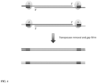
- the transposase can then be removed from each fragment followed by a gap fill-in step, by for example, a polymerase extension step.
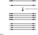
- the resulting double stranded nucleic acid fragment sequence can then be amplified, for example using multiplex PCR amplification.
- the fragments can then be sequenced and the sequence of the genomic DNA can be determined.
- the transposon DNA of the transposome can include sequences facilitating amplification methods, such as specific primer sequences or transcription promoter sequences which can be attached to the fragments so that the fragments can be amplified prior to sequencing, such as by PCR or RNA transcription using methods known to those of skill in the art. It is to be understood that the present disclosure contemplates different amplification methods for amplifying the fragments and different sequencing methods for sequencing the amplicons are not limited to any particular amplification or sequencing method.
- Embodiments of the present disclosure are directed to a method of multiplex end-tagging amplification of nucleic acids, such as genomic DNA, such as a low or small amount of genomic DNA or a limited amount of DNA such as a genomic sequence or genomic sequences obtained from a single cell or a plurality of cells of the same cell type or from a tissue, fluid or blood sample obtained from an individual or a substrate.
- nucleic acids such as genomic DNA, such as a low or small amount of genomic DNA or a limited amount of DNA such as a genomic sequence or genomic sequences obtained from a single cell or a plurality of cells of the same cell type or from a tissue, fluid or blood sample obtained from an individual or a substrate.
- the methods described herein can be performed in a single tube with a single reaction mixture.
- the nucleic acid sample can be within an unpurified or unprocessed lysate from a single cell.
- Nucleic acids to be subjected to the methods disclosed herein need not be purified, such as by column purification, prior to being contacted with the various reagents and under the various conditions as described herein.
- the methods described herein reduce the loss rate, i.e., loss of the original target nucleic acid so as to assist in providing substantial and uniform coverage of the entire genome of a single cell producing amplified DNA for high-throughput sequencing.
- Embodiments of the present invention relate in general to methods and compositions for making DNA fragments, for example, DNA fragments from the whole genome of a single cell which may then be subjected to amplification and sequencing methods known to those of skill in the art and as described herein.
- methods of making nucleic acid fragments described herein utilize a transposome library.
- a transposase as part of a transposome is used to create a set of double stranded genomic DNA fragments.
- the transposases have the capability to bind to transposon DNA and dimerize when contacted together, such as when being placed within a reaction vessel or reaction volume, forming a transposase/transposon DNA complex dimer called a transposome.
- Each transposon DNA of the transposome includes a double stranded transposase binding site and a first nucleic acid sequence including a priming site sequence and optionally functional sequences such as a transcription promoter site.
- the first nucleic acid sequence may be in the form of a single stranded extension.
- Each transposome of the transposome library includes a unique and different priming site sequence that are different from the priming site sequences of each remaining member of the transposome library.
- each transposome of the transposome library includes two unique and different priming site sequence that are different from the priming site sequences of each remaining member of the transposome library.
- the transposomes have the capability to randomly bind to target locations along double stranded nucleic acids, such as double stranded genomic DNA, forming a complex including the transposome and the double stranded genomic DNA.
- the transposases in the transposome cleave the double stranded genomic DNA, with one transposase cleaving the upper strand and one transposase cleaving the lower strand.
- Each of the transposon DNA in the transposome is attached to the double stranded genomic DNA at each end of the cut site, i.e. one transposon DNA of the transposome is attached to the left hand cut site and the other transposon DNA of the transposome is attached to the right hand cut site.
- transposon DNA of the transposome When the transposon DNA of the transposome each have different primer binding site sequences, the left hand cut site and the right hand cut site are "barcoded" with a different and unique barcode, i.e. priming site, sequences.
- the transposon DNA of the transposome When the transposon DNA of the transposome each have the same primer binding site sequence, the left hand cut site and the right hand cut site are "barcoded” with the same barcode, i.e. priming site, sequence.
- adjacent transposomes used to make a fragment each have a different and unique primer binding site sequence, the resulting fragment will have a different and unique primer binding site on each end of the fragment.
- a plurality of transposase/transposon DNA complex dimers i.e.
- transposomes bind to a corresponding plurality of target locations along a double stranded genomic DNA, for example, and then cleave the double stranded genomic DNA into a plurality of double stranded fragments with each fragment having transposon DNA with a different barcode sequence attached at each end of the double stranded fragment.
- the transposon DNA is attached to the double stranded genomic DNA and a single stranded gap exists between one strand of the genomic DNA and one strand of the transposon DNA.
- gap extension is carried out to fill the gap and create a double stranded connection between the double stranded genomic DNA and the double stranded transposon DNA.
- a nucleic acid sequence including the transposase binding site and the priming site sequence is attached at each end of the double stranded fragment.
- the transposase is attached to the transposon DNA which is attached at each end of the double stranded fragment.
- the transposases are removed from the transposon DNA which is attached at each end of the double stranded genomic DNA fragments.
- the double stranded genomic DNA fragments which have the transposon DNA with different priming site sequences attached at each end of the double stranded genomic DNA fragments are then gap filled and extended using the transposon DNA as a template. Accordingly, a double stranded nucleic acid extension product is produced which includes the double stranded genomic DNA fragment and a double stranded transposon DNA including a different priming site sequence at each end of the double stranded genomic DNA.
- the double stranded nucleic acid extension products including the genomic DNA fragment, the different priming site sequences at each end can be amplified using methods known to those of skill in the art to produce amplicons of the genomic DNA fragment and the different primer binding site at each end.
- PCR primer sequences and reagents can be used for amplification.
- the transposons as described herein may also include an RNA polymerase binding site for production of RNA transcripts which may then be reverse transcribed into cDNA for linear amplification.
- the double stranded nucleic acid extension products including the genomic DNA fragment and the different priming site sequences at each end can be combined with amplification reagents and the double stranded genomic nucleic acid fragment may then be amplified using methods known to those of skill in the art to produce amplicons of the double stranded genomic nucleic acid fragment.
- the amplicons can then be collected and/or purified prior to further analysis.
- the amplicons can be sequenced using methods known to those of skill in the art. Once sequenced, the sequences can be computationally analyzed to identify the genomic DNA.
- Embodiments of the present disclosure are directed to a method of amplifying DNA using multiplex end-tagging, wherein the DNA is a low or small amount of genomic DNA or a limited amount of DNA such as a genomic sequence or genomic sequences obtained from a single cell or a plurality of cells of the same cell type or from a tissue, fluid or blood sample obtained from an individual or a substrate.
- the methods described herein can be performed in a single tube to create the fragments having different and unique sequences at each end which are then amplified and sequenced using high throughput sequencing platforms known to those of skill in the art.
- the transposome fragmentation and barcoding method described herein is useful for amplifying and then sequencing of low or small or limited amounts of DNA.
- Methods described herein have particular application in biological systems or tissue samples characterized by highly heterogeneous cell populations such as tumor and neural masses.
- the methods described herein can utilize varied sources of DNA materials, including genetically heterogeneous tissues (e.g. cancers), rare and precious samples (e.g. embryonic stem cells), and non-dividing cells (e.g. neurons) and the like, as well as, sequencing platforms and genotyping methods known to those of skill in the art.
- a method of single cell whole genome amplification, sequencing and assembly includes contacting double stranded genomic DNA from a single cell with transposases, such as Tn5 transposases, each bound to a transposon DNA, wherein the transposon DNA includes a double-stranded 19 bp transposase (Tnp) binding site and a first nucleic acid sequence including a unique and different priming site sequence to form a transposase/transposon DNA complex dimer called a transposome.
- the first nucleic acid sequence may be in the form of a single stranded extension.
- the first nucleic acid sequence may be an overhang, such as a 5' overhang, wherein the overhang includes a unique and different priming site sequence.
- the overhang may include other functional sequences as desired.
- the overhang can be of any length suitable to include a priming site sequence, or other functional sequences as desired.
- the transposome bind to target locations along the double stranded genomic DNA and cleave the double stranded genomic DNA into a plurality of double stranded fragments, with each double stranded fragment having a first complex attached to an upper strand by the Tnp binding site and a second complex attached to a lower strand by the Tnp binding site.
- the transposon binding site, and therefore the transposon DNA along with the primer binding site is attached to each 5' end of the double stranded fragment.
- the Tn5 transposases are removed from the complex.
- the double stranded fragments include one or more cytosine or one or more methylcytosine and are extended along the transposon DNA to make a double stranded extension product having dissimilar or different or unique priming site sequences at each end of the double stranded extension product.
- a gap which may result from attachment of the Tn5 transposase binding site to the double stranded genomic DNA fragment may be filled.
- the gap filled double stranded extension product is then subjected to chemical treatment in the presence of carrier DNA to convert cytosine to uracil.
- the gap filled double stranded extension product is mixed with amplification reagents, and the double stranded genomic DNA fragment is amplified.
- the amplicons which include a dissimilar or different or unique priming site sequence (which may function as a barcode sequence) at each end, are sequenced using, for example, high-throughput sequencing methods known to those of skill in the art.
- embodiments are directed to methods for the amplification, sequencing and assembly of substantially the entire genome without loss of representation of specific sites (herein defined as "whole genome amplification").
- whole genome amplification comprises amplification of substantially all fragments or all fragments of a genomic library.
- substantially entire or substantially all refers to about 80%, about 85%, about 90%, about 95%, about 97%, or about 99% of all sequences in a genome.
- biological sample is intended to include, but is not limited to, tissues, cells, biological fluids and isolates thereof, isolated from a subject, as well as tissues, cells and fluids present within a subject.
- in vitro has its art recognized meaning, e.g., involving purified reagents or extracts, e.g., cell extracts.
- in vivo also has its art recognized meaning, e.g., involving living cells, e.g., immortalized cells, primary cells, cell lines, and/or cells in an organism.
- the terms “complementary” and “complementarity” are used in reference to nucleotide sequences related by the base-pairing rules.
- sequence 5'-AGT-3' is complementary to the sequence 5'-ACT-3'.
- Complementarity can be partial or total. Partial complementarity occurs when one or more nucleic acid bases is not matched according to the base pairing rules. Total or complete complementarity between nucleic acids occurs when each and every nucleic acid base is matched with another base under the base pairing rules. The degree of complementarity between nucleic acid strands has significant effects on the efficiency and strength of hybridization between nucleic acid strands.
- hybridization refers to the pairing of complementary nucleic acids. Hybridization and the strength of hybridization (i.e., the strength of the association between the nucleic acids) is impacted by such factors as the degree of complementary between the nucleic acids, stringency of the conditions involved, the T m of the formed hybrid, and the G:C ratio within the nucleic acids. A single molecule that contains pairing of complementary nucleic acids within its structure is said to be “self-hybridized.”
- T m refers to the melting temperature of a nucleic acid.
- the melting temperature is the temperature at which a population of double-stranded nucleic acid molecules becomes half dissociated into single strands.
- stringency refers to the conditions of temperature, ionic strength, and the presence of other compounds such as organic solvents, under which nucleic acid hybridizations are conducted.
- Low stringency conditions when used in reference to nucleic acid hybridization, comprise conditions equivalent to binding or hybridization at 42 °C in a solution consisting of 5x SSPE (43.8 g/l NaCl, 6.9 g/l NaH 2 PO 4 (H 2 O) and 1.85 g/l EDTA, pH adjusted to 7.4 with NaOH), 0.1% SDS, 5x Denhardt's reagent (50x Denhardt's contains per 500 ml: 5 g Ficoll (Type 400, Pharmacia), 5 g BSA (Fraction V; Sigma)) and 100 mg/ml denatured salmon sperm DNA followed by washing in a solution comprising 5x SSPE, 0.1% SDS at 42 °C when a probe of about 500 nucleotides in length is employed.
- 5x SSPE 43.8 g/l NaCl, 6.9 g/l NaH 2 PO 4 (H 2 O) and 1.85 g/l EDTA, pH adjusted to 7.4 with NaOH
- “Medium stringency conditions,” when used in reference to nucleic acid hybridization, comprise conditions equivalent to binding or hybridization at 42 °C in a solution consisting of 5x SSPE (43.8 g/l NaCl, 6.9 g/l NaH 2 PO 4 (H 2 O) and 1.85 g/l EDTA, pH adjusted to 7.4 with NaOH), 0.5% SDS, 5x Denhardt's reagent and 100 mg/ml denatured salmon sperm DNA followed by washing in a solution comprising 1.0x SSPE, 1.0% SDS at 42 °C when a probe of about 500 nucleotides in length is employed.
- High stringency conditions when used in reference to nucleic acid hybridization, comprise conditions equivalent to binding or hybridization at 42 °C in a solution consisting of 5x SSPE (43.8 g/l NaCl, 6.9 g/l NaH 2 PO 4 (H 2 O) and 1.85 g/l EDTA, pH adjusted to 7.4 with NaOH), 0.5% SDS, 5x Denhardt's reagent and 100 mg/ml denatured salmon sperm DNA followed by washing in a solution comprising 0.1x SSPE, 1.0% SDS at 42 °C when a probe of about 500 nucleotides in length is employed.
- genomic as used herein is defined as the collective gene set carried by an individual, cell, or organelle.
- genomic DNA as used herein is defined as DNA material comprising the partial or full collective gene set carried by an individual, cell, or organelle.
- nucleoside refers to a molecule having a purine or pyrimidine base covalently linked to a ribose or deoxyribose sugar.
- exemplary nucleosides include adenosine, guanosine, cytidine, uridine and thymidine.
- Additional exemplary nucleosides include inosine, 1-methyl inosine, pseudouridine, 5,6-dihydrouridine, ribothymidine, 2N-methylguanosine and 2,2N,N-dimethylguanosine (also referred to as "rare" nucleosides).
- nucleotide refers to a nucleoside having one or more phosphate groups joined in ester linkages to the sugar moiety.
- exemplary nucleotides include nucleoside monophosphates, diphosphates and triphosphates.
- polynucleotide oligonucleotide
- nucleic acid molecule are used interchangeably herein and refer to a polymer of nucleotides, either deoxyribonucleotides or ribonucleotides, of any length joined together by a phosphodiester linkage between 5' and 3' carbon atoms.
- Polynucleotides can have any three-dimensional structure and can perform any function, known or unknown.
- polynucleotides a gene or gene fragment (for example, a probe, primer, EST or SAGE tag), exons, introns, messenger RNA (mRNA), transfer RNA, ribosomal RNA, ribozymes, cDNA, recombinant polynucleotides, branched polynucleotides, plasmids, vectors, isolated DNA of any sequence, isolated RNA of any sequence, nucleic acid probes and primers.
- a polynucleotide can comprise modified nucleotides, such as methylated nucleotides and nucleotide analogs. The term also refers to both double- and single-stranded molecules.
- any embodiment of this invention that comprises a polynucleotide encompasses both the double-stranded form and each of two complementary single-stranded forms known or predicted to make up the double-stranded form.
- a polynucleotide is composed of a specific sequence of four nucleotide bases: adenine (A); cytosine (C); guanine (G); thymine (T); and uracil (U) for thymine when the polynucleotide is RNA.
- A adenine
- C cytosine
- G guanine
- T thymine
- U uracil
- polynucleotide sequence is the alphabetical representation of a polynucleotide molecule. This alphabetical representation can be input into databases in a computer having a central processing unit and used for bioinformatics applications such as functional genomics and homology searching.
- DNA DNA molecule
- deoxyribonucleic acid molecule refers to a polymer of deoxyribonucleotides.
- DNA can be synthesized naturally (e.g., by DNA replication). RNA can be post-transcriptionally modified. DNA can also be chemically synthesized. DNA can be single-stranded (i.e., ssDNA) or multi-stranded (e.g., double stranded, i.e., dsDNA).
- nucleotide analog refers to a non-standard nucleotide, including non-naturally occurring ribonucleotides or deoxyribonucleotides.
- nucleotide analogs are modified at any position so as to alter certain chemical properties of the nucleotide yet retain the ability of the nucleotide analog to perform its intended function.
- positions of the nucleotide which may be derivitized include the 5 position, e.g., 5-(2-amino)propyl uridine, 5-bromo uridine, 5-propyne uridine, 5-propenyl uridine, etc.; the 6 position, e.g., 6-(2-amino) propyl uridine; the 8-position for adenosine and/or guanosines, e.g., 8-bromo guanosine, 8-chloro guanosine, 8-fluoroguanosine, etc.
- 5 position e.g., 5-(2-amino)propyl uridine, 5-bromo uridine, 5-propyne uridine, 5-propenyl uridine, etc.
- the 6 position e.g., 6-(2-amino) propyl uridine
- the 8-position for adenosine and/or guanosines e.g
- Nucleotide analogs also include deaza nucleotides, e.g., 7-deaza-adenosine; O- and N-modified (e.g., alkylated, e.g., N6-methyl adenosine, or as otherwise known in the art) nucleotides; and other heterocyclically modified nucleotide analogs such as those described in Herdewijn, Antisense Nucleic Acid Drug Dev., 2000 Aug. 10(4):297-310 .
- Nucleotide analogs may also comprise modifications to the sugar portion of the nucleotides.
- the 2' OH-group may be replaced by a group selected from H, OR, R, F, Cl, Br, I, SH, SR, NH 2 , NHR, NR 2 , COOR, or OR, wherein R is substituted or unsubstituted C 1 -C 6 alkyl, alkenyl, alkynyl, aryl, etc.Other possible modifications include those described in U.S. Pat. Nos. 5,858,988 , and 6,291,438 .
- the phosphate group of the nucleotide may also be modified, e.g., by substituting one or more of the oxygens of the phosphate group with sulfur (e.g., phosphorothioates), or by making other substitutions which allow the nucleotide to perform its intended function such as described in, for example, Eckstein, Antisense Nucleic Acid Drug Dev. 2000 Apr. 10(2):117-21 , Rusckowski et al. Antisense Nucleic Acid Drug Dev. 2000 Oct. 10(5):333-45 , Stein, Antisense Nucleic Acid Drug Dev. 2001 Oct. 11(5): 317-25 , Vorobjev et al. Antisense Nucleic Acid Drug Dev. 2001 Apr.
- Nucleic acids processed by methods described herein may be DNA and they may be obtained from any useful source, such as, for example, a human sample.
- a double stranded DNA molecule is further defined as comprising a genome, such as, for example, one obtained from a sample from a human.
- the sample may be any sample from a human, such as blood, serum, plasma, cerebrospinal fluid, cheek scrapings, nipple aspirate, biopsy, semen (which may be referred to as ejaculate), urine, feces, hair follicle, saliva, sweat, immunoprecipitated or physically isolated chromatin, and so forth.
- the sample comprises a single cell.
- the sample includes only a single cell.
- the DNA sample is genomic DNA, micro dissected chromosome DNA, yeast artificial chromosome (YAC) DNA, plasmid DNA, cosmid DNA, phage DNA, P1 derived artificial chromosome (PAC) DNA, or bacterial artificial chromosome (BAC) DNA, mitochondrial DNA, chloroplast DNA, forensic sample DNA, or other DNA from natural or artificial sources to be tested.
- the DNA sample is mammalian DNA, plant DNA, yeast DNA, viral DNA, or prokaryotic DNA.
- the DNA sample is obtained from a human, bovine, porcine, ovine, equine, rodent, avian, fish, shrimp, plant, yeast, virus, or bacteria.
- the DNA sample is genomic DNA.
- cells are identified and then a single cell or a plurality of cells is isolated.
- Cells within the scope of the present disclosure include any type of cell where understanding the DNA content is considered by those of skill in the art to be useful.
- a cell according to the present disclosure includes a cancer cell of any type, hepatocyte, oocyte, embryo, stem cell, iPS cell, ES cell, neuron, erythrocyte, melanocyte, astrocyte, germ cell, oligodendrocyte, kidney cell and the like.
- the methods of the present invention are practiced with the cellular DNA from a single cell.
- a plurality of cells includes from about 2 to about 1,000,000 cells, about 2 to about 10 cells, about 2 to about 100 cells, about 2 to about 1,000 cells, about 2 to about 10,000 cells, about 2 to about 100,000 cells, about 2 to about 10 cells or about 2 to about 5 cells.
- a "single cell” refers to one cell.
- Single cells useful in the methods described herein can be obtained from a tissue of interest, or from a biopsy, blood sample, or cell culture. Additionally, cells from specific organs, tissues, tumors, neoplasms, or the like can be obtained and used in the methods described herein. Furthermore, in general, cells from any population can be used in the methods, such as a population of prokaryotic or eukaryotic single celled organisms including bacteria or yeast.
- a single cell suspension can be obtained using standard methods known in the art including, for example, enzymatically using trypsin or papain to digest proteins connecting cells in tissue samples or releasing adherent cells in culture, or mechanically separating cells in a sample.
- Single cells can be placed in any suitable reaction vessel in which single cells can be treated individually. For example a 96-well plate, such that each single cell is placed in a single well.
- FACS fluorescence activated cell sorting
- flow cytometry cytometry
- micromanipulation e.g. the Quixell TM cell transfer system from Stoelting Co.
- Semi-automated cell pickers e.g. the Quixell TM cell transfer system from Stoelting Co.
- Individual cells can, for example, be individually selected based on features detectable by microscopic observation, such as location, morphology, or reporter gene expression.
- a combination of gradient centrifugation and flow cytometry can also be used to increase isolation or sorting efficiency.
- the cell is lysed to release cellular contents including DNA, using methods known to those of skill in the art.
- the cellular contents are contained within a vessel or a collection volume.
- cellular contents such as genomic DNA
- Lysis can be achieved by, for example, heating the cells, or by the use of detergents or other chemical methods, or by a combination of these.
- any suitable lysis method known in the art can be used. For example, heating the cells at 72°C for 2 minutes in the presence of Tween-20 is sufficient to lyse the cells.
- cells can be heated to 65°C for 10 minutes in water ( Esumi et al., Neurosci Res 60(4):439-51 (2008 )); or 70°C for 90 seconds in PCR buffer II (Applied Biosystems) supplemented with 0.5% NP-40 ( Kurimoto et al., Nucleic Acids Res 34(5):e42 (2006 )); or lysis can be achieved with a protease such as Proteinase K or by the use of chaotropic salts such as guanidine isothiocyanate ( U.S. Publication No. 2007/0281313 ).
- a protease such as Proteinase K
- chaotropic salts such as guanidine isothiocyanate
- Amplification of genomic DNA according to methods described herein can be performed directly on cell lysates, such that a reaction mix can be added to the cell lysates.
- the cell lysate can be separated into two or more volumes such as into two or more containers, tubes or regions using methods known to those of skill in the art with a portion of the cell lysate contained in each volume container, tube or region.
- Genomic DNA contained in each container, tube or region may then be amplified by methods described herein or methods known to those of skill in the art.
- a nucleic acid used in the invention can also include native or non-native bases.
- a native deoxyribonucleic acid can have one or more bases selected from the group consisting of adenine, thymine, cytosine or guanine and a ribonucleic acid can have one or more bases selected from the group consisting of uracil, adenine, cytosine or guanine.
- Exemplary non-native bases that can be included in a nucleic acid, whether having a native backbone or analog structure include, without limitation, inosine, xathanine, hypoxathanine, isocytosine, isoguanine, 5-methylcytosine, 5-hydroxymethyl cytosine, 2-aminoadenine, 6-methyl adenine, 6-methyl guanine, 2-propyl guanine, 2-propyl adenine, 2-thioLiracil, 2-thiothymine, 2- thiocytosine, 15 - halouracil, 15 -halocytosine, 5-propynyl uracil, 5-propynyl cytosine, 6-azo uracil, 6-azo cytosine, 6-azo thymine, 5-uracil, 4-thiouracil, 8-halo adenine or guanine, 8- amino adenine or guanine, 8-thiol adenine or guanine,
- the present invention is based in part on methods for making nucleic acid fragment templates, such as from DNA or genomic DNA, using a transposase or transposome to fragment the original or starting nucleic acid sequence, such as genomic DNA, and to attach a different priming site sequence to each end of a cut or fragmentation site to thereby produce a set of fragments with each member of the set having two unique and different priming site sequences.
- the nucleic acid fragment templates are amplified to produce amplicons.
- the amplicons of the nucleic acid fragment templates may be collected and sequenced. The collected amplicons form a library of amplicons of the fragments of the original nucleic acid, such as genomic DNA.
- methods are described of making nucleic acid fragments using an enzyme such as Tn5. Such methods are known in the art and include those practiced using the illumina Nextera kit. According to one exemplary aspect, methods described herein utilize a transposome library and a method referred to as "tagmentation" to the extent that fragments are created from a larger dsDNA sequence where the fragments are tagged with primers to be used in single primer extension and amplification.
- a genomic DNA such as genomic nucleic acid obtained from a lysed single cell
- a plurality or library of transposomes is used to cut the genomic DNA into double stranded fragments.
- Each transposome of the plurality or library is a dimer of a transposase bound to a transposon DNA, i.e. each transposome includes two separate transposon DNA.
- Each transposon DNA of a transposome includes a transposase binding site and a primer binding site sequence.
- the primer binding site sequence is unique to the transposome.
- the priming site sequence of each transposon of a transposome could be unique and/or different.
- the priming site sequence of each transposon of a transposome could be the same.
- the majority of the transposome has two transposon DNA that has different priming site sequences and only a small fraction of the transposome has two transposon DNA that has the same priming site sequence.
- the priming site sequence of the two transposon DNA of each transposome member can be the same, but the priming site sequence or sequences of the transposon DNA from different transposome members are unique and different.
- the transposon nucleic acid sequences having priming site sequences that bind to the transposase may be methylated at every cytosine so the priming site sequence is not changed after cytosine conversion required for methylation detection.
- the priming site sequences of each transposon DNA of a transposome is unique and different.
- the priming site sequence or sequences of the transposon DNA of a transposome is unique and different from the remaining members of the transposome plurality or library.
- each transposome of the plurality or library of transposomes has its own unique and different priming site sequences which are different from the remaining members of the transposome plurality or library and may have two unique and different priming site sequences which are different from the remaining members of the transposome plurality or library.
- the transposon DNA becomes attached to the upper and lower strands of each double stranded fragment at each cut or fragmentation site.
- the cut or fragmentation site is tagged with different priming site sequences. Since the priming site sequence may be the same for each transposon DNA, the cut or fragmentation site is tagged with the same priming site sequence. Where adjacent transposomes used to generate a fragment each have different primer binding site sequences associated therewith, the fragment has different primer binding site sequences at each end of the fragment. Accordingly, the fragment will have two unique and different primer binding site sequences.
- each transposome has its own unique and/or different priming site sequence associated therewith (and may have two unique and/or different priming site sequences associated therewith), and a library of transposomes are used to create many cut or fragmentation sites, each cut or fragmentation site will have a different and unique priming site sequence attached at either end of the cut site and each fragment will have different and/or unique priming site sequences on each end of the fragment. Accordingly, many fragments from the original nucleic acid sequence are created by the library of transposomes with each fragment having a dissimilar priming site sequence at each end of the fragment. The double stranded fragments are then processed to fill gaps. The fragments are amplified using suitable amplification reagents, such as a primer sequences, DNA polymerase and nucleotides for PCR amplification and are sequenced using methods known to those of skill in the art.
- suitable amplification reagents such as a primer sequences, DNA polymerase and nucleotides for PCR amplification and
- an exemplary transposon system includes Tn5 transposase, Mu transposase, Tn7 transposase or IS5 transposase and the like.
- Other useful transposon systems are known to those of skill in the art and include Tn3 transposon system (see Maekawa, T., Yanagihara, K., and Ohtsubo, E. (1996), A cell-free system of Tn3 transposition and transposition immunity, Genes Cells 1, 1007-1016 ), Tn7 transposon system (see Craig, N.L. (1991), Tn7: a target site-specific transposon, Mol. Microbiol.
- Tn10 tranposon system see Chalmers, R., Sewitz, S., Lipkow, K., and Crellin, P. (2000), Complete nucleotide sequence of Tn10, J. Bacteriol 182, 2970-2972
- Piggybac transposon system see Li, X., Burnight, E.R., Cooney, A.L., Malani, N., Brady, T., Sander, J.D., Staber, J., Wheelan, S.J., Joung, J.K., McCray, P.B., Jr., et al. (2013), PiggyBac transposase tools for genome engineering, Proc. Natl. Acad. Sci.
- Tn5 transposition systems are described and are available to those of skill in the art. See Goryshin, I.Y. and W.S. Reznikoff, Tn5 in vitro transposition. The Journal of biological chemistry, 1998. 273(13): p. 7367-74 ; Davies, D.R., et al., Three-dimensional structure of the Tn5 synaptic complex transposition intermediate. Science, 2000. 289(5476): p. 77-85 ; Goryshin, I.Y., et al., Insertional transposon mutagenesis by electroporation of released Tn5 transposition complexes. Nature biotechnology, 2000. 18(1): p. 97-100 and Stier-White, M., I.
- Kits utilizing a Tn5 transposition system for DNA library preparation and other uses are known. See Adey, A., et al., Rapid, low-input, low-bias construction of shotgun fragment libraries by high-density in vitro transposition. Genome biology, 2010. 11(12): p. R119 ; Marine, R., et al., Evaluation of a transposase protocol for rapid generation of shotgun high-throughput sequencing libraries from nanogram quantities of DNA. Applied and environmental microbiology, 2011.
- the transposon may include one or more methylated cytosines, such as methylated cytosine adapters or primers as described herein.
- the gap filling step includes using methylated dCTP instead of dCTP in the dNTP mix so as to create fully methylated double stranded adapters.
- the gap filled fragments are then combined with carrier DNA.
- Carrier DNA can be any dsDNA fragments having a length between 100 base pairs (bp) to 4 kilo base pairs.
- the carrier DNA may be a DNA type that is different from the target DNA.
- the carrier DNA may be a DNA type that is the same as the target DNA.
- the carrier DNA is sonicated lambda DNA.
- carrier DNA does not include Illumina sequencing adapters.
- the carrier DNA serves to protect the target DNA from the harsh conditions of chemical treatment and to reduce damage to the target DNA or loss of the target DNA.
- exemplary non-bisulfite conversion utilizes a strong oxidation reagent Fe(II), which will cause DNA damage by producing hydroxyl radicals.
- the carrier DNA which is added to the reaction medium in an amount that is 100 to 10000 times (e.g., 100 to 1000 times) more in amount than the sample DNA, serves to occupy excess hydroxyl radicals and the prevent or limit their interaction with the target DNA.
- 20ng sonicated lambda carrier DNA is provided for a 6pg sample DNA from a single cell.
- the conversion reaction uses a specialized buffer and purifications (buffer exchange while preserving DNA) before or even in between different steps.
- a purification step to remove chemical reagents may be required before PCR amplification.
- Two or three purification steps may be carried out as a result of the cytosine conversion process.
- Each DNA purification step can result in 50% - 90% DNA loss if the input DNA amount is low, such as DNA from a single cell. But if carrier DNA is added to the sample DNA, such as 20ng lambda DNA added to 6pg sample DNA, the DNA purification will only cause a 10% loss, resulting in the preservation of 90% of the sample DNA although mixed with carrier DNA. According to the present disclosure, efficiency of the DNA purification increases as the amount of input DNA increases.
- the reaction medium including the gap-filled double stranded segments and carrier DNA can be purified by DNA spin-columns or beads-based DNA purification, or other purification methods known to those of skill in the art, before DNA conversion required for methylation detection.
- the reaction medium can proceed directly to chemical conversion.
- the gap filled fragments combined with carrier DNA proceeds to mix with chemical reagents that chemically change a cytosine to a uracil.
- chemical reagents are described in US 2013/0244237 and may be available from New England Biolabs.
- Other enzymatic reagents will become apparent to those of skill in the art based on the present disclosure.
- the reagent is not bisulfite or excludes bisulfite or the reagent converts cytosine to uracil with the proviso that the reagent is not bisulfite.
- the reaction medium including the chemically converted fragments can be purified by DNA spin-columns or beads-based DNA purification, or other purification methods known to those of skill in the art, before amplification.
- the reaction medium can proceed directly to amplification.
- amplification of only the target fragments is carried out by using primers that target the adapter sequences incorporated into the fragments by the transposomes.
- Carrier DNA is not amplified and is becomes ssDNA due to denaturation.
- DNA to be amplified refers to a process by which extra or multiple copies of a particular polynucleotide are formed.
- DNA to be amplified may be obtained from a single cell or a small population of cells.
- Methods described herein allow DNA to be amplified from any species or organism in a reaction mixture, such as a single reaction mixture carried out in a single reaction vessel.
- methods described herein include sequence independent amplification of DNA from any source including but not limited to human, animal, plant, yeast, viral, eukaryotic and prokaryotic DNA.
- Microdroplets may be formed as an emulsion of an oil phase and an aqueous phase.
- An emulsion may include aqueous droplets or isolated aqueous volumes within a continuous oil phase.
- Emulsion whole genome amplification methods are described using small volume aqueous droplets in oil to isolate each fragment for uniform amplification of a single cell's genome. By distributing each fragment into its own droplet or isolated aqueous reaction volume, each droplet is allowed to reach saturation of DNA amplification. The amplicons within each droplet are then merged by demulsification resulting in an even amplification of all of the fragments of the whole genome of the single cell.
- PCR is a reaction in which replicate copies are made of a target polynucleotide using a pair of primers or a set of primers consisting of an upstream and a downstream primer, and a catalyst of polymerization, such as a DNA polymerase, and typically a thermally-stable polymerase enzyme.
- Methods for PCR are well known in the art, and taught, for example in MacPherson et al. (1991) PCR 1: A Practical Approach (IRL Press at Oxford University Press ).
- the term “polymerase chain reaction” (“PCR") of Mullis U.S. Pat. Nos.
- 4,683,195 , 4,683,202 , and 4,965,188 refers to a method for increasing the concentration of a segment of a target sequence without cloning or purification.
- This process for amplifying the target sequence includes providing oligonucleotide primers with the desired target sequence and amplification reagents, followed by a precise sequence of thermal cycling in the presence of a polymerase (e.g., DNA polymerase).
- the primers are complementary to their respective strands ("primer binding sequences") of the double stranded target sequence.
- primer binding sequences e.g., DNA polymerase
- the primers are extended with a polymerase so as to form a new pair of complementary strands.
- the steps of denaturation, primer annealing, and polymerase extension can be repeated many times (i.e., denaturation, annealing and extension constitute one "cycle;” there can be numerous “cycles") to obtain a high concentration of an amplified segment of the desired target sequence.
- the length of the amplified segment of the desired target sequence is determined by the relative positions of the primers with respect to each other, and therefore, this length is a controllable parameter.
- the method is referred to as the “polymerase chain reaction” (hereinafter "PCR") and the target sequence is said to be “PCR amplified.”
- PCR With PCR, it is possible to amplify a single copy of a specific target sequence in genomic DNA to a level detectable by several different methodologies (e.g., hybridization with a labeled probe; incorporation of biotinylated primers followed by avidin-enzyme conjugate detection; incorporation of 32P-labeled deoxynucleotide triphosphates, such as dCTP or dATP, into the amplified segment).
- any oligonucleotide or polynucleotide sequence can be amplified with the appropriate set of primer molecules.
- the amplified segments created by the PCR process itself within each microdroplet are, themselves, efficient templates for subsequent PCR amplifications.
- a primer can also be used as a probe in hybridization reactions, such as Southern or Northern blot analyses.
- Amplification refers to a process by which extra or multiple copies of a particular polynucleotide are formed.
- Amplification includes methods such as PCR, ligation amplification (or ligase chain reaction, LCR) and other amplification methods. These methods are known and widely practiced in the art. See, e.g., U.S. Patent Nos. 4,683,195 and 4,683,202 and Innis et al., "PCR protocols: a guide to method and applications” Academic Press, Incorporated (1990 ) (for PCR); and Wu et al. (1989) Genomics 4:560-569 (for LCR).
- the PCR procedure describes a method of gene amplification which is comprised of (i) sequence-specific hybridization of primers to specific genes within a DNA sample (or library), (ii) subsequent amplification involving multiple rounds of annealing, elongation, and denaturation using a DNA polymerase, and (iii) screening the PCR products for a band of the correct size.
- the primers used are oligonucleotides of sufficient length and appropriate sequence to provide initiation of polymerization, i.e. each primer is specifically designed to be complementary to each strand of the genomic locus to be amplified.
- Primers useful to amplify sequences from a particular gene region are preferably complementary to, and hybridize specifically to sequences in the target region or in its flanking regions and can be prepared using methods known to those of skill in the art. Nucleic acid sequences generated by amplification can be sequenced directly.
- a double-stranded polynucleotide can be complementary or homologous to another polynucleotide, if hybridization can occur between one of the strands of the first polynucleotide and the second.
- Complementarity or homology is quantifiable in terms of the proportion of bases in opposing strands that are expected to form hydrogen bonding with each other, according to generally accepted base-pairing rules.
- PCR product refers to the resultant mixture of compounds after two or more cycles of the PCR steps of denaturation, annealing and extension are complete. These terms encompass the case where there has been amplification of one or more segments of one or more target sequences.
- amplification reagents may refer to those reagents (deoxyribonucleotide triphosphates, buffer, etc.), needed for amplification except for primers, nucleic acid template, and the amplification enzyme.
- amplification reagents along with other reaction components are placed and contained in a reaction vessel (test tube, microwell, etc.).
- Amplification methods include PCR methods known to those of skill in the art and also include rolling circle amplification ( Blanco et al., J. Biol. Chem., 264, 8935-8940, 1989 ), hyperbranched rolling circle amplification ( Lizard et al., Nat.
- an emulsion PCR reaction is created by vigorously shaking or stirring a "water in oil” mix to generate millions of micron-sized aqueous compartments.
- Microfluidic chips may be equipped with a device to create an emulsion by shaking or stirring an oil phase and a water phase.
- aqueous droplets may be spontaneously formed by combining a certain oil with an aqueous phase or introducing an aqueous phase into an oil phase.
- the DNA library to be amplified is mixed in a limiting dilution prior to emulsification.
- microdroplet size, and amount of microdroplets created limiting dilution of the DNA fragment library to be amplified is used to generate compartments containing, on average, just one DNA molecule.
- up to 3 ⁇ 10 9 individual PCR reactions per ⁇ l can be conducted simultaneously in the same tube. Essentially each little aqueous compartment microdroplet in the emulsion forms a micro PCR reactor.
- the average size of a compartment in an emulsion ranges from sub- micron in diameter to over a 100 microns, or from 1 picoliter to 1000 picoliters or from 1 nanoliter to 1000 nanoliters or from 1 picoliter to 1 nanoliter or from 1 picoliter to 1000 nanoliters depending on the emulsification conditions.
- modified primers are used in a PCR-like template and enzyme dependent synthesis.
- the primers may be modified by labeling with a capture moiety (e.g., biotin) and/or a detector moiety (e.g., enzyme).
- a capture moiety e.g., biotin
- a detector moiety e.g., enzyme
- an excess of labeled probes are added to a sample.
- the probe binds and is cleaved catalytically. After cleavage, the target sequence is released intact to be bound by excess probe. Cleavage of the labeled probe signals the presence of the target sequence.
- Suitable amplification methods include “race and "one-sided PCR.”. ( Frohman, In: PCR Protocols: A Guide To Methods And Applications, Academic Press, N.Y., 1990 , each herein incorporated by reference). Methods based on ligation of two (or more) oligonucleotides in the presence of nucleic acid having the sequence of the resulting "di-oligonucleotide,” thereby amplifying the di-oligonucleotide, also may be used to amplify DNA in accordance with the present disclosure ( Wu et al., Genomics 4:560-569, 1989 , incorporated herein by reference).
- the term "primer” generally includes an oligonucleotide, either natural or synthetic, that is capable, upon forming a duplex with a polynucleotide template, of acting as a point of initiation of nucleic acid synthesis, such as a sequencing primer, and being extended from its 3' end along the template so that an extended duplex is formed.
- the sequence of nucleotides added during the extension process is determined by the sequence of the template polynucleotide.
- primers are extended by a DNA polymerase. Primers usually have a length in the range of between 3 to 36 nucleotides, also 5 to 24 nucleotides, also from 14 to 36 nucleotides.
- Primers within the scope of the invention include orthogonal primers, amplification primers, constructions primers and the like. Pairs of primers can flank a sequence of interest or a set of sequences of interest. Primers and probes can be degenerate or quasi-degenerate in sequence. Primers within the scope of the present invention bind adjacent to a target sequence.
- a "primer” may be considered a short polynucleotide, generally with a free 3' -OH group that binds to a target or template potentially present in a sample of interest by hybridizing with the target, and thereafter promoting polymerization of a polynucleotide complementary to the target.
- Primers of the instant invention are comprised of nucleotides ranging from 17 to 30 nucleotides.
- the primer is at least 17 nucleotides, or alternatively, at least 18 nucleotides, or alternatively, at least 19 nucleotides, or alternatively, at least 20 nucleotides, or alternatively, at least 21 nucleotides, or alternatively, at least 22 nucleotides, or alternatively, at least 23 nucleotides, or alternatively, at least 24 nucleotides, or alternatively, at least 25 nucleotides, or alternatively, at least 26 nucleotides, or alternatively, at least 27 nucleotides, or alternatively, at least 28 nucleotides, or alternatively, at least 29 nucleotides, or alternatively, at least 30 nucleotides, or alternatively at least 50 nucleotides, or alternatively at least 75 nucleotides or alternatively at least 100 nucleotides.
- the reaction medium including the amplified fragments can be purified by DNA spin-columns or beads-based DNA purification, or other purification methods known to those of skill in the art, before sequencing. DNA purification followed after amplification, such as by PCR reaction, will remove most single-stranded carrier DNA, which results in a pure amplified target DNA library ready for sequencing.
- the DNA amplified according to the methods described herein may be sequenced and analyzed using methods known to those of skill in the art. Determination of the sequence of a nucleic acid sequence of interest can be performed using a variety of sequencing methods known in the art including, but not limited to, sequencing by hybridization (SBH), sequencing by ligation (SBL) ( Shendure et al. (2005) Science 309:1728 ), quantitative incremental fluorescent nucleotide addition sequencing (QIFNAS), stepwise ligation and cleavage, fluorescence resonance energy transfer (FRET), molecular beacons, TaqMan reporter probe digestion, pyrosequencing, fluorescent in situ sequencing (FISSEQ), FISSEQ beads ( U.S. Pat. No.
- SBH sequencing by hybridization
- SBL sequencing by ligation
- QIFNAS quantitative incremental fluorescent nucleotide addition sequencing
- FRET fluorescence resonance energy transfer
- molecular beacons TaqMan reporter probe digestion, pyrosequencing, fluorescent in situ sequencing (FISSEQ), F
- allele-specific oligo ligation assays e.g., oligo ligation assay (OLA), single template molecule OLA using a ligated linear probe and a rolling circle amplification (RCA) readout, ligated padlock probes, and/or single template molecule OLA using a ligated circular padlock probe and a rolling circle amplification (RCA) readout
- OLA oligo ligation assay
- RCA rolling circle amplification
- ligated padlock probes ligated padlock probes
- RCA rolling circle amplification
- Polonator platforms can also be utilized.
- a variety of light-based sequencing technologies are known in the art ( Landegren et al. (1998) Genome Res. 8:769-76 ; Kwok (2000) Pharmacogenomics 1:95-100 ; and Shi (2001) Clin. Chem. 47:164-172 ).
- the amplified DNA can be sequenced by any suitable method.
- the amplified DNA can be sequenced using a high-throughput screening method, such as Applied Biosystems' SOLiD sequencing technology, or Illumina's Genome Analyzer.
- the amplified DNA can be shotgun sequenced.
- the number of reads can be at least 10,000, at least 1 million, at least 10 million, at least 100 million, or at least 1000 million.
- the number of reads can be from 10,000 to 100,000, or alternatively from 100,000 to 1 million, or alternatively from 1 million to 10 million, or alternatively from 10 million to 100 million, or alternatively from 100 million to 1000 million.
- a "read” is a length of continuous nucleic acid sequence obtained by a sequencing reaction.
- “Shotgun sequencing” refers to a method used to sequence very large amount of DNA (such as the entire genome).
- the DNA to be sequenced is first shredded into smaller fragments which can be sequenced individually.
- the sequences of these fragments are then reassembled into their original order based on their overlapping sequences, thus yielding a complete sequence.
- “Shredding" of the DNA can be done using a number of difference techniques including restriction enzyme digestion or mechanical shearing. Overlapping sequences are typically aligned by a computer suitably programmed. Methods and programs for shotgun sequencing a cDNA library are well known in the art.
- one aspect of the present invention relates to diagnostic assays for determining the genomic DNA in order to determine whether an individual is at risk of developing a disorder and/or disease. Such assays can be used for prognostic or predictive purposes to thereby prophylactically treat an individual prior to the onset of the disorder and/or disease. Accordingly, in certain exemplary embodiments, methods of diagnosing and/or prognosing one or more diseases and/or disorders using one or more of expression profiling methods described herein are provided.
- electronic apparatus readable media comprising one or more genomic DNA sequences described herein.
- electronic apparatus readable media refers to any suitable medium for storing, holding or containing data or information that can be read and accessed directly by an electronic apparatus.
- Such media can include, but are not limited to: magnetic storage media, such as floppy discs, hard disc storage medium, and magnetic tape; optical storage media such as compact disc; electronic storage media such as RAM, ROM, EPROM, EEPROM and the like; general hard disks and hybrids of these categories such as magnetic/optical storage media.
- the medium is adapted or configured for having recorded thereon one or more expression profiles described herein.
- the term "electronic apparatus” is intended to include any suitable computing or processing apparatus or other device configured or adapted for storing data or information.
- Examples of electronic apparatuses suitable for use with the present invention include stand-alone computing apparatus; networks, including a local area network (LAN), a wide area network (WAN) Internet, Intranet, and Extranet; electronic appliances such as a personal digital assistants (PDAs), cellular phone, pager and the like; and local and distributed processing systems.
- recorded refers to a process for storing or encoding information on the electronic apparatus readable medium.
- Those skilled in the art can readily adopt any of the presently known methods for recording information on known media to generate manufactures comprising one or more expression profiles described herein.
- a variety of software programs and formats can be used to store the genomic DNA information of the present invention on the electronic apparatus readable medium.
- the nucleic acid sequence can be represented in a word processing text file, formatted in commercially-available software such as WordPerfect and MicroSoft Word, or represented in the form of an ASCII file, stored in a database application, such as DB2, Sybase, Oracle, or the like, as well as in other forms.
- Any number of data processor structuring formats e.g., text file or database
- a transposition system is used to make nucleic acid fragments for chemical methylation treatment including carrier DNA, amplification, and sequencing as desired.
- a transposition system is used to fragment genomic DNA into double stranded genomic DNA fragments with the transposon DNA having different priming site sequences inserted therein.
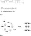
- a transposon DNA includes a double stranded transposase binding site and a unique and different priming site sequence M.
- the transposon DNA can include one or more 5-methylcytosines.
- the double stranded transposase binding site may be a double-stranded 19 bp Tn5 transposase (Tnp) binding site which is linked or connected, such as by covalent bond, to a single-stranded overhang including a priming site sequence, such as at one end of the overhang.
- Tnp Tn5 transposase
- the transposon DNA is inserted into the genomic DNA of a single cell while creating fragments using a transposase. After transposase removal and gap fill-in, the genomic DNA fragments having dissimilar or different or unique priming site sequences at each end of the fragment are amplified using primers together with a DNA polymerase, nucleotides and amplification reagents to PCR amplify the whole genome of the single cell.
- a DNA column purification step is not carried out so as to maximize the small amount ( ⁇ 6 pg) of genomic DNA that can be obtained from within a single cell prior to amplification.
- the DNA can be amplified directly from a cell lysate or other impure condition. Accordingly, the DNA sample may be impure, unpurified, or not isolated. Accordingly, aspects of the present method allow one to maximize genomic DNA for amplification and reduce loss due fragments having the same priming site sequence on each end as with other methods, i.e. non-multiplex methods.
- methods described herein may utilize amplification methods other than PCR.
- transposase (Tnp, grey circles) and the transposon DNA each having unique and different priming site sequences illustrated by different pattern overhang sequences are combined to form a plurality of transposomes.
- Each transposome has two different and unique priming site sequences.
- Each transposome has two different and unique priming site sequences compared to each other transposome within the plurality.
- the 20 transposon pool is mixed with Tn5 transposase at equal molar ratio and incubated at room temperature for 30 minutes.
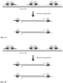
- transposomes of the transposome library randomly capture or otherwise bind to the target single-cell genomic DNA as dimers.
- Representative transposomes are numbered 1, 2 and 3, though the number of transposome members can be greater depending on the desired application.
- a representative number of transposons having different and/or unique primer binding site sequences is between 5 and 50.
- Each transposome includes two unique and/or different priming site sequences.
- transposome 1 includes two unique and/or different priming site sequences
- transposome 2 includes two unique and/or different priming site sequences
- transposome 3 includes two unique and/or different priming site sequences, etc.
- the unique and/or different priming site sequence is within each transposon DNA of the transposome.
- the transposases in the transposome cut the genomic DNA with one transposase cutting an upper strand and one transposase cutting a lower strand to create a genomic DNA fragment.
- the fragment can include one or more cytosines or one or more 5-methylcytosines.
- the plurality of transposomes creates a plurality of genomic DNA fragments, with one or more fragments including one or more cytosines or one or more 5-methylcytosines.
- transposon DNA from the transposon DNA dimer is thus attached to each end of the cut site or fragmentation site, i.e., one transposon DNA from transposome 1 is attached to the left hand cut site and the other transposon DNA from transposome 1 is attached to the right hand cut site.
- each fragment will have a dissimilar priming site sequence at each end of the fragment. This is represented by the two exemplary fragments where the upper fragment has unique and different priming site sequence 1 on one end and unique and different priming site sequence 2 on the other end. Likewise, the lower fragment has unique and different priming site sequence 2 on one end and unique and different priming site sequence 3 on the other end.
- the cut site between the two fragments is produced by transposome 2 and the left hand cut site (i.e. viewing the right side of the upper fragment in Fig. 3 ) includes the one transposon with unique and different priming site sequence 2 while the right hand cut site (i.e. viewing the left side of the lower fragment in Fig. 3 ) includes unique and different priming site sequence 2 (with "2" referring to transposome 2).
- 100nM transposome is added to cell lysate and the transposition reaction mixture is incubated at 55 degree for 10 minutes with magnesium final concentration of 5mM. After removing the transposase, the genomic DNA is cut into millions of small DNA fragments, each tagged with one of the 20 transposon sequences at each end. ( Fig. 3A )
- the transposome library may include 20 different and/or unique primer binding site sequences as described herein while the members of the transposome library may approach millions of members.
- transposomes of the transposome library randomly capture or otherwise bind to the target single-cell genomic DNA as dimers.
- Representative transposomes are numbered 1, 2 and 3, though the number of transposome members can be greater depending on the desired application.
- a representative number of transposons having different and/or unique primer binding site sequences is between 5 and 50.
- Each transposome includes the same unique and/or different primer binding site sequence at each transposon of the transposome.
- transposome 1 includes the same primer binding site sequence on each transposon
- transposome 2 includes the same primer binding site sequence on each transposon
- transposome 3 includes the same primer binding site sequence on each transposon, etc.
- each transposome has a unique and different primer binding site associated therewith, such that each transposome has a different primer binding site associated therewith compared to other members of the transposome library.
- the transposases in the transposome cut the genomic DNA with one transposase cutting an upper strand and one transposase cutting a lower strand to create a genomic DNA fragment.
- the plurality of transposomes creates a plurality of genomic DNA fragments with one or more fragments including one or more cytosines or one or more 5-methylcytosines for chemical treatment to change cytosines to uracils.
- transposon DNA from the transposon DNA dimer is thus attached to each end of the cut site or fragmentation site, i.e., one transposon DNA from transposome 1 is attached to the left hand cut site and the other transposon DNA from transposome 1 is attached to the right hand cut site.
- the transposome library cuts the nucleic acid into fragments, each fragment will have a dissimilar priming site sequence at each end of the fragment, since adjacent transposomes bound to the nucleic acid which create the fragment each have different primer binding site sequences. This is represented by the two exemplary fragments where the upper fragment has unique and different priming site sequence 1 on one end and unique and different priming site sequence 2 on the other end.
- the lower fragment has unique and different priming site sequence 2 on one end (which is the same primer binding site sequence as on the right end of the upper fragment) and unique and different priming site sequence 3 on the other end.
- the cut site between the two fragments is produced by transposome 2 and the left hand cut site (i.e. viewing the right side of the upper fragment in Fig. 3 ) includes the one transposon with unique and different priming site sequence 2 while the right hand cut site (i.e. viewing the left side of the lower fragment in Fig. 3 ) includes unique and different priming site sequence 2 (with "2" referring to transposome 2). Accordingly, even where the transposome has the same primer binding site sequence on each transposon, the method results in a fragment having different primer binding site sequences at each end of the fragment.
- the fragmentation of the genomic DNA and insertion of the methyl transposons leaves a gap on both ends of the transposition/insertion site.
- the gap may have any length but a 9 base gap is exemplary.
- the result is a genomic DNA fragment with a transposon DNA Tnp binding site attached to the 5' position of an upper strand and a transposon DNA Tnp binding site attached to the 5' position of a lower strand. Gaps resulting from the attachment or insertion of the transposon DNA are shown.
- the transposase is removed and gap extension is performed to fill the gap and complement the single-stranded overhang originally designed in the transposon DNA as shown in Fig. 4 .
- the gap-filled fragments are subject to chemical treatment to convert cytosine to uracil in the presence of carrier DNA. These chemically processed fragments can then be subject to amplification and purification as described herein.
- a DNA polymerase reaction mixture containing 200uM each dNTPs, 1X NEB Q5 reaction buffer, 125nM each of 20 primers and 0.02U/uL Q5 DNA polymerase is then added and incubated at 72 0 C for 3 minutes to fill the gap left by the transposition ( Fig. 4 ).
- the fragments shown in Fig. 4 are subject to multiplex PCR amplification to produce amplicons. 15 cycles of PCR reactions are performed as: 98 0 C 30s, 65 0 C 1min, 72 0 C 2min to amplify the target genomic DNA.
- the amplification products are then purified by Zymo DNA purification column.
- Tn5 transposition is carried out using a transposome library including transposomes each with a different and unique primer binding site sequence (or each with two different and unique primer binding site sequences) as described herein and including methyl transposons and transposition buffer are added to the cell lysis which is mixed well and is incubated at 55°C for 10 minutes. 1mg/ml protease is added after the transposition to remove the transposase from binding to the single cell genomic DNA.
- Q5 DNA polymerase, dNTP, PCR reaction buffer and primers are added to the reaction mixture which is heated to 72°C for 10min to fill in the gap generated from the transposon insertion.
- Carrier DNA such as sonicated lambda DNA between 100 bp and 4000 bp, and chemical reagents or enzymatic reagents for converting cytosine to uracil are added and cytosine to uracil conversion is carried out.
- This step can be carried out without prior purification.
- the chemically processed fragments are subjected to 5 to 25 cycles of PCR reaction to amplify the single cell genomic DNA.
- This step can be carried out without purification.
- the amplification products are purified for further analysis such as by high through put deep sequencing.
- a cell is selected, cut from a culture dish, and dispensed in a tube using a laser dissection microscope (LMD-6500, Leica) as follows.
- LMD-6500 laser dissection microscope
- the cells are plated onto a membrane-coated culture dish and observed using bright field microscopy with a 10x objective (Leica).
- a UV laser is then used to cut the membrane around an individually selected cell such that it falls into the cap of a PCR tube.
- the tube is briefly centrifuged to bring the cell down to the bottom of the tube.
- 3 - 5 ⁇ l lysis buffer (30mM Tris-Cl PH 7.8, 2mM EDTA, 20mM KCl, 0.2% Triton X-100, 500 ⁇ g/ml Qiagen Protease) is added to the side of the PCR tube and span down.
- the captured cell is then thermally lysed using the following temperature schedule on PCR machine: 50°C 3 hours, 75°C 30 minutes.
- mouth pipette a single cell into a low salt lysis buffer containing EDTA and protease such as QIAGEN protease (QIAGEN) at a concentration of 10 - 5000 ⁇ g/mL.
- QIAGEN QIAGEN protease
- the incubation would be 37-55°C for 1 - 4 hrs.
- the protease is then heat inactivated up to 80°C and further inactivated by specific protease inhibitors such as 4-(2-Aminoethyl) benzenesulfonyl fluoride hydrochloride (AEBSF) or phenylmethanesulfonyl fluoride (PMSF) (Sigma Aldrich).
- AEBSF 4-(2-Aminoethyl) benzenesulfonyl fluoride hydrochloride
- PMSF phenylmethanesulfonyl fluoride
- An exemplary single cell lysis procedure is as follows:
- the single cell lysis and the transposome library are mixed in a buffer system containing 1 - 100 mM Mg 2+ and optionally 1 - 100 mM Mn 2+ or Co 2+ or Ca 2+ as well and incubate at 37 - 55°C for 5 - 240 minutes.
- the reaction volume varies depending on the cell lysis volume.
- the amount of transposome library added in the reaction could be readily tuned depending on the desired fragmentation size.
- the transposition reaction is stopped by chelating Mg 2+ using EDTA and optionally EGTA or other chelating agents for ions.
- short double stranded DNA could be added to the mixture as a spike-in.
- the residue transposome is inactivated by protease digestion such as QIAGEN protease at a final concentration 1 - 500 ⁇ g/mL at 37 - 55°C for 10 - 60 minutes.
- protease digestion such as QIAGEN protease at a final concentration 1 - 500 ⁇ g/mL at 37 - 55°C for 10 - 60 minutes.
- the protease is then inactivated by heat and/or protease inhibitor, such as AEBSF.
- Nextera transposons have one strand of 5'-/Phos/-CTGTCTCTTATACACATCT-3' (SEQ ID NO: 1) and one 5mC modified strand of either 5'-TMGTMGGMAGMGTMAGATGTGTATAAGAGAMAG-3' ("P5") (SEQ ID NO: 2) or 5'-GTMTMGTGGGMTMGGAGATGTGTATAAGAGAMAG-3' ("P7") (SEQ ID NO: 3) (IDT, purification: PAGE).
- Nextera XT Illumina
- M stands for methylated cytosine.
- Nextera index primers (IDT, purification: standard desalting; then dissolved in 0.1 X TE to 5 uM and stored at -20 C) are in the format of
- Tn5 transposon is constructed from 5'-/Phos/ CTGTCTCTTATACACATCT-3', while the other strand was in the form of 5'-[tag]-AGATGTGTATAAGAGAMAG-3'.
- Each of the oligos (IDT, purification: PAGE) was dissolved in 0.1 X TE to a final concentration of 100 uM.
- For each of the n 20 tags, two strands were annealed at a final concentration of 5 uM each. The 20 annealed transposons were then pooled with equal volumes.
- the transposase was purified after expression from the pTXB1-Tn5 plasmid (Addgene).
- Transposome was assembled at a final concentration of 1.25 uM dimer (2.5 uM monomer), 1:10 diluted (125 nM dimer, or 250 nM monomer), and aliquoted for single uses and store at -80 C.
- 20-primer Mix (for use in PCR Mix 1) was in the form of 5'-[tag]AGATGTGTATAAG-3'.
- Each of the oligos (IDT, purification: standard desalting) was dissolved in 0.1 X TE to a final concentration of 100 uM, and combined with equal volumes (100 uM total, or 5 uM each).
- 40-primer Mix (for use in PCR Mix 2) was in the form of
- the transposome reaction is stopped using a stop buffer as follows: Prepare 0.2mg/ml QP; prepare stop buffer; 1ul 2x stop buffer; 1ul 2mg/ml QP (final 100ug/ml). Transposition is stopped or halted by running (a 12 uL volume) under conditions of a) 50 C for 40 min; b) 70 C for 20 min, and c) 4 C constant thereafter.
- a PCR reaction mixture including Mg 2+ , dNTP mix, primers and a thermal stable DNA polymerase such as Deepvent exo-DNA polymerase (New England Biolabs) is added to the solution at a suitable temperature and for a suitable time period to fill the 9 bp gap left by the transposition reaction.
- the gap filling incubation temperature and time depends on the specific DNA polymerase used.
- the DNA polymerase is optionally inactivated by heating and/or protease treatment such as QIAGEN protease.
- the protease if used, is then inactivated by heat and/or protease inhibitor.
- Carrier DNA (dsDNA) is then added to the reaction medium.
- Carrier DNA present includes DNA fragments between 100 bp and 4000 bp in length such as is created by sonicating lambda DNA.
- Carrier DNA is present within the reaction medium in an amount of at least 20ng and between 20 ng and 50 ng.
- An exemplary gap-filling procedure is as follows:
- Gap fill by running (35 uL volume) under the following conditions. a) 4 C for 3 min (to allow the lid to pre-heat); b) 65 C for 3 min; c) 4 C store.
- Carrier DNA (dsDNA) is then added to the reaction medium along with chemical reagents to convert cytosines to uracils.
- Carrier DNA present during the chemical or enzymatic conversion of cytosine to uracil includes DNA fragments between 100 bp and 4000 bp in length such as is created by sonicating lambda DNA.
- Carrier DNA is present within the reaction medium in an amount of at least 20ng and between 20 ng and 50 ng.
- general methods known to those of skill in the art are used to amplify a DNA fragment.
- the chemically converted fragments from the above example are combined with PCR reaction reagents in an aqueous medium.
- the aqueous medium is then subject to PCR conditions to PCR amplify each DNA fragment.
- the fragments are sequenced using methods known to those of skill in the art and the sequences are stored in computer readable memory.
- the sequences then can be compared an assembled into genomic sequences using methods, including software methods, known to those of skill in the art.
- FIG. 7 One aspect of the present disclosure is shown in Fig. 7 .
- the method described herein using the transposome approach with carrier DNA resulted in higher genomic coverage compared to an existing single-cell whole methylome sequencing technique.
- the method described herein using the transposome approach with carrier DNA resulted in higher mapping rate compared to an existing single-cell whole methylome sequencing technique.
- the method described herein using the transposome approach with carrier DNA resulted in higher accuracy compared to an existing single-cell whole methylome sequencing technique.
- Techniques include scWGBS " Smallwood, S. A., Lee, H. J., Angermueller, C., Krueger, F., Saadeh, H., Peat, J., ...
- TET2 Reaction Buffer (included with TET2) to one tube of TET2 Reaction Buffer Supplement (included with TET2) and mix well, then store at-20°C.
- the volume of the reaction for the denaturation step and ABOPEC reaction is 40ul and may be between 30 ul and 60 ul. This volume advantageously allows avoidance of a DNA purification step after treatment with APOBEC.
- PCR can be directly performed by adding 40ul of PCR buffer mix including DNA polymerase and primers to the 40ul ABOPEC reaction.
- PCR was performed by the addition of 16.5 uL PCR Mix 2 (15ul Q5U 2x master mix and 1.5uL 40-primer Mix) and incubation at 98 C for 30 s, 2 cycles of 98 C for 10 s + 62 C for 30 s + 65 C for 1 min, and 65 C for 5 min.
- primers were similarly removed by the addition of 1 uL 20 U/uL ExoI (NEB M0293S) and incubation at 37 C for 30 min, 72 C for 20 min.
- PCR was similarly performed by the addition of 2.5 uLNEB Index Primer (NEB E7335S, E7500S, E7710S, E7730S) and 6.5 uL PCR Mix 3 (5 uL Q5U 2x master mix (NEB), 1.25 uL water, 0.25 uL Universal Primer (IDT, purification: PAGE)) and incubation at 98 C for 30 s, 2 or more cycles of 98 C for 10 s + 62 C for 30 s + 65 C for 1 min, and 65 C for 5 min. Libraries could be pooled at this step or at any step afterwards. 1.2x AMpure beads is used for size selection.
- kits of the present disclosure generally will include at least the transposome (consists of transposase enzyme and transposon DNA), nucleotides, DNA polymerase, carrier DNA and chemical reagents to convert cytosine to uracil or 5-methylcytosine to uracil, necessary to carry out the claimed method along with primer sets as needed.
- the kit will also contain directions for amplifying DNA from DNA samples. Exemplary kits are those suitable for use in amplifying whole genomic DNA.
- the kits will preferably have distinct containers for each individual reagent, enzyme or reactant. Each agent will generally be suitably aliquoted in their respective containers.
- the container means of the kits will generally include at least one vial or test tube. Flasks, bottles, and other container means into which the reagents are placed and aliquoted are also possible.
- the individual containers of the kit will preferably be maintained in close confinement for commercial sale. Suitable larger containers may include injection or blow-molded plastic containers into which the desired vials are retained. Instructions are preferably provided with the kit.
Landscapes
- Chemical & Material Sciences (AREA)
- Life Sciences & Earth Sciences (AREA)
- Health & Medical Sciences (AREA)
- Organic Chemistry (AREA)
- Genetics & Genomics (AREA)
- Engineering & Computer Science (AREA)
- Zoology (AREA)
- Wood Science & Technology (AREA)
- Bioinformatics & Cheminformatics (AREA)
- Biotechnology (AREA)
- General Engineering & Computer Science (AREA)
- Molecular Biology (AREA)
- Biochemistry (AREA)
- Physics & Mathematics (AREA)
- Analytical Chemistry (AREA)
- Biophysics (AREA)
- Microbiology (AREA)
- Biomedical Technology (AREA)
- Proteomics, Peptides & Aminoacids (AREA)
- General Health & Medical Sciences (AREA)
- Chemical Kinetics & Catalysis (AREA)
- Crystallography & Structural Chemistry (AREA)
- Bioinformatics & Computational Biology (AREA)
- Virology (AREA)
- Plant Pathology (AREA)
- Immunology (AREA)
- General Chemical & Material Sciences (AREA)
- Medicinal Chemistry (AREA)
- Measuring Or Testing Involving Enzymes Or Micro-Organisms (AREA)
- Apparatus Associated With Microorganisms And Enzymes (AREA)
Claims (17)
- Procédé d'analyse de caractéristiques de méthylation d'ADN génomique cible comprenantla mise en contact d'ADN génomique avec une banque de transposomes, chaque transposome de la banque ayant deux transposases et deux ADN transposons, dans lequel chaque ADN transposon comporte un site de liaison à une transposase et une séquence de site de liaison à une amorce, dans lequel la séquence de site de liaison à une amorce est différente du site de liaison à une amorce d'autres membres de la banque de transposomes, dans lequel chaque ADN transposon comporte une ou plusieurs 5-méthylcytosines, dans lequel la banque de transposomes se lie à des emplacements cibles le long de l'ADN génomique et la transposase clive l'ADN génomique en une pluralité de fragments d'ADN génomique double brin représentant une banque de fragments d'ADN génomique, chaque fragment d'ADN génomique double brin comportant une ou plusieurs cytosines et/ou une ou plusieurs 5-méthylcytosines et une séquence de site de liaison à une amorce unique et/ou différente sur chaque extrémité du fragment d'ADN génomique,le remplissage d'un espace entre l'ADN transposon et le fragment d'ADN génomique pour former une banque de produits d'extension de fragment d'ADN génomique double brin ayant des séquences de site de liaison à une amorce uniques et/ou différentes à chaque extrémité,le traitement de la banque de produits d'extension de fragment d'ADN génomique double brin pour convertir la cytosine en uracile en présence d'ADN porteur, etl'amplification des produits d'extension de fragment d'ADN génomique double brin pour produire des amplicons,et le séquençage des amplicons.
- Procédé selon la revendication 1, dans lequel chaque transposome dans la banque de transposomes comporte deux séquences de site de liaison à une amorce différentes.
- Procédé selon la revendication 1, dans lequel chaque transposome dans la banque de transposomes comporte deux séquences de site de liaison à une amorce identiques sur chaque transposon du transposome, qui sont différentes des séquences de site de liaison à une amorce dans d'autres transposomes de la banque de transposomes.
- Procédé selon la revendication 1, dans lequel l'ADN génomique est un ADN génomique entier obtenu à partir d'une cellule unique.
- Procédé selon la revendication 1, dans lequel la transposase est la transposase Tn5, transposase Mu, transposase Tn7 ou transposase IS5.
- Procédé selon la revendication 1, dans lequel l'ADN transposon comporte un site de liaison à Tnp double brin de 19 pb et un surplomb, dans lequel le surplomb comporte une séquence de site de liaison à une amorce unique et/ou différente à l'extrémité 5' du surplomb.
- Procédé selon la revendication 1, dans lequel des transposases liées sont éliminées des fragments double brin avant le remplissage d'espace et l'extension des fragments d'ADN génomique double brin.
- Procédé selon la revendication 1, dans lequel l'ADN génomique provient d'une cellule prénatale, d'une cellule cancéreuse, ou d'une cellule tumorale circulante ;
de préférence, l'ADN génomique provient d'une cellule prénatale unique, d'une cellule cancéreuse unique ou d'une cellule tumorale circulante unique. - Procédé selon la revendication 1, dans lequel la séquence de site de liaison à une amorce unique et différente est un site de liaison à une amorce PCR spécifique.
- Procédé selon la revendication 1, dans lequel la banque de transposomes comporte 1 à 100, 1 à 10, 5 à 50, 30 à 100, 15 à 25, 100 à 1000, 1000 à 10 000 ou 10 000 à 100 000 séquences de site de liaison à une amorce uniques et différentes.
- Procédé selon la revendication 1, dans lequel les séquences de site de liaison à une amorce différentes sont orthogonales.
- Procédé selon la revendication 1, dans lequel l'ADN transposon est méthylé au niveau de chaque cytosine.
- Procédé selon la revendication 1, dans lequel l'ADN transposon comporte des adaptateurs de cytosine méthylés ; de préférence, l'étape de remplissage d'espace comporte l'utilisation de dCTP méthylée au lieu de dCTP dans le mélange de dNTP.
- Procédé selon la revendication 1, qui est caractérisé par un ou plusieurs des éléments suivants :(i) l'ADN porteur est choisi dans le groupe constitué de fragments d'ADNds (double brin) ayant une longueur comprise entre 100 paires de bases (pb) et 4 kilo-paires de bases ;(ii) l'ADN porteur est un type d'ADN qui est différent de l'ADN cible, ou identique à celui-ci ;(iii) l'ADN porteur est de l'ADN lambda traité aux ultrasons ;(iv) l'ADN porteur ne comporte pas d'adaptateurs de séquençage Illumina ;(v) l'ADN porteur est ajouté au milieu réactionnel selon une quantité qui est 100 à 10 000 fois supérieure à la quantité de l'ADN échantillon.
- Procédé selon la revendication 1, comportant en outre une étape après l'étape de remplissage d'espace, mais avant l'étape de conversion : la purification du milieu réactionnel comportant les segments double brin et ADN porteur à espace rempli ; ou, le milieu réactionnel comportant les segments double brin et ADN porteur à espace rempli est transféré directement à l'étape de conversion sans purification ;
de préférence, l'étape de purification est effectuée par des colonnes de centrifugation d'ADN ou par purification d'ADN à base de billes. - Procédé selon la revendication 1, dans lequel le réactif pour convertir la cytosine en uracile n'est pas du bisulfite ou exclut du bisulfite.
- Procédé selon la revendication 1, comportant en outre une étape après l'étape de conversion, mais avant l'étape d'amplification : la purification du milieu réactionnel comportant les fragments chimiquement convertis ;
ou, le milieu réactionnel comportant les fragments chimiquement convertis est directement transféré à l'étape d'amplification sans purification.
Applications Claiming Priority (1)
| Application Number | Priority Date | Filing Date | Title |
|---|---|---|---|
| PCT/CN2019/113387 WO2021077415A1 (fr) | 2019-10-25 | 2019-10-25 | Détection et analyse de méthylation d'adn de mammifère |
Publications (4)
| Publication Number | Publication Date |
|---|---|
| EP4048808A1 EP4048808A1 (fr) | 2022-08-31 |
| EP4048808A4 EP4048808A4 (fr) | 2023-08-09 |
| EP4048808B1 true EP4048808B1 (fr) | 2024-12-11 |
| EP4048808C0 EP4048808C0 (fr) | 2024-12-11 |
Family
ID=75619645
Family Applications (1)
| Application Number | Title | Priority Date | Filing Date |
|---|---|---|---|
| EP19949899.9A Active EP4048808B1 (fr) | 2019-10-25 | 2019-10-25 | Détection et analyse de méthylation d'adn de mammifère |
Country Status (6)
| Country | Link |
|---|---|
| US (1) | US12553044B2 (fr) |
| EP (1) | EP4048808B1 (fr) |
| JP (1) | JP7489455B2 (fr) |
| CN (1) | CN114391043B (fr) |
| ES (1) | ES3009684T3 (fr) |
| WO (1) | WO2021077415A1 (fr) |
Families Citing this family (3)
| Publication number | Priority date | Publication date | Assignee | Title |
|---|---|---|---|---|
| CN114438184B (zh) * | 2022-04-08 | 2022-07-12 | 昌平国家实验室 | 游离dna甲基化测序文库构建方法及应用 |
| JP2025534400A (ja) * | 2022-09-30 | 2025-10-15 | 北京昌平▲実▼▲験▼室 | 二本鎖dnaデアミナーゼを用いたフットプリンティングによりゲノムワイドdna結合タンパク質結合部位を決定するための方法 |
| CN118308491A (zh) * | 2023-01-06 | 2024-07-09 | 北京昌平实验室 | 单细胞dna甲基化检测方法 |
Family Cites Families (39)
| Publication number | Priority date | Publication date | Assignee | Title |
|---|---|---|---|---|
| US2703908A (en) | 1954-05-12 | 1955-03-15 | Joseph J Stracker | Heat deflector for pot handle |
| US4683195A (en) | 1986-01-30 | 1987-07-28 | Cetus Corporation | Process for amplifying, detecting, and/or-cloning nucleic acid sequences |
| US4683202A (en) | 1985-03-28 | 1987-07-28 | Cetus Corporation | Process for amplifying nucleic acid sequences |
| US4965188A (en) | 1986-08-22 | 1990-10-23 | Cetus Corporation | Process for amplifying, detecting, and/or cloning nucleic acid sequences using a thermostable enzyme |
| AU622104B2 (en) | 1987-03-11 | 1992-04-02 | Sangtec Molecular Diagnostics Ab | Method of assaying of nucleic acids, a reagent combination and kit therefore |
| JP2856804B2 (ja) | 1988-03-24 | 1999-02-10 | ユニバーシティ オブ アイオワ リサーチ ファウンデイション | 標識された相補的核酸プローブを開裂する触媒反応の補因子としての活性に基づく核酸配列検出のための触媒ハイブリッド形成系 |
| US5858988A (en) | 1993-02-24 | 1999-01-12 | Wang; Jui H. | Poly-substituted-phenyl-oligoribo nucleotides having enhanced stability and membrane permeability and methods of use |
| US6291438B1 (en) | 1993-02-24 | 2001-09-18 | Jui H. Wang | Antiviral anticancer poly-substituted phenyl derivatized oligoribonucleotides and methods for their use |
| US5681702A (en) | 1994-08-30 | 1997-10-28 | Chiron Corporation | Reduction of nonspecific hybridization by using novel base-pairing schemes |
| US5684143A (en) | 1996-02-21 | 1997-11-04 | Lynx Therapeutics, Inc. | Oligo-2'-fluoronucleotide N3'->P5' phosphoramidates |
| US5925545A (en) | 1996-09-09 | 1999-07-20 | Wisconsin Alumni Research Foundation | System for in vitro transposition |
| JP2001519538A (ja) | 1997-10-10 | 2001-10-23 | プレジデント・アンド・フェローズ・オブ・ハーバード・カレッジ | 核酸アレイのレプリカ増幅 |
| US6485944B1 (en) | 1997-10-10 | 2002-11-26 | President And Fellows Of Harvard College | Replica amplification of nucleic acid arrays |
| US6511803B1 (en) | 1997-10-10 | 2003-01-28 | President And Fellows Of Harvard College | Replica amplification of nucleic acid arrays |
| AU2005216549A1 (en) | 2004-02-27 | 2005-09-09 | President And Fellows Of Harvard College | Polony fluorescent in situ sequencing beads |
| WO2006073504A2 (fr) | 2004-08-04 | 2006-07-13 | President And Fellows Of Harvard College | Sequençage des oscillations dans l'anticodon |
| WO2007120208A2 (fr) | 2005-11-14 | 2007-10-25 | President And Fellows Of Harvard College | Séquençage de l'adn en cercle tournant sur nanogrille |
| JP5073967B2 (ja) | 2006-05-30 | 2012-11-14 | 株式会社日立製作所 | 単一細胞の遺伝子発現定量方法 |
| US20080102452A1 (en) * | 2006-10-31 | 2008-05-01 | Roberts Douglas N | Control nucleic acid constructs for use in analysis of methylation status |
| US20080269068A1 (en) | 2007-02-06 | 2008-10-30 | President And Fellows Of Harvard College | Multiplex decoding of sequence tags in barcodes |
| CN101802223A (zh) | 2007-08-15 | 2010-08-11 | 香港大学 | 用于高通量亚硫酸氢盐dna-测序的方法和组合物及其用途 |
| EP2310401B1 (fr) | 2008-07-22 | 2015-11-25 | The Trustees of Columbia University in the City of New York | Procédés d établissement de profils de méthylation universels |
| US9080211B2 (en) | 2008-10-24 | 2015-07-14 | Epicentre Technologies Corporation | Transposon end compositions and methods for modifying nucleic acids |
| CA2750054C (fr) | 2008-10-24 | 2018-05-29 | Epicentre Technologies Corporation | Compositions terminales de transposon et procede de modification d'acides nucleiques |
| FR2957821B1 (fr) | 2010-03-24 | 2014-08-29 | Inst Francais Du Petrole | Nouvelle zone de regeneration du catalyseur divisee en secteurs pour unites catalytiques regeneratives |
| CN103443338B (zh) | 2011-02-02 | 2017-09-22 | 华盛顿大学商业化中心 | 大规模平行邻接作图 |
| US9005935B2 (en) | 2011-05-23 | 2015-04-14 | Agilent Technologies, Inc. | Methods and compositions for DNA fragmentation and tagging by transposases |
| US20130017978A1 (en) | 2011-07-11 | 2013-01-17 | Finnzymes Oy | Methods and transposon nucleic acids for generating a dna library |
| JP6224689B2 (ja) | 2012-03-15 | 2017-11-01 | ニユー・イングランド・バイオレイブス・インコーポレイテツド | シトシンとこれの修飾物とを識別するための、およびメチローム分析のための方法および組成物 |
| EP3102691B1 (fr) * | 2014-02-03 | 2019-09-11 | Thermo Fisher Scientific Baltics UAB | Procédé de fragmentation régulée de l'adn |
| RU2736728C2 (ru) | 2014-10-17 | 2020-11-19 | Иллумина Кембридж Лимитед | Транспозиция с сохранением сцепления генов |
| US10815478B2 (en) | 2014-11-05 | 2020-10-27 | Illumina, Inc. | Method of sequential tagmentation with transposase compositions for reduction of insertion bias |
| WO2016191618A1 (fr) * | 2015-05-27 | 2016-12-01 | Jianbiao Zheng | Procédés d'insertion de codes à barres moléculaires |
| WO2018057779A1 (fr) * | 2016-09-23 | 2018-03-29 | Jianbiao Zheng | Compositions de transposons synthétiques et leurs procédés d'utilisation |
| CA3055817A1 (fr) | 2017-03-08 | 2018-09-13 | President And Fellows Of Harvard College | Procedes d'amplification d'adn pour maintenir un etat de methylation |
| MX2019013993A (es) | 2017-05-23 | 2020-07-28 | Harvard College | Amplificación de etiquetado extremo múltiple de ácidos nucleicos. |
| DK3635136T3 (da) * | 2017-06-07 | 2022-01-10 | Univ Oregon Health & Science | Enkeltcelle-helgenombiblioteker til methyleringssekvensering |
| CN108300766A (zh) * | 2018-01-16 | 2018-07-20 | 四川大学 | 利用转座酶对染色质开放区和线粒体甲基化研究的方法 |
| DE102018221710A1 (de) | 2018-12-13 | 2020-06-18 | Volkswagen Aktiengesellschaft | Dachkonsole für ein Fahrzeug |
-
2019
- 2019-10-25 US US17/771,265 patent/US12553044B2/en active Active
- 2019-10-25 ES ES19949899T patent/ES3009684T3/es active Active
- 2019-10-25 CN CN201980099399.4A patent/CN114391043B/zh active Active
- 2019-10-25 WO PCT/CN2019/113387 patent/WO2021077415A1/fr not_active Ceased
- 2019-10-25 EP EP19949899.9A patent/EP4048808B1/fr active Active
- 2019-10-25 JP JP2022524572A patent/JP7489455B2/ja active Active
Also Published As
| Publication number | Publication date |
|---|---|
| JP7489455B2 (ja) | 2024-05-23 |
| CN114391043A (zh) | 2022-04-22 |
| CN114391043B (zh) | 2024-03-15 |
| EP4048808C0 (fr) | 2024-12-11 |
| EP4048808A4 (fr) | 2023-08-09 |
| JP2023507876A (ja) | 2023-02-28 |
| US20220389412A1 (en) | 2022-12-08 |
| EP4048808A1 (fr) | 2022-08-31 |
| ES3009684T3 (en) | 2025-03-31 |
| US12553044B2 (en) | 2026-02-17 |
| WO2021077415A1 (fr) | 2021-04-29 |
Similar Documents
| Publication | Publication Date | Title |
|---|---|---|
| US20230203563A1 (en) | Multiplex End-Tagging Amplification of Nucleic Acids | |
| US20190203204A1 (en) | Methods of De Novo Assembly of Barcoded Genomic DNA Fragments | |
| EP3325665B1 (fr) | Procédés d'amplification de séquences d'acides nucléiques | |
| US20200063213A1 (en) | Methods of Amplifying DNA to Maintain Methylation Status | |
| EP4090766B1 (fr) | Procédés de séquençage ciblé | |
| AU2016421196A1 (en) | Methods of whole genome digital amplification | |
| CN113025687A (zh) | 用于核酸扩增的样品制备 | |
| EP4048808B1 (fr) | Détection et analyse de méthylation d'adn de mammifère | |
| HK40071357A (en) | Methylation detection and analysis of mammalian dna | |
| HK40071357B (en) | Methylation detection and analysis of mammalian dna | |
| HK40072305A (en) | Methylation detection and analysis of mammalian dna | |
| WO2024146540A1 (fr) | Procédé de détection de méthylation d'adn d'une cellule unique |
Legal Events
| Date | Code | Title | Description |
|---|---|---|---|
| STAA | Information on the status of an ep patent application or granted ep patent |
Free format text: STATUS: THE INTERNATIONAL PUBLICATION HAS BEEN MADE |
|
| PUAI | Public reference made under article 153(3) epc to a published international application that has entered the european phase |
Free format text: ORIGINAL CODE: 0009012 |
|
| STAA | Information on the status of an ep patent application or granted ep patent |
Free format text: STATUS: REQUEST FOR EXAMINATION WAS MADE |
|
| 17P | Request for examination filed |
Effective date: 20220525 |
|
| AK | Designated contracting states |
Kind code of ref document: A1 Designated state(s): AL AT BE BG CH CY CZ DE DK EE ES FI FR GB GR HR HU IE IS IT LI LT LU LV MC MK MT NL NO PL PT RO RS SE SI SK SM TR |
|
| REG | Reference to a national code |
Ref country code: HK Ref legal event code: DE Ref document number: 40071357 Country of ref document: HK |
|
| DAV | Request for validation of the european patent (deleted) | ||
| DAX | Request for extension of the european patent (deleted) | ||
| A4 | Supplementary search report drawn up and despatched |
Effective date: 20230710 |
|
| RIC1 | Information provided on ipc code assigned before grant |
Ipc: C12Q 1/68 20180101AFI20230704BHEP |
|
| STAA | Information on the status of an ep patent application or granted ep patent |
Free format text: STATUS: EXAMINATION IS IN PROGRESS |
|
| 17Q | First examination report despatched |
Effective date: 20240229 |
|
| GRAP | Despatch of communication of intention to grant a patent |
Free format text: ORIGINAL CODE: EPIDOSNIGR1 |
|
| STAA | Information on the status of an ep patent application or granted ep patent |
Free format text: STATUS: GRANT OF PATENT IS INTENDED |
|
| INTG | Intention to grant announced |
Effective date: 20240801 |
|
| GRAS | Grant fee paid |
Free format text: ORIGINAL CODE: EPIDOSNIGR3 |
|
| GRAA | (expected) grant |
Free format text: ORIGINAL CODE: 0009210 |
|
| STAA | Information on the status of an ep patent application or granted ep patent |
Free format text: STATUS: THE PATENT HAS BEEN GRANTED |
|
| AK | Designated contracting states |
Kind code of ref document: B1 Designated state(s): AL AT BE BG CH CY CZ DE DK EE ES FI FR GB GR HR HU IE IS IT LI LT LU LV MC MK MT NL NO PL PT RO RS SE SI SK SM TR |
|
| REG | Reference to a national code |
Ref country code: GB Ref legal event code: FG4D |
|
| REG | Reference to a national code |
Ref country code: CH Ref legal event code: EP |
|
| REG | Reference to a national code |
Ref country code: IE Ref legal event code: FG4D |
|
| REG | Reference to a national code |
Ref country code: DE Ref legal event code: R096 Ref document number: 602019063540 Country of ref document: DE |
|
| U01 | Request for unitary effect filed |
Effective date: 20250107 |
|
| U07 | Unitary effect registered |
Designated state(s): AT BE BG DE DK EE FI FR IT LT LU LV MT NL PT RO SE SI Effective date: 20250115 |
|
| REG | Reference to a national code |
Ref country code: ES Ref legal event code: FG2A Ref document number: 3009684 Country of ref document: ES Kind code of ref document: T3 Effective date: 20250331 |
|
| PG25 | Lapsed in a contracting state [announced via postgrant information from national office to epo] |
Ref country code: HR Free format text: LAPSE BECAUSE OF FAILURE TO SUBMIT A TRANSLATION OF THE DESCRIPTION OR TO PAY THE FEE WITHIN THE PRESCRIBED TIME-LIMIT Effective date: 20241211 |
|
| PG25 | Lapsed in a contracting state [announced via postgrant information from national office to epo] |
Ref country code: NO Free format text: LAPSE BECAUSE OF FAILURE TO SUBMIT A TRANSLATION OF THE DESCRIPTION OR TO PAY THE FEE WITHIN THE PRESCRIBED TIME-LIMIT Effective date: 20250311 |
|
| PG25 | Lapsed in a contracting state [announced via postgrant information from national office to epo] |
Ref country code: GR Free format text: LAPSE BECAUSE OF FAILURE TO SUBMIT A TRANSLATION OF THE DESCRIPTION OR TO PAY THE FEE WITHIN THE PRESCRIBED TIME-LIMIT Effective date: 20250312 |
|
| PG25 | Lapsed in a contracting state [announced via postgrant information from national office to epo] |
Ref country code: RS Free format text: LAPSE BECAUSE OF FAILURE TO SUBMIT A TRANSLATION OF THE DESCRIPTION OR TO PAY THE FEE WITHIN THE PRESCRIBED TIME-LIMIT Effective date: 20250311 |
|
| PG25 | Lapsed in a contracting state [announced via postgrant information from national office to epo] |
Ref country code: SM Free format text: LAPSE BECAUSE OF FAILURE TO SUBMIT A TRANSLATION OF THE DESCRIPTION OR TO PAY THE FEE WITHIN THE PRESCRIBED TIME-LIMIT Effective date: 20241211 |
|
| PG25 | Lapsed in a contracting state [announced via postgrant information from national office to epo] |
Ref country code: PL Free format text: LAPSE BECAUSE OF FAILURE TO SUBMIT A TRANSLATION OF THE DESCRIPTION OR TO PAY THE FEE WITHIN THE PRESCRIBED TIME-LIMIT Effective date: 20241211 |
|
| PG25 | Lapsed in a contracting state [announced via postgrant information from national office to epo] |
Ref country code: IS Free format text: LAPSE BECAUSE OF FAILURE TO SUBMIT A TRANSLATION OF THE DESCRIPTION OR TO PAY THE FEE WITHIN THE PRESCRIBED TIME-LIMIT Effective date: 20250411 |
|
| PG25 | Lapsed in a contracting state [announced via postgrant information from national office to epo] |
Ref country code: SK Free format text: LAPSE BECAUSE OF FAILURE TO SUBMIT A TRANSLATION OF THE DESCRIPTION OR TO PAY THE FEE WITHIN THE PRESCRIBED TIME-LIMIT Effective date: 20241211 |
|
| PG25 | Lapsed in a contracting state [announced via postgrant information from national office to epo] |
Ref country code: CZ Free format text: LAPSE BECAUSE OF FAILURE TO SUBMIT A TRANSLATION OF THE DESCRIPTION OR TO PAY THE FEE WITHIN THE PRESCRIBED TIME-LIMIT Effective date: 20241211 |
|
| PGFP | Annual fee paid to national office [announced via postgrant information from national office to epo] |
Ref country code: GB Payment date: 20250925 Year of fee payment: 7 |
|
| PLBE | No opposition filed within time limit |
Free format text: ORIGINAL CODE: 0009261 |
|
| STAA | Information on the status of an ep patent application or granted ep patent |
Free format text: STATUS: NO OPPOSITION FILED WITHIN TIME LIMIT |
|
| REG | Reference to a national code |
Ref country code: CH Ref legal event code: L10 Free format text: ST27 STATUS EVENT CODE: U-0-0-L10-L00 (AS PROVIDED BY THE NATIONAL OFFICE) Effective date: 20251022 |
|
| REG | Reference to a national code |
Ref country code: CH Ref legal event code: U11 Free format text: ST27 STATUS EVENT CODE: U-0-0-U10-U11 (AS PROVIDED BY THE NATIONAL OFFICE) Effective date: 20251101 |
|
| U20 | Renewal fee for the european patent with unitary effect paid |
Year of fee payment: 7 Effective date: 20251008 |
|
| 26N | No opposition filed |
Effective date: 20250912 |
|
| PGFP | Annual fee paid to national office [announced via postgrant information from national office to epo] |
Ref country code: CH Payment date: 20251101 Year of fee payment: 7 |
|
| PGFP | Annual fee paid to national office [announced via postgrant information from national office to epo] |
Ref country code: ES Payment date: 20251107 Year of fee payment: 7 |