EP4056582A1 - Vaccin contre le zika/la dengue et son application - Google Patents
Vaccin contre le zika/la dengue et son application Download PDFInfo
- Publication number
- EP4056582A1 EP4056582A1 EP20884983.6A EP20884983A EP4056582A1 EP 4056582 A1 EP4056582 A1 EP 4056582A1 EP 20884983 A EP20884983 A EP 20884983A EP 4056582 A1 EP4056582 A1 EP 4056582A1
- Authority
- EP
- European Patent Office
- Prior art keywords
- protein
- antigen
- mutation
- vaccine
- virus
- Prior art date
- Legal status (The legal status is an assumption and is not a legal conclusion. Google has not performed a legal analysis and makes no representation as to the accuracy of the status listed.)
- Pending
Links
Images


Classifications
-
- A—HUMAN NECESSITIES
- A61—MEDICAL OR VETERINARY SCIENCE; HYGIENE
- A61K—PREPARATIONS FOR MEDICAL, DENTAL OR TOILETRY PURPOSES
- A61K39/00—Medicinal preparations containing antigens or antibodies
- A61K39/12—Viral antigens
-
- A—HUMAN NECESSITIES
- A61—MEDICAL OR VETERINARY SCIENCE; HYGIENE
- A61P—SPECIFIC THERAPEUTIC ACTIVITY OF CHEMICAL COMPOUNDS OR MEDICINAL PREPARATIONS
- A61P31/00—Antiinfectives, i.e. antibiotics, antiseptics, chemotherapeutics
- A61P31/12—Antivirals
-
- A—HUMAN NECESSITIES
- A61—MEDICAL OR VETERINARY SCIENCE; HYGIENE
- A61P—SPECIFIC THERAPEUTIC ACTIVITY OF CHEMICAL COMPOUNDS OR MEDICINAL PREPARATIONS
- A61P31/00—Antiinfectives, i.e. antibiotics, antiseptics, chemotherapeutics
- A61P31/12—Antivirals
- A61P31/14—Antivirals for RNA viruses
-
- C—CHEMISTRY; METALLURGY
- C07—ORGANIC CHEMISTRY
- C07K—PEPTIDES
- C07K14/00—Peptides having more than 20 amino acids; Gastrins; Somatostatins; Melanotropins; Derivatives thereof
- C07K14/005—Peptides having more than 20 amino acids; Gastrins; Somatostatins; Melanotropins; Derivatives thereof from viruses
-
- C—CHEMISTRY; METALLURGY
- C07—ORGANIC CHEMISTRY
- C07K—PEPTIDES
- C07K16/00—Immunoglobulins [IG], e.g. monoclonal or polyclonal antibodies
- C07K16/08—Immunoglobulins [IG], e.g. monoclonal or polyclonal antibodies against material from viruses
- C07K16/10—RNA viruses
- C07K16/116—Togaviridae (F); Matonaviridae (F); Flaviviridae (F)
-
- C—CHEMISTRY; METALLURGY
- C12—BIOCHEMISTRY; BEER; SPIRITS; WINE; VINEGAR; MICROBIOLOGY; ENZYMOLOGY; MUTATION OR GENETIC ENGINEERING
- C12N—MICROORGANISMS OR ENZYMES; COMPOSITIONS THEREOF; PROPAGATING, PRESERVING, OR MAINTAINING MICROORGANISMS; MUTATION OR GENETIC ENGINEERING; CULTURE MEDIA
- C12N15/00—Mutation or genetic engineering; DNA or RNA concerning genetic engineering, vectors, e.g. plasmids, or their isolation, preparation or purification; Use of hosts therefor
- C12N15/09—Recombinant DNA-technology
- C12N15/63—Introduction of foreign genetic material using vectors; Vectors; Use of hosts therefor; Regulation of expression
- C12N15/79—Vectors or expression systems specially adapted for eukaryotic hosts
- C12N15/85—Vectors or expression systems specially adapted for eukaryotic hosts for animal cells
- C12N15/86—Viral vectors
-
- C—CHEMISTRY; METALLURGY
- C12—BIOCHEMISTRY; BEER; SPIRITS; WINE; VINEGAR; MICROBIOLOGY; ENZYMOLOGY; MUTATION OR GENETIC ENGINEERING
- C12N—MICROORGANISMS OR ENZYMES; COMPOSITIONS THEREOF; PROPAGATING, PRESERVING, OR MAINTAINING MICROORGANISMS; MUTATION OR GENETIC ENGINEERING; CULTURE MEDIA
- C12N7/00—Viruses; Bacteriophages; Compositions thereof; Preparation or purification thereof
-
- C—CHEMISTRY; METALLURGY
- C12—BIOCHEMISTRY; BEER; SPIRITS; WINE; VINEGAR; MICROBIOLOGY; ENZYMOLOGY; MUTATION OR GENETIC ENGINEERING
- C12Q—MEASURING OR TESTING PROCESSES INVOLVING ENZYMES, NUCLEIC ACIDS OR MICROORGANISMS; COMPOSITIONS OR TEST PAPERS THEREFOR; PROCESSES OF PREPARING SUCH COMPOSITIONS; CONDITION-RESPONSIVE CONTROL IN MICROBIOLOGICAL OR ENZYMOLOGICAL PROCESSES
- C12Q1/00—Measuring or testing processes involving enzymes, nucleic acids or microorganisms; Compositions therefor; Processes of preparing such compositions
- C12Q1/70—Measuring or testing processes involving enzymes, nucleic acids or microorganisms; Compositions therefor; Processes of preparing such compositions involving virus or bacteriophage
- C12Q1/701—Specific hybridization probes
-
- G—PHYSICS
- G01—MEASURING; TESTING
- G01N—INVESTIGATING OR ANALYSING MATERIALS BY DETERMINING THEIR CHEMICAL OR PHYSICAL PROPERTIES
- G01N33/00—Investigating or analysing materials by specific methods not covered by groups G01N1/00 - G01N31/00
- G01N33/48—Biological material, e.g. blood, urine; Haemocytometers
- G01N33/50—Chemical analysis of biological material, e.g. blood, urine; Testing involving biospecific ligand binding methods; Immunological testing
- G01N33/53—Immunoassay; Biospecific binding assay; Materials therefor
- G01N33/569—Immunoassay; Biospecific binding assay; Materials therefor for microorganisms, e.g. protozoa, bacteria, viruses
- G01N33/56983—Viruses
-
- G—PHYSICS
- G01—MEASURING; TESTING
- G01N—INVESTIGATING OR ANALYSING MATERIALS BY DETERMINING THEIR CHEMICAL OR PHYSICAL PROPERTIES
- G01N33/00—Investigating or analysing materials by specific methods not covered by groups G01N1/00 - G01N31/00
- G01N33/48—Biological material, e.g. blood, urine; Haemocytometers
- G01N33/50—Chemical analysis of biological material, e.g. blood, urine; Testing involving biospecific ligand binding methods; Immunological testing
- G01N33/68—Chemical analysis of biological material, e.g. blood, urine; Testing involving biospecific ligand binding methods; Immunological testing involving proteins, peptides or amino acids
- G01N33/6854—Immunoglobulins
-
- A—HUMAN NECESSITIES
- A61—MEDICAL OR VETERINARY SCIENCE; HYGIENE
- A61K—PREPARATIONS FOR MEDICAL, DENTAL OR TOILETRY PURPOSES
- A61K39/00—Medicinal preparations containing antigens or antibodies
- A61K2039/51—Medicinal preparations containing antigens or antibodies comprising whole cells, viruses or DNA/RNA
- A61K2039/525—Virus
- A61K2039/5254—Virus avirulent or attenuated
-
- A—HUMAN NECESSITIES
- A61—MEDICAL OR VETERINARY SCIENCE; HYGIENE
- A61K—PREPARATIONS FOR MEDICAL, DENTAL OR TOILETRY PURPOSES
- A61K39/00—Medicinal preparations containing antigens or antibodies
- A61K2039/51—Medicinal preparations containing antigens or antibodies comprising whole cells, viruses or DNA/RNA
- A61K2039/525—Virus
- A61K2039/5256—Virus expressing foreign proteins
-
- A—HUMAN NECESSITIES
- A61—MEDICAL OR VETERINARY SCIENCE; HYGIENE
- A61K—PREPARATIONS FOR MEDICAL, DENTAL OR TOILETRY PURPOSES
- A61K39/00—Medicinal preparations containing antigens or antibodies
- A61K2039/51—Medicinal preparations containing antigens or antibodies comprising whole cells, viruses or DNA/RNA
- A61K2039/53—DNA (RNA) vaccination
-
- A—HUMAN NECESSITIES
- A61—MEDICAL OR VETERINARY SCIENCE; HYGIENE
- A61K—PREPARATIONS FOR MEDICAL, DENTAL OR TOILETRY PURPOSES
- A61K39/00—Medicinal preparations containing antigens or antibodies
- A61K2039/57—Medicinal preparations containing antigens or antibodies characterised by the type of response, e.g. Th1, Th2
- A61K2039/575—Medicinal preparations containing antigens or antibodies characterised by the type of response, e.g. Th1, Th2 humoral response
-
- C—CHEMISTRY; METALLURGY
- C07—ORGANIC CHEMISTRY
- C07K—PEPTIDES
- C07K2317/00—Immunoglobulins specific features
- C07K2317/30—Immunoglobulins specific features characterized by aspects of specificity or valency
- C07K2317/33—Crossreactivity, e.g. for species or epitope, or lack of said crossreactivity
-
- C—CHEMISTRY; METALLURGY
- C07—ORGANIC CHEMISTRY
- C07K—PEPTIDES
- C07K2317/00—Immunoglobulins specific features
- C07K2317/30—Immunoglobulins specific features characterized by aspects of specificity or valency
- C07K2317/34—Identification of a linear epitope shorter than 20 amino acid residues or of a conformational epitope defined by amino acid residues
-
- C—CHEMISTRY; METALLURGY
- C07—ORGANIC CHEMISTRY
- C07K—PEPTIDES
- C07K2317/00—Immunoglobulins specific features
- C07K2317/70—Immunoglobulins specific features characterized by effect upon binding to a cell or to an antigen
-
- C—CHEMISTRY; METALLURGY
- C07—ORGANIC CHEMISTRY
- C07K—PEPTIDES
- C07K2317/00—Immunoglobulins specific features
- C07K2317/70—Immunoglobulins specific features characterized by effect upon binding to a cell or to an antigen
- C07K2317/76—Antagonist effect on antigen, e.g. neutralization or inhibition of binding
-
- C—CHEMISTRY; METALLURGY
- C12—BIOCHEMISTRY; BEER; SPIRITS; WINE; VINEGAR; MICROBIOLOGY; ENZYMOLOGY; MUTATION OR GENETIC ENGINEERING
- C12N—MICROORGANISMS OR ENZYMES; COMPOSITIONS THEREOF; PROPAGATING, PRESERVING, OR MAINTAINING MICROORGANISMS; MUTATION OR GENETIC ENGINEERING; CULTURE MEDIA
- C12N2710/00—MICROORGANISMS OR ENZYMES; COMPOSITIONS THEREOF; PROPAGATING, PRESERVING, OR MAINTAINING MICROORGANISMS; MUTATION OR GENETIC ENGINEERING; CULTURE MEDIA dsDNA viruses
- C12N2710/00011—Details
- C12N2710/10011—Adenoviridae
- C12N2710/10311—Mastadenovirus, e.g. human or simian adenoviruses
- C12N2710/10334—Use of virus or viral component as vaccine, e.g. live-attenuated or inactivated virus, VLP, viral protein
-
- C—CHEMISTRY; METALLURGY
- C12—BIOCHEMISTRY; BEER; SPIRITS; WINE; VINEGAR; MICROBIOLOGY; ENZYMOLOGY; MUTATION OR GENETIC ENGINEERING
- C12N—MICROORGANISMS OR ENZYMES; COMPOSITIONS THEREOF; PROPAGATING, PRESERVING, OR MAINTAINING MICROORGANISMS; MUTATION OR GENETIC ENGINEERING; CULTURE MEDIA
- C12N2710/00—MICROORGANISMS OR ENZYMES; COMPOSITIONS THEREOF; PROPAGATING, PRESERVING, OR MAINTAINING MICROORGANISMS; MUTATION OR GENETIC ENGINEERING; CULTURE MEDIA dsDNA viruses
- C12N2710/00011—Details
- C12N2710/10011—Adenoviridae
- C12N2710/10311—Mastadenovirus, e.g. human or simian adenoviruses
- C12N2710/10341—Use of virus, viral particle or viral elements as a vector
- C12N2710/10343—Use of virus, viral particle or viral elements as a vector viral genome or elements thereof as genetic vector
-
- C—CHEMISTRY; METALLURGY
- C12—BIOCHEMISTRY; BEER; SPIRITS; WINE; VINEGAR; MICROBIOLOGY; ENZYMOLOGY; MUTATION OR GENETIC ENGINEERING
- C12N—MICROORGANISMS OR ENZYMES; COMPOSITIONS THEREOF; PROPAGATING, PRESERVING, OR MAINTAINING MICROORGANISMS; MUTATION OR GENETIC ENGINEERING; CULTURE MEDIA
- C12N2770/00—MICROORGANISMS OR ENZYMES; COMPOSITIONS THEREOF; PROPAGATING, PRESERVING, OR MAINTAINING MICROORGANISMS; MUTATION OR GENETIC ENGINEERING; CULTURE MEDIA ssRNA viruses positive-sense
- C12N2770/00011—Details
- C12N2770/24011—Flaviviridae
- C12N2770/24111—Flavivirus, e.g. yellow fever virus, dengue, JEV
- C12N2770/24122—New viral proteins or individual genes, new structural or functional aspects of known viral proteins or genes
-
- C—CHEMISTRY; METALLURGY
- C12—BIOCHEMISTRY; BEER; SPIRITS; WINE; VINEGAR; MICROBIOLOGY; ENZYMOLOGY; MUTATION OR GENETIC ENGINEERING
- C12N—MICROORGANISMS OR ENZYMES; COMPOSITIONS THEREOF; PROPAGATING, PRESERVING, OR MAINTAINING MICROORGANISMS; MUTATION OR GENETIC ENGINEERING; CULTURE MEDIA
- C12N2770/00—MICROORGANISMS OR ENZYMES; COMPOSITIONS THEREOF; PROPAGATING, PRESERVING, OR MAINTAINING MICROORGANISMS; MUTATION OR GENETIC ENGINEERING; CULTURE MEDIA ssRNA viruses positive-sense
- C12N2770/00011—Details
- C12N2770/24011—Flaviviridae
- C12N2770/24111—Flavivirus, e.g. yellow fever virus, dengue, JEV
- C12N2770/24134—Use of virus or viral component as vaccine, e.g. live-attenuated or inactivated virus, VLP, viral protein
-
- G—PHYSICS
- G01—MEASURING; TESTING
- G01N—INVESTIGATING OR ANALYSING MATERIALS BY DETERMINING THEIR CHEMICAL OR PHYSICAL PROPERTIES
- G01N2333/00—Assays involving biological materials from specific organisms or of a specific nature
- G01N2333/005—Assays involving biological materials from specific organisms or of a specific nature from viruses
- G01N2333/08—RNA viruses
- G01N2333/18—Togaviridae; Flaviviridae
- G01N2333/183—Flaviviridae, e.g. pestivirus, mucosal disease virus, bovine viral diarrhoea virus, classical swine fever virus (hog cholera virus) or border disease virus
- G01N2333/185—Flaviviruses or Group B arboviruses, e.g. yellow fever virus, japanese encephalitis, tick-borne encephalitis, dengue
-
- G—PHYSICS
- G01—MEASURING; TESTING
- G01N—INVESTIGATING OR ANALYSING MATERIALS BY DETERMINING THEIR CHEMICAL OR PHYSICAL PROPERTIES
- G01N2469/00—Immunoassays for the detection of microorganisms
- G01N2469/20—Detection of antibodies in sample from host which are directed against antigens from microorganisms
-
- Y—GENERAL TAGGING OF NEW TECHNOLOGICAL DEVELOPMENTS; GENERAL TAGGING OF CROSS-SECTIONAL TECHNOLOGIES SPANNING OVER SEVERAL SECTIONS OF THE IPC; TECHNICAL SUBJECTS COVERED BY FORMER USPC CROSS-REFERENCE ART COLLECTIONS [XRACs] AND DIGESTS
- Y02—TECHNOLOGIES OR APPLICATIONS FOR MITIGATION OR ADAPTATION AGAINST CLIMATE CHANGE
- Y02A—TECHNOLOGIES FOR ADAPTATION TO CLIMATE CHANGE
- Y02A50/00—TECHNOLOGIES FOR ADAPTATION TO CLIMATE CHANGE in human health protection, e.g. against extreme weather
- Y02A50/30—Against vector-borne diseases, e.g. mosquito-borne, fly-borne, tick-borne or waterborne diseases whose impact is exacerbated by climate change
Definitions
- the present disclosure relates to the field of biotechnology, specifically, to a field of Zika/dengue vaccine and its application thereof.
- Zika virus is a mosquito-borne virus, belonging to the genus Flavivirus of the family Flaviviridae.
- no vaccines and drugs are available so far.
- ZIKV still poses a threat to people living in endemic areas. Therefore, development of a ZIKV vaccine is urgent.
- DENV virus dengue virus, DENV
- the structures of ZIKV virus and DENV virus are relatively similar, both of which are icosahedral spherical structures with an envelope.
- the surface of the envelope contains an envelope (Envelope, E) protein.
- the internal viral genome is a single-stranded positive-stranded RNA, about 11kb in length, with only one open reading frame.
- the translated polyprotein can be cleaved into 3 structural proteins (C, prM, and E) and 7 non-structural proteins (NS1, NS2A, NS2B, NS3, NS4A, NS4B, and NS5).
- the E protein is about 53 kD in size and is the main protein on the surface of ZIKV and DENV, which mediates entry of a virus into cells via membrane fusion. Thus, it is an important target for activating neutralizing antibodies. At the same time, E protein is also an important target protein when designing vaccines.
- the E protein has 504 amino acids and exists as a dimer. Each monomer has three domains, DI, DII, and DIII, respectively.
- the head of DII contains a highly conserved fusion loop (FL), in which the sequences of the FL region in both ZIKV virus and DENV virus are completely identical, being D98-R99-G100-W101-G102-N103-G104-C105-G106-L107-F108-G109.
- FL highly conserved fusion loop
- the FL region plays a key role in the membrane fusion process of virus invasion. During virus infection, immune cells will produce a large number of antibodies against FL.
- prM protein The size of prM protein is about 26kD, which assists in the correct folding of the E protein.
- the transmembrane region at the 3' end of prM/E serves as an endoplasmic reticulum retention signal to assist prM and E to form a heterodimer.
- One of the main functions of the prM protein is to maintain the stability of the E protein.
- the pr polypeptide In immature virus particles, the pr polypeptide is located at the tip of the E protein, forming a pr-E spike, which hides the fusion peptide of the E protein. At this time, prM is not easily to be contacted with and cleaved by furin due to steric hindrance.
- the acidic environment in the Golgi body induces a rearrangement reaction that exposes the cleavage site of furin, and the prM protein is cleaved by furin into the M protein.
- the cleaved pr polypeptide is not immediately dissociated from the virion. Instead, the pr polypeptide needs to be exposed to a neutral pH cellular environment before it is released and presents a mature virion.
- ADE refers to an antibody enhances viral infection when the antibody is insufficient to neutralize the virus or at a sub-neutralizing concentration (Beltramello et al., 2010; Dejnirattisai et al., 2010). Although epidemiological investigations are still insufficient, pre-existing ZIKV antibodies from human beings, monkeys, and mice have all been shown to enhance DENV infection in cellular experiments (George et al., 2017; Richner et al., 2017; Stettler et al., 2016; Valiant et al., 2018).
- ZIKV vaccine may have an ADE effect on future DENV infection after immunization during ZIKV vaccine design.
- Antibodies that elicit ADE responses are mainly induced by the FL fusion region of the virus (Beltramello et al., 2010; Dejnirattisai et al., 2010). In Flavivirus infections, such antibodies account for a large proportion of the total induced antibodies. These antibodies often cross-react between different serotypes due to highly conserved epitopes. Most of the antibodies also have low neutralizing activity, which easily lead to ADE reaction. However, most of the antibodies with high neutralizing activity bind to other epitopes of the E protein.
- ZIKV neutralizing monoclonal antibodies targeting Domain I (DI), Domain II (DII) and Domain III (DIII) or quaternary epitopes of the E protein have been identified (Barba-Spaeth et al., 2016; Stettler et al., 2016; Wang et al., 2017; Wang et al., 2016; Zhao et al., 2016). Therefore, an ideal ZIKV vaccine design strategy is to transfer the hot spot epitopes of the immune response from the FL region to other neutralizing epitopes.
- Antibodies are absorbed by cells through binding to virions and then binding to the Fc y receptor protein on the surface of myeloid cells, which subsequently promote viral infection. Since DENV has four serotypes, ADE is likely to occur when someone is infected with DENV a second time with a different serotype, which explains the more severe disease phenomenon in human beings after DENV infection (Katzelnick et al., 2017).
- ADE is used to explain the application limitations of the only currently approved DENV vaccine, Dengvaxia ® , which is recommended only for use in DENV seropositive individuals, while an injection of the vaccine can actually exacerbate the risk of dengue infection for seronegative individuals (Rey et al., 2018; Slon-Campos et al., 2019). Therefore, it is also a challenge to avoid ADE during DENV vaccine development.
- the application aims to provide a Zika/dengue vaccine and its application to avoid ADE effect.
- the present application has obtained the epitope information of an antibody that causes ADE effect using crystal structure analysis and other structural and functional analysis.
- the present application provides antigens, for which some mutations are introduced into the E-protein FL fusion region of either a Zika virus or a dengue virus. Antigens with said mutations are unable to bind to antibodies that causes ADE (FLE antibody).
- One embodiment of the present application also provides a vaccine, which can avoid the production of antibodies induced by the FL epitope after immunization, thereby reducing or eliminating the ADE effect.
- the examples provides an antigen, having an E protein FL fusion region of Zika virus or dengue virus, wherein the E protein FL fusion region comprises one of the following mutations:
- D98 site, W101 site, N103 site, G106 site, L107 site or F108 site is located in the E protein FL fusion region.
- the FL (fusion region) sequence of genus Flavivirus is highly conservative, and the FL sequences of ZIKV virus and DENV virus are completely identical as D98-R99-G100-W101-G102-N103-G104-C105-G106-L107-F108-G109.
- the numbering of D98, W101, N103, G106, L107 and F108 sites refers to the position in the E protein sequences of Zika virus and dengue virus.
- examples can be refered to the 98, 101, 103, 106, 107 and 108 sites of E protein of the Zika virus shown in SEQ ID NO. 1 (e.g. ZIKVFSS13025 strain, GenBank: JN860885.1).
- the mutation of the E protein FL fusion region is selected from any one or a combination of the following groups consisting of different mutation forms: Mutation site Mutation form Five-site mutations of D98/N103/G106/L107/ F108 D98N/N103T/G106F/L107E/F108W D98N/N103T/G106F/L107K/F108W D98N/N103T/G106L/L107E/F108W Three site mutations of G106/L107/ F108 G106F/L107E/F108W G106F/L107K/F108W G106L/L107E/F108W Double site mutations of G106/L107 G106L/L107E G106L/L107K G106F/L107E G106F/L107K G106F/L107R Double site mutations of G106/F108 G106F/F108W G106L/F108W G106F/F108H G106Y/F108W G106
- the antigen comprises the E protein FL fusion region of Zika virus, the antigen further comprises a full sequence or a partial sequence of M protein of Zika virus ; preferably, the antigen further comprises a full sequence of M protein of Zika virus; when the antigen comprises the E protein FL fusion region of dengue virus, the antigen further comprises a full sequence or a partial sequence of M protein of dengue virus; preferably, the antigen further comprises a full sequence of M protein of dengue virus.
- M protein is formed when prM structural protein is cleaved by furin.
- the full sequence or the partial sequence of M protein refers to 0.5%-100%, 50-100%, 60-100%, 70-100%, 80-100%, 90-100% sequence similarity of the M protein, the sequence can be either a sequence selected continuously from the M protein or a combination of fragments selected separately from the M protein.
- the antigen comprises the E protein FL fusion region of Zika virus, the antigen further comprises a full sequence or a partial sequence of prM protein of Zika virus; preferably, the antigen further comprises a full sequence of prM protein of Zika virus; when the antigen comprises the E protein FL fusion region of dengue virus, the antigen further comprises a full sequence or a partial sequence of prM protein of dengue virus; preferably, the antigen further comprises a full sequence of prM protein of dengue virus.
- the prM protein is a structural protein of Zika virus or dengue virus, with a size of about 26 kD, and is used for assisting in the correct folding of the E protein.
- the full sequence or the partial sequence of prM protein refers to 0.5%-100%, 50-100%, 60-100%, 70-100%, 80-100%, 90-100% sequence similarity of the prM protein, the sequence can be either a sequence selected continuously from the prM protein or a combination of fragments selected separately from the prM protein.
- the antigen comprises the E protein FL fusion region of Zika virus, the antigen further comprises a full sequence or a partial sequence of E protein of Zika virus ; preferably, the antigen further comprises a full sequence of E protein of Zika virus; when the antigen comprises the E protein FL fusion region of dengue virus, the antigen further comprises a full sequence or a partial sequence of E protein of dengue virus; preferably, the antigen further comprises a full sequence of E protein of dengue virus.
- the full sequence or the partial sequence of E protein refers to 0.5%-100%, 50-100%, 60-100%, 70-100%, 80-100%, 90-100% sequence similarity of the E protein, the sequence can be either a sequence selected continuously from the E protein, or a combination of fragments selected separately from the E protein.
- the E protein, prM protein, and M protein sequences of Zika virus can be obtained according to the full sequences of Zika virus strain disclosed in NCBI and the prior art.
- the E protein, prM protein, M protein sequences of dengue virus can be obtained according to the full sequences of four serotypes of dengue virus strain disclosed in NCBI and the prior art.
- the Zika virus includes all Zika virus strains, such as ZIKV FSS13025 strain (GenBank: JN860885.1) and ZIKK SMGC-1 strain.
- the dengue virus includes four serotypes of dengue virus strains, such as DENV1 (Hawaii strain, GenBank: KM204119), DENV2 (New Guinea C strain, GenBank: KM204118.1), DENV3 (YN02 strain, GenBank: KF824903) and DENV4 (B5 strain, Guangzhou, China, GenBank: AF289029).
- DENV1 HiFii strain, GenBank: KM204119
- DENV2 New Guinea C strain, GenBank: KM204118.1
- DENV3 YN02 strain, GenBank: KF824903
- DENV4 B5 strain, Guangzhou, China, GenBank: AF289029.
- One embodiment of the present disclosure also provides an antigen binding epitope of E protein FL fusion region of Zika virus, wherein the E protein FL fusion region of Zika virus is the amino acid sequence of D98-R99-G100-W101-G102-N103-G104-C105-G106-L107-F108-G109, which comprises one of the following mutations:
- One embodiment of the present disclosure also provides a Zika virus antigen comprising the above-mentioned antigen binding epitope.
- the above-mentioned Zika virus antigen also comprises one or more of the following sequences:
- One embodiment of the present disclosure also provides an antigen binding epitope of E protein FL fusion region of dengue virus, wherein the E protein FL fusion region of dengue virus is the amino acid sequence of D98-R99-G100-W101-G102-N103-G104-C105-G106-L107-F108-G109, which comprises one of the following mutations:
- One embodiment of the present disclosure also provides a dengue virus antigen comprising the above-mentioned antigen binding epitope.
- the above-mentioned dengue virus antigen also comprises one or more of the following sequences:
- One embodiment of the present disclosure also provides an antibody that obtained from the above-mentioned antigen, the above-mentioned Zika virus antigen, and the above-mentioned dengue virus antigen.
- One embodiment of the present disclosure also provides a polynucleotide encoding the above-mentioned antigen, the above-mentioned antigen binding epitope, the above-mentioned Zika virus antigen, and the above-mentioned dengue virus antigen.
- One embodiment of the present disclosure also provides an expression cassette, recombinant vector, transgenic cell line, recombinant bacteria, adenovirus, lentivirus or viral particle comprising the above-mentioned polynucleotide.
- One embodiment of the present disclosure also provides an mRNA encoding the above-mentioned antigen, the above-mentioned antigen binding epitope, the above-mentioned Zika virus antigen, and the above-mentioned dengue virus antigen.
- One embodiment of the present disclosure also provides a vaccine, that comprises the above-mentioned antigen, the above-mentioned Zika virus antigen, the above-mentioned dengue virus antigen, the above-mentioned polynucleotide, the above-mentioned expression cassette, recombinant vector, transgenic cell line, recombinant bacteria, adenovirus viruses, lentiviruses or virus particles, or the above-mentioned mRNA as active ingredients.
- a vaccine that comprises the above-mentioned antigen, the above-mentioned Zika virus antigen, the above-mentioned dengue virus antigen, the above-mentioned polynucleotide, the above-mentioned expression cassette, recombinant vector, transgenic cell line, recombinant bacteria, adenovirus viruses, lentiviruses or virus particles, or the above-mentioned mRNA as active ingredients.
- the vaccine is one or more of an inactivated vaccine, an attenuated vaccine, a DNA vaccine, an mRNA vaccine, an adenovirus vaccine, other viral vector vaccines, a subunit vaccine or viral particles.
- the vaccine is an adenovirus vaccine.
- the vaccine further comprises a pharmaceutically or veterinarily acceptable vehicle, diluent, adjuvant or excipient.
- One embodiment of the present disclosure also provides use of the above-mentioned antigens, the above-mentioned antigen binding epitopes, the above-mentioned antibodies, the above-mentioned polynucleotides, the above-mentioned expression cassettes, recombinant vectors, transgenic cell lines, recombinant bacteria, adenoviruses, lentiviruses or virus particles, or the above-mentioned mRNAs in the manufacture of a vaccine for preventing and/or treating infections of viruses of genus Flavivirus.
- One embodiment of the present disclosure also provides use of the above-mentioned antigens, the above-mentioned antigen binding epitopes, the above-mentioned antibodies, the above-mentioned polynucleotides, the above-mentioned expression cassettes, recombinant vectors, transgenic cell lines, recombinant bacteria, adenoviruses, lentiviruses or virus particles, or the above-mentioned mRNAs in the manufacture of detection reagents or kits for detecting infections of viruses of genus Flavivirus.
- Example 1 Detection of humoral immune responses in mice induced by recombinant chimpanzee adenovirus vaccine constructed with ZIKV wild-type M/E and prM/E antigens
- the M/E antigen of ZIKV FSS13025 virus strain (GenBank: JN860885.1) was constructed into type 7 chimpanzee adenovirus vector. After packaging, culture and purification of the adenovirus, the recombinant adenovirus vaccine AdC7-M/E-WT was obtained (the recombinant adenovirus vaccine AdC7-M/E-WT: Xu et al. (2018) Journal of virology. vol. 92, 6 e01722-17. 26 Feb . as the construction control). Experimental results show that it had a good protective effect on mice. It has been reported in literatures that the adenovirus vaccine constructed with ZIKV prM/E antigen also has protective effect.
- the prM/E antigen of ZIKV-SMGC-1 virus strain was constructed into type 7 chimpanzee adenovirus vector, and the recombinant adenovirus AdC7- prM/E-WT was obtained after packaging (Recombinant adenovirus AdC7-prM/E-WT was constructed according to Hassan, Ahmed O et al. (2019) Cell reports, vol. 28, 10: 2634-2646.e4 .), as a control for subsequent experiments.
- the process of the micro-neutralization assay was as follows: VERO cells were plated in a 96-well plate one day in advance. The serum was diluted with gradient in 1% FBS (Gibco, 10270-106) in DMEM medium (Invitrogen, C11995500BT) using a 96-well plate the next day. The virus was also diluted with 1% FBS in DMEM medium. The serum and the virus solution were mixed. 100 FFU ZIKV-SMGC-1 was added to each well, and incubated at 37°C for 2 hours. The supernatant of the medium was removed from the VERO cell plate. A mixture of serum and virus was added, and cultured for 2 hours. After that, DMEM containing 10% FBS was supplemented.
- FBS Gibco, 10270-106
- DMEM medium Invitrogen, C11995500BT
- the cells were cultured in a 37 °C incubator for 4 days. After 4 days, the cell culture plate was taken out. All supernatant was discarded. The precipitate was washed once with PBS. 150 ⁇ l methanol was added for fixation, which was placed in -20 °C refrigerator for 15-20 minutes, and then washed twice with PBS. 2% nonfat milk (blocking solution) in PBS was used for blocking for 30 minutes at room temperature, and then primary antibody was added.
- the primary antibody was Z6 antibody that binds to ZIKV E protein, diluted to a working concentration of 5 ⁇ g/mL with blocking solution, incubated at room temperature for 2 hours, and then washed 3 times with PBST. After that, secondary antibody was added.
- the secondary antibody was a goat anti-human antibody conjugated to HRP (Proteintech, SA00001-17), which was diluted 1500-fold with blocking solution, incubated at room temperature for 2 hours, and then washed 4 times with PBST. 50 ⁇ l of TMB chromogenic solution (Biyuntian, P0209) was added, which was incubated at room temperature, and reacted for about 20 minutes. The color change was monitored, and 50 ⁇ l of 2 M hydrochloric acid was added to stop the reaction. The OD450 absorbance value was read on a microplate reader.
- MN 50 neutralization titer value
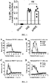
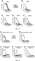
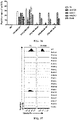
- the results of the neutralization assay were shown in FIG. 1 .
- the group immunized with PBS was the negative control group, Sham group (represented by Sham in the figure), and no neutralizing antibodies can be detected in the serum of this group.
- the log MN 50 average value of neutralizing antibodies can be detected in mice in both AdC7-M/E-WT group (indicated by M/E in the figure) and the AdC7-prM/E-WT group (indicated by prM/E in the figure), which were between 2 and 2.5.
- Example 2 In vitro experiments to detect the ADE of DENV in serums from mice immunized with wild-type recombinant adenovirus vaccine AdC7-M/E-WT and AdC7-prM/E-WT
- fusion loop (FL) sequence of the envelope (E) protein of viruses of genus Flavivirus is highly conserved, infection with ZIKV or immunization with a vaccine expressing the ZIKV E protein can induce the production of antibodies that cross-react with DENV, resulting in an antibody-dependent enhanced response to DENV (ADE) ( Stettler, K., et al. (2016) Science: science. aaf8505 .). Therefore, it was tested whether the serums from mice immunized with the recombinant adenovirus vaccines AdC7-M/E-WT and AdC7-prM/E-WT could produce ADE against DENV.
- ADE antibody-dependent enhanced response to DENV
- Serum obtained from mice immunized with recombinant adenovirus vaccine in Example 1 was gradiently diluted with RPMI-1640 medium (Invitrogen, C11875500BT) containing 1% FBS. The diluted samples were added to a 96-well plate, 10 ⁇ l per well. Then, the corresponding DENV (DENV2, GenBank: KM204118.1; DENV3, GenBank: KF824903; DENV4, GenBank: AF289029; DENV1 was a virus strain that isolated from a sample of an infected patient in Shenzhen Third People's Hospital) was added to each well and incubated in a 37°C cell incubator for 1 hour.
- RPMI-1640 medium Invitrogen, C11875500BT
- the cultured K562 cells expressing Fc ⁇ RIIA receptors on the cell surface were centrifuged at 800 g for 5 minutes, resuspended in RPMI-1640 medium containing 1% FBS, and then counted. After that, the cell density was adjusted to 3 ⁇ 10 6 cells per ml, added to the mixture comprising virus and serum in 10 ⁇ l per well, and incubated in a 37°C cell incubator for 2 hours. 100 ⁇ l of RPMI-1640 medium containing 2% FBS was supplemented to each well, and the incubation was continued in a 37 °C cell incubator for 4 days. After 4 days, the cells were transferred to a 96-well plate, and centrifuged at 800 g for 5 minutes.
- K562 cell expressing the Fc ⁇ RIIA receptor on the cell surface were used as a cell infection model in the experiment.
- Serums from mice immunized with recombinant adenovirus vaccines in group of AdC7-M/E-WT (indicated by M/E in the figure) and in group of AdC7-prM/E-WT (indicated by prM/E in the figure) showed enhanced effects on DENV-infected K562 cells, and the serum of the control group Sham group (indicated by Sham in the figure) had no ADE.
- Z6 antibody is a monoclonal antibody binding to ZIKV E protein which was obtained from B cells isolated from the blood of a patient with ZIKV infection by sequencing the antibody sequence, recombinant expressing and purifying. Previous experiments have shown that it mainly bound to the FL epitope of ZIKV E protein ( Wang, Qihui, et al. Science translational medicine 8.369(2016):369ra179 .). Since the FL sequences of ZIKV and DENV were very conservative and Z6 antibody had low neutralizing activity ( Wang, Qihui, et al.
- Literatures had reported two complex structures of antibodies and antigens of the FL epitope of genus Flavivirus, namely the structure of 2A10G6 antibody and ZIKV soluble E protein (sE) ( Dai, Lianpan, et al. Cell Host & Microbe (2016): S1931312816301494 .), and the structure of E53 antibody and WNV E protein ( Cherier, Mickael V, et al. The EMBO Journal 28.20 (2009): 3269-3276 .).
- sE ZIKV soluble E protein
- 2A10G6 was a Flavivirus broad-spectrum neutralizing antibody that can neutralize DENV1-4, WNV, YFV and ZIKV, which bound to the FL and bc loop of ZIKV E protein ( Dai, Lianpan, et al. Cell Host & Microbe (2016): S1931 312 816 301 494 .).
- Example 5 Construction of chimeric virus antigen protein using the FL of a virus of genus Flavivirus with a large evolutionary distance
- the primers used in the construction of the pCAGGS-M/E-MutA/B/C plasmids were shown in Table 2. Taking the construction of MutA as an example, the plasmid pCAGGS-M/E-WT was used as a template, and WT-F and mutA-R were used as primers to obtain the product mutA-1 by PCR. Plasmid pCAGGS-M/E-WT was used as a template, and WT-R and mutA-F were used as primers, to obtain the product mutA-2 by PCR.
- mutA-1 and mutA-2 were mixed in a molar ratio of 1:1 as a template, and WT-F and WT-R were used as primers for PCR to obtain the PCR product mutA.
- the pCAGGS plasmid was restricted with Xhol and EcoRI to obtain a linear plasmid with double cohesive ends.
- the digested linear plasmid and the PCR product mutA were mixed in a molar ratio of 1:5.
- the In-Fusion kit was used for recombination.
- the recombinant product was transformed into DH5 ⁇ competent cells, which was spread on an ampicillin-resistant plate and cultured at 37 °C.
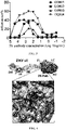
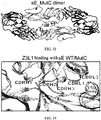
- 293T cells were transfected with wild-type plasmid pCAGGS-M/E-WT and mutant plasmids pCAGGS-M/E-MutA, pCAGGS-M/E-MutB and pCAGGS-M/E-MutC, respectively. After 48 hours, cells were collected, digested into single cells, fixed and permeabilized, incubated with ZIKV E-binding antibody, incubated with Goat Anti-Human FITC secondary antibody, and finally detected the positive proportion of samples by flow cytometry. The results were shown in FIG. 9 .
- Z3L1, Z20 and Z23 were ZIKV-specific antibodies with high neutralizing activity, which bind to DI, DII and DIII of ZIKV E protein, respectively ( Wang, Qihui, et al. (2016) Science translational medicine 8.369:369 ra179 .).
- the antibodies (Z6 and 2A10G6) that bind to the FL epitope of the ZIKV E protein can bind to cells expressing the wild-type M/E-WT antigen, but none of them bound to cells expressing the 3 mutant antigens.
- the Z3L1, Z23 and Z20 antibodies with high neutralizing activity that bound to non-FL epitopes bind both to cells expressing the wild-type M/E-WT antigen and to cells expressing the 3 mutant antigens, indicating that the FL epitopes on M/E-MutA, M/E-MutB and M/E-MutC antigens were destructed, so that it was impossible for the corresponding antibodies to bind.
- the epitopes bound by other strong ZIKV neutralizing antibodies did not change, and the corresponding antibodies were still able to bind.
- M/E-MutA, M/E-MutB and M/E-MutC antigens can induce less or no antibodies that bind to DENV FL, thereby reducing ADE on DENV.
- M/E-MutA, M/E-MutB and M/E-MutC antigens do not affect other antibody epitopes.
- Example 7 Construction of recombinant chimpanzee adenovirus vaccines AdC7-M/E-MutB and AdC7-M/E-MutC using M/E-MutB and M/E-MutC antigens of ZIKV
- the M/E-MutB and M/E-MutC antigens were cloned into the pshuttle vector. Plasmid pCAGGS-M/E-MutB or pCAGGS-M/E-MutC was used as template, and to_pshuttle-F and to_pshuttle-R were used as primers to carry out PCR reaction to obtain PCR product to-pshuttle-mutB and PCR product to_pshuttle-mutC.
- the pshuttle plasmid was restricted with Xbal (Thermo, FD0684) and Kpnl (Thermo, FD0524) to obtain a linear plasmid with double cohesive ends.
- the digested linear plasmid was mixed with the PCR product to_pshuttle-mutB or to_pshuttle-mutC in a molar ratio of 1:5.
- the In-Fusion kit was used for recombination.
- the recombinant product was transformed into DH5 ⁇ competent cells, spread on kanamycin-resistant plate, and cultured at 37 °C. After that, the clones were picked for PCR identification and sequencing identification, and then the plasmid was extracted. Then the cassettes expressing M/E-MutB and M/E-MutC on pshuttle plasmids were constructed into AdC7 vector.
- For the above construction steps see: Xu, Kun et al. (2016) Journal of virology. vol. 92,6 e01722-17. 26 Feb .
- the PCR products to_AdC7-MutB and to_AdC7-MutC were obtained by PCR reaction with plasmid pshuttle-M/E-MutB or MutC as template and to_AdC7-F and to_AdC7-R as primers.
- the AdC7 plasmid was restricted with PI-SceI (NEB, R0696S) and I-Ceul (NEB, R0699S) to obtain a linear plasmid with double cohesive ends.
- the digested linear plasmid was mixed with the PCR product to_AdC7-MutB or to_AdC7-MutC in a molar ratio of 1:5.
- the In-Fusion kit was used for recombination.
- the recombinant product was transformed into stbl2 competent cells, spread on ampicillin-resistant cells, and cultured at 30°C. After that, the clones were picked for PCR identification and sequencing identification, and then the plasmid was extracted. The construction flow of the recombinant plasmid was shown in FIG. 10 .
- the primer sequences used were shown in Table 3.
- Table 3 Primers for construction of chimpanzee adenovirus ZIKV mutant vaccine with MutB and MutC antigens Name of primer Primer sequence (5'-3') to_pshuttle-F AAACGGGCCCTCTAGAGCCACCATGGGCAAGAGGAGC (SEQ ID NO.
- Plasmids were transfected into HEK293 cells using Fugene-6 Transfection Reagent (Promega, E2691), cultured in a 37°C incubator for at least 7 days, and then checked by microscopy every day for the appearance of plaques. All cells and supernatant were collected as the first-generation recombinant adenovirus when plaque cells fell off.
- the culture can be scaled up in sequence according to the ratio of 1:10, until the culture reaches 40 plates of cells. All cells were collected, and the cells were lysed by freezing and thawing 3 times to release the virus. After that, cesium chloride was used for density gradient centrifugation. Polyacrylamide gel (Bio-Gel P-6 DG Media, BIO-RAD, 1500738) was used for desalting and purification. OD260 of the sample was detected by NANODROP. The concentration of the sample was the value of OD260 multiplied by 1.1 ⁇ 10 12 , with the unit being vp (viral particle)/ml. Aliquots were stored in - 80°C.
- Example 8 Evaluation of humoral immune response in BALB/c mice induced by recombinant adenovirus vaccines AdC7-M/E-MutB and AdC7-M/E-MutC
- mice 24 BALB/c mice were randomly divided into 4 groups and immunized by intramuscular injection with 3 recombinant adenovirus vaccines, namely AdC7-M/E-WT, AdC7-M/E-MutB and AdC7-M/E-MutC.
- the dose of adenovirus vaccine immunization was 1.6 ⁇ 10 11 vp. 1 group of mice was immunized with PBS as a negative control. After 4 weeks, blood was collected to separate serum, and the neutralizing antibody titer in serum was detected by microneutralization assay.
- Sham referred to the PBS-immunized group
- M/E-WT referred to the AdC7-M/E-WT vaccine group
- M/E-MutB referred to the AdC7-M/E-MutB vaccine group
- M/E-MutC referred to the AdC7-M/E-MutC-immunized vaccine group.
- Ifnar1 - / - mice were randomly divided into 4 groups and respectively injected with AdC7-M/E-WT, AdC7-M/E-MutB and AdC7-M/E-MutC adenovirus vaccines by intramuscular injection.
- the dose for immunization with the adenovirus vaccine was 1.6 ⁇ 10 11 vp, and 1 group of mice was immunized with PBS as a negative control. After 28 days, blood was collected to separate serum, and the titer level of neutralizing ZIKV antibody in the serum of Ifnar1 - / - mice was detected by microneutralization assay. The results were shown in FIG. 12 .
- both AdC7-M/E-MutB and AdC7-M/E-MutC vaccines can induce higher levels of neutralizing antibody differences in mice.
- Sham referred to the PBS-immunized group
- M/E-WT referred to the AdC7-M/E-WT vaccine group
- M/E-MutB referred to the AdC7-M/E-MutB vaccine group
- M/E-MutC referred to the AdC7-M/E-MutC-immunized vaccine group.
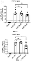
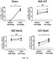
- mice immunized in Example 9 were challenged with ZIKV virus on Day 30 after immunization, by intraperitoneally injecting and 5 ⁇ 10 6 PFU ZIKV (SMGC-1 strain). The results were shown in Figure 13 .
- mice immunized with AdC7-M/E-WT, AdC7-M/E-MutB and AdC7-M/E-MutC did not suffer a weight loss after challenge (Panel B in FIG. 13 ). None of the three groups of mice died (Panel A in FIG. 13 ).
- AdC7-M/E-MutB and AdC7-M/E-MutC vaccines could exhibit a complete protective effect during mice challenge, which was the same as the wild-type AdC7-M/E-WT vaccine.
- Sham referred to the PBS-immunized group
- M/E-WT referred to the AdC7-M/E-WT vaccine group
- M/E-MutB referred to the AdC7-M/E-MutB vaccine group
- M/E-MutC referred to the AdC7-M/E-MutC-immunized vaccine group.
- AdC7-M/E-MutB and AdC7-M/E-MutC vaccines can protect mice against viremia induced by viral infection
- the blood of mice on Day 3 and Day 6 after the challenge were collected to separated serum.
- RNA was extracted using MagaBio Plus Viral RNA Kit (Bori Technology, BSC58S1B), and then FastKing One-Step Reverse Transcription-Fluorescence Quantitation Kit (Tiangen Biochemical Technology, FP314) was used for viral RNA Quantification.
- the probe and the primer sequences used in quantification were shown in Table 4. The quantitative results were shown in FIG. 14 .
- Table 4 Probe and primer sequences used for RT-PCR quantification of ZIKV-SMGC-1 nucleic acid Name Sequence (5'-3') ZIKV-probe FAM - CCACACCTCTGCCGGCACAC (SEQ ID NO.14) - TAMRA ZIKV-F TTGGCTGGCCTATCAGGTTG (SEQ ID NO.15) ZIKV-R CACCTCGGTTTGAGCACTCT (SEQ ID NO.16)
- Sham referred to the PBS-immunized group
- M/E-WT referred to the AdC7-M/E-WT vaccine group
- M/E-MutB referred to the AdC7-M/E-MutB vaccine group
- M/E-MutC referred to the AdC7-M/E-MutC-immunized vaccine group.
- adenovirus vaccine immunization was 1.6 ⁇ 10 11 vp. 1 group of mice was immunized with PBS as a negative control. On day 28 after immunization, blood was collected to separate serum. On the 30th day after immunization, 5 ⁇ 10 4 FFU of ZIKV (SMGC-1 strain) was intraperitoneally injected. Blood was collected again on day 6 after ZIKV was challenged.
- Sham referred to the PBS-immunized group
- M/E-WT referred to the AdC7-M/E-WT vaccine group
- M/E-MutB referred to the AdC7-M/E-MutB vaccine group
- M/E-MutC referred to the AdC7-M/E-MutC-immunized vaccine group.
- Serum neutralizing antibody titers were detected on day 28 after immunization and on day 7 after challenge using a microneutralization assay. The results were shown in FIG. 16 .
- Sham referred to the PBS-immunized group
- M/E-WT referred to the AdC7-M/E-WT vaccine group
- M/E-MutB referred to the AdC7-M/E-MutB vaccine group
- M/E-MutC referred to the AdC7-M/E-MutC-immunized vaccine group.
- FIG. 16 sequentially showed the results of the Sham group, the M/E-WT group, the M/E-MutB group, and the M/E-MutC group from left to right.
- mutant disrupts the E protein FL epitope and reduced the amount of antibodies induced by this epitope, thus showing a weakened binding ability of the serums from mice immunized with AdC7-M/E-MutB and AdC7-M/E-MutC adenovirus vaccine to bind DENV E protein.
- Panel A in FIG. 17 showed the results of binding ability of mouse serum to Zika virus (ZIKV) E protein.
- Panel B showed the result of binding ability of mouse serum to serotype 1 dengue virus (DENV1) E protein.
- Panel C was the result of binding ability between mouse serum and serotype 2 dengue virus (DENV2) E protein.
- Panel D showed the result of binding ability between mouse serum and serotype 3 dengue virus (DENV3) E protein.
- Panel E showed the result of binding ability between mouse serum and serotype 4 dengue virus (DENV4) E protein.
- Example 13 In vitro experiments to detect the ADE to DENV of serum from BALB/c mice immunized with ZIKV vaccine
- Example 12 demonstrated that the AdC7-M/E-MutB and AdC7-M/E-MutC vaccines reduced the induction of cross-antibodies aginst DENV.
- the specific steps were as follows.
- the serums of the immunized BALB/c mice in Example 8 were diluted in gradient, incubated with DENV1, DENV2, DENV3 and DENV4, respectively, and then K562 cells were added. After 4 days of culture, FITC-labeled Z6 antibody was used for staining, followed by flow cytometry to detect the proportion of positive cells. The results were shown in FIG. 18 .
- DENV virus could not infect K562 cells without antibody mediation.
- the infection rate of the detected samples was at the background level.
- the serums from the mice in the M/E-WT group could mediate four serotypes of DENV virus to infect K562 cells.
- the infection rates of both groups of the M/E-MutB and M/E-MutC samples were significantly lower than those of the M/E-WT group, showing a weakened or even eliminated ADE to DENV. This indicates that AdC7-M/E-MutB and AdC7-M/E-MutC vaccines achieved a good effect of reducing ADE.
- Sham referred to the PBS-immunized group
- M/E-WT referred to the AdC7-M/E-WT vaccine group
- M/E-MutB referred to the AdC7-M/E-MutB vaccine group
- M/E-MutC referred to the AdC7-M/E-MutC-immunized vaccine group.
- Panel A shows the detection of enhancement on ZIKV infection of K562 cells by mouse serum.
- Panel B shows the detection of enhancement on DENV1 infection of K562 cells by mouse serum.
- Panel C shows the detection of enhancement on DENV2 infection of K562 cells by mouse serum.
- Panel D shows the detection of enhancement on DENV3 infection of K562 cells by mouse serum.
- Panel E shows the detection of enhancement on DENV4 infection of K562 cells by mouse serum.
- Example 14 In vivo experiments to detect the ADE to DENV in serum from BALB/c mice immunized with ZIKV vaccine
- AdC7-M/E-MutB and AdC7-M/E-MutC vaccines can reduce ADE to DENV under physiological conditions.
- mice 80 BALB/c mice were randomly divided into 4 groups of 20 mice each and immunized with AdC7-M/E-WT, AdC7-M/E-MutB and AdC7-M/E-MutC adenovirus vaccine and PBS, respectively.
- the dose of adenovirus vaccine immunization was 1.6 ⁇ 10 11 vp each mouse.
- Blood was collected after 4 weeks to seperate serum, which was heated at 56 °C for 30 minutes, and the serums from 20 mice in each group were mixed together for subsequent passive immunization of Ifn ⁇ / ⁇ r - / - mice .
- mice were randomly divided into 4 groups and intraperitoneally injected with serums from BALB/c mice immunized with AdC7-M/E-WT, AdC7-M/E-MutB and AdC7-M/E-MutC adenovirus vaccine or PBS, respectively (serums were diluted with PBS at a ratio of 1:10, and each mouse was injected with 200 ⁇ l of the diluted serum).
- DENV2 virus was injected subcutaneously at 5000 FFU per mouse 24 hours later, and each mouse was weighed before injection. After that, the status, survival and body weight of the mice were observed every day, and the results were shown in FIG. 19 .
- mice in the M/E-WT group died earlier, indicating that the serum of the mice immunized with the AdC7-M/E-WT vaccine enhanced the incidence of DENV. Additionally, the time and trend of death of mice in the M/E-MutB and M/E-MutC groups were consistent with those in the Sham group (panel A in FIG. 19 ), indicating that mutations to the ZIKV E protein reduced the production of antibodies that led to ADE effect against DENV, such that the performance of mice in M/E-MutB and M/E-MutC groups were similar to that in the Sham group.
- Sham referred to the PBS-immunized group
- M/E-WT referred to the AdC7-M/E-WT vaccine group
- M/E-MutB referred to the AdC7-M/E-MutB vaccine group
- M/E-MutC referred to the AdC7-M/E-MutC-immunized vaccine group.
- mice were randomly divided into 3 groups, and were immunized with AdC7-M/E-WT adenovirus vaccine (WT group), AdC7-M/E-MutB adenovirus vaccine (MutB group) and AdC7-M/E- MutC adenovirus vaccine (MutC group) by intramuscular injection, 1.6 ⁇ 10 11 vp per mouse.
- WT group AdC7-M/E-MutB adenovirus vaccine
- MotC group AdC7-M/E- MutC adenovirus vaccine
- the lymph nodes were dissected out and placed in 1640 medium containing 1% FBS.
- the lymph nodes of all mice in each vaccine group were mixed together, ground using the rough side of a glass slide, and followed by filtration with a 0.45 ⁇ m filter.
- Lymphocytes were centrifuged at 400 g for 15 minutes at 4 °C, the supernatant was discarded and the precipitate was resuspended in 1 ml of FACS buffer.
- FACS buffer was a PBS solution comprising 0.5% FBS. After resuspension, it was transferred to a 1.5 ml EP tube, centrifuged at 400 g for 10 minutes at 4°C. The supernatant was discarded. The cells were resuspended in 200 ⁇ l FACS buffer. 4 ⁇ g biotin-labeled mixture of ZIKV E monomeric protein and dimeric protein were added, and incubated at 4°C for 30 min in the dark.
- FACS buffer 1 ml was added, mixed well, and centrifuged to precipitate cells. After that, 1 ml of FACS buffer was added to wash again, and then antibody was added for staining. The antibody was diluted with FACS buffer.
- Each 200 ⁇ l of antibody solution contained: FITC - GL7, 2 ⁇ l (BD, 553666); PE - CD138, 4 ⁇ l ( BD, 553714); PE/CY7 - CD38, 4 ⁇ l (BioLegend, 102718); APC - CD93, 4 ⁇ l (BioLegend, 136510); BV421 - B220, 16 ⁇ l (BioLegend, 103240); BV510 - IgD, 2 ⁇ l ( BD, 563110); BV711, 4 ⁇ l (BD, 563262).
- WT referred to the group immunized with AdC7-M/E-WT vaccine
- MutB referred to the group immunized with AdC7-M/E-MutB vaccine
- MutC referred to the group immunized with AdC7-M/E-MutC vaccine.
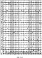
- the AdC7-M/E-WT vaccine activated the HV:LV of GC B cell clones in mice with a clear preference, in which IGHV9-2-1:IGKV10-96 (29.9%) , IGHV1-22:IGKV14-111 (14.4%) and IGKV1-22:IGKV6-23 (7.5%) had the highest frequency. The sum of these three was about 50%.
- HV:LVs of GC B cell clones in mice activated with AdC7-M/E-MutB and AdC7-M/E-MutC vaccines were more diverse and more dispersed in frequency distribution.
- the monoclonal antibodies derived from the AdC7-M/E-WT vaccine group (represented as M/E-WT in Table 6) were named as ZWT.1-10, and the monoclonal antibodies defived from the AdC7-M/E-MutB vaccine group (represented as M/E-MutB in Table 6) and AdC7-M/E-MutC vaccine group (represented as M/E-MutC in Table 6) were named as ZMutB.1-8 and ZMutC.1-13, respectively (Table 6).
- ELISA detects the binding ability of each monoclonal antibody to ZIKV E (monomer or dimer). When the OD450 of the sample was higher than 5 times of the value of the negative control, it was considered to be binding, otherwise it was considered to be non-binding.
- the experimental method of ELISA was as follows: Dilute the protein with ELISA coating solution (sodium carbonate-sodium bicarbonate buffer, pH 9.6) to 3 ⁇ g/ml, add 100 ⁇ l to each well of 96-well ELISA plate, and leave standing overnight at 4°C. The next day, discard the coating solution, and block the ELISA plate with 5% nonfat milk in PBS, and leave at room temperature for 1 hour. Pour off the blocking solution, add 100 ⁇ l of the culture supernatant expressing monoclonal antibody to each well of the ELISA plate, incubate at room temperature for 2 hours, and wash 3 times with PBST.
- ELISA coating solution sodium carbonate-sodium bicarbonate buffer, pH 9.6
- Goat Anti-Mouse HRP (ab6789) secondary antibody diluted 1:2000 in blocking solution, incubate at room temperature for 1.5 hours, and wash 4 times with PBST.
- Add 50 ⁇ l of TMB chromogenic solution to develop color add 50 ⁇ l of 2M hydrochloric acid after 30 minutes to stop the reaction, and detect the OD450 value on a microplate reader.
- the ELISA test results were shown in FIG. 25 .
- Most of the monoclonal antibodies could bind to the monomer or dimer form of the ZIKV sE protein.
- the positive rate of binding reached 90% (9/10) in the ME-WT group, 87.5% in ME-MutB group (7/8), 76.9% in ME-MutC group (10/13), respectively.
- the FLE monoclonal antibodies derived from the ME-WT group were mainly composed of four types of HV:LV genes.
- FIG. 27 shows that the FLE monoclonal antibodies we isolated from the ME-WT group were more similar to the loci and sequences of these four reported monoclonal antibodies, as shown in FIG. 27 , FIG. 28-1 and FIG. 28-2 .
- 6B6C-1 and 4G2 use the same HV:LV gene pair as ZWT.1-3 and ZWT.4-5, respectively, and have a high sequence similarity; 2A10G6 and E53 use the same HV gene as ZWT.6 and ZWT.8, respectively.
- the LV genes used by 2A10G6 and ZWT.6 were different, the LV sequences of the two antibodies were somewhat similar. As can be seen from FIG. 28-2 , the CDRL3 and FR4 of 2A10G6 and ZWT.6 were identical.
- the FLE monoclonal antibody cloned from the lymph node GC B cells of mice immunized with the AdC7-M/E-WT vaccine had a locus that was close to or even the same as the reported mouse FLE monoclonal antibody, and the sequences were relatively similar. It showed that the locus used to induce antibodies that bound to the FL epitope in mice had a preference, and the characteristics of the produced FLE antibodies were also relatively similar.
- FIG. 28-1 A showed the sequence alignment analysis of the antibody heavy chains of ZWT.1, ZWT.2, ZWT.3, and 6B6C-1 and the mouse locus
- B showed the sequence alignment of the antibody light chain of ZWT.1, ZWT.2, ZWT.3, and 6B6C-1 and the mouse locus
- C showed the sequence alignment of the antibody heavy chain of ZWT.4, ZWT.5, ZWT.6, 4G2, and 2A10G6 and the mouse locus.
- D showed the sequence alignment of antibody light chain of ZWT.4, ZWT.5, and 4G2 and the mouse locus
- E showed the sequence alignment of antibody light chain of ZWT.6, and 2A10G6 and the mouse locus.
- Example 17 SPR assay to detect the affinity of wild-type and mutant ZIKV E proteins to ZIKV antibodies
- AdC7-M/E-MutB and AdC7-M/E-MutC vaccines were able to provide complete protection in mice while avoiding ADE against DENV, we expressed purified soluble sE-MutC protein as a representative to compare with sE-WT to explain the underlying molecular mechanism.
- BIOCORE8000 was based on the principle of Surface Plasmon Resonance (SPR), which can detect the interaction between molecules, reflect the dynamic changes in the process of molecular binding in real time, and obtain the kinetic parameters of the interaction.
- SPR Surface Plasmon Resonance
- FIG. 29 from A to D were the binding results of ZIKV sE-WT protein and 2A10G6 antibody (sE-WT-2A10G6), the binding results of ZIKV sE-MutC protein and 2A10G6 antibody (sE-MutC-2A10G6), binding results of ZIKV sE-WT protein and Z6 antibody (sE-WT-Z6), and binding results of ZIKV sE-MutC protein and Z6 antibody (sE-MutC-Z6), respectively.
- the affinity of 2A10G6 antibody binding to ZIKV sE-WT protein was 9.13 nM.
- FIG. 30 from A to D were the binding results of ZIKV sE-WT protein and Z3L1 antibody (sE-WT-Z3L1), the binding results of ZIKV sE-MutC protein and Z3L1 antibody (sE-MutC-Z3L1), binding results of ZIKV sE-WT protein and Z23 antibody (sE-WT-Z23), and binding results of ZIKV sE-MutC protein and Z23 antibody (sE-MutC-Z23), respectively.
- Z3L1 and Z23 antibodies bind to ZIKV E-WT protein with affinities of 9.48 ⁇ M and 0.625 ⁇ M, respectively. From panel B and panel D in FIG. 30 , it can be seen that Z3L1 and Z23 antibodies bind to ZIKV sE-MutC protein with affinities of 8.01 ⁇ M and 0.701 ⁇ M, respectively.
- the affinity of the mutant protein ZIKV sE-MutC to Z23 and Z3L1 was almost unchanged compared with that of the wild-type ZIKV sE-WT protein. This indicated that the designed mutant could maintain the overall conformation of the protein and no change was made to epitopes other than the mutated site.
- the folded form of sE-MutC was very similar to that of the wild-type protein, showing normal secondary, tertiary and quaternary epitope structures.
- the ZIKV wild-type protein sE-WT and mutant protein sE-MutC have the same binding mode to Z3L1 antibody, and the binding sites were mainly D0, E0 and F0 strands and 150 loop in DI, as well as kl hairpin in DII (Wang, et al. (2016) Science translational medicine 8.369:369ra179.).
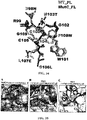
- the FL region of sE-MutC and sE-WT were overlapped and analyzed for comparison. The results were shown in FIG. 34 . It can be seen from FIG. 34 that the conformations of the FL epitopes of both were very similar, and only differs in the side chains of the mutation site.
- FIG. 35 the complex structures of Z6 antibody and ZIKV sE protein, 2A10G6 antibody and ZIKV sE protein (PDB: 5JHL), and E53 antibody and WNV sE protein (PDB: 3150) were shown in A to C, respectively.
- PDB 2A10G6 antibody and ZIKV sE protein
- PB E53 antibody and WNV sE protein
- the F108W mutation creates a steric hindrance that affects the interaction between the FL epitope of sE-MutC and the 2A10G6 antibody. Therefore, on the FL epitope of ZIKV E protein, the three key amino acid mutations G106, L107 and F108 achieve a synergistic effect, eliminating the induction of FLE antibodies.
- the signal peptide gene (SEQ ID NO. 17) derived from JEV (Japanese encephalitis virus) and the M/E gene (SEQ ID NO. 18) expressing DENV2 of New Guinea C strain (GenBank: KM204118.1) were constructed into pshuttle vector to obtain plasmid pshuttle-DV2-M/E-WT expressing wild-type DENV2 M/E.
- the signal peptide gene (SEQ ID NO. 17) derived from JEV and the gene expressing the prM/E protein of DENV2 New Guinea C strain (SEQ ID NO. 19) were constructed into pshuttle vector to obtain the wild-type DENV2 prM/E plasmid pshuttle-DV2-prM/E-WT.
- MutA (D98N, N103T, G106F, L107E and F108W)
- MutB (D98N, N103T, G106F, L107K and F108W)
- MutC (D98N, N103T, G106L, L107E and F108W) mutant plasmids were constructed using pshuttle-DV2-M/E-WT plasmid as template. The primer sequences used in the construction process were shown in Table 9.
- 293T cells were transfected with plasmids pshuttle-DV2-M/E-WT and pshuttle-DV2-prM/E-WT expressing wild-type protein, and three mutant plasmids, respectively. After 48 hours, the supernatant was removed. The cells were washed once with PBS, then trypsinized into single cells, centrifuged, resuspended in DMEM medium, and washed again with DMEM medium. Then, the Fixation and Permeabilization solution from BD Company was added, and placing on ice for 20 minutes. Then, the cells were collected by centrifugation at 800 g for 10 minutes and washed twice with 1 ⁇ Perm/Wash buffer from BD company.
- Each sample was divided into 5 parts, adding Z6, 2A10G6 and mAb11 antibodies that bound to FL epitopes and mAb513 and D448 antibodies that bound to non-FL epitopes, respectively, and placing in a refrigerator at 4°C for 1 hour.
- the cells were harvested by centrifugation and washed twice with 1 ⁇ Perm/Wash buffer.
- Goat Anti-Human FITC (Proteintech, 00003-12) antibody was added, and placing in a refrigerator at 4°C for 1 hour.
- the cells were harvested by centrifugation and washed twice with 1 ⁇ Perm/Wash buffer.
- the cells were resuspended in PBS (200 ⁇ l per well). The positive proportion of the samples was detected by flow cytometry.
- the experimental results were shown in FIG. 36 .
- mAb513 and D448 antibodies that bound to non-FL epitopes with higher neutralizing activity, they could still bind to the M/E-MutA or M/E-MutB or M/E-MutC proteins of DENV2 introduced with MutA, MutB and MutC mutations, indicating that the mutation of the FL epitope of DENV2 E protein had no significant effect on other epitopes.
- the above results showed that these three mutation combinations could be used in DENV vaccines, and the vaccine obtained based on the DENV2 E protein antigen with the combination of G106, L107 and F108 site mutations may have reduced ADE effect caused by subsequent DENV virus infection after vaccine immunization.
- W101 was the amino acid where E protein interacts most with antibodies. It had also been reported in the literature that most FLE antibodies bound to E protein by W101 ( Dejnirattisai, W. et al (2015). Nat Immunol 16, 170-177 .). Therefore, we tried to mutate W101 into other 19 amino acids, and then detected whether the epitope of the antigen expressed by cells was changed by cytometry to screen out the most suitable mutation.
- the signal peptide gene (SEQ ID NO. 17) derived from JEV and the M/E gene (SEQ ID NO. 31) of wild-type ZIKV were constructed into pCAGGS vector (Addgene) to obtain the plasmid pCAGGS-M/E-WT that could express M/E protein of wild-type ZIKV Using this plasmid as a template, tryptophan at position 101 of E protein was mutated into other 19 amino acids. Taking the mutation of tryptophan to alanine as an example, using plasmid pCAGGS-M/E-WT as a template, and using W101-WT-F and W101-1A-R as primers, PCR product W101-1A-1 was obtained.
- PCR product W101-1A-2 was obtained. Then, W101-1A-1 and W101-1A-2 were mixed in a molar ratio of 1:1 as a template, and W101-WT-F and W101-WT-R were used as primers for PCR to obtain PCR product W101-1A.
- the pCAGGS vector was restricted with Xhol (Thermo, FD0694) and EcoRI (Thermo, FD0274) to obtain a linear plasmid with double cohesive ends.
- the digested linear plasmid was mixed with W101-1A according to a molar ratio of 1:5, and the In-Fusion kit (Takara, 639648) was used for recombination.
- the recombinant product was transformed into DH5 ⁇ competent cells, spread on ampicillin-resistant plates, and cultured at 37 °C. After that, the clones were picked for PCR identification and sequencing identification.
- 293T cells were transfected with wild-type plasmids and 19 mutant plasmids, respectively. After 48 hours, the supernatant was removed. The cells were washed once with PBS, then trypsinized into single cells, centrifuged, resuspended in DMEM medium, and washed again with DMEM medium. Then, the Fixation and Permeabilization solution from BD Company was added, and placing on ice for 20 minutes. Then, the cells were collected by centrifugation at 800 g for 10 minutes and washed twice with 1 ⁇ Perm/Wash buffer from BD company.
- Each sample was divided into 5 parts, adding Z6 and 2A10G6 antibodies respectively, and placing in a refrigerator at 4 °C for 1 hour.
- the cells were harvested by centrifugation and washed twice with 1 ⁇ Perm/Wash buffer.
- Goat Anti-Human FITC (Proteintech, 00003-12) antibody was added, and placing in a refrigerator at 4°C for 1 hour.
- the cells were harvested by centrifugation and washed twice with 1 ⁇ Perm/Wash buffer.
- the cells were resuspended in PBS (200 ⁇ l per well). The positive proportion of the samples was detected by flow cytometry.
- the experimental results were shown in FIG. 37 .
- ZIKV wild-type M/E antigen could bind to Z6 and 2A10G6 antibodies, while all 19 mutants can hardly bind to Z6 and 2A10G6 antibodies, indicating that tryptophan at position 101 of ZIKV E protein was important for activating antibodies targeting FL.
- the vaccine prepared by the W101 site mutation will reduce or avoid the production of FL epitope-induced antibodies, thereby avoiding the ADE effect on DENV after vaccine immunization.
- the wild-type plasmid pCAGGS-ZIKV-M/E-WT and the successfully constructed mutant plasmid were transfected into 293T cells respectively. After 48 hours, the cells were harvested, digested into single cells, fixed and permeabilized, incubated with FL epitope-binding ADE antibodies Z6 and 2A10G6, and incubated with Goat Anti-Human (mouse) FITC secondary antibody. Finally, the positive proportion of samples was measured by flow cytometry. If the positive proportion was lower than 10% of the wild-type positive rate, it was considered as non-binding, and if 10%-50% of the wild-type positive rate, it was considered as weak binding. The results were shown in Table 12.
- G106F G106F-F TTCTTATTCGGAAAGGGAGGCATCGTGACTTG (SEQ ID NO.134)
- G106F-R GAATAAGAAGCATCCATTGCCCCAGCCTCTGT (SEQ ID NO.135 )
- G106W G106W-F TGGTTATTCGGAAAGGGAGGCATCGTGACTTG (SEQ ID NO.136)
- G106W-R GAATAACCAGCATCCATTGCCCCAGCCTCTGT (SEQ ID NO.137)
- G106Y G106Y-F TACTTATTCGGAAAGGGAGGCATCGTGACTTG (SEQ ID NO.138)
- G106Y-R GAATAAGTAGCATCCATTGCCCCAGCCTCTGT( SEQ ID NO.139)
- G106I G106I-F ATCTTATTCGGAAAGGGAGGCATCGTGACTTG( SEQ ID NO.140 )
- G106I-R GAATAAGATGCATCCATTGCCCCAGCCTCTGT (SEQ ID NO.141) L107
- F108A F108A-F GGTTTAGCCGGAAAGGGAGGCATCGTGACTTG (SEQ ID NO.160)
- F108A-R GGCTAAACCGCATCCATTGCCCCAGCCTCTGT (SEQ ID NO.161) G106/L107 mutation G106L/L107E G106L/L107K G106F/L107E G106F/L107K G106F/L107R G106/F108 mutation G106F/F108W G106L/F108W G106F/F108H G106Y/F108W G106W/F108Y L107/F108 mutation L107E/F108W L107K/F108W L107R/F108W L107D/F108W L107K/F108Y
- 293T cells were transfected with the wild-type plasmids pCAGGS-ZIKV-M/E-WT, pCAGGS-DENV2-M/E-WT and the mutant constructs in the above table, respectively. After 48 hours, the cells were harvested, digested into single cells, fixed and permeabilized, incubated with FL epitope-binding ADE antibodies Z6 and 2A10G6, and incubated with Goat Anti-Human (mouse) FITC secondary antibody. Finally, the positive proportion of samples was measured by flow cytometry. If the positive proportion was lower than 10% of the wild-type positive rate, it was considered as non-binding, and if 10%-50% of the wild-type positive rate, it was considered as weak binding. The results were shown in Table 14.
- Examples in the present disclosure relates to a Zika/dengue vaccine and its application thereof.
- the present application has obtained the epitope information of an antibody that causes ADE effect based on crystal structure analysis and other structural and functional analysis.
- the present disclosure provides antigens, in which some mutations are introduced into the E-protein FL fusion region of the Zika virus or dengue virus. All antigens with said mutations are unable to bind to antibodies causing ADE (FLE antigen). After immunization with the vaccine of the present disclosure acquired from the said antigens, production of FL epitope-induced antibodies can be prevented, thereby reducing or eliminating the ADE effect.
Landscapes
- Health & Medical Sciences (AREA)
- Life Sciences & Earth Sciences (AREA)
- Chemical & Material Sciences (AREA)
- Virology (AREA)
- Organic Chemistry (AREA)
- Engineering & Computer Science (AREA)
- General Health & Medical Sciences (AREA)
- Immunology (AREA)
- Molecular Biology (AREA)
- Medicinal Chemistry (AREA)
- Genetics & Genomics (AREA)
- Biochemistry (AREA)
- Microbiology (AREA)
- Biomedical Technology (AREA)
- Biotechnology (AREA)
- Proteomics, Peptides & Aminoacids (AREA)
- Zoology (AREA)
- Wood Science & Technology (AREA)
- Biophysics (AREA)
- Bioinformatics & Cheminformatics (AREA)
- Veterinary Medicine (AREA)
- Public Health (AREA)
- Animal Behavior & Ethology (AREA)
- Pharmacology & Pharmacy (AREA)
- Physics & Mathematics (AREA)
- Hematology (AREA)
- Urology & Nephrology (AREA)
- General Engineering & Computer Science (AREA)
- Analytical Chemistry (AREA)
- Communicable Diseases (AREA)
- Nuclear Medicine, Radiotherapy & Molecular Imaging (AREA)
- Gastroenterology & Hepatology (AREA)
- General Chemical & Material Sciences (AREA)
- Mycology (AREA)
- Epidemiology (AREA)
- Chemical Kinetics & Catalysis (AREA)
- Oncology (AREA)
- Pathology (AREA)
- General Physics & Mathematics (AREA)
- Cell Biology (AREA)
Applications Claiming Priority (2)
| Application Number | Priority Date | Filing Date | Title |
|---|---|---|---|
| CN201911082867 | 2019-11-07 | ||
| PCT/CN2020/127614 WO2021089055A1 (fr) | 2019-11-07 | 2020-11-09 | Vaccin contre le zika/la dengue et son application |
Publications (2)
| Publication Number | Publication Date |
|---|---|
| EP4056582A1 true EP4056582A1 (fr) | 2022-09-14 |
| EP4056582A4 EP4056582A4 (fr) | 2024-02-07 |
Family
ID=74033067
Family Applications (1)
| Application Number | Title | Priority Date | Filing Date |
|---|---|---|---|
| EP20884983.6A Pending EP4056582A4 (fr) | 2019-11-07 | 2020-11-09 | Vaccin contre le zika/la dengue et son application |
Country Status (8)
| Country | Link |
|---|---|
| US (1) | US20240173395A1 (fr) |
| EP (1) | EP4056582A4 (fr) |
| JP (1) | JP7725465B2 (fr) |
| KR (1) | KR20220093197A (fr) |
| CN (3) | CN114907456B (fr) |
| BR (1) | BR112022008865A2 (fr) |
| MX (1) | MX2022005498A (fr) |
| WO (1) | WO2021089055A1 (fr) |
Families Citing this family (7)
| Publication number | Priority date | Publication date | Assignee | Title |
|---|---|---|---|---|
| CN116082499B (zh) * | 2021-11-08 | 2023-10-27 | 东莞市朋志生物科技有限公司 | 一种抗登革ns1蛋白的抗体及其应用 |
| CN114544949A (zh) * | 2022-01-04 | 2022-05-27 | 天津亿泰微科生物科技发展有限公司 | 一种检测寨卡病毒包膜蛋白e的elisa试剂盒 |
| KR102844490B1 (ko) * | 2022-12-30 | 2025-08-08 | 전북대학교 산학협력단 | 뎅기 바이러스 외피 단백질의 4차 구조 에피토프를 모방한 재조합 항원 및 상기 항원을 유효성분으로 포함하는 백신 조성물 |
| CN116694686B (zh) * | 2023-06-08 | 2025-04-29 | 宁波大学 | 乙型脑炎重组腺病毒穿梭质粒、腺病毒质粒、病毒及其制备方法和应用 |
| CN116855514A (zh) * | 2023-07-14 | 2023-10-10 | 中国科学院微生物研究所 | 编码寨卡病毒抗原肽的mRNA、基于其的mRNA疫苗及其制备方法 |
| CN120271717A (zh) * | 2024-01-05 | 2025-07-08 | 北京昌平实验室 | 一种黄病毒疫苗抗原的发现方法及其应用 |
| WO2025146159A1 (fr) * | 2024-01-05 | 2025-07-10 | 北京昌平实验室 | Antigène recombinant dirigé contre le virus zika/dengue, composition vaccinale et utilisation associées |
Family Cites Families (10)
| Publication number | Priority date | Publication date | Assignee | Title |
|---|---|---|---|---|
| EP2984171A2 (fr) | 2013-04-10 | 2016-02-17 | Skau Aps | Peptides ayant des domaines immunosuppresseurs pour la transfection |
| WO2016037985A1 (fr) * | 2014-09-08 | 2016-03-17 | Ruprecht-Karls-Universität Heidelberg | Construction pour l'administration d'une molécule dans le cytoplasme d'une cellule |
| WO2016130321A1 (fr) * | 2015-02-09 | 2016-08-18 | Academia Sinica | Vaccin à épitope substitué destiné à l'amélioration de la sécurité d'emploi et de l'immunogénicité contre les virus de la dengue |
| GB2550418A (en) | 2016-05-20 | 2017-11-22 | Laing Peter | An improved vaccine against flaviviruses avoiding elicitation or stimulation of infection-enhancing antibodies |
| GB201610162D0 (en) | 2016-06-10 | 2016-07-27 | Imp Innovations Ltd And Inst Pasteur | Methods |
| WO2018010789A1 (fr) | 2016-07-13 | 2018-01-18 | Humabs Biomed Sa | Nouveaux anticorps se liant spécifiquement aux épitopes du virus zika et leurs utilisations |
| AU2017405996B2 (en) * | 2017-03-27 | 2025-03-27 | The University Of Queensland | Chimeric insect-specific flaviviruses |
| US11078491B2 (en) * | 2017-05-24 | 2021-08-03 | Arizona Board Of Regents On Behalf Of Arizona State University | Vaccines against Zika virus based on Zika structure proteins |
| MX2019015925A (es) * | 2017-06-23 | 2020-08-06 | American Type Culture Collection Atcc | Partículas similares a virus que comprenden el antígeno del zika. |
| WO2019042555A1 (fr) | 2017-08-31 | 2019-03-07 | Humabs Biomed Sa | Anticorps multispécifiques se liant spécifiquement aux épitopes du virus zika et leurs utilisations |
-
2020
- 2020-11-09 KR KR1020227018876A patent/KR20220093197A/ko active Pending
- 2020-11-09 WO PCT/CN2020/127614 patent/WO2021089055A1/fr not_active Ceased
- 2020-11-09 EP EP20884983.6A patent/EP4056582A4/fr active Pending
- 2020-11-09 CN CN202210545896.6A patent/CN114907456B/zh active Active
- 2020-11-09 CN CN202011243025.6A patent/CN112194712B/zh active Active
- 2020-11-09 US US17/775,094 patent/US20240173395A1/en active Pending
- 2020-11-09 BR BR112022008865A patent/BR112022008865A2/pt unknown
- 2020-11-09 JP JP2022526316A patent/JP7725465B2/ja active Active
- 2020-11-09 CN CN202210547283.6A patent/CN114907457B/zh active Active
- 2020-11-09 MX MX2022005498A patent/MX2022005498A/es unknown
Also Published As
| Publication number | Publication date |
|---|---|
| MX2022005498A (es) | 2022-08-11 |
| CN112194712B (zh) | 2022-06-14 |
| JP7725465B2 (ja) | 2025-08-19 |
| BR112022008865A2 (pt) | 2022-09-20 |
| EP4056582A4 (fr) | 2024-02-07 |
| CN114907457B (zh) | 2025-05-30 |
| CN114907456B (zh) | 2025-05-30 |
| US20240173395A1 (en) | 2024-05-30 |
| WO2021089055A1 (fr) | 2021-05-14 |
| KR20220093197A (ko) | 2022-07-05 |
| CN112194712A (zh) | 2021-01-08 |
| JP2023500358A (ja) | 2023-01-05 |
| CN114907457A (zh) | 2022-08-16 |
| CN114907456A (zh) | 2022-08-16 |
Similar Documents
| Publication | Publication Date | Title |
|---|---|---|
| EP4056582A1 (fr) | Vaccin contre le zika/la dengue et son application | |
| Zhang et al. | Development and structural basis of a two-MAb cocktail for treating SARS-CoV-2 infections | |
| US20220106364A1 (en) | Complexes of cytomegalovirus proteins | |
| JP2025172747A (ja) | 抗デング熱ワクチン及び抗体 | |
| EP4282880A1 (fr) | Anticorps 76e1 neutralisant entièrement humain à large spectre contre le coronavirus, et utilisation associée | |
| WO2022206222A1 (fr) | Vaccin à base de protéine s-rbd trimérique contre le nouveau coronavirus, son procédé de préparation et son utilisation | |
| JP2014502262A (ja) | Hiv治療用の融合タンパク質 | |
| CN118271460A (zh) | 痘病毒多抗原嵌合疫苗及其用途 | |
| US12064476B2 (en) | Zika virus immunogenic compositions | |
| CN107973850A (zh) | 重组猪瘟病毒e2蛋白猪源化单克隆抗体及其制备方法和应用 | |
| AU2015313650A1 (en) | Flavivirus virus like particle | |
| WO2023217206A1 (fr) | Nouveau vaccin à base d'acide nucléique chimérique contre le coronavirus et son utilisation | |
| AU2021331410A1 (en) | Nucleic acids encoding a polypeptide comprising a modified fc region of a human IGG1 and at least one heterologous antigen | |
| CN115925892B (zh) | 一种抗新型冠状病毒的中和抗体d9及其应用 | |
| CN115925893B (zh) | 一种抗新型冠状病毒的中和抗体d7及其应用 | |
| Pantano | Development of BSL-2 platforms for the identification of pan-neutralizing human monoclonal antibodies against Coronaviridae | |
| Li et al. | Single amino acid substitution in Hendra virus attachment glycoprotein induces cross-neutralizing antibodies against Nipah virus | |
| CN106749672B (zh) | 一种丙型肝炎病毒融合抗原蛋白及其应用 | |
| Donnellan et al. | A broadly-neutralizing antibody against Orthoebolavirus glycoprotein that potentiates the breadth and neutralization of other antibodies | |
| TW202309069A (zh) | 糖鏈修飾rbd及其用途 | |
| CN117295761A (zh) | 与sars-cov-2刺突蛋白的受体结合结构域偶联或融合的抗体以及其用于疫苗目的的用途 | |
| HK40116654A (zh) | 抗原性多肽及其用途 | |
| CN114736304A (zh) | 一种融合蛋白、核酸分子、载体、宿主细胞及应用 | |
| Rubio et al. | A human bi-specific antibody against Zika virus with high therapeutic potential |
Legal Events
| Date | Code | Title | Description |
|---|---|---|---|
| STAA | Information on the status of an ep patent application or granted ep patent |
Free format text: STATUS: THE INTERNATIONAL PUBLICATION HAS BEEN MADE |
|
| PUAI | Public reference made under article 153(3) epc to a published international application that has entered the european phase |
Free format text: ORIGINAL CODE: 0009012 |
|
| STAA | Information on the status of an ep patent application or granted ep patent |
Free format text: STATUS: REQUEST FOR EXAMINATION WAS MADE |
|
| 17P | Request for examination filed |
Effective date: 20220607 |
|
| AK | Designated contracting states |
Kind code of ref document: A1 Designated state(s): AL AT BE BG CH CY CZ DE DK EE ES FI FR GB GR HR HU IE IS IT LI LT LU LV MC MK MT NL NO PL PT RO RS SE SI SK SM TR |
|
| DAV | Request for validation of the european patent (deleted) | ||
| DAX | Request for extension of the european patent (deleted) | ||
| RIC1 | Information provided on ipc code assigned before grant |
Ipc: A61P 31/14 20060101ALI20231102BHEP Ipc: A61K 39/12 20060101ALI20231102BHEP Ipc: C12N 15/63 20060101ALI20231102BHEP Ipc: C12N 15/86 20060101ALI20231102BHEP Ipc: C12N 15/40 20060101ALI20231102BHEP Ipc: C07K 16/10 20060101ALI20231102BHEP Ipc: C07K 14/18 20060101AFI20231102BHEP |
|
| A4 | Supplementary search report drawn up and despatched |
Effective date: 20240109 |
|
| RIC1 | Information provided on ipc code assigned before grant |
Ipc: A61P 31/14 20060101ALI20240103BHEP Ipc: A61K 39/12 20060101ALI20240103BHEP Ipc: C12N 15/63 20060101ALI20240103BHEP Ipc: C12N 15/86 20060101ALI20240103BHEP Ipc: C12N 15/40 20060101ALI20240103BHEP Ipc: C07K 16/10 20060101ALI20240103BHEP Ipc: C07K 14/18 20060101AFI20240103BHEP |