EP4057007A2 - Verfahren zur diagnose von brustkrebs - Google Patents
Verfahren zur diagnose von brustkrebs Download PDFInfo
- Publication number
- EP4057007A2 EP4057007A2 EP22152294.9A EP22152294A EP4057007A2 EP 4057007 A2 EP4057007 A2 EP 4057007A2 EP 22152294 A EP22152294 A EP 22152294A EP 4057007 A2 EP4057007 A2 EP 4057007A2
- Authority
- EP
- European Patent Office
- Prior art keywords
- cyr61
- sample
- breast cancer
- cells
- cell
- Prior art date
- Legal status (The legal status is an assumption and is not a legal conclusion. Google has not performed a legal analysis and makes no representation as to the accuracy of the status listed.)
- Granted
Links
Images










Classifications
-
- G—PHYSICS
- G01—MEASURING; TESTING
- G01N—INVESTIGATING OR ANALYSING MATERIALS BY DETERMINING THEIR CHEMICAL OR PHYSICAL PROPERTIES
- G01N33/00—Investigating or analysing materials by specific methods not covered by groups G01N1/00 - G01N31/00
- G01N33/48—Biological material, e.g. blood, urine; Haemocytometers
- G01N33/50—Chemical analysis of biological material, e.g. blood, urine; Testing involving biospecific ligand binding methods; Immunological testing
- G01N33/53—Immunoassay; Biospecific binding assay; Materials therefor
- G01N33/575—Immunoassay; Biospecific binding assay; Materials therefor for cancer
- G01N33/57515—Immunoassay; Biospecific binding assay; Materials therefor for cancer of the breast
-
- G—PHYSICS
- G01—MEASURING; TESTING
- G01N—INVESTIGATING OR ANALYSING MATERIALS BY DETERMINING THEIR CHEMICAL OR PHYSICAL PROPERTIES
- G01N33/00—Investigating or analysing materials by specific methods not covered by groups G01N1/00 - G01N31/00
- G01N33/48—Biological material, e.g. blood, urine; Haemocytometers
- G01N33/50—Chemical analysis of biological material, e.g. blood, urine; Testing involving biospecific ligand binding methods; Immunological testing
- G01N33/53—Immunoassay; Biospecific binding assay; Materials therefor
- G01N33/575—Immunoassay; Biospecific binding assay; Materials therefor for cancer
- G01N33/5758—Immunoassay; Biospecific binding assay; Materials therefor for cancer involving compounds serving as markers for tumours, cancers or neoplasias, e.g. cellular determinants, receptors, heat shock/stress proteins, A-protein, oligosaccharides or metabolites
-
- G—PHYSICS
- G01—MEASURING; TESTING
- G01N—INVESTIGATING OR ANALYSING MATERIALS BY DETERMINING THEIR CHEMICAL OR PHYSICAL PROPERTIES
- G01N2800/00—Detection or diagnosis of diseases
- G01N2800/54—Determining the risk of relapse
Definitions
- the present invention relates to methods detecting cancer cells, in particular the detection of circulating and/or disseminating cancer cells in liquid samples obtained from a subject suspected of having cancer.
- the metastatic cascade in cancer comprises the release of primary breast tumour cells into the blood (circulating tumour cells or CTC) followed by the settlement of such tumour cells at secondary organs (disseminated tumour cells or DTC) and their later metastatic outgrowth (1).
- CTC circulating tumour cells
- DTC dissected tumour cells
- BM bone marrow
- hypoxia is not restricted to the primary tumour, but can also occur in the human bone marrow, where values of only 1 % O 2 (hypoxia) were detected in the areas of haematopoietic stem cell niches.
- O 2 hypoxia
- DTC have shown to occupy these niches which supports the idea that bone marrow DTC can be subjected to hypoxia.
- microenvironmental stress tolerant mDTC in the bone marrow.
- Such DTC express epithelial differentiation markers at low level, but are strongly positive for proteins of the hypoxia response program unfolded protein response (UPR).
- CTC and DTC thus have some characteristics in common with the cancer from which they originate, epithelial characteristics of the original cancer are lost and at the same time mesenchymal properties are acquired in the course of CTC and DTC transformation (5).
- Cancer stem cells which are the precursor cells of tumor cells with epithelial differentiation (carcinomas), may also lack epithelial characteristics and exhibit mesenchymal attributes.
- CTC/DTC may resemble properties of cancer stem cells. This applies in particular to the expression of cancer markers, i.e. sequences differentially expressed by cancer cells.
- CYR61 is a signalling protein which is a member of the CCN family.
- the protein can be secreted or associated with the extracellular matrix (ECM).
- ECM extracellular matrix
- CYR61 is known to contribute to regulation of different of cellular activities by interaction with cell surface integrin receptors. Different Cyr61 activities have been associated with cancer.
- (6) discloses methods of preventing or inhibiting breast cancer cell proliferation using compositions that interfere with or block sex steroid or growth factor binding to and inducing Cyr61 gene. The publication further mentions methods which screen for ligands regulating Cyr61 protein expression.
- (7) discloses the detection of Cyr61 protein in urinary samples as a quick and easy screen for the diagnosis and prognosis of cancer in a patient.
- the present invention provides methods of detecting cancer or cancer cells comprising:
- Cyr61 represents a highly advantageous marker for cancer and cancer cells and cell fragments in blood, plasma, serum and/or bone marrow aspirate, as the marker is expressed on cancer cells, but not on other cells present in these liquid samples.
- the methods of detecting a pathologic condition according to the present invention are particularly suitable for the detection of circulating tumor cells and/or disseminating tumor cells.
- breast cancer or breast cancer cells are detected if the concentration of Cyr61 in the test sample is higher than in the reference sample.
- the present invention provides methods for the diagnosis or differential diagnosis of cancer comprising a method of detecting cancer cells in a liquid sample as characterized above.
- the present invention provides methods for screening an anti-cancer drug, comprising:
- the present invention provides methods of detecting cancer or cancer cells comprising:
- the liquid sample is obtained from a subject, which is preferably a human, but can also be an animal.
- a cancer cell is a single tumor cell or part of a larger aggregate of tumor cells, i.e. a cell cluster in the context of CTCs or micrometastases in the context of DTC. Cancer cells are heterogeneous in nature and it is difficult to provide a definition that encompasses all cancer cells.
- a cancer cell is a cell that expresses epithelial cytokeratins and does not express the leukocyte marker CD45.
- the method is most frequently carried out using a liquid sample obtained from a subject suspected of having cancer or a subject previously diagnosed with cancer. Accordingly, the methods of the present invention are preferably carried out using a liquid sample from a subject previously diagnosed with cancer using a method that differs from the methods of the present invention. As will be explained in more detail below, in this embodiment the methods of the present invention will provide a differential diagnosis of the cancer or inflammatory status.
- Methods of obtaining a liquid sample from a subject wherein the liquid sample is selected from blood, plasma, serum and/or bone marrow aspirate are well known in the art and any one of these methods can be used in carrying out the methods of the present invention.
- the liquid sample obtained is a simple blood sample.
- Methods of obtaining a serum sample from a whole blood or plasma sample are well known in the art.
- Methods of obtaining a bone marrow aspirate are equally well known in the art.
- the methods of the present invention are in vitro methods for detecting cancer cells or for providing differential cancer diagnosis.
- the methods of the present invention can be used for the detection of soluble and/or cell surface associated Cyr61.
- soluble Cyr61 is used in the present application to refer to the protein and identifiable fragments thereof as present in the liquid fraction of the sample, such as in the serum of a blood sample.
- cell surface associated Cyr61 is used in the present application to refer to the protein or an identifiable fragment thereof when present on the surface of cells, microvesicles and/or exosomes or as an integral part thereof.
- the invention provides methods of detecting cancer or cancer cells comprising:
- the invention thus provides methods of detecting cancer or cancer cells comprising:
- the present invention provides methods, methods of detecting cancer, wherein the sample is a blood sample, the serum is isolated and Cyr61 is detected in the serum.
- the detection may comprise immunocytochemical analysis.
- the detection comprises detecting Cyr61 in serum with an ELISA.
- CTC are tumor cells that expresses epithelial cytokeratins and which do not express the leukocyte marker CD45 which were released from a cancerous origin and are thus present in body liquids.
- the detection methods comprise contacting the cells, microvesicles and/or exosomes or the homogenate thereof with at least one further antibody selected from an anti-cytokeratin antibody and an anti-CD45 antibody.
- DTC are tumor cells or cells that expresses epithelial cytokeratins and which do not express the leukocyte marker CD45 and which are present outside from a cancerous origin in the body and are present in secondary sites such as bone marrow, liver, lungs, or brain. Secondary sites can be any site in the human body that is not the place of the cancerous origin or sites that were defined as sources for CTCs. DTC are thus cancer cells expressing Cyr61 which are obtained from a site in the human body that is not the place of the cancerous origin.
- the method of detecting breast cancer or breast cancer cells of the present invention use a sandwich ELISA using two different anti-human Cyr61 antibodies binding to distinct Cyr61 epitopes.
- the sample is preferably blood, serum is isolated and Cyr61 is detected in the serum using a sandwich ELISA using two different anti-human Cyr61 antibodies binding distinct Cyr61 epitopes.
- the distinct Cyr61 epitopes bound by the different anti-human Cyr61 antibodies are at least 50 amino acids apart, or at least 75, 85 or 100 amino acids apart. This means that each of the Cyr61 epitopes bound by the anti-human Cyr61 antibodies is separated from the other Cyr61 epitope by at least 75 amino acids.
- the method of detecting breast cancer or breast cancer cells in a subject according to the present invention comprises the determination of the concentration of Cyr61 in the test sample. In one aspect this method further comprises a step of comparing the concentration of Cyr61 in the test sample to the concentration of Cyr61 in a reference sample.
- the concentration of Cyr61 in the reference sample can be obtained by determining the concentration of Cyr61 in a single reference sample, for example in a single sample of a healthy woman over fifty years of age.
- the concentration of Cyr61 in the reference sample can be determined as the average or mean value of the concentration of Cyr61 in several reference samples, for example as the average value of the concentration of Cyr61 in several reference samples of healthy women over fifty years of age.
- the methods of detecting breast cancer as described above detect breast cancer or breast cancer cells if the concentration of Cyr61 in the test sample is higher than in the reference sample.
- the step of detecting a pathologic condition may comprise a step, wherein the cells, microvesicles and/or exosomes present in the liquid sample are homogenized, and Cyr61 is detected in the homogenate.
- Cells in a sample are homogenized by breaking the cell membranes and releasing the cell organelles and the cytoplasm. Numerous methods for homogenization of cells are known in the art and can be used in the context of the present invention to detect Cyr61 located in the cells. In an alternative approach, cell surface associated Cyr61 is detected and this approach obviously does not require cell homogenization.
- the cells, microvesicles and/or exosomes may be separated from the liquid sample before determining the presence of Cyr61. This can be done for example by centrifugation.
- the separation process may also encompass methods of concentrating the cells or cell fragments either in relation to the liquid sample as such or in relation to other cells not expressing Cyr61.
- the detection of the presence of Cyr61 may comprise the detection of the presence or the determination of the concentration of Cyr61.
- Numerous different methods for detecting the presence or determining the concentration of Cyr61 are known in the art and any analytical approach that is suitable to detect Cyr61 in a liquid sample on nucleic acid ( cyr61 ) or protein level can be used in the context of the present invention.
- the concentration of Cyr61 is determined using an antibody or antibody derivatives to detect Cyr61 levels.
- the methods of detecting cancer cells of the present invention may be characterized by a step, wherein the detection comprises contacting the cells, microvesicles and/or exosomes or the homogenate thereof with an anti-Cyr61 antibody.
- Anti-Cyr61 antibodies are known in the art and can be used for the purposes of the present invention.
- the methods may comprise antibody based immunocytochemical analysis methods well known in the art.
- methods of detecting cancer or cancer cells of the present invention further comprise a step, wherein the cells, microvesicles and/or exosomes or the homogenate thereof are contacted with an anti-Cyr61 antibody and with at least one further antibody selected from an anti-cytokeratin antibody and an anti-CD45 antibody.
- an anti-Cyr61 antibody and with at least one further antibody selected from an anti-cytokeratin antibody and an anti-CD45 antibody.
- Numerous anti-cytokeratin and anti-CD45 antibodies are known in the art and can be used for the purposes of the present invention.
- CTC are tumor cells that express epithelial cytokeratins but do not express the leukocyte marker CD45. The method using several antibodies thus provides a more complete characterization of the cell types analyzed as part of the detection process.
- the method of detecting cancer or cancer cells comprises:
- the method of detecting cancer or cancer cells comprises:
- the signal emitted by the binding of the second antibody may be a fluorescent signal and may be detected by automated detection devices.
- the antibody may bind the entire protein or a part or fragment of Cyr61.
- Further methods of detection can be also used and include FACS, ELISA, Western-Blot, enzymatic assays, PCR, hybridization and array technologies. Further included are methods based on biochemical or biophysical detection or analysis, including microscopic analysis, such as entire wavelength range analysis, chromatographic or electrophoretic methods, labeling/derivatization techniques, for example using isotopes or chemical compounds, gas chromatography, atomic force microscopy, mass spectrometry, for example coupled with or without chromatography and other spectrometric methods, nuclear magnetic resonance spectroscopy and other spectrometric methods.
- the methods of detecting cancer cells according to the present invention may comprise a comparison of the value obtained with an internal or external standard.
- oligonucleotides DNA or RNA
- proteins or peptides having the sequence of Cyr61 can be used in a known concentration as the standard.
- the methods of detecting a pathologic condition according to the present invention may also comprise a step, wherein the concentration of Cyr61 in the sample is compared to the concentration of Cyr61 in a reference sample.
- the concentration of Cyr61 in a reference sample may but need not be detected in the same assay as the concentration of Cyr61 in the sample to be tested.
- the concentration of Cyr61 in the reference sample may have been obtained as a standard long before determining the concentration in the sample to be tested and the comparison of the concentration in the sample to be tested and the concentration in the reference sample may be carried out automatically by a device analyzing the concentration of Cyr61 in the sample to be tested.
- the methods of detecting cancer cells according to the present invention are particularly suitable for the diagnosis of cancer. Accordingly, the present invention provides a method for the diagnosis of cancer, which comprises a method of detecting cancer cells as described above.
- any diagnosis form of cancer can be used in the methods of the present invention.
- the methods of the present invention are used for the differential diagnosis of cancer, such as for the identification of circulating tumor cells and/or disseminating tumor cells.
- the methods for the diagnosis or differential diagnosis of cancer according to the present invention may comprise the detection of further cancer markers, for example including the detection of HIF-1 and/or PD-L1.
- the present application for the first time shows that in certain cancer types Cyr61 expression in cancer cells present in liquid samples is closely related to the expression of HIF-1 and/or PD-L1.
- Numerous different alternatives for detecting the expression of HIF-1 and/or PD-L1 are known in the art and can be used in the methods of the present invention for detecting of HIF-1 and/or PD-L1.
- the detection of Cyr61, of HIF-1 and/or PD-L1 can be carried out in a single assay or in completely unrelated assays.
- the diagnosis of cancer as provided by the present invention represents the diagnosis of a cancer cell which is an epithelial cancer cell or a mesenchymal cancer cell type.
- the methods for the diagnosis of cancer are particularly suitable for the differential diagnosis of the metastatic potential of a breast cancer cell, a prostate cancer cell, a lung cancer cell, etc.
- the present invention may be used to identify the treatment regimen for a patient who has been diagnosed with cancer, including patients who have been diagnosed with cancer and from which the primary tumor has been removed by surgical procedures.
- the methods of detecting cancer cells according to the present invention are directed to the diagnosis of an early cancer inflammatory condition. It is well known that certain inflammatory conditions induce adverse microenvironmental conditions, like hypoxia, which allow the detection of early malignant cells, for example in conditions such as pancreatitis or colitis ulcerosa.
- the present invention provides methods for screening an anti-cancer drug, comprising:
- Example 1 provides an overview over the Materials and Methods used in subsequent Examples 2 to 11.
- Bone Marrow Specimens Bone Marrow Specimens. Bone marrow was aspirated bilaterally from both the anterior and posterior iliac crests (10 ml/site) from healthy volunteers. The following procedures were accomplished under sterile conditions. Bone marrow aspirates were washed in HBSS (Biochrom AG, Berlin, Germany) and diluted in PBS (Gibco/Life Technologies, Carlsbad, USA) and separated by density centrifugation using Ficoll Paque Plus (GE Healthcare, Kunststoff, Germany). Mononuclear cells were collected from the interphase layer and washed twice in PBS with 10% fetal calf serum (Biological Industries, Kibbutz Beit Haemek, Israel).
- Cytospins were prepared by centrifuging the bone marrow mononuclear cells down onto glass slides (Superfrost plus, Glaswarenfabrik Karl Hecht KG, Sondheim, Germany; 7 ⁇ 10 5 mononuclear cells per slide). The slides were air-dried overnight and stored at -80°C. Blood samples were processed as described for bone marrow samples, and for spiking experiments cell lines were spiked into the blood or bone marrow from healthy individuals and processed as described.
- DTC cell lines and culture conditions standard cell culture condition. The cultivation of the DTC cell lines was essentially performed as described (11). A detailed overview of the generation, authentication and the properties of the DTC cell lines BC-M1 (obtained from the bone marrow of a breast cancer patient), LC-M1 (obtained from the bone marrow of a lung cancer patient) and PC-E1 and PC-M1 (obtained from the bone marrow of a prostate cancer patient) has been reported before (12). For detailed characterization of the cell lines see refs (12, 11). In brief, the DTC cell lines were cultured at 37 °C in a humidified environment with 5% of carbon dioxide and 10 % of oxygen.
- the culture medium was RPMI 1640 supplemented with 10% fetal bovine serum, 2 mM L-glutamine, 10 mg/l insulin, 5.5 mg/l transferrin (all from Life Technologies, Darmstadt, Germany), 50 ⁇ g/l epidermal growth factor (EGF; Miltenyi Biotec, Bergisch Gladbach, Germany) and 10 ⁇ g/l human basic fibroblast growth factor (b-FGF, Miltenyi Biotec).
- the breast cancer cell line MCF-7 was obtained from the American Type Culture Collection.
- the breast cancer cell lines MDA-MB-468 (MDA-468), MDA-MB-231 (MDA-231) and BT-20 were purchased from Cell Lines Service (Eppelheim, Germany).
- the breast cancer cell line Hs578t was kindly provided by Thomas Dittmar (university of Witten/Herdecke, Germany).
- the bone metastatic sublines of MDA-MB-231 MDA-MB-231 SA (13) and MDA-MB-231 B02 (14) were cultivated in DMEM with 10% FCS and 2 mM L-glutamine.
- MDA-MB-231 B02 was kindly provided by Philippe Clézardin and MDA-MB-231 SA was kindly provided by Theresa A.
- Guise The brain metastatic (in mice) subline of MDA-MB-231, MDA-MB-231 BR, was a kind gift of Frank Winkler (German Cancer Research Centre, Heidelberg, Germany) and was cultured as described for MDA-231 (see below).
- MDA-468, MCF-7, PC-3, LNCAP, Du145, Hs578t, MDA-231, SCC25, Cal27 and BT-20 were cultivated in Dulbecco's Modified Eagle's Medium (DMEM) supplemented with 10% fetal calf serum and 2 mM L-glutamine (all from Life Technologies).
- DMEM Dulbecco's Modified Eagle's Medium
- MDA-468 cells that overexpress ErbB-2 (MDA-468 ErbB-2), the corresponding G418 resistant control cells carrying an expression vector without insert (MDA-468 control) and the YF-mutant of the ErbB-2 expression vector lacking the Y1248 phosphorylation site (MDA-468 PM) [23] were cultivated as described for MDA-468 with supplementation of 400 ⁇ g/ml G418 to the medium. All cell lines were cultured at 37 °C in a humidified environment. Cell lines that were cultivated in RPMI were kept in presence of 5 % of CO 2 , and cell lines that were cultured in DMEM were kept in presence of 10 % CO 2 . With the exception of the DTC cell lines the remaining gas mixture was atmospheric air. These cell culture conditions refer as to "standard cell culture condition" in this work. Protein samples were generated within six months after resuscitation of the cell lines.
- Cultivation of the cell lines in presence of 1 % O 2 , under glucose starvation conditions or in presence of cobalt cloride was performed using the incubator Heracell 15 (Thermo Fisher Scientific, Waltham, USA). The oxygen partial pressure was adjusted by N 2 .
- the cell lines were cultured in medium that contains no glucose (Glu 0 ), DMEM, no glucose and RPMI, no glucose (both Life Technologies) were used.
- Glu 0 glucose
- DMEM no glucose
- RPMI no glucose (both Life Technologies) were used.
- glucose starvation experiments in presence of 1 % O 2 both conditions were combined.
- the other culture conditions were the same as for standard culture conditions.
- For stabilization of HIF-1a the cells were incubated with 150 ⁇ M cobalt chloride. For these experiments, all other parameters were the same as for standard cell culture conditions.
- Brefeldin A treatment was applied to analyze the Cyr61 secretion in cultured cells.
- BFA was purchased from Merck (Calbiochem, Darmstadt, Germany) and was dissolved in DMSO in a concentration of 10 mg / ml (stock solution). The stock solution was then diluted in 12 ml of cell culture medium without FCS to a final concentration of 5 ⁇ g /ml for each 75 cm 2 cell culture flask. The cells were incubated with BFA for 18 under standard cell culture conditions. For the control cells DMSO without BFA was applied.
- Centrifugation steps were performed at 0 °C until the proteins of culture medium were not dissolved in lysis buffer (9.8 M urea, 15 mM EDTA, 30 mM Tris).
- the culture medium was collected and centrifuged at 2000 ⁇ g for 5 min.
- the cell pellet (detached cells) was washed with 8 ml PBS and the cells were lysed with lysis buffer and processed as described in the section below.
- the supernatant (12 ml per cell culture flask) was concentrated by ultraspin centrifugal devices (Vivaspin 4, 10,000 Da MWCO, PES membrane; Sartorius-Stedim, Göttingen, Germany) to a volume of 200 ⁇ l.
- Isolated tumor cells were harvested, cytospun on a slide (SuperFrost/Plus), and stained as described below.
- Fresh blood was received from the Institute for Transfusion Medicine, University Medical Center Hamburg-Eppendorf.
- Clinical samples from breast cancer patients were drawn from breast cancer patients positive for distant metastases.
- Immunocytochemical Cyr61 detection in cell lines, blood and bone marrow samples were spiked into blood or bone marrow samples of healthy control persons. The samples were processed as described for the analysis of the bone marrow specimen or for size based CTC enrichment. Immunocytochemical double staining was performed applying the anti-Cyr61 antibody (H78) in combination with a Cytokeratin specific antibody cocktail.
- the Cytokeratin antibody cocktail consisted of the antibody AE1/AE3 (mouse monoclonal; affymetrix eBioscience, Frankfurt, Germany) and C11 (mouse monoclonal; Cell Signaling Technology, Danvers, USA).
- the secondary Alexa 488 rabbit anti-mouse fluorochrome antibody (Molecular Probes) was added in a 1:200 dilution in 10 % AB-Serum and incubated for 45 min. After another washing step (3 x with PBS), slides were covered with Vectashield Mounting Medium containing Dapi (Vector Laboratories, Burlingame, USA). Staining controls were run in parallel, using dilution media instead of the primary and secondary antibody, Slides were evaluated manually using the microscope Axioplan 2 (Carl Zeiss AG, Oberkochen, Germany).
- TMA tissue mircroarray
- IHC immunohistochemical staining
- a breast cancer tissue microarray (TMA, see ref. [24]) was used for the Cyr61 expression analysis in primary tumors.
- TMA generation 0.6-mm cores were taken from invasive parts of the tumours, which had been selected by a pathologist and placed on 3 separate arrays. Sections of 6 ⁇ m were cut from the TMA blocks.
- the postoperative diagnosis including evaluation of histological type and grading, was assessed by pathologists on whole formalin-fixed paraffin-embedded tissue sections. As negative control, the primary antibody was omitted in parallel sections.
- tumor samples became eligible for evaluation, in which for 182 primary tumors clinicopathological data were available.
- Cyr61 immunostaining was evaluated in blinded fashion by the pathologist Sabine Riethdorf using immunoreactive score based on staining intensity and percentage of positive cells.
- the fraction of immunostained tumor cells was recorded, and the staining intensity was estimated on a three-step scale (0, 1, 2). Tumors were then initially categorized according to arbitrarily predefined criteria into three groups, including very low, strongly positive, and one intermediate group.
- Cyr61 immunohistochemical staining For the IHC staining of the TMA the anti-Cyr61 antibody (H78) rabbit polyclonal (Santa Cruz Biotechnology, Santa Cruz, USA) was applied. This antibody was applied previously by another working group for TMA staining in prostate cancer[39].
- Paraffin-embedded specimens on microscope slides of breast cancer patients were applied. The paraffin wax was removed by incubating the specimen at 60 °C for two hours followed by incubation of the samples two times in xylene for 10 min each. To remove the xylene the slides were incubated in 99% ethanol, followed by incubation in 96 % ethanol and in 80 % ethanol.
- each step was performed twice, and the incubation time was 2 min for each step.
- the samples were autoclaved at 120° C in citrate buffer (pH 6.0) for 5 minutes. Thereafter, sections were rinsed with TBST for 5 min.
- a peroxidase treatment was performed using the Dako REAL Peroxidase-Blocking Solution (DAKO, Glostrup, Denmark) for five minutes.
- the anti-Cyr61 antibody was applied. The antibody was used in a 1:750 dilution using the Dako Antibody Diluent (DAKO) and incubated at 4 °C over night. Subsequently, three washing steps with TBST for 3 minutes were performed.
- DAKO Dako Antibody Diluent
- DAKO REAL Detection system Peroxidase/DAB (DAKO #K5001) according to the manufacturer's instructions.
- DAB 3,3'-Diaminobenzidine
- the specimens were dried by incubation in 80 % ethanol, followed by incubation in 96 % ethanol, 99 % ethanol and xylene. Each step was performed twice for two minutes each.
- Eukitt mounting medium (Kindler, Freiburg, Germany) was used. The staining of the bone marrow metastases was performed as described for the TMA.
- the anti-Vimentin antibody (BD Pharmingen, Erembodegem, Belgium; mouse monoclonal clone RV202) that was also used for western blot was applied.
- the anti-Vimentin antibody was diluted 1:100 in 10 % AB-Serum in PBS and incubated for 60 min.
- the diluent was Dako Antibody Diluent with Background Reducing Components (DAKO).
- DAKO Background Reducing Components
- Alexa 546 goat anti-mouse fluorochrome antibody was diluted 1:200 in 10 % AB-Serum in PBS and incubated for 45 min.
- PBMCs Peripheral blood mononuclear cells
- the cells were then incubated in lysis mix and homogenized by ultrasonic treatment. Subsequently proteins were purified by precipitation using 600 ⁇ l of precipitant (component of the 2-D Quant Kit, GE Healthcare, Uppsala, Sweden) per 250 ⁇ l of the sample and co-precipitant (component of the 2-D Quant Kit) in the same amount (17).
- the purified proteins were dissolved in 100 ⁇ l of 9.8 M urea and solubilized for 1 h at room temperature.
- Protein precipitation from blood plasma samples was performed as described before for cell lines (33). The precipitated proteins were dissolved in 9.8 M urea.
- the protein concentration was determined using the Pierce BCA Protein Assay Kit (Pierce, Rockford, USA) according to the manufacturer's instructions and using BSA as the standard. The samples were stored at -80 °C. The sample quality and the quality of the BCA-test results were confirmed by colloidal Coomassie stained SDS gels. The staining procedure was performed according to Neuhoff (18).
- the proteins were transferred to Immobilon-P SQ membranes (Millipore GmbH, Schwalbach, Germany).
- the proteins were transferred by tank blot using the mini VE vertical electrophoresis system equipped with tank blot transfer units (GE Healthcare, Uppsala, Sweden).
- the primary antibodies were applied according to the instructions of the manufacturer using the dilutions described in the section "Antibodies used for western blot". If no viable information from the supplier was available, the membrane was blocked with 5 % lowfat powdered milk (Roth, Düsseldorf, Germany) in TBST (blocking buffer) for one hour with gentle agitation. In these cases the primary antibodies were diluted in blocking buffer and applied to the membranes at 4 °C with gentle agitation over night.
- the appropriate secondary antibodies conjugated with horseradish peroxidase (all DAKO, Glostrup, Denmark) were used at dilutions from 1:500 to 1:10,000 depending on the signal intensity for the individual experiment. All secondary antibodies were diluted with blocking buffer. Bands were visualized using the ECL Prime Western Blotting Detection Reagent and X-ray films (both GE Healthcare) in accordance to the manufacturer's instructions. X-ray films were digitized using the GS-700 imaging densitometer (Bio-Rad). Densitometric analysis was performed using the Quantity one software (Bio-Rad). Each reaction was performed in biological triplicate.
- the membranes were stripped using the following stripping buffer: 7.56 g Tris, 20 g SDS, 7.8 g 2-mercaptoethanol, adjustment to pH 9.5 using HCl, H 2 O ad 1 l. Prior to use 0.1 g DTT was added to 25 ml of the stripping buffer. The membranes were incubated at room temperature with gentle agitation for one hour. After washing with TBST the membranes were incubated with blocking buffer for one hour and a different primary antibody was applied.
- Antibodies used for western blot were purchased from the following suppliers: Anti-AE1/AE3 antibody, mouse monoclonal, dilution 1:10,000. Affymetrix eBioscience, Frankfurt, Germany. BD Biosciences, Franklin Lakes, USA: anti-Integrin ⁇ v (CD51), mouse monoclonal, clone Clone 21/CD51, dilution 1:500. BD Pharmingen, Erembodegem, Belgium: Anti-Vimentin antibody, mouse monoclonal clone RV202, dilution 1:5,000. Cell Signaling Technology, Danvers, USA: anti-alpha-Tubulin (11H10) antibody, rabbit monoclonal, dilution 1:10,000.
- Anti-BiP (C50B12) antibody rabbit monoclonal (BiP is a synonym for Grp78), dilution 1:1,000.
- Anti-Cyr61 antibody rabbit polyclonal, dilution 1:1,000 (MDA-MB-231, Hs578t, BC-M1, LC-M1) other cell lines 1:500.
- Anti-HIF-la D2U3T
- Anti-Lamin A/C antibody rabbit polyclonal, dilution 1:1000.
- Anti-PD-Ll (E1L3N) antibody rabbit monoclonal, dilution 1:1000.
- the anti-Integrin antibodies were taken from the Integrin antibody sampler kit (Cell Signaling Technology), with the exception of anti-Integrin ⁇ v.
- the applied dilutions were: anti-Integrin ⁇ 5: 1:1,000, Integrin ⁇ 4 (D8P6C) XP 1:1,000, Integrin ⁇ 4 (D2E1) XP 1:500, Integrin ⁇ 1 (D2E5) 1:2,000, Integrin ⁇ 3 (D7X3P) XP 1:2,000, Integrin ⁇ 5 (D24A5) 1:1,000.
- Anti-ErbB-2 antibody mouse monoclonal (clone CB11), dilution 1:500.
- Anti-CXCR4 antibody rabbit polyclonal, dilution 1:4,000.
- BD biosciences Heidelberg, Germany: Anti-Integrin ⁇ v antibody, mouse monoclonal (clone 21/CD51), dilution 1:500.
- Cell Signaling Technology Anti-AKT antibody, rabbit polyclonal, dilution 1:5,000.
- Antiphospho AKT antibody Ser473
- rabbit monoclonal rabbit monoclonal (clone 193H12), dilution 1:500.
- Anti-alpha-Tubulin antibody rabbit monoclonal (clone 11H10), dilution 1:10,000.
- Anti-BiP antibody rabbit monoclonal (clone C50B12) (BiP is a synonym for Grp78), dilution 1:1,000.
- Anti-Cyr61 antibody rabbit polyclonal, dilution 1:1,000 (MDA-MB-231, Hs578t, BC-M1, LC-M1) other cell lines 1:500.
- Anti-EGF receptor antibody rabbit monoclonal (clone D38B1), dilution 1:1,000.
- Anti-HIF-la rabbit monoclonal (clone D2U3T), dilution 1:1,000.
- Anti-Integrin ⁇ 5 antibody rabbit polyclonal, dilution 1:1,000.
- Anti-Integrin ⁇ 4 antibody rabbit monoclonal (clone D8P6C), dilution 1:1,000.
- Anti-Integrin ⁇ 4 antibody rabbit monoclonal (clone D2E1), dilution 1:500.
- Anti-Lamin A/C antibody rabbit polyclonal, dilution 1:1,000.
- Anti-PD-Ll antibody rabbit monoclonal (clone E1L3N), dilution 1:2,000. Epitomics, Burlingame, USA: Anti-E-Cadherin antibody, rabbit monoclonal (clone EP700Y), dilution 1:20,000. Merck (Calbiochem), Darmstadt, Germany: Anti-c-ErbB-2/c-Neu (Ab-3) antibody, mouse monoclonal (clone 3B5), dilution 1:4,000. Micromet, Kunststoff, Germany: Anti-pan-cytokeratin antibody, mouse monoclonal (clone A45/BB3), dilution 1:10,000.
- SILAC Stable isotope labelling with amino acids in cell culture
- BC-M1 RPMI Media for SILAC and for MDA-468 DMEM Media for SILAC were used and were supplemented with 10 % dialyzed fetal bovine serum (all from Pierce Biotechnology) and with 2 mM L-glutamine (Life Technologies).
- the RPMI was further supplemented with 10 mg/l insulin, 5.5 mg/l transferrin (both from Life Technologies), 50 ⁇ g/l EGF and 10 ⁇ g/l b-FGF (both Miltenyi Biotec).
- the proteins of BC-M1 were labelled using 13 C 6 -Arginine-HCl and 13 C 6 -Lysine-2HCl (both Pierce Biotechnology) in a concentration of 100 mg per litre RPMI.
- DMEM for MDA-468 was supplemented with Arginine-HCl and Lysine-2HCl (both Pierce Biotechnology).
- the amino acids were added to the media after sterile filtration.
- these media are called SILAC-media (heavy and light). Morphological analysis of the cells by microscopy revealed no detectable morphologic alteration of the cells cultured in SILAC medium compared with the cells cultured under standard cell culture condition until passage 7 (including; that is ⁇ 18 days).
- Preparative sample preparation for mass spectrometry For preparative labelling of the proteins the cells were cultured for 3 passages in SILAC media in 75 cm 2 cell culture flasks under standard conditions. Depending on the cell growth the cells were split in 1:3 or 1:4 ratio every 2-3 days. Four flasks each of passage three that were cultivated at different dates were analyzed. The following protocol is described for one flask.
- the cell harvest was performed as described for Western Blot and the cells were lysed in 400 ⁇ l of lysis mix (8 M urea/15 mM EDTA/30 mM Tris). The lysate was homogenized by ultrasound treatment using the ultrasonic device UP50H (3 identical steps (amplitude 100%; 10 s).
- the proteins were allowed to solve by incubation of the sample at room temperature for 1 h followed by centrifugation (12.000 g at room temperature for 5 min) and collection of the supernatant. After that the sample was supplemented with 0.05 volume of 1M DTT/1M Tris and incubated at 56 °C for 20 min with vigorous shaking. DTT also served as a scavenger for the potential formation of isocyanate ions from urea.
- the irreversible blocking of the SH-groups was carried out by addition of IAA to a final concentration of 200 mM. The pH value was adjusted to pH 9.5 using 2M Tris. Then the sample was incubated at room temperature and protected from light for 30 min with vigorous shaking.
- the proteins were purified by precipitation using 1.2 ml of precipitant (component of the 2-D Quant Kit, GE Healthcare) per 500 ⁇ l of the sample and co-precipitant (component of the 2-D Quant Kit) in the same amount as the precipitant (17).
- the proteins were pelleted by centrifugation (16.000 g for 5 min) and washed with 1 ml of 80 mM Tris in 80 % acetone. Residual contaminating substances were removed by an additional washing step with 1 ml of 80 mM acetone.
- the purified proteins were dissolved in 100 ⁇ l of 9.8 M urea and solubilized for 1 h at room temperature. This was followed by determination of the protein concentration using the BCA test.
- Samples were diluted with 9 volumes of 100 mM NH 4 HCO 3 pH 8.3.
- the proteins were digested by Trypsin (Trypsin sequencing grade, Roche, Mannheim, Germany) by addition of 1 ⁇ g Trypsin per 20 ⁇ g of protein of the sample.
- Trypsin Trypsin sequencing grade, Roche, Mannheim, Germany
- 380 ⁇ g of protein of each cell line (MDA-468 and BC-M1) was combined in a 1:1 ratio. Thereafter the sample was incubated at 37 °C over night with vigorous shaking.
- OFFGEL fractionation The desalted peptides were fractionated according to their isoelectric points using an OFFGEL fractionator device (Agilent Technologies, 3100 OFFGEL fractionator, Waldbronn Germany).
- IPF isoelectric focussing
- the sample was dissolved in 3 ml loading buffer (20% MeOH, 1% IPG-buffer [pH 4-7], dissolved in HPLC-H 2 O) and 150 ⁇ L sample volume was added in each well.
- loading buffer 20% MeOH, 1% IPG-buffer [pH 4-7], dissolved in HPLC-H 2 O
- fractions 2 and 23 50 ⁇ L of loading buffer was added.
- the IEF was performed with a maximum current of 50 ⁇ A and typical voltages ranging from 300 V to 2500 V until a total of 50 kVh was attained. After the IEF the fractions were collected and dried using a speed vac system.
- nanoUPLC-ESI-QTOF-MS/MS analysis Mass spectrometric analyses were performed on a QTOF mass spectrometer (QTOF Premier, Micromass/Waters, Manchester, UK) equipped with a nanoUPLC (nanoAcQUITY, Waters, Manchester, UK). For QTOF measurements concentrated samples were dissolved in 30 ⁇ l 0.1% FA and 4 ⁇ L were loaded on a trapping column (nanoAcquity UPLC column, C18, 180 ⁇ m x 20 mm, Waters, Manchester, UK). After trapping, the trapping column was washed for 5 min with 2% ACN, 0.1% FA (5 ⁇ L/min).
- the peptides were eluted onto the separation column (nanoAcquity UPLC column, BEH 130 C18, Waters; 100 ⁇ m x 100 mm, 500 nL/min, gradient: A, 0.1% formic acid in HPLC-H 2 O; B, 0.1% formic acid in acetonitrile, 2-50% B in 87 min).
- the spray was generated from a fused-silica emitter (I.D. 10 ⁇ m, New Objective, Woburn, USA) at a capillary voltage of 1520 V, a source temperature of 100 °C and a cone voltage of 40 V in positive ion mode. Data were recorded in data dependant acquisition mode (DDA).
- MS survey scans were performed over an m/z range from 400-1500 with a scan-time of 0.6 s and an interscan delay of 0.05 s. The three most abundant signals were used for fragmentation.
- MS/MS spectra were obtained from 100-1500 m/z with a scan-time of 3.2 sec and a collision ramp from 15-35 eV.
- An online exclusion was used to prevent multiple fragmentation events (exclusion time: 60 sec, exclusion window: +/- 0.1 m/z).
- a lockspray spectrum was recorded every 10 seconds (1 pMol/ ⁇ L [Glu1] Fibrinopeptide B (Sigma, Kunststoff, Germany)) over an m/z range from 100-1500 using a collision energy of 22 eV.
- MS/MS spectra were obtained from 100-1500 m/z with a scan-time of 0.95 s and a collision ramp from 20-32 eV.
- Cyr61 detection an inclusion list was used: m/z 1180.6389 (+/- 0.1 Da), retention time: 4174.8 s (+/- 90 s), scan-time: 4.95 s, interscan delay: 0.05 s, collison ramp: 20-32 eV.
- the mzML files were searched against a human decoy database (uniprot, protein know-ledgebase (UniProtKB), www.uniprot.org) using two different search engines (the open mass spectrometry search algorithm (OMSSA) (21), XTANDEM (22) with the following parameters: precursor mass tolerance was set to 35 ppm and fragment mass tolerance was set to 0.1 Da. Furthermore, one missed cleavage was allowed and an oxidation of methionine, a carbami-domethylation on cysteine as well as 13 C 6 -label on both lysine and arginine were considered as variable modifications.
- OMSSA open mass spectrometry search algorithm
- SILAC quantification For SILAC quantification the raw data files were converted to mzXML files in profile mode using the massWolf file converter. Signal processing was performed using TOPPAS (19). First mzXML files were converted to mzML files using the FileConverter tool. For subsequent data processing the mzML files were filtered with the FileFilter tool (only MS1 level, rt-range [s]: 1800-5000). The filtered raw data were smoothed using NoiseFilterSGolay (frame length: 13, polynomial order: 4). The SILAC pairs were identified and quantified using the SILACAnalyzer tool using the following parameters. The retention time threshold was set to 50 seconds and an intensity cutoff of 5 counts was used.
- the intensity correlation was set to 0.6 and a model deviation of 2.5 was considered.
- a peptide required at least three isotopic peaks and maximal seven isotopic peaks to be take into account by the SILACAnalyzer.
- the label was set to 13 C 6 for lysine and arginine and for detection one missed cleavage was allowed.
- the results were exported as consensusXML files.
- the data set of the identified peptides, which was handled as idXML-files, were matched to the corresponding SILAC pairs using the IDMapper tool and exported as csv files.
- CSV files were loaded into Mathematica. For each SILAC pair, the ratio was calculated by dividing the intensity of the light peptide by the intensity of the heavy. Peptide ratios were grouped by the protein they were assigned to and the mean ratio of peptides was used as the protein ratio.
- tumor cells were first cultivated in 6-well plates under standard conditions. 15.000 to 25.000 cells were employed per well and allowed to grow for one day.
- Enzyme-linked immunosorbent assay (ELISA). Sample procurement: Blood plasma from healthy control persons or breast cancer patients was obtained by centrifugation of fresh blood samples at 2500 x g for 15 min. Cell culture supernatant was clarified by centrifugation at 2500 x g for 15 min. For whole cell lysate of cell lines, the cell were lysed with LPIP buffer (140 mM NaCl, 50 mM Tris HCl, pH 7.5, 1 mM EDTA, 0.05 % NP40, 10 % glycerol) and the cell lysates were applied in an appropriate dilution. The diluent was PBS. In case of plasma and cell culture supernatant 100 ⁇ l of undiluted sample volume was applied. Recombinant human Cyr61 protein was purchased from Abnova (Taipei, Taiwan). The protein concentration was determined using the Pierce BCA Protein Assay Kit.
- the secondary antibody (goat anti-rabbit IgG-AP, Santa Cruz Biotechnology) was diluted 1: 3,000 with blocking buffer and 30 ⁇ l of the dilution was added to each well followed by incubation for one hour at room temperature. The wells were washed with thee washing steps as described. After addition of 100 ⁇ l of prewarmed substrate solution (para-Nitrophenyl phosphate one component microwell substrate solution, Southern Biotech, Birmingham, USA) to each well, the reaction was incubated in the dark at room temperature for 30 minutes. The reaction was stopped by addition of 15 ⁇ l of 3 N NaOH).
- prewarmed substrate solution para-Nitrophenyl phosphate one component microwell substrate solution, Southern Biotech, Birmingham, USA
- Cyr61 ELISA A modification of the Cyr61 ELISA to a sandwich ELISA was recently established which was performed as follows.
- the assay was performed in F8 maxisorp loose stripes (Thermo Fisher, Waltham, USA).
- the anti-Cyr61 antibody (H78) was diluted to a concentration of 6 ⁇ g / ml in PBS and 25 ⁇ l of the diluted antibody was added to each well.
- the assay was incubated at 4 °C over night. Next, the wells were washed with 100 ⁇ l PBS and two times with 100 ⁇ l PBS with 0.02 % Tween 20 (Roth). Subsequently, the wells were blocked with blocking buffer using 30 ⁇ l per well and incubated for 2 h at room temperature.
- the wells were washed again with 100 ⁇ l PBS and two times with 100 ⁇ l PBS with 0.02 % Tween 20 (Roth). After that, 100 ⁇ l of sample, for example undiluted plasma, were added to each well and incubated at room temperature for two hours followed by three washing steps as described.
- sample for example undiluted plasma
- 100 ⁇ l of sample for example undiluted plasma
- 100 ⁇ l of sample for example undiluted plasma
- a polyclonal goat anti-mouse antibody coupled with horseradish peroxidase (P0447, Dako, Glostrup, Denmark) was diluted 1:250 with blocking buffer and 30 ⁇ l of the dilution were applied to each well.
- the secondary antibody was incubated at room temperature for one hour. The assay was washed three times as described.
- As substrate 100 ⁇ l of TMB one component HRP microwell substrate (E102, Bethyl Laboratories, Montgomery, USA) was added to each well. The incubation was carried out protected from light for 15 min followed by stopping the reaction by addition of 100 ⁇ l of 1 N sulphuric acid. The extinction at 450 nm was detected using the ELISA reader NanoQuant infinite M200 pro.
- Cyr61 For the precipitation of Cyr61, 20 ⁇ l of the anti-Cyr61 antibody (anti-Cyr61 antibody (H78) rabbit polyclonal, Santa Cruz Biotechnology) was applied.
- anti-Cyr61 antibody (H78) rabbit polyclonal For the IgG control assay, 10 ⁇ l of normal rabbit IgG-AC (Santa Cruz Biotechnology) was used. The samples were incubated in a rotator at 4 °C. After 3 h of incubation 25 ⁇ l beads (Protein A Sepharose, CL - 4B / GE Healthcare) were added to each assay and incubated on a rotator at 4 °C for one additional hour.
- the bead-antibody conjugates were washed using 1 ml of LPIP buffer for each assay, followed by centrifugation at 2.000 rpm at 4° C. The washing was repeated four times.
- the antibodies were cleaved by application of 50 ⁇ l sample buffer and 5 ⁇ l of reducing agent (NuPAGE LDS Sample Buffer 4X, NuPAGE Sample Reducing Agent 10 X both Gibco/Life Technologies) to the sample followed by incubation at 70 °C in a thermocycler. After spinning down at 2000 rpm at 4 °C for 1 minute the samples were separated by SDS-PAGE.
- Sample procurement Blood plasma from healthy control persons or breast cancer patients was obtained by centrifugation of fresh blood samples at 2500 x g for 15 min. Cell culture supernatant was clarified by centrifugation at 2500 x g for 15 min.
- LPIP buffer 140 mM NaCl, 50 mM Tris HCl, pH 7.5, 1 mM EDTA, 0.05 % NP40, 10 % glycerol
- the diluent was PBS (Gibco/Life Technologies).
- Recombinant human Cyr61 protein was purchased from Abnova (Taipei, Taiwan). Protein concentrations were determined using the Pierce BCA Protein Assay Kit.
- the pipeting steps were performed with a 100 ⁇ l multichannel pipette using TC-Plattes 96 Well, Standard, F (Sarstedt).
- the anti-Cyr61 antibody H2 from Santa Cruz Biotechnology
- the antibody was diluted 1:250 in Dulbecco's Modified Eagle Medium (DMEM) with 10 % FCS and 35 ⁇ l of the diluted antibody was applied to each well.
- DMEM Dulbecco's Modified Eagle Medium
- FCS Dulbecco's Modified Eagle Medium
- 35 ⁇ l of the diluted antibody was applied to each well.
- the plate was incubated at 4 °C over night with gentle agitation.
- To remove unbound antibody the wells were washed with 100 ⁇ l of volume three times. The first time the wells were washed with PBS, followed by two steps using PBS with 0.02 % Tween 20 (Roth, Düsseldorf, Germany).
- the unspecific binding was blocked with blocking buffer (5 % nonfat dry milk [Roth], in PBS with 0.02 % Tween) using 100 ⁇ l of blocking buffer per well.
- the assay was incubated with gentle agitation at 4 °C over night.
- three washing steps using 100 ⁇ l of volume each as described were performed.
- 2.5 ⁇ l of plasma diluted in 97.5 ⁇ l DMEM with 10 % FCS were added to the wells and incubated for two hours at room temperature with gentle agitation. This was followed by three washing steps using 100 ⁇ l of volume each as described.
- the anti-Cyr61 antibody H78 (Santa Cruz Biotechnology) was added to the wells.
- the anti-Cyr61 antibody was diluted 1:500 in DMEM with 10 % FCS and 35 ⁇ l of the diluted antibody was added to each well followed by incubation at room temperature with gentle agitation for two hours. This was followed by three washing steps using 100 ⁇ l of volume each as described.
- a polyclonal goat anti-rabbit immunoglobulin antibody coupled with horseradish peroxidase (Dako, Glostrup, Denmark) was diluted 1:250 with blocking buffer and 35 ⁇ l of the dilution were applied to each well. The reaction was incubated at room temperature for one hour with gentle agitation. This was followed by three washing steps using 100 ⁇ l of volume each as described.
- TMB one component HRP microwell substrate (Bethyl Laboratories, Montgomery, USA) was added to each well. The incubation was carried out protected from light at room temperature for 15 min. The reaction was stopped by addition of 100 ⁇ l of Stop Solution for TMB Substrates (Immunochemistry Technologies, Bloomington, USA) and incubation in the dark with gentle agitation for 15 min. The extinction at 450 / 620 nm was detected using the ELISA reader NanoQuant infinite M200 pro (Tecan, Switzerland). The OD values were converted to Cyr61 concentrations using recombinant and purified Cyr61 protein as a standard.
- Cyr61-immunoprecipitation 100 ⁇ l of freshly harvested cell culture supernatant that was 72 h on MDA-MB-231 cells was used.
- the beads Protein A Sepharose, CL - 4B; GE Healthcare, Kunststoff, Germany
- LPIP buffer with cOmplete ULTRA Tablets protease inhibitor from Roche, Mannheim, Germany.
- 4 ⁇ g of the mouse monoclonal anti-Cyr61 antibody H2 was added.
- the anti-Cyr61 antibody was replaced by normal mouse IgG-AC (Santa Cruz Biotechnology).
- the assays were incubated on a rotator at 4 °C overnight. To remove unbound antibody, the beads were washed five times with 1 ml of IP buffer (2,000 x g for 1 minute). The beads were suspended in 100 ⁇ l of cell culture supernatant and 900 ⁇ l of cold IP buffer and incubated on a rotator at 4 °C for three days. Subsequently, the bead-antibody conjugates were washed using 1 ml of IP buffer for each assay, followed by centrifugation (2,000 x g for 1 minute). The washing was repeated four times.
- the antibodies were cleaved by application of 100 ⁇ l Laemmli sample buffer to the pelleted beads and heated to 70 °C for ten minutes. After centrifugation (10,000 x g for 1 minute) an aliquot of the supernatant was analyzed by Western Blot for Cyr61 using the anti-Cyr61 antibody H78 (Santa Cruz Biotechnology) for the detection of Cyr61.
- BC-M1 breast cancer
- LC-M1 lung cancer
- PC-E1 and PC-M1 prostate cancer
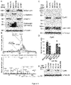
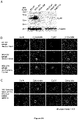
- LC-M1 exhibited mesenchymal characteristics, such as high expression of vimentin but retained some epithelial attributes such as low expression of certain cytokeratins ( Fig. 1A ).
- basal-like MDA-468 breast cancer cells have a predominant epithelial phenotype but contain a subpopulation of tumour cells that are enriched for mesenchymal marker proteins like vimentin (see below).
- HIF-1 ⁇ hypoxia-inducible factor 1-alpha
- PD-L1 programmed cell death 1 ligand 1
- CTC CTC
- PD-L1 can be induced by 0.5 % of O 2 via HIF-1a (26)
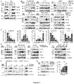
- a potential PD-L1 induction for O 2 concentrations relevant for the bone marrow (1 % O 2 ) or by Co 2+- treatment We observed a PD-L1 induction in BC-M1 and Hs578t under 1 % O 2 ( Fig.
- PD-L1 is transiently induced ( Fig. 5D ) in response to Co 2+- treatment. Further, extraordinary high levels of PD-L1 were observed in MDA-231 BR ( Fig. 5E ). This shows that DTC with a phenotype like BC-M1 are able to maintain PD-L1 levels under hypoxic conditions as present in the bone marrow in vivo and confirms that our model is compatible with the situation in patients in this respect.
- pancreatic cancer In contrast, in pancreatic cancer the metastatic spread frequently occurs via peritoneal dissemination (27).
- pancreatic cancer cell lines we observed an inverse correlation between the Cyr61 and PD-L1 levels ( Fig. 6B ).
- pancreatic cancer is one example of tumour cell entities, that can rise from inflammation (pancreatitis) leading to the dissemination of pre-malignant cells (28).
- pancreatitis inflammation
- pre-malignant cells 28.
- ErbB-2 positive tumours show only a low number of individual tumour cells with mesenchymal attributes (4).
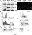
- ErbB-2 might affect the acquisition of mesenchymal attributes and Cyr61 levels ( Fig. 7A, B ).
- ErbB-2 was overexpressed in MDA-468 and the resulting cell line was named MDA-468 ErbB-2.
- MDA-468 control cells carried an expression vector without insert and MDA-468 PM expressed an ErbB-2 protein in which tyrosine 1248 was replaced by phenylalanine (12). Under standard cell culture conditions, the Cyr61 expression was reduced in MDA-468 ErbB-2 compared with MDA-468 control.
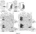
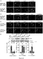
- MDA-ErbB-2 After exposition to hypoxic conditions the Cyr61 expression values in MDA-ErbB-2 decreased even further. Compared with MDA-468 control, MDA-ErbB-2 and MDA-468 PM showed an attenuation of the vimentin induction under hypoxic conditions. This was confirmed on the cellular level by immunofluorescent staining for vimentin ( Fig. 7C, D ).
- Cyr61 receptors For the induction of a biological function of secreted Cyr61, the cells need the presence of suitable Cyr61 receptors.
- Cyr61 receptors the heterodimer of integrin ⁇ v and integrin ⁇ 3 (integrin ⁇ v ⁇ 3) in one of the best investigated Cyr61 receptors (34).
- Cyr61 receptor integrin ⁇ v ⁇ 3 integrin ⁇ v ⁇ 3
- MCF-7 where integrin ⁇ 3 was not detectable, all investigated cell lines have the structural prerequisites for the formation of a functional Cyr61-integrin ⁇ v ⁇ 3 complex.
- the DTC cell lines exhibit very high levels of integrin ⁇ v and integrin ⁇ 3.
- the bone metastatic sublines of MDA-231, MDA-231 SA and MDA-231 B02 display elevated levels of integrin ⁇ v and integrin ⁇ 3 compared with the parental cell line.
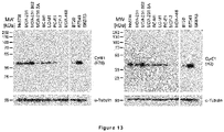
- H78 the quality of the anti-Cyr61 antibody (H78) was compared with an anti-Cyr61 antibody from Cell Signalling (CST) by Western Blot ( Fig. 10A ).
- CST Cell Signalling
- Fig. 10A Western Blot
- Both antibodies detected a signal at a molecular mass of approx. 40 kDa, which was in both cases the predominant signal in the Western Blots.
- the H78 antibody provided clearer signals with fewer background signals compared with the CST antibody, in particular in cell lysates with low Cyr61 levels like MDA-MB-468.
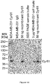
- the H78 antibody was further analyzed by application of human recombinant Cyr61 in a dilution series ( Fig. 10B ).
- the Western Blots showed a band at a molecular mass of approximately 40 kDa both in the cell lysates and in the samples of the recombinant Cyr61 protein.
- the observed molecular masses are compatible with a predicted molecular mass of processed (cleaved signal peptide) of approx. 39.5 kDa.
- MCF-7 is a cell line with no detectable Cyr61 signals, but with some degree of unspecific background.
- Cyr61 was not detectable in MCF-7 cell lysates analyzed by Western Blot, we assumed that Cyr61 is not synthesized in MCF-7 (or only at very low levels). Therefore, it is probable to assume that Cyr61 is not secreted in MCF-7 so that the supernatant can be applied for background subtraction similar to the experiments performed on the cell lysates.
- BC-M1 whole cell lysate contain 62.2 ng ⁇ 0.6 ng Cyr61.
- the inherent advantage of the Western Blot approach is the ability to discriminate between specific Cyr61 signals and unspecific background signals due to protein separation in the SDS PAGE.
- the ELISA approach does not contain a similar fractionation step so that it is more difficult to discriminate between true Cyr61 signals and unspecific background. This issue could be handled by the application of the highly specific anti-Cyr61 (H78) antibody and by introduction of a background noise reduction step. Therefore it has to be kept in mind that the Cyr61 values for MCF-7 are zero by definition, even though that after massive over exposition of the X-ray films after Western Blot very faint signals in the MCF-7 cell lysate were detected. However, these signals could also be unspecific binding.
- Cyr61 ELISA Biological analyses on cell lines by the Cyr61 ELISA. Measurements of Cyr61 values in cell lines by Western Blot refer to the applied protein amount on the SDS PAGE. Since this approach is not applicable to secreted proteins, an alternative standardized reference system for secreted Cyr61 was established. This system includes the cell number and the cultivation time so that the Cyr61 secretion rate can be determined as the Cyr61 secretion rate in number of Cyr61 molecules per cell and day.
- the cells quickly secreted Cyr61 into the medium (0 h vs. 24 h). After that, the Cyr61 concentration is remarkably constant over time and appears to be specific for the individual cell line.
- BC-M1 and MDA-MB-231 Due to the unexpected rapid secretion of Cyr61, the experiment was repeated for BC-M1 and MDA-MB-231 with modifications ( Fig 12B ). In that case, after every 12 h the cell culture medium was replaced by fresh medium and the harvested medium was analyzed for Cyr61.
- the Cyr61 secretion rate increased in MDA-MB-231 after repeated medium withdrawal over time ( Fig. 12B ).
- the cells secreted within 12 h almost the Cyr61 amount detected in Fig. 12A for 24 h ( Fig 12A : 0 h vs. 24 h 999 ng / ml and Fig. 12B : 48 h 954 ng / ml).
- cysteine-rich angiogenic inducer 61 cysteine-rich angiogenic inducer 61 (Cyr61)
- two anti-Cyr61 antibodies were applied.
- One antibody was the rabbit polyclonal anti-Cyr61 antibody H78 which was raised against the amino acids 163-240 of human Cyr61.
- the other antibody was the mouse monoclonal anti-Cyr61 antibody H2 which was raised against the amino acids 345-381 of human Cyr61. Both antibodies were from Santa Cruz Biotechnology, Santa Cruz, USA.
- the Western Blots showed one single band at a molecular mass of approximately 40 kDa in the cell lysates for both antibodies.
- the observed molecular masses are compatible with a predicted molecular mass of processed (cleaved signal peptide) of approx. 39.5 kDa.
- additional signals were observed at molecular mass of approx. 80 kDa. Since Cyr61 contains five disulfide bridges, these signals might be Cyr61 dimers linked by disulfide bridges that could not be completely cleaved in the sample preparation.
- the detected Cyr61 amounts by ELISA could be compared with those detected by Western Blot.
- MDA-231 and Hs578t approximately 100 pg / ⁇ g Cyr61 were detected using the H78 antibody, and using the H2 antibody, the Cyr61 amounts were 250 pg / ⁇ g Cyr61 by Western Blot.
- the ELISA provided values of approximately 83 pg / ⁇ g Cyr61 for MDA-231 and Hs578t.
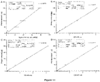
- Equation 2 was used for the determination of the Cyr61 amounts in cell lysates and cell culture supernatants of a set of test samples of MCF-7 ( Fig. 19 ).
- MDA-231 cell lysates The analyses for the MDA-231 cell lysates are shown in Fig. 21A and for the culture supernatants are shown in Fig. 21B .
- One microgram MDA-231 cell lysate contained 80 pg Cyr61 in these measurements ( Fig. 21A ), which may serve as a reference value for the spiking experiments with recombinant Cyr61.
- Fig. 21A contained 80 pg Cyr61 in these measurements
- Cyr61 For example, after spiking of 50 pg recombinant Cyr61 into 1 ⁇ g MDA-231 cell lysate 140 pg Cyr61 were detected in the sample, which is 10 pg more than expected.
- the cell culture supernatants Fig.
- the ELISA provided slightly higher values than expected when the Cyr61 amount is low. This could be seen in the dilution series of MDA-231 culture supernatant, where an increase of the sample volume by factor 10 led to less than an increase by factor 10 of the calculated Cyr61 amount.
- Cyr61-ELISA Since plasma samples may contain other proteins than cell culture samples, we investigated if the established Cyr61-ELISA can be applied to the analysis of patient samples.
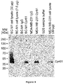
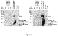
- the Cyr61 status in the plasma of healthy control individuals was tested by Western Blot using the H78 antibody ( Fig. 22 ). This may provide information of unspecific binding of the anti-Cyr61 antibody to other proteins.
- the application of 100 ⁇ l of blood plasma resulted in massive distortion of the SDS-gel bands (not shown), we applied a protein purification step. To confirm that Cyr61 is recovered in this purification step, 100 ng of recombinant Cyr61 was spiked to one aliquot of the patient samples.
- the Cyr61 ELISA was applied to the analysis of plasma samples from healthy donors.
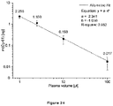
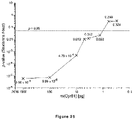
- the detection limit of the Cyr61 ELISA was determined using plasma samples of healthy female individuals over the age of 50 years. Since for the analysis of breast cancer patients 2.5 ⁇ l of plasma were applied, the experiments shown in Fig. 25 were also performed using 2.5 ⁇ l of plasma. In this experiment, recombinant Cyr61 was spiked into plasma samples of healthy individuals and the detected OD value was compared with OD values of plasma without spiked Cyr61. These analyses showed that the probability of a correct discrimination of Cyr61 positive samples from negative samples decreased with decreasing amount of Cyr61.
- Cyr61 concentration of 520 pg/ml The application of 1.3 pg Cyr61 in 2.5 ⁇ l of plasma is a Cyr61 concentration of 520 pg/ml.
- the calculated Cyr61 concentration was 1.707 ng/ml.
- the stability of Cyr61 was analyzed in plasma samples of breast cancer patients.
- theses measurements provided first insights into the Cyr61 concentration in the plasma of breast cancer patients ( Fig. 26 ).
- the first sample set comprised plasma samples of breast cancer patients with no visible signs of overt metastases (stage M0) which were acquired in the years 1998 and 1999 ( Fig. 26A ).
- stage M0 overt metastases
- a second sample set was analyzed.
- the second set consisted of fresh plasma samples from metastatic breast cancer patients at the time of diagnosis (M1, Fig. 26B ) that were analyzed immediately after procurement of the blood samples in October 2016. All samples were stored at -80 °C and were thawed for the analysis of the Cyr61 concentration by application of 2.5 ⁇ l of plasma.
- Cyr61 concentrations ranged from 58.3 ng/ml to 286.9 ng/ml with an average value of 141.1 ⁇ 78.1 ng/ml.
- the Cyr61 concentration in freshly acquired plasma samples was determined at the time of acquisition (October 2016), in April 2017 and August 2017 ( Fig. 26B ). Overall, the Cyr61 concentration remained remarkably constant over time and was similar to the initial Cyr61 concentration (October 2016) in the subsequent measurements. The initial Cyr61 concentration in the samples that were acquired 1998 and 1999 ( Fig. 26A ) was unknown. Indeed, the Cyr61 concentrations determined in October 2016, April 2017 and August 2017 were similar in each sample for each time point. For the majority of the samples a slight increase of the Cyr61 concentrations was observed in the repeated measurements, which may be due to gradual advances in the handling of the ELISA over time.
- the established Cyr61 ELISA is suitable for the detection of Cyr61 from cell line samples (whole cell lysate and culture supernatants) as well from patient samples.
- the critical components of an ELISA - the two primary antibodies H2 and H78 - showed specific binding to Cyr61 in these samples using recombinant human Cyr61 protein as a control.
- both antibodies provided similar quantitative results in Western Blots compared with the ELISA showing that we have established a very homogeneous and robust system that can be used in different applications.
- the H2 antibody As the catching antibody that binds to the Cyr61 present in the sample. Using immunoprecipitation we could show that the H2 is able to catch approximately 100 % of the Cyr61 in present in the sample.
- ELISA shows a linear dynamic range over several orders of magnitude, e. g. from 1 to 1000 pg Cyr61, which allows the reliable quantification of Cyr61 from very dilute and concentrated samples in one single experiment.
- the detection limit of the ELISA is lower than 1.3 pg of Cyr61 in plasma samples. To our experience, this sensitivity is by far sufficient to analyze plasma samples from a variety of different cancers like breast, prostate, lung or liver cancer.
- EXAMPLE 10 Analyses of the Cyr61 levels in blood plasma of breast cancer patients by the Cyr61 ELISA. Since the proteome of blood samples differs from those of cancer cell lines, we investigated if the established Cyr61-ELISA can be applied to the analysis of blood samples. For the detection of soluble Cyr61 in the blood, the blood plasma was analyzed.
- Cyr61 status in the plasma of healthy control individuals was tested by Western Blot ( Fig. 12C ). Similar to the analyses on cell lines, this may provide information of unspecific binding of the anti-Cyr61 antibody. Since the application of 100 ⁇ l of blood plasma resulted in massive distortion of the SDS-gel bands, we applied a protein purification step for the samples analyzed here. To confirm that Cyr61 is recovered in this purification step, 100 ng of recombinant Cyr61 was spiked to one aliquot of the patient samples.
- the Cyr61-ELISA was applied as previously described to blood plasma samples ( Fig. 27 ).
- 100 ⁇ l of plasma were analyzed in triplicates and the values were calculated to the Cyr61 concentration [ng / ml].
- the patient samples were analyzed without protein purification step.
- the plasma from 14 arbitrarily chosen irrespective of sex, age...; Fig. 27A
- healthy individuals was analyzed.
- 100 ng of recombinant Cyr61 was analyzed.
- the calculated Cyr61 concentrations in the samples from the healthy control persons ranged from 2.27 ⁇ 0.35 ng / ml to 5.71 ⁇ 0.60 ng / ml.
- the average value of all 14 samples was 3.47 ⁇ 1.40 ng / ml.
- the Cyr61 concentrations in the plasma ranged from 1.93 ⁇ 0.39 ng / ml to 3.98 ⁇ 0.66 ng / ml.
- the average value of all 20 samples was 3.07 ⁇ 0.26 ng / ml.
- the Cyr61 ELISA can be applied to the analysis of plasma samples from cancer patients.
- Plasma samples of healthy control persons - in particular female individuals with an age over 50 years - showed very low Cyr61 values. These values are comparable to the values of cell culture medium without tumour cells, which was analyzed as an unspecific control. Nevertheless, we have not performed a background subtraction for the analyses on plasma samples so that the actual values for human samples might be slightly lower than calculated.
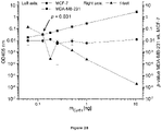
- the cell culture supernatants of MDA-MB-231 and MCF-7 were applied in a dilution series.
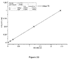
- a calibration curve was derived. This calibration curve was applied to a sample of cell culture medium in which 0.5 ng of recombinant Cyr61 was spiked. Application of the calibration curve to the extinction of this sample provided a value of 0.48 ng Cyr61.
- Cyr61 assay As a first application of this Cyr61 assay we compared samples of five breast cancer patients from the time of diagnosis and no signs of overt metastasis with five samples of healthy individuals. For the beast cancer patients we detected an average value of 10.3 ng / ml ⁇ 9.2 ng / ml and for the healthy individuals a value of 1.0 ng / ml ⁇ 0.4 ng / ml was observed.
- Cyr61 For the detection of Cyr61 by ELISA, the mayor components are the anti-Cyr61 antibody (H78) rabbit polyclonal (Santa Cruz Biotechnology, Santa Cruz, USA), the anti-Cyr61 antibody, mouse monoclonal (clone 365108) from R & D Systems (Minneapolis, USA) and an ELISA reader (e. g. NanoQuant infinite M200 pro, Tecan, Gurnnedorf, Switzerland at wavelengths suitable for the individual substrate applied). Positive controls are cell culture supernatants from MDA-MB-231 or Hs578t or commercially available recombinant human Cyr61 (Abnova, Taipei, Taiwan).
- the values for the supernatants from these cell lines should range between 0.9-1.4 ⁇ l / ml.
- the cell culture supernatants should be kept at least for 48 h on the cells.
- Negative controls may be cell culture supernatants from MCF-7 or GI101 or fresh cell culture medium.
- An appropriate approach for the analysis of human plasma sample is the generation of a dilution series using recombinant Cyr61 that is spiked into blood plasma of healthy control persons which is compared with blood plasma alone.
- the Cyr61 concentrations in blood plasma usually range from 1 ng / ml (healthy persons), 10 ng / ml (breast cancer without metastasis) to 100 ng / ml (metastatic breast cancer).
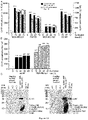
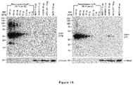
- Cyr61 expression in DTC and CTC freshly obtained from cancer patients was compared with those of mononuclear blood or bone marrow cells from healthy volunteers ( Fig. 29 ).
- Cyr61 levels of breast cancer cells were compared with those of mononuclear blood or bone marrow cells from healthy volunteers ( Fig. 29 ).
- Fig. 29A mononuclear blood cells by western blot
- cell lines were spiked as an internal positive control to mononuclear blood (MDA-231 or BC-M1) or bone marrow cells (MDA-231) from healthy volunteers.
- the Cyr61 signals in MDA-231 and BC-M1 were clearly distinct from the blood or bone marrow cells ( Fig. 29B ).
- Technical evaluation of the method on a small set of three bone marrow samples of DTC positive breast cancer patients revealed that few DTC show Cyr61 signals, whereas the majority of the DTC show very weak or no detectable Cyr61 signals ( Fig. 29C ).
- Sensitive Cyr61 detection was obtained by a novel label-free technique that enriches CTCs by their size and allows a reliable subsequent microscopic detection of even low Cyr61 signal intensities ( Fig. 30 ).
- Tumor cells were detected by cytokeratin staining ( Fig. 30A ) and additional counterstaining of leukocytes with CD45 antibodies ensured assay specificity, even in the case when cytokeratin expression was weak ( Fig. 30B ) which is important to detect CTCs undergoing an EMT.
- Cyr61 weakly positive or Cyr61 negative CTC commonly showed a filamentous cytokeratin pattern with high signal intensity that spans through the entire cytoplasm ( Fig. 30C lower panel).
- MDA-468 and MDA-231 kept the Cyr61 levels nearly constant, suggesting that corresponding CTC phenotypes would reach secondary sites only with minor alterations in the Cyr61 levels.
- BC-M1 down regulates Cyr61 in response to reoxygenation to 16 % (1 % O 2 for 14 days + 4 h 10 % O 2 vs. 14 days 1 % O 2 ) suggesting that cells with a BC-M1 phenotype may be able to reach distant sites with other Cyr61 phenotypes than those they started their passage with.
- Cyr61 as a relevant protein that sensitively responds to microenvironmental stress conditions such as hypoxia and glucose starvation in disseminating cancer cells.
- Cyr61 concentrations in the blood plasma are already elevated in early stages of breast cancer compared with sex and age matched healthy control individuals.
- Our ELISA assay also shows superior performance compared to mammography as the current standard tool for breast cancer screening. For the DCIS subgroup, sensitivities of 58%-81% and specificities of 72%-79% using mammography were recently reported (40). Hence, analysis of only 2.5 ⁇ l of blood plasma is sufficient for our Cyr61 ELISA to discriminate between a "healthy" status and an early stage of breast cancer.
- the Cyr61 ELISA assay is a simple and inexpensive alternative to established liquid biopsy assays. In the future, the assay may be a useful tool which could complement and guide the use of mammography in women in breast cancer screening programs.
- Cyr61 is a 42 kDa cytoplasmic protein that can be secreted to the extracellular space and increased Cyr61 levels can be already observed in early breast cancer without any signs of distant overt metastases. We could show that the detection of secreted Cyr61 in blood plasma is of potential diagnostic use for early detection and risk assessment of breast cancer (see below).
- Cyr61 is involved in differentiation, migration and in the induction of angiogenesis or control of cell proliferation.
- breast cancer elevated Cyr61 expression was reported to be associated with high invasiveness in cell lines and with formation of metastasis after injection of these cell lines in mice.
- proteins may be regulated under microenvironmental stress like hypoxia, which also affects the induction of mesenchymal attributes and tumor cell dissemination and metastasis.
- hypoxia a microenvironmental stress
- 1 % of O 2 sufficient to stabilize HIF-1 ⁇ in most human cells.
- BC-M1 and Hs578t all analyzed cell lines downregulated Cyr61 under these conditions. In BC-M1 and Hs578t, Cyr61 downregulation was only observed upon strong stabilization of HIF-1 ⁇ by Co 2+ -treatment.
- Cyr61 might not solely be implicated into the metastasis to the bone, but also to other distant sites, in particular the brain.
- Brain metastasis is of special interest in the therapy of breast cancer metastasis, since therapeutic antibodies like Herceptin are unable to pass the blood-brain barrier.
- Cyr61 detection in CTC and DTC might also be a suitable surrogate marker for the capacity of CTC and DTC to escape from immune cell destruction.
- pancreatic cancer cell lines were positive for Cyr61, suggesting frequent expression of Cyr61 in pancreatic cancer.
- the metastatic spread frequently occurs via peritoneal dissemination in pancreatic cancer.
- EMT and dissemination can be already occur at the inflammatory stage of pancreatitis, thus in a pre-malignant stage of pancreatic cancer.
- Cyr61 is up-regulated during EMT at the stage of pancreatitis and that Cyr61 is suitable to detect pancreatic diseases already at the inflammatory stage.
- Cyr61 protein levels in clinical specimens were assessed by immunohistochemistry. Scoring staining intensity revealed that 24 % of primary breast tumours exhibited a strong Cyr61 expression, which is similar to previous observations by other groups. Cyr61 expression was not correlated to the detection of DTC in bone marrow, which can be explained by the versatile regulation of Cyr61 depending on the actual microenvironmental conditions.
- Cyr61 was subsequently focused on the expression of Cyr61 in disseminating cancer cells of breast cancer patients.
- CTC CTC
- sensitive multiplex immunostaining assays were developed, which allowed us to detect even tumor cells with low Cyr61 levels.
- Cyr61 levels were marked heterogeneity of Cyr61 levels in CTCs both with regard to inter and intra-patient variability. This finding might reflect the fact that CTC are derived from various sites and may have encounter various microenvironmental conditions affecting Cyr61 expression.
- cytokeratin detection in CTC/DTC we applied here a sensitive and broad range pan-cytokeratin antibody cocktail.
- This cocktail is suitable to detect cytokeratin in the DTC cell lines with downregulated expression of cytokeratins typical for adenocarcinomas (12, 29).
- cytokeratins typical for adenocarcinomas (12, 29).
- no Cyr61 positive/cytokeratin negative cells were found, supporting a previous finding that breast cancer cells with a complete lack of all cytokeratin proteins are very rare (32). It furthermore underlines the absence of Cyr61 in hematopoietic cells and points to the potential usefulness of Cyr61 as marker for CTC/DTC.
- Cyr61 detection in tumour cells with mesenchymal attributes might support the identification of mCTC/mDTC.
- the blood plasma of age-matched healthy persons displayed very low levels of secreted Cyr61 detected with the Cyr61 ELISA as compared to significantly higher values in breast cancer patients. This important finding suggests that elevated Cyr61 levels in the blood plasma are an attribute of breast cancer cells.
- tumour cells may secrete considerable amounts of Cyr61 into the extracellular space; in particular under conditions of persistent fluid exchange elevated amounts of Cyr61 are secreted. In patients, this would resemble to a situation where tumour cells are in proximity to blood vessels. This may lead to a constant withdrawal of Cyr61 and induction of Cyr61 secretion by the tumour cells. Under these conditions, nascent Cyr61 might be immediately secreted so that Cyr61 cannot accumulate within the cells.
- the sensitivity of the ELISA together with the Cyr61 secretion under liquid exchange conditions may allow the detection of Cyr61 in the blood plasma even when the tumour cells show low levels of Cyr61 or when tumour cell colonies are relatively small.
- ELISA-based detection of Cyr61 in blood plasma may be suitable for the early detection of breast cancer, which could help to reduce the number of mammographies in current screening programs of women over the age of 50 years. This would considerably reduce health costs and potential side effects caused by radiation.
- CYR61 levels increase with tumour progression (i.e., higher values in M1 vs. Mo patients), indicating a potential use as blood-based marker to assess and monitor the risk of progression towards metastasis - the leading cause of cancer-related death - and monitor the efficacy of antimetastatic therapies (36).
Landscapes
- Health & Medical Sciences (AREA)
- Life Sciences & Earth Sciences (AREA)
- Immunology (AREA)
- Engineering & Computer Science (AREA)
- Urology & Nephrology (AREA)
- Biomedical Technology (AREA)
- Hematology (AREA)
- Chemical & Material Sciences (AREA)
- Molecular Biology (AREA)
- Cell Biology (AREA)
- Biotechnology (AREA)
- Microbiology (AREA)
- Food Science & Technology (AREA)
- Medicinal Chemistry (AREA)
- Physics & Mathematics (AREA)
- Analytical Chemistry (AREA)
- Biochemistry (AREA)
- General Health & Medical Sciences (AREA)
- General Physics & Mathematics (AREA)
- Pathology (AREA)
- Measuring Or Testing Involving Enzymes Or Micro-Organisms (AREA)
- Investigating Or Analysing Biological Materials (AREA)
- Micro-Organisms Or Cultivation Processes Thereof (AREA)
Applications Claiming Priority (4)
| Application Number | Priority Date | Filing Date | Title |
|---|---|---|---|
| EP17157020.3A EP3364190A1 (de) | 2017-02-20 | 2017-02-20 | Verfahren zum nachweis von krebs oder krebszellen |
| PCT/EP2018/054052 WO2018150031A1 (en) | 2017-02-20 | 2018-02-19 | Method of detecting cancer or cancer cells |
| EP18705153.7A EP3583425B1 (de) | 2017-02-20 | 2018-02-19 | Verfahren zur diagnose von brustkrebs |
| EP21200664.7 | 2021-10-04 |
Related Parent Applications (2)
| Application Number | Title | Priority Date | Filing Date |
|---|---|---|---|
| EP18705153.7A Division EP3583425B1 (de) | 2017-02-20 | 2018-02-19 | Verfahren zur diagnose von brustkrebs |
| EP21200664.7 Division | 2017-02-20 | 2021-10-04 |
Publications (4)
| Publication Number | Publication Date |
|---|---|
| EP4057007A2 true EP4057007A2 (de) | 2022-09-14 |
| EP4057007A3 EP4057007A3 (de) | 2022-10-12 |
| EP4057007C0 EP4057007C0 (de) | 2024-11-13 |
| EP4057007B1 EP4057007B1 (de) | 2024-11-13 |
Family
ID=58098534
Family Applications (3)
| Application Number | Title | Priority Date | Filing Date |
|---|---|---|---|
| EP17157020.3A Withdrawn EP3364190A1 (de) | 2017-02-20 | 2017-02-20 | Verfahren zum nachweis von krebs oder krebszellen |
| EP18705153.7A Active EP3583425B1 (de) | 2017-02-20 | 2018-02-19 | Verfahren zur diagnose von brustkrebs |
| EP22152294.9A Active EP4057007B1 (de) | 2017-02-20 | 2018-02-19 | Verfahren zur diagnose von brustkrebs |
Family Applications Before (2)
| Application Number | Title | Priority Date | Filing Date |
|---|---|---|---|
| EP17157020.3A Withdrawn EP3364190A1 (de) | 2017-02-20 | 2017-02-20 | Verfahren zum nachweis von krebs oder krebszellen |
| EP18705153.7A Active EP3583425B1 (de) | 2017-02-20 | 2018-02-19 | Verfahren zur diagnose von brustkrebs |
Country Status (5)
| Country | Link |
|---|---|
| US (1) | US12584920B2 (de) |
| EP (3) | EP3364190A1 (de) |
| JP (1) | JP7177096B2 (de) |
| ES (2) | ES3006332T3 (de) |
| WO (1) | WO2018150031A1 (de) |
Families Citing this family (5)
| Publication number | Priority date | Publication date | Assignee | Title |
|---|---|---|---|---|
| EP3364190A1 (de) | 2017-02-20 | 2018-08-22 | Panka Cancer Research AG | Verfahren zum nachweis von krebs oder krebszellen |
| EP3938788A4 (de) | 2019-02-12 | 2023-01-04 | Medpacto, Inc. | Verfahren zur diagnose von krebs mittels anti-bag2-antikörper |
| CN110361536A (zh) * | 2019-07-04 | 2019-10-22 | 昆山汇先医药技术有限公司 | 一种肿瘤细胞表面标志分子pd-l1的检测方法 |
| CN114277138B (zh) * | 2020-03-30 | 2022-06-24 | 中国医学科学院肿瘤医院 | 外泌体arpc5、mboat2等在肺癌诊断中的应用 |
| CN111537726A (zh) * | 2020-05-29 | 2020-08-14 | 武汉大学 | 高效定量检测细胞外囊泡中pd-l1水平的方法、elisa试剂盒及使用方法 |
Citations (2)
| Publication number | Priority date | Publication date | Assignee | Title |
|---|---|---|---|---|
| US20040086504A1 (en) | 2001-06-21 | 2004-05-06 | Deepak Sampath | Cyr61 as a target for treatment and diagnosis of breast cancer |
| US20110189700A1 (en) | 2005-02-18 | 2011-08-04 | Moses Marsha A | Cyr61 as a biomarker for diagnosis and prognosis of cancers of epithelial origin |
Family Cites Families (17)
| Publication number | Priority date | Publication date | Assignee | Title |
|---|---|---|---|---|
| CA2423413A1 (en) | 2000-09-29 | 2002-04-04 | Wyeth | Use of cyr61 in the treatment and diagnosis of human uterine leiomyomas |
| ATE459886T1 (de) | 2003-07-11 | 2010-03-15 | Robert Zeillinger | Verfahren zur vorhersage der metastasefähigkeit von mammakarzinom |
| CA2561338A1 (en) | 2004-03-26 | 2005-10-20 | Merck & Co., Inc. | Method and biomarkers for detecting tumor endothelial cell proliferation |
| US20050241951A1 (en) | 2004-04-30 | 2005-11-03 | Kenneth Crouse | Selective catalytic activation of non-conductive substrates |
| US20100035240A1 (en) | 2004-08-10 | 2010-02-11 | Cardiff Biologicals Limited | Methods and kit for the prognosis of breast cancer |
| US20070099209A1 (en) | 2005-06-13 | 2007-05-03 | The Regents Of The University Of Michigan | Compositions and methods for treating and diagnosing cancer |
| EP2706358A3 (de) | 2008-12-23 | 2014-05-07 | Merck Patent GmbH | Biomarker für Hemmer mit antiangiogener Aktivität |
| TW201102086A (en) | 2009-06-04 | 2011-01-16 | Hoffmann La Roche | Antibodies against human CCN1 and uses thereof |
| US20110223616A1 (en) | 2010-03-12 | 2011-09-15 | The Curators Of The University Of Missouri | HuR-Associated Biomarkers |
| EP2678448A4 (de) * | 2011-02-22 | 2014-10-01 | Caris Life Sciences Luxembourg Holdings S A R L | Zirkulierende biomarker |
| US9526800B2 (en) | 2012-03-28 | 2016-12-27 | Massachusetts Institute Of Technology | Cancer-related extracellular matrix signatures and related methods and products |
| CN103940997B (zh) * | 2014-03-21 | 2017-01-04 | 上海柏慧康生物科技有限公司 | 一种乳腺癌循环肿瘤细胞检测系统及试剂盒 |
| RU2715038C2 (ru) * | 2014-07-11 | 2020-02-21 | Дженентек, Инк. | Антитела анти-pd-l1 и способы их диагностического применения |
| CA2990687A1 (en) | 2015-06-29 | 2017-01-05 | Syndax Pharmaceuticals, Inc. | Combination of hdac inhibitor and anti-pd-l1 antibody for treatment of cancer |
| US9567399B1 (en) * | 2016-06-20 | 2017-02-14 | Kymab Limited | Antibodies and immunocytokines |
| US12130290B2 (en) * | 2016-11-29 | 2024-10-29 | Trustees Of Tufts College | Compositions and methods for diagnosing breast cancer |
| EP3364190A1 (de) | 2017-02-20 | 2018-08-22 | Panka Cancer Research AG | Verfahren zum nachweis von krebs oder krebszellen |
-
2017
- 2017-02-20 EP EP17157020.3A patent/EP3364190A1/de not_active Withdrawn
-
2018
- 2018-02-19 EP EP18705153.7A patent/EP3583425B1/de active Active
- 2018-02-19 ES ES22152294T patent/ES3006332T3/es active Active
- 2018-02-19 JP JP2019565978A patent/JP7177096B2/ja active Active
- 2018-02-19 EP EP22152294.9A patent/EP4057007B1/de active Active
- 2018-02-19 US US16/486,913 patent/US12584920B2/en active Active
- 2018-02-19 WO PCT/EP2018/054052 patent/WO2018150031A1/en not_active Ceased
- 2018-02-19 ES ES18705153T patent/ES2902749T3/es active Active
Patent Citations (2)
| Publication number | Priority date | Publication date | Assignee | Title |
|---|---|---|---|---|
| US20040086504A1 (en) | 2001-06-21 | 2004-05-06 | Deepak Sampath | Cyr61 as a target for treatment and diagnosis of breast cancer |
| US20110189700A1 (en) | 2005-02-18 | 2011-08-04 | Moses Marsha A | Cyr61 as a biomarker for diagnosis and prognosis of cancers of epithelial origin |
Non-Patent Citations (38)
| Title |
|---|
| ALIX-PANABIERESPANTEL: "Challenges in Circulating Tumor Cell Research", NATURE REVIEWS, vol. 14, 2014, pages 623 - 631, XP055360601 |
| AMINOLOLAMA-SHAKERI S ET AL.: "Can Radiologists Predict the Presence of Ductal Carcinoma In Situ and Invasive Breast Cancer?", AJR AM J ROENTGENOL, vol. 208, no. 4, April 2017 (2017-04-01), pages 933 - 939 |
| BARDELLI APANTEL K: "Liquid Biopsies, What We Do Not Know (Yet", CANCER CELL, vol. 31, no. 2, 2017, pages 172 - 179, XP029919259, DOI: 10.1016/j.ccell.2017.01.002 |
| BARSOUM IBSMALLWOOD CASIEMENS DR ET AL.: "A mechanism of hypoxia-mediated escape from adaptive immunity in cancer cells", CANCER RES, vol. 74, 2014, pages 665 - 674, XP055528003, DOI: 10.1158/0008-5472.CAN-13-0992 |
| BARTKOWIAK KEFFENBERGER KEHARDER SANDREAS ABUCK FPETER-KATALINIC J ET AL.: "Discovery of a novel unfolded protein response phenotype of cancer stem/progenitor cells from the bone marrow of breast cancer patients", J PROTEOME RES, vol. 9, no. 6, 2010, pages 3158 - 68, XP002602188, DOI: 10.1021/PR100039D |
| BARTKOWIAK KWIECZOREK MBUCK FHARDER SMOLDENHAUER JEFFENBERGER KE ET AL.: "Two-dimensional differential gel electrophoresis of a cell line derived from a breast cancer micrometastasis revealed a stem/ progenitor cell protein profile", J PROTEOME RES, vol. 8, no. 4, 2009, pages 2004 - 14 |
| BARTKOWIAK, K.KWIATKOWSKI, M.BUCK, F.GORGES, T. M. ET AL.: "Disseminated Tumor Cells Persist in the Bone Marrow of Breast Cancer Patients through Sustained Activation of the Unfolded Protein Response", CANCER RES, vol. 75, 2015, pages 5367 - 5377 |
| BENDASBORSICK: "Cancer Cell Adhesion and Metastasis, Selectins, Integrins and the Inhibitory Potential of Heparins", INTERNATIONAL JOURNAL OF CELL BIOLOGY, vol. 2012, pages 1 - 10 |
| BIDARD, F.C., PEETERS, D.J., FEHM, T., NOLE, F., GISBERT-CRIADO, R., MAVROUDIS, D., GRI-SANTI, S., GENERALI, D., GARCIA-SAENZ, J.A: "Clinical validity of circulating tumour cells in patients with metastatic breast cancer: a pooled analysis of individual patient data", LANCET ONCOL, vol. 15, 2014, pages 406 - 414 |
| BRAUN SVOGL FDNAUME BJANNI WOSBORNE MPCOOMBES RC ET AL.: "A pooled analysis of bone marrow micrometastasis in breast cancer", N ENGL J MED, vol. 353, no. 8, 2005, pages 793 - 802 |
| DERY MA, JODOIN J, URSINI-SIEGEL J, ALEYNIKOVA O, FERRARIO C, HASSAN S: "Endoplasmic reticulum stress induces PRNP prion protein gene expression in breast cancer", BREAST CANCER RES, vol. 15, no. 2, 2013, pages R22, XP021146266, DOI: 10.1186/bcr3398 |
| DITTMAR THUSEMANN ASCHEWE YNOFER JRNIGGEMANN BZANKER KS ET AL.: "Induction of cancer cell migration by epidermal growth factor is initiated by specific phosphorylation of tyrosine 1248 of c-erbB-2 receptor via EGFR", FASEB J, vol. 16, no. 13, 2002, pages 1823 - 5 |
| EFFENBERGER KEBORGEN EEULENBURG CZBARTKOWIAK KGROSSER ASYNNESTVEDT M ET AL.: "Detection and clinical relevance of early disseminated breast cancer cells depend on their cytokeratin expression pattern", BREAST CANCER RES TREAT, vol. 125, no. 3, 2011, pages 729 - 38, XP019875055, DOI: 10.1007/s10549-010-0911-2 |
| FENYO DBEAVIS RC: "A method for assessing the statistical significance of mass spectrometry-based protein identifications using general scoring schemes", ANAL CHEM, vol. 75, 2003, pages 768 - 774, XP002317156, DOI: 10.1021/ac0258709 |
| GEER LYMARKEY SPKOWALAK JA ET AL.: "Open mass spectrometry search algorithm", J PROTEOME RES, vol. 3, 2004, pages 958 - 964 |
| HEIDI LEDFORD: "Simple blood test detects eight different kinds of cancer", NATURE, 18 January 2018 (2018-01-18) |
| J. D. COHEN ET AL., SCIENCE, 2018 |
| JANNI WVOGL FDWIEDSWANG GSYNNESTVEDT MFEHM TJUCKSTOCK J ET AL.: "Persistence of disseminated tumor cells in the bone marrow of breast cancer patients predicts increased risk for relapse--a European pooled analysis", CLIN CANCER RES, vol. 17, no. 9, 2011, pages 2967 - 76 |
| KOHLBACHER OREINERT KGROPL C ET AL.: "TOPP--the OpenMS proteomics pipeline", BIOINFORMATICS, vol. 23, 2007, pages e191 - 197 |
| LEHTINEN LVAINIO PWIKMAN HREEMTS JHILVO MISSA R ET AL.: "15-Hydroxyprostaglandin dehydrogenase associates with poor prognosis in breast cancer, induces epithelial-mesenchymal transition, and promotes cell migration in cultured breast cancer cells", J PATHOL, vol. 226, no. 4, 2012, pages 674 - 86 |
| LIN ET AL.: "Elevated Expression of Cyr61 Enhances Peritoneal Dissemination of Gastric Cancer Cells Through Integrin a2(31", THE JOURNAL OF BIOLOGICAL CHEMISTRY, vol. 282, 2007, pages 34594 - 34604, XP002565726, DOI: 10.1074/jbc.M706600200 |
| MAZEL MJACOT WPANTEL KBARTKOWIAK KTOPART DCAYREFOURCQ LROSSILLE DMAUDELONDE TFEST TALIX-PANABIERES C: "Frequent expression of PD-L1 on circulating breast cancer cells", MOL ONCOL, 9 June 2015 (2015-06-09) |
| MENG, S.TRIPATHY, D.FRENKEL, E.P.SHETE, S.NAFTALIS, E.ZHUTH, J.F.BEITSCH, P.D.LEITCH, M.HOOVER, S.EUHUS, D.: "Circulating tumor cells in patients with breast cancer dormancy", CLIN CANCER RES, vol. 10, 2004, pages 8152 - 8162, XP055694080, DOI: 10.1158/1078-0432.CCR-04-1110 |
| NAHNSEN SBERTSCH ARAHNENFUHRER J ET AL.: "Probabilistic consensus scoring improves tandem mass spectrometry peptide identification", J PROTEOME RES, vol. 10, 2011, pages 3332 - 3343 |
| NEUHOFF V, AROLD N, TAUBE D: "Improved staining of proteins in polyacrylamide gels including isoelectric focusing gels with clear background at nanogram sensitivity using Coomassie Brilliant Blue G-250 and R-250", ELECTROPHORESIS, vol. 9, 1988, pages 255 - 262, XP001031894, DOI: 10.1002/elps.1150090603 |
| PANTEL KALIX-PANABIERES CRIETHDORF S: "Cancer micrometastases", NAT REV CLIN ONCOL, vol. 6, no. 6, 2009, pages 339 - 51 |
| PANTEL KBRAKENHOFF RH: "Dissecting the metastatic cascade", NAT REV CANCER, vol. 4, no. 6, 2004, pages 448 - 56 |
| PANTEL KDICKMANNS AZIPPELIUS AKLEIN CSHI JHOECHTLEN-VOLLMAR W ET AL.: "Establishment of micrometastatic carcinoma cell lines: a novel source of tumor cell vaccines", J NATL CANCER INST, vol. 87, no. 15, 1995, pages 1162 - 8, XP000611729, DOI: 10.1093/jnci/87.15.1162 |
| PEYRUCHAUD 0WINDING BPECHEUR I ET AL.: "Early detection of bone metastases in a murine model using fluorescent human breast cancer cells: application to the use of the bisphosphonate zoledronic acid in the treatment of osteolytic lesions", J BONE MINER, vol. 16, 2001, pages 2027 - 2034 |
| POLLARI SKAKONEN SMEDGREN H ET AL.: "Enhanced serine production by bone metastatic breast cancer cells stimulates osteoclastogenesis", BREAST CANCER RES TREAT, vol. 125, 2012, pages 421 - 430, XP019868281, DOI: 10.1007/s10549-010-0848-5 |
| RHIM ADMIREK ETAIELLO NMMAITRA ABAILEY JMMCALLISTER FREICHERT MBEATTY GLRUSTGI AKVONDERHEIDE RH: "EMT and dissemination precede pancreatic tumor formation", CELL, vol. 148, no. 1-2, 20 January 2012 (2012-01-20), pages 349 - 61, XP055223014, DOI: 10.1016/j.cell.2011.11.025 |
| STURM MBERTSCH AGROPL C ET AL.: "OpenMS - an open-source software framework for mass spectrometry", BMC BIOINFORMATICS, vol. 9, 2008, pages 163, XP021031732 |
| SUN ET AL.: "Involvement of Cyr61 in Growth, Migration and Metastasis of Prostate Cancer Cells", BRITISH JOURNAL OF CANCER, vol. 99, 2008, pages 1656 - 1667, XP055094309, DOI: 10.1038/sj.bjc.6604712 |
| VELLON, L.MENENDEZ, J.A.LUPU, R: "AlphaVbeta3 integrin regulates heregulin (HRG)-induced cell proliferation and survival in breast cancer", ONCOGENE, vol. 24, 2005, pages 3759 - 3773 |
| WILLIPINSKI-STAPELFELDT BRIETHDORF SASSMANN VWOELFLE URAU TSAUTER G ET AL.: "Changes in Cytoskeletal Protein Composition Indicative of an Epithelial-Mesenchymal Transition in Human Micrometastatic and Primary Breast Carcinoma Cells", CLIN CANCER, vol. 11, no. 22, 2005, pages 8006 - 14, XP055180265, DOI: 10.1158/1078-0432.CCR-05-0632 |
| YACHIDA SIACOBUZIO-DONAHUE CA: "The pathology and genetics of metastatic pancreatic cancer", ARCH PATHOL LAB MED, vol. 133, no. 3, March 2009 (2009-03-01), pages 413 - 22 |
| YU MBARDIA AWITTNER BSSTOTT SLSMAS METING DT ET AL.: "Circulating breast tumor cells exhibit dynamic changes in epithelial and mesenchymal composition", SCIENCE, vol. 339, no. 6119, 2013, pages 580 - 4 |
| YUAN YHILLIARD GFERGUSON T ET AL.: "Cobalt inhibits the interaction between hypoxia-inducible factor-alpha and von Hippel-Lindau protein by direct binding to hypoxia-inducible factor-alpha", J BIOL CHEM, vol. 278, 2003, pages 15911 - 15916 |
Also Published As
| Publication number | Publication date |
|---|---|
| JP2020510844A (ja) | 2020-04-09 |
| JP7177096B2 (ja) | 2022-11-22 |
| ES2902749T3 (es) | 2022-03-29 |
| EP3364190A1 (de) | 2018-08-22 |
| US20200057067A1 (en) | 2020-02-20 |
| EP4057007C0 (de) | 2024-11-13 |
| EP4057007A3 (de) | 2022-10-12 |
| WO2018150031A1 (en) | 2018-08-23 |
| EP3583425B1 (de) | 2021-10-06 |
| US12584920B2 (en) | 2026-03-24 |
| ES3006332T3 (en) | 2025-03-18 |
| EP3583425A1 (de) | 2019-12-25 |
| EP4057007B1 (de) | 2024-11-13 |
Similar Documents
| Publication | Publication Date | Title |
|---|---|---|
| Rossi et al. | PHGDH heterogeneity potentiates cancer cell dissemination and metastasis | |
| Gao et al. | Acetylation-dependent regulation of PD-L1 nuclear translocation dictates the efficacy of anti-PD-1 immunotherapy | |
| Arrieta et al. | ERK1/2 phosphorylation predicts survival following anti-PD-1 immunotherapy in recurrent glioblastoma | |
| Hsu et al. | STT3-dependent PD-L1 accumulation on cancer stem cells promotes immune evasion | |
| Harris et al. | Exosomes released from breast cancer carcinomas stimulate cell movement | |
| EP4057007B1 (de) | Verfahren zur diagnose von brustkrebs | |
| Di Vizio et al. | Oncosome formation in prostate cancer: association with a region of frequent chromosomal deletion in metastatic disease | |
| Vardaki et al. | Periostin is identified as a putative metastatic marker in breast cancer-derived exosomes | |
| Leiphrakpam et al. | Ezrin expression and cell survival regulation in colorectal cancer | |
| Dianzani et al. | Extracellular proteasome-osteopontin circuit regulates cell migration with implications in multiple sclerosis | |
| Samuel et al. | Ewing sarcoma family of tumors-derived small extracellular vesicle proteomics identify potential clinical biomarkers | |
| Iwahashi et al. | Cytoplasmic p53 aggregates accumulated in p53-mutated cancer correlate with poor prognosis | |
| JP5893037B2 (ja) | Brafv600eに特異的に結合する抗体を使用する癌の診断のための手段及び方法 | |
| Ke et al. | Mechanical signal modulates prostate cancer immune escape by USP8-mediated ubiquitination-dependent degradation of PD-L1 and MHC-1 | |
| Wang et al. | HER2 status of CTCs by peptide-functionalized nanoparticles as the diagnostic biomarker of breast cancer and predicting the efficacy of anti-HER2 treatment | |
| Qin et al. | Exosomal PSM-E inhibits macrophage M2 polarization to suppress prostate cancer metastasis through the RACK1 signaling axis | |
| Hyeon et al. | Proteogenomic characterization of molecular and cellular targets for treatment-resistant subtypes in locally advanced cervical cancers | |
| Xue et al. | Peptidomic analysis of endometrial tissue from patients with ovarian endometriosis | |
| US20190154694A1 (en) | Detection and treatment of cancer | |
| Ziegler et al. | Proteomic surfaceome analysis of mesothelioma | |
| Zhao et al. | A four amino acid intracellular motif of VISTA blocks growth receptor signaling in cancer cells to induce tumor suppression | |
| US20160313335A1 (en) | Methods for the Prognosis of Breast Cancer | |
| Ji et al. | Dual-target CAR-T therapy for ovarian cancer: synergistic targeting of MSLN and B7H3 enhances anti-tumor efficacy and overcomes antigen heterogeneity | |
| US20180030145A1 (en) | Methods of detecting cancer | |
| Zhang et al. | LAG3 as a marker of immune activation in esophageal squamous carcinoma treated with concurrent chemoradiotherapy |
Legal Events
| Date | Code | Title | Description |
|---|---|---|---|
| PUAI | Public reference made under article 153(3) epc to a published international application that has entered the european phase |
Free format text: ORIGINAL CODE: 0009012 |
|
| STAA | Information on the status of an ep patent application or granted ep patent |
Free format text: STATUS: THE APPLICATION HAS BEEN PUBLISHED |
|
| PUAL | Search report despatched |
Free format text: ORIGINAL CODE: 0009013 |
|
| AC | Divisional application: reference to earlier application |
Ref document number: 3583425 Country of ref document: EP Kind code of ref document: P |
|
| AK | Designated contracting states |
Kind code of ref document: A2 Designated state(s): AL AT BE BG CH CY CZ DE DK EE ES FI FR GB GR HR HU IE IS IT LI LT LU LV MC MK MT NL NO PL PT RO RS SE SI SK SM TR |
|
| AK | Designated contracting states |
Kind code of ref document: A3 Designated state(s): AL AT BE BG CH CY CZ DE DK EE ES FI FR GB GR HR HU IE IS IT LI LT LU LV MC MK MT NL NO PL PT RO RS SE SI SK SM TR |
|
| RIC1 | Information provided on ipc code assigned before grant |
Ipc: G01N 33/574 20060101AFI20220905BHEP |
|
| STAA | Information on the status of an ep patent application or granted ep patent |
Free format text: STATUS: REQUEST FOR EXAMINATION WAS MADE |
|
| 17P | Request for examination filed |
Effective date: 20230412 |
|
| RBV | Designated contracting states (corrected) |
Designated state(s): AL AT BE BG CH CY CZ DE DK EE ES FI FR GB GR HR HU IE IS IT LI LT LU LV MC MK MT NL NO PL PT RO RS SE SI SK SM TR |
|
| STAA | Information on the status of an ep patent application or granted ep patent |
Free format text: STATUS: EXAMINATION IS IN PROGRESS |
|
| 17Q | First examination report despatched |
Effective date: 20230704 |
|
| GRAP | Despatch of communication of intention to grant a patent |
Free format text: ORIGINAL CODE: EPIDOSNIGR1 |
|
| STAA | Information on the status of an ep patent application or granted ep patent |
Free format text: STATUS: GRANT OF PATENT IS INTENDED |
|
| INTG | Intention to grant announced |
Effective date: 20240605 |
|
| GRAS | Grant fee paid |
Free format text: ORIGINAL CODE: EPIDOSNIGR3 |
|
| GRAA | (expected) grant |
Free format text: ORIGINAL CODE: 0009210 |
|
| STAA | Information on the status of an ep patent application or granted ep patent |
Free format text: STATUS: THE PATENT HAS BEEN GRANTED |
|
| AC | Divisional application: reference to earlier application |
Ref document number: 3583425 Country of ref document: EP Kind code of ref document: P |
|
| AK | Designated contracting states |
Kind code of ref document: B1 Designated state(s): AL AT BE BG CH CY CZ DE DK EE ES FI FR GB GR HR HU IE IS IT LI LT LU LV MC MK MT NL NO PL PT RO RS SE SI SK SM TR |
|
| REG | Reference to a national code |
Ref country code: GB Ref legal event code: FG4D |
|
| REG | Reference to a national code |
Ref country code: CH Ref legal event code: EP |
|
| REG | Reference to a national code |
Ref country code: DE Ref legal event code: R096 Ref document number: 602018076647 Country of ref document: DE |
|
| REG | Reference to a national code |
Ref country code: IE Ref legal event code: FG4D |
|
| U01 | Request for unitary effect filed |
Effective date: 20241204 |
|
| U07 | Unitary effect registered |
Designated state(s): AT BE BG DE DK EE FI FR IT LT LU LV MT NL PT RO SE SI Effective date: 20241213 |
|
| REG | Reference to a national code |
Ref country code: ES Ref legal event code: FG2A Ref document number: 3006332 Country of ref document: ES Kind code of ref document: T3 Effective date: 20250318 |
|
| U20 | Renewal fee for the european patent with unitary effect paid |
Year of fee payment: 8 Effective date: 20250224 |
|
| PG25 | Lapsed in a contracting state [announced via postgrant information from national office to epo] |
Ref country code: HR Free format text: LAPSE BECAUSE OF FAILURE TO SUBMIT A TRANSLATION OF THE DESCRIPTION OR TO PAY THE FEE WITHIN THE PRESCRIBED TIME-LIMIT Effective date: 20241113 Ref country code: IS Free format text: LAPSE BECAUSE OF FAILURE TO SUBMIT A TRANSLATION OF THE DESCRIPTION OR TO PAY THE FEE WITHIN THE PRESCRIBED TIME-LIMIT Effective date: 20250313 |
|
| PG25 | Lapsed in a contracting state [announced via postgrant information from national office to epo] |
Ref country code: NO Free format text: LAPSE BECAUSE OF FAILURE TO SUBMIT A TRANSLATION OF THE DESCRIPTION OR TO PAY THE FEE WITHIN THE PRESCRIBED TIME-LIMIT Effective date: 20250213 |
|
| PG25 | Lapsed in a contracting state [announced via postgrant information from national office to epo] |
Ref country code: GR Free format text: LAPSE BECAUSE OF FAILURE TO SUBMIT A TRANSLATION OF THE DESCRIPTION OR TO PAY THE FEE WITHIN THE PRESCRIBED TIME-LIMIT Effective date: 20250214 |
|
| PG25 | Lapsed in a contracting state [announced via postgrant information from national office to epo] |
Ref country code: PL Free format text: LAPSE BECAUSE OF FAILURE TO SUBMIT A TRANSLATION OF THE DESCRIPTION OR TO PAY THE FEE WITHIN THE PRESCRIBED TIME-LIMIT Effective date: 20241113 |
|
| PG25 | Lapsed in a contracting state [announced via postgrant information from national office to epo] |
Ref country code: RS Free format text: LAPSE BECAUSE OF FAILURE TO SUBMIT A TRANSLATION OF THE DESCRIPTION OR TO PAY THE FEE WITHIN THE PRESCRIBED TIME-LIMIT Effective date: 20250213 |
|
| PG25 | Lapsed in a contracting state [announced via postgrant information from national office to epo] |
Ref country code: SM Free format text: LAPSE BECAUSE OF FAILURE TO SUBMIT A TRANSLATION OF THE DESCRIPTION OR TO PAY THE FEE WITHIN THE PRESCRIBED TIME-LIMIT Effective date: 20241113 |
|
| PG25 | Lapsed in a contracting state [announced via postgrant information from national office to epo] |
Ref country code: SK Free format text: LAPSE BECAUSE OF FAILURE TO SUBMIT A TRANSLATION OF THE DESCRIPTION OR TO PAY THE FEE WITHIN THE PRESCRIBED TIME-LIMIT Effective date: 20241113 |
|
| PG25 | Lapsed in a contracting state [announced via postgrant information from national office to epo] |
Ref country code: CZ Free format text: LAPSE BECAUSE OF FAILURE TO SUBMIT A TRANSLATION OF THE DESCRIPTION OR TO PAY THE FEE WITHIN THE PRESCRIBED TIME-LIMIT Effective date: 20241113 |
|
| PLBE | No opposition filed within time limit |
Free format text: ORIGINAL CODE: 0009261 |
|
| STAA | Information on the status of an ep patent application or granted ep patent |
Free format text: STATUS: NO OPPOSITION FILED WITHIN TIME LIMIT |
|
| 26N | No opposition filed |
Effective date: 20250814 |
|
| REG | Reference to a national code |
Ref country code: CH Ref legal event code: U11 Free format text: ST27 STATUS EVENT CODE: U-0-0-U10-U11 (AS PROVIDED BY THE NATIONAL OFFICE) Effective date: 20260301 |
|
| U20 | Renewal fee for the european patent with unitary effect paid |
Year of fee payment: 9 Effective date: 20260224 |
|
| PGFP | Annual fee paid to national office [announced via postgrant information from national office to epo] |
Ref country code: GB Payment date: 20260219 Year of fee payment: 9 |
|
| PGFP | Annual fee paid to national office [announced via postgrant information from national office to epo] |
Ref country code: ES Payment date: 20260319 Year of fee payment: 9 |
|
| PGFP | Annual fee paid to national office [announced via postgrant information from national office to epo] |
Ref country code: IE Payment date: 20260218 Year of fee payment: 9 |
|
| PGFP | Annual fee paid to national office [announced via postgrant information from national office to epo] |
Ref country code: CH Payment date: 20260301 Year of fee payment: 9 |