EP4060601A1 - Prétraitement d'image pour une image fundoscopique - Google Patents
Prétraitement d'image pour une image fundoscopique Download PDFInfo
- Publication number
- EP4060601A1 EP4060601A1 EP21196275.8A EP21196275A EP4060601A1 EP 4060601 A1 EP4060601 A1 EP 4060601A1 EP 21196275 A EP21196275 A EP 21196275A EP 4060601 A1 EP4060601 A1 EP 4060601A1
- Authority
- EP
- European Patent Office
- Prior art keywords
- image
- fundoscopic
- region
- interest
- eyeball
- Prior art date
- Legal status (The legal status is an assumption and is not a legal conclusion. Google has not performed a legal analysis and makes no representation as to the accuracy of the status listed.)
- Granted
Links
Images


Classifications
-
- G—PHYSICS
- G06—COMPUTING OR CALCULATING; COUNTING
- G06T—IMAGE DATA PROCESSING OR GENERATION, IN GENERAL
- G06T5/00—Image enhancement or restoration
- G06T5/70—Denoising; Smoothing
-
- G—PHYSICS
- G06—COMPUTING OR CALCULATING; COUNTING
- G06T—IMAGE DATA PROCESSING OR GENERATION, IN GENERAL
- G06T5/00—Image enhancement or restoration
- G06T5/90—Dynamic range modification of images or parts thereof
-
- G—PHYSICS
- G06—COMPUTING OR CALCULATING; COUNTING
- G06T—IMAGE DATA PROCESSING OR GENERATION, IN GENERAL
- G06T5/00—Image enhancement or restoration
- G06T5/73—Deblurring; Sharpening
-
- G—PHYSICS
- G06—COMPUTING OR CALCULATING; COUNTING
- G06N—COMPUTING ARRANGEMENTS BASED ON SPECIFIC COMPUTATIONAL MODELS
- G06N20/00—Machine learning
-
- G—PHYSICS
- G06—COMPUTING OR CALCULATING; COUNTING
- G06T—IMAGE DATA PROCESSING OR GENERATION, IN GENERAL
- G06T5/00—Image enhancement or restoration
- G06T5/50—Image enhancement or restoration using two or more images, e.g. averaging or subtraction
-
- G—PHYSICS
- G06—COMPUTING OR CALCULATING; COUNTING
- G06T—IMAGE DATA PROCESSING OR GENERATION, IN GENERAL
- G06T5/00—Image enhancement or restoration
- G06T5/90—Dynamic range modification of images or parts thereof
- G06T5/94—Dynamic range modification of images or parts thereof based on local image properties, e.g. for local contrast enhancement
-
- G—PHYSICS
- G06—COMPUTING OR CALCULATING; COUNTING
- G06T—IMAGE DATA PROCESSING OR GENERATION, IN GENERAL
- G06T7/00—Image analysis
- G06T7/0002—Inspection of images, e.g. flaw detection
- G06T7/0012—Biomedical image inspection
-
- G—PHYSICS
- G06—COMPUTING OR CALCULATING; COUNTING
- G06T—IMAGE DATA PROCESSING OR GENERATION, IN GENERAL
- G06T7/00—Image analysis
- G06T7/10—Segmentation; Edge detection
- G06T7/11—Region-based segmentation
-
- G—PHYSICS
- G06—COMPUTING OR CALCULATING; COUNTING
- G06T—IMAGE DATA PROCESSING OR GENERATION, IN GENERAL
- G06T7/00—Image analysis
- G06T7/10—Segmentation; Edge detection
- G06T7/13—Edge detection
-
- G—PHYSICS
- G06—COMPUTING OR CALCULATING; COUNTING
- G06V—IMAGE OR VIDEO RECOGNITION OR UNDERSTANDING
- G06V10/00—Arrangements for image or video recognition or understanding
- G06V10/20—Image preprocessing
- G06V10/25—Determination of region of interest [ROI] or a volume of interest [VOI]
-
- G—PHYSICS
- G06—COMPUTING OR CALCULATING; COUNTING
- G06T—IMAGE DATA PROCESSING OR GENERATION, IN GENERAL
- G06T2207/00—Indexing scheme for image analysis or image enhancement
- G06T2207/10—Image acquisition modality
- G06T2207/10004—Still image; Photographic image
-
- G—PHYSICS
- G06—COMPUTING OR CALCULATING; COUNTING
- G06T—IMAGE DATA PROCESSING OR GENERATION, IN GENERAL
- G06T2207/00—Indexing scheme for image analysis or image enhancement
- G06T2207/20—Special algorithmic details
- G06T2207/20004—Adaptive image processing
- G06T2207/20012—Locally adaptive
-
- G—PHYSICS
- G06—COMPUTING OR CALCULATING; COUNTING
- G06T—IMAGE DATA PROCESSING OR GENERATION, IN GENERAL
- G06T2207/00—Indexing scheme for image analysis or image enhancement
- G06T2207/20—Special algorithmic details
- G06T2207/20024—Filtering details
-
- G—PHYSICS
- G06—COMPUTING OR CALCULATING; COUNTING
- G06T—IMAGE DATA PROCESSING OR GENERATION, IN GENERAL
- G06T2207/00—Indexing scheme for image analysis or image enhancement
- G06T2207/20—Special algorithmic details
- G06T2207/20081—Training; Learning
-
- G—PHYSICS
- G06—COMPUTING OR CALCULATING; COUNTING
- G06T—IMAGE DATA PROCESSING OR GENERATION, IN GENERAL
- G06T2207/00—Indexing scheme for image analysis or image enhancement
- G06T2207/20—Special algorithmic details
- G06T2207/20172—Image enhancement details
- G06T2207/20182—Noise reduction or smoothing in the temporal domain; Spatio-temporal filtering
-
- G—PHYSICS
- G06—COMPUTING OR CALCULATING; COUNTING
- G06T—IMAGE DATA PROCESSING OR GENERATION, IN GENERAL
- G06T2207/00—Indexing scheme for image analysis or image enhancement
- G06T2207/20—Special algorithmic details
- G06T2207/20172—Image enhancement details
- G06T2207/20192—Edge enhancement; Edge preservation
-
- G—PHYSICS
- G06—COMPUTING OR CALCULATING; COUNTING
- G06T—IMAGE DATA PROCESSING OR GENERATION, IN GENERAL
- G06T2207/00—Indexing scheme for image analysis or image enhancement
- G06T2207/30—Subject of image; Context of image processing
- G06T2207/30004—Biomedical image processing
- G06T2207/30041—Eye; Retina; Ophthalmic
-
- G—PHYSICS
- G06—COMPUTING OR CALCULATING; COUNTING
- G06T—IMAGE DATA PROCESSING OR GENERATION, IN GENERAL
- G06T2207/00—Indexing scheme for image analysis or image enhancement
- G06T2207/30—Subject of image; Context of image processing
- G06T2207/30004—Biomedical image processing
- G06T2207/30096—Tumor; Lesion
-
- G—PHYSICS
- G06—COMPUTING OR CALCULATING; COUNTING
- G06V—IMAGE OR VIDEO RECOGNITION OR UNDERSTANDING
- G06V40/00—Recognition of biometric, human-related or animal-related patterns in image or video data
- G06V40/10—Human or animal bodies, e.g. vehicle occupants or pedestrians; Body parts, e.g. hands
- G06V40/18—Eye characteristics, e.g. of the iris
- G06V40/193—Preprocessing; Feature extraction
Definitions
- the disclosure relates to an image processing technology, and particularly relates to an image pre-processing method and an image processing apparatus for a fundoscopic image.
- Medical images are images taken of specific parts of organisms, and these images may be used to assess the risk or severity of a disease. For example, through fundoscopic photographic examinations, diseases such as retinopathy, glaucoma, macular disease or the like may be detected early. Generally speaking, most doctors judge lesions in the medical images manually. Although computer-assisted evaluation of medical images is now possible, breakthroughs are needed in terms of indicators such as efficiency, complexity, and accuracy.
- Embodiments of the disclosure provide an image pre-processing method and an image processing apparatus for a fundoscopic image, in which features can be enhanced, thereby improving accuracy of subsequent identification of lesions or other features.
- the image pre-processing method includes (but is not limited to) the following steps.
- a region of interest is obtained from a fundoscopic image to generate a first image.
- the region of interest is focused on an eyeball in the fundoscopic image.
- a smoothing process is performed on the first image to generate a second image.
- a value difference between multiple neighboring pixels is increased to generate a third image.
- the third image is used for image recognition.
- the image processing apparatus includes (but is not limited to) a storage and a processor.
- the storage stores a code.
- the processor is coupled to the storage.
- the processor loads and executes the code to be configured to obtain a region of interest from a fundoscopic image to generate a first image, perform a smoothing process on the first image to generate a second image, and increase a value difference between multiple neighboring pixels in the second image to generate a third image.
- the region of interest is focused on an eyeball in the fundoscopic image.
- the third image is used for image recognition.
- the image pre-processing method and the image processing apparatus for a fundoscopic image according to the embodiments of the disclosure, before image recognition is performed, the region of interest is cut out from an initial fundoscopic image, and the smoothing process and value enhancement are further performed. In this way, features may be enhanced, noises may be reduced, and accuracy of the subsequent image recognition may be improved.
- FIG. 1 is a block diagram of components of an image processing apparatus 100 according to an embodiment of the disclosure.
- the image processing apparatus 100 includes (but is not limited to) a storage 110 and a processor 130.
- the image processing apparatus 100 may be a desktop computer, a notebook computer, a smart phone, a tablet computer, a server, a medical testing instrument, or other computing apparatuses.
- the storage 110 may be any type of fixed or removable random access memory (RAM), read only memory (ROM), flash memory, hard disk drive (HDD), solid-state drive (SSD) or similar components.
- the storage 110 is configured to record a code, a software module, a configuration, data (for example, an image, a value, a reference value, a distance, etc.) or a file, and the embodiments thereof will be described in detail later.
- the processor 130 is coupled to the storage 110.
- the processor 130 may be a central processing unit (CPU), a graphic processing unit (GPU), or other programmable general-purpose or special-purpose microprocessor, digital signal processor (DSP), programmable controller, field programmable gate array (FPGA), application-specific integrated circuit (ASIC), neural network accelerator or other similar components or a combination of the above components.
- the processor 130 is configured to execute all or part of operations of the image processing apparatus 100, and may load and execute each code, the software module, the file, and the data recorded by the storage 110.
- FIG. 2 is a flowchart of an image pre-processing method according to an embodiment of the disclosure.
- the processor 130 may obtain a region of interest from a fundoscopic image to generate a first image (step S210).
- the fundoscopic image is an image obtained by fundoscopic photography of a human or other organism.
- the processor 130 may obtain the fundoscopic image through a built-in or external image capturing apparatus, or the processor 130 may download the fundoscopic image from a server, a computer, or a storage medium. It should be noted that fundoscopic images from different sources may have different shapes or sizes. In order to normalize these fundoscopic images, the processor 130 may first cut out the region of interest considered as important or useful information.
- the region of interest according to the embodiments of the disclosure is focused on an eyeball in the fundoscopic image.
- the processor 130 may locate a center of the eyeball from the fundoscopic image.
- the processor 130 may take a point where the most straight lines in a gradient direction of pixels intersect in the fundoscopic image as a position of the eyeball.
- the processor 130 may perform the Hough transformation on the fundoscopic image to select a circle that best meets the requirements, and determine a center of circle of a contour of the region of interest accordingly.
- the processor 130 may further determine the region of interest according to the center of the eyeball.
- the processor 130 may set a circle that relatively or most conforms to the contour of the eyeball according to the center, and use the contour of the circle as a boundary of the region of interest.
- a circle obtained by the Hough transformation may be used as the region of interest by the processor 130.
- the processor 130 may search for a boundary of the eyeball from the outside to the center of the fundoscopic image. For example, the processor 130 scans the fundoscopic image sequentially from four sides toward the center, and determines lightness of a scanned region. It should be noted that a lightness value (or brightness, that is, the lightness of a color) on one side of the boundary of the eyeball is higher than the other side of the boundary of the eyeball. Generally speaking, a region outside the eyeball in the fundoscopic image has a low lightness value and may be in black.
- the processor 130 may determine that an outermost edge of the region of interest on this side has been found.
- the processor 130 may use the outermost edges respectively found on the four sides of the fundoscopic image as boundary lines. That is, a quadrilateral is formed.
- the processor 130 may use a length of a shortest side of the quadrilateral as a diameter of a circle (taken as the eyeball), and use the circle formed by the diameter as the boundary of the eyeball.
- the center of the circle is the center of the quadrilateral.
- the processor 130 multiplies half the length of the shortest side of the quadrilateral by a floating point number greater than 0 and less than 1 to obtain a radius length, and uses the radius length to obtain a circle. In addition, the center of the circle is still at the center of the quadrilateral. Then, the processor 130 may determine the region of interest according to the boundary of the eyeball. That is, the boundary of the eyeball is taken as the boundary of the region of interest.
- the processor 130 may cut out the region of interest from the fundoscopic image. That is, the processor 130 may delete a region in the fundoscopic image that is not the region of interest.
- the processor 130 may further add a background color outside the region of interest to form the first image. This background color will be considered as useless information in subsequent image recognition (for example, for lesion identification or for severity identification) of the fundoscopic image.
- the useless information may be excluded by feature extraction or may have a relatively low value.
- red, green and blue background colors all consist of values of 128, 64, or 0, but are not limited thereto.
- the size, shape, and/or ratio of the first image may be fixed, thereby normalizing different fundoscopic images.
- the circle may be changed to an ellipse or other geometric figures.
- FIG. 3 is a schematic diagram of the first image according to an embodiment of the disclosure.
- a circular region i.e., the region of interest
- the eyeball corresponds to the eyeball.
- the processor 130 may perform a smoothing process on the first image to generate a second image (step S230).
- the smoothing process is a spatial domain filtering technology capable of directly blurring a pixel in an image and removing noises. For example, a value difference (also referred to as distance) between neighboring pixels may be reduced.
- the smoothing process is Gaussian blur.
- the processor 130 may perform Gaussian blur on the first image. For example, the processor 130 performs a convolution operation on each pixel in the first image by using a Gaussian convolution kernel, and then sums convolution results to obtain the second image.
- FIG. 4 is a schematic diagram of the second image according to an embodiment of the disclosure.
- FIG. 4 shows that details of some noises are blurred by Gaussian blur, while edges of blood vessels, macula, veins and/or arteries remain visible.
- the smoothing processing may also be median filtering, mean filtering, box filtering or other processing.
- the processor 130 may increase a value difference between multiple neighboring pixels in the second image to generate a third image (step S250). Specifically, the smoothing process may narrow the value difference between the neighboring pixels.
- the processor 130 may increase (i.e., update or change) the value difference in proportion to a distance between the value difference between the neighboring pixels and a reference value. For example, when the reference value is 128, the processor 130 may calculate an original value difference of red, green, and blue channel values between each pixel and its neighboring pixel, and compare the distances between the original value differences and the reference value. As the distance is increased, the processor 130 may increase the magnitude of an increase in the value difference.
- the processor 130 may reduce the magnitude of an increase in the value difference.
- the proportion may be 1, 2, 5, or 10 times.
- the processor 130 may change the values of the corresponding pixels so that the value difference between two pixels matches the updated value difference.
- a value to be changed has an upper limit or lower limit.
- the upper limit may be 255 and the lower limit may be 0.
- the changed value exceeds the upper limit or the lower limit, it may be set as a specific value (for example, the upper limit, the lower limit or other values).
- a mathematical relationship between the original value difference and the updated value difference is not limited to a proportional relationship.
- the processor 130 may also adopt a linear relationship, an exponential relationship, or other mathematical relationships depending on actual needs.
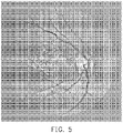
- FIG. 5 is a schematic diagram of the third image according to an embodiment of the disclosure.
- the blood vessels, the macula, the optic disc, the veins, the arteries or other objects in FIG. 5 are more clear.
- the third image may be used for image recognition.
- the processor 130 inputs the third image to a detection model based on a machine learning algorithm (for example, deep neural network (DNN), multi-layer perceptron (MLP), support vector machine (SVM) or other machine learning models).
- the detection model may be used for image recognition.
- the third image may be used in pre-processing in a training phase and/or inference phase of the detection model.
- an initial fundoscopic image is usually immediately subjected to feature extraction without undergoing image pre-processing.
- the image pre-processing according to the embodiments of the disclosure may facilitate identification of a part such as blood vessels, macula, or veins, but the disclosure is not limited to the above.
- the image recognition may be based on scale-invariant feature transform (SIFT), a Haar-like feature, AdaBoost, or other recognition technologies.
- SIFT scale-invariant feature transform
- AdaBoost AdaBoost
- the region of interest in the fundoscopic image is determined, the smoothing process is performed on the image, and the value difference is increased. In this way, features may be enhanced, and the subsequent image recognition, model training, or other image applications may be facilitated.
Landscapes
- Engineering & Computer Science (AREA)
- Theoretical Computer Science (AREA)
- General Physics & Mathematics (AREA)
- Physics & Mathematics (AREA)
- Computer Vision & Pattern Recognition (AREA)
- Medical Informatics (AREA)
- Software Systems (AREA)
- Health & Medical Sciences (AREA)
- Quality & Reliability (AREA)
- Radiology & Medical Imaging (AREA)
- Nuclear Medicine, Radiotherapy & Molecular Imaging (AREA)
- General Health & Medical Sciences (AREA)
- Mathematical Physics (AREA)
- Artificial Intelligence (AREA)
- Evolutionary Computation (AREA)
- Data Mining & Analysis (AREA)
- General Engineering & Computer Science (AREA)
- Computing Systems (AREA)
- Multimedia (AREA)
- Eye Examination Apparatus (AREA)
- Image Analysis (AREA)
- Image Processing (AREA)
- Color Image Communication Systems (AREA)
- Measuring And Recording Apparatus For Diagnosis (AREA)
- Apparatus For Radiation Diagnosis (AREA)
Applications Claiming Priority (1)
| Application Number | Priority Date | Filing Date | Title |
|---|---|---|---|
| TW110109989A TWI775356B (zh) | 2021-03-19 | 2021-03-19 | 用於眼底圖的影像前處理方法及影像處理裝置 |
Publications (2)
| Publication Number | Publication Date |
|---|---|
| EP4060601A1 true EP4060601A1 (fr) | 2022-09-21 |
| EP4060601B1 EP4060601B1 (fr) | 2025-12-10 |
Family
ID=77739002
Family Applications (1)
| Application Number | Title | Priority Date | Filing Date |
|---|---|---|---|
| EP21196275.8A Active EP4060601B1 (fr) | 2021-03-19 | 2021-09-13 | Prétraitement d'image mis en oeuvre par ordinateur pour une image fundoscopique |
Country Status (6)
| Country | Link |
|---|---|
| US (1) | US11954824B2 (fr) |
| EP (1) | EP4060601B1 (fr) |
| JP (1) | JP7337124B2 (fr) |
| CN (1) | CN115115528B (fr) |
| PH (1) | PH12021050543A1 (fr) |
| TW (1) | TWI775356B (fr) |
Cited By (1)
| Publication number | Priority date | Publication date | Assignee | Title |
|---|---|---|---|---|
| EP4557213A1 (fr) * | 2023-11-15 | 2025-05-21 | Koninklijke Philips N.V. | Amélioration de la qualité d'image d'images médicales et fourniture de données d'image médicale à un utilisateur |
Citations (4)
| Publication number | Priority date | Publication date | Assignee | Title |
|---|---|---|---|---|
| US20050169553A1 (en) * | 2000-09-29 | 2005-08-04 | Maurer Ron P. | Image sharpening by variable contrast stretching |
| US20070248277A1 (en) * | 2006-04-24 | 2007-10-25 | Scrofano Michael A | Method And System For Processing Image Data |
| US20150110368A1 (en) * | 2013-10-22 | 2015-04-23 | Eyenuk, Inc. | Systems and methods for processing retinal images for screening of diseases or abnormalities |
| WO2019013779A1 (fr) * | 2017-07-12 | 2019-01-17 | Mohammed Alauddin Bhuiyan | Détection et quantification automatisées de caractéristiques de vaisseaux sanguins pour classement d'image rétinienne et dépistage de maladies |
Family Cites Families (14)
| Publication number | Priority date | Publication date | Assignee | Title |
|---|---|---|---|---|
| JP3618877B2 (ja) | 1996-02-05 | 2005-02-09 | キヤノン株式会社 | 眼科画像処理装置 |
| JP4190221B2 (ja) * | 2002-07-09 | 2008-12-03 | Hoya株式会社 | 画像輪郭強調装置 |
| KR20050025927A (ko) * | 2003-09-08 | 2005-03-14 | 유웅덕 | 홍채인식을 위한 동공 검출 방법 및 형상기술자 추출방법과 그를 이용한 홍채 특징 추출 장치 및 그 방법과홍채인식 시스템 및 그 방법 |
| JP4636841B2 (ja) * | 2004-09-29 | 2011-02-23 | キヤノン株式会社 | 眼科画像撮影装置及び撮影方法 |
| US8320641B2 (en) * | 2004-10-28 | 2012-11-27 | DigitalOptics Corporation Europe Limited | Method and apparatus for red-eye detection using preview or other reference images |
| JP2006263127A (ja) | 2005-03-24 | 2006-10-05 | Gifu Univ | 眼底画像診断支援システム、及び眼底画像診断支援プログラム |
| JP2007117154A (ja) | 2005-10-25 | 2007-05-17 | Pentax Corp | 電子内視鏡システム |
| JP2011035477A (ja) * | 2009-07-29 | 2011-02-17 | Kyocera Corp | 画像処理装置、撮像装置、ノイズ除去方法、並びにプログラム |
| US9454804B2 (en) * | 2012-03-27 | 2016-09-27 | Hitachi Medical Corporation | Image processing device and image processing method |
| JP2015051054A (ja) | 2013-09-05 | 2015-03-19 | キヤノン株式会社 | 画像処理装置、画像処理システム及び画像処理方法 |
| PH12018500541B1 (en) * | 2015-09-11 | 2024-04-12 | Jumio Corp | Method and system for determining a quality metric in biometric authentication |
| CN108229252B (zh) * | 2016-12-15 | 2020-12-15 | 腾讯科技(深圳)有限公司 | 一种瞳孔定位方法及系统 |
| CN108537155B (zh) * | 2018-03-29 | 2021-01-26 | Oppo广东移动通信有限公司 | 图像处理方法、装置、电子设备及计算机可读存储介质 |
| CN111833334A (zh) * | 2020-07-16 | 2020-10-27 | 上海志唐健康科技有限公司 | 一种基于孪生网络架构的眼底影像特征处理分析方法 |
-
2021
- 2021-03-19 TW TW110109989A patent/TWI775356B/zh active
- 2021-04-16 CN CN202110411128.7A patent/CN115115528B/zh active Active
- 2021-04-21 US US17/235,938 patent/US11954824B2/en active Active
- 2021-07-20 JP JP2021119515A patent/JP7337124B2/ja active Active
- 2021-09-13 EP EP21196275.8A patent/EP4060601B1/fr active Active
- 2021-10-25 PH PH1/2021/050543A patent/PH12021050543A1/en unknown
Patent Citations (4)
| Publication number | Priority date | Publication date | Assignee | Title |
|---|---|---|---|---|
| US20050169553A1 (en) * | 2000-09-29 | 2005-08-04 | Maurer Ron P. | Image sharpening by variable contrast stretching |
| US20070248277A1 (en) * | 2006-04-24 | 2007-10-25 | Scrofano Michael A | Method And System For Processing Image Data |
| US20150110368A1 (en) * | 2013-10-22 | 2015-04-23 | Eyenuk, Inc. | Systems and methods for processing retinal images for screening of diseases or abnormalities |
| WO2019013779A1 (fr) * | 2017-07-12 | 2019-01-17 | Mohammed Alauddin Bhuiyan | Détection et quantification automatisées de caractéristiques de vaisseaux sanguins pour classement d'image rétinienne et dépistage de maladies |
Non-Patent Citations (2)
| Title |
|---|
| BESENCZI REN�T� ET AL: "A review on automatic analysis techniques for color fundus photographs", COMPUTATIONAL AND STRUCTURAL BIOTECHNOLOGY JOURNAL, vol. 14, 6 October 2016 (2016-10-06), Sweden, pages 371 - 384, XP055891577, ISSN: 2001-0370, DOI: 10.1016/j.csbj.2016.10.001 * |
| vol. 2526, 10 October 2002, SPRINGER INTERNATIONAL PUBLISHING, ISBN: 978-3-030-41298-2, ISSN: 0302-9743, article WALTER THOMAS ET AL: "Automatic Detection of Microaneurysms in Color Fundus Images of the Human Retina by Means of the Bounding Box Closing", pages: 210 - 220, XP055892492, DOI: 10.1007/3-540-36104-9_23 * |
Cited By (2)
| Publication number | Priority date | Publication date | Assignee | Title |
|---|---|---|---|---|
| EP4557213A1 (fr) * | 2023-11-15 | 2025-05-21 | Koninklijke Philips N.V. | Amélioration de la qualité d'image d'images médicales et fourniture de données d'image médicale à un utilisateur |
| WO2025103839A1 (fr) | 2023-11-15 | 2025-05-22 | Koninklijke Philips N.V. | Amélioration de la qualité d'image d'images médicales et fourniture de données d'image médicale à un utilisateur |
Also Published As
| Publication number | Publication date |
|---|---|
| US20220301111A1 (en) | 2022-09-22 |
| TW202238514A (zh) | 2022-10-01 |
| EP4060601B1 (fr) | 2025-12-10 |
| CN115115528A (zh) | 2022-09-27 |
| CN115115528B (zh) | 2025-02-28 |
| TWI775356B (zh) | 2022-08-21 |
| PH12021050543A1 (en) | 2023-01-09 |
| JP2022145411A (ja) | 2022-10-04 |
| JP7337124B2 (ja) | 2023-09-01 |
| US11954824B2 (en) | 2024-04-09 |
Similar Documents
| Publication | Publication Date | Title |
|---|---|---|
| Sevastopolsky | Optic disc and cup segmentation methods for glaucoma detection with modification of U-Net convolutional neural network | |
| Adem | Exudate detection for diabetic retinopathy with circular Hough transformation and convolutional neural networks | |
| BahadarKhan et al. | A morphological hessian based approach for retinal blood vessels segmentation and denoising using region based otsu thresholding | |
| Rebouças Filho et al. | Automatic histologically-closer classification of skin lesions | |
| CN109753978B (zh) | 图像分类方法、装置以及计算机可读存储介质 | |
| Hsu et al. | Chronic wound assessment and infection detection method | |
| CN107835654B (zh) | 图像处理装置、图像处理方法和记录介质 | |
| Sopharak et al. | Machine learning approach to automatic exudate detection in retinal images from diabetic patients | |
| CN111127425A (zh) | 基于视网膜眼底图像的目标检测定位方法和装置 | |
| CN111882566B (zh) | 视网膜图像的血管分割方法、装置、设备及存储介质 | |
| US20220374947A1 (en) | Artificial intelligence-based system and method for grading collectible trading cards | |
| WO2021136368A1 (fr) | Procédé et appareil pour détecter automatiquement une zone principale pectorale dans une image cible de molybdène | |
| Tavakoli et al. | Unsupervised automated retinal vessel segmentation based on Radon line detector and morphological reconstruction | |
| Toptaş et al. | Detection of optic disc localization from retinal fundus image using optimized color space | |
| US11690569B2 (en) | Blood vessel detecting apparatus and image-based blood vessel detecting method | |
| US11954824B2 (en) | Image pre-processing method and image processing apparatus for fundoscopic image | |
| Tang et al. | Selective search and intensity context based retina vessel image segmentation | |
| Santos et al. | Deep neural network model based on one-stage detector for identifying fundus lesions | |
| Chiamaka Okafor et al. | The effect of image preprocessing algorithms on diabetic foot ulcer classification | |
| Reddy et al. | Implementation of InceptionV3 Model for Skin Disease Detection and Classification | |
| CN118135292A (zh) | 眼底图像分类方法和装置、电子设备及存储介质 | |
| Vasu et al. | Optimal Anisotropic Guided Filtering in retinal fundus imaging: A dual approach to enhancement and segmentation | |
| Sathananthavathi et al. | Improvement of thin retinal vessel extraction using mean matting method | |
| Sabri et al. | Detection, analysis and classification of skin lesions: challenges and opportunities | |
| Pirhala et al. | Segmentation of Significant Regions in Retinal Images: Perspective of U-Net Network Through a Comparative Approach |
Legal Events
| Date | Code | Title | Description |
|---|---|---|---|
| PUAI | Public reference made under article 153(3) epc to a published international application that has entered the european phase |
Free format text: ORIGINAL CODE: 0009012 |
|
| STAA | Information on the status of an ep patent application or granted ep patent |
Free format text: STATUS: THE APPLICATION HAS BEEN PUBLISHED |
|
| STAA | Information on the status of an ep patent application or granted ep patent |
Free format text: STATUS: REQUEST FOR EXAMINATION WAS MADE |
|
| AK | Designated contracting states |
Kind code of ref document: A1 Designated state(s): AL AT BE BG CH CY CZ DE DK EE ES FI FR GB GR HR HU IE IS IT LI LT LU LV MC MK MT NL NO PL PT RO RS SE SI SK SM TR |
|
| 17P | Request for examination filed |
Effective date: 20220826 |
|
| RBV | Designated contracting states (corrected) |
Designated state(s): AL AT BE BG CH CY CZ DE DK EE ES FI FR GB GR HR HU IE IS IT LI LT LU LV MC MK MT NL NO PL PT RO RS SE SI SK SM TR |
|
| STAA | Information on the status of an ep patent application or granted ep patent |
Free format text: STATUS: EXAMINATION IS IN PROGRESS |
|
| 17Q | First examination report despatched |
Effective date: 20250411 |
|
| GRAP | Despatch of communication of intention to grant a patent |
Free format text: ORIGINAL CODE: EPIDOSNIGR1 |
|
| STAA | Information on the status of an ep patent application or granted ep patent |
Free format text: STATUS: GRANT OF PATENT IS INTENDED |
|
| RIC1 | Information provided on ipc code assigned before grant |
Ipc: G06T 5/00 20060101AFI20250626BHEP Ipc: G06T 5/73 20240101ALI20250626BHEP Ipc: G06T 5/94 20240101ALI20250626BHEP Ipc: G06T 7/13 20170101ALI20250626BHEP Ipc: G06T 7/00 20170101ALN20250626BHEP Ipc: A61B 3/12 20060101ALN20250626BHEP |
|
| INTG | Intention to grant announced |
Effective date: 20250710 |
|
| GRAS | Grant fee paid |
Free format text: ORIGINAL CODE: EPIDOSNIGR3 |
|
| GRAA | (expected) grant |
Free format text: ORIGINAL CODE: 0009210 |
|
| STAA | Information on the status of an ep patent application or granted ep patent |
Free format text: STATUS: THE PATENT HAS BEEN GRANTED |
|
| AK | Designated contracting states |
Kind code of ref document: B1 Designated state(s): AL AT BE BG CH CY CZ DE DK EE ES FI FR GB GR HR HU IE IS IT LI LT LU LV MC MK MT NL NO PL PT RO RS SE SI SK SM TR |
|
| REG | Reference to a national code |
Ref country code: CH Ref legal event code: F10 Free format text: ST27 STATUS EVENT CODE: U-0-0-F10-F00 (AS PROVIDED BY THE NATIONAL OFFICE) Effective date: 20251210 Ref country code: GB Ref legal event code: FG4D |
|
| REG | Reference to a national code |
Ref country code: DE Ref legal event code: R096 Ref document number: 602021044005 Country of ref document: DE |
|
| REG | Reference to a national code |
Ref country code: IE Ref legal event code: FG4D |
|
| PG25 | Lapsed in a contracting state [announced via postgrant information from national office to epo] |
Ref country code: ES Free format text: LAPSE BECAUSE OF FAILURE TO SUBMIT A TRANSLATION OF THE DESCRIPTION OR TO PAY THE FEE WITHIN THE PRESCRIBED TIME-LIMIT Effective date: 20251210 |
|
| REG | Reference to a national code |
Ref country code: LT Ref legal event code: MG9D |
|
| PG25 | Lapsed in a contracting state [announced via postgrant information from national office to epo] |
Ref country code: NO Free format text: LAPSE BECAUSE OF FAILURE TO SUBMIT A TRANSLATION OF THE DESCRIPTION OR TO PAY THE FEE WITHIN THE PRESCRIBED TIME-LIMIT Effective date: 20260310 |
|
| PG25 | Lapsed in a contracting state [announced via postgrant information from national office to epo] |
Ref country code: HR Free format text: LAPSE BECAUSE OF FAILURE TO SUBMIT A TRANSLATION OF THE DESCRIPTION OR TO PAY THE FEE WITHIN THE PRESCRIBED TIME-LIMIT Effective date: 20251210 Ref country code: FI Free format text: LAPSE BECAUSE OF FAILURE TO SUBMIT A TRANSLATION OF THE DESCRIPTION OR TO PAY THE FEE WITHIN THE PRESCRIBED TIME-LIMIT Effective date: 20251210 |
|
| REG | Reference to a national code |
Ref country code: NL Ref legal event code: MP Effective date: 20251210 |
|
| PG25 | Lapsed in a contracting state [announced via postgrant information from national office to epo] |
Ref country code: RS Free format text: LAPSE BECAUSE OF FAILURE TO SUBMIT A TRANSLATION OF THE DESCRIPTION OR TO PAY THE FEE WITHIN THE PRESCRIBED TIME-LIMIT Effective date: 20260310 |
|
| PG25 | Lapsed in a contracting state [announced via postgrant information from national office to epo] |
Ref country code: LV Free format text: LAPSE BECAUSE OF FAILURE TO SUBMIT A TRANSLATION OF THE DESCRIPTION OR TO PAY THE FEE WITHIN THE PRESCRIBED TIME-LIMIT Effective date: 20251210 |