EP4062888A1 - Procédé et appareil d'ajustement de la fonction visuelle - Google Patents
Procédé et appareil d'ajustement de la fonction visuelle Download PDFInfo
- Publication number
- EP4062888A1 EP4062888A1 EP20891162.8A EP20891162A EP4062888A1 EP 4062888 A1 EP4062888 A1 EP 4062888A1 EP 20891162 A EP20891162 A EP 20891162A EP 4062888 A1 EP4062888 A1 EP 4062888A1
- Authority
- EP
- European Patent Office
- Prior art keywords
- subregional
- retinal
- eyes
- area
- preset
- Prior art date
- Legal status (The legal status is an assumption and is not a legal conclusion. Google has not performed a legal analysis and makes no representation as to the accuracy of the status listed.)
- Pending
Links
Images


Classifications
-
- A—HUMAN NECESSITIES
- A61—MEDICAL OR VETERINARY SCIENCE; HYGIENE
- A61H—PHYSICAL THERAPY APPARATUS, e.g. DEVICES FOR LOCATING OR STIMULATING REFLEX POINTS IN THE BODY; ARTIFICIAL RESPIRATION; MASSAGE; BATHING DEVICES FOR SPECIAL THERAPEUTIC OR HYGIENIC PURPOSES OR SPECIFIC PARTS OF THE BODY
- A61H5/00—Exercisers for the eyes
- A61H5/005—Exercisers for training the stereoscopic view
-
- G—PHYSICS
- G02—OPTICS
- G02B—OPTICAL ELEMENTS, SYSTEMS OR APPARATUS
- G02B27/00—Optical systems or apparatus not provided for by any of the groups G02B1/00 - G02B26/00, G02B30/00
- G02B27/01—Head-up displays
- G02B27/017—Head mounted
- G02B27/0172—Head mounted characterised by optical features
-
- A—HUMAN NECESSITIES
- A61—MEDICAL OR VETERINARY SCIENCE; HYGIENE
- A61H—PHYSICAL THERAPY APPARATUS, e.g. DEVICES FOR LOCATING OR STIMULATING REFLEX POINTS IN THE BODY; ARTIFICIAL RESPIRATION; MASSAGE; BATHING DEVICES FOR SPECIAL THERAPEUTIC OR HYGIENIC PURPOSES OR SPECIFIC PARTS OF THE BODY
- A61H2201/00—Characteristics of apparatus not provided for in the preceding codes
- A61H2201/50—Control means thereof
- A61H2201/5023—Interfaces to the user
- A61H2201/5043—Displays
-
- G—PHYSICS
- G02—OPTICS
- G02B—OPTICAL ELEMENTS, SYSTEMS OR APPARATUS
- G02B27/00—Optical systems or apparatus not provided for by any of the groups G02B1/00 - G02B26/00, G02B30/00
- G02B27/01—Head-up displays
- G02B27/0101—Head-up displays characterised by optical features
- G02B2027/0127—Head-up displays characterised by optical features comprising devices increasing the depth of field
-
- G—PHYSICS
- G02—OPTICS
- G02B—OPTICAL ELEMENTS, SYSTEMS OR APPARATUS
- G02B27/00—Optical systems or apparatus not provided for by any of the groups G02B1/00 - G02B26/00, G02B30/00
- G02B27/01—Head-up displays
- G02B27/0101—Head-up displays characterised by optical features
- G02B2027/0132—Head-up displays characterised by optical features comprising binocular systems
- G02B2027/0134—Head-up displays characterised by optical features comprising binocular systems of stereoscopic type
-
- G—PHYSICS
- G02—OPTICS
- G02B—OPTICAL ELEMENTS, SYSTEMS OR APPARATUS
- G02B27/00—Optical systems or apparatus not provided for by any of the groups G02B1/00 - G02B26/00, G02B30/00
- G02B27/01—Head-up displays
- G02B27/0179—Display position adjusting means not related to the information to be displayed
- G02B2027/0185—Displaying image at variable distance
Definitions
- the present application relates to the field of electronic technology, and in particular, to a visual function adjustment method and device, and a virtual reality head-mounted display device (HMD).
- HMD virtual reality head-mounted display device
- Eye vision is undoubtedly extremely important, and the retina is a vital part of imaging in an eye.
- Each binocular cell in the visual cortex receives input from the retinas of the left and right eyes.
- the visual images formed by the retina of an eye are encoded by neurons in different visual centers to form neural images in the visual cortex.
- Human visual perception relies heavily on the latter.
- the visual cortex neural circuit has strong plasticity, and appropriate visual stimulation on the retina can gradually restore the visual function of the neural circuit in the corresponding cortical area.
- the retina is divided into a foveal and a peripheral area; the foveal is the area of the retina where vision (color discrimination, resolution) is most acute.
- the macula there is a yellow area called the macula about 3.5mm from the temporal side of the optic disc, and the depression in the center of the macula is the fovea.
- many ophthalmic diseases such as glaucoma, AMD, RP (retinitis pigmentosa) and DR (diabetic retinopathy) can cause localized retinal lesions due to various reasons, the corresponding visual field areas of the cerebral cortex were unable to form normal neural images.
- the area with localized damage to the retina is defined as: at least a part of the retina is damaged, rather than the entire retina is damaged; and "at least a part” means that one or more of the retina is damaged at localized damage to the area.
- the purpose of the embodiments of the present application is to provide a visual function adjustment method and device, and a virtual reality head-mounted display device, which can improve and restore the visual function abnormality in the corresponding region of the cerebral cortex caused by subregional retinal damage. And then achieve the purpose of stimulating the retina to regulate the neural circuit of the brain to adjust the visual function.
- one embodiment of the present application provides a visual function adjustment method, including:
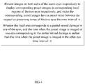
- performing imaging on both eyes of the user to display corresponding preset images respectively in corresponding subregional regions of the two eyes including: Use the video streams to image the two eyes respectively, and the corresponding frames of the video streams of the two eyes include corresponding preset images corresponding to the corresponding localized area positions; and the corresponding frames of the video streams of the two eyes There is a preset time interval ⁇ , so that the preset images of the corresponding frames in the video streams of the two eyes, the imaging time of the corresponding one eye with subregional retinal damage is longer than that of the preset image in the other eye The time of imaging is earlier than the time interval ⁇ .
- the method includes: the frames of each video stream have intervening frames, and the corresponding intervening frames of the video streams of the two sides have the same time interval ⁇ .
- the inserted frame is a blank image frame; or the inserted frame is a frame of a specific image, and the specific image is different from the preset image.
- the method further includes: when the preset image is played, a prompt is output, and feedback from the user for the prompt is received, and the user's concentration is determined according to the feedback.
- the method includes: The prompt is a recognizable image, or the prompt is a recognizable sound.
- the localized area is determined by:
- an embodiment of the present invention provides a method for adjusting the binocular visual function, including: when both eyes have regional retinal damage, determine each regional retinal damage in both eyes separately, and perform the following steps for each regional retinal damage:
- the method includes:
- the method includes: The first subregional region is imaged on both eyes of the user by the preset device, so that the same first preset image is displayed in the corresponding first subregional regions of the two eyes respectively, and after the alternating time interval ⁇ , the second subregional region is imaged respectively.
- the user's eyes are respectively imaged by the preset device to display the same second preset image respectively in the corresponding second subregional regions of the two eyes, and last for alternating time intervals ⁇ ; where time interval ⁇ ⁇ alternate time interval ⁇ .
- the method includes: the first preset image and the second preset image are the same but have different positions; or, the first preset image and the second preset image are different in image. location.
- the localized area is determined by setting P number of points in the non-foveal area of the visual field of one eye, and the luminance range of each point; a fixation target is displayed for the foveal area of the visual field of one eye of the user, and the user is asked to fixate at the fixation target; then the luminance is continuously adjusted at any of the P points to determine whether the user is reached luminance threshold at the point; determine the difference between the luminance of the two eyes at the same corresponding point, and use the luminance difference in the corresponding points of the two eyes to determine the localized area corresponding to the regional retinal damage.
- an embodiment of the present invention provides a method for adjusting a multi-region viewing function, including:
- the localized area corresponds to a subregional retinal damage in one of the eyes, and the time when the preset image is imaged in one eye corresponding to the subregional retinal damage is earlier than the time interval ⁇ when the preset image is imaged in the other eye.
- the user's eyes are imaged respectively to display corresponding preset images in corresponding subregional regions of both eyes respectively, and the corresponding preset images are imaged on both sides of the eyes respectively.
- There is a preset time interval ⁇ between times including:
- the method includes: With respect to the n th subregional area, imaging is performed on both eyes of the user respectively to display the corresponding n th preset image in the corresponding n th subregional area of the two sides of the eyes, respectively; after completing the adjustment of the visual function of both sides of the n th subregional area, for the n+1 th subregional area, the user's two eyes are respectively imaged to display the corresponding images in the n+1 th subregional area of the two eyes, respectively.
- the method includes: The n th subregional region is imaged on both eyes of the user by the preset device to display the corresponding nth preset image respectively in the corresponding subregional regions of the two eyes, and after the alternating time interval ⁇ continues, the n+1 th The subregional area is imaged on both eyes of the user by the preset device to display the corresponding n+1 th preset image in the corresponding subregional area of the two eyes respectively, and last for alternating time intervals ⁇ ; where time interval ⁇ ⁇ alternate time interval ⁇ .
- the method includes:
- the localized area is determined by:
- an embodiment of the present invention provides a method for adjusting a multi-region viewing function, including:
- the user's eyes are respectively imaged to display corresponding preset images in corresponding subregional areas of both eyes respectively, and the corresponding preset images are imaged on the two eyes respectively.
- the method includes:
- the method includes: The n th subregional region is imaged on both eyes of the user by the preset device to display the corresponding n th preset image respectively in the corresponding subregional regions of the two eyes, and after the alternating time interval ⁇ continues, the n+1 th subregional area is imaged on both eyes of the user by the preset device, so as to display the corresponding n+1 th preset image in the corresponding subregional area of the two eyes respectively, and last for alternating time intervals ⁇ ; where time interval ⁇ ⁇ alternate time interval ⁇ .
- the method includes:
- the localized area is determined by:
- an embodiment of the present invention provides a visual function adjustment method using a graph sequence, including:
- the m subregional retinal lesions are located on one eye, or the m subregional retinal lesions are located on both eyes, respectively.
- the image sequence is used to perform imaging on both sides of the user's eyes respectively, so as to display corresponding preset images in corresponding subregional regions of the two eyes respectively, and make the corresponding preset images, there is a preset time interval ⁇ between the respective imaging times of the two eyes, including:
- the method includes:
- the method includes: For the n th subregional area, the user's eyes are imaged respectively to display the corresponding n th preset images in the corresponding subregional areas of the two eyes, and after the alternating time interval ⁇ , the n+1 th subregional area is passed through.
- the preset device performs imaging on the user's eyes respectively to display the corresponding n+1 th preset image in the corresponding subregional regions of the two eyes, and continues the alternating time interval ⁇ ; where time interval ⁇ ⁇ alternate time interval ⁇ .
- the method includes:
- the method further includes: A prompt is output when the sequence of pictures is played, and feedback from the user for the prompt is received, and the user's concentration is determined according to the feedback.
- the method includes: Each of the graph sequences has interpolated frames, and the corresponding interpolated frames of the graph sequences of the two eyes have the same time interval ⁇ between them.
- each point on the binocular area of the retina has a corresponding representative area on the visual cortex; and the representative area gradually becomes larger as the degree of eccentricity decreases.
- the representative area of the fovea is maximized (the 0-0.5° foveal area corresponds to the 142mm 2 area of the primary visual cortex!); at this moment, each point on the fovea is a large representative area in the visual cortex.
- the utilization rate of this area is not very high under normal circumstances, so the embodiment of the present application can adjust the vision.
- the visual cortex neural circuit corresponding to the retinal damage area can be regulated, so that the synapses of the neurons in this circuit can be reshaped, so that the input provided by a small number of remaining retinal neurons can be efficiently used, and finally, to a certain extent Reconstruction of visual function.
- the retina will have localized lesions due to various reasons, so that the position corresponding to the localized retinal damage in the visual field cannot be imaged.
- the problem of visual impairment caused by subregional retinal damage is mostly to eliminate the subregional retinal damage by means of drug and surgical treatment, and to treat or eliminate in this way.
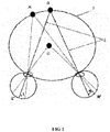
- the stereoscopic vision of people's binocular is formed in this way: both eyes are fixed on an object at the same time, and the eyes of both eyes intersect at a point, which is called a fixation point, and the reflection from the fixation point returns to the retinas of the left and right eyes.
- the light point is called the corresponding point (Corresponding Point). Since there is a distance between the two pupils of a person (the average value is 50-75mm), the relative positions of the left and right eyes are different for the same scene, which leads to different perspectives for the eyes to see external objects, which results in Binocular Disparity, that is, the left and right eyes see images with different viewing angles.
- This visual information from both eyes is transferred to the primary visual cortex through the lateral geniculate body, and then processed and integrated by the visual centers at all levels to obtain a neural image with a sense of three-dimensional depth. Specifically, once the eyes focus on the fixation point, not only can this point be clearly seen, but also the distance, depth, convexity and concavity, contrast, chromatic aberration, contour, etc. between this point and surrounding objects can be distinguished.
- the image is a stereoscopic image, and the stereoscopic function of people's binocular vision is referred to as stereopsis ( Stereopsis ).
- Stereoscopic vision gives a sense of depth to one's perception of the surrounding visual world.
- the eyes have the ability to perceive the depth of the captured external image (Depth Perception), and this ability is based on the depth information (Depth Cue) that the left and right eyes can extract from the scene.
- Depth Perception the ability to perceive the depth of the captured external image
- Depth Cue the depth information that the left and right eyes can extract from the scene.
- the reason why the human eye can have these capabilities mainly depends on the following four basic functions of the eyes:
- Binocular parallax describes the difference in perspective that occurs at the horizontal position where the image of the object falls on the retinas of both eyes when the left and right eyes stare at the same object due to the distance between the two eyes, referred to as binocular parallax.
- the brain uses binocular disparity to extract depth information from 2D retinal images.
- the adaptive adjustment of the eyes mainly refers to the active focusing behavior of the eyes. Mainly used for close-range focusing.
- the focal length of the eye is finely adjusted by changing the curvature of the intraocular lens (Lens). When looking at an object, the curvature of the intraocular lens changes so that the eyes can focus on seeing objects at different distances and different parts of the same scene.
- the adjustment of the lens is achieved through the contraction and relaxation of its attached transcribed muscle.
- Depth perception is determined by the direction of eye gaze. Your eyes form an angle when you look at an object. The closer the object is to you, the larger the angle is, and the farther the object is from you, the smaller the angle. As the object moves further and further away from you, the line of sight of the eyes tends to be parallel.
- Parallel parallax creates a sense of depth through the relative motion between the observer and the observed object. Close one eye and you can only see the outside world through one eye. Extending your thumb when looking at an object with one eye can block part of your field of view, but when you turn your head, you can see that part of the blocked scene. Specifically, when you use parallel parallax to obtain depth-sensing information, the brain uses calculations to determine the relative distance between you and two objects.
- Both glaucoma and macular disease cause localized damage to the retina, where macular disease produces localized retinal damage in the fovea of the retina, and glaucoma produces localized retinal damage in the peripheral area of the fovea.
- macular disease produces localized retinal damage in the fovea of the retina
- glaucoma produces localized retinal damage in the peripheral area of the fovea.
- maculopathy patients will fail to image the localized central region of the central region within the visual field, while glaucoma will fail to image the localized peripheral region in the peripheral region outside the central region of the visual field.
- the embodiment of the present application proposes a visual function adjustment method and device, and a virtual reality head-mounted display device (HMD). Based on the aforementioned visual imaging principle, physical means are used to visually stimulate the visually imaged retina, thereby stimulating the cortex. Adjustment of visual function to gradually improve or even eliminate the impact of localized retinal damage on visual imaging.
- the visual function adjustment method and device, and the HMD device proposed in the embodiments of the present application stimulate the retina by means of localized stimulation.
- the visual function adjustment method includes:
- the corresponding preset image refers to that when the adjustment of the visual function is performed through a plane image, the corresponding preset image refers to the exact same image, that is, the eyes on both sides are, at an interval ⁇ , the same preset image is displayed.
- the corresponding preset images are two images including disparity. This is because in the imaging mechanism of the human cerebral cortex, if the disparity issue is considered, the images displayed on both eyes need to contain a certain disparity to achieve stereoscopic imaging.
- This principle can refer to the existing production principles for movies in HMD, that is, to allow users to form stereoscopic perception through the HMD, it is necessary to display the video images for both eyes. Only after certain disparity processing is performed on the image, the user can generate a stereoscopic perception when viewing the image; that is, the corresponding preset images with disparity cues are displayed by the two eyes respectively.
- an image of an existing HMD simultaneously displays corresponding preset images for both eyes.
- corresponding preset images are displayed on both eyes at the positions corresponding to the regional retinal damage; and the corresponding preset images are displayed on the retinas of both eyes following a sequential order.
- the sequential imaging refers to that the retina position corresponding to the area of subregional retinal damage on one eye with subregional retinal damage is imaged first, and the retina corresponding to the area of subregional retinal damage of the first eye is imaged next (this retinal area corresponding to the location of localized retinal damage in the first eye with no or lesser lesions); and the two are shown with an interval ⁇ .
- the retina for visual imaging can be stimulated, so that the preset image is imaged first in the eye with subregional retinal damage, and then the preset image is presented in the other eye.
- this method can well use localized visual stimulation to stimulate the cerebral cortex through the retina. After a period of stimulation, repeated visual stimulation from the retina will repair the cortical areas that cannot normally form neural images within the visual field of the cerebral cortex corresponding to the subregionally damaged area of the retina, thereby gradually improving or even restoring vision.
- the corresponding localized areas of both eyes refer to the same position of both eyes. For example, if a subregional retinal damage occurs in a localized area of one eye, when using the method of the embodiment of the present application to adjust the visual function, the same localized area of the two eyes is used one after the other. The same preset image is displayed with the localized area corresponding to the localized damage to the retina of one of the eyes.
- a visual function adjustment method including: When both eyes have subregional retinal damage, determine each subregional retinal damage in both eyes separately, and perform the following steps for each subregional retinal damage:
- an image of an existing HMD simultaneously displays corresponding preset images for both eyes.
- corresponding preset images are displayed on both eyes at the position corresponding to the subregional retinal damage; and the corresponding preset images are displayed on the retinas of both eyes.
- the sequential imaging refers to that the retinal position corresponding to the area of subregional retinal damage on one eye is imaged first, and the retinal position corresponding to the area of normal or less damaged on another eye is imaged next, i.e., the other eye corresponding to the location of localized retinal damage in the first eye with no or lesser lesions; and the two are shown with an interval ⁇ .
- the retina for visual imaging can be stimulated, so that the preset image is presented first in the eye with subregional retinal damage, and then the preset image is presented in the other eye. Due to the visual imaging mechanism of the human eye, it is only necessary to set the time interval ⁇ well, so that the human brain will not feel that it is displayed successively, which will cause discomfort.
- this method delivers localized visual stimulation from the retina to stimulate binocular cells in the visual cortex. After a period of stimulation, the retinal visual stimulation will repair the cortical areas that cannot normally form neural images within the visual field of the cerebral cortex corresponding to the subregionally damaged area of the retina, thereby gradually improving or even restoring vision.
- the corresponding localized areas of both eyes refer to the same position of both eyes. For example, if a subregional retinal damage occurs in a localized area of one eye, when using the method of the embodiment of the present application to adjust the visual function, the same localized area of the two eyes is used one after the other. The same preset image is displayed with the localized area corresponding to the localized damage to the retina of one of the eyes.
- the treatment method for subregional retinal damage in both eyes can be consistent with the aforementioned case of multiple subregional retinal damage in one eye, that is, visual function adjustment is performed for each subregional retinal damage in turn, or the time interval ⁇ is used as a cycle. Visual adjustment was performed on multiple retinal localized lesions in turn.
- the user has two subregional retinal damages L1 and L2 in the left eye, and one subregional retinal damage R1 in the right eye; the visual function adjustment at this time can be divided into three stages: the first stage is for the first part of the left eye.
- Subregional retinal damage L1 the playing time is from t0 to t1;
- the second segment is for the subregional retinal damage R1 of the right eye, and the playing time is from t1 to t2;
- the third segment is for the second subregional retinal damage L2 of the left eye, and the playing time is from t1 to t2. t2 to t3. Then go back to the first paragraph.
- a graph sequence can be prepared in advance, and the graph sequence includes the above-mentioned three different preset maps at different positions, so as to adjust the visual function for each subregional retinal damage respectively.
- it can be played in a loop, and the sequence of the three sub-pictures in each loop is not limited in any way.
- the positions of the preset images displayed are different; at the same time, each sub-image can include multiple sub-images displayed at the same position. Different graph sequences.
- the graph sequence produced includes three subgraphs; this is just an example.
- the graph sequence may include N segments of sub-images. That is, the method includes:
- the visual function adjustment is performed on the N subregional regions in turn. That is: after the subregional retinal damage area of one eye is divided into N subregional areas, the corresponding preset image is played for the first subregional area in the eye with subregional retinal damage at time t0, and then at time t0+ ⁇ , the corresponding preset image is also played in the first subregional area of the other eye.
- the corresponding preset image is played in the eye with subregional retinal damage at the t0+ ⁇ th time, and then the corresponding preset image is also presented in the second subregional area of the other eye at the t0+ ⁇ + ⁇ th time, and so on.
- visual function adjustment can be performed on N localized regions in turn.
- an embodiment of the present application proposes a method for adjusting a visual function.
- the visual function adjustment method includes:
- the corresponding preset images are two images with disparity. This is because in the imaging mechanism of the human visual cortex, if the disparity issue is considered, the images displayed when imaging both eyes need to contain a certain disparity to achieve stereoscopic perception.
- This principle can refer to the existing production principles for movies in HMD; that is, in order to allow users to form stereoscopic images through the HMD, it is necessary to display the video for both eyes. Only after certain processing is performed on the image, the user can generate a stereoscopic perception when viewing the image; that is, the corresponding preset images containing disparity are displayed by the two eyes respectively.
- an image of an existing virtual reality head-mounted display device simultaneously displays corresponding preset images for both eyes.
- corresponding preset images are displayed on both eyes at the position corresponding to the subregional retinal damage; and the corresponding preset pattern is sequentially displayed on the retinas of both eyes.
- the sequential imaging refers to that the retina position corresponding to the area of subregional retinal damage on one eye with subregional retinal damage is imaged first, and the retina position corresponding to the area of less damaged or normal on the other eye is imaged next, i.e., the other eye corresponding to the location of localized retinal damage in the first eye with no or lesser lesions; and the two are shown with an interval ⁇ .
- the retina for visual imaging can be stimulated, so that the preset image is presented first in the eye with subregional retinal damage, and then the preset image is displayed in the other eye. Due to the visual imaging mechanism of the human eye, it is only necessary to set the time interval ⁇ well, so that the human brain will not feel that it is displayed successively, which will cause discomfort.
- this method can well use localized visual stimulation to pass through the retina and stimulate the visual cortex. After a period of repeated stimulation, the retinal visual stimulation will repair the cortical visual areas that cannot form normal neural images within the visual field of the visual cortex corresponding to the subregionally damaged area of the retina, thereby gradually improving or even restoring vision.
- the corresponding localized areas of both eyes refer to the same position of both eyes. For example, if a subregional retinal damage occurs in a localized area of one eye, when using the method of the embodiment of the present application to adjust the visual function, the same localized area of the two eyes is stimulated one after the other. The same preset image is displayed with the localized area corresponding to the localized damage to the retina of one of the eyes.
- visual function adjustment can only be performed for one retinal subregional damage in one eye in each time period. This is because the human retinal imaging can only adjust the visual function for one retinal subregional damage in one eye at a time, otherwise the imaging system will be confused. Therefore, after the visual function adjustment of one localized area is completed, the visual function adjustment of another localized area may be performed; and two or more localized areas may be adjusted in turn within the preset time ⁇ .
- the above method includes the following steps:
- the above method may also include other steps, and these other steps may be any one or more steps in other embodiments of the present application, or may not be mentioned in the embodiments of the present application and any steps, and these methods should also fall within the protection scope of the claims of the present application.
- the corresponding preset image refers to that when the adjustment of the visual function is performed through a plane image, the corresponding preset image refers to the exact same image, that is, the eyes on both sides are at the interval ⁇ , the same preset map is displayed.
- the corresponding preset images are two images including disparity. This is because in the imaging mechanism of the human cerebral cortex, if the disparity issue is considered, the images displayed when imaging both eyes need to contain a certain disparity to achieve stereoscopic imaging.
- This principle can refer to the existing production principles for videos in HMD, in order to allow users to form a three-dimensional perception through the HMD, it is necessary to display images for both sides of the eye. Certain processing is performed to enable the user to generate a stereoscopic image when viewing the image; that is, the corresponding preset images including disparity are displayed on both sides of the eyes respectively.
- the pictures of the existing HMD respectively display corresponding preset pictures for both eyes at the same time.
- the embodiment of the present application also proposes another method for adjusting vision, including: Determine the scope of subregional retinal damage in one eye, and divide the scope into N subregional retinal damage areas; and determine a subregional area for displaying a corresponding preset map in both eyes for one of the subregional retinal damage areas. That is to say, a relatively large area of subregional retinal damage can be divided into N subregional retinal damage areas, and visual function adjustment is performed only for one of the N subregional retinal damage areas at a time. That is, the method includes:
- images are presented on both sides of the user's eyes to display the corresponding images in the corresponding n+1 th subregional areas of both sides of the eyes.
- the method of the embodiment of the present application includes: The n th subregional region is imaged on both eyes of the user by the preset device to display the corresponding n th preset image respectively in the corresponding subregional regions of the two eyes, and after the alternating time interval ⁇ continues, the n+1 th The subregional area is imaged on both eyes of the user by the preset device to display the corresponding n+1 th preset image in the corresponding subregional area of the two eyes respectively, and last for alternating time intervals ⁇ ; where time interval ⁇ ⁇ alternate time interval ⁇ .
- the visual function adjustment is performed on the N subregional regions in turn. That is: after the subregional retinal damage area of one eye is divided into N subregional areas, the corresponding preset image is played for the first subregional area in the eye with subregional retinal damage at time t0, and then at time t0+ ⁇ . The corresponding preset image is also played in the first subregional area of the other eye.
- the corresponding preset image is played in the eye with subregional retinal damage at the t0+ ⁇ th time, and then the corresponding preset image is also played in the second subregional area of the other eye at the t0+ ⁇ + ⁇ th time, and so on.
- visual function restoration can be performed on N localized regions in turn.
- the same preset pattern can be used for each localized area. That is, the n th preset image is the same as the n+1 th preset image and has a different location. Or, in order to improve the effect of adjusting the visual function, different images may be displayed for each localized area, that is, the images and positions of the n th preset image and the n+1 th preset image are different.
- the visual function adjustment can be divided into two sections: the first section is for the first section.
- the second segment is for the second subregional retinal injury L1, and the playing time is from t0 to t1; while the second segment is for the second subregional retinal injury L2, and the playing time is from t1 to t2. Then go back to the first paragraph, and so on.
- different preset patterns can be used for each localized area; that is, different preset patterns can be used in two sections.
- a graph sequence can be prepared in advance, and the graph sequence includes the above-mentioned two different preset maps at different positions, so as to adjust the visual function for each subregional retinal damage respectively.
- it can be played in a loop, and the sequence of the two sub-pictures in each loop is not limited in any way.
- the positions of the preset images displayed are different; at the same time, each sub-image can include multiple sub-images displayed at the same position. Different graph sequences.
- the embodiment of the present application proposes a visual function adjustment method and device using a graph sequence, and a HMD.
- a visual function adjustment method and device using a graph sequence, and a HMD.
- physical means are used to visually stimulate the retina of visual imaging, so as to adjust the cortical visual function to gradually improve or even eliminate the impact of localized retinal damage on visual imaging.
- the visual function adjustment method and device, and the HMD proposed in the embodiments of the present application stimulate the retina by means of localized stimulation.
- the visual function adjustment method using the graph sequence includes:
- the visual adjustment is performed on the positions of m subregional retinal lesions among the M subregional retinal lesions by using a graph sequence, including:
- a picture of an existing HMD displays corresponding preset pictures for both eyes at the same time.
- corresponding preset images are displayed on both eyes at the position corresponding to the subregional retinal damage; and the corresponding preset images are sequentially displayed on the retinas of both eyes.
- the sequential imaging refers to that the retina position corresponding to the area of subregional retinal damage on one eye with subregional retinal damage is presented first, and the retina corresponding to the area of subregional retinal damage of the first eye is presented on the other eye next, i.e., the other eye corresponding to the location of localized retinal damage in the first eye with no or lesser lesions; and the two are shown with an interval ⁇ .
- the preset image is first presented in the eye with subregional retinal damage, and then the preset image is imaged in the other eye. Due to the visual imaging mechanism of the human eye, it is only necessary to set the time interval ⁇ very well, so that the human brain will not feel that the display is successively displayed and cause discomfort.
- the corresponding localized areas of both eyes refer to the same position of both eyes. For example, if a subregional retinal damage occurs in a localized area of one eye, when using the method of the embodiment of the present application to adjust the visual function, the same localized area of the two eyes is used one after the other. The same preset map is shown with the localized area corresponding to the localized retinal damage in one of the eyes.
- the m subregional retinal lesions are located on one eye, or the m subregional retinal lesions are located on both eyes, respectively. That is to say: there may be only one eye with subregional retinal damage, or there may be subregional retinal damage in both eyes; and there may be too much scope of subregional retinal damage in any one eye.
- the localized area where the preset map is displayed is uniquely corresponding to the position of the subregional retinal damage of one eye.
- the corresponding preset map is displayed in and only in the predetermined area of the two eyes corresponding to the subregional retinal damage, so as to stimulate the two eyes corresponding to the subregional retinal damage position through the preset map.
- the embodiments of the present application propose a variety of solutions, so as to restore the visual function according to different situations of subregional retinal damage.
- visual function restoration can only be performed for one retinal subregional damage in one eye in each time period. This is because the human retinal imaging can only adjust the visual function for one retinal subregional damage in one eye at a time, otherwise it will cause confusion in the imaging system. Therefore, after the visual function adjustment of one localized area is completed, the visual function adjustment of another localized area may be performed; and two or more localized areas may be adjusted in turn within the preset time ⁇ .
- if two or more localized retinal damages are determined that is, two or more localized areas, in order to improve the effect of visual function adjustment, different localized areas may be damaged.
- Different preset maps are used, that is, a preset map is determined for each subregional retinal damage.
- the image sequence is used to perform imaging on both sides of the user's eyes respectively, so as to display corresponding preset images in corresponding subregional regions of the two eyes respectively, and make the corresponding preset images.
- a graph sequence can be used to first perform visual function adjustment for a localized area, and then perform visual function adjustment for another localized area after completing the visual function adjustment of the localized area, that is, the method includes:
- the visual function restoration can also be performed for each localized area in turn by using a graph sequence, that is, the method includes:
- one or more consecutive frames of a predetermined image are added at a predetermined position in the video stream: for example, a blank frame, Or a frame with a predetermined logo image.
- Adding frames with a predetermined map can provide a buffer for retinal imaging, and can also be used as an interpolation to increase the number of frames in the video stream.
- the ordinary video stream is generally 24 or 25 frames per second or other predetermined frame numbers
- a HMD such as 3D glasses
- the current Mainstream 3D glasses are 90-120 frames per second. Interpolating between the frames of the video stream at this point can turn a video stream of 24 or 25 frames per year into a video stream of 90-120 frames per second.
- the time interval a is 8.33 milliseconds.
- the reason for this setting is that the method of the present application can be implemented by using 3D glasses, because 3D glasses have a natural advantage and can limit the field of view of each glasses of a person to a specific area, which can prevent peripheral light from looking at each other.
- the function adjustment process has an impact; at the same time, the working principle of the 3D glasses is to display images for both eyes separately, so that visual stimulation can be separately applied to the left and right eye channels to realize any of the solutions in the embodiments of the present application.
- the minimum time interval shown in the figure is 1/120 of a second, that is, 8.33 milliseconds.
- time interval ⁇ can be adjusted according to the operating frequency of the device.
- the limited time interval ⁇ must be perfectly corresponding to the operating frequency of the device, and the solution of the embodiment of the present application can also be implemented with a slight difference.
- HMD virtual reality head-mounted display device
- the aforementioned time interval ⁇ of 8.33 milliseconds is only an example, and does not limit the protection scope of the present application.
- the 3D glasses with the working frequency of 120 Hz are just an example, and do not limit the protection scope of the present application.
- the device for implementing the embodiments of the present application may be any device that can display a preset image, which is not limited in the embodiments of the present application.
- a prompt is performed when the corresponding preset image or the corresponding preset image sequence is played, and it is determined whether corresponding feedback from the user for the prompt is received.
- This mechanism may be referred to as a focus detection mechanism to determine that the user's attention is focused on viewing the preset image. This condition is to prevent users from having problems concentrating during long-term use. Because once the user is inattentive, the played preset image cannot produce a predetermined visual function adjustment visual function adjustment effect.
- the prompt may be an icon with a direction prompt displayed in a preset diagram, and user feedback on the icon with a direction prompt is received, so as to determine that the user has made correct feedback.
- a "convex"-shaped graphic is regularly displayed, and the protrusions of the graphic can be oriented in different directions; the user makes a preset action according to the direction of the protrusions in the graphic (for example, draw a predetermined direction on the touchpad, use the Press the corresponding key on the keyboard with the arrow keys, ....).
- the protrusions of the "convex"-shaped figure can be directed to the left or right, and changed in sequence; at the same time, the user can press the left or right mouse button correspondingly.
- the prompt can also be a prompt tone that is played on a regular basis, so that the user can give feedback when the prompt tone is heard.
- the correspondence between the position of the subregional retinal damage and the position where the preset image is displayed can be obtained by various methods.
- the following example is given in the embodiment of the present application for illustration: In this example, the following steps are performed for each eye:
- the minimum adjustment step size may be predetermined, or determined depending on the adjustable parameters of the device, or determined in any other feasible manner.
- the respective contrast difference values of the P points corresponding to the two eyes can be determined, and the point with the largest contrast difference value among the P points can be used as the depending on the site of functional adjustment.
- the maximum value of luminance, and the luminance values in all embodiments of the present application are relative values based on the display luminance interval. Do the following steps for each eye:
- the corresponding relationship between the position of the user's retinal damage and the position where the preset image is displayed can be determined, so as to determine the position where the preset image is displayed.
- the above-mentioned method for determining localized retinal damage is just an example, which is a relatively accurate and repeatable manner.
- Those skilled in the art can understand that other methods can also be used to determine. For example: displaying a subregional map at different positions within the visual field of the user's monocular, and judging whether the user sees the subregional map, so as to determine the location of subregional retinal damage; that is, the exhaustive method.
- the retinal map can be used to determine the location of localized retinal damage.
- the localized area where the preset image is displayed can be determined correspondingly; therefore, the position of the localized area where the preset image is displayed can be determined in many ways.
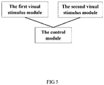
- an embodiment of the present application further proposes a visual function adjustment device, including: a first imaging mechanism for imaging the left eye, a second imaging mechanism for imaging the right eye, and a device for controlling a control mechanism for the first imaging mechanism and the second imaging mechanism;
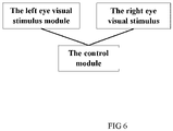
- an embodiment of the present application further proposes a virtual reality head-mounted display device, including: a left-eye imaging mechanism for imaging the left eye, a right-eye imaging mechanism for imaging the right eye, a control mechanism for controlling the left-eye imaging mechanism and the right-eye imaging mechanism;
- the visual function adjustment device shown in FIG 5 and the virtual reality head-mounted display device shown in FIG 6 in the embodiment of the present application can be used in any of the foregoing embodiments. Therefore, the principles, functions, and execution steps of the visual function adjustment device and the virtual reality head-mounted display device in the embodiments of the present application are all corresponding to the visual function adjustment method in any of the foregoing embodiments. Therefore, it will not be repeated here.
Landscapes
- Health & Medical Sciences (AREA)
- Physics & Mathematics (AREA)
- Life Sciences & Earth Sciences (AREA)
- Pain & Pain Management (AREA)
- Physical Education & Sports Medicine (AREA)
- Rehabilitation Therapy (AREA)
- Epidemiology (AREA)
- Animal Behavior & Ethology (AREA)
- General Health & Medical Sciences (AREA)
- Public Health (AREA)
- Veterinary Medicine (AREA)
- Ophthalmology & Optometry (AREA)
- General Physics & Mathematics (AREA)
- Optics & Photonics (AREA)
- Eye Examination Apparatus (AREA)
Applications Claiming Priority (5)
| Application Number | Priority Date | Filing Date | Title |
|---|---|---|---|
| CN201911140283.9A CN110882139B (zh) | 2019-11-20 | 2019-11-20 | 一种利用图序列的视功能调整方法及装置 |
| CN201911140282.4A CN110850596B (zh) | 2019-11-20 | 2019-11-20 | 两侧眼视功能调整装置及虚拟现实头戴式显示设备 |
| CN201911139300.7A CN110812145B (zh) | 2019-11-20 | 2019-11-20 | 一种视功能调整方法及装置、虚拟现实头戴式显示设备 |
| CN201911140281.XA CN110812146B (zh) | 2019-11-20 | 2019-11-20 | 多区域视功能调整方法及装置、虚拟现实头戴式显示设备 |
| PCT/CN2020/125906 WO2021098498A1 (fr) | 2019-11-20 | 2020-11-02 | Procédé et appareil d'ajustement de la fonction visuelle |
Publications (2)
| Publication Number | Publication Date |
|---|---|
| EP4062888A1 true EP4062888A1 (fr) | 2022-09-28 |
| EP4062888A4 EP4062888A4 (fr) | 2023-12-13 |
Family
ID=75981262
Family Applications (1)
| Application Number | Title | Priority Date | Filing Date |
|---|---|---|---|
| EP20891162.8A Pending EP4062888A4 (fr) | 2019-11-20 | 2020-11-02 | Procédé et appareil d'ajustement de la fonction visuelle |
Country Status (3)
| Country | Link |
|---|---|
| US (1) | US20220276495A1 (fr) |
| EP (1) | EP4062888A4 (fr) |
| WO (1) | WO2021098498A1 (fr) |
Families Citing this family (1)
| Publication number | Priority date | Publication date | Assignee | Title |
|---|---|---|---|---|
| AU2021352417A1 (en) * | 2020-09-30 | 2023-04-06 | Acucela Inc. | Myopia prediction, diagnosis, planning, and monitoring device |
Family Cites Families (16)
| Publication number | Priority date | Publication date | Assignee | Title |
|---|---|---|---|---|
| CA2475042C (fr) * | 2002-02-08 | 2011-05-10 | Novavision, Inc. | Procede et dispositif perfectionnes d'entrainement de la vision humaine |
| GB0210288D0 (en) * | 2002-05-04 | 2002-06-12 | Univ Nottingham | Ocular display apparatus for assessment and measurement of and for treatment of ocular disorders, and methods therefor |
| DK3329838T3 (da) * | 2007-10-23 | 2020-11-09 | Univ Mcgill | Vurdering og behandling af binokulært syn |
| CN103239347B (zh) * | 2013-05-09 | 2015-05-13 | 北京大学 | 一种采用调控眼优势治疗视觉功能障碍的系统 |
| TWI559730B (zh) * | 2014-08-25 | 2016-11-21 | 群創光電股份有限公司 | 立體畫面顯示系統及其方法 |
| KR102656342B1 (ko) * | 2015-07-23 | 2024-04-09 | 뉴저지 인스티튜트 오브 테크놀로지 | 사용자의 눈 움직임에 반응하여 시각적 치유 비디오 게임을 제어하기 위한 방법, 시스템 및 장치 |
| US10500124B2 (en) * | 2015-09-03 | 2019-12-10 | University Of Rochester | Methods, devices and systems for orthoptics |
| EP3420889B1 (fr) * | 2017-06-28 | 2020-01-15 | Vestel Elektronik Sanayi ve Ticaret A.S. | Appareil permettant de vérifier la vision d'un utilisateur |
| CN107929006A (zh) * | 2017-11-17 | 2018-04-20 | 广州视景医疗软件有限公司 | 一种视觉训练方法、装置和设备 |
| CN107920244A (zh) * | 2017-11-17 | 2018-04-17 | 广州视景医疗软件有限公司 | 一种视觉训练方法、装置和设备 |
| KR102029423B1 (ko) * | 2018-03-16 | 2019-10-07 | 김선호 | 시각장애 예방 및 치료장치 |
| CN108852766B (zh) * | 2018-04-03 | 2022-11-01 | 山东省看看视力矫治科技有限公司 | 视力矫正装置 |
| CN110812145B (zh) * | 2019-11-20 | 2022-02-22 | 精准视光(北京)医疗技术有限公司 | 一种视功能调整方法及装置、虚拟现实头戴式显示设备 |
| CN110812146B (zh) * | 2019-11-20 | 2022-02-22 | 精准视光(北京)医疗技术有限公司 | 多区域视功能调整方法及装置、虚拟现实头戴式显示设备 |
| CN110850596B (zh) * | 2019-11-20 | 2022-02-18 | 精准视光(北京)医疗技术有限公司 | 两侧眼视功能调整装置及虚拟现实头戴式显示设备 |
| CN110882139B (zh) * | 2019-11-20 | 2022-02-18 | 精准视光(北京)医疗技术有限公司 | 一种利用图序列的视功能调整方法及装置 |
-
2020
- 2020-11-02 EP EP20891162.8A patent/EP4062888A4/fr active Pending
- 2020-11-02 WO PCT/CN2020/125906 patent/WO2021098498A1/fr not_active Ceased
-
2022
- 2022-05-19 US US17/749,069 patent/US20220276495A1/en active Pending
Also Published As
| Publication number | Publication date |
|---|---|
| EP4062888A4 (fr) | 2023-12-13 |
| WO2021098498A1 (fr) | 2021-05-27 |
| US20220276495A1 (en) | 2022-09-01 |
Similar Documents
| Publication | Publication Date | Title |
|---|---|---|
| US12016629B2 (en) | Screening apparatus and method | |
| US20170296421A1 (en) | Head-mounted apparatus and methods for treatment and enhancement of visual function | |
| CN110292515A (zh) | 一种视功能训练的方法及系统 | |
| CN114903760B (zh) | 一种斜视训练设备 | |
| JPWO2018055618A5 (fr) | ||
| Hoffman et al. | Focus information is used to interpret binocular images | |
| CN115280219B (zh) | 强化视力的系统与方法 | |
| WO2022051688A1 (fr) | Systèmes et procédés pour améliorer la vision binoculaire | |
| CN108271011A (zh) | 用于治疗弱视的3d图像系统的参数处理方法及系统和装置 | |
| Iatsun et al. | Investigation of visual fatigue/discomfort generated by S3D video using eye-tracking data | |
| CN113081718A (zh) | 基于生物机制刺激配合的综合视觉训练系统 | |
| CN110850596B (zh) | 两侧眼视功能调整装置及虚拟现实头戴式显示设备 | |
| CN110812145B (zh) | 一种视功能调整方法及装置、虚拟现实头戴式显示设备 | |
| US20220276495A1 (en) | Visual function adjustment method and apparatus | |
| CN110812146B (zh) | 多区域视功能调整方法及装置、虚拟现实头戴式显示设备 | |
| CN110882139B (zh) | 一种利用图序列的视功能调整方法及装置 | |
| CN116942074B (zh) | 基于多屏深度多焦点堆栈模式的视功能评估训练方法 | |
| CN105739090A (zh) | 一种立体观看景深的计算方法 | |
| RU2283071C2 (ru) | Способ восстановления бинокулярного зрения | |
| US20260021009A1 (en) | Multi-tiled plenoptic system for multiple vision treatments and training for the induction of hyper visual acuity | |
| Ishio et al. | Importance of visual distance adjustment for AR application of binocular see-through smart glasses | |
| CN115645238A (zh) | 基于光场和分光场景的多维视野刺激系统及方法 | |
| DIAMOND | –Sensory Status in Strabismus | |
| Vienne | Understanding and Improving the Quality of Experience in 3D media perception: Accommodation/Vergence conflict in Stereopsis | |
| FERNANDEZ-MALOIGNE | Iana IATSUN |
Legal Events
| Date | Code | Title | Description |
|---|---|---|---|
| STAA | Information on the status of an ep patent application or granted ep patent |
Free format text: STATUS: THE INTERNATIONAL PUBLICATION HAS BEEN MADE |
|
| PUAI | Public reference made under article 153(3) epc to a published international application that has entered the european phase |
Free format text: ORIGINAL CODE: 0009012 |
|
| STAA | Information on the status of an ep patent application or granted ep patent |
Free format text: STATUS: REQUEST FOR EXAMINATION WAS MADE |
|
| 17P | Request for examination filed |
Effective date: 20220620 |
|
| AK | Designated contracting states |
Kind code of ref document: A1 Designated state(s): AL AT BE BG CH CY CZ DE DK EE ES FI FR GB GR HR HU IE IS IT LI LT LU LV MC MK MT NL NO PL PT RO RS SE SI SK SM TR |
|
| DAV | Request for validation of the european patent (deleted) | ||
| DAX | Request for extension of the european patent (deleted) | ||
| A4 | Supplementary search report drawn up and despatched |
Effective date: 20231113 |
|
| RIC1 | Information provided on ipc code assigned before grant |
Ipc: A61H 5/00 20060101AFI20231107BHEP |
|
| STAA | Information on the status of an ep patent application or granted ep patent |
Free format text: STATUS: EXAMINATION IS IN PROGRESS |
|
| 17Q | First examination report despatched |
Effective date: 20250313 |