EP4079224A1 - Dispositif d'analyse électrocardiographique, procédé d'analyse électrocardiographique et programme associé - Google Patents
Dispositif d'analyse électrocardiographique, procédé d'analyse électrocardiographique et programme associé Download PDFInfo
- Publication number
- EP4079224A1 EP4079224A1 EP21784075.0A EP21784075A EP4079224A1 EP 4079224 A1 EP4079224 A1 EP 4079224A1 EP 21784075 A EP21784075 A EP 21784075A EP 4079224 A1 EP4079224 A1 EP 4079224A1
- Authority
- EP
- European Patent Office
- Prior art keywords
- machine learning
- paroxysmal
- electrocardiogram
- person
- analyzed
- Prior art date
- Legal status (The legal status is an assumption and is not a legal conclusion. Google has not performed a legal analysis and makes no representation as to the accuracy of the status listed.)
- Granted
Links
Images


Classifications
-
- A—HUMAN NECESSITIES
- A61—MEDICAL OR VETERINARY SCIENCE; HYGIENE
- A61B—DIAGNOSIS; SURGERY; IDENTIFICATION
- A61B5/00—Measuring for diagnostic purposes; Identification of persons
- A61B5/0002—Remote monitoring of patients using telemetry, e.g. transmission of vital signals via a communication network
- A61B5/0004—Remote monitoring of patients using telemetry, e.g. transmission of vital signals via a communication network characterised by the type of physiological signal transmitted
- A61B5/0006—ECG or EEG signals
-
- A—HUMAN NECESSITIES
- A61—MEDICAL OR VETERINARY SCIENCE; HYGIENE
- A61B—DIAGNOSIS; SURGERY; IDENTIFICATION
- A61B5/00—Measuring for diagnostic purposes; Identification of persons
- A61B5/24—Detecting, measuring or recording bioelectric or biomagnetic signals of the body or parts thereof
- A61B5/316—Modalities, i.e. specific diagnostic methods
- A61B5/318—Heart-related electrical modalities, e.g. electrocardiography [ECG]
- A61B5/346—Analysis of electrocardiograms
- A61B5/349—Detecting specific parameters of the electrocardiograph cycle
- A61B5/361—Detecting fibrillation
-
- A—HUMAN NECESSITIES
- A61—MEDICAL OR VETERINARY SCIENCE; HYGIENE
- A61B—DIAGNOSIS; SURGERY; IDENTIFICATION
- A61B5/00—Measuring for diagnostic purposes; Identification of persons
- A61B5/24—Detecting, measuring or recording bioelectric or biomagnetic signals of the body or parts thereof
- A61B5/316—Modalities, i.e. specific diagnostic methods
- A61B5/318—Heart-related electrical modalities, e.g. electrocardiography [ECG]
- A61B5/346—Analysis of electrocardiograms
- A61B5/349—Detecting specific parameters of the electrocardiograph cycle
- A61B5/363—Detecting tachycardia or bradycardia
-
- A—HUMAN NECESSITIES
- A61—MEDICAL OR VETERINARY SCIENCE; HYGIENE
- A61B—DIAGNOSIS; SURGERY; IDENTIFICATION
- A61B5/00—Measuring for diagnostic purposes; Identification of persons
- A61B5/72—Signal processing specially adapted for physiological signals or for diagnostic purposes
- A61B5/7235—Details of waveform analysis
- A61B5/7264—Classification of physiological signals or data, e.g. using neural networks, statistical classifiers, expert systems or fuzzy systems
- A61B5/7267—Classification of physiological signals or data, e.g. using neural networks, statistical classifiers, expert systems or fuzzy systems involving training the classification device
Definitions
- the present disclosure relates to an electrocardiogram analysis apparatus, a method for analyzing an electrocardiogram, and a program.
- Patent Document 1 a Holter monitor that can be worn on the body of a patient to measure an electrocardiogram over a long period is known (see Patent Document 1, for example).
- Patent Document 1 Japanese Unexamined Patent Application Publication No. 2007-195693
- a waveform abnormality that can be evaluated with an electrocardiogram includes arrhythmia that occurs paroxysmally.
- paroxysmal arrhythmia episodes occur infrequently such as once a day to once every few months, for example.
- paroxysmal arrhythmias specifically, there are ventricular fibrillation and paroxysmal tachycardia such as paroxysmal atrial fibrillation, ventricular tachycardia, supraventricular tachycardia, atrial flutter, or the like.
- the electrocardiogram exhibits an electrocardiogram waveform in sinus rhythm when no episode is occurring.
- an electrocardiogram of a subject person of electrocardiogram analysis is measured for a long period (e.g., 24 hours) with a Holter monitor, the electrocardiogram does not necessarily include a waveform of an episode. Therefore, there were cases where it was difficult to specify whether the subject person had paroxysmal arrhythmia.
- the present disclosure focuses on this point, and its object is to make it easier to specify whether or not a subject person of electrocardiogram analysis has paroxysmal arrhythmia.
- An electrocardiogram analysis apparatus includes: a machine learning part that has a machine learning model realized by machine learning that uses training electrocardiogram data of a patient with paroxysmal arrhythmia during a non-paroxysmal period during which no episode of the paroxysmal arrhythmia occurs; an input processing part that inputs electrocardiogram data of a person to be analyzed, which is a subject of analysis, into the machine learning model; and an output control part that outputs, to an information terminal, abnormality information which is to be output from the machine learning model and is about whether the person to be analyzed has the paroxysmal arrhythmia.
- the machine learning part may have the machine learning model that is realized by machine learning that uses the training electrocardiogram data in the non-paroxysmal period which is a predetermined period of at least one of before or after a paroxysmal period, which is specified as a period during which an episode of the paroxysmal arrhythmia occurs.
- the machine learning part may have the machine learning model realized by machine learning that uses the training electrocardiogram data of the patient having at least one of paroxysmal atrial fibrillation, ventricular tachycardia, supraventricular tachycardia, atrial flutter, or ventricular fibrillation as the paroxysmal arrhythmia.
- the input processing part may input the electrocardiogram data that has been measured at the same sampling rate as the training electrocardiogram data to the machine learning model.
- the output control part may output, to the information terminal, the abnormality information indicating a probability that the person to be analyzed has the paroxysmal arrhythmia.
- the input processing part may input a plurality of pieces of the electrocardiogram data measured in a plurality of different periods to the machine learning model, and the output control part may output, to the information terminal, the abnormality information about changes in the probability in the plurality of different periods.
- the input processing part may input, to the machine learning model, the electrocardiogram data measured by an electrocardiograph worn by the person to be analyzed living daily life.
- the output control part may output, to the information terminal, the abnormality information representing at least one of (i) a value, a level, or a score of the probability that the person to be analyzed has the paroxysmal arrhythmia, (ii) a value, a level, or a score of the probability that the person to be analyzed does not have the paroxysmal arrhythmia, (iii) whether there is the paroxysmal arrhythmia in the person to be analyzed, or (iv) whether there are signs of the paroxysmal arrhythmia in the person to be analyzed.
- the machine learning part may receive, from the information terminal to which the abnormality information is output, determination information indicating whether or not the person to be analyzed has the paroxysmal arrhythmia, and may regenerate the machine learning model by performing machine learning using the determination information and the electrocardiogram data.
- a method for analyzing an electrocardiogram executed by a computer, includes the steps of: acquiring a machine learning model that is realized by machine learning that uses training electrocardiogram data of a patient with paroxysmal arrhythmia during a non-paroxysmal period during which no episode of the paroxysmal arrhythmia occurs; inputting electrocardiogram data of a person to be analyzed, which is a subject of analysis, into the machine learning model; and outputting, to an information terminal, abnormality information which is to be output from the machine learning model and is about whether the person to be analyzed has the paroxysmal arrhythmia.
- a program causes a computer to function as: a machine learning part that has a machine learning model realized by machine learning that uses training electrocardiogram data of a patient with paroxysmal arrhythmia during a non-paroxysmal period during which no episode of the paroxysmal arrhythmia occurs; an input processing part that inputs electrocardiogram data of a person to be analyzed, which is a subject of analysis, into the machine learning model; and an output control part that outputs, to an information terminal, abnormality information which is to be output from the machine learning model and is about whether the person to be analyzed has the paroxysmal arrhythmia.
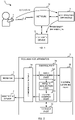
- FIG. 1 illustrates an outline of an electrocardiogram analysis system S according to the present embodiment.
- the electrocardiogram analysis system S includes an electrocardiograph 1, a doctor's device 2, and an electrocardiogram analysis apparatus 3. A plurality of electrocardiographs 1 and a plurality of doctor's devices 2 may be provided.
- the electrocardiogram analysis system S may include equipment such as other servers and terminals.
- the electrocardiograph 1 is an electrocardiograph worn by a person, and is electrocardiogram measurement equipment that generates electrocardiogram data which indicates his/her electrocardiogram by measuring a potential while being worn on his/her wrist, palm, chest, or the like, for example. That is, the electrocardiograph 1 is a Holter monitor (also referred to as a wearable electrocardiograph or a continuously mounted electrocardiograph).
- the electrocardiograph 1 transmits the generated electrocardiogram data to the electrocardiogram analysis apparatus 3 via a network N including a wireless communication line.
- the electrocardiogram data generated by the electrocardiograph 1 may be sent to the electrocardiogram analysis apparatus 3 using a storage medium without passing through the network N, for example.
- the doctor's device 2 is an information terminal used by a medical worker such as a doctor who examines a person, and includes a display (display device) and a computer, for example.
- the doctor's device 2 is associated in advance with a medical worker who uses the doctor's device 2 with an ID or the like given to the medical worker.
- the doctor's device 2 outputs abnormality information output by the electrocardiogram analysis apparatus 3 on the basis of the electrocardiogram data generated by the electrocardiograph 1.
- the doctor's device 2 may display the abnormality information on the display and may output voice indicating the abnormality information from a speaker.
- the electrocardiogram analysis apparatus 3 is an apparatus that outputs abnormality information about whether a person to be analyzed has paroxysmal arrhythmia on the basis of the electrocardiogram data generated by the electrocardiograph 1, and is a server, for example.
- the abnormality information is (i) information representing a value, a level, or a score of the probability that the person to be analyzed has paroxysmal arrhythmia and (ii) whether there is paroxysmal arrhythmia or signs of paroxysmal arrhythmia in the person to be analyzed. Further, the abnormality information may be information representing a value, a level, or a score of the probability that the person to be analyzed has no paroxysmal arrhythmia.
- the abnormality information may be information representing whether there is paroxysmal arrhythmia in the person to be analyzed. Further, the abnormality information may be information about an occurrence of other paroxysmal arrhythmias. Paroxysmal arrhythmia is at least one of ventricular fibrillation or paroxysmal tachycardia such as paroxysmal atrial fibrillation, ventricular tachycardia, supraventricular tachycardia, atrial flutter, or the like.
- FIG. 2 is a block diagram of the electrocardiogram analysis system S according to the present embodiment. Arrows indicate main data flows in FIG. 2 , and there may be data flows not shown in FIG. 2 .
- each block is not a hardware (device) unit but a functional unit.
- each block indicates a configuration of a function unit, not a configuration of a hardware (device) unit. Therefore, the blocks shown in FIG. 2 may be implemented in a single device or separately in a plurality of devices. The transfer of data between the blocks may be performed via any means, such as a data bus, a network, a portable storage medium, or the like.
- the electrocardiogram analysis apparatus 3 includes a communication part 31, a storage part 32, a machine learning part 33, and a control part 34.
- the control part 34 includes an input processing part 341, a result acquisition part 342, and an output control part 343.
- the communication part 31 has a communication controller for transmitting and receiving data between the electrocardiograph 1 and the doctor's device 2 via the network N.
- the communication part 31 notifies the control part 34 of the data received from the electrocardiograph 1 and the doctor's device 2 via the network N.
- the communication part 31 transmits the data output from the control part 34 to the doctor's device 2 via the network N.
- the storage part 32 is a storage medium including a read only memory (ROM), a random access memory (RAM), a hard disk drive, and the like.
- the storage part 32 stores in advance a program executed by the control part 34.
- the storage part 32 may be provided outside the electrocardiogram analysis apparatus 3, and in such a case, the storage part may transmit and receive the data to and from the control part 34 via the network N.
- the machine learning part 33 By learning on the basis of electrocardiogram data for training (hereinafter, training electrocardiogram data), which is to be used as training data, the machine learning part 33 generates a machine learning model that outputs abnormality information about whether paroxysmal arrhythmia is included in electrocardiogram data that is input to the machine learning part 33 and holds the generated machine learning model.
- the machine learning model is a model generated by machine learning (i) normal electrocardiogram data (i.e., electrocardiogram data of a person who has no paroxysmal arrhythmia) and (ii) abnormal electrocardiogram data (i.e., electrocardiogram data of a person with paroxysmal arrhythmia) as training data.
- the machine learning part 33 may hold a machine learning model generated outside the electrocardiogram analysis apparatus 3.
- An internal configuration of the machine learning model is any desired configuration, and it includes a convolutional neural network (CNN) or a recurrent neural network (RNN), for example.
- the machine learning part 33 includes a processor that executes various calculations using the CNN, and a memory that stores coefficients of the CNN, for example.
- the machine learning model of the machine learning part 33 outputs the abnormality information about whether the person to be analyzed has paroxysmal arrhythmia on the basis of the input electrocardiogram data.
- the machine learning part 33 may include only a memory for storing the machine learning model generated outside the electrocardiogram analysis apparatus 3. At least some of the functions of the machine learning part 33 may be built into the control part 34.
- the control part 34 includes a processor such as a central processing unit (CPU) and functions as an input processing part 341, a result acquisition part 342, and an output control part 343 by executing the program stored in the storage part 32, for example. At least some of the functions of the control part 34 may be executed by an electric circuit. Further, at least some of the functions of the control part 34 may be executed by the control part 34 executing a program executed via a network.
- a processor such as a central processing unit (CPU) and functions as an input processing part 341, a result acquisition part 342, and an output control part 343 by executing the program stored in the storage part 32, for example.
- a processor such as a central processing unit (CPU) and functions as an input processing part 341, a result acquisition part 342, and an output control part 343 by executing the program stored in the storage part 32, for example.
- At least some of the functions of the control part 34 may be executed by an electric circuit. Further, at least some of the functions of the control part 34 may be executed by
- the input processing part 341 inputs electrocardiogram data of a person to be analyzed, which is the subject of analysis, into the machine learning model of the machine learning part 33.
- the result acquisition part 342 acquires information output by the machine learning model of the machine learning part 33.
- the output control part 343 outputs, to the doctor's device 2, abnormality information which is to be output from the machine learning model of the machine learning part 33 and corresponds to whether the person to be analyzed has paroxysmal arrhythmia.
- Detailed processing executed by the input processing part 341, the result acquisition part 342, and the output control part 343 will be described later.
- the electrocardiogram analysis system S is not limited to the specific configuration shown in FIG. 2 .
- the electrocardiograph 1, the doctor's device 2, and the electrocardiogram analysis apparatus 3 may be configured by connecting two or more physically separated devices in a wired or wireless manner.
- the electrocardiogram analysis apparatus 3 may be formed by a single computer, may be formed by a plurality of computers cooperating with each other, or may be formed by a cloud which is a collection of computer resources. Two or more of the electrocardiograph 1, the doctor's device 2, and the electrocardiogram analysis apparatus 3 may be formed as one device.
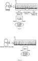
- FIG. 3 is a schematic diagram illustrating a method in which the electrocardiogram analysis apparatus 3 performs machine learning.
- the electrocardiograph 1 measures an electrocardiogram of a patient.
- the patient is a person who has been diagnosed by a doctor as having paroxysmal arrhythmia (for example, at least one of ventricular fibrillation or paroxysmal tachycardia such as paroxysmal atrial fibrillation, ventricular tachycardia, supraventricular tachycardia, atrial flutter, or the like).
- the electrocardiograph 1 generates electrocardiogram data H0 indicating the measured electrocardiogram and transmits the electrocardiogram data H0 to the electrocardiogram analysis apparatus 3 via the network N.
- the input processing part 341 acquires the electrocardiogram data H0 transmitted by the electrocardiograph 1.
- the input processing part 341 acquires the electrocardiogram data H0 transmitted by the electrocardiograph 1.
- the doctor can specify the paroxysmal period T1 by observing the electrocardiogram.
- the non-paroxysmal period T0 is a predetermined period of at least one of before or after the paroxysmal period T1, which is specified by the doctor as a period during which an episode of arrhythmia occurs and is a period which is not specified by the doctor as the period during which the episode of arrhythmia occurs.
- the non-paroxysmal period T0 is preferably 7 days before or after the paroxysmal period T1, more preferably 24 hours before or after the paroxysmal period T1.
- the input processing part 341 inputs, as training electrocardiogram data of a person who has paroxysmal arrhythmia, the non-paroxysmal period T0 portion within the electrocardiogram data H0 to the machine learning part 33. Further, the input processing part 341 inputs, as training electrocardiogram data of a person who has no paroxysmal arrhythmia, electrocardiogram data of a person who has been diagnosed by the doctor as not having paroxysmal arrhythmia to the machine learning part 33.
- the input processing part 341 uses, as the training electrocardiogram data, electrocardiogram data of a predetermined period of time during which a person is at rest (e.g., during sleep). Further, it is desirable that the input processing part 341 uses, as the training electrocardiogram data, electrocardiogram data of a period in which the doctor has specified that symptoms different from paroxysmal arrhythmia to be analyzed have not occurred. By doing this, the electrocardiogram analysis system S can exclude data that may be noise and improve the accuracy of machine learning.
- the machine learning part 33 generates a machine learning model that outputs abnormality information about whether the input electrocardiogram data is the electrocardiogram data of a person with paroxysmal arrhythmia, by performing known machine learning (e.g., the CNN or the RNN) using the input training electrocardiogram data.
- the machine learning part 33 may acquire a machine learning model generated with an external apparatus (server or the like) by the above-described machine learning method.
- FIG. 4 is a schematic diagram illustrating a method in which the electrocardiogram analysis apparatus 3 analyzes electrocardiogram data.
- the electrocardiograph 1 measures an electrocardiogram of the person to be analyzed.
- the electrocardiograph 1 generates electrocardiogram data H1 indicating the measured electrocardiogram, and transmits the electrocardiogram data H1 to the electrocardiogram analysis apparatus 3 via the network N.
- the input processing part 341 acquires the electrocardiogram data H1 transmitted by the electrocardiograph 1. It is desirable that the input processing part 341 sequentially acquires the electrocardiogram data H1 measured by the electrocardiograph 1 (i.e., the Holter monitor or a patch electrocardiograph) worn by the person to be analyzed living daily life. By doing this, the electrocardiogram analysis system S can promptly notify the medical worker of the information about whether or not the person to be analyzed has paroxysmal arrhythmia.
- the electrocardiogram analysis system S can promptly notify the medical worker of the information about whether or not the person to be analyzed has paroxysmal arrhythmia.
- the input processing part 341 may acquire electrocardiogram data indicating the electrocardiogram of the person to be analyzed measured in advance in a hospital or the like, electrocardiogram data acquired from an electrocardiograph mounted on a steering wheel of a car, electrocardiogram data acquired from a 12-lead electrocardiograph, electrocardiogram data acquired from an electrocardiograph mounted on a smart watch, or the like.
- the input processing part 341 inputs the acquired electrocardiogram data HI, as electrocardiogram data to be analyzed, to the machine learning model of the machine learning part 33.
- the input processing part 341 inputs, to the machine learning model, electrocardiogram data to be analyzed that has been measured at the same sampling rate (for example, 1000 Hz) as the training electrocardiogram data.
- the electrocardiogram analysis apparatus 3 can prevent an analysis result from being affected by a difference between sampling rates of electrocardiogram data.
- the input processing part 341 may perform a process of converting the sampling rate on the electrocardiogram data to be analyzed, and then input the processed data to the machine learning model.
- the machine learning model of the machine learning part 33 When the electrocardiogram data is input, the machine learning model of the machine learning part 33 outputs the abnormality information about whether the input electrocardiogram data is electrocardiogram data of a person with paroxysmal arrhythmia. For example, the machine learning model outputs, as the abnormality information, abnormality information about whether a person (i.e., the person to be analyzed), who is a measurement source of the input electrocardiogram data, has paroxysmal arrhythmia.
- the machine learning model outputs, as the abnormal information, at least one of (i) the value of the probability that the person to be analyzed has paroxysmal arrhythmia, (ii) the level of the probability that the person to be analyzed has paroxysmal arrhythmia, (iii) the score of the probability that the person to be analyzed has paroxysmal arrhythmia, (iv) whether there is paroxysmal arrhythmia in the person to be analyzed, or (v) whether there are signs of paroxysmal arrhythmia in the person to be analyzed.
- the result acquisition part 342 acquires, as the analysis result, the abnormality information about whether the person to be analyzed has paroxysmal arrhythmia, which is output from the machine learning model of the machine learning part 33.
- the output control part 343 outputs the abnormality information acquired by the result acquisition part 342 to the doctor's device 2.
- the abnormal information is represented by the value, the level, or the score of the probability that the person to be analyzed has paroxysmal arrhythmia. Further, the abnormality information may be information indicating the value, the level, or the score of the probability that the person to be analyzed has no paroxysmal arrhythmia.
- the level of the probability is represented by a letter or symbol such as H (high), L (low), o, ⁇ or the like associated with each range of probability values, for example.
- the score of the probability is expressed by, for example, a score obtained by converting the probability by a predetermined equation.
- the abnormality information may be information indicating whether there is paroxysmal arrhythmia or any sign of paroxysmal arrhythmia in the person to be analyzed. Whether there is arrhythmia or not is indicated by a character or a symbol that indicates a determination result of whether there is arrhythmia or any sign of arrhythmia by the machine learning model, for example. Also, the abnormality information may be output for the overall electrocardiogram data or may be output in association with each of a plurality of periods forming the electrocardiogram data. The abnormality information may be information on occurrence of other paroxysmal arrhythmias.
- the doctor's device 2 may display the abnormality information on the display, and may output a voice indicating the abnormality information from the speaker.
- the doctor's device 2 may directly display the abnormality information, or may convert the abnormality information into other information ("Reexamination required,” “Medication required,” or the like if the probability is equal to or greater than a predetermined value) and then output the information.
- FIG. 5 is a schematic diagram of an analysis result screen displayed by the doctor's device 2.
- the analysis result screen includes an electrocardiogram H and abnormality information P.
- the electrocardiogram H represents at least a part of the electrocardiogram data used for analysis.
- the abnormality information P represents the value or the level of the probability that the person to be analyzed has paroxysmal arrhythmia, which is output from the machine learning model, for example.
- the electrocardiogram analysis system S can detect signs of paroxysmal arrhythmia in a non-paroxysmal period that are difficult for a doctor to find with his/her eyes, and display the signs as the information about whether the person to be analyzed has paroxysmal arrhythmia.
- the doctor can easily specify whether the person to be analyzed has paroxysmal arrhythmia, and can determine whether to do an additional examination or the like as needed.
- the electrocardiogram analysis apparatus 3 may display changes in the probability in a plurality of different periods on the doctor's device 2.
- the input processing part 341 inputs, as the electrocardiogram data to be analyzed, a plurality of pieces of electrocardiogram data indicating electrocardiograms of the person to be analyzed measured by the electrocardiograph 1 during the plurality of different periods (for example, a specific day of respective months) to the machine learning model of the machine learning part 33.
- the result acquisition part 342 acquires, in association with each of the plurality of different periods, the abnormality information about whether the person to be analyzed has paroxysmal arrhythmia output by the machine learning model.
- the output control part 343 causes the doctor's device 2 to display the abnormality information about the changes in the probability of the plurality of different periods acquired by the result acquisition part 342.
- the abnormality information represents the changes in the probability by a graph that shows change over time or the level (difference, rate of change, or the like) of the change in the probability, for example.
- the analysis result screen may include an area (e.g., a button or a selection box) for inputting a diagnosis on whether the person to be analyzed has paroxysmal arrhythmia diagnosed by a doctor.
- the doctor's device 2 transmits the input contents to the electrocardiogram analysis apparatus 3 as determination information on whether or not the person to be analyzed has paroxysmal arrhythmia.
- the electrocardiogram analysis apparatus 3 receives the determination information transmitted by the doctor's device 2. By performing the above-described machine learning using the received determination information and the electrocardiogram data of the person to be analyzed, the machine learning part 33 regenerates the machine learning model that outputs the abnormality information about whether the input electrocardiogram data is the electrocardiogram data of the person with paroxysmal arrhythmia. By doing this, the electrocardiogram analysis system S can accept feedback on the diagnosis by the doctor and improve the accuracy of the abnormality information output by the machine learning model.
- FIG. 6 is a flowchart of an electrocardiogram analysis method executed by the electrocardiogram analysis system S according to the present embodiment.
- the present embodiment includes the electrocardiogram analysis method illustrated in FIG. 6 , a program for executing said electrocardiogram analysis method, and a computer-readable storage medium storing the program.
- the input processing part 341 acquires electrocardiogram data transmitted by the electrocardiograph 1 of a patient with paroxysmal arrhythmia (S11).
- the input processing part 341 inputs a non-paroxysmal period portion within the patient's electrocardiogram data to the machine learning part 33 as training electrocardiogram data of a person who has paroxysmal arrhythmia.
- the input processing part 341 inputs electrocardiogram data of a person who has been diagnosed by a doctor as not having paroxysmal arrhythmia to the machine learning part 33 as training electrocardiogram data of a person who has no paroxysmal arrhythmia.
- the machine learning part 33 generates a machine learning model that outputs abnormality information about whether the input electrocardiogram data is electrocardiogram data of a person with paroxysmal arrhythmia by machine learning using the input training electrocardiogram data (S12).
- steps S 11 to S12 may be omitted.
- the input processing part 341 acquires electrocardiogram data to be analyzed, which is transmitted by the electrocardiograph 1 of a person to be analyzed (S 13).
- the input processing part 341 inputs the acquired electrocardiogram data as the electrocardiogram data to be analyzed into the machine learning model of the machine learning part 33 (S14).
- the machine learning model of the machine learning part 33 outputs the abnormality information about whether the input electrocardiogram data is electrocardiogram data of a person with paroxysmal arrhythmia.
- the result acquisition part 342 acquires, as an analysis result, the abnormality information about whether the person to be analyzed has paroxysmal arrhythmia, which is output from the machine learning model of the machine learning part 33 (S15).
- the output control part 343 causes the doctor's device 2 to display the abnormality information about whether or not the result acquisition part 342 has acquired the abnormality information (S16).
- a patient's electrocardiogram measured within a limited period of time does not always include a waveform pertaining to an episode because episodes occur infrequently in paroxysmal arrhythmia.
- an electrocardiogram of a patient with paroxysmal arrhythmia shows signs of arrhythmia, which are difficult for a doctor to detect with his/her eyes even in the non-paroxysmal period during which no episode occurs. Therefore, the electrocardiogram analysis system S inputs the electrocardiogram data of the person to be analyzed into a machine learning model obtained by machine learning the electrocardiogram data of the patient with paroxysmal arrhythmia during his/her non-paroxysmal period, thereby outputting the abnormality information about whether the person to be analyzed has paroxysmal arrhythmia. By doing this, the electrocardiogram analysis system S can make it easier to specify whether the person to be analyzed has paroxysmal arrhythmia.
- the electrocardiogram analysis apparatus 3 is used for supporting a doctor to make a diagnosis, but the electrocardiogram analysis apparatus 3 may be used for other uses.
- the electrocardiogram analysis apparatus 3 may be used in (i) a medical checkup result display system that displays whether there is a possibility of occurrence of paroxysmal arrhythmia in a person to be analyzed and whether a reexamination or a thorough checkup is required, on the basis of the abnormality information, (ii) an insurance examination support system in which the abnormality information is included in the eligibility criteria of an insurance plan of the person to be analyzed, and (iii) an insurance contract document apparatus in which the abnormality information is included in display items of insurance contract documents of the person to be analyzed. Therefore, the electrocardiogram analysis apparatus 3 can support screening an insurance application or the like on the basis of health conditions of the person to be analyzed.
- the electrocardiogram analysis apparatus 3 may be used in a clinical trial participant selection system in which the abnormality information is included in display items of information concerning candidates for clinical trials. Therefore, the electrocardiogram analysis apparatus 3 can support making a determination such as excluding candidates who are likely to have paroxysmal arrhythmia from participants of a clinical trial on an evaluation of an electrocardiograph, for example.
- the electrocardiogram analysis apparatus 3 may be used as a drug administration determination support system or a drug administration contraindication determination support system which make use of the abnormality information. Therefore, the electrocardiogram analysis apparatus 3 can (i) support determination of the advisability of administering a drug or the like, which cannot be administered when there is arrhythmia, to a patient who is likely to have paroxysmal arrhythmia and (ii) use the abnormality information as a digital biomarker that stratifies patients who have paroxysmal disease, for example.
- the electrocardiogram analysis apparatus 3 may be used in a car driving control apparatus for safely stopping a vehicle when an episode of paroxysmal arrhythmia is detected while the vehicle is being driven, on the basis of the abnormality information.
- the electrocardiogram analysis apparatus 3 may be used in an alert system which recommends visiting a medical institution by displaying, on a smart device such as a smartphone, onset of paroxysmal arrhythmia on the basis of the abnormality information.
- the processor of the electrocardiogram analysis apparatus 3 performs each step (process) included in the electrocardiogram analysis method shown in FIG. 6 . That is, the processor of the electrocardiogram analysis apparatus 3 reads, from the storage part, a program for executing the electrocardiogram analysis method shown in FIG. 6 and executes the program to control each unit of the electrocardiogram analysis system S, thereby executing the electrocardiogram analysis method shown in FIG. 6 .
- the steps included in the electrocardiogram analysis method shown in FIG. 6 may be partially omitted, the order among the steps may be changed, or a plurality of steps may be performed in parallel.
Landscapes
- Health & Medical Sciences (AREA)
- Life Sciences & Earth Sciences (AREA)
- Engineering & Computer Science (AREA)
- Cardiology (AREA)
- Physics & Mathematics (AREA)
- Animal Behavior & Ethology (AREA)
- Biomedical Technology (AREA)
- Heart & Thoracic Surgery (AREA)
- Medical Informatics (AREA)
- Molecular Biology (AREA)
- Surgery (AREA)
- Biophysics (AREA)
- General Health & Medical Sciences (AREA)
- Public Health (AREA)
- Veterinary Medicine (AREA)
- Pathology (AREA)
- Artificial Intelligence (AREA)
- Physiology (AREA)
- Computer Networks & Wireless Communication (AREA)
- Evolutionary Computation (AREA)
- Fuzzy Systems (AREA)
- Mathematical Physics (AREA)
- Computer Vision & Pattern Recognition (AREA)
- Psychiatry (AREA)
- Signal Processing (AREA)
- Measurement And Recording Of Electrical Phenomena And Electrical Characteristics Of The Living Body (AREA)
Applications Claiming Priority (2)
| Application Number | Priority Date | Filing Date | Title |
|---|---|---|---|
| JP2020069705 | 2020-04-08 | ||
| PCT/JP2021/008444 WO2021205788A1 (fr) | 2020-04-08 | 2021-03-04 | Dispositif d'analyse électrocardiographique, procédé d'analyse électrocardiographique et programme associé |
Publications (4)
| Publication Number | Publication Date |
|---|---|
| EP4079224A1 true EP4079224A1 (fr) | 2022-10-26 |
| EP4079224A4 EP4079224A4 (fr) | 2023-06-21 |
| EP4079224B1 EP4079224B1 (fr) | 2025-07-23 |
| EP4079224C0 EP4079224C0 (fr) | 2025-07-23 |
Family
ID=78022561
Family Applications (1)
| Application Number | Title | Priority Date | Filing Date |
|---|---|---|---|
| EP21784075.0A Active EP4079224B1 (fr) | 2020-04-08 | 2021-03-04 | Dispositif d'analyse électrocardiographique, procédé d'analyse électrocardiographique et programme associé |
Country Status (4)
| Country | Link |
|---|---|
| US (1) | US12582315B2 (fr) |
| EP (1) | EP4079224B1 (fr) |
| JP (3) | JP7002168B1 (fr) |
| WO (1) | WO2021205788A1 (fr) |
Families Citing this family (13)
| Publication number | Priority date | Publication date | Assignee | Title |
|---|---|---|---|---|
| CA2797980C (fr) | 2010-05-12 | 2015-08-18 | Irhythm Technologies, Inc. | Caracteristiques de dispositif et elements de construction pour adherence a long terme |
| JP6198849B2 (ja) | 2013-01-24 | 2017-09-20 | アイリズム・テクノロジーズ・インコーポレイテッドiRhythm Technologies,Inc. | 生理学的信号をモニターする電子装置、及び該電子装置の部分を除去交換する方法 |
| US20160120434A1 (en) | 2014-10-31 | 2016-05-05 | Irhythm Technologies, Inc. | Wireless physiological monitoring device and systems |
| WO2022032117A1 (fr) | 2020-08-06 | 2022-02-10 | Irhythm Technologies, Inc. | Dispositif adhésif de surveillance physiologique |
| JP7796108B2 (ja) | 2020-08-06 | 2026-01-08 | アイリズム・テクノロジーズ・インコーポレイテッド | 生理学的モニタリング装置の電気部品 |
| USD1063079S1 (en) | 2021-08-06 | 2025-02-18 | Irhythm Technologies, Inc. | Physiological monitoring device |
| KR102788715B1 (ko) * | 2022-05-04 | 2025-04-01 | 주식회사 뷰노 | 심전도 데이터를 분석하기 위한 방법 및 장치 |
| EP4550352A4 (fr) * | 2022-07-29 | 2025-09-10 | Medical Ai Co Ltd | Procédé, programme et dispositif pour fournir un service d'interprétation d'ecg |
| JP7672033B2 (ja) * | 2023-02-10 | 2025-05-07 | 公立大学法人横浜市立大学 | 心不全推定装置、心不全推定システム、心不全推定方法、及び心不全推定プログラム |
| WO2024191200A1 (fr) * | 2023-03-13 | 2024-09-19 | 주식회사 메디컬에이아이 | Procédé, programme et dispositif pour identifier une fibrillation auriculaire potentielle sur la base d'un modèle d'intelligence artificielle |
| KR102877334B1 (ko) * | 2023-10-17 | 2025-10-29 | (주)씨어스테크놀로지 | 온라인 머신러닝 알고리즘을 이용한 심전도 판독장치 |
| JP7493293B1 (ja) | 2024-03-13 | 2024-05-31 | 株式会社カルディオインテリジェンス | 心電図解析のための支援装置、支援システム、およびプログラム |
| KR102832591B1 (ko) * | 2024-08-30 | 2025-07-10 | 주식회사 휴이노 | 심전도 신호에서 이상 파형의 발생을 예측하기 위한 방법 및 시스템 |
Family Cites Families (14)
| Publication number | Priority date | Publication date | Assignee | Title |
|---|---|---|---|---|
| JP2007195693A (ja) | 2006-01-25 | 2007-08-09 | Matsushita Electric Works Ltd | 携帯型心電計測装置 |
| US8909330B2 (en) | 2009-05-20 | 2014-12-09 | Sotera Wireless, Inc. | Body-worn device and associated system for alarms/alerts based on vital signs and motion |
| US9314178B2 (en) | 2013-03-14 | 2016-04-19 | Greatbach, Ltd. | Cardiac signal recording using dynamically generated detection thresholds |
| CN107847154B (zh) * | 2015-05-13 | 2021-07-16 | 阿利弗克公司 | 不一致监测 |
| US10779744B2 (en) * | 2015-10-27 | 2020-09-22 | Cardiologs Technologies Sas | Automatic method to delineate or categorize an electrocardiogram |
| CN107516075B (zh) * | 2017-08-03 | 2020-10-09 | 安徽华米智能科技有限公司 | 心电信号的检测方法、装置及电子设备 |
| WO2019046294A1 (fr) | 2017-08-28 | 2019-03-07 | University Of South Florida | Système et procédé pour prédire une fibrillation atriale (fa) |
| US20240099593A1 (en) * | 2017-10-06 | 2024-03-28 | Alivecor, Inc. | Machine learning health analysis with a mobile device |
| JP2019115618A (ja) * | 2017-12-27 | 2019-07-18 | オムロンヘルスケア株式会社 | 情報処理装置、情報処理方法、及び情報処理プログラム |
| CN108577823B (zh) * | 2018-04-27 | 2021-02-02 | 京东方科技集团股份有限公司 | 一种心律失常检测装置和心律失常检测系统 |
| JP6483890B1 (ja) * | 2018-04-27 | 2019-03-13 | 国立大学法人滋賀医科大学 | 診断支援装置、機械学習装置、診断支援方法、機械学習方法および機械学習プログラム |
| US11723577B2 (en) * | 2019-05-06 | 2023-08-15 | Medtronic, Inc. | Visualization of arrhythmia detection by machine learning |
| CN110403600B (zh) * | 2019-07-26 | 2022-02-08 | 武汉海星通技术股份有限公司 | 基于差值时间散点图的阵发性房颤智能分析方法及系统 |
| US12224052B2 (en) * | 2019-10-03 | 2025-02-11 | Rom Technologies, Inc. | System and method for using AI, machine learning and telemedicine for long-term care via an electromechanical machine |
-
2021
- 2021-03-04 WO PCT/JP2021/008444 patent/WO2021205788A1/fr not_active Ceased
- 2021-03-04 JP JP2021544452A patent/JP7002168B1/ja active Active
- 2021-03-04 EP EP21784075.0A patent/EP4079224B1/fr active Active
- 2021-12-20 JP JP2021205708A patent/JP7701048B2/ja active Active
-
2022
- 2022-07-11 US US17/811,860 patent/US12582315B2/en active Active
-
2025
- 2025-06-12 JP JP2025098324A patent/JP2025128317A/ja active Pending
Also Published As
| Publication number | Publication date |
|---|---|
| EP4079224B1 (fr) | 2025-07-23 |
| JP2025128317A (ja) | 2025-09-02 |
| US20220346647A1 (en) | 2022-11-03 |
| JP7002168B1 (ja) | 2022-01-20 |
| EP4079224C0 (fr) | 2025-07-23 |
| JP2022037153A (ja) | 2022-03-08 |
| EP4079224A4 (fr) | 2023-06-21 |
| WO2021205788A1 (fr) | 2021-10-14 |
| JP7701048B2 (ja) | 2025-07-01 |
| JPWO2021205788A1 (fr) | 2021-10-14 |
| US12582315B2 (en) | 2026-03-24 |
Similar Documents
| Publication | Publication Date | Title |
|---|---|---|
| US12582315B2 (en) | Electrocardiogram analysis apparatus, electrocardiogram analyzing method, and non-transitory computer-readable storage medium | |
| AU2022201530B2 (en) | Apparatus, systems and methods for predicting, screening and monitoring of encephalopathy/delirium | |
| US20170156592A1 (en) | Healthcare systems and monitoring method for physiological signals | |
| US11412978B2 (en) | Information processing system and program | |
| Faragli et al. | Cardiovascular screening in low-income settings using a novel 4-lead smartphone-based electrocardiograph (D-Heart®) | |
| WO2023074341A1 (fr) | Dispositif de traitement d'informations, support d'enregistrement, et procédé de traitement d'informations | |
| Khatun et al. | Detection of myocardial infarction and arrhythmia from single-lead ECG data using bagging trees classifier | |
| Sabar et al. | A study to assess a novel automated electrocardiogram technology in screening for atrial fibrillation | |
| US8103065B2 (en) | Assessment of medical conditions | |
| Sara et al. | Heart rate non linear dynamics in patients with persistent vegetative state: a preliminary report | |
| Van Heuverswyn et al. | Clinical validation of a 13-lead electrocardiogram derived from a self-applicable 3-lead recording for diagnosis of myocardial supply ischaemia and common non-ischaemic electrocardiogram abnormalities at rest | |
| KR102545129B1 (ko) | 원격 상담 서비스 제공 방법 및 시스템 | |
| JP7537826B2 (ja) | Ecg信号に基づいて指標値を変換する方法およびシステム | |
| JP7479106B1 (ja) | プログラム、出力装置及び出力方法 | |
| WO2025227126A1 (fr) | Détection d'arythmie cardiaque et d'ischémie myocardique à partir de données d'électrocardiographie à l'aide de l'apprentissage automatique | |
| Hughes-Noehrer et al. | Increasing the accuracy of ST-segment elevation detection in an ECG through automated visual rules | |
| WO2024106005A1 (fr) | Programme, dispositif de sortie et procédé de sortie | |
| WO2025227125A1 (fr) | Détection d'une insuffisance cardiaque congestive à partir de données d'électrocardiographie à l'aide de l'apprentissage automatique | |
| BR112018011326B1 (pt) | Sistema para rastreamento de delírio de paciente |
Legal Events
| Date | Code | Title | Description |
|---|---|---|---|
| STAA | Information on the status of an ep patent application or granted ep patent |
Free format text: STATUS: THE INTERNATIONAL PUBLICATION HAS BEEN MADE |
|
| PUAI | Public reference made under article 153(3) epc to a published international application that has entered the european phase |
Free format text: ORIGINAL CODE: 0009012 |
|
| STAA | Information on the status of an ep patent application or granted ep patent |
Free format text: STATUS: REQUEST FOR EXAMINATION WAS MADE |
|
| 17P | Request for examination filed |
Effective date: 20220718 |
|
| AK | Designated contracting states |
Kind code of ref document: A1 Designated state(s): AL AT BE BG CH CY CZ DE DK EE ES FI FR GB GR HR HU IE IS IT LI LT LU LV MC MK MT NL NO PL PT RO RS SE SI SK SM TR |
|
| A4 | Supplementary search report drawn up and despatched |
Effective date: 20230524 |
|
| RIC1 | Information provided on ipc code assigned before grant |
Ipc: A61B 5/00 20060101ALI20230517BHEP Ipc: A61B 5/363 20210101ALI20230517BHEP Ipc: A61B 5/361 20210101ALI20230517BHEP Ipc: A61B 5/346 20210101AFI20230517BHEP |
|
| DAV | Request for validation of the european patent (deleted) | ||
| DAX | Request for extension of the european patent (deleted) | ||
| STAA | Information on the status of an ep patent application or granted ep patent |
Free format text: STATUS: EXAMINATION IS IN PROGRESS |
|
| 17Q | First examination report despatched |
Effective date: 20240214 |
|
| GRAP | Despatch of communication of intention to grant a patent |
Free format text: ORIGINAL CODE: EPIDOSNIGR1 |
|
| STAA | Information on the status of an ep patent application or granted ep patent |
Free format text: STATUS: GRANT OF PATENT IS INTENDED |
|
| INTG | Intention to grant announced |
Effective date: 20250306 |
|
| RIN1 | Information on inventor provided before grant (corrected) |
Inventor name: TANIGUCHI, TADAHIRO Inventor name: TAKATA, TOMOHIRO Inventor name: TANIGUCHI, HIROHISA Inventor name: TAMURA, YUICHI |
|
| GRAS | Grant fee paid |
Free format text: ORIGINAL CODE: EPIDOSNIGR3 |
|
| GRAA | (expected) grant |
Free format text: ORIGINAL CODE: 0009210 |
|
| STAA | Information on the status of an ep patent application or granted ep patent |
Free format text: STATUS: THE PATENT HAS BEEN GRANTED |
|
| AK | Designated contracting states |
Kind code of ref document: B1 Designated state(s): AL AT BE BG CH CY CZ DE DK EE ES FI FR GB GR HR HU IE IS IT LI LT LU LV MC MK MT NL NO PL PT RO RS SE SI SK SM TR |
|
| REG | Reference to a national code |
Ref country code: GB Ref legal event code: FG4D |
|
| REG | Reference to a national code |
Ref country code: CH Ref legal event code: EP |
|
| REG | Reference to a national code |
Ref country code: DE Ref legal event code: R096 Ref document number: 602021034705 Country of ref document: DE |
|
| REG | Reference to a national code |
Ref country code: IE Ref legal event code: FG4D |
|
| U01 | Request for unitary effect filed |
Effective date: 20250819 |
|
| U07 | Unitary effect registered |
Designated state(s): AT BE BG DE DK EE FI FR IT LT LU LV MT NL PT RO SE SI Effective date: 20250822 |
|
| PG25 | Lapsed in a contracting state [announced via postgrant information from national office to epo] |
Ref country code: IS Free format text: LAPSE BECAUSE OF FAILURE TO SUBMIT A TRANSLATION OF THE DESCRIPTION OR TO PAY THE FEE WITHIN THE PRESCRIBED TIME-LIMIT Effective date: 20251123 |
|
| PG25 | Lapsed in a contracting state [announced via postgrant information from national office to epo] |
Ref country code: NO Free format text: LAPSE BECAUSE OF FAILURE TO SUBMIT A TRANSLATION OF THE DESCRIPTION OR TO PAY THE FEE WITHIN THE PRESCRIBED TIME-LIMIT Effective date: 20251023 |
|
| PG25 | Lapsed in a contracting state [announced via postgrant information from national office to epo] |
Ref country code: HR Free format text: LAPSE BECAUSE OF FAILURE TO SUBMIT A TRANSLATION OF THE DESCRIPTION OR TO PAY THE FEE WITHIN THE PRESCRIBED TIME-LIMIT Effective date: 20250723 |
|
| PG25 | Lapsed in a contracting state [announced via postgrant information from national office to epo] |
Ref country code: GR Free format text: LAPSE BECAUSE OF FAILURE TO SUBMIT A TRANSLATION OF THE DESCRIPTION OR TO PAY THE FEE WITHIN THE PRESCRIBED TIME-LIMIT Effective date: 20251024 |
|
| PG25 | Lapsed in a contracting state [announced via postgrant information from national office to epo] |
Ref country code: PL Free format text: LAPSE BECAUSE OF FAILURE TO SUBMIT A TRANSLATION OF THE DESCRIPTION OR TO PAY THE FEE WITHIN THE PRESCRIBED TIME-LIMIT Effective date: 20250723 |
|
| PG25 | Lapsed in a contracting state [announced via postgrant information from national office to epo] |
Ref country code: RS Free format text: LAPSE BECAUSE OF FAILURE TO SUBMIT A TRANSLATION OF THE DESCRIPTION OR TO PAY THE FEE WITHIN THE PRESCRIBED TIME-LIMIT Effective date: 20251023 |
|
| PG25 | Lapsed in a contracting state [announced via postgrant information from national office to epo] |
Ref country code: ES Free format text: LAPSE BECAUSE OF FAILURE TO SUBMIT A TRANSLATION OF THE DESCRIPTION OR TO PAY THE FEE WITHIN THE PRESCRIBED TIME-LIMIT Effective date: 20250723 |
|
| U20 | Renewal fee for the european patent with unitary effect paid |
Year of fee payment: 6 Effective date: 20260116 |
|
| PG25 | Lapsed in a contracting state [announced via postgrant information from national office to epo] |
Ref country code: SM Free format text: LAPSE BECAUSE OF FAILURE TO SUBMIT A TRANSLATION OF THE DESCRIPTION OR TO PAY THE FEE WITHIN THE PRESCRIBED TIME-LIMIT Effective date: 20250723 |
|
| PG25 | Lapsed in a contracting state [announced via postgrant information from national office to epo] |
Ref country code: CZ Free format text: LAPSE BECAUSE OF FAILURE TO SUBMIT A TRANSLATION OF THE DESCRIPTION OR TO PAY THE FEE WITHIN THE PRESCRIBED TIME-LIMIT Effective date: 20250723 |
|
| PG25 | Lapsed in a contracting state [announced via postgrant information from national office to epo] |
Ref country code: SK Free format text: LAPSE BECAUSE OF FAILURE TO SUBMIT A TRANSLATION OF THE DESCRIPTION OR TO PAY THE FEE WITHIN THE PRESCRIBED TIME-LIMIT Effective date: 20250723 |