EP4086626A1 - Système d'analyse d'échantillon et procédé de gestion de précision automatique associé - Google Patents
Système d'analyse d'échantillon et procédé de gestion de précision automatique associé Download PDFInfo
- Publication number
- EP4086626A1 EP4086626A1 EP19958734.6A EP19958734A EP4086626A1 EP 4086626 A1 EP4086626 A1 EP 4086626A1 EP 19958734 A EP19958734 A EP 19958734A EP 4086626 A1 EP4086626 A1 EP 4086626A1
- Authority
- EP
- European Patent Office
- Prior art keywords
- quality control
- sample
- test result
- blood
- smear
- Prior art date
- Legal status (The legal status is an assumption and is not a legal conclusion. Google has not performed a legal analysis and makes no representation as to the accuracy of the status listed.)
- Granted
Links
Images


Classifications
-
- G—PHYSICS
- G01—MEASURING; TESTING
- G01N—INVESTIGATING OR ANALYSING MATERIALS BY DETERMINING THEIR CHEMICAL OR PHYSICAL PROPERTIES
- G01N35/00—Automatic analysis not limited to methods or materials provided for in any single one of groups G01N1/00 - G01N33/00; Handling materials therefor
- G01N35/00584—Control arrangements for automatic analysers
- G01N35/00594—Quality control, including calibration or testing of components of the analyser
- G01N35/00613—Quality control
- G01N35/00623—Quality control of instruments
-
- G—PHYSICS
- G01—MEASURING; TESTING
- G01N—INVESTIGATING OR ANALYSING MATERIALS BY DETERMINING THEIR CHEMICAL OR PHYSICAL PROPERTIES
- G01N1/00—Sampling; Preparing specimens for investigation
- G01N1/28—Preparing specimens for investigation including physical details of (bio-)chemical methods covered elsewhere, e.g. G01N33/50, C12Q
- G01N1/2813—Producing thin layers of samples on a substrate, e.g. smearing, spinning-on
-
- G—PHYSICS
- G01—MEASURING; TESTING
- G01N—INVESTIGATING OR ANALYSING MATERIALS BY DETERMINING THEIR CHEMICAL OR PHYSICAL PROPERTIES
- G01N1/00—Sampling; Preparing specimens for investigation
- G01N1/28—Preparing specimens for investigation including physical details of (bio-)chemical methods covered elsewhere, e.g. G01N33/50, C12Q
- G01N1/30—Staining; Impregnating ; Fixation; Dehydration; Multistep processes for preparing samples of tissue, cell or nucleic acid material and the like for analysis
- G01N1/31—Apparatus therefor
- G01N1/312—Apparatus therefor for samples mounted on planar substrates
-
- G—PHYSICS
- G01—MEASURING; TESTING
- G01N—INVESTIGATING OR ANALYSING MATERIALS BY DETERMINING THEIR CHEMICAL OR PHYSICAL PROPERTIES
- G01N15/00—Investigating characteristics of particles; Investigating permeability, pore-volume or surface-area of porous materials
- G01N15/02—Investigating particle size or size distribution
- G01N15/0205—Investigating particle size or size distribution by optical means
- G01N15/0227—Investigating particle size or size distribution by optical means using imaging; using holography
-
- G—PHYSICS
- G01—MEASURING; TESTING
- G01N—INVESTIGATING OR ANALYSING MATERIALS BY DETERMINING THEIR CHEMICAL OR PHYSICAL PROPERTIES
- G01N15/00—Investigating characteristics of particles; Investigating permeability, pore-volume or surface-area of porous materials
- G01N15/10—Investigating individual particles
- G01N15/14—Optical investigation techniques, e.g. flow cytometry
- G01N15/1429—Signal processing
-
- G—PHYSICS
- G01—MEASURING; TESTING
- G01N—INVESTIGATING OR ANALYSING MATERIALS BY DETERMINING THEIR CHEMICAL OR PHYSICAL PROPERTIES
- G01N15/00—Investigating characteristics of particles; Investigating permeability, pore-volume or surface-area of porous materials
- G01N15/10—Investigating individual particles
- G01N15/14—Optical investigation techniques, e.g. flow cytometry
- G01N15/1429—Signal processing
- G01N15/1433—Signal processing using image recognition
-
- G—PHYSICS
- G01—MEASURING; TESTING
- G01N—INVESTIGATING OR ANALYSING MATERIALS BY DETERMINING THEIR CHEMICAL OR PHYSICAL PROPERTIES
- G01N35/00—Automatic analysis not limited to methods or materials provided for in any single one of groups G01N1/00 - G01N33/00; Handling materials therefor
- G01N35/00029—Automatic analysis not limited to methods or materials provided for in any single one of groups G01N1/00 - G01N33/00; Handling materials therefor provided with flat sample substrates, e.g. slides
-
- G—PHYSICS
- G01—MEASURING; TESTING
- G01N—INVESTIGATING OR ANALYSING MATERIALS BY DETERMINING THEIR CHEMICAL OR PHYSICAL PROPERTIES
- G01N15/00—Investigating characteristics of particles; Investigating permeability, pore-volume or surface-area of porous materials
- G01N2015/0092—Monitoring flocculation or agglomeration
-
- G—PHYSICS
- G01—MEASURING; TESTING
- G01N—INVESTIGATING OR ANALYSING MATERIALS BY DETERMINING THEIR CHEMICAL OR PHYSICAL PROPERTIES
- G01N15/00—Investigating characteristics of particles; Investigating permeability, pore-volume or surface-area of porous materials
- G01N15/01—Investigating characteristics of particles; Investigating permeability, pore-volume or surface-area of porous materials specially adapted for biological cells, e.g. blood cells
- G01N2015/012—Red blood cells
-
- G—PHYSICS
- G01—MEASURING; TESTING
- G01N—INVESTIGATING OR ANALYSING MATERIALS BY DETERMINING THEIR CHEMICAL OR PHYSICAL PROPERTIES
- G01N15/00—Investigating characteristics of particles; Investigating permeability, pore-volume or surface-area of porous materials
- G01N15/01—Investigating characteristics of particles; Investigating permeability, pore-volume or surface-area of porous materials specially adapted for biological cells, e.g. blood cells
- G01N2015/016—White blood cells
-
- G—PHYSICS
- G01—MEASURING; TESTING
- G01N—INVESTIGATING OR ANALYSING MATERIALS BY DETERMINING THEIR CHEMICAL OR PHYSICAL PROPERTIES
- G01N15/00—Investigating characteristics of particles; Investigating permeability, pore-volume or surface-area of porous materials
- G01N15/01—Investigating characteristics of particles; Investigating permeability, pore-volume or surface-area of porous materials specially adapted for biological cells, e.g. blood cells
- G01N2015/018—Platelets
-
- G—PHYSICS
- G01—MEASURING; TESTING
- G01N—INVESTIGATING OR ANALYSING MATERIALS BY DETERMINING THEIR CHEMICAL OR PHYSICAL PROPERTIES
- G01N15/00—Investigating characteristics of particles; Investigating permeability, pore-volume or surface-area of porous materials
- G01N15/02—Investigating particle size or size distribution
- G01N2015/0294—Particle shape
-
- G—PHYSICS
- G01—MEASURING; TESTING
- G01N—INVESTIGATING OR ANALYSING MATERIALS BY DETERMINING THEIR CHEMICAL OR PHYSICAL PROPERTIES
- G01N15/00—Investigating characteristics of particles; Investigating permeability, pore-volume or surface-area of porous materials
- G01N15/10—Investigating individual particles
- G01N15/14—Optical investigation techniques, e.g. flow cytometry
- G01N2015/1497—Particle shape
-
- G—PHYSICS
- G01—MEASURING; TESTING
- G01N—INVESTIGATING OR ANALYSING MATERIALS BY DETERMINING THEIR CHEMICAL OR PHYSICAL PROPERTIES
- G01N35/00—Automatic analysis not limited to methods or materials provided for in any single one of groups G01N1/00 - G01N33/00; Handling materials therefor
- G01N35/00029—Automatic analysis not limited to methods or materials provided for in any single one of groups G01N1/00 - G01N33/00; Handling materials therefor provided with flat sample substrates, e.g. slides
- G01N2035/00039—Transport arrangements specific to flat sample substrates, e.g. pusher blade
-
- G—PHYSICS
- G01—MEASURING; TESTING
- G01N—INVESTIGATING OR ANALYSING MATERIALS BY DETERMINING THEIR CHEMICAL OR PHYSICAL PROPERTIES
- G01N35/00—Automatic analysis not limited to methods or materials provided for in any single one of groups G01N1/00 - G01N33/00; Handling materials therefor
- G01N35/00029—Automatic analysis not limited to methods or materials provided for in any single one of groups G01N1/00 - G01N33/00; Handling materials therefor provided with flat sample substrates, e.g. slides
- G01N2035/00099—Characterised by type of test elements
- G01N2035/00138—Slides
Definitions
- the disclosure relates to the field of medical devices, and in particular, to a sample analysis system and an automated precision management method therefor.
- a hematology analyzer, a smear preparation apparatus (also referred to as a smearing and staining machine), a cell image analysis apparatus, and the like are often used in blood analysis. Regular quality control is required to ensure proper function and performance of instruments such as the smearing and staining machine and the cell image analysis apparatus. In the prior art, quality control for the smearing and staining machine and the cell image analysis apparatus is performed manually, which involves complex and cumbersome operations and consumes a lot of time and labor, resulting in low efficiency.
- the disclosure mainly provides a sample analysis system and an automated precision management method therefor.
- a precision management method for a sample analysis system includes the following steps:
- the preset condition for triggering quality control includes at least one of: completion of testing of N th sample after the hematology analyzer initially starts testing in each cycle, a preset time, a preset load, and a preset test result, wherein N is a natural number not less than 1.
- comparing the first test result with the second test result of the quality control sample includes:
- the method further includes: performing quality control on the hematology analyzer; and if the hematology analyzer passes the quality control, testing the samples to obtain the first test results of the samples.
- the method further includes: if the deviation between the first test result value and the second test result value of the preset type of cells in the quality control sample is outside the preset range, outputting prompt information indicating that the cell image analysis apparatus fails to pass quality control.
- the first test result of the quality control sample includes abnormality information
- comparing the first test result with the second result of the quality control sample includes:
- the abnormality information includes at least one of malaria parasites, platelet aggregation, red blood cell aggregation, hematocrit exceeding a preset threshold, uneven size of red blood cells, and abnormal white blood cells.
- the method further includes: determining quality of smearing and/or staining of the quality control blood smear based on test information related to smearing and/or staining of the quality control blood smear in the second test result.
- the test information related to smearing in the second test result includes at least one of blood film morphology and cell distribution of the quality control blood smear that are obtained by the cell image analysis apparatus.
- the test information related to staining in the second test result includes at least one of blood film color and color shade of the quality control blood smear that are obtained by the cell image analysis apparatus.
- comparing the first test result with the second test result of the quality control sample includes: comparing the first test result with the second test result of the quality control sample after the smearing quality and the staining quality are determined to be satisfactory.
- selecting a quality control sample based on the sample test results obtained by the hematology analyzer includes: selecting a sample, which has a first test result containing a test value of a preset type of cells that falls within a preset range, as the quality control sample.
- the test value of the preset type of cells in the first test result that falls within a preset range includes at least one of: a white blood cell count that falls within a preset first range, a red blood cell count that falls within a preset second range, and a mean corpuscular volume that falls within a preset third range.
- selecting a quality control sample based on the sample test results obtained by the hematology analyzer includes: selecting a sample having a first test result containing abnormality information as the quality control sample, wherein preferably the abnormality information includes at least one of malaria parasites, platelet aggregation, red blood cell aggregation, hematocrit exceeding a preset threshold, uneven size of red blood cells, and abnormal white blood cells.
- the method further includes:
- the method further includes: if a deviation between corresponding results in the first test result and in the second test result of the quality control sample is outside a preset range, reselecting a quality control sample from the samples to be analyzed that have been tested.
- the method further includes: printing a quality control marker on the quality control sample, and the cell image analysis apparatus switches to a quality control mode to image and analyze the quality control sample when identifying the quality control marker.
- a sample analysis system in an embodiment and includes:
- the smear preparation apparatus includes:
- the cell image analysis apparatus includes an identification apparatus configured to identify the quality control marker on the blood smear of the quality control sample.
- the second test result includes test information based on cell imaging and analysis, and test information related to quality of smearing and/or staining of the cell image analysis apparatus.
- the controller is further configured to determine quality of smearing and/or staining of the smear preparation apparatus based on the test information related to quality of smearing and/or staining of the cell image analysis apparatus in the second test result.
- the hematology analyzer is further configured to tests the sample to obtain the first test result of the sample, only after being subjected to and passing quality control.
- the controller is further configured to: if a deviation between the second test result and the first test result of the quality control sample is outside a preset range, control the display apparatus to output quality control prompt information of the cell image analysis apparatus.
- an automated precision management method for a sample analysis system includes the following steps:
- the method further includes: providing an automated quality control option on a display interface for selection by a user, and after an instruction for selecting the automated quality control option is received, performing the automated quality control start step.
- a sample analysis system in an embodiment and includes:
- the system further includes:
- the smear preparation apparatus includes a printing apparatus configured to print sample information on the blood smear of the quality control sample, wherein the sample information includes a quality control marker.
- the controller is configured to: identify blood film morphology and/or cell distribution of the quality control blood smear based on the test information related to smearing in the second test result; and determine smearing quality of the smearing machine based on the blood film morphology and/or cell distribution of the quality control blood smear; wherein the test information related to smearing in the second test result includes at least one of blood film morphology and cell distribution of the quality control blood smear that are obtained by the cell image analysis apparatus.
- the controller is further configured to: identify at least one of blood film color and color shade of the quality control blood smear based on test information related to staining in the second test result; and determine staining quality of the smear preparation apparatus based on at least one of the blood film color and the color shade of the quality control blood smear, wherein the test information related to staining in the second test result includes at least one of blood film color and color shade of the quality control blood smear that are obtained by the cell image analysis apparatus.
- the controller is further configured to: compare corresponding result information in the first test result of the quality control sample with that in the second test result of the quality control sample; and control a display apparatus to output a result of the comparison.
- a sample analysis system in an embodiment and includes:
- a computer-readable storage medium in an embodiment and includes a program, wherein the program is capable of being executed by a processor to implement the foregoing methods.
- a quality control sample may be automatically selected, the quality control sample is prepared into a quality control blood smear, and then a cell image analysis apparatus images and analyzes the quality control blood smear to obtain a test result based on the imaging and analysis.
- the test result of the cell image analysis apparatus is compared with a test result of a hematology analyzer, and a comparison result is output, thereby automatically completing precision management for instruments such as the cell image analysis apparatus or the hematology analyzer.
- connection or “coupling” mentioned in the disclosure includes both direct connection and indirect connection (coupling).
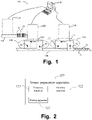
- FIG. 1 is a schematic structural diagram of a sample analysis system 100 according to an embodiment of the disclosure.
- the sample analysis system 100 may be a blood analysis pipeline system, which includes at least one hematology analyzer 110, at least one smear preparation apparatus 120, at least one cell image analysis apparatus 130, and a control apparatus 140.
- the hematology analyzer 110 is configured to perform blood routine examination on a sample to be analyzed
- the smear preparation apparatus 120 is configured to prepare a smear of the sample to be analyzed
- the cell image analysis apparatus 130 is configured to image and analyze cells in the smear
- the control apparatus 140 is communicatively connected to the hematology analyzer 110, the smear preparation apparatus 120, and the cell image analysis apparatus 130.
- the sample analysis system 100 further includes a first transfer track 150 and a second transfer track 160.
- the first transfer track 150 is configured to transfer a test tube rack 10 capable of holding a plurality of tubes 11 containing samples to be analyzed from the hematology analyzer 110 to the smear preparation apparatus 120.
- the second transfer track 150 is configured to transfer a slide basket 20 capable of holding a plurality of prepared smears 21 from the smear preparation apparatus 120 to the cell image analysis apparatus 130.
- the control apparatus 140 is electrically connected to the first transfer track 150 and the second transfer track 160 and controls actions thereof.
- the sample analysis system 100 further includes feeding mechanisms 170 and 180 that respectively corresponds to the hematology analyzer 110 and the smear preparation apparatus 120.
- the feeding mechanisms 170 and 180 respectively include loading buffer regions 171 and 181, feeding test regions 172 and 183, and unloading buffer regions 173 and 183.
- the test tube rack 20 When a sample to be analyzed on the test tube rack 20 needs to be transferred to the hematology analyzer 110 for testing, the test tube rack 20 is first transferred from the first transfer track 150 to the loading buffer region 171, and is then transferred from the loading buffer region 171 to the feeding test region 172 for testing by the hematology analyzer 110. After the test is completed, the test tube rack is unloaded from the feeding test region 172 to the unloading buffer region 173, and finally enters the first transfer track 150 from the unloading buffer region 173.
- test tube rack 20 when a sample to be analyzed on the test tube rack 20 needs to be examined via microscopy, the test tube rack 20 needs to be transferred to the smear preparation apparatus 120 for smear preparation.
- the test tube rack 20 is first transferred from the first transfer track 150 to the loading buffer region 181, and is then transferred from the loading buffer region 181 to the feeding test region 182, so that the smear preparation apparatus 120 prepares a smear.
- the test tube rack is unloaded from the feeding test region 182 to the unloading buffer region 183, and finally enters the first transfer track 150 from the unloading buffer region 183.
- the smear preparation apparatus 120 places the prepared smear in the slide basket 20, and transfers the slide basket 20 containing the smear to be analyzed to the cell image analysis apparatus 130 via the second transfer track 160, and the cell image analysis apparatus 130 images and analyzes cells in the sample on the smear to be analyzed.
- the sample analysis system 100 also includes a display apparatus (not shown) configured to display a sample test result and an input apparatus configured to provide input.
- the display apparatus may be a liquid crystal screen, a touchscreen, an LED screen, or the like.
- the control apparatus 140 may output processed data as image display data such as analysis data and instrument operating parameters to the display apparatus for display.
- the input apparatus may be a mouse, a keyboard, various keys, a trackball, or the like, to facilitate input operations of a user.
- the display apparatus may be disposed on the hematology analyzer 110, the cell image analysis apparatus 130, the control apparatus 140, or elsewhere.
- the smear preparation apparatus 120 may be configured for smear preparation for blood, body fluid, and the like. As shown in FIG. 2 , the smear preparation apparatus 120 includes a smearing machine 121, a staining machine 122, and a printing apparatus 123.
- the smearing machine 121 is configured to smear a sample based on a preset smearing parameter.
- a smearing process includes dispensing the sample to a slide and spreading the sample on the slide into a blood film.
- the smearing machine 121 includes a sampling mechanism for drawing a sample, a slide loading mechanism for moving a slide to a working line, a sample adding mechanism for adding the sample onto the slide, a smearing mechanism for smearing the sample on the slide, and a drying mechanism for drying a blood film on the slide.
- sample aspiration may be puncture aspiration (a sample container has a lid, and the sampling apparatus passes through the lid of the sample container) or may be open aspiration (a sample container is open, and the sampling apparatus directly aspirates the sample from an opening).
- a micro sample feeding mechanism is also included. The micro sample feeding mechanism may directly move a test tube placed by an operator toward the sampling apparatus, or the sampling apparatus may move toward a test tube placed by an operator.
- the micro sample feeding mechanism may alternatively move a test tube toward the sample adding mechanism directly, or the sample adding mechanism may move toward a test tube placed by an operator and directly adds the sample after the sample adding mechanism (such as a blood-dropping needle) aspirates the blood sample, without drawing blood via the sampling mechanism, thereby reducing the need of blood sample and further realizing micro sample feeding and priority sample feeding.
- the sample adding mechanism prepares to drop the blood onto the slide.
- the slide loading mechanism extracts a slide and loads the slide into a corresponding position to facilitate blood dropping operation.
- operations such as detection of left and right sides of the slide and cleaning of the slide may be further performed, and then the slide is loaded.
- the loaded slide may be printed with related information, and operations such as detection of front and back of the slide may also be performed.
- a blood-dropping needle of the sample adding mechanism drops the sample onto the slide and then smearing operation is performed.
- the smearing mechanism smears the blood on the slide into a shape of a blood film.
- the blood film on the slide may be dried to maintain morphology of the blood film.
- the slide is shown in FIG. 2 .
- the slide may be driven to turn over before drying the blood film, so as to satisfy a corresponding need.
- the dried blood smear may further be subjected to drying examination to determine a drying effect of the blood film.
- the dried blood smear may further be subjected to blood film spreading examination to determine whether the blood film is spread and whether a spreading state is satisfactory.
- the staining machine 122 is configured to stain the slide, and specifically, stain the blood film on the slide based on a preset staining parameter.
- the printing apparatus 123 is configured to print sample information on the blood smear of the sample. As shown in FIG. 3 , a label region is provided on the blood smear, and the sample information is printed on the label region of the blood smear.
- the sample information printed on the label region may include information such as a sample number, a barcode, and a quality control marker.
- the cell image analysis apparatus 130 such as a cell image analyzer, at least includes an imaging apparatus 131, a smear moving apparatus 132, and an image analysis apparatus 133.
- the imaging apparatus 131 includes a camera 1312 and a lens group 1311, and is configured to image cells in the sample smeared on the smear.
- the smear moving apparatus 132 is configured to move the smear relative to the imaging apparatus 131, so that the imaging apparatus 131 captures cell images in a specific region of the smear.
- the image analysis apparatus 133 is configured to analyze the cell images of the smear.
- the lens group 1311 may include a first objective lens, a second objective lens, and an ocular lens.
- the first objective lens may be, for example, a 10x objective lens
- the second objective lens may be, for example, a 100x objective lens.
- the lens group 1311 may further include a third objective lens, which may be, for example, a 40x objective lens.
- the cell image analysis apparatus 130 further includes an identification apparatus 134, a slide gripping apparatus 135, and a smear recycle apparatus 136.
- the identification apparatus 134 is configured to identify identity information of the smear.
- the slide gripping apparatus 135 is configured to grip the smear from the identification apparatus 134 to the smear moving apparatus 132 for testing.
- the smear recycle apparatus 136 is configured to hold a smear that has been subjected to testing.
- the cell image analysis apparatus 130 further includes a slide basket loading apparatus 137 configured to load a slide basket containing smears to be analyzed.
- the slide gripping apparatus 135 is further configured to grip a smear to be analyzed in the slide basket loaded on the slide basket loading apparatus 137 to the identification apparatus 134 for identification of identity information.
- the slide basket loading apparatus 137 is connected to the first transfer track 160, so that the smear prepared by the smear preparation apparatus 120 can be transferred to the cell image analysis apparatus 130.
- the blood analysis pipeline system provided in the disclosure includes one, two or more hematology analyzers 110, and one, two or more cell image analysis apparatuses 130.
- FIG. 8 an example in which the blood analysis pipeline system includes two hematology analyzers 110 and two cell image analysis apparatuses 130 is used for description.
- FIG. 8 is only used as an example, and does not limit the number of instruments and apparatuses.
- the hematology analyzers 110, the cell image analysis apparatuses 130, and the smear preparation apparatus have been described above, and therefore are not further described herein.
- the dispatching apparatus is configured to dispatch the sample and the blood smear, and includes the foregoing first transfer track 150, second transfer track 160, feeding mechanism 170, and feeding mechanism 180.
- the foregoing display apparatus includes a display, and the display is disposed on the control apparatus 140.
- the control apparatus 140 further includes a controller 142 and a memory 143.
- the memory 143 is configured to store various data, such as computer programs, test results of the hematology analyzer, test results of the cell image analysis apparatus, and data generated by the smear preparation apparatus.
- the controller 142 includes a processor, which may be configured to execute a program to perform precision management on the blood analysis pipeline system.
- the precision management may include, for example, determination of accuracy and quality of a result provided by an instrument, and may be also referred to as quality control.
- an automated precision management method in which the sample analysis system 100 is a blood analysis pipeline system includes the following steps: Step 1: A sample for quality control is obtained. There is a plurality of methods of obtaining the sample for quality control, one of which is shown in FIG. 7 , including the following steps:
- the preset condition for triggering quality control includes at least one of: completion of testing of N th sample after the hematology analyzer initially starts testing in each cycle, a preset time, a preset load and a preset test result, wherein N is a natural number not less than 1.
- the controller selects a quality control sample every day after the first sample has been tested, or the controller selects a quality control sample at 9:00 a.m. every day, or the controller selects a quality control sample when a load of the sample analysis system is below a preset value, or the controller determines whether the first test results of the samples obtained by the hematology analyzer 110 are test results that satisfy a preset condition, and if yes, the controller selects a quality control sample.
- the test result that satisfies the preset condition may be that various blood routine examination result values all fall within normal ranges, or may be a test result including abnormality information.
- the former may be used for determining whether result values of a normal sample tested by the hematology analyzer and those of the normal sample tested by the cell image analysis apparatus are consistent, and the latter may be used for determining whether abnormality information of a sample tested by the hematology analyzer and that of the sample tested by the cell image analysis apparatus is consistent.
- the foregoing trigger condition may be set by a user, and after the preset condition for triggering quality control is satisfied, a quality control process may be automatically performed.
- the controller selects a quality control sample based on the sample test results obtained by the hematology analyzer 110, wherein the selected quality control sample may be a sample with a normal result. Specifically, the controller selects a sample with a test value of a preset type of cells in the first test result that falls within a preset range, as a quality control sample. For example, a sample with a white blood cell count (WBC) that falls within a first preset range is selected as a quality control sample, a sample with a red blood cell count (RBC) that falls within a second preset range is selected as a quality control sample, or a sample with a mean corpuscular volume (MCV) that falls within a third range preset is selected as a quality control sample.
- WBC white blood cell count
- RBC red blood cell count
- MCV mean corpuscular volume
- the quality control sample may be selected, at least one of the three types of samples, which may be specifically determined based on a quality control need.
- the first range, the second range, and the third range may be set based on a quality control need.
- the first range is from 7.0 ⁇ 10 9 to 10.0 ⁇ 10 9 /L
- the second range is from 4.0 ⁇ 10 12 to 5.5 ⁇ 10 12 /L
- the third range is from 85 to 95 fL.
- the selected quality control sample may alternatively be a sample with an abnormal result.
- the controller selects a sample having a first test result containing abnormality information as a quality control sample.
- the abnormality information includes at least one of malaria parasites, platelet aggregation, red blood cell aggregation, hematocrit (HCT) exceeding a preset threshold, uneven size of red blood cells, and abnormal white blood cells.
- selected samples may include both a sample with a normal result and a sample with an abnormal result, so that quality control can be performed from two perspectives, thereby satisfying various quality control needs.
- Step 1.3 The controller controls the dispatching apparatus, for example, the first track 150, to transfer the quality control sample to the smearing machine of the smear preparation apparatus 120.
- Another method of obtaining a sample for quality control includes the following steps:
- Still another method of obtaining a sample for quality control includes the following steps:
- the automated precision management method may further include: providing, by the controller, an automated quality control option on a display interface of the display apparatus for selection by a user, and after an instruction for selecting the automated quality control option is received, performing the automated quality control start step, that is, step 1.2, step 1.2', or step 1.2".
- the automated precision management method in the disclosure may be enabled or disabled by the user. If disabled, the user may manually perform the quality control operation.
- Step 2 The controller controls the smearing machine of the smear preparation apparatus 120 to prepare a blood smear from the quality control sample. Specifically, the controller controls the smearing machine to smear the quality control sample based on a preset smearing parameter, wherein a smearing process includes dispensing the quality control sample onto a slide and spreading the sample on the slide into a blood film.
- the smearing parameter may include width, length, tail shape, and the like of the blood film, and the smearing machine may perform smearing based on the preset smearing parameter, to prepare a blood smear that satisfies the preset smearing parameter.
- the prepared blood smear is shown in FIG. 2 .
- One or more blood smears may be prepared from a same sample, and the specific number of blood smears depends on a quality control need, thereby satisfying various quality control needs.
- the controller further controls the printing apparatus to provide sample information on the blood smear of the quality control sample, wherein the sample information includes a quality control marker.
- the printing apparatus prints, sprays, attaches, writes, or draws the quality control marker on the label region shown in FIG. 2 , so that the quality control blood smear has the quality control marker, which facilitates identification. Because the quality control marker is provided, in subsequent staining and imaging, the quality control blood smear is treated differently from regular samples, and corresponding staining and imaging may be performed for the quality control blood smear based on quality control needs, to realize better quality control.
- Step 3 The controller controls the staining machine to stain the quality control blood smear, for example, the controls the staining machine to stain the blood film on the slide based on a preset staining parameter or a preset staining mode.
- the preset staining parameter may be a parameter corresponding to a characteristic (blood or body fluid) of the sample, or may be a parameter corresponding to a staining mode of the cell image analysis apparatus 130.
- the smearing machine and the staining machine both select a preset parameter or a preset mode for smearing and staining for the quality control sample, thereby preparing a blood smear matching the preset parameter or the preset mode.
- test result may be used for determining quality of smearing and staining of the cell image analysis apparatus 130. It can be seen that sample selection, blood smear preparation and staining in the disclosure may all be performed based on quality control needs, so that quality control can be completed quickly.
- the controller controls the dispatching apparatus, for example, the second transfer track 160, to transfer the quality control blood smear to the cell image analysis apparatus 130.
- Step 4 The controller controls the cell image analysis apparatus 130 to image and analyze the quality control blood smear to obtain a second test result. Specifically, the controller controls working of the cell image analysis apparatus 130, the identification apparatus of the cell image analysis apparatus 130 identifies the quality control marker on the blood smear of the quality control sample, and therefore the cell image analysis apparatus 130 learns that this blood smear is a blood smear of the quality control sample, and then switches to a quality control mode and images and analyzes the quality control blood smear in the quality control mode to obtain the second test result.
- the cell image analysis apparatus 130 may use a slide scanner, an image captured in the quality control mode of the slide scanner is more comprehensive than an image captured in a regular mode, and a second test result obtained through analysis in the quality control mode is also more comprehensive than that obtained for a regular sample, thereby satisfying quality control needs. That is, the cell image analysis apparatus 130 can perform imaging and analysis based on quality control needs.
- working modes of the cell image analysis apparatus 130 include a white blood cell analysis mode, a red blood cell analysis mode, a platelet analysis mode, and the like, which may be used to specifically analyze and test white blood cells, red blood cells, and platelets respectively.
- the quality control mode may be a combination of these three modes, and can be used to image and analyze various types of cells more comprehensively.
- Step 5 The controller obtains the second test result of the quality control blood smear from the cell image analysis apparatus 130, compares the first test result and the second test result of the quality control sample, and controls the display apparatus to output a result of the comparison.
- the controller obtains the second test result of the quality control blood smear from the cell image analysis apparatus 130, compares the first test result and the second test result of the quality control sample, and controls the display apparatus to output a result of the comparison.
- the first test result is used as a baseline, then it can be determined through the comparison whether the second test result is accurate, so as to perform quality control on the cell image analysis apparatus 130; if the second test result is used as a baseline, it can be determined through the comparison whether the first test result is accurate, so as to perform quality control on the hematology analyzer 110. Details are described below.
- step 1 may be started after the hematology analyzer 110 passes quality control.
- quality control may be performed on the cell image analysis apparatus 130 by using the first test result obtained by the hematology analyzer 110 as a baseline.
- the quality control for the hematology analyzer 110 may include quality control performed on the hematology analyzer by using a standard control product, or quality control performed by comparing results of a plurality of hematology analyzers.
- a test result obtained by the hematology analyzer 110 for the standard control product is within a preset range, or if a test result obtained by the hematology analyzer 110 and a test result obtained by another analyzer for a same sample are consistent, the hematology analyzer 110 is deemed to pass quality control.
- Comparing the first test result with the second test result of the quality control sample may specifically include: comparing a first test result value of a preset type of cells in the quality control sample in the first test result with a second test result value of the preset type of cells in the second test result, for example, comparing result values of lymphocytes and neutrophils in the first test result with those in the second test result; if a deviation between the first test result value and the second test result value of the preset type of cells of the quality control sample is outside a preset range, outputting prompt information.
- a lymphocyte ratio of the quality control sample in the first test result is 20%
- a lymphocyte ratio of the quality control sample in the second test result is 30%
- a preset deviation range of lymphocytes is from -5% to 5%
- a deviation between the lymphocyte ratios in the first test result and the second test result is outside the preset range
- a display interface may display the deviation of 10% between the lymphocyte ratios, or may prompt that the deviation between the lymphocyte ratios is outside the preset range, which indicates that at least one of the results of the hematology analyzer and the cell image analysis apparatus is inaccurate, and at least one of the hematology analyzer and the cell image analysis apparatus fails to pass quality control.
- the selected quality control sample is a normal sample, which, for example, has a WBC (white blood cell) count between 7.0 ⁇ 10 9 /L and 10.0 ⁇ 10 9 /L, an RBC (red blood cell) count between 4.0 ⁇ 10 12 /L and 5.5 ⁇ 10 12 /L, and an MCV (mean corpuscular volume) between 85 fL and 95 fL
- the controller compares a white blood cell count of the quality control sample in the first test result with that in the second test result, and if a deviation between the two white blood cell counts is not outside the preset range, it indicates that the test of the white blood cell count conducted by the cell image analysis apparatus 130 satisfies a precision requirement.
- the controller compares a red blood cell count of the quality control sample in the first test result and that in the second test result, and if a deviation between the two red blood cell counts is not outside the preset range, it indicates that the test of the red blood cell count conducted by the cell image analysis apparatus 130 satisfies a precision requirement.
- the controller compares a mean corpuscular volume of red blood cells in the quality control sample in the first test result and that in the second test result, if a deviation between the two mean corpuscular volumes is not outside the preset range, it indicates that the test of the mean corpuscular volume conducted by the cell image analysis apparatus 130 satisfies a precision requirement.
- a deviation between a result of a preset parameter in the first test result and a result of the preset parameter in the second test result may be obtained through comparison to determine whether the test conducted by the cell image analysis apparatus 130 satisfies requirements, and if the deviation is outside the preset range, prompt information indicating that the cell image analysis apparatus 130 fails to pass quality control is output.
- a ratio of the deviation between the result of the preset parameter in the first test result and the result of the preset parameter in the second test result may further be obtained through comparison to determine whether the test conducted by the cell image analysis apparatus 130 satisfies requirements.
- the controller obtains a ration or percentage of deviation between the second test result and the first test result of the quality control sample through comparison.
- the ratio of deviation may be obtained by multiplying by 100% a quotient of dividing the deviation by the first test result value. If the ratio of deviation between the second test result and the first test result of the quality control sample is outside a corresponding preset range, the controller outputs, via the display apparatus, prompt information indicating that the cell image analysis apparatus 130 fails to pass quality control.
- the prompt information may be more specific, for example, prompt information indicating that a test result of a specific type of cells obtained by the cell image analysis apparatus 130 does not satisfy the precision requirement is output.
- determining whether the ratio of deviation between the first test result and the second test result of the quality control sample is outside a preset range may include: comparing result values of a plurality of preset parameters.
- the result values of the preset parameters may be test values of various types of cells such as neutrophilic segmented granulocytes, lymphocytes, and eosinophils. If a deviation between the result values of any type of cells in the first test result and in the second test result is outside a range, it is indicated that the ratio of deviation between the first test result and the second test result of the quality control sample is outside the preset range. If deviations of the result values of all preset parameters in the first test result and in the second test result are not outside the preset range, it is indicated that the ratio of deviation between the first test result and the second test result of the quality control sample is not outside the preset range.
- the controller controls the dispatching apparatus to transfer the sample to be analyzed (the quality control sample) to another hematology analyzer for retesting, to obtain a first retest result using said hematology analyzer; the controller compares the first retest result with the second test result, to determine whether a deviation between the first retest result value and the second test result value is outside a preset range, whether a ration of deviation between the first retest result and the second test result is outside a preset range, and so on.
- a specific process is the same as the foregoing comparison process of the first test result and the second test result, and details are not described herein again. Retesting by another hematology analyzer instead of the hematology analyzer can eliminate interference caused by the hematology analyzer itself to quality control for the cell image analysis apparatus 130.
- the controller may alternatively control the dispatching apparatus to transfer the sample to be analyzed (quality control sample) to another cell image analysis apparatus for retesting, to obtain a second retest result using said cell image analysis apparatus; and then compare the second retest result with the first test result, to determine whether a deviation between the first test result value and the second retest result value is outside a preset range, whether a ratio of deviation between the first test result and the second retest result is outside a preset range, and so on.
- a specific process is the same as the foregoing comparison process of the first test result and the second test result, and details are not described herein again.
- Retesting by another cell image analysis apparatus instead of the cell image analysis apparatus 130 can eliminate interference caused by the cell image analysis apparatus 130 itself to quality control for the cell analyzer.
- the controller may alternatively control the dispatching apparatus to transfer the sample to be analyzed (quality control sample) to the original hematology analyzer and the original cell image analysis apparatus for retesting, to obtain a first retest result using the original hematology analyzer and a second retest result using the original cell image analysis apparatus; and further compare the first retest result with the second retest result.
- a specific comparison process thereof is the same as the foregoing comparison process of the first test result and the second test result, and details are not described herein again.
- retesting of the quality control sample includes retesting by a new hematology analyzer, retesting by a new cell image analysis apparatus, or retesting by the original hematology analyzer or cell image analysis apparatus. Retesting of the quality control sample is performed again upon failure of initial quality control, and quality control determination may be performed again to obtain a more accurate quality control result.
- the controller may reselect a quality control sample from the samples to be analyzed that have been tested to conduct retesting, and perform quality control determination again to improve accuracy.
- the cell image analysis apparatus 130 and the hematology analyzer 110 have their respective advantages in sample testing.
- the cell image analysis apparatus 130 is more accurate than the hematology analyzer 110 in terms of morphology testing of cells. Therefore, test results obtained by the cell image analysis apparatus 130 may be used for quality control determination for the hematology analyzer 110.
- the first test result of the quality control sample includes abnormality information that satisfies the preset condition for triggering quality control. For example, in step 1, a sample with abnormality information is selected as a quality control sample, and the quality control sample is further used for quality control on the hematology analyzer 110.
- the controller determines, through comparison, whether abnormality information in the first test result of the quality control sample is consistent with abnormality information in the second test result of the quality control sample; and if the abnormality information in the first test result of the quality control sample is inconsistent with the abnormality information in the second test result of the quality control sample, outputting corresponding prompt information via the display apparatus, for example, outputting prompt information indicating that testing performed by the hematology analyzer 110 for abnormality information is unsatisfactory.
- prompt information indicating that testing performed by the hematology analyzer 110 for abnormality information is satisfactory is outputted via the display apparatus.
- the cell image analysis apparatus 130 can generally obtain more accurate corresponding test results for abnormality information such as malaria parasites, PLT aggregation, red blood cell aggregation, and abnormal cell warning.
- precision management may be performed on the hematology analyzer 110 based on the test results obtained by the cell image analyzer 130.
- automated quality control is implemented for both the cell image analysis apparatus 130 and the hematology analyzer 110.
- quality of smearing and staining of the quality control blood smear may be further determined based on the second test result obtained by the cell image analysis apparatus 130, that is, quality control may also be determined for the smearing machine and the staining machine.
- the second test result includes test information based on cell imaging and analysis, and test information related to quality of smearing and staining of the cell image analysis apparatus.
- the cell image analysis apparatus 130 may capture images of the quality control blood smear, and may identify blood film shape, blood film color, and the like based on an image identification algorithm; or the cell image analysis apparatus 130 may scan the blood smear by using its objective lens to identify information such as cell distribution, blood film boundary, blood film shape, and blood film color of the quality control blood smear. After the information related to smearing and staining is obtained, whether the quality of smearing and staining meets expectation may be determined.
- the quality control blood smear is prepared based on the preset smearing parameter and the preset staining parameter or the preset staining mode, blood film size and shape, cell distribution, color, and the like of the quality control blood smear are supposed to match the preset smearing parameter and the preset staining parameter or the preset staining mode. If it is found through the cell image analysis apparatus 130 that the blood film size and shape, cell distribution, color, or the like of the quality control blood smear does not match the preset smearing parameter and the preset staining parameter or the preset staining mode, it may be determined that the smear preparation apparatus fails to pass quality control.
- the controller may identify at least one of blood film morphology and cell distribution of the quality control blood smear based on the test information related to smearing in the second test result; and determine smearing quality of the smearing machine based on at least one of the blood film morphology and cell distribution of the quality control blood smear.
- the test information related to smearing in the second test result includes at least one of blood film morphology and cell distribution of the quality control blood smear that are obtained by the cell image analysis apparatus 130, for example, a blood film morphology image and a cell distribution image of the quality control blood smear that may be obtained by the image analysis apparatus 130.
- the controller may identify the blood film morphology in the image based on the blood film morphology image of the quality control blood smear.
- the controller identifies the cell distribution based on the cell distribution image of the quality control blood smear, for example, to determine whether positional distribution of specified cells in the blood film is satisfactory, if not, it indicates that the smearing is unqualified.
- the controller may further identify at least one of blood film color and color shade of the quality control blood smear based on the test information related to staining in the second test result; and determine staining quality of the smear preparation apparatus based on at least one of the blood film color and the color shade of the quality control blood smear.
- the test information related to staining in the second test result includes at least one of blood film color and color shade of the quality control blood smear that are obtained by the cell image analysis apparatus 130.
- the controller identifies the blood film color (such as chromatic value) and the color shade (such as gray-scale value) based on the blood film image in the second test result, or certainly, may obtain the blood film color and the color shade of the quality control blood smear directly by using the cell image analysis apparatus 130; determines whether the blood film color is a preset color, and if the blood film color is not the preset color, it indicates that the staining performed by the staining machine is unqualified; and determines whether the color shade of the blood film is consistent with a preset depth, and if the color shade of the blood film is not consistent with the preset depth, it indicates that the staining is unqualified.
- the blood film color such as chromatic value
- the color shade such as gray-scale value
- step 4 after the second test result is obtained, the controller determines the smearing quality and the staining quality, and step 5 is performed after it is determined that the smearing quality and the staining quality is qualified.
- quality control is performed on the cell image analysis apparatus 130 after it is determined that the smearing machine and staining machine are functioning properly in performing smearing and staining. In this way, interference caused by the smearing quality and the staining quality to the quality control for the cell image analysis apparatus 130 is avoided, thereby obtaining high quality control accuracy.
- quality control is implemented for the smearing machine, the staining machine, and the slide scanner based on the entire blood analysis pipeline system.
- the test results may also reflect consistency and correlation of the entire pipeline system, thereby facilitating comprehensive determination on the pipeline system. Because a regular sample is tested only based on test items to obtain a result, a test process is simple, and the result can reflect only some functions and features of the system. In the disclosure, however, processes such as sample selection, smearing, staining, and slide scanning are all performed in accordance with quality control needs, with functions and features of the system all covered, and quality control determination is thus comprehensive with high accuracy.
- an automated precision management method for a sample analysis system is provided and applied to a blood analysis pipeline system including a hematology analyzer, a smear preparation apparatus and a cell image analysis apparatus.
- the method includes the following steps: Step S1: After the pipeline system is ready, a user places samples to be analyzed.
- the pipeline system being ready means that software and hardware of the pipeline system are ready in a state capable of testing samples after the pipeline system is turned on or quits hibernation. After the samples to be analyzed are placed, the system may start a process of automated sample transfer and testing.
- Samples in a batch are samples placed into one or more sample racks by the user, and the hematology analyzer may test these samples in sequence.
- the sample rack is configured to hold at least one sample, and usually is capable of holding a plurality of samples.
- Step S2 The hematology analyzer tests a current sample at its test position to obtain a first test result of the sample, which is a blood routine examination result in this embodiment.
- Step S3 A controller automatically selects a sample for preparing a quality control blood smear based on the blood routine examination result.
- the sample needs to satisfy a specific parameter requirement, for example, the sample has a WBC count between 7.0 ⁇ 10 9 /L and 10.0 ⁇ 10 9 /L, an RBC count between 4.0 ⁇ 10 12 /L and 5.5 ⁇ 10 12 /L, an MCV value between 85 fL and 95 fL, and so on. If a blood routine examination result of a specific sample satisfies this requirement, the sample may be automatically selected as a quality control sample. There may be one quality control sample. Alternatively, there may be a plurality of quality control samples.
- some quality control samples are samples with all test items falling within a normal range, or some quality control samples are samples with some parameters being outside a limit, for example, samples whose blood routine examination results include high HCT, uneven size of RBCs, abnormal white blood cells, and the like.
- Steps S4 The controller controls the dispatching apparatus to automatically transfer the quality control sample to the smearing machine.
- Step S5 The controller controls the smear preparation apparatus to prepare a quality control blood smear, namely to collect a blood sample, drop the blood sample onto a slide, and spread the blood sample into a blood film.
- a method for preparing the quality control blood smear includes the following steps:
- Step S6 The staining machine or staining module stains the quality control blood smear. As shown in FIG. 11 , staining may specifically include the following steps:
- the controller also determines whether some test results obtained by the hematology analyzer satisfy expectation based on the morphology analysis result obtained by the cell image analysis apparatus (an instrument classification result obtained by the cell image analysis apparatus or an accurate result obtained through manual adjustment). For example, for some items that cannot be tested through regular quality control for the hematology analyzer, for example, accuracy of malaria parasites, PLT aggregation, red blood cell aggregation, abnormal cell warning, and the like, regular quality control for the hematology analyzer generally cannot lead to an accurate quality control result.
- the cell image analysis apparatus can more intuitively and accurately test the items such as malaria parasites, PLT aggregation, red blood cell aggregation, and abnormal cell warning. In this case, test results obtained by the cell image analysis apparatus may be used for quality control on test results of these items obtained by the hematology analyzer.
- a method for automated precision management on various instruments in a pipeline system by using a quality control blood smear may be derived. As shown in FIG. 12 , the method specifically includes the following steps:
- the two results are consistent or the deviation therebetween is within the acceptable range, it indicates that accuracy of the classification result of the cell image analysis apparatus is satisfactory. If the five-classification result of white blood cells obtained by the hematology analyzer and the pre-classification result of the cell image analysis apparatus are consistent or the deviation is within the acceptable range, it indicates that accuracy of the classification result of the cell image analysis apparatus is satisfactory.
- the sample analysis system may be a blood analysis pipeline system including a hematology analyzer, a smear preparation apparatus and a cell image analysis apparatus.
- the method includes the following steps:
- step S9' the controller determines quality of smearing and staining of the quality control blood smear based on the second test result.
- the program may be stored in a computer-readable storage medium which may include: a read-only memory, a random access memory, a magnetic disk, an optical disc, a hard disk, or the like.
- a computer executes the program to implement the foregoing functions.
- the program is stored in a memory of a device, and when the program in the memory is executed by a processor, all or some of the foregoing functions may be implemented.
- the program may alternatively be stored in a storage medium such as a server, another computer, a magnetic disk, an optical disc, a flash drive, or a removable hard disk, and downloaded or replicated for storage in a memory of a local device or version update is performed on a system of the local device.
- a processor executes the program in the memory, all or some of the functions in the foregoing embodiments may be implemented.
- any tangible non-transitory computer-readable storage medium may be used, including a magnetic storage device (a hard disk, a floppy disk, or the like), an optical storage device (CD-ROM, DVD, Blu-ray disc, or the like), a flash memory, and/or the like.
- These computer program instructions may be loaded on a general-purpose computer, a special-purpose computer, or other programmable data processing devices to form a machine, such that these instructions, when executed on a computer or other programmable data processing apparatuses, may produce a means for implementing a specified function.
- These computer program instructions may alternatively be stored in a computer-readable memory.
- the computer-readable memory may instruct a computer or other programmable data processing devices to operate in a particular manner, such that the instructions stored in the computer-readable memory may produce an article of manufacture which includes a means for implementing a specified function.
- the computer program instructions may alternatively be loaded onto a computer or other programmable data processing devices to perform a series of operational steps on the computer or other programmable devices to produce a computer-implemented process, such that the instructions, when executed on the computer or other programmable devices, may provide steps for implementing a specified function.
- the term "include” used herein and any other variations thereof all refer to a non-exclusive inclusion, such that a process, method, article, or device including a list of elements includes not only these elements, but also other elements that are not expressly listed or not inherent to the process, method, system, article, or device.
- the term “couple” used herein and any other variations thereof refer to a physical connection, an electrical connection, a magnetic connection, an optical connection, a communication connection, a functional connection, and/or any other connections.
Landscapes
- Chemical & Material Sciences (AREA)
- Health & Medical Sciences (AREA)
- Life Sciences & Earth Sciences (AREA)
- Biochemistry (AREA)
- General Physics & Mathematics (AREA)
- Pathology (AREA)
- Immunology (AREA)
- Analytical Chemistry (AREA)
- Physics & Mathematics (AREA)
- General Health & Medical Sciences (AREA)
- Engineering & Computer Science (AREA)
- Dispersion Chemistry (AREA)
- Quality & Reliability (AREA)
- Signal Processing (AREA)
- Biomedical Technology (AREA)
- Molecular Biology (AREA)
- Investigating Or Analysing Biological Materials (AREA)
Priority Applications (1)
| Application Number | Priority Date | Filing Date | Title |
|---|---|---|---|
| EP25184344.7A EP4600657A3 (fr) | 2019-12-31 | 2019-12-31 | Système d'analyse d'échantillon et procédé de gestion de précision automatique associé |
Applications Claiming Priority (1)
| Application Number | Priority Date | Filing Date | Title |
|---|---|---|---|
| PCT/CN2019/130898 WO2021134664A1 (fr) | 2019-12-31 | 2019-12-31 | Système d'analyse d'échantillon et procédé de gestion de précision automatique associé |
Related Child Applications (1)
| Application Number | Title | Priority Date | Filing Date |
|---|---|---|---|
| EP25184344.7A Division EP4600657A3 (fr) | 2019-12-31 | 2019-12-31 | Système d'analyse d'échantillon et procédé de gestion de précision automatique associé |
Publications (4)
| Publication Number | Publication Date |
|---|---|
| EP4086626A1 true EP4086626A1 (fr) | 2022-11-09 |
| EP4086626A4 EP4086626A4 (fr) | 2023-01-25 |
| EP4086626B1 EP4086626B1 (fr) | 2025-07-23 |
| EP4086626C0 EP4086626C0 (fr) | 2025-07-23 |
Family
ID=76686233
Family Applications (2)
| Application Number | Title | Priority Date | Filing Date |
|---|---|---|---|
| EP25184344.7A Pending EP4600657A3 (fr) | 2019-12-31 | 2019-12-31 | Système d'analyse d'échantillon et procédé de gestion de précision automatique associé |
| EP19958734.6A Active EP4086626B1 (fr) | 2019-12-31 | 2019-12-31 | Système d'analyse d'échantillon et procédé de gestion de précision automatique associé |
Family Applications Before (1)
| Application Number | Title | Priority Date | Filing Date |
|---|---|---|---|
| EP25184344.7A Pending EP4600657A3 (fr) | 2019-12-31 | 2019-12-31 | Système d'analyse d'échantillon et procédé de gestion de précision automatique associé |
Country Status (3)
| Country | Link |
|---|---|
| EP (2) | EP4600657A3 (fr) |
| CN (1) | CN114829933B (fr) |
| WO (1) | WO2021134664A1 (fr) |
Cited By (2)
| Publication number | Priority date | Publication date | Assignee | Title |
|---|---|---|---|---|
| EP4242623A1 (fr) * | 2022-03-09 | 2023-09-13 | Sysmex Corporation | Procédé et appareil de génération d'informations de contrôle de qualité pour un frottis de sang |
| WO2025245510A1 (fr) * | 2024-05-24 | 2025-11-27 | Beckman Coulter, Inc. | Évaluation d'analyseur reposant sur un échantillon témoin et de patient |
Families Citing this family (3)
| Publication number | Priority date | Publication date | Assignee | Title |
|---|---|---|---|---|
| JP2025509397A (ja) * | 2022-03-11 | 2025-04-11 | ベックマン コールター, インコーポレイテッド | コントロールおよび分析器におけるその使用 |
| KR102869509B1 (ko) * | 2022-06-02 | 2025-10-14 | (주)유아이엠디 | 혈액 샘플의 이미지 분석 정보 제공 방법 |
| CN117173698A (zh) * | 2023-07-25 | 2023-12-05 | 中国人民解放军陆军军医大学第二附属医院 | 一种远程辅助的血细胞智能识别方法、装置及储存介质 |
Family Cites Families (10)
| Publication number | Priority date | Publication date | Assignee | Title |
|---|---|---|---|---|
| US10514385B2 (en) * | 2011-07-22 | 2019-12-24 | Sysmex Corporation | Hematology analyzer, method, and system for quality control measurements |
| CN202770842U (zh) * | 2012-05-21 | 2013-03-06 | 长沙高新技术产业开发区爱威科技实业有限公司 | 一种尿液分析装置 |
| CN102662048B (zh) * | 2012-05-21 | 2014-07-16 | 爱威科技股份有限公司 | 一种尿液分析方法及尿液分析装置 |
| WO2015139276A1 (fr) * | 2014-03-20 | 2015-09-24 | 深圳迈瑞生物医疗电子股份有限公司 | Machine de teinture à poussoir, procédé pour commander le poussoir et dispositif associé |
| JP2015222214A (ja) * | 2014-05-23 | 2015-12-10 | 東ソー株式会社 | 自動分析装置 |
| CN105136795B (zh) * | 2015-07-20 | 2018-12-11 | 爱威科技股份有限公司 | 血液样本检测装置、方法和系统 |
| KR20170014689A (ko) * | 2015-07-31 | 2017-02-08 | 가톨릭대학교 산학협력단 | 혈구분석기의 자동 정도 관리 장치 |
| EP3391058A4 (fr) * | 2015-12-18 | 2019-08-28 | Abbott Laboratories | Systèmes et procédés pour analyse automatisée |
| US10395368B2 (en) * | 2015-12-18 | 2019-08-27 | Abbott Laboratories | Methods and systems for assessing histological stains |
| JP6875137B2 (ja) * | 2017-01-31 | 2021-05-19 | シスメックス株式会社 | 精度管理方法、精度管理システム、管理装置、分析装置および精度管理異常判定方法 |
-
2019
- 2019-12-31 WO PCT/CN2019/130898 patent/WO2021134664A1/fr not_active Ceased
- 2019-12-31 CN CN201980103170.3A patent/CN114829933B/zh active Active
- 2019-12-31 EP EP25184344.7A patent/EP4600657A3/fr active Pending
- 2019-12-31 EP EP19958734.6A patent/EP4086626B1/fr active Active
Cited By (3)
| Publication number | Priority date | Publication date | Assignee | Title |
|---|---|---|---|---|
| EP4242623A1 (fr) * | 2022-03-09 | 2023-09-13 | Sysmex Corporation | Procédé et appareil de génération d'informations de contrôle de qualité pour un frottis de sang |
| US12530769B2 (en) | 2022-03-09 | 2026-01-20 | Sysmex Corporation | Method of generating quality control information, apparatus for generating quality control information, and program |
| WO2025245510A1 (fr) * | 2024-05-24 | 2025-11-27 | Beckman Coulter, Inc. | Évaluation d'analyseur reposant sur un échantillon témoin et de patient |
Also Published As
| Publication number | Publication date |
|---|---|
| EP4086626B1 (fr) | 2025-07-23 |
| CN114829933B (zh) | 2026-03-20 |
| EP4086626C0 (fr) | 2025-07-23 |
| EP4600657A2 (fr) | 2025-08-13 |
| EP4086626A4 (fr) | 2023-01-25 |
| EP4600657A3 (fr) | 2025-11-12 |
| WO2021134664A1 (fr) | 2021-07-08 |
| CN114829933A (zh) | 2022-07-29 |
Similar Documents
| Publication | Publication Date | Title |
|---|---|---|
| EP4086626B1 (fr) | Système d'analyse d'échantillon et procédé de gestion de précision automatique associé | |
| JP5426181B2 (ja) | 検体処理システム、細胞画像分類装置、及び検体処理方法 | |
| JP5470625B2 (ja) | 粒子画像解析方法および装置 | |
| US9726584B2 (en) | Sample imaging apparatus | |
| CN105136795B (zh) | 血液样本检测装置、方法和系统 | |
| CN101968493B (zh) | 尿液工作台管理方法和尿液工作台管理器 | |
| JPS60162955A (ja) | 血球自動分析装置 | |
| EP4067881A1 (fr) | Dispositif de préparation de frottis, système d'analyse d'échantillon et procédé associé | |
| US12481146B2 (en) | Sample image capturing system and method, and computer-readable storage medium | |
| JPH0652263B2 (ja) | 細胞分析装置 | |
| CN114858796A (zh) | 样本图像分析系统和玻片质量检测方法 | |
| WO2021109152A1 (fr) | Système et procédé d'analyse d'échantillons, analyseur d'images de cellules et support de stockage | |
| JP7438345B2 (ja) | 診断分析器のモデルを訓練する装置および方法 | |
| US20220334100A1 (en) | Sample analyzing system, method and cell image analyzing device | |
| JP2023501939A (ja) | 管アセンブリタイプを識別する装置および方法 | |
| KR101895760B1 (ko) | 혈구 분석 시스템 및 그의 제어방법 | |
| JP2023131196A (ja) | 精度管理情報の生成方法、精度管理情報の生成装置、及びプログラム | |
| CN114858795A (zh) | 样本图像获取系统、样本图像获取方法及可读存储介质 | |
| KR102869509B1 (ko) | 혈액 샘플의 이미지 분석 정보 제공 방법 | |
| CN118730646B (zh) | 一种血液推片机及方法 | |
| US20260126426A1 (en) | Sample analyzing system, method and cell image analyzing device | |
| CN119104546A (zh) | 阅片装置、阅片方法及可读存储介质 | |
| CN114324920A (zh) | 样本分析方法、系统及存储介质 | |
| JPS61258167A (ja) | 血球自動分類装置 |
Legal Events
| Date | Code | Title | Description |
|---|---|---|---|
| STAA | Information on the status of an ep patent application or granted ep patent |
Free format text: STATUS: THE INTERNATIONAL PUBLICATION HAS BEEN MADE |
|
| PUAI | Public reference made under article 153(3) epc to a published international application that has entered the european phase |
Free format text: ORIGINAL CODE: 0009012 |
|
| STAA | Information on the status of an ep patent application or granted ep patent |
Free format text: STATUS: REQUEST FOR EXAMINATION WAS MADE |
|
| 17P | Request for examination filed |
Effective date: 20220727 |
|
| AK | Designated contracting states |
Kind code of ref document: A1 Designated state(s): AL AT BE BG CH CY CZ DE DK EE ES FI FR GB GR HR HU IE IS IT LI LT LU LV MC MK MT NL NO PL PT RO RS SE SI SK SM TR |
|
| A4 | Supplementary search report drawn up and despatched |
Effective date: 20230103 |
|
| RIC1 | Information provided on ipc code assigned before grant |
Ipc: G01N 35/00 20060101ALI20221221BHEP Ipc: G01N 33/48 20060101AFI20221221BHEP |
|
| DAV | Request for validation of the european patent (deleted) | ||
| DAX | Request for extension of the european patent (deleted) | ||
| GRAP | Despatch of communication of intention to grant a patent |
Free format text: ORIGINAL CODE: EPIDOSNIGR1 |
|
| STAA | Information on the status of an ep patent application or granted ep patent |
Free format text: STATUS: GRANT OF PATENT IS INTENDED |
|
| INTG | Intention to grant announced |
Effective date: 20241115 |
|
| GRAJ | Information related to disapproval of communication of intention to grant by the applicant or resumption of examination proceedings by the epo deleted |
Free format text: ORIGINAL CODE: EPIDOSDIGR1 |
|
| STAA | Information on the status of an ep patent application or granted ep patent |
Free format text: STATUS: REQUEST FOR EXAMINATION WAS MADE |
|
| GRAP | Despatch of communication of intention to grant a patent |
Free format text: ORIGINAL CODE: EPIDOSNIGR1 |
|
| STAA | Information on the status of an ep patent application or granted ep patent |
Free format text: STATUS: GRANT OF PATENT IS INTENDED |
|
| INTC | Intention to grant announced (deleted) | ||
| INTG | Intention to grant announced |
Effective date: 20250331 |
|
| GRAS | Grant fee paid |
Free format text: ORIGINAL CODE: EPIDOSNIGR3 |
|
| GRAA | (expected) grant |
Free format text: ORIGINAL CODE: 0009210 |
|
| STAA | Information on the status of an ep patent application or granted ep patent |
Free format text: STATUS: THE PATENT HAS BEEN GRANTED |
|
| AK | Designated contracting states |
Kind code of ref document: B1 Designated state(s): AL AT BE BG CH CY CZ DE DK EE ES FI FR GB GR HR HU IE IS IT LI LT LU LV MC MK MT NL NO PL PT RO RS SE SI SK SM TR |
|
| REG | Reference to a national code |
Ref country code: GB Ref legal event code: FG4D |
|
| REG | Reference to a national code |
Ref country code: CH Ref legal event code: EP |
|
| REG | Reference to a national code |
Ref country code: DE Ref legal event code: R096 Ref document number: 602019073113 Country of ref document: DE |
|
| REG | Reference to a national code |
Ref country code: IE Ref legal event code: FG4D |
|
| U01 | Request for unitary effect filed |
Effective date: 20250805 |
|
| U07 | Unitary effect registered |
Designated state(s): AT BE BG DE DK EE FI FR IT LT LU LV MT NL PT RO SE SI Effective date: 20250818 |
|
| PG25 | Lapsed in a contracting state [announced via postgrant information from national office to epo] |
Ref country code: IS Free format text: LAPSE BECAUSE OF FAILURE TO SUBMIT A TRANSLATION OF THE DESCRIPTION OR TO PAY THE FEE WITHIN THE PRESCRIBED TIME-LIMIT Effective date: 20251123 |
|
| PG25 | Lapsed in a contracting state [announced via postgrant information from national office to epo] |
Ref country code: NO Free format text: LAPSE BECAUSE OF FAILURE TO SUBMIT A TRANSLATION OF THE DESCRIPTION OR TO PAY THE FEE WITHIN THE PRESCRIBED TIME-LIMIT Effective date: 20251023 |
|
| PG25 | Lapsed in a contracting state [announced via postgrant information from national office to epo] |
Ref country code: HR Free format text: LAPSE BECAUSE OF FAILURE TO SUBMIT A TRANSLATION OF THE DESCRIPTION OR TO PAY THE FEE WITHIN THE PRESCRIBED TIME-LIMIT Effective date: 20250723 |
|
| PG25 | Lapsed in a contracting state [announced via postgrant information from national office to epo] |
Ref country code: GR Free format text: LAPSE BECAUSE OF FAILURE TO SUBMIT A TRANSLATION OF THE DESCRIPTION OR TO PAY THE FEE WITHIN THE PRESCRIBED TIME-LIMIT Effective date: 20251024 |
|
| PG25 | Lapsed in a contracting state [announced via postgrant information from national office to epo] |
Ref country code: PL Free format text: LAPSE BECAUSE OF FAILURE TO SUBMIT A TRANSLATION OF THE DESCRIPTION OR TO PAY THE FEE WITHIN THE PRESCRIBED TIME-LIMIT Effective date: 20250723 |
|
| PG25 | Lapsed in a contracting state [announced via postgrant information from national office to epo] |
Ref country code: RS Free format text: LAPSE BECAUSE OF FAILURE TO SUBMIT A TRANSLATION OF THE DESCRIPTION OR TO PAY THE FEE WITHIN THE PRESCRIBED TIME-LIMIT Effective date: 20251023 |
|
| PG25 | Lapsed in a contracting state [announced via postgrant information from national office to epo] |
Ref country code: ES Free format text: LAPSE BECAUSE OF FAILURE TO SUBMIT A TRANSLATION OF THE DESCRIPTION OR TO PAY THE FEE WITHIN THE PRESCRIBED TIME-LIMIT Effective date: 20250723 |
|
| U20 | Renewal fee for the european patent with unitary effect paid |
Year of fee payment: 7 Effective date: 20251230 |
|
| PG25 | Lapsed in a contracting state [announced via postgrant information from national office to epo] |
Ref country code: SM Free format text: LAPSE BECAUSE OF FAILURE TO SUBMIT A TRANSLATION OF THE DESCRIPTION OR TO PAY THE FEE WITHIN THE PRESCRIBED TIME-LIMIT Effective date: 20250723 |
|
| PG25 | Lapsed in a contracting state [announced via postgrant information from national office to epo] |
Ref country code: CZ Free format text: LAPSE BECAUSE OF FAILURE TO SUBMIT A TRANSLATION OF THE DESCRIPTION OR TO PAY THE FEE WITHIN THE PRESCRIBED TIME-LIMIT Effective date: 20250723 |
|
| PG25 | Lapsed in a contracting state [announced via postgrant information from national office to epo] |
Ref country code: SK Free format text: LAPSE BECAUSE OF FAILURE TO SUBMIT A TRANSLATION OF THE DESCRIPTION OR TO PAY THE FEE WITHIN THE PRESCRIBED TIME-LIMIT Effective date: 20250723 |