EP4102461A2 - Verfahren und vorrichtung zur registrierung medizinischer bilder - Google Patents
Verfahren und vorrichtung zur registrierung medizinischer bilder Download PDFInfo
- Publication number
- EP4102461A2 EP4102461A2 EP22178200.6A EP22178200A EP4102461A2 EP 4102461 A2 EP4102461 A2 EP 4102461A2 EP 22178200 A EP22178200 A EP 22178200A EP 4102461 A2 EP4102461 A2 EP 4102461A2
- Authority
- EP
- European Patent Office
- Prior art keywords
- image
- dimensional
- medical image
- rigid
- registration
- Prior art date
- Legal status (The legal status is an assumption and is not a legal conclusion. Google has not performed a legal analysis and makes no representation as to the accuracy of the status listed.)
- Pending
Links
Images







Classifications
-
- G—PHYSICS
- G06—COMPUTING OR CALCULATING; COUNTING
- G06T—IMAGE DATA PROCESSING OR GENERATION, IN GENERAL
- G06T7/00—Image analysis
- G06T7/30—Determination of transform parameters for the alignment of images, i.e. image registration
-
- A—HUMAN NECESSITIES
- A61—MEDICAL OR VETERINARY SCIENCE; HYGIENE
- A61B—DIAGNOSIS; SURGERY; IDENTIFICATION
- A61B6/00—Apparatus or devices for radiation diagnosis; Apparatus or devices for radiation diagnosis combined with radiation therapy equipment
- A61B6/02—Arrangements for diagnosis sequentially in different planes; Stereoscopic radiation diagnosis
- A61B6/03—Computed tomography [CT]
- A61B6/032—Transmission computed tomography [CT]
-
- A—HUMAN NECESSITIES
- A61—MEDICAL OR VETERINARY SCIENCE; HYGIENE
- A61B—DIAGNOSIS; SURGERY; IDENTIFICATION
- A61B6/00—Apparatus or devices for radiation diagnosis; Apparatus or devices for radiation diagnosis combined with radiation therapy equipment
- A61B6/52—Devices using data or image processing specially adapted for radiation diagnosis
- A61B6/5211—Devices using data or image processing specially adapted for radiation diagnosis involving processing of medical diagnostic data
- A61B6/5229—Devices using data or image processing specially adapted for radiation diagnosis involving processing of medical diagnostic data combining image data of a patient, e.g. combining a functional image with an anatomical image
- A61B6/5235—Devices using data or image processing specially adapted for radiation diagnosis involving processing of medical diagnostic data combining image data of a patient, e.g. combining a functional image with an anatomical image combining images from the same or different ionising radiation imaging techniques, e.g. PET and CT
-
- G—PHYSICS
- G06—COMPUTING OR CALCULATING; COUNTING
- G06T—IMAGE DATA PROCESSING OR GENERATION, IN GENERAL
- G06T7/00—Image analysis
- G06T7/0002—Inspection of images, e.g. flaw detection
- G06T7/0012—Biomedical image inspection
-
- G—PHYSICS
- G06—COMPUTING OR CALCULATING; COUNTING
- G06T—IMAGE DATA PROCESSING OR GENERATION, IN GENERAL
- G06T7/00—Image analysis
- G06T7/10—Segmentation; Edge detection
- G06T7/11—Region-based segmentation
-
- G—PHYSICS
- G16—INFORMATION AND COMMUNICATION TECHNOLOGY [ICT] SPECIALLY ADAPTED FOR SPECIFIC APPLICATION FIELDS
- G16H—HEALTHCARE INFORMATICS, i.e. INFORMATION AND COMMUNICATION TECHNOLOGY [ICT] SPECIALLY ADAPTED FOR THE HANDLING OR PROCESSING OF MEDICAL OR HEALTHCARE DATA
- G16H30/00—ICT specially adapted for the handling or processing of medical images
- G16H30/40—ICT specially adapted for the handling or processing of medical images for processing medical images, e.g. editing
-
- G—PHYSICS
- G06—COMPUTING OR CALCULATING; COUNTING
- G06T—IMAGE DATA PROCESSING OR GENERATION, IN GENERAL
- G06T2207/00—Indexing scheme for image analysis or image enhancement
- G06T2207/10—Image acquisition modality
- G06T2207/10072—Tomographic images
- G06T2207/10081—Computed x-ray tomography [CT]
-
- G—PHYSICS
- G06—COMPUTING OR CALCULATING; COUNTING
- G06T—IMAGE DATA PROCESSING OR GENERATION, IN GENERAL
- G06T2207/00—Indexing scheme for image analysis or image enhancement
- G06T2207/10—Image acquisition modality
- G06T2207/10116—X-ray image
-
- G—PHYSICS
- G06—COMPUTING OR CALCULATING; COUNTING
- G06T—IMAGE DATA PROCESSING OR GENERATION, IN GENERAL
- G06T2207/00—Indexing scheme for image analysis or image enhancement
- G06T2207/30—Subject of image; Context of image processing
- G06T2207/30004—Biomedical image processing
- G06T2207/30096—Tumor; Lesion
Definitions
- the invention relates to a medical image registration method and an apparatus therefor, and more particularly, to a method of registering a two-dimensional medical image, such as a radiographic image, and a three-dimensional medical image, such as a computed tomography (CT) image, and an apparatus therefor.
- a two-dimensional medical image such as a radiographic image
- a three-dimensional medical image such as a computed tomography (CT) image
- a radiographic image (X-ray image), a computed tomography (CT) image, and a magnetic resonance imaging (MRI) image are used for lesion diagnosis.
- a radiographic image is a two-dimensional image, which is easy to take and inexpensive, but it is difficult to identify the detailed structure of the inside of the human body from the two-dimensional image.
- a CT image or an MRI image has the advantage of identifying the cross-sectional structure of the inside of the human body, but has disadvantages in that it takes a long of time and is expensive to take the CT or MRI image.
- a radiographic image is an image of a two-dimensional coordinate system and a CT image is an image of a three-dimensional coordinate system, and thus, it is difficult to register these two images.
- the invention solves this problem by providing a medical image registration method having the features of claim 1, and a medical image registration apparatus having the features of claim 5.
- Advantageous developments of the invention are mentioned in the dependent claims, the wording of which is herewith incorporated into the description by reference.
- One or more embodiments of the invention include a method and apparatus for segmenting a two-dimensional medical image and a three-dimensional medical image into rigid tissue and soft tissue and registering the same.
- a medical image registration method performed by a medical image registration apparatus, includes: generating a two-dimensional rigid image obtained by two-dimensionally projecting rigid tissue segmented from a three-dimensional medical image; generating a two-dimensional soft image obtained by two-dimensionally projecting the three-dimensional medical image or a two-dimensional soft image obtained by two-dimensionally projecting soft tissue segmented from the three-dimensional medical image; registering a two-dimensional medical image with the two-dimensional rigid image; and registering the two-dimensional medical image with the two-dimensional soft image.
- a medical image registration apparatus includes: a rigid image generator configured to generate a two-dimensional rigid image obtained by two-dimensionally projecting rigid tissue segmented from a three-dimensional medical image; a soft image generator configured to generate a two-dimensional soft image obtained by two-dimensionally projecting the three-dimensional medical image or a two-dimensional soft image obtained by two-dimensionally projecting soft tissue segmented from the three-dimensional medical image; a rigid registration unit configured to register a two-dimensional medical image with the two-dimensional rigid image; and a soft registration unit configured to register the two-dimensional medical image with the two-dimensional soft image.
- FIG. 1 illustrates a configuration of an example of a medical image registration apparatus according to an embodiment of the invention.
- a medical image registration apparatus 100 upon receiving a two-dimensional medical image 110 and a three-dimensional medical image 120, registers the two images.
- registration refers to making the two images, which are taken separately, appear in one coordinate system.
- the two-dimensional medical image 110 refers to an image composed of pixels in a two-dimensional coordinate system
- the three-dimensional medical image 120 refers to an image composed of voxels in a three-dimensional coordinate system.
- a radiographic image is used as an example of the two-dimensional medical image 110
- a computed tomography (CT) image is used as an example of the three-dimensional medical image 120, but the two-dimensional medical image 110 and the three-dimensional medical image 120 are not limited thereto and may include other various types of images.
- the two-dimensional medical image 110 or the three-dimensional medical image 120 may be data stored according to the Digital Imaging and Communications in Medicine (DICOM) standard.
- the two-dimensional medical image 110 or the three-dimensional medical image 120 may be stored in other various types of image storage formats.
- the two-dimensional medical image 110 or the three-dimensional medical image 120 may be stored in various systems or databases including a Picture Archiving and Communication System (PACS).
- PACS Picture Archiving and Communication System
- the medical image registration apparatus 100 may read the two-dimensional medical image 110 and the three-dimensional medical image 120 stored in the PACS, register the two images, and store a result of the registration in the PACS.
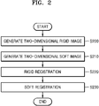
- FIG. 2 is a flowchart of an example of a medical image registration method according to an embodiment of the invention.
- the medical image registration apparatus 100 (hereinafter, referred to as 'apparatus') generates a two-dimensional rigid image obtained by two-dimensionally projecting rigid tissue segmented from a three-dimensional medical image (e.g., a CT image of the chest) in operation S200.
- the rigid tissue refers to a hard part of the human body, and may be, for example, bone.
- a two-dimensional rigid image is a two-dimensional image of a bone region.
- the apparatus 100 may segment a bone region, which is rigid tissue, by using the feature that each human tissue (bone, organ, muscle, fat, etc.) has different voxel brightness values (HU, Hounsfield units) from each other in a three-dimensional medical image.
- a three-dimensional bone region may be extracted by connecting voxels having brightness values corresponding to bone.
- various methods according to the related art, of segmenting each human tissue from a three-dimensional medical image such as segmenting rigid tissue by using an artificial intelligence model for segmenting human tissue, may be applied to the present embodiment, and the rigid tissue segmentation method is not limited to a specific method.
- the apparatus 100 generates a two-dimensional soft image obtained by two-dimensionally projecting a three-dimensional medical image and/or a two-dimensional soft image obtained by two-dimensionally projecting soft tissue segmented from a three-dimensional medical image in operation S210.
- the two-dimensional soft image may be an image generated using a three-dimensional medical image as it is or an image generated using a soft image segmented from a three-dimensional medical image.
- a two-dimensional soft image obtained from a three-dimensional medical image and a two-dimensional soft image obtained from soft tissue segmented from a three-dimensional medical image may be used together.
- the soft tissue may be a tissue, the movement of which is different from that of rigid tissue, according to respiration or motion of a subject.
- the apparatus 100 may segment a three-dimensional lung region from a three-dimensional medical image, based on a voxel brightness value of the lung.
- Examples of a human tissue segmentation method are: Korean Patent Registration No. 10-1482247 titled “Airway Segmentation Method and Apparatus therefor", Korean Patent Registration No. 10-1514003 titled “Lunglobe Extraction Method and Apparatus", and Korean Patent Registration No.
- the positions of the bones and the lung may be different depending on the respiration or motion of a subject.
- a state of the respiration (for example, inhalation or exhalation) or motion of the subject when capturing a two-dimensional medical image is different from the state of respiration or motion of the subject when capturing a three-dimensional medical image
- the positional relationship between the bones and the lung in the two-dimensional medical image may be different from the positional relationship between the bones and the lung in the three-dimensional medical image.
- the apparatus 100 separately performs a registration process of rigid tissue and a registration process of soft tissue, as described below.
- the apparatus 100 registers a two-dimensional medical image (e.g., a radiographic image of the chest) with the two-dimensional rigid image obtained above in operation S200.
- a two-dimensional medical image e.g., a radiographic image of the chest
- various human tissue such as bones, organs, and muscles, are all superimposed on a two-dimensional plane.
- the apparatus 100 performs a registration process of adjusting a position, direction, size, etc. of the two-dimensional medical image and/or the two-dimensional rigid image such that a bone region in the two-dimensional rigid image and a bone region in the two-dimensional medical image match each other.
- a mapping relationship between each pixel of the two-dimensional medical image and each voxel of the three-dimensional medical image may be identified.
- the apparatus 100 may fix a two-dimensional medical image in a coordinate system and enlarge/reduce, rotate, or translate the two-dimensional rigid image in the coordinate system so that the bone regions of the two images match each other.
- the apparatus 100 may superimpose a two-dimensional medical image and a two-dimensional rigid image in one coordinate system, and then register the two images by repeating a process of adjusting a size and a position of the two-dimensional rigid image such that a difference between a pixel brightness value of a bone region of the two-dimensional rigid image and a pixel brightness value of a region of the two-dimensional medical image corresponding to the bone region is minimized.
- Other various registration algorithms for registering two images may be applied to the present embodiment.
- the apparatus 100 registers the two-dimensional medical image with the two-dimensional soft image in operation S230.
- the apparatus 100 may fix the two-dimensional medical image in a coordinate system and register the two-dimensional medical image with the two-dimensional soft image by repeating a process of enlarging/reducing, translating, or rotating the two-dimensional soft image until an error (e.g., a root mean square (RMS) error, etc.) is minimized.
- an error e.g., a root mean square (RMS) error, etc.
- the apparatus 100 may display the two-dimensional rigid image registered with the two-dimensional medical image in a superimposed manner or display the two-dimensional soft image registered with the two-dimensional medical image in a superimposed manner.
- the apparatus 100 may display information about various tissue (e.g., bone, lung, lesion, etc.) identified from a three-dimensional medical image (e.g., size, volume, etc. of various tissue) on a two-dimensional medical image or provide the information to other devices along with the two-dimensional medical image.
- the apparatus 100 may superimpose and display a lesion region inside and outside the rigid tissue or the soft tissue of the three-dimensional medical image on the two-dimensional medical image or provide the lesion region to another device along with the two-dimensional medical image.
- the registration of the rigid image (S220) and the registration of the soft image (S230) may be performed in the reverse order or in parallel.
- FIG. 3 illustrates types of registration according to an embodiment of the present invention.
- the apparatus 100 performs rigid registration 310, soft registration 312, and lesion registration 314 on the three-dimensional medical image 120 and the two-dimensional medical image 110.
- the lesion registration 314 may be omitted.
- an embodiment in which the lesion registration 314 is included is described.
- the three-dimensional medical image 120 is a three-dimensional coordinate system image composed of voxel data
- the two-dimensional medical image 110 is a two-dimensional coordinate system image composed of pixel data
- the apparatus 100 may reduce the dimension of the three-dimensional medical image 120 to two dimensions to generate a two-dimensional rigid image 300, a two-dimensional soft image 302, and a two-dimensional lesion image 304.
- the two-dimensional rigid image 300 is generated by two-dimensionally projecting a three-dimensional rigid tissue region segmented from the three-dimensional medical image 120.
- the two-dimensional rigid image 300 is a two-dimensional image of the bone.
- the two-dimensional soft image 302 is generated by two-dimensionally projecting a three-dimensional soft tissue region segmented from the three-dimensional medical image 120.
- the two-dimensional soft image 302 is a two-dimensional image of the lung.
- the two-dimensional soft image 302 may be an image obtained by two-dimensionally projecting the three-dimensional medical image 120 as it is as a whole.
- the two-dimensional lesion image 304 is an image generated by two-dimensionally projecting a lesion region segmented from the three-dimensional medical image 120.
- the lesion region may be various regions indicating a lesion, such as a tumor or inflammation.
- various algorithms for segmenting a lesion region from the three-dimensional medical image 120 may be applied to the present embodiment.
- various artificial intelligence models for recognizing lesions may be applied to the present embodiment.
- the rigid registration 310 is a process of registering the two-dimensional medical image 110 with the two-dimensional rigid image 300 by using various registration algorithms according to the related art, as described with reference to FIG. 2, and the soft registration 312 is a process of registering the two-dimensional medical image 110 with the two-dimensional soft image 302 by using various registration algorithms according to the related art.
- the apparatus 100 does not directly apply a registration algorithm to the two-dimensional medical image 110 and the two-dimensional lesion image 304, but performs the lesion registration 314 of identically applying, to the two-dimensional lesion image 304, values for enlarging/reducing, translating or rotating the two-dimensional rigid image 300 or the two-dimensional soft image 302 for the rigid registration 310 or the soft registration 312.
- the lesion registration 314 is a process of applying, to the two-dimensional lesion image 304, values of enlargement/reduction, translation, or rotation, which are applied to the two-dimensional rigid image 300 or the two-dimensional soft image 302.
- the apparatus 100 applies, to the two-dimensional lesion image 304, values for enlargement/reduction, translation, or rotation, which are applied to the two-dimensional soft image 302 during the soft registration process 312.
- the apparatus 100 applies, to the two-dimensional lesion image 304, values for enlargement/reduction, translation, or rotation, which are applied to the two-dimensional rigid image 300 during the rigid registration 310.
- FIG. 4 illustrates an example of a three-dimensional medical image according to an embodiment of the invention.
- a three-dimensional medical image 400 includes a plurality of tomographic images.
- the plurality of tomographic images are composed of voxel data. That is, the three-dimensional medical image 400 is composed of a plurality of pieces of voxel data.
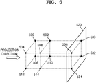
- FIGS. 5 and 6 illustrate an example of a method of two-dimensionally projecting a three-dimensional medical image, according to an embodiment of the invention.
- a three-dimensional medical image is composed of a plurality of voxels 500, 502, 504, 506, 508, 510, 512, and 514 respectively including brightness values as illustrated in FIG. 4.
- the three-dimensional medical image may be a three-dimensional rigid tissue region or a three-dimensional soft tissue region, which is segmented from the three-dimensional medical image.
- the regions will be described as a three-dimensional medical image below.
- the apparatus 100 projects the three-dimensional medical image onto a virtual plane 520 in a certain direction in order to reduce the dimensionality of the three-dimensional medical image to two-dimensions.
- An image projected onto the virtual plane 520 is a two-dimensional image.
- the apparatus 100 generates a brightness value of the two-dimensional image by averaging brightness values of voxels superimposed in a projection direction.
- the apparatus 100 may average brightness values of the first voxel 500 and the second voxel 502 that are superimposed in the projection direction, and generate a brightness value of a first pixel 530 of the two-dimensional image projected onto the virtual plane 520.
- the apparatus 100 may generate a brightness value of a second pixel 536 by averaging brightness values of the third and fourth voxels 504 and 506, generate a brightness value of a third pixel 532 by averaging brightness values of the fifth and sixth voxels 508 and 510, and generate a brightness value of a third pixel 534 by averaging brightness values of the seventh and eighth voxels 512 and 514.
- a two-dimensional soft image 605 may be generated by two-dimensionally projecting a three-dimensional medical image 600 as it is (A), or a two-dimensional rigid image 615 may be generated by two-dimensionally projecting a region 610 of rigid tissue (e.g., bone) segmented from the three-dimensional medical image 600 (B), or a two-dimensional soft image 625 may be generated by two-dimensionally projecting a region 620 of soft tissue (e.g., a lung) segmented from the three-dimensional medical image 600, or a two-dimensional lesion image 635 may be generated by two-dimensionally projecting a lesion region 630 segmented from the three-dimensional medical image 600 (D).
- a two-dimensional soft image 615 may be generated by two-dimensionally projecting a region 610 of rigid tissue (e.g., bone) segmented from the three-dimensional medical image 600 (B)
- a two-dimensional soft image 625 may be generated by two-dimensionally projecting a region 620 of soft
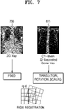
- FIG. 7 illustrates an example of rigid registration according to an embodiment of the invention.
- the apparatus 100 may fix a two-dimensional medical image 700 (e.g., a radiographic image), and register the two-dimensional medical image 700 with the two-dimensional rigid image 615 by translating, rotating, or scaling the two-dimensional rigid image 615.
- a two-dimensional medical image 700 e.g., a radiographic image
- FIG. 8 illustrates an example of soft registration according to an embodiment of the invention.
- the apparatus 100 may fix the two-dimensional medical image 700 (e.g., a radiographic image), and register the two-dimensional medical image 700 with the two-dimensional soft image 605 or 625 by translating, rotating, or scaling the two-dimensional soft image 605 or 625.
- the two-dimensional medical image 700 e.g., a radiographic image
- the apparatus 100 may also register the two-dimensional lesion image 635 obtained by two-dimensionally projecting a lesion region (e.g., a tumor, inflammation, etc.) segmented from a three-dimensional medical image, with the two-dimensional medical image 700. While the present embodiment shows an example in which there is a lesion region in soft tissue, the lesion region may also be in rigid tissue.
- the apparatus 100 does not perform a registration process by directly comparing the two-dimensional medical image 700 with the two-dimensional lesion image 635, but register the two-dimensional lesion image 635 by using a result of performing a process of registering the two-dimensional medical image 700 with the two-dimensional soft image 605 or 625. The above method is illustrated in FIG. 9.
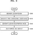
- FIG. 9 is a flowchart of an example of a lesion registration method according to an embodiment of the invention.
- the apparatus 100 segments a lesion region from a three-dimensional medical image in operation S900.
- the apparatus 100 generates a two-dimensional lesion image by two-dimensionally projecting a three-dimensional lesion region segmented from the three-dimensional medical image, by using the projection method as illustrated in FIG. 5 in operation S910.
- the apparatus 100 generates a lesion registration image by applying, to the two-dimensional lesion region, values for translation, rotation, or scaling of a two-dimensional rigid image or a two-dimensional soft image, identified from a process of rigid registration between a two-dimensional medical image and the two-dimensional rigid image or a process of soft registration between the two-dimensional medical image and the two-dimensional soft image in operation S920.
- the apparatus 100 when there is a lesion region in rigid tissue, for rigid registration, the apparatus 100 performs lesion registration by using the values for translation, rotation, and scaling of the two-dimensional rigid image, which are applied to the two-dimensional rigid image, and when there is a lesion region in soft tissue, the apparatus 100 performs, for soft registration, lesion registration by using the values for translation, rotation, and scaling of the two-dimensional soft image, which are applied to the two-dimensional soft image.
- the apparatus 100 may superimpose and display the lesion registration image on the two-dimensional medical image in operation S930.
- An example of superimposing the lesion registration image on the two-dimensional medical image is shown in FIG. 10.
- FIG. 10 illustrates lesion of a three-dimensional medical image, superimposed and displayed on a two-dimensional medical image through a registration process according to an embodiment of the invention.
- the apparatus 100 may detect and display a location of a lesion region 1010 in a two-dimensional medical image 1000.
- the apparatus 100 may register a two-dimensional lesion image obtained from a three-dimensional medical image, by the method of FIG. 9, with a two-dimensional medical image, and then display a position of the registered two-dimensional lesion image on the two-dimensional medical image.
- the apparatus 100 may display various types of information 1020 (e.g., size and volume of a lesion) identified from the three-dimensional medical image together with the two-dimensional medical image 1000.
- FIG. 11 illustrates a configuration of an example of a medical image registration apparatus according to an embodiment of the invention.
- the medical image registration apparatus 100 includes a rigid image generator 1100, a soft image generator 1110, a rigid registration unit 1120, a soft registration unit 1130, a lesion registration unit 1140, and an image processing unit 1150.
- the apparatus 100 may be implemented by a computing device including a memory and a processor, and each component may be mounted in the memory and executed by the processor.
- each component may be mounted in the memory and executed by the processor.
- some of the components of the present embodiment e.g., the lesion registration unit 1140 and the image processing unit 1150 may be omitted.
- the rigid image generator 1100 generates a two-dimensional rigid image obtained by two-dimensionally projecting rigid tissue segmented from a three-dimensional medical image.
- the rigid image generator 1100 may segment a bone region, which is rigid tissue, from a three-dimensional medical image, and then generate a two-dimensional rigid image obtained by two-dimensionally projecting a three-dimensional bone region.
- the soft image generator 1110 generates a two-dimensional soft image obtained by two-dimensionally projecting a three-dimensional medical image or a two-dimensional soft image obtained by two-dimensionally projecting soft tissue segmented from a three-dimensional medical image.
- the soft image generator 1110 may segment a lung region, which is soft tissue, from a three-dimensional medical image, and then generate a two-dimensional soft image obtained by two-dimensionally projecting a three-dimensional lung region.
- the rigid registration unit 1120 registers a two-dimensional medical image with a two-dimensional rigid image.
- the rigid registration unit 1120 may fix a two-dimensional medical image, and register the two-dimensional medical image with a two-dimensional rigid image by translating, rotating, or scaling the two-dimensional rigid image.
- the soft registration unit 1130 registers a two-dimensional medical image with a two-dimensional soft image.
- the soft registration unit 1130 may fix a two-dimensional medical image, and register the two-dimensional medical image with a two-dimensional soft image by translating, rotating, or scaling the two-dimensional soft image.
- An image registration algorithm is well known, and various registration algorithms according to the related art may be used in rigid registration and/or soft registration.
- the lesion registration unit 1140 registers a two-dimensional medical image with a two-dimensional lesion image.
- the lesion registration unit 1140 registers a two-dimensional lesion image with a two-dimensional medical image by using a registration result of the rigid registration unit 1120 or the soft registration unit 1130, instead of using a registration algorithm according to the related art.
- the lesion registration unit 1140 may generate a lesion registration image by applying, to a two-dimensional lesion image, values for translating, rotating, or scaling the two-dimensional soft image by the soft registration unit 1130 for registration of the two-dimensional soft image.
- the image processing unit 1150 may generate an image obtained by excluding rigid or soft tissue from a two-dimensional medical image. For example, when the two-dimensional medical image and the two-dimensional rigid image are registered, the image processing unit 1150 may generate an image obtained by removing rigid tissue (e.g., bone) of the two-dimensional rigid image, from the two-dimensional medical image. The image processing unit 1150 may correct a pixel brightness of a rigid tissue region removed from the two-dimensional medical image to be the same as a pixel brightness of tissue around the bone. As another example, when a two-dimensional medical image and a two-dimensional soft image are registered, the image processing unit 1150 may generate an image obtained by removing a region corresponding to soft tissue of the two-dimensional soft image, from the two-dimensional medical image.
- rigid tissue e.g., bone
- the image processing unit 1150 may correct a pixel brightness of a rigid tissue region removed from the two-dimensional medical image to be the same as a pixel brightness of tissue around the bone.
- the present disclosure may also be implemented as computer-readable code on a computer-readable recording medium.
- the computer-readable recording medium includes all types of recording devices in which data readable by a computer system is stored. Examples of computer-readable recording media include read only memory (ROM), random access memory (RAM), a compact disk ROM (CD-ROM), a solid state disk (SSD), and optical data storage devices.
- ROM read only memory
- RAM random access memory
- CD-ROM compact disk ROM
- SSD solid state disk
- optical data storage devices optical data storage devices.
- the computer-readable recording medium is distributed in a network-connected computer system so that the computer-readable code can be stored and executed in a distributed manner.
- a two-dimensional medical image and a three-dimensional medical image may be registered.
- a two-dimensional medical image and a three-dimensional medical image which differ from each other due to respiration or motion of a subject, may be accurately registered.
- a location of a lesion may be accurately marked on a two-dimensional medical image by using a three-dimensional medical image.
Landscapes
- Engineering & Computer Science (AREA)
- Health & Medical Sciences (AREA)
- Life Sciences & Earth Sciences (AREA)
- Medical Informatics (AREA)
- Physics & Mathematics (AREA)
- Computer Vision & Pattern Recognition (AREA)
- Theoretical Computer Science (AREA)
- General Health & Medical Sciences (AREA)
- Nuclear Medicine, Radiotherapy & Molecular Imaging (AREA)
- Radiology & Medical Imaging (AREA)
- General Physics & Mathematics (AREA)
- Public Health (AREA)
- High Energy & Nuclear Physics (AREA)
- Molecular Biology (AREA)
- Veterinary Medicine (AREA)
- Optics & Photonics (AREA)
- Pathology (AREA)
- Biomedical Technology (AREA)
- Heart & Thoracic Surgery (AREA)
- Biophysics (AREA)
- Surgery (AREA)
- Animal Behavior & Ethology (AREA)
- Quality & Reliability (AREA)
- Pulmonology (AREA)
- Epidemiology (AREA)
- Primary Health Care (AREA)
- Apparatus For Radiation Diagnosis (AREA)
- Measuring And Recording Apparatus For Diagnosis (AREA)
Applications Claiming Priority (1)
| Application Number | Priority Date | Filing Date | Title |
|---|---|---|---|
| KR1020210075641A KR102383753B1 (ko) | 2021-06-10 | 2021-06-10 | 의료영상 정합 방법 및 그 장치 |
Publications (2)
| Publication Number | Publication Date |
|---|---|
| EP4102461A2 true EP4102461A2 (de) | 2022-12-14 |
| EP4102461A3 EP4102461A3 (de) | 2023-01-04 |
Family
ID=81182941
Family Applications (1)
| Application Number | Title | Priority Date | Filing Date |
|---|---|---|---|
| EP22178200.6A Pending EP4102461A3 (de) | 2021-06-10 | 2022-06-09 | Verfahren und vorrichtung zur registrierung medizinischer bilder |
Country Status (3)
| Country | Link |
|---|---|
| US (1) | US12423838B2 (de) |
| EP (1) | EP4102461A3 (de) |
| KR (1) | KR102383753B1 (de) |
Families Citing this family (15)
| Publication number | Priority date | Publication date | Assignee | Title |
|---|---|---|---|---|
| GB2536650A (en) | 2015-03-24 | 2016-09-28 | Augmedics Ltd | Method and system for combining video-based and optic-based augmented reality in a near eye display |
| US12458411B2 (en) | 2017-12-07 | 2025-11-04 | Augmedics Ltd. | Spinous process clamp |
| US12521201B2 (en) | 2017-12-07 | 2026-01-13 | Augmedics Ltd. | Spinous process clamp |
| EP3787543A4 (de) | 2018-05-02 | 2022-01-19 | Augmedics Ltd. | Registrierung einer bezugsmarke für ein system der erweiterten realität |
| US11766296B2 (en) | 2018-11-26 | 2023-09-26 | Augmedics Ltd. | Tracking system for image-guided surgery |
| US12178666B2 (en) | 2019-07-29 | 2024-12-31 | Augmedics Ltd. | Fiducial marker |
| US11382712B2 (en) | 2019-12-22 | 2022-07-12 | Augmedics Ltd. | Mirroring in image guided surgery |
| US11389252B2 (en) | 2020-06-15 | 2022-07-19 | Augmedics Ltd. | Rotating marker for image guided surgery |
| US12502163B2 (en) | 2020-09-09 | 2025-12-23 | Augmedics Ltd. | Universal tool adapter for image-guided surgery |
| US12239385B2 (en) | 2020-09-09 | 2025-03-04 | Augmedics Ltd. | Universal tool adapter |
| US12150821B2 (en) | 2021-07-29 | 2024-11-26 | Augmedics Ltd. | Rotating marker and adapter for image-guided surgery |
| US12475662B2 (en) | 2021-08-18 | 2025-11-18 | Augmedics Ltd. | Stereoscopic display and digital loupe for augmented-reality near-eye display |
| WO2023203521A1 (en) | 2022-04-21 | 2023-10-26 | Augmedics Ltd. | Systems and methods for medical image visualization |
| JP2025531829A (ja) | 2022-09-13 | 2025-09-25 | オーグメディックス リミテッド | 画像誘導医療介入のための拡張現実アイウェア |
| CN120355764B (zh) * | 2025-06-24 | 2025-09-09 | 深圳惟德精准医疗科技有限公司 | 组织图像的配准方法及相关产品 |
Citations (4)
| Publication number | Priority date | Publication date | Assignee | Title |
|---|---|---|---|---|
| KR101482247B1 (ko) | 2013-08-01 | 2015-01-14 | 서울대학교산학협력단 | 기도 추출 방법 및 그 장치 |
| KR101514003B1 (ko) | 2013-10-17 | 2015-04-24 | 서울대학교산학협력단 | 폐엽 추출 방법 및 그 장치 |
| KR20180098984A (ko) | 2017-02-28 | 2018-09-05 | 메디컬아이피 주식회사 | 의료영상의 영역 분리 방법 및 그 장치 |
| KR20210075641A (ko) | 2019-12-13 | 2021-06-23 | 삼성전자주식회사 | 정보 제공 방법 및 이를 지원하는 전자 장치 |
Family Cites Families (10)
| Publication number | Priority date | Publication date | Assignee | Title |
|---|---|---|---|---|
| US7330578B2 (en) * | 2005-06-23 | 2008-02-12 | Accuray Inc. | DRR generation and enhancement using a dedicated graphics device |
| US20080037843A1 (en) * | 2006-08-11 | 2008-02-14 | Accuray Incorporated | Image segmentation for DRR generation and image registration |
| US8948487B2 (en) * | 2011-09-28 | 2015-02-03 | Siemens Aktiengesellschaft | Non-rigid 2D/3D registration of coronary artery models with live fluoroscopy images |
| CN103854276B (zh) * | 2012-12-04 | 2018-02-09 | 东芝医疗系统株式会社 | 图像配准及分割装置和方法,以及医学图像设备 |
| KR101806512B1 (ko) * | 2016-01-19 | 2017-12-07 | 한국외국어대학교 연구산학협력단 | 배경 영상 정보를 이용한 혈관의 비강체 영상정합 장치 및 영상정합 방법 |
| KR101876643B1 (ko) * | 2016-11-23 | 2018-07-13 | 한국과학기술원 | 2d 형광 투시 영상과 3d ct 영상 정합 기반의 치료 가이딩 시스템 및 방법 |
| US10102640B2 (en) * | 2016-11-29 | 2018-10-16 | Optinav Sp. Z O.O. | Registering three-dimensional image data of an imaged object with a set of two-dimensional projection images of the object |
| KR101929412B1 (ko) * | 2017-02-28 | 2018-12-14 | 연세대학교 산학협력단 | 영상 데이터베이스 기반 2차원 x-선 영상 및 3차원 ct 영상의 실시간 정합 방법 및 그 장치 |
| KR101981202B1 (ko) * | 2018-12-11 | 2019-05-22 | 메디컬아이피 주식회사 | 의료영상 재구성 방법 및 그 장치 |
| CN111402305B (zh) * | 2020-06-05 | 2020-09-11 | 嘉兴太美医疗科技有限公司 | 医学影像配准方法、系统和计算机可读介质 |
-
2021
- 2021-06-10 KR KR1020210075641A patent/KR102383753B1/ko active Active
-
2022
- 2022-06-09 EP EP22178200.6A patent/EP4102461A3/de active Pending
- 2022-06-09 US US17/836,948 patent/US12423838B2/en active Active
Patent Citations (4)
| Publication number | Priority date | Publication date | Assignee | Title |
|---|---|---|---|---|
| KR101482247B1 (ko) | 2013-08-01 | 2015-01-14 | 서울대학교산학협력단 | 기도 추출 방법 및 그 장치 |
| KR101514003B1 (ko) | 2013-10-17 | 2015-04-24 | 서울대학교산학협력단 | 폐엽 추출 방법 및 그 장치 |
| KR20180098984A (ko) | 2017-02-28 | 2018-09-05 | 메디컬아이피 주식회사 | 의료영상의 영역 분리 방법 및 그 장치 |
| KR20210075641A (ko) | 2019-12-13 | 2021-06-23 | 삼성전자주식회사 | 정보 제공 방법 및 이를 지원하는 전자 장치 |
Also Published As
| Publication number | Publication date |
|---|---|
| US20220398752A1 (en) | 2022-12-15 |
| US12423838B2 (en) | 2025-09-23 |
| EP4102461A3 (de) | 2023-01-04 |
| KR102383753B1 (ko) | 2022-04-08 |
Similar Documents
| Publication | Publication Date | Title |
|---|---|---|
| EP4102461A2 (de) | Verfahren und vorrichtung zur registrierung medizinischer bilder | |
| KR101981202B1 (ko) | 의료영상 재구성 방법 및 그 장치 | |
| US10803354B2 (en) | Cross-modality image synthesis | |
| US9741131B2 (en) | Anatomy aware articulated registration for image segmentation | |
| EP2189942A2 (de) | Verfahren und System zur Registrierung eines medizinischen Bilds | |
| US20090129650A1 (en) | System for presenting projection image information | |
| US20160371861A1 (en) | Prior image based three dimensional imaging | |
| US9675317B2 (en) | Interface identification apparatus and method | |
| US9135696B2 (en) | Implant pose determination in medical imaging | |
| US20090074276A1 (en) | Voxel Matching Technique for Removal of Artifacts in Medical Subtraction Images | |
| EP3201876B1 (de) | Vorrichtung zur verarbeitung medizinischer bilder, verfahren zur verarbeitung medizinischer bilder | |
| CN104346821A (zh) | 用于医学成像的自动规划 | |
| JP2005521502A (ja) | 胸部および腹部の画像モダリティの重ね合わせ | |
| JP2016539701A (ja) | 三次元(3d)プレスキャンに基づく体積画像データ処理 | |
| US9691157B2 (en) | Visualization of anatomical labels | |
| CN111757701A (zh) | 用于检测解剖区域的系统和方法 | |
| EP1652122B1 (de) | Automatische registration von medizinischen intra-modalitäts-volumenbildern durch verwendung einer affinen transformation | |
| CN107220646B (zh) | 一种去背景干扰的医学图像文字识别增强方法 | |
| CN108430376B (zh) | 提供投影数据集 | |
| US11138736B2 (en) | Information processing apparatus and information processing method | |
| US9361684B2 (en) | Feature validation using orientation difference vector | |
| Wang et al. | X-Recon: learning-based patient-specific high-resolution CT reconstruction from orthogonal X-ray images | |
| CN118985004A (zh) | 根据展开扫描图像的自动肋骨骨折检测 | |
| US20220092745A1 (en) | Information processing apparatus, information processing method, and non-transitory recording medium | |
| US11495346B2 (en) | External device-enabled imaging support |
Legal Events
| Date | Code | Title | Description |
|---|---|---|---|
| PUAI | Public reference made under article 153(3) epc to a published international application that has entered the european phase |
Free format text: ORIGINAL CODE: 0009012 |
|
| STAA | Information on the status of an ep patent application or granted ep patent |
Free format text: STATUS: THE APPLICATION HAS BEEN PUBLISHED |
|
| PUAL | Search report despatched |
Free format text: ORIGINAL CODE: 0009013 |
|
| AK | Designated contracting states |
Kind code of ref document: A2 Designated state(s): AL AT BE BG CH CY CZ DE DK EE ES FI FR GB GR HR HU IE IS IT LI LT LU LV MC MK MT NL NO PL PT RO RS SE SI SK SM TR |
|
| AK | Designated contracting states |
Kind code of ref document: A3 Designated state(s): AL AT BE BG CH CY CZ DE DK EE ES FI FR GB GR HR HU IE IS IT LI LT LU LV MC MK MT NL NO PL PT RO RS SE SI SK SM TR |
|
| RIC1 | Information provided on ipc code assigned before grant |
Ipc: G06T 7/30 20170101AFI20221125BHEP |
|
| STAA | Information on the status of an ep patent application or granted ep patent |
Free format text: STATUS: REQUEST FOR EXAMINATION WAS MADE |
|
| 17P | Request for examination filed |
Effective date: 20230630 |
|
| RBV | Designated contracting states (corrected) |
Designated state(s): AL AT BE BG CH CY CZ DE DK EE ES FI FR GB GR HR HU IE IS IT LI LT LU LV MC MK MT NL NO PL PT RO RS SE SI SK SM TR |
|
| STAA | Information on the status of an ep patent application or granted ep patent |
Free format text: STATUS: THE APPLICATION IS DEEMED TO BE WITHDRAWN |