EP4104863A1 - Procédé de production de biomatériau décellularisé, biomatériau décellularisé et utilisation de celui-ci - Google Patents
Procédé de production de biomatériau décellularisé, biomatériau décellularisé et utilisation de celui-ci Download PDFInfo
- Publication number
- EP4104863A1 EP4104863A1 EP21754359.4A EP21754359A EP4104863A1 EP 4104863 A1 EP4104863 A1 EP 4104863A1 EP 21754359 A EP21754359 A EP 21754359A EP 4104863 A1 EP4104863 A1 EP 4104863A1
- Authority
- EP
- European Patent Office
- Prior art keywords
- biomaterial
- decellularized
- tissue
- rpm
- buffer
- Prior art date
- Legal status (The legal status is an assumption and is not a legal conclusion. Google has not performed a legal analysis and makes no representation as to the accuracy of the status listed.)
- Withdrawn
Links
- 239000012620 biological material Substances 0.000 title claims abstract description 248
- 238000004519 manufacturing process Methods 0.000 title claims description 24
- 238000000034 method Methods 0.000 claims abstract description 142
- 210000001519 tissue Anatomy 0.000 claims abstract description 116
- 210000003786 sclera Anatomy 0.000 claims abstract description 58
- 210000002744 extracellular matrix Anatomy 0.000 claims abstract description 29
- 102000010834 Extracellular Matrix Proteins Human genes 0.000 claims abstract description 28
- 108010037362 Extracellular Matrix Proteins Proteins 0.000 claims abstract description 28
- 239000000126 substance Substances 0.000 claims abstract description 23
- 210000002808 connective tissue Anatomy 0.000 claims abstract description 19
- 239000013543 active substance Substances 0.000 claims abstract description 14
- 241000282414 Homo sapiens Species 0.000 claims abstract description 11
- 230000009885 systemic effect Effects 0.000 claims abstract description 9
- 210000000988 bone and bone Anatomy 0.000 claims description 66
- 230000008569 process Effects 0.000 claims description 62
- LFQSCWFLJHTTHZ-UHFFFAOYSA-N Ethanol Chemical compound CCO LFQSCWFLJHTTHZ-UHFFFAOYSA-N 0.000 claims description 45
- 239000000872 buffer Substances 0.000 claims description 43
- 239000000243 solution Substances 0.000 claims description 39
- 210000002435 tendon Anatomy 0.000 claims description 33
- XLYOFNOQVPJJNP-UHFFFAOYSA-N water Chemical compound O XLYOFNOQVPJJNP-UHFFFAOYSA-N 0.000 claims description 33
- KCXVZYZYPLLWCC-UHFFFAOYSA-N EDTA Chemical compound OC(=O)CN(CC(O)=O)CCN(CC(O)=O)CC(O)=O KCXVZYZYPLLWCC-UHFFFAOYSA-N 0.000 claims description 32
- 238000003756 stirring Methods 0.000 claims description 31
- 101710163270 Nuclease Proteins 0.000 claims description 30
- 238000005406 washing Methods 0.000 claims description 24
- 150000001875 compounds Chemical class 0.000 claims description 23
- FAPWRFPIFSIZLT-UHFFFAOYSA-M Sodium chloride Chemical compound [Na+].[Cl-] FAPWRFPIFSIZLT-UHFFFAOYSA-M 0.000 claims description 21
- 239000007983 Tris buffer Substances 0.000 claims description 20
- 238000004659 sterilization and disinfection Methods 0.000 claims description 20
- 239000003599 detergent Substances 0.000 claims description 19
- 230000001954 sterilising effect Effects 0.000 claims description 19
- 238000005202 decontamination Methods 0.000 claims description 18
- 239000000835 fiber Substances 0.000 claims description 18
- 230000017423 tissue regeneration Effects 0.000 claims description 18
- 230000003588 decontaminative effect Effects 0.000 claims description 17
- 230000001857 anti-mycotic effect Effects 0.000 claims description 15
- 230000018044 dehydration Effects 0.000 claims description 15
- 238000006297 dehydration reaction Methods 0.000 claims description 15
- 239000003814 drug Substances 0.000 claims description 15
- 230000003115 biocidal effect Effects 0.000 claims description 13
- 239000011159 matrix material Substances 0.000 claims description 13
- 239000002543 antimycotic Substances 0.000 claims description 12
- 102000016942 Elastin Human genes 0.000 claims description 11
- 108010014258 Elastin Proteins 0.000 claims description 11
- 229920002549 elastin Polymers 0.000 claims description 11
- 238000011049 filling Methods 0.000 claims description 11
- 239000008363 phosphate buffer Substances 0.000 claims description 11
- 238000011069 regeneration method Methods 0.000 claims description 10
- 239000003242 anti bacterial agent Substances 0.000 claims description 9
- 238000012937 correction Methods 0.000 claims description 9
- 239000003102 growth factor Substances 0.000 claims description 9
- 230000001105 regulatory effect Effects 0.000 claims description 9
- 241000283690 Bos taurus Species 0.000 claims description 8
- TWRXJAOTZQYOKJ-UHFFFAOYSA-L Magnesium chloride Chemical compound [Mg+2].[Cl-].[Cl-] TWRXJAOTZQYOKJ-UHFFFAOYSA-L 0.000 claims description 8
- 229940088710 antibiotic agent Drugs 0.000 claims description 8
- 230000001965 increasing effect Effects 0.000 claims description 8
- 239000002207 metabolite Substances 0.000 claims description 8
- 235000015097 nutrients Nutrition 0.000 claims description 8
- 238000012545 processing Methods 0.000 claims description 8
- 230000011664 signaling Effects 0.000 claims description 8
- 102000012422 Collagen Type I Human genes 0.000 claims description 7
- 108010022452 Collagen Type I Proteins 0.000 claims description 7
- IAYPIBMASNFSPL-UHFFFAOYSA-N Ethylene oxide Chemical compound C1CO1 IAYPIBMASNFSPL-UHFFFAOYSA-N 0.000 claims description 7
- 239000008367 deionised water Substances 0.000 claims description 7
- 229910021641 deionized water Inorganic materials 0.000 claims description 7
- 239000005556 hormone Substances 0.000 claims description 7
- 229940088597 hormone Drugs 0.000 claims description 7
- 210000003491 skin Anatomy 0.000 claims description 7
- 239000011780 sodium chloride Substances 0.000 claims description 7
- 102000001187 Collagen Type III Human genes 0.000 claims description 5
- 108010069502 Collagen Type III Proteins 0.000 claims description 5
- 241000283973 Oryctolagus cuniculus Species 0.000 claims description 5
- 210000001503 joint Anatomy 0.000 claims description 5
- 229910001629 magnesium chloride Inorganic materials 0.000 claims description 4
- 230000005855 radiation Effects 0.000 claims description 4
- 238000004140 cleaning Methods 0.000 claims description 3
- 238000011031 large-scale manufacturing process Methods 0.000 claims description 3
- 238000002791 soaking Methods 0.000 claims description 2
- 239000002738 chelating agent Substances 0.000 claims 1
- 229940096422 collagen type i Drugs 0.000 claims 1
- 241001465754 Metazoa Species 0.000 abstract description 49
- 239000000969 carrier Substances 0.000 abstract description 4
- 239000007943 implant Substances 0.000 description 71
- 210000004027 cell Anatomy 0.000 description 44
- 210000001508 eye Anatomy 0.000 description 33
- 238000004458 analytical method Methods 0.000 description 26
- 238000002513 implantation Methods 0.000 description 25
- 239000000463 material Substances 0.000 description 23
- 238000005520 cutting process Methods 0.000 description 22
- 238000011534 incubation Methods 0.000 description 22
- 102000008186 Collagen Human genes 0.000 description 20
- 108010035532 Collagen Proteins 0.000 description 20
- 229920001436 collagen Polymers 0.000 description 20
- 235000019441 ethanol Nutrition 0.000 description 20
- 239000012634 fragment Substances 0.000 description 19
- 238000001356 surgical procedure Methods 0.000 description 19
- 230000003902 lesion Effects 0.000 description 15
- 238000012360 testing method Methods 0.000 description 15
- 206010022562 Intermittent claudication Diseases 0.000 description 14
- 208000024980 claudication Diseases 0.000 description 14
- 230000003993 interaction Effects 0.000 description 14
- 239000000523 sample Substances 0.000 description 14
- 108010053770 Deoxyribonucleases Proteins 0.000 description 13
- 102000016911 Deoxyribonucleases Human genes 0.000 description 13
- RYMZZMVNJRMUDD-UHFFFAOYSA-N SJ000286063 Natural products C12C(OC(=O)C(C)(C)CC)CC(C)C=C2C=CC(C)C1CCC1CC(O)CC(=O)O1 RYMZZMVNJRMUDD-UHFFFAOYSA-N 0.000 description 13
- 230000010354 integration Effects 0.000 description 13
- 239000012528 membrane Substances 0.000 description 13
- RYMZZMVNJRMUDD-HGQWONQESA-N simvastatin Chemical compound C([C@H]1[C@@H](C)C=CC2=C[C@H](C)C[C@@H]([C@H]12)OC(=O)C(C)(C)CC)C[C@@H]1C[C@@H](O)CC(=O)O1 RYMZZMVNJRMUDD-HGQWONQESA-N 0.000 description 13
- 229960002855 simvastatin Drugs 0.000 description 13
- 208000005156 Dehydration Diseases 0.000 description 12
- PMMYEEVYMWASQN-DMTCNVIQSA-N Hydroxyproline Chemical compound O[C@H]1CN[C@H](C(O)=O)C1 PMMYEEVYMWASQN-DMTCNVIQSA-N 0.000 description 12
- 210000004204 blood vessel Anatomy 0.000 description 12
- PMMYEEVYMWASQN-UHFFFAOYSA-N dl-hydroxyproline Natural products OC1C[NH2+]C(C([O-])=O)C1 PMMYEEVYMWASQN-UHFFFAOYSA-N 0.000 description 12
- 229960002591 hydroxyproline Drugs 0.000 description 12
- FGMPLJWBKKVCDB-UHFFFAOYSA-N trans-L-hydroxy-proline Natural products ON1CCCC1C(O)=O FGMPLJWBKKVCDB-UHFFFAOYSA-N 0.000 description 12
- 239000013504 Triton X-100 Substances 0.000 description 11
- 229920004890 Triton X-100 Polymers 0.000 description 11
- 210000002950 fibroblast Anatomy 0.000 description 11
- 210000004379 membrane Anatomy 0.000 description 11
- KFSLWBXXFJQRDL-UHFFFAOYSA-N Peracetic acid Chemical compound CC(=O)OO KFSLWBXXFJQRDL-UHFFFAOYSA-N 0.000 description 10
- 108010083644 Ribonucleases Proteins 0.000 description 10
- 102000006382 Ribonucleases Human genes 0.000 description 10
- UCSJYZPVAKXKNQ-HZYVHMACSA-N streptomycin Chemical compound CN[C@H]1[C@H](O)[C@@H](O)[C@H](CO)O[C@H]1O[C@@H]1[C@](C=O)(O)[C@H](C)O[C@H]1O[C@@H]1[C@@H](NC(N)=N)[C@H](O)[C@@H](NC(N)=N)[C@H](O)[C@H]1O UCSJYZPVAKXKNQ-HZYVHMACSA-N 0.000 description 10
- 238000011282 treatment Methods 0.000 description 10
- 239000008187 granular material Substances 0.000 description 9
- WSFSSNUMVMOOMR-UHFFFAOYSA-N Formaldehyde Chemical compound O=C WSFSSNUMVMOOMR-UHFFFAOYSA-N 0.000 description 8
- 108010034546 Serratia marcescens nuclease Proteins 0.000 description 8
- 102000004142 Trypsin Human genes 0.000 description 8
- 108090000631 Trypsin Proteins 0.000 description 8
- 239000000047 product Substances 0.000 description 8
- 238000011002 quantification Methods 0.000 description 8
- 230000008439 repair process Effects 0.000 description 8
- 238000004626 scanning electron microscopy Methods 0.000 description 8
- 239000012588 trypsin Substances 0.000 description 8
- 230000015572 biosynthetic process Effects 0.000 description 7
- 229960003964 deoxycholic acid Drugs 0.000 description 7
- KXGVEGMKQFWNSR-LLQZFEROSA-N deoxycholic acid Chemical compound C([C@H]1CC2)[C@H](O)CC[C@]1(C)[C@@H]1[C@@H]2[C@@H]2CC[C@H]([C@@H](CCC(O)=O)C)[C@@]2(C)[C@@H](O)C1 KXGVEGMKQFWNSR-LLQZFEROSA-N 0.000 description 7
- 238000002474 experimental method Methods 0.000 description 7
- 102000004169 proteins and genes Human genes 0.000 description 7
- 108090000623 proteins and genes Proteins 0.000 description 7
- 238000007920 subcutaneous administration Methods 0.000 description 7
- 206010015548 Euthanasia Diseases 0.000 description 6
- WZUVPPKBWHMQCE-UHFFFAOYSA-N Haematoxylin Chemical compound C12=CC(O)=C(O)C=C2CC2(O)C1C1=CC=C(O)C(O)=C1OC2 WZUVPPKBWHMQCE-UHFFFAOYSA-N 0.000 description 6
- 238000003556 assay Methods 0.000 description 6
- 210000000845 cartilage Anatomy 0.000 description 6
- 230000008021 deposition Effects 0.000 description 6
- 230000012010 growth Effects 0.000 description 6
- 230000002757 inflammatory effect Effects 0.000 description 6
- 210000000056 organ Anatomy 0.000 description 6
- 230000011164 ossification Effects 0.000 description 6
- 230000009467 reduction Effects 0.000 description 6
- 229930182555 Penicillin Natural products 0.000 description 5
- JGSARLDLIJGVTE-MBNYWOFBSA-N Penicillin G Chemical compound N([C@H]1[C@H]2SC([C@@H](N2C1=O)C(O)=O)(C)C)C(=O)CC1=CC=CC=C1 JGSARLDLIJGVTE-MBNYWOFBSA-N 0.000 description 5
- 208000038016 acute inflammation Diseases 0.000 description 5
- 230000006022 acute inflammation Effects 0.000 description 5
- 230000001413 cellular effect Effects 0.000 description 5
- 238000006243 chemical reaction Methods 0.000 description 5
- 238000011156 evaluation Methods 0.000 description 5
- 230000004054 inflammatory process Effects 0.000 description 5
- 229940049954 penicillin Drugs 0.000 description 5
- 229960005322 streptomycin Drugs 0.000 description 5
- QJYNZEYHSMRWBK-NIKIMHBISA-N 1,2,3,4,6-pentakis-O-galloyl-beta-D-glucose Chemical compound OC1=C(O)C(O)=CC(C(=O)OC[C@@H]2[C@H]([C@H](OC(=O)C=3C=C(O)C(O)=C(O)C=3)[C@@H](OC(=O)C=3C=C(O)C(O)=C(O)C=3)[C@H](OC(=O)C=3C=C(O)C(O)=C(O)C=3)O2)OC(=O)C=2C=C(O)C(O)=C(O)C=2)=C1 QJYNZEYHSMRWBK-NIKIMHBISA-N 0.000 description 4
- VEXZGXHMUGYJMC-UHFFFAOYSA-N Hydrochloric acid Chemical compound Cl VEXZGXHMUGYJMC-UHFFFAOYSA-N 0.000 description 4
- 210000001188 articular cartilage Anatomy 0.000 description 4
- 239000003153 chemical reaction reagent Substances 0.000 description 4
- 238000010790 dilution Methods 0.000 description 4
- 239000012895 dilution Substances 0.000 description 4
- 239000012153 distilled water Substances 0.000 description 4
- 229940079593 drug Drugs 0.000 description 4
- 230000005021 gait Effects 0.000 description 4
- 210000004195 gingiva Anatomy 0.000 description 4
- 239000001963 growth medium Substances 0.000 description 4
- 238000001727 in vivo Methods 0.000 description 4
- 230000008595 infiltration Effects 0.000 description 4
- 238000001764 infiltration Methods 0.000 description 4
- 239000002609 medium Substances 0.000 description 4
- 230000004048 modification Effects 0.000 description 4
- 238000012986 modification Methods 0.000 description 4
- 210000004409 osteocyte Anatomy 0.000 description 4
- 230000010412 perfusion Effects 0.000 description 4
- 210000003516 pericardium Anatomy 0.000 description 4
- BDERNNFJNOPAEC-UHFFFAOYSA-N propan-1-ol Chemical compound CCCO BDERNNFJNOPAEC-UHFFFAOYSA-N 0.000 description 4
- 230000001172 regenerating effect Effects 0.000 description 4
- 210000004876 tela submucosa Anatomy 0.000 description 4
- 230000007838 tissue remodeling Effects 0.000 description 4
- 210000003932 urinary bladder Anatomy 0.000 description 4
- QKNYBSVHEMOAJP-UHFFFAOYSA-N 2-amino-2-(hydroxymethyl)propane-1,3-diol;hydron;chloride Chemical compound Cl.OCC(N)(CO)CO QKNYBSVHEMOAJP-UHFFFAOYSA-N 0.000 description 3
- 239000004255 Butylated hydroxyanisole Substances 0.000 description 3
- PEDCQBHIVMGVHV-UHFFFAOYSA-N Glycerine Chemical compound OCC(O)CO PEDCQBHIVMGVHV-UHFFFAOYSA-N 0.000 description 3
- 206010020565 Hyperaemia Diseases 0.000 description 3
- 206010061218 Inflammation Diseases 0.000 description 3
- 239000000020 Nitrocellulose Substances 0.000 description 3
- 208000006735 Periostitis Diseases 0.000 description 3
- 241000700159 Rattus Species 0.000 description 3
- 230000004075 alteration Effects 0.000 description 3
- 230000033115 angiogenesis Effects 0.000 description 3
- 238000010171 animal model Methods 0.000 description 3
- 229940043253 butylated hydroxyanisole Drugs 0.000 description 3
- 235000019282 butylated hydroxyanisole Nutrition 0.000 description 3
- CZBZUDVBLSSABA-UHFFFAOYSA-N butylated hydroxyanisole Chemical compound COC1=CC=C(O)C(C(C)(C)C)=C1.COC1=CC=C(O)C=C1C(C)(C)C CZBZUDVBLSSABA-UHFFFAOYSA-N 0.000 description 3
- 210000004087 cornea Anatomy 0.000 description 3
- 238000011161 development Methods 0.000 description 3
- 230000018109 developmental process Effects 0.000 description 3
- 230000029087 digestion Effects 0.000 description 3
- YQGOJNYOYNNSMM-UHFFFAOYSA-N eosin Chemical compound [Na+].OC(=O)C1=CC=CC=C1C1=C2C=C(Br)C(=O)C(Br)=C2OC2=C(Br)C(O)=C(Br)C=C21 YQGOJNYOYNNSMM-UHFFFAOYSA-N 0.000 description 3
- 210000000744 eyelid Anatomy 0.000 description 3
- 230000002349 favourable effect Effects 0.000 description 3
- 238000007710 freezing Methods 0.000 description 3
- 230000008014 freezing Effects 0.000 description 3
- 210000003734 kidney Anatomy 0.000 description 3
- 230000014759 maintenance of location Effects 0.000 description 3
- 244000005700 microbiome Species 0.000 description 3
- 229920001220 nitrocellulos Polymers 0.000 description 3
- 210000000963 osteoblast Anatomy 0.000 description 3
- 230000000278 osteoconductive effect Effects 0.000 description 3
- 239000000137 peptide hydrolase inhibitor Substances 0.000 description 3
- 210000003460 periosteum Anatomy 0.000 description 3
- YBYRMVIVWMBXKQ-UHFFFAOYSA-N phenylmethanesulfonyl fluoride Chemical compound FS(=O)(=O)CC1=CC=CC=C1 YBYRMVIVWMBXKQ-UHFFFAOYSA-N 0.000 description 3
- 238000000053 physical method Methods 0.000 description 3
- 238000007634 remodeling Methods 0.000 description 3
- 238000010257 thawing Methods 0.000 description 3
- 239000001974 tryptic soy broth Substances 0.000 description 3
- 238000002525 ultrasonication Methods 0.000 description 3
- KIUKXJAPPMFGSW-DNGZLQJQSA-N (2S,3S,4S,5R,6R)-6-[(2S,3R,4R,5S,6R)-3-Acetamido-2-[(2S,3S,4R,5R,6R)-6-[(2R,3R,4R,5S,6R)-3-acetamido-2,5-dihydroxy-6-(hydroxymethyl)oxan-4-yl]oxy-2-carboxy-4,5-dihydroxyoxan-3-yl]oxy-5-hydroxy-6-(hydroxymethyl)oxan-4-yl]oxy-3,4,5-trihydroxyoxane-2-carboxylic acid Chemical compound CC(=O)N[C@H]1[C@H](O)O[C@H](CO)[C@@H](O)[C@@H]1O[C@H]1[C@H](O)[C@@H](O)[C@H](O[C@H]2[C@@H]([C@@H](O[C@H]3[C@@H]([C@@H](O)[C@H](O)[C@H](O3)C(O)=O)O)[C@H](O)[C@@H](CO)O2)NC(C)=O)[C@@H](C(O)=O)O1 KIUKXJAPPMFGSW-DNGZLQJQSA-N 0.000 description 2
- APKFDSVGJQXUKY-KKGHZKTASA-N Amphotericin-B Natural products O[C@H]1[C@@H](N)[C@H](O)[C@@H](C)O[C@H]1O[C@H]1C=CC=CC=CC=CC=CC=CC=C[C@H](C)[C@@H](O)[C@@H](C)[C@H](C)OC(=O)C[C@H](O)C[C@H](O)CC[C@@H](O)[C@H](O)C[C@H](O)C[C@](O)(C[C@H](O)[C@H]2C(O)=O)O[C@H]2C1 APKFDSVGJQXUKY-KKGHZKTASA-N 0.000 description 2
- 206010002091 Anaesthesia Diseases 0.000 description 2
- IJGRMHOSHXDMSA-UHFFFAOYSA-N Atomic nitrogen Chemical compound N#N IJGRMHOSHXDMSA-UHFFFAOYSA-N 0.000 description 2
- 102000008143 Bone Morphogenetic Protein 2 Human genes 0.000 description 2
- 108010049931 Bone Morphogenetic Protein 2 Proteins 0.000 description 2
- 241000282472 Canis lupus familiaris Species 0.000 description 2
- 108010042407 Endonucleases Proteins 0.000 description 2
- 102000004533 Endonucleases Human genes 0.000 description 2
- MHAJPDPJQMAIIY-UHFFFAOYSA-N Hydrogen peroxide Chemical compound OO MHAJPDPJQMAIIY-UHFFFAOYSA-N 0.000 description 2
- 206010029113 Neovascularisation Diseases 0.000 description 2
- PXIPVTKHYLBLMZ-UHFFFAOYSA-N Sodium azide Chemical compound [Na+].[N-]=[N+]=[N-] PXIPVTKHYLBLMZ-UHFFFAOYSA-N 0.000 description 2
- 208000021945 Tendon injury Diseases 0.000 description 2
- 210000001361 achilles tendon Anatomy 0.000 description 2
- 150000001413 amino acids Chemical class 0.000 description 2
- APKFDSVGJQXUKY-INPOYWNPSA-N amphotericin B Chemical compound O[C@H]1[C@@H](N)[C@H](O)[C@@H](C)O[C@H]1O[C@H]1/C=C/C=C/C=C/C=C/C=C/C=C/C=C/[C@H](C)[C@@H](O)[C@@H](C)[C@H](C)OC(=O)C[C@H](O)C[C@H](O)CC[C@@H](O)[C@H](O)C[C@H](O)C[C@](O)(C[C@H](O)[C@H]2C(O)=O)O[C@H]2C1 APKFDSVGJQXUKY-INPOYWNPSA-N 0.000 description 2
- 229960003942 amphotericin b Drugs 0.000 description 2
- 230000037005 anaesthesia Effects 0.000 description 2
- 230000003444 anaesthetic effect Effects 0.000 description 2
- 230000001760 anti-analgesic effect Effects 0.000 description 2
- 230000003110 anti-inflammatory effect Effects 0.000 description 2
- 230000000890 antigenic effect Effects 0.000 description 2
- 230000008901 benefit Effects 0.000 description 2
- 210000005252 bulbus oculi Anatomy 0.000 description 2
- 230000012292 cell migration Effects 0.000 description 2
- 230000008859 change Effects 0.000 description 2
- 239000013522 chelant Substances 0.000 description 2
- VDQQXEISLMTGAB-UHFFFAOYSA-N chloramine T Chemical compound [Na+].CC1=CC=C(S(=O)(=O)[N-]Cl)C=C1 VDQQXEISLMTGAB-UHFFFAOYSA-N 0.000 description 2
- 210000001612 chondrocyte Anatomy 0.000 description 2
- 208000037976 chronic inflammation Diseases 0.000 description 2
- 230000006020 chronic inflammation Effects 0.000 description 2
- 210000003683 corneal stroma Anatomy 0.000 description 2
- 230000003247 decreasing effect Effects 0.000 description 2
- 230000007547 defect Effects 0.000 description 2
- 230000015961 delipidation Effects 0.000 description 2
- 238000005115 demineralization Methods 0.000 description 2
- 230000002328 demineralizing effect Effects 0.000 description 2
- 230000004069 differentiation Effects 0.000 description 2
- 229940042399 direct acting antivirals protease inhibitors Drugs 0.000 description 2
- 238000001035 drying Methods 0.000 description 2
- 230000000694 effects Effects 0.000 description 2
- 238000001493 electron microscopy Methods 0.000 description 2
- 230000002255 enzymatic effect Effects 0.000 description 2
- 239000012467 final product Substances 0.000 description 2
- 238000002695 general anesthesia Methods 0.000 description 2
- 210000003630 histaminocyte Anatomy 0.000 description 2
- 229920002674 hyaluronan Polymers 0.000 description 2
- 229960003160 hyaluronic acid Drugs 0.000 description 2
- 230000002706 hydrostatic effect Effects 0.000 description 2
- 230000008105 immune reaction Effects 0.000 description 2
- 230000006872 improvement Effects 0.000 description 2
- 238000000338 in vitro Methods 0.000 description 2
- 230000036512 infertility Effects 0.000 description 2
- 230000028709 inflammatory response Effects 0.000 description 2
- 210000003041 ligament Anatomy 0.000 description 2
- 210000004185 liver Anatomy 0.000 description 2
- 230000005499 meniscus Effects 0.000 description 2
- BDAGIHXWWSANSR-UHFFFAOYSA-N methanoic acid Natural products OC=O BDAGIHXWWSANSR-UHFFFAOYSA-N 0.000 description 2
- 230000002906 microbiologic effect Effects 0.000 description 2
- 230000011278 mitosis Effects 0.000 description 2
- 239000000203 mixture Substances 0.000 description 2
- 230000000877 morphologic effect Effects 0.000 description 2
- 210000004877 mucosa Anatomy 0.000 description 2
- 210000004165 myocardium Anatomy 0.000 description 2
- 210000004663 osteoprogenitor cell Anatomy 0.000 description 2
- 206010033675 panniculitis Diseases 0.000 description 2
- 239000002245 particle Substances 0.000 description 2
- VLTRZXGMWDSKGL-UHFFFAOYSA-N perchloric acid Chemical compound OCl(=O)(=O)=O VLTRZXGMWDSKGL-UHFFFAOYSA-N 0.000 description 2
- 230000002093 peripheral effect Effects 0.000 description 2
- 230000000704 physical effect Effects 0.000 description 2
- 230000035755 proliferation Effects 0.000 description 2
- 230000001681 protective effect Effects 0.000 description 2
- 230000008929 regeneration Effects 0.000 description 2
- 210000002027 skeletal muscle Anatomy 0.000 description 2
- 238000000527 sonication Methods 0.000 description 2
- 238000002798 spectrophotometry method Methods 0.000 description 2
- 238000007619 statistical method Methods 0.000 description 2
- 238000003860 storage Methods 0.000 description 2
- 210000004304 subcutaneous tissue Anatomy 0.000 description 2
- 239000001973 thioglycolate broth Substances 0.000 description 2
- 238000012795 verification Methods 0.000 description 2
- 102000040650 (ribonucleotides)n+m Human genes 0.000 description 1
- 108091032973 (ribonucleotides)n+m Proteins 0.000 description 1
- HSTOKWSFWGCZMH-UHFFFAOYSA-N 3,3'-diaminobenzidine Chemical compound C1=C(N)C(N)=CC=C1C1=CC=C(N)C(N)=C1 HSTOKWSFWGCZMH-UHFFFAOYSA-N 0.000 description 1
- UMCMPZBLKLEWAF-BCTGSCMUSA-N 3-[(3-cholamidopropyl)dimethylammonio]propane-1-sulfonate Chemical compound C([C@H]1C[C@H]2O)[C@H](O)CC[C@]1(C)[C@@H]1[C@@H]2[C@@H]2CC[C@H]([C@@H](CCC(=O)NCCC[N+](C)(C)CCCS([O-])(=O)=O)C)[C@@]2(C)[C@@H](O)C1 UMCMPZBLKLEWAF-BCTGSCMUSA-N 0.000 description 1
- OSWFIVFLDKOXQC-UHFFFAOYSA-N 4-(3-methoxyphenyl)aniline Chemical compound COC1=CC=CC(C=2C=CC(N)=CC=2)=C1 OSWFIVFLDKOXQC-UHFFFAOYSA-N 0.000 description 1
- BGNGWHSBYQYVRX-UHFFFAOYSA-N 4-(dimethylamino)benzaldehyde Chemical compound CN(C)C1=CC=C(C=O)C=C1 BGNGWHSBYQYVRX-UHFFFAOYSA-N 0.000 description 1
- SQDAZGGFXASXDW-UHFFFAOYSA-N 5-bromo-2-(trifluoromethoxy)pyridine Chemical compound FC(F)(F)OC1=CC=C(Br)C=N1 SQDAZGGFXASXDW-UHFFFAOYSA-N 0.000 description 1
- 238000009631 Broth culture Methods 0.000 description 1
- OKTJSMMVPCPJKN-UHFFFAOYSA-N Carbon Chemical compound [C] OKTJSMMVPCPJKN-UHFFFAOYSA-N 0.000 description 1
- 206010007710 Cartilage injury Diseases 0.000 description 1
- 229920002567 Chondroitin Polymers 0.000 description 1
- 229920001287 Chondroitin sulfate Polymers 0.000 description 1
- 102000000503 Collagen Type II Human genes 0.000 description 1
- 108010041390 Collagen Type II Proteins 0.000 description 1
- 229920002307 Dextran Polymers 0.000 description 1
- 102000004190 Enzymes Human genes 0.000 description 1
- 108090000790 Enzymes Proteins 0.000 description 1
- 102000016359 Fibronectins Human genes 0.000 description 1
- 108010067306 Fibronectins Proteins 0.000 description 1
- 206010017577 Gait disturbance Diseases 0.000 description 1
- 229920002683 Glycosaminoglycan Polymers 0.000 description 1
- 208000031226 Hyperlipidaemia Diseases 0.000 description 1
- 229930193140 Neomycin Natural products 0.000 description 1
- 239000004677 Nylon Substances 0.000 description 1
- CTQNGGLPUBDAKN-UHFFFAOYSA-N O-Xylene Chemical compound CC1=CC=CC=C1C CTQNGGLPUBDAKN-UHFFFAOYSA-N 0.000 description 1
- 102000016387 Pancreatic elastase Human genes 0.000 description 1
- 108010067372 Pancreatic elastase Proteins 0.000 description 1
- 241001494479 Pecora Species 0.000 description 1
- 102000057297 Pepsin A Human genes 0.000 description 1
- 108090000284 Pepsin A Proteins 0.000 description 1
- 102000003992 Peroxidases Human genes 0.000 description 1
- 206010036590 Premature baby Diseases 0.000 description 1
- 229940124158 Protease/peptidase inhibitor Drugs 0.000 description 1
- RTAQQCXQSZGOHL-UHFFFAOYSA-N Titanium Chemical compound [Ti] RTAQQCXQSZGOHL-UHFFFAOYSA-N 0.000 description 1
- 208000027418 Wounds and injury Diseases 0.000 description 1
- 238000002835 absorbance Methods 0.000 description 1
- 239000008351 acetate buffer Substances 0.000 description 1
- FXAGBTBXSJBNMD-UHFFFAOYSA-N acetic acid;2-hydroxypropane-1,2,3-tricarboxylic acid Chemical compound CC(O)=O.OC(=O)CC(O)(C(O)=O)CC(O)=O FXAGBTBXSJBNMD-UHFFFAOYSA-N 0.000 description 1
- 230000006978 adaptation Effects 0.000 description 1
- 239000000853 adhesive Substances 0.000 description 1
- 230000001070 adhesive effect Effects 0.000 description 1
- 210000001789 adipocyte Anatomy 0.000 description 1
- 210000000577 adipose tissue Anatomy 0.000 description 1
- 230000002411 adverse Effects 0.000 description 1
- 230000001476 alcoholic effect Effects 0.000 description 1
- 210000001691 amnion Anatomy 0.000 description 1
- 229940035676 analgesics Drugs 0.000 description 1
- 239000000730 antalgic agent Substances 0.000 description 1
- 229940121363 anti-inflammatory agent Drugs 0.000 description 1
- 239000002260 anti-inflammatory agent Substances 0.000 description 1
- 230000000123 anti-resoprtive effect Effects 0.000 description 1
- 230000003260 anti-sepsis Effects 0.000 description 1
- 238000004500 asepsis Methods 0.000 description 1
- 230000002238 attenuated effect Effects 0.000 description 1
- 230000000975 bioactive effect Effects 0.000 description 1
- 239000012472 biological sample Substances 0.000 description 1
- 210000004369 blood Anatomy 0.000 description 1
- 239000008280 blood Substances 0.000 description 1
- 210000000601 blood cell Anatomy 0.000 description 1
- 230000037396 body weight Effects 0.000 description 1
- 230000008468 bone growth Effects 0.000 description 1
- 210000001185 bone marrow Anatomy 0.000 description 1
- 210000002805 bone matrix Anatomy 0.000 description 1
- 230000010478 bone regeneration Effects 0.000 description 1
- 239000002041 carbon nanotube Substances 0.000 description 1
- 229910021393 carbon nanotube Inorganic materials 0.000 description 1
- 230000000747 cardiac effect Effects 0.000 description 1
- 108010015046 cell aggregation factors Proteins 0.000 description 1
- 238000004113 cell culture Methods 0.000 description 1
- 239000006143 cell culture medium Substances 0.000 description 1
- 230000004663 cell proliferation Effects 0.000 description 1
- 210000000080 chela (arthropods) Anatomy 0.000 description 1
- 238000001311 chemical methods and process Methods 0.000 description 1
- DLGJWSVWTWEWBJ-HGGSSLSASA-N chondroitin Chemical compound CC(O)=N[C@@H]1[C@H](O)O[C@H](CO)[C@H](O)[C@@H]1OC1[C@H](O)[C@H](O)C=C(C(O)=O)O1 DLGJWSVWTWEWBJ-HGGSSLSASA-N 0.000 description 1
- 229940059329 chondroitin sulfate Drugs 0.000 description 1
- 210000001136 chorion Anatomy 0.000 description 1
- 210000000078 claw Anatomy 0.000 description 1
- 238000005056 compaction Methods 0.000 description 1
- 238000010835 comparative analysis Methods 0.000 description 1
- 238000010276 construction Methods 0.000 description 1
- 238000011109 contamination Methods 0.000 description 1
- 230000009089 cytolysis Effects 0.000 description 1
- 230000006378 damage Effects 0.000 description 1
- 230000006735 deficit Effects 0.000 description 1
- 238000007872 degassing Methods 0.000 description 1
- 210000004207 dermis Anatomy 0.000 description 1
- 238000001514 detection method Methods 0.000 description 1
- 238000006073 displacement reaction Methods 0.000 description 1
- 238000009826 distribution Methods 0.000 description 1
- 230000005489 elastic deformation Effects 0.000 description 1
- 238000001962 electrophoresis Methods 0.000 description 1
- 230000008030 elimination Effects 0.000 description 1
- 238000003379 elimination reaction Methods 0.000 description 1
- 238000005538 encapsulation Methods 0.000 description 1
- 230000002708 enhancing effect Effects 0.000 description 1
- 238000006911 enzymatic reaction Methods 0.000 description 1
- 229940088598 enzyme Drugs 0.000 description 1
- 210000000981 epithelium Anatomy 0.000 description 1
- 230000001605 fetal effect Effects 0.000 description 1
- 235000019253 formic acid Nutrition 0.000 description 1
- 238000004108 freeze drying Methods 0.000 description 1
- 239000000417 fungicide Substances 0.000 description 1
- 229960005150 glycerol Drugs 0.000 description 1
- BBKFSSMUWOMYPI-UHFFFAOYSA-N gold palladium Chemical compound [Pd].[Au] BBKFSSMUWOMYPI-UHFFFAOYSA-N 0.000 description 1
- 210000001564 haversian system Anatomy 0.000 description 1
- 210000005003 heart tissue Anatomy 0.000 description 1
- 210000003709 heart valve Anatomy 0.000 description 1
- 238000007490 hematoxylin and eosin (H&E) staining Methods 0.000 description 1
- 238000010562 histological examination Methods 0.000 description 1
- 239000000815 hypotonic solution Substances 0.000 description 1
- 238000007654 immersion Methods 0.000 description 1
- 230000009851 immunogenic response Effects 0.000 description 1
- 230000002779 inactivation Effects 0.000 description 1
- 230000006698 induction Effects 0.000 description 1
- 239000012678 infectious agent Substances 0.000 description 1
- 208000015181 infectious disease Diseases 0.000 description 1
- 210000004969 inflammatory cell Anatomy 0.000 description 1
- 208000014674 injury Diseases 0.000 description 1
- 238000011081 inoculation Methods 0.000 description 1
- 239000002054 inoculum Substances 0.000 description 1
- 230000000968 intestinal effect Effects 0.000 description 1
- 230000001788 irregular Effects 0.000 description 1
- 230000002262 irrigation Effects 0.000 description 1
- 238000003973 irrigation Methods 0.000 description 1
- 210000003127 knee Anatomy 0.000 description 1
- 230000000670 limiting effect Effects 0.000 description 1
- 239000007788 liquid Substances 0.000 description 1
- 210000004698 lymphocyte Anatomy 0.000 description 1
- 210000002540 macrophage Anatomy 0.000 description 1
- 238000012423 maintenance Methods 0.000 description 1
- 230000035800 maturation Effects 0.000 description 1
- 238000005259 measurement Methods 0.000 description 1
- WSFSSNUMVMOOMR-NJFSPNSNSA-N methanone Chemical compound O=[14CH2] WSFSSNUMVMOOMR-NJFSPNSNSA-N 0.000 description 1
- 230000005012 migration Effects 0.000 description 1
- 238000013508 migration Methods 0.000 description 1
- 239000008267 milk Substances 0.000 description 1
- 230000003278 mimic effect Effects 0.000 description 1
- 210000005087 mononuclear cell Anatomy 0.000 description 1
- 210000002200 mouth mucosa Anatomy 0.000 description 1
- 230000002107 myocardial effect Effects 0.000 description 1
- 229960004927 neomycin Drugs 0.000 description 1
- 229910052757 nitrogen Inorganic materials 0.000 description 1
- 239000002736 nonionic surfactant Substances 0.000 description 1
- 239000011824 nuclear material Substances 0.000 description 1
- 235000016709 nutrition Nutrition 0.000 description 1
- 230000035764 nutrition Effects 0.000 description 1
- 229920001778 nylon Polymers 0.000 description 1
- 230000003287 optical effect Effects 0.000 description 1
- 230000008520 organization Effects 0.000 description 1
- 230000003204 osmotic effect Effects 0.000 description 1
- 210000002997 osteoclast Anatomy 0.000 description 1
- 210000005009 osteogenic cell Anatomy 0.000 description 1
- 230000002188 osteogenic effect Effects 0.000 description 1
- 230000002138 osteoinductive effect Effects 0.000 description 1
- 238000013021 overheating Methods 0.000 description 1
- 210000004923 pancreatic tissue Anatomy 0.000 description 1
- 239000012188 paraffin wax Substances 0.000 description 1
- 210000004417 patella Anatomy 0.000 description 1
- 229940111202 pepsin Drugs 0.000 description 1
- 108040007629 peroxidase activity proteins Proteins 0.000 description 1
- 210000002826 placenta Anatomy 0.000 description 1
- 210000004180 plasmocyte Anatomy 0.000 description 1
- 230000004983 pleiotropic effect Effects 0.000 description 1
- 150000008442 polyphenolic compounds Chemical class 0.000 description 1
- 235000013824 polyphenols Nutrition 0.000 description 1
- 229920000136 polysorbate Polymers 0.000 description 1
- 239000013641 positive control Substances 0.000 description 1
- 230000002980 postoperative effect Effects 0.000 description 1
- 239000000843 powder Substances 0.000 description 1
- 238000007781 pre-processing Methods 0.000 description 1
- 239000002244 precipitate Substances 0.000 description 1
- 238000002360 preparation method Methods 0.000 description 1
- 230000000750 progressive effect Effects 0.000 description 1
- 230000009103 reabsorption Effects 0.000 description 1
- 238000011084 recovery Methods 0.000 description 1
- 230000002829 reductive effect Effects 0.000 description 1
- 238000011160 research Methods 0.000 description 1
- 230000004044 response Effects 0.000 description 1
- 238000012552 review Methods 0.000 description 1
- 238000007789 sealing Methods 0.000 description 1
- 210000002966 serum Anatomy 0.000 description 1
- 230000035939 shock Effects 0.000 description 1
- 210000000813 small intestine Anatomy 0.000 description 1
- 210000004872 soft tissue Anatomy 0.000 description 1
- 241000894007 species Species 0.000 description 1
- 238000010186 staining Methods 0.000 description 1
- 230000003068 static effect Effects 0.000 description 1
- 238000013190 sterility testing Methods 0.000 description 1
- 230000004936 stimulating effect Effects 0.000 description 1
- 210000005065 subchondral bone plate Anatomy 0.000 description 1
- 238000003786 synthesis reaction Methods 0.000 description 1
- 229920002994 synthetic fiber Polymers 0.000 description 1
- 210000002303 tibia Anatomy 0.000 description 1
- 230000008354 tissue degradation Effects 0.000 description 1
- 230000025366 tissue development Effects 0.000 description 1
- 230000009772 tissue formation Effects 0.000 description 1
- 229910052719 titanium Inorganic materials 0.000 description 1
- 239000010936 titanium Substances 0.000 description 1
- LENZDBCJOHFCAS-UHFFFAOYSA-N tris Chemical compound OCC(N)(CO)CO LENZDBCJOHFCAS-UHFFFAOYSA-N 0.000 description 1
- 108010050327 trypticase-soy broth Proteins 0.000 description 1
- 238000002604 ultrasonography Methods 0.000 description 1
- 230000003612 virological effect Effects 0.000 description 1
- 230000000007 visual effect Effects 0.000 description 1
- 239000008096 xylene Substances 0.000 description 1
Images


Classifications
-
- A—HUMAN NECESSITIES
- A61—MEDICAL OR VETERINARY SCIENCE; HYGIENE
- A61L—METHODS OR APPARATUS FOR STERILISING MATERIALS OR OBJECTS IN GENERAL; DISINFECTION, STERILISATION OR DEODORISATION OF AIR; CHEMICAL ASPECTS OF BANDAGES, DRESSINGS, ABSORBENT PADS OR SURGICAL ARTICLES; MATERIALS FOR BANDAGES, DRESSINGS, ABSORBENT PADS OR SURGICAL ARTICLES
- A61L27/00—Materials for grafts or prostheses or for coating grafts or prostheses
- A61L27/36—Materials for grafts or prostheses or for coating grafts or prostheses containing ingredients of undetermined constitution or reaction products thereof, e.g. transplant tissue, natural bone, extracellular matrix
- A61L27/3683—Materials for grafts or prostheses or for coating grafts or prostheses containing ingredients of undetermined constitution or reaction products thereof, e.g. transplant tissue, natural bone, extracellular matrix subjected to a specific treatment prior to implantation, e.g. decellularising, demineralising, grinding, cellular disruption/non-collagenous protein removal, anti-calcification, crosslinking, supercritical fluid extraction, enzyme treatment
- A61L27/3687—Materials for grafts or prostheses or for coating grafts or prostheses containing ingredients of undetermined constitution or reaction products thereof, e.g. transplant tissue, natural bone, extracellular matrix subjected to a specific treatment prior to implantation, e.g. decellularising, demineralising, grinding, cellular disruption/non-collagenous protein removal, anti-calcification, crosslinking, supercritical fluid extraction, enzyme treatment characterised by the use of chemical agents in the treatment, e.g. specific enzymes, detergents, capping agents, crosslinkers, anticalcification agents
-
- A—HUMAN NECESSITIES
- A61—MEDICAL OR VETERINARY SCIENCE; HYGIENE
- A61K—PREPARATIONS FOR MEDICAL, DENTAL OR TOILETRY PURPOSES
- A61K38/00—Medicinal preparations containing peptides
-
- A—HUMAN NECESSITIES
- A61—MEDICAL OR VETERINARY SCIENCE; HYGIENE
- A61K—PREPARATIONS FOR MEDICAL, DENTAL OR TOILETRY PURPOSES
- A61K31/00—Medicinal preparations containing organic active ingredients
- A61K31/33—Heterocyclic compounds
- A61K31/335—Heterocyclic compounds having oxygen as the only ring hetero atom, e.g. fungichromin
- A61K31/365—Lactones
- A61K31/366—Lactones having six-membered rings, e.g. delta-lactones
-
- A—HUMAN NECESSITIES
- A61—MEDICAL OR VETERINARY SCIENCE; HYGIENE
- A61K—PREPARATIONS FOR MEDICAL, DENTAL OR TOILETRY PURPOSES
- A61K35/00—Medicinal preparations containing materials or reaction products thereof with undetermined constitution
- A61K35/12—Materials from mammals; Compositions comprising non-specified tissues or cells; Compositions comprising non-embryonic stem cells; Genetically modified cells
- A61K35/30—Nerves; Brain; Eyes; Corneal cells; Cerebrospinal fluid; Neuronal stem cells; Neuronal precursor cells; Glial cells; Oligodendrocytes; Schwann cells; Astroglia; Astrocytes; Choroid plexus; Spinal cord tissue
-
- A—HUMAN NECESSITIES
- A61—MEDICAL OR VETERINARY SCIENCE; HYGIENE
- A61K—PREPARATIONS FOR MEDICAL, DENTAL OR TOILETRY PURPOSES
- A61K47/00—Medicinal preparations characterised by the non-active ingredients used, e.g. carriers or inert additives; Targeting or modifying agents chemically bound to the active ingredient
- A61K47/06—Organic compounds, e.g. natural or synthetic hydrocarbons, polyolefins, mineral oil, petrolatum or ozokerite
- A61K47/16—Organic compounds, e.g. natural or synthetic hydrocarbons, polyolefins, mineral oil, petrolatum or ozokerite containing nitrogen, e.g. nitro-, nitroso-, azo-compounds, nitriles, cyanates
-
- A—HUMAN NECESSITIES
- A61—MEDICAL OR VETERINARY SCIENCE; HYGIENE
- A61K—PREPARATIONS FOR MEDICAL, DENTAL OR TOILETRY PURPOSES
- A61K47/00—Medicinal preparations characterised by the non-active ingredients used, e.g. carriers or inert additives; Targeting or modifying agents chemically bound to the active ingredient
- A61K47/30—Macromolecular organic or inorganic compounds, e.g. inorganic polyphosphates
- A61K47/42—Proteins; Polypeptides; Degradation products thereof; Derivatives thereof, e.g. albumin, gelatin or zein
-
- A—HUMAN NECESSITIES
- A61—MEDICAL OR VETERINARY SCIENCE; HYGIENE
- A61L—METHODS OR APPARATUS FOR STERILISING MATERIALS OR OBJECTS IN GENERAL; DISINFECTION, STERILISATION OR DEODORISATION OF AIR; CHEMICAL ASPECTS OF BANDAGES, DRESSINGS, ABSORBENT PADS OR SURGICAL ARTICLES; MATERIALS FOR BANDAGES, DRESSINGS, ABSORBENT PADS OR SURGICAL ARTICLES
- A61L27/00—Materials for grafts or prostheses or for coating grafts or prostheses
- A61L27/36—Materials for grafts or prostheses or for coating grafts or prostheses containing ingredients of undetermined constitution or reaction products thereof, e.g. transplant tissue, natural bone, extracellular matrix
- A61L27/3604—Materials for grafts or prostheses or for coating grafts or prostheses containing ingredients of undetermined constitution or reaction products thereof, e.g. transplant tissue, natural bone, extracellular matrix characterised by the human or animal origin of the biological material, e.g. hair, fascia, fish scales, silk, shellac, pericardium, pleura, renal tissue, amniotic membrane, parenchymal tissue, fetal tissue, muscle tissue, fat tissue, enamel
- A61L27/3633—Extracellular matrix [ECM]
-
- A—HUMAN NECESSITIES
- A61—MEDICAL OR VETERINARY SCIENCE; HYGIENE
- A61L—METHODS OR APPARATUS FOR STERILISING MATERIALS OR OBJECTS IN GENERAL; DISINFECTION, STERILISATION OR DEODORISATION OF AIR; CHEMICAL ASPECTS OF BANDAGES, DRESSINGS, ABSORBENT PADS OR SURGICAL ARTICLES; MATERIALS FOR BANDAGES, DRESSINGS, ABSORBENT PADS OR SURGICAL ARTICLES
- A61L27/00—Materials for grafts or prostheses or for coating grafts or prostheses
- A61L27/36—Materials for grafts or prostheses or for coating grafts or prostheses containing ingredients of undetermined constitution or reaction products thereof, e.g. transplant tissue, natural bone, extracellular matrix
- A61L27/3683—Materials for grafts or prostheses or for coating grafts or prostheses containing ingredients of undetermined constitution or reaction products thereof, e.g. transplant tissue, natural bone, extracellular matrix subjected to a specific treatment prior to implantation, e.g. decellularising, demineralising, grinding, cellular disruption/non-collagenous protein removal, anti-calcification, crosslinking, supercritical fluid extraction, enzyme treatment
- A61L27/3691—Materials for grafts or prostheses or for coating grafts or prostheses containing ingredients of undetermined constitution or reaction products thereof, e.g. transplant tissue, natural bone, extracellular matrix subjected to a specific treatment prior to implantation, e.g. decellularising, demineralising, grinding, cellular disruption/non-collagenous protein removal, anti-calcification, crosslinking, supercritical fluid extraction, enzyme treatment characterised by physical conditions of the treatment, e.g. applying a compressive force to the composition, pressure cycles, ultrasonic/sonication or microwave treatment, lyophilisation
-
- A—HUMAN NECESSITIES
- A61—MEDICAL OR VETERINARY SCIENCE; HYGIENE
- A61L—METHODS OR APPARATUS FOR STERILISING MATERIALS OR OBJECTS IN GENERAL; DISINFECTION, STERILISATION OR DEODORISATION OF AIR; CHEMICAL ASPECTS OF BANDAGES, DRESSINGS, ABSORBENT PADS OR SURGICAL ARTICLES; MATERIALS FOR BANDAGES, DRESSINGS, ABSORBENT PADS OR SURGICAL ARTICLES
- A61L27/00—Materials for grafts or prostheses or for coating grafts or prostheses
- A61L27/50—Materials characterised by their function or physical properties, e.g. injectable or lubricating compositions, shape-memory materials, surface modified materials
- A61L27/54—Biologically active materials, e.g. therapeutic substances
-
- A—HUMAN NECESSITIES
- A61—MEDICAL OR VETERINARY SCIENCE; HYGIENE
- A61L—METHODS OR APPARATUS FOR STERILISING MATERIALS OR OBJECTS IN GENERAL; DISINFECTION, STERILISATION OR DEODORISATION OF AIR; CHEMICAL ASPECTS OF BANDAGES, DRESSINGS, ABSORBENT PADS OR SURGICAL ARTICLES; MATERIALS FOR BANDAGES, DRESSINGS, ABSORBENT PADS OR SURGICAL ARTICLES
- A61L27/00—Materials for grafts or prostheses or for coating grafts or prostheses
- A61L27/50—Materials characterised by their function or physical properties, e.g. injectable or lubricating compositions, shape-memory materials, surface modified materials
- A61L27/56—Porous materials, e.g. foams or sponges
-
- A—HUMAN NECESSITIES
- A61—MEDICAL OR VETERINARY SCIENCE; HYGIENE
- A61L—METHODS OR APPARATUS FOR STERILISING MATERIALS OR OBJECTS IN GENERAL; DISINFECTION, STERILISATION OR DEODORISATION OF AIR; CHEMICAL ASPECTS OF BANDAGES, DRESSINGS, ABSORBENT PADS OR SURGICAL ARTICLES; MATERIALS FOR BANDAGES, DRESSINGS, ABSORBENT PADS OR SURGICAL ARTICLES
- A61L27/00—Materials for grafts or prostheses or for coating grafts or prostheses
- A61L27/50—Materials characterised by their function or physical properties, e.g. injectable or lubricating compositions, shape-memory materials, surface modified materials
- A61L27/58—Materials at least partially resorbable by the body
-
- A—HUMAN NECESSITIES
- A61—MEDICAL OR VETERINARY SCIENCE; HYGIENE
- A61L—METHODS OR APPARATUS FOR STERILISING MATERIALS OR OBJECTS IN GENERAL; DISINFECTION, STERILISATION OR DEODORISATION OF AIR; CHEMICAL ASPECTS OF BANDAGES, DRESSINGS, ABSORBENT PADS OR SURGICAL ARTICLES; MATERIALS FOR BANDAGES, DRESSINGS, ABSORBENT PADS OR SURGICAL ARTICLES
- A61L31/00—Materials for other surgical articles, e.g. stents, stent-grafts, shunts, surgical drapes, guide wires, materials for adhesion prevention, occluding devices, surgical gloves, tissue fixation devices
- A61L31/04—Macromolecular materials
- A61L31/06—Macromolecular materials obtained otherwise than by reactions only involving carbon-to-carbon unsaturated bonds
-
- A—HUMAN NECESSITIES
- A61—MEDICAL OR VETERINARY SCIENCE; HYGIENE
- A61L—METHODS OR APPARATUS FOR STERILISING MATERIALS OR OBJECTS IN GENERAL; DISINFECTION, STERILISATION OR DEODORISATION OF AIR; CHEMICAL ASPECTS OF BANDAGES, DRESSINGS, ABSORBENT PADS OR SURGICAL ARTICLES; MATERIALS FOR BANDAGES, DRESSINGS, ABSORBENT PADS OR SURGICAL ARTICLES
- A61L31/00—Materials for other surgical articles, e.g. stents, stent-grafts, shunts, surgical drapes, guide wires, materials for adhesion prevention, occluding devices, surgical gloves, tissue fixation devices
- A61L31/14—Materials characterised by their function or physical properties, e.g. injectable or lubricating compositions, shape-memory materials, surface modified materials
- A61L31/16—Biologically active materials, e.g. therapeutic substances
-
- A—HUMAN NECESSITIES
- A61—MEDICAL OR VETERINARY SCIENCE; HYGIENE
- A61P—SPECIFIC THERAPEUTIC ACTIVITY OF CHEMICAL COMPOUNDS OR MEDICINAL PREPARATIONS
- A61P19/00—Drugs for skeletal disorders
-
- A—HUMAN NECESSITIES
- A61—MEDICAL OR VETERINARY SCIENCE; HYGIENE
- A61L—METHODS OR APPARATUS FOR STERILISING MATERIALS OR OBJECTS IN GENERAL; DISINFECTION, STERILISATION OR DEODORISATION OF AIR; CHEMICAL ASPECTS OF BANDAGES, DRESSINGS, ABSORBENT PADS OR SURGICAL ARTICLES; MATERIALS FOR BANDAGES, DRESSINGS, ABSORBENT PADS OR SURGICAL ARTICLES
- A61L2430/00—Materials or treatment for tissue regeneration
- A61L2430/02—Materials or treatment for tissue regeneration for reconstruction of bones; weight-bearing implants
-
- A—HUMAN NECESSITIES
- A61—MEDICAL OR VETERINARY SCIENCE; HYGIENE
- A61L—METHODS OR APPARATUS FOR STERILISING MATERIALS OR OBJECTS IN GENERAL; DISINFECTION, STERILISATION OR DEODORISATION OF AIR; CHEMICAL ASPECTS OF BANDAGES, DRESSINGS, ABSORBENT PADS OR SURGICAL ARTICLES; MATERIALS FOR BANDAGES, DRESSINGS, ABSORBENT PADS OR SURGICAL ARTICLES
- A61L2430/00—Materials or treatment for tissue regeneration
- A61L2430/06—Materials or treatment for tissue regeneration for cartilage reconstruction, e.g. meniscus
-
- A—HUMAN NECESSITIES
- A61—MEDICAL OR VETERINARY SCIENCE; HYGIENE
- A61L—METHODS OR APPARATUS FOR STERILISING MATERIALS OR OBJECTS IN GENERAL; DISINFECTION, STERILISATION OR DEODORISATION OF AIR; CHEMICAL ASPECTS OF BANDAGES, DRESSINGS, ABSORBENT PADS OR SURGICAL ARTICLES; MATERIALS FOR BANDAGES, DRESSINGS, ABSORBENT PADS OR SURGICAL ARTICLES
- A61L2430/00—Materials or treatment for tissue regeneration
- A61L2430/10—Materials or treatment for tissue regeneration for reconstruction of tendons or ligaments
-
- A—HUMAN NECESSITIES
- A61—MEDICAL OR VETERINARY SCIENCE; HYGIENE
- A61L—METHODS OR APPARATUS FOR STERILISING MATERIALS OR OBJECTS IN GENERAL; DISINFECTION, STERILISATION OR DEODORISATION OF AIR; CHEMICAL ASPECTS OF BANDAGES, DRESSINGS, ABSORBENT PADS OR SURGICAL ARTICLES; MATERIALS FOR BANDAGES, DRESSINGS, ABSORBENT PADS OR SURGICAL ARTICLES
- A61L2430/00—Materials or treatment for tissue regeneration
- A61L2430/34—Materials or treatment for tissue regeneration for soft tissue reconstruction
-
- A—HUMAN NECESSITIES
- A61—MEDICAL OR VETERINARY SCIENCE; HYGIENE
- A61L—METHODS OR APPARATUS FOR STERILISING MATERIALS OR OBJECTS IN GENERAL; DISINFECTION, STERILISATION OR DEODORISATION OF AIR; CHEMICAL ASPECTS OF BANDAGES, DRESSINGS, ABSORBENT PADS OR SURGICAL ARTICLES; MATERIALS FOR BANDAGES, DRESSINGS, ABSORBENT PADS OR SURGICAL ARTICLES
- A61L2430/00—Materials or treatment for tissue regeneration
- A61L2430/40—Preparation and treatment of biological tissue for implantation, e.g. decellularisation, cross-linking
Definitions
- This patent application refers to the field of tissue engineering. It particularly refers to a method of producing decellularized biomaterial from biological tissue, as well as to the decellularized biomaterial produced by said method comprising biological extracellular matrix. Particularly, said biological tissue derives from animal or human eye's connective tissue. More particularly, it refers to a method of producing decellularized biomaterial from animal sclera for producing a biological scaffold, prostheses and/or as carriers of active substances for systemic or localized release, as well as biomaterial itself and uses thereof.
- a biomaterial can be defined as a material intended to interface with biological systems in order to evaluate, treat, increase or replace an organ, tissue or function of the organism.
- regenerative medicine is based on the use of a matrix or scaffold that will function as a support framework for cells of interest in order to assist in the regeneration or construction of a new tissue, as well as allowing the transportation of nutrients, metabolites, growth factors, signaling molecules, other regulatory molecules, and medicaments, among others.
- Scaffolds can be synthetic or biological.
- the use of synthetic scaffolds is advantageous, as their chemical and physical properties can be easily manipulated according to the objective proposed and according to the cell type to be used.
- biological scaffolds unlike synthetic ones, are compounds of natural extracellular matrix derived from tissues, such as tendons, ligaments, submucosa of small intestine, bladder and liver (BADYLAK, 2008; MENG et al., 2014).
- biological scaffolds can retain growth factors and structural elements such as elastin, fibronectin and collagen, providing a favorable environment for adherence, differentiation and cell proliferation (PARENTEAU-BAREIL et al. 2010).
- Scaffolds offer structural, biochemical, and biomechanical properties capable of directing and regulating cell behavior and tissue development (GILPIN & YANG, 2017).
- Cells that are used in tissue engineering can be from the patient himself (autologous), from another individual (allogenic) or individuals of different (xenogenic) species (ABBAS; LICHTMAN; PILLAI, 2012).
- decellularization is a widely used technique, as it eliminates the presence of possible antigenic particles (BADYLAK et al., 2002).
- Decellularization can be performed by chemical, enzymatic, physical, and combinative procedures. This strategy seeks to remove the cells from the tissue at issue, and eliminates the genetic material present there, to prevent possible immune reactions and maintain the elements of the extracellular matrix preserving the mechanical properties necessary to maintain scaffold functionality (GILPIN & YANG, 2017).
- decellularization is a process used, there are different methods for carrying out the same. Each method has its particularities, advantages and disadvantages, and are applied in different biological materials from different origins.
- Patent CN107007886 describes decontamination of bovine or porcine intestinal submucosa using peracetic acid and ethanol and, after viral inactivation, using trypsin, PBS, EDTA and submucosa ultrasonication for decellularization. This patent does not use detergents nor nuclease and it uses an expensive equipment, which is the ultrasonicator, and it does not mention any eye tissue.
- Patent CN105944142 describes three methods of decellularization of tendon or ligament, using a protective solution (cell culture medium, chondroitin, dextran, antibiotic and hyaluronic acid, under pressure) before and after treatment with detergent and enzymes. Its nuclease incubation temperature is 25 or 30°C. However, it does not use chelant (EDTA) nor hypotonic buffer, nor it uses RNase in the decellularization process, nor DNAse standard temperature, which is 37°C, and it does not mention the possibility of using it in eye tissue.
- EDTA chelant
- hypotonic buffer nor hypotonic buffer
- RNase RNase in the decellularization process
- DNAse standard temperature which is 37°C, and it does not mention the possibility of using it in eye tissue.
- Patent CN105833353 presents a process for decellularization of porcine skin, which uses protective solution (chondroitin sulfate, hyaluronic acid, glycerol and neomycin) and a condition of high static pressure, followed by incubation with nucleases that can be performed simultaneously or separately from incubation with detergent. It does not mention the use of a chelant (EDTA) nor the use of hypotonic buffer, and it does not mention any eye tissue.
- protective solution chondroitin sulfate, hyaluronic acid, glycerol and neomycin
- Patent CN101947144 describes a decellularized corneal scaffold. For the decellularization process, it performs freezing and thawing cycles, followed by incubation in hypotonic solution, digestion with nuclease, and electrophoresis followed by scaffold rehydration. The patent does not use sclera and predicts the use only for replacement of other corneas (transplants).
- Patent EP3412322 describes methods of preparing and using membranes consisting of collagen and submitted to dehydration and decellularization, from a human placenta (amniotic and chorionic membranes).
- the decellularization process is based on the use of detergents, like Triton X-100 or SDS, washings preferably under vacuum, and treatment with cell adhesion factors and fetal bovine/human serum that facilitates cell culture in the biomaterial. It neither mentions the use of nucleases nor the use of EDTA, or of any eye tissue.
- Patent RU2016135703 presents a decellularization process to obtain kidney matrix from rats and rabbits.
- the decellularization is detergent-enzymatic using a high performance and perfusion system and a bioreactor, as well as solutions (hypotonic, containing EDTA, for osmotic shock and detergents - SDS, SDC, Triton X-100).
- the process involves decellularization of the entire organ and it does not mention any eye tissue.
- Patent application CN106730005 proposes a method for preparing a corneal stroma for decellularization, so that the collagen structure is not damaged.
- the invention presents as its product the obtainment of corneal stroma that comprises a cornea and a sclera linked to the corneal periphery, and the scleral tissue with a width of only 2-3 mm. Decellularization itself and the use of sclera are not in the focus of the invention.
- Patent application US2018256784 presents several methods of bovine intervertebral disc decellularization, which uses ultrasonication with decellularization solutions (nonionic surfactant and protease inhibitor), washings with water and 70% ethanol solution and nuclease digestion for 48 hours. It also demonstrates the methods of evaluating the efficiency of decellularization as a quantification of residual DNA and glycosaminoglycans. It reports incubation with nucleases for extended time, requires the ultrasonicator and does not mention ocular tissues.
- Patent application KR20180133172 presents a method for producing an injectable adhesive to fix bone grafts from a decellularized extracellular matrix.
- the sample tissue was the tibia, incubated for previous demineralization, followed by decellularization with trypsin, and degassed by stirring. It does not provide details of the solutions, nor it mentions any tissue of eye origin.
- Patent application US201816121969 describes decellularization and delipidation of adipose tissue and various other types of soft tissues.
- the document suggests several formats that the decellularized tissue can assume, various types of preparation and preprocessing of material, stages of delipidation, decellularization and disinfection. However, it does not specifically mention sclera, much less the use of endonucleases.
- Patent application KR20180003879 describes a decellularization process by degassing using a bioreactor. Although the title refers to the use in several organs, the results presented refer only to kidneys. The method described in that document differs from the method proposed by this patent application.
- Porcine bladders were washed and oxidized for decellularization by immersion in peracetic acid (at different concentrations) for 4 hours. Then, they were treated with Triton X-100 for 2 days and washed with distilled water for another 2 days.
- the decellularization process does not mention the use of detergents or nucleases and is carried out with the entire organ, without reference to eye tissues.
- the study performed by Dahl et al. (2003) describes three methods of porcine carotid decellularization for use as scaffolds.
- the first method uses buffer with 1% Triton X-100, 0.02% EDTA and 0.2 mg/mL of DNase for 24 hours.
- the second method describes 11-hour incubation with hypotonic buffer with TRIS HCl, EDTA, PMSF (phenylmethylsulfonyl fluoride) and BHA (butylated hydroxyanisole) followed by 11-hour incubation of hypertonic buffer with TRIS HCl, NaCl, EDTA, PMSF and BHA.
- the third treatment comprises 11-hour incubation with a zwitterionic detergent solution (CHAPS) with NaCl, EDTA in PBS and followed by incubation with an anionic detergent solution (SDS) with NaCl and EDTA in PBS. All treatments were performed under 10% CO2 and 37°C under constant stirring and were washed with PBS for 5 minutes after decellularization. The processes do not mention the use of nucleases nor cite eye tissues.
- CHAPS zwitterionic detergent solution
- SDS anionic detergent solution
- the scaffolds were prepared from decellularized pericardium and treated with PGG (penta-galloyl glucose), a collagen-binding polyphenol.
- PGG penta-galloyl glucose
- the pericardium was washed in sterile saline solution and stored in water at 4°C overnight. Then, it was treated with SDC and Triton X-100, EDTA and NaN3 in TRIS HCl buffer (pH 7.8) with moderate stirring for 6 days at 22°C and the solution was changed after 3 days.
- the organs were submitted to perfusion for blood withdraw and to perform decellularization with solutions containing distilled water (1-3h), SDS, Triton X-100, PAA (peracetic acid) and Na-DOC (sodium deoxycholate) (2-18 to 24h).
- the protocol did not use nucleases and, unlike the proposed one, it requires equipment to carry out the perfusion, as it is made with an entire organ.
- the study performed by Martinello et al. (2012) presents a recellularized scaffold of cadaveric tissue in tendon lesions.
- the decellularization protocol uses TRIS-EDTA and protease inhibitors for 2 hours, followed by 0.1% SDS for 5 hours and incubation with DNase for 2 hours. It does not mention the use of eye tissue.
- pancreatic tissue was initially decellularized using two 48-hour incubations with sodium deoxycholate/2.5 mM PBS exchange. After this incubation, the tissue was rinsed with water and washed in 1X PBS supplemented with antibiotics for 72 hours, rinsed with water every 24 hours and the replacement of in natura antibiotics was performed daily. The resulting decellularized pancreatic matrix was freeze dried and stored at -80°C for future use. Decellularization does not mention the use of SDS or EDTA and there is no mention of eye tissues.
- biomaterials for use such as biological scaffolds
- methods known for obtaining biomaterials for use comprise time-consuming treatments, they do not have an appropriate degree of decellularization and/or they are not reproductible when applied on large-scale. Therefore, the field of tissue engineering seeks to develop improved biomaterials for use as a scaffold or prosthesis, as well as developing a method for producing these materials, that can ensure greater efficiency and lower variability of the final product.
- the present patent application aims at developing a method for producing decellularized biomaterial comprising biological extracellular matrix and capable of producing said decellularized extracellular matrix on a larger scale, shorter time, with a lower degree of variability and greater reproducibility of the final product.
- Another object of this application is providing a biomaterial comprising the decellularized biological extracellular matrix with improved properties to be used as scaffold, in the production of prosthesis or as carriers of substances.
- Another object of this application is providing a biomaterial comprising, in addition to the biological decellularized extracellular matrix with improved properties, at least one additional substance for systemic or local release.
- Another object of this application is providing the use of this biomaterial in the production of a product as a scaffold, in the production of prosthesis or of a carrier of active substances for systemic or local release in an individual in need thereof, preferably for use in processes of tissue repairing and/or regeneration, such as for bone tissue, for cartilaginous tissue, for joints, for tendon, for skin, for prosthetic purposes in implant dentistry and/or bucomaxillofacial traumatology, for surgical procedures of filling and/or anatomic repair, among others.
- Another object of this invention is to provide the use of this biomaterial in the production of a carrier product of water or alcohol soluble substances, nutrients, metabolites, growth factors, signaling molecules, regulatory molecules, hormones and medicines or pharmaceutically active compounds, among others, in an individual or animal in need thereof.
- This patent application describes a method for producing decellularized biomaterial comprising biological decellularized extracellular matrix, particularly in which the biological tissue is derived from eye connective tissue, more particularly scleras from porcine, bovine, rabbit or human individuals.
- This method presents a better cost-benefit ratio, allowing the reduction of the processing time, with efficiency in the decellularization process, reducing variability between final products and enhancing reproducibility of the method proposed herein.
- biomaterials comprising biological decellularized extracellular matrix with improved properties, as those produced by the proposed method, to be used as scaffold in the production of prosthesis or as carriers of substances for systemic or local release.
- Another embodiment of this application comprises the biomaterials as produced by the proposed method, which comprise, in addition to the decellularized extracellular matrix of biological origin with improved properties, at least one additional active substance for systemic or local release.
- the refered substance can be selected from the group consisting of: water or alcohol soluble substances, nutrients, metabolites, growth factors, signaling molecules, regulatory molecules, hormones and medicaments, or pharmaceutically active compounds.
- Another embodiment of this application is the use of this biomaterial in the production of a scaffold, in the production of prosthesis and/or in the production of a carrier of active substances for systemic or local release in an individual in need thereof, preferably for use in processes of tissue repair and/or regeneration, such as for bone tissue, for cartilaginous tissue, for joints, for skin, for prosthetic purposes in implant dentistry and/or bucomaxillofacial traumatology, for surgical procedures of filling and/or anatomic repair, among others.
- tissue repair and/or regeneration such as for bone tissue, for cartilaginous tissue, for joints, for skin, for prosthetic purposes in implant dentistry and/or bucomaxillofacial traumatology, for surgical procedures of filling and/or anatomic repair, among others.
- the present application describes a method for producing a decellularized biomaterial from biological tissue, comprising the steps of:
- Said method further comprises a dehydration step, which can preferably occur after a decontamination and before the sterilization step, and a rehydration step of the decellularized biomaterial.
- the biological tissue used in this method is derived from an eye's connective tissue, even more particularly scleras from porcine, bovine, rabbit or human origin.
- biomaterial comprises the decellularized extracellular matrix of biological origin with improved properties, in which said matrix comprises type I and III collagen fibers and elastin.
- this application describes the use of said decellularized biomaterial produced as scaffold, prosthesis or carrier of active substances in an individual in need thereof.
- Example 1 Collection and processing of samples from animal origin.
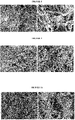
- Animal eyeball samples were collected in cold storages. In the example shown herein, these are porcine eyeball samples collected in slaughterhouses that process exclusively porcines ( Figure 1A ). Immediately after collection, the samples were immersed in saline solution (0.9% NaCl) and kept refrigerated in a thermal container until received by the laboratory. The transport time until the laboratory should be as short as possible, and always less than 10h.
- TBS solution buffered TRIS saline solution
- running water Figures 2A and 2B
- Example 2- Decellularization protocol of biological material.
- Decellularization is a strategy that seeks reducing or eliminating the chances of adverse immunogenic reaction due to the use of biological scaffolds. This technique acts in the removal of cells from the tissue under discussion, and in the elimination of the genetic material present therein, but seeking to maintain the elements of the extracellular matrix and preserving the mechanical properties necessary for the maintenance of the scaffold functionality.
- the samples were washed with deionized water, for 30 minutes-4 hours, at a temperature between 4-37°C, under stirring between 80-200 rpm and washed with isotonic buffer (TBS solution) for 5-15 minutes, at a temperature between 4-37°C and under stirring between 80-200 rpm.
- TRIS solution isotonic buffer
- the protocol followed with incubation of biological samples with nuclease buffer (10-50 mM TRIS, 2-10 mM MgCl 2 , 1-80 U/mL DNAse and 0.05-80 U/mL RNAse), for 30 minutes-3h, under stirring between 100-200 rpm, and at a temperature between 30-40°C.
- the samples were submitted to final washes in saline-phosphate buffer (PBS) containing 0.05-1% w/v EDTA, for up to 5 minutes at a temperature between 25-37°C, under stirring between 80-200 rpm.
- PBS saline-phosphate buffer
- protocol A strictly followed the description in the study carried out by Kasbekar et al. (2018).
- the difference of the proposed protocol B is on the concentration of benzonase used in the nuclease buffer, which is raised from 1 U/mL to 5 U/mL.
- Protocol C raises this concentration to 10 U/mL.
- Protocol D by its turn, replaces benzonase with DNase and RNase (80 U/mL), while protocol E uses 8 U/mL of DNase and RNase, and reduces all rotations during the process, preferably from 80 rpm to 200 rpm, more preferably from between 90 and 180 rpm, and even more preferably 100 rpm, and add a phase comprising washings with deionized water after incubation with detergent buffer, preferably for 4 hours, at a temperature of 25°C and rotation of 100 rpm, with two exchanges in this interval.
- DNase and RNase 80 U/mL
- protocol E uses 8 U/mL of DNase and RNase, and reduces all rotations during the process, preferably from 80 rpm to 200 rpm, more preferably from between 90 and 180 rpm, and even more preferably 100 rpm, and add a phase comprising washings with deionized water after incubation with detergent buffer, preferably for 4 hours, at a temperature of 25°C and rotation
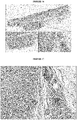
- the decellularization efficiency criteria covers an number of less than 50 ng of DNA/mg of extracellular matrix (dry weight), and the lack of intact DNA and visible nuclear material in histological analysis.
- protocols A, B and C did not reach the DNA/mg tissue limit required for efficient decellularization.
- These protocols used benzonase ( Serratia marcescens nuclease), a promiscuous endonuclease that attacks and degrades all forms of DNA and RNA (Nestle and Roberts, 1969). Therefore, the combined use of nucleases (DNase/RNase) was proposed under high concentration, replacing benzonase (protocol D). This protocol was the first to meet the criteria for decellularization efficiency.
- Example 3a Decontamination protocol of the decellularized biomaterial.
- the decellularized biomaterial was decontaminated by performing an initial wash in sterile PBS and then soaked in 1x sterile PBS (approximately 30 mL) containing concentrated antibiotic and/or antimycotic solution.
- the concentrated solution of antibiotic and/or antimycotic solution comprises at least one compound with antibiotic action and at least one compound with antimycotic action, but it particularly comprises: penicillin, streptomycin and amphotericin B.
- the samples were stored in this solution for about 36h-60h, preferably about 48 h, at a temperature of about 2°C-8°C, preferably 4°C.
- the said method was proven to be effective both as an initial and preliminar decontamination step of the decellularized biomaterial, that can further be subjected to an additional sterilization step (using ethylene oxide or any other method known by one person skilled in the art, for example, gamma radiation) .
- Example 3b Sterilization protocol of the decellularized biomaterial.
- the sterilization step was carried out using ethylene oxide.
- the samples of the biomaterial were then cut into specific dimensions for carrying out the tests, and dried at room temperature for about 30 minutes to 1 hour. After drying, they were packed and sealed in surgical grade paper, ensuring identification and sealing to verify ethylene oxide sterilization later.
- the samples were then subjected to an ethylene oxide sterilization process and aerated for 1 day. After that, analyzes were carried out to investigate the microbiological growth for 14 days in thioglycolate broth culture medium (30°C to 35°C) and tryptic soy broth - TSB (20°C to 25°C).
- Example 4 Collagen quantification.
- Quantification of hydroxyproline aims at indirect determination of collagen content of the biomaterial produced.
- 5-50 mg of the samples were homogenized in 100-400 ⁇ L of deionized water.
- an amino acid dilution curve HP was performed.
- Figure 5 shows on the left side, in triplicate, the standard hydroxyproline curve as control, with decreasing dilutions of this substance in vertical. The darker it is, the higher is the hydroxyproline concentration, which is one of the main components of collagen. On the right side, three samples of individual decellularized biomaterial had the hydroxyproline quantification carried out in 3 decreasing dilutions in vertical.
- the reaction was prepared in 96-well plates, in which 5-50 ⁇ L of the samples were added to the wells, as well as 5-50 ⁇ L of each solution of the hydroxyproline dilution curve, and the test was performed in triplicate. The plates were dried for removing hydrochloric acid. After drying, 50-500 ⁇ L of the chloramine T reagent (citrate-acetate buffer, 50% n-propanol and chloramine T, pH 6.5) were added. The plates were incubated at room temperature from 2-10 minutes.
- the mean concentration was 103.76 ⁇ g of hydroxyproline per milligram of biomaterial, which corresponds approximately to 769 ⁇ g of collagen per milligram of biomaterial.
- the collagen content of the decellularized biomaterial produced corresponds to 76.9% its weight.
- Example 5 Dehydration and rehydration test of the decellularized biomaterial.
- This experiment aimed to know and measure the reabsorption capacity of aqueous or alcoholic solutions from the dehydrated biomaterial.
- fragments of different sizes of the decellularized biomaterial e.g.: 0.6 x 0.6 cm; 1.0 x 1.0 cm; 1.5 x 1.5 cm
- 0.6 x 0.6 cm; 1.0 x 1.0 cm; 1.5 x 1.5 cm were initially weighed and then dehydrated in ethanol at increasing concentrations (45%, 55%, 65%, 75%, 85%, 95% and 100% volume/volume), incubating them for at least 3 hours in each concentration.
- the fragments were again weighed and rehydrated, and incubated for about 20 minutes-40 minutes, with at least one selected substance from the group that consists of: water, alcohol, water or alcohol soluble substances, nutrients, metabolites, growth factors, signaling molecules, regulatory molecules, hormone, medicaments and/or pharmaceutically active compounds.
- the biomaterial was submitted to rehydration by adding fixed volumes of water and incubated for up to 30 minutes at room temperature (with quantification of the volume absorbed every 15 minutes). After rehydration, the fragments were weighed again, and the reabsorbed volume was measured (tables 3 and 4).
- the dehydration step occurs after the decellularization and decontamination steps of the biomaterial.
- the decontamination process is carried out with an antibiotic and/or antimycotic solution.
- the dehydration step can be carried out after the decellularization of the biomaterial, with an initial decontamination step taking place or not, but before subjecting it to the sterilization step.
- the sterilization process being carried out with ethylene oxide or any other method known by one person skilled in the art, for example, gamma radiation.
- the product then obtained by said process comprises a dehydrated, decontaminated and/or sterilized decellularized biomaterial, which can be stored, and can then go through a rehydration and/or association step with at least one active compound only when it is used.
- Example 6 Analysis of type I collagen, type III collagen and elastin contents.
- the objective was identifying, qualitatively, the presence of type I collagen, type III collagen and elastin in the decellularized biomaterial produced. Briefly, three nitrocellulose membranes were sensitized with small fragments of in natura scleras, dehydrated and rehydrated scleras and the decellularized biomaterial produced. Primary antibodies against each protein were used as positive controls of the reactions. The protein-free binding sites present in the nitrocellulose membrane were blocked by the addition of 1-3% PBS-milk buffer, until covering the membranes entirely, under stirring, at 20-40°C, for 1-5h.
- the membranes were washed 1-7 times with 1x PBS buffer, and each membrane was incubated with a specific primary antibody and diluted in 1x PBS, under stirring at 20-40°C, for 1-5h.
- PBS-T 1x PBS-Tween buffer
- the respective IgG-specific secondary antibodies marked with peroxidase and diluted in PBS, were incubated, under stirring, at 20-40°C, for 1-5h.
- the membranes were washed again 1-7 times with 0.05% PBS-T buffer and the reaction was revealed with DAB (3,3'-diaminobenzidine) solution.
- Figure 6 shows the dot blot of the proteins analyzed, showing the presence of each type of protein, with figure 6A showing the experiment layout.
- Figures 6B, 6C and 6D are representative images of dot blot for type I collagen, type III collagen and elastin, respectively.
- the morphological analysis of the decellularized biomaterial produced aimed to evaluate its three-dimensional structure, the presence and quality of collagen fibers and the verification of the presence/absence of cells in the different materials analyzed.
- Samples of in natura sclera, dehydrated sclera, dehydrated and rehydrated sclera and decellularized biomaterial produced were washed with buffer and fixed in a fixing solution suitable for electron microscopy. Then, the fixer was removed, and the samples were submitted to progressive dehydration in solutions with increasing concentrations of ethyl alcohol. After dehydration, the samples were dried and stored. The samples received 3 cycles of gold-palladium cover in the metallizer and were visualized using a Zeiss EVO MA10 microscope. All analyses were performed in triplicate, in several magnifications, using 5-15 kV exposures.
- Figures 7A and 7B show scanning electron microscopy of in natura sclera samples, under magnifications of 833 times and 2900 times, respectively. The presence of cells can be clearly visualized as indicated by the arrow in figure 7B.
- Figures 8A and 8B show scanning electron microscopy of dehydrated sclera samples, under magnifications of 833 times and 7100 times, respectively. The presence of cells can be clearly visualized as indicated by the arrow in figure 7B.
- Figures 9A and 9B show scanning electron microscopy of dehydrated and rehydrated sclera samples, under magnifications of 1250 times and 2640 times, respectively. The presence of cells can be clearly visualized as indicated by the arrows in figure 7B.
- Figures 10A and 10B show scanning electron microscopy of samples of the decellularized biomaterial produced, under magnifications of 923 times and 5520 times, respectively. The presence of cells has not been identified, and the collagen and elastin fibers remained without apparent or relevant alteration.
- Electron microscopy analyses showed that the decellularization method used, led to cell removal from the biomaterial, while in the other groups where the biological material did not go through the decellularization process, the presence of cells was clearly observed.
- the fibers were kept healthy in the decellularized biomaterial, showing that the biomaterial comprises a decellularized fibrous matrix that maintained the characteristics of a biological matrix without the cell's removal presenting relevant alterations in the formation and quality of the fibrous matrix, when compared to in natura, dehydrated or dehydrated/rehydrated sclera.
- Example 8 Biomechanical assay (elasticity and maximum strain modules).
- a biomechanical assay was performed to assess the resistance of scleras and the produced biomaterials under different conditions, in order to evaluate the physical properties of this biomaterial and enabling the indication of possible applications in the context of regenerative medicine.
- approximately 12 fragments of sclera samples were used under the following conditions: in natura sclera, decellularized biomaterial produced, dehydrated decellularized biomaterial (in increasing concentrations of ethanol) and dehydrated and rehydrated decellularized biomaterial (dehydrated in increasing concentrations of ethanol and rehydrated in water for 30-50 min). All fragments were obtained from the central region from each sclera in natura, with approximate measurements of 5 x 20 x 1 mm (width x length x thickness).
- the specimens were submitted to a tensile assay using a WDW 38/56 - 1000E Universal Test Machine from TIME Group Inc. To achieve the forces exerted, a load cell with a maximum capacity of 50 kgf was used and the deformations were registered by the machine's internal displacement sensors. The specimen was fixed in two claws apart from 5 mm. The force application speed was 2 mm/min.
- Figure 11A shows a scatter dot plot of the Young elasticity module (GPa) for in natura sclera samples (In natura), samples of decellularized biomaterial (Decellularized), samples of decellularized and dehydrated biomaterial (Dehydrated) and samples of decellularized, dehydrated and rehydrated biomaterial (Rehydrated).
- the Young elasticity module used in this study describes the ratio between strain and deformation in the direction of the applied load, being the maximum strain that a material supports without undergoing permanent deformation.
- E the elasticity parameter
- Figure 11B shows a scatter dot plot of the maximum strain (GPa) for in natura sclera samples (In natura), samples of decellularized biomaterial (Decellularized), samples of decellularized and dehydrated biomaterial (Dehydrated) and samples of decellularized, dehydrated and rehydrated biomaterial (Rehydrated).
- MPa maximum strain parameter
- Example 9 Subcutaneous implantation of biomaterial produced to analyze local inflammation.
- the animals were anesthetized with isofluorane, trichotomy and antisepsis were performed in the region and, soon after, longitudinal incisions with a scalpel were performed.
- the subcutaneous region was divulsed using a blunt-tip scissors and then a fragment of the decellularized biomaterial of 0.5 cm x 0.5 cm x 0.2 cm was introduced in one of the incisions and, in another incision, only the divulsion of the tissues was made without introducing biomaterial and used as a control.
- the incisions were sutured, and the animals were kept under observation until the end of anesthesia. Animal euthanasia was performed after 5 and 15 days (acute and chronic inflammation, respectively) of the material implantation.
- the implantation region was removed, fixed in 10% formaldehyde for histological analysis and stained with HE (images representing this procedure are shown in figure 12 ). Histological examination aimed to evaluate the intensity of inflammatory reaction of surrounding tissue, presence of vessels, fibroblasts, multinucleated giant cells and tissue integration.
- Figures 12 A-D illustrate the procedure for implanting the biomaterial produced in subcutaneous tissue in rats.
- Figure 12A shows the animals under anesthesia with isofluorane mask.
- Figure 12B shows the implant of the biomaterial.
- Figure 12C shows the suture of the incisions.
- Figure 12D shows the removal of the implantation region after 5 or 15 days of surgery.
- the analyses were performed by 4 independent evaluators in all selected fields. The mean of the scores of each animal was calculated, assigned by each evaluator and a comparison between the results of 5 and 15 days was performed.
- Figure 13 shows a representative image of the histological cutting from the control group after 5 days.
- figure 14 shows representative images of the histological cutting of the subcutaneous biomaterial implant after 5 days of implantation.
- the insert shows a specific field, showing the contact area of the inflammatory infiltrate with biomaterial.
- Figure 15 shows a representative image of the histological field of the implant of subcutaneous biomaterial after 5 days of implantation. Arrows indicate fibroblasts infiltrated in the biomaterial (material evidenced on the right side) and the dotted circle is an example of blood vessel.
- Figure 16 shows representative images of the histological section of the subcutaneous biomaterial implant after 15 days of implantation.
- the insert shows a specific field, showing the contact area of the inflammatory infiltrate with the implanted biomaterial.
- Figure 17 shows a representative image of the histological field of the subcutaneous implant of biomaterial after 15 days of implantation.
- the arrows indicate fibroblasts infiltrated in the biomaterial (material evidenced on the left side).
- the dotted circles indicate blood vessels.
- the dotted rectangles indicate multinucleated giant cells.
- Example 10- Implantation of the biomaterial in a tendon injury.
- the experiment sought to analyze the effect of implanting the dehydrated decellularized biomaterial produced in ram tendon injury.
- the implant made of the biomaterial produced by the method described in the present application was placed and fixed to the bone with a titanium screw and suture at the end of the tendon.
- the postoperative period was monitored and the animals received antibiotics, anti-inflammatories and analgesics.
- videos were made to assess claudication (gait impairment assessment), 48 hours after surgery and periodically until euthanasia.
- the animals were randomly divided into two groups with five animals each, to be euthanized in the first experimental time of 49 days (D49) and second experimental time of 114 days (D114) postoperatively. On the day of euthanasia, the animals were clinically assessed, presenting normal mucosa, vital signs and behavior.
- the surgical specimen was collected and stored in a solution containing 10% formaldehyde and sent for histological slides stained with hematoxylin and eosin.
- claudication To assess claudication, the following parameters were considered: absent, when the animal trots without claudication; light, when the animal walks with slight imbalance; moderate, when claudication is interspersed with walking; severe, when the animal cannot steady its paw while walking.
- Figure 19 (A) shows the diffuse inflammatory infiltrate and implanted decellularized biomaterial in the D49 experimental group and Figure 19 (B) shows the less diffuse and more defined inflammatory infiltrate (indicated by ⁇ ⁇ ) around the implanted decellularized biomaterial (indicated by *) in the second experimental group D114.
- the region between the injured tendon and the implant presented disorganized, hypercellularized fibers, different from what is observed in a healthy native tendon ( Fig. 20 ).
- Figure 20 (A) shows the region of the implanted biomaterial (indicated by *); injured native tendon (indicated by ⁇ ) and region intermediate to the native tendon and the implant (highlighted).
- Figure 20 (B) shows the tissue adjacent to the injured native tendon, with disorganized and hypercellularized fibers.
- Figure (C) shows the healthy native tendon that was removed for replacement by the implant.
- Figure (D) shows a region of healthy native tendon at higher magnification.
- Figure 21 shows the presence of blood vessels and hyperemia in the region of interaction of the native tissue with the biomaterial implant in the first experimental time D49 (indicated by the arrows).
- Figure 22 shows a microscopic image of the loose connective tissue (indicated by ]) overlying the injured tendon region (indicated by #).
- Figures 23 (A) and (B) show the decellularized biomaterial implant in interaction with the native tendon in the second experimental time.
- the implant (indicated by *) is observed to be infiltrated by host cells; with a predominance of those with fibroblast characteristics (indicated by thick arrows).
- the fibers in the implant-tendon interaction region (indicated by the black arrows) tend to organize themselves with the increase in compaction and the parallel arrangement between them; becoming, in this region of the implant, with histological aspect of tissue organization similar to that of the native tendon (indicated by #).
- Figures 24 (A) and (B) show the profile of predominant cells that infiltrated the implant: first experimental time D49 (A) and second experimental time D114 (B) .
- the decellularized biomaterial implant produced by the method described in the present application acted efficiently to allow cellular infiltration and also provided a favorable environment so that these cells used this scaffold in an attempt to induce repair and/or remodeling of the lesion, although the experimental time was not sufficient to visualize the complete integration of the implant or complete repair of the lesion.
- the clinical, macroscopic and histological aspects showed no signs of infection and/or rejection of the biomaterial, suggesting a good interaction of this implant during the assessed experimental times.
- the decellularized biomaterial implant showed the potential for tissue remodeling, allowing recellularization mainly in its periphery and allowed the vascularization of the region and favored the deposition of extracellular matrix, which are essential factors for the remodeling of the injured tissue
- Example 11 Implantation of the biomaterial in the bone.
- an in vivo bone implant experiment was carried out, more specifically, an implant on the buccal surface of the maxillary alveolar bone of dogs.
- the assay consisted of three experimental groups. In the first group called CONTROL, the standard commercialized material for bone regeneration was used, 0.25 g Geistlich Bio-Oss ® ; in the second group, the decellularized dehydrated biomaterial, called BIOMATERIAL group, was used; and in the third, the decellularized dehydrated sclera, carrying 2.5% simvastatin, called BIOMATERIAL WITH ACTIVE COMPOUND group.
- Fragments from the surgical site were removed after 49 (D49) and 130 (D130) days.
- the samples were fixed in a solution containing 10% formaldehyde, dehydrated in increasing concentrations of ethanol, diaphanized in xylene, infiltrated, embedded in paraffin and stained with hematoxylin and eosin for subsequent histological analysis under a light microscope.
- Figures 25 show the oral mucosa membrane with the presence of granules from the implanted commercial bone graft (indicated by *).
- A it is possible to observe the dense connective tissue where the graft is implanted (indicated by #) and loose connective tissue at the top (initiated by a square bracket).
- B the commercial graft granule is highlighted (indicated by *) with the formation of the adjacent bone blastema, this process being enlarged and detailed in (C) and (D), in the control group D49.
- Figures 26 show the D130 control group.
- (A) there is the presence of commercial bone graft granules (indicated by *) in the membrane, external to the periosteum of the alveolar bone (indicated by [), in addition to the presence of the highlighted granule included in the newly formed bone (indicated by #).
- the area highlighted in (B) was enlarged in (C) for analysis, where growth lines are visualized (indicated by arrows) close to the biomaterial region and the formation of Havers Systems (indicated by **).
- H Havers Systems
- BIOMATERIAL D49 group it was possible to observe the intimate contact of the decellularized biomaterial with the alveolar bone of the animal, with a high number of cells interspersing the sclera region and angiogenesis within the biomaterial, characteristics that were remarkable at this experimental time. Areas with significant inflammatory infiltrate were not visualized (Figure 27).
- Figures 27 show the BIOMATERIAL group D49.
- A the attached gingiva membrane region and bone tissue of the experimental animal (indicated by [), implantation of decellularized biomaterial (indicated by *) adjacent to the alveolar bone of the animal (indicated by [).
- B by magnification of (A), details of the implant and bone tissue in formation (indicated by #) can be observed, where it is possible to verify the interaction of the biomaterial with the bone and probable osteoprogenitor cells invading the biomaterial and the presence of cells in the decellularized biomaterial as well as blood vessels (indicated by arrows).
- BIOMATERIAL D130 group the presence of numerous blood vessels is evident, proposing moderate angiogenesis inside the decellularized biomaterial implant and intimate contact of the referred implant with a region that suggests discrete bone formation. Bone formation showed a disorganized or slightly organized pattern, not differing from the initial time, D49. In the biomaterial, interspersed cells were also observed inside, and in the region adjacent to the bone of the experimental animal ( Figure 28 )
- Figures 28 show the BIOMATERIAL group D130.
- A areas of close integration are observed between the biomaterial implant and the animal's bone (indicated by [) and implant and loose connective tissue (indicated by ⁇ ).
- the decellularized biomaterial implant (indicated by #) now has possible osteoprogenitor cells inside and the presence of a small cellular infiltrate interspersing its fibers.
- B the presence of possible adipocytes is highlighted, with probable origin from the bone marrow (indicated by arrows).
- C and (D) we can see the deposition of the newly formed bone (indicated by *) on the implanted biomaterial.
- an osteoconductive role of decellularized biomaterials is suggested as produced by the method described by the present patent application.
- BIOMATERIAL WITH ACTIVE COMPOUND group where the ability of the biomaterial to be used in combination with active substances was assessed, also working as a carrier, in this case using simvastatin, the histological assessment showed the presence of greater bone volume compared to the other groups ( Figure 29 ), where it is possible to observe the alveolar bone and adjacent areas of new bone formation.
- Figure 29 shows the group BIOMATERIAL WITH ACTIVE COMPOUND D49.
- the image is a photomicrograph demonstrating the presence of the gingival mucosal epithelium (indicated by an arrow), membrane (indicated by [) of the mucosa and the region where the biomaterial with simvastatin was positioned (indicated by ⁇ ) adjacent to the alveolar bone of the animal (indicated by *).
- the region of bone neoformation (#) is highlighted in the rectangle.
- the area of bone formation in the BIOMATERIAL WITH ACTIVE COMPOUND D49 group showed integrated bone trabeculae, characteristic of the bone maturation process. In addition, it is observed in an immature bone region, with a large number of osteocytes and the presence of small medullary cavities containing blood vessels ( Figure 30 ).
- Figures 30 show the group BIOMATERIAL WITH ACTIVE COMPOUND D49. Region of interaction between the biomaterial with simvastatin and alveolar bone (A) and connective tissue (initiated by [). There is a greater volume of newly formed bone (indicated by *) adjacent and integrated with alveolar bone (indicated by #), cellular infiltrate and blood vessels within the newly formed bone trabeculae (indicated by *). Newly formed bone has disorganized collagen fibers as shown in image (B) and growth lines (indicated by arrows in A and B) that distinguish it from alveolar bone.
- Figures 31 show the group BIOMATERIAL WITH ACTIVE COMPOUND D130.
- mast cells indicated by ⁇
- other cell types inside the biomaterial with simvastatin. Lines of growth (indicated by arrows) separate mature alveolar bone (indicated by [) from newly formed (indicated by *). Connective tissue from the animal itself (indicated by ⁇ ).
- the decellularized biomaterial produced by the method described in the present patent application had an osteoconductive and osteoinductive role, in addition to favoring cell migration.
- Simvastatin is a drug used in the treatment of hyperlipidemia and has pleiotropic osteogenic and antiresorptive effects already studied in vitro (showing changes in cellular dynamics resulting from the effects of simvastatin on bone tissue, which are associated with increased expression of bone morphogenetic protein-2 (BMP-2), stimulating the proliferation and differentiation of osteoblasts).
- BMP-2 bone morphogenetic protein-2
- Example 12- Implantation of the biomaterial in cartilage lesion.
- an implant of the decellularized biomaterial produced by the method described by the present patent application in injury caused in cartilaginous tissue in vivo, more specifically an implantation of the biomaterial in a lesion caused in the knees of sheep.
- the animals were euthanized by pre-anesthetic medication and the surgical site was removed.
- the surgical specimen was fixed in 4% formalin and demineralized with formic acid solution.
- the procedures for performing the histological slides were followed, stained with Hematoxylin and Eosin.
- Figure 32 shows a histological section of the group that suffered cartilage injury and received the implant, at an experimental time of 30 days post-surgery.
- the dashed line indicates the approximate delimitation of the implant at the lesion site.
- the square bracket (]) indicates the region of native articular cartilage and the arrows illustrate the interaction with bone tissue (subchondral region).
- Figures 33 show a histological cutting of the group that suffered the cartilage lesion and received the implant, at an experimental time of 30 days post-surgery.
- Figure (A) is a representative image of the histological cutting with the demarcated regions that have been enlarged for a better analysis in (B) and (C).
- the arrows indicate the region of integration between the articular cartilage and the neo-formed connective tissue, indicating important integration with the implant. Circles indicate chondrocyte mitosis figures.
- Figure (C) shows the region of the biomaterial implant with fibroblast infiltration, deposition of new extracellular matrix, which leads to the loss of border delimitation, due to good integration between the tissues (lower left).
- inflammatory cells Adjacent to the biomaterial implant, inflammatory cells were also found, with a predominance of mononuclear cells, such as lymphocytes, macrophages and plasma cells, which corresponds to a stage of the repair process. There were no signs of rejection of the biomaterial, such as the presence of multinucleated giant cells (Langerhans giant cells) or encapsulation process, in any of the animals analyzed.
- mononuclear cells such as lymphocytes, macrophages and plasma cells
- Example 13 Implantation of the biomaterial for filling and anatomical correction.
- an implant of the biomaterial produced in a human being who had eyelid retention in one eye was performed (indicated by an arrow in Figure 34 A) .
- the biomaterial implant was then prepared in the proper size to be surgically implanted in the desired eyelid ( Figure 34 B-D ).
- Figure 34 (E) shows the result of the surgery right after the procedure.
- Figure 34 (F) shows the result of the filling surgery and anatomical correction using the decellularized biomaterial of the present patent application, after approximately 10 months of the aforementioned surgical procedure. The patient did not reject the implanted biomaterial, and the filling and anatomical correction was a success, aesthetically recovering the symmetry of the patient's eyes.
- this application describes a method of producing decellularized biomaterial, comprising the steps of:
- Said isotonic buffer of steps (a) and (e) is a buffered TRIS saline solution (TBS)comprising 10-100 mM TRIS and 100-200 mM NaCl.
- TRIS buffered TRIS saline solution
- the hypotonic buffer of step (b) comprises from 5-20 mM of TRIS; 0.05-1% w/v of EDTA and pH between 6-8.
- the detergent buffer of step (c) comprises from 5-20 mM TRIS; 0.05-1% w/v of EDTA and 0.05-2% w/v SDS.
- Nuclease buffer from step (f) comprises 10-50 mM TRIS; 2-10 mM MgCl 2 ; 1-80 U/mL DNAse; and/or 0.05-80 U/mL RNAse.
- Step (d) comprises at least one wash water exchange in half the total time. This method can be performed in a semi-automated way for large-scale production, and preferably steps (a)-(g) can be performed in a bioreactor.
- the decontamination step of the decellularized biomaterial comprises at least the use of one antibiotic and/or antimycotics.
- the decontamination step of the decellularized biomaterial using antibiotics and/or antimycotics comprises:
- the concentrated solution of antibiotic and/or antimycotic comprises at least one compound with antibiotic activity and at least one compound with antimycotic activity.
- the concentrated solution of antibiotic and/or antimycotic comprises: penicillin, streptomycin and amphotericin B.
- the sterilization step of the decellularized biomaterial comprises the use of ethylene oxide or any other method known by one person skilled in the art, for example, gamma radiation.
- Said method of producing decellularized biomaterial further comprises a dehydration step that may occur before or after the decontamination and/or sterilization step.
- the dehydration step comprises subjecting the decellularized biomaterial or the decontaminated and decellularized biomaterial to a dehydration process in which said biomaterial is incubated with increasing concentrations of an alcohol, preferably wherein the alcohol concentration increases by 45%, 55%, 65%, 75%, 85%, 95% up to 100% volume/volume, and wherein said biomaterial is incubated for at least 3 hours in each of the so-called alcohol concentrations.
- the method may further comprise a rehydration step of the dehydrated biomaterial, which can occur preferably after the sterilization step, wherein it comprises incubating said dehydrated biomaterial for about 20-40 minutes, with at least one substance selected from the group that consists of: water, alcohol, water or alcohol soluble substances, nutrients, metabolites, growth factors, signaling molecules, regulatory molecules, hormone, medicaments and/or pharmaceutically active compounds (e.g.: simvastatin).
- a rehydration step of the dehydrated biomaterial which can occur preferably after the sterilization step, wherein it comprises incubating said dehydrated biomaterial for about 20-40 minutes, with at least one substance selected from the group that consists of: water, alcohol, water or alcohol soluble substances, nutrients, metabolites, growth factors, signaling molecules, regulatory molecules, hormone, medicaments and/or pharmaceutically active compounds (e.g.: simvastatin).
- the application also describes the decellularized biomaterial produced by this method, wherein it comprises a biological decellularized extracellular matrix, wherein said matrix predominantly comprises types I and III collagen fibers and elastin.
- the biomaterial may be used in combination with at least one active substance or it may comprise at least one active substances selected from the group that consists of: water or alcohol soluble substances, nutrients, metabolites, growth factors, signaling molecules, regulatory molecules, hormones, medicines and/or pharmaceutically active compounds (e.g.: simvastatin).
- the biomaterial is for use as or for production of a scaffold product, prosthesis or carrier of active substances for systemic or localized release in an individual in need thereof.
- a scaffold product for use as or for production of a scaffold product, prosthesis or carrier of active substances for systemic or localized release in an individual in need thereof.
- tissue repair and/or regeneration preferably of bone tissue, cartilaginous tissue, joint, tendon, skin, for prosthetic purposes in implant dentistry and/or bucomaxillofacial traumatology, for surgical procedures of filling and/or anatomic repair, or in the production of prosthesis.
Landscapes
- Health & Medical Sciences (AREA)
- Life Sciences & Earth Sciences (AREA)
- Chemical & Material Sciences (AREA)
- Medicinal Chemistry (AREA)
- Veterinary Medicine (AREA)
- Public Health (AREA)
- General Health & Medical Sciences (AREA)
- Animal Behavior & Ethology (AREA)
- Epidemiology (AREA)
- Engineering & Computer Science (AREA)
- Biomedical Technology (AREA)
- Chemical Kinetics & Catalysis (AREA)
- Dermatology (AREA)
- Oral & Maxillofacial Surgery (AREA)
- Transplantation (AREA)
- Molecular Biology (AREA)
- Botany (AREA)
- Pharmacology & Pharmacy (AREA)
- Zoology (AREA)
- Urology & Nephrology (AREA)
- Surgery (AREA)
- Vascular Medicine (AREA)
- Heart & Thoracic Surgery (AREA)
- General Chemical & Material Sciences (AREA)
- Immunology (AREA)
- Proteomics, Peptides & Aminoacids (AREA)
- Bioinformatics & Cheminformatics (AREA)
- Developmental Biology & Embryology (AREA)
- Cell Biology (AREA)
- Dispersion Chemistry (AREA)
- Neurosurgery (AREA)
- Biophysics (AREA)
- Neurology (AREA)
- Ophthalmology & Optometry (AREA)
- Biotechnology (AREA)
- Virology (AREA)
- Inorganic Chemistry (AREA)
- Oil, Petroleum & Natural Gas (AREA)
- Organic Chemistry (AREA)
- Nuclear Medicine, Radiotherapy & Molecular Imaging (AREA)
Applications Claiming Priority (2)
| Application Number | Priority Date | Filing Date | Title |
|---|---|---|---|
| US202062976956P | 2020-02-14 | 2020-02-14 | |
| PCT/BR2021/050069 WO2021159198A1 (fr) | 2020-02-14 | 2021-02-12 | Procédé de production de biomatériau décellularisé, biomatériau décellularisé et utilisation de celui-ci |
Publications (2)
| Publication Number | Publication Date |
|---|---|
| EP4104863A1 true EP4104863A1 (fr) | 2022-12-21 |
| EP4104863A4 EP4104863A4 (fr) | 2024-02-21 |
Family
ID=77291950
Family Applications (1)
| Application Number | Title | Priority Date | Filing Date |
|---|---|---|---|
| EP21754359.4A Withdrawn EP4104863A4 (fr) | 2020-02-14 | 2021-02-12 | Procédé de production de biomatériau décellularisé, biomatériau décellularisé et utilisation de celui-ci |
Country Status (10)
| Country | Link |
|---|---|
| US (1) | US20240216580A1 (fr) |
| EP (1) | EP4104863A4 (fr) |
| JP (1) | JP2023517179A (fr) |
| KR (1) | KR20220161290A (fr) |
| AU (1) | AU2021220753A1 (fr) |
| BR (1) | BR112022016166A2 (fr) |
| CA (1) | CA3171328A1 (fr) |
| IL (1) | IL295582A (fr) |
| MX (1) | MX2022009961A (fr) |
| WO (1) | WO2021159198A1 (fr) |
Cited By (1)
| Publication number | Priority date | Publication date | Assignee | Title |
|---|---|---|---|---|
| KR102859281B1 (ko) * | 2024-05-16 | 2025-09-16 | 주식회사 엠젠솔루션 | 생체모방형 이종 조직 유래 이식재 제조를 위한 세포성분 제거 기술 |
Families Citing this family (5)
| Publication number | Priority date | Publication date | Assignee | Title |
|---|---|---|---|---|
| CN114796615B (zh) * | 2022-04-20 | 2023-08-25 | 诺一迈尔(苏州)医学科技有限公司 | 一种软骨脱细胞基质及其制备方法 |
| CN115837096B (zh) * | 2022-08-19 | 2024-10-01 | 山东第一医科大学附属眼科研究所(山东省眼科研究所、山东第一医科大学附属青岛眼科医院) | 一种基于透明化巩膜的角膜修复材料及其制备方法和应用 |
| CN118903559A (zh) * | 2024-07-30 | 2024-11-08 | 北京佰仁医疗科技股份有限公司 | 一种消化外科生物补片及其制备方法 |
| CN119258276B (zh) * | 2024-10-14 | 2026-01-13 | 中国人民解放军空军军医大学 | 一种人脱细胞牙槽骨基质微粒的制备方法 |
| CN119199096B (zh) * | 2024-10-30 | 2025-11-07 | 杭州百凌生物科技有限公司 | 一种细胞清洗液及其应用 |
Family Cites Families (19)
| Publication number | Priority date | Publication date | Assignee | Title |
|---|---|---|---|---|
| AU5574101A (en) * | 2000-04-28 | 2001-11-12 | Univ Emory | Decellularized vascular prostheses |
| US20030187515A1 (en) | 2002-03-26 | 2003-10-02 | Hariri Robert J. | Collagen biofabric and methods of preparing and using the collagen biofabric |
| US20110212524A1 (en) * | 2006-12-04 | 2011-09-01 | Body Organ Biomedical Corporation | Biomaterial and preparation method thereof |
| CN101977622A (zh) * | 2008-03-24 | 2011-02-16 | 欧几里得系统公司 | 增加角膜上皮的通透性和使基质胶原纤维网络不稳定的方法 |
| ES2719816T3 (es) * | 2010-02-26 | 2019-07-16 | Decell Tech Inc | Métodos para la descelularización de tejidos |
| CN101947144B (zh) | 2010-09-29 | 2012-07-04 | 厦门大学 | 一种板层组织工程角膜支架及其制作方法与应用 |
| BR102014003817B1 (pt) * | 2014-02-19 | 2023-09-26 | Fundação Universidade Federal De São Carlos | Processo de recobrimento descontínuo utilizando um biomaterial bioabsorvível e bioativo aplicado sobre substratos sólidos, recobrimento descontínuo e seu uso |
| WO2017044570A1 (fr) | 2015-09-08 | 2017-03-16 | Clemson University | Biomatériau décellularisé et son procédé de formation |
| CN105833353B (zh) | 2016-05-09 | 2018-04-20 | 拜欧迪赛尔(北京)生物科技有限公司 | 一种生物工程脱细胞真皮基质的制备及用途 |
| CN105944142B (zh) | 2016-05-09 | 2018-07-13 | 拜欧迪赛尔(北京)生物科技有限公司 | 一种脱细胞肌腱或韧带支架的制备方法 |
| KR20180003879A (ko) | 2016-07-01 | 2018-01-10 | 순천향대학교 산학협력단 | 탈세포화용 생물반응기 시스템과, 이를 이용한 기관의 탈세포화 방법 |
| RU2653489C2 (ru) | 2016-09-02 | 2018-05-08 | Федеральное государственное бюджетное учреждение "Государственный научный центр Российской Федерации - Федеральный медицинский биофизический центр имени А.И. Бурназяна Федерального медико-биологического агентства" (ФГБУ "ГНЦ ФМБЦ им. А.И. Бурназяна ФМБА России") | Способ получения децеллюляризированных матриксов паренхиматозных органов лабораторных животных |
| CN106362212A (zh) * | 2016-10-21 | 2017-02-01 | 华中科技大学同济医学院附属协和医院 | 一种用于去除组织细胞的亲脂性去细胞溶液、试剂盒和方法 |
| CN106614519B (zh) * | 2016-11-24 | 2019-11-19 | 山东省眼科研究所 | 一种脱细胞角膜缘保护液及脱细胞角膜缘的制备方法 |
| CN106729999B (zh) * | 2016-11-24 | 2019-11-19 | 山东省眼科研究所 | 一种构建组织工程角膜缘的方法 |
| CN106730005A (zh) | 2016-12-22 | 2017-05-31 | 深圳艾尼尔角膜工程有限公司 | 一种角膜基质及其制备方法 |
| CN107007886B (zh) | 2017-03-03 | 2018-02-06 | 北京博辉瑞进生物科技有限公司 | 一种生物组织基质材料、制备方法及其用途 |
| KR20180133172A (ko) | 2017-06-05 | 2018-12-13 | 주식회사 티앤알바이오팹 | 골 이식재 고정용 주사형 접착제 조성물의 제조방법 및 이를 통해 제조된 골 이식재 고정용 주사형 접착제 조성물 |
| CN108888804B (zh) * | 2018-07-27 | 2021-05-14 | 山东省眼科研究所 | 一种软组织修复材料及其制备方法 |
-
2021
- 2021-02-12 KR KR1020227031839A patent/KR20220161290A/ko not_active Withdrawn
- 2021-02-12 MX MX2022009961A patent/MX2022009961A/es unknown
- 2021-02-12 US US17/904,184 patent/US20240216580A1/en not_active Abandoned
- 2021-02-12 CA CA3171328A patent/CA3171328A1/fr active Pending
- 2021-02-12 AU AU2021220753A patent/AU2021220753A1/en not_active Abandoned
- 2021-02-12 WO PCT/BR2021/050069 patent/WO2021159198A1/fr not_active Ceased
- 2021-02-12 EP EP21754359.4A patent/EP4104863A4/fr not_active Withdrawn
- 2021-02-12 BR BR112022016166A patent/BR112022016166A2/pt not_active IP Right Cessation
- 2021-02-12 IL IL295582A patent/IL295582A/en unknown
- 2021-02-12 JP JP2022549335A patent/JP2023517179A/ja active Pending
Cited By (1)
| Publication number | Priority date | Publication date | Assignee | Title |
|---|---|---|---|---|
| KR102859281B1 (ko) * | 2024-05-16 | 2025-09-16 | 주식회사 엠젠솔루션 | 생체모방형 이종 조직 유래 이식재 제조를 위한 세포성분 제거 기술 |
Also Published As
| Publication number | Publication date |
|---|---|
| IL295582A (en) | 2022-10-01 |
| WO2021159198A1 (fr) | 2021-08-19 |
| BR112022016166A2 (pt) | 2022-10-04 |
| MX2022009961A (es) | 2022-11-14 |
| JP2023517179A (ja) | 2023-04-24 |
| US20240216580A1 (en) | 2024-07-04 |
| CA3171328A1 (fr) | 2021-08-19 |
| EP4104863A4 (fr) | 2024-02-21 |
| KR20220161290A (ko) | 2022-12-06 |
| AU2021220753A1 (en) | 2022-10-06 |
Similar Documents
| Publication | Publication Date | Title |
|---|---|---|
| EP4104863A1 (fr) | Procédé de production de biomatériau décellularisé, biomatériau décellularisé et utilisation de celui-ci | |
| US11660376B2 (en) | Decellularized biomaterial from non-mammalian tissue | |
| CN103108661B (zh) | 再生性组织支架的制备 | |
| Beck et al. | Approaching the compressive modulus of articular cartilage with a decellularized cartilage-based hydrogel | |
| Xia et al. | Decellularized cartilage as a prospective scaffold for cartilage repair | |
| CN106456837B (zh) | 最终消毒的来自细胞外基质的水凝胶的制备方法 | |
| Parmaksiz et al. | Decellularization of bovine small intestinal submucosa and its use for the healing of a critical‐sized full‐thickness skin defect, alone and in combination with stem cells, in a small rodent model | |
| CN103002927B (zh) | 再生性组织支架 | |
| CN102238970B (zh) | 利用动物组织粉末制造多孔立体支架的方法及其多孔立体支架 | |
| US20230079113A1 (en) | Biomaterial for Articular Cartilage Maintenance and Treatment of Arthritis | |
| Güngörmüş et al. | Histopathological and biomechanical evaluation of tenocyte seeded allografts on rat Achilles tendon regeneration | |
| Khajavi et al. | Fish cartilage: A promising source of biomaterial for biological scaffold fabrication in cartilage tissue engineering | |
| CN1775304A (zh) | 具有生物活性的人工生物瓣瓣膜的制备方法 | |
| Wang et al. | The mitochondrial protectant SS31 optimized decellularized Wharton's jelly scaffold improves allogeneic chondrocyte implantation-mediated articular cartilage repair | |
| Kumaresan et al. | Development of Human Umbilical cord based scaffold for tissue engineering application | |
| Yusof et al. | Decellularization strategies to engineer fibrocartilage bioscaffolds: a review | |
| Chen et al. | Optimal number of chemical extraction treatments for maintaining the biological properties of an allogeneic tendon | |
| Askari et al. | Potential of human amniotic membrane application for articular cartilage regeneration: a review | |
| WO2017127227A1 (fr) | Biomatériau pour xénogreffes | |
| HK1233551A1 (en) | Methods for preparation of a terminally sterilized hydrogel derived from extracellular matrix |
Legal Events
| Date | Code | Title | Description |
|---|---|---|---|
| STAA | Information on the status of an ep patent application or granted ep patent |
Free format text: STATUS: THE INTERNATIONAL PUBLICATION HAS BEEN MADE |
|
| PUAI | Public reference made under article 153(3) epc to a published international application that has entered the european phase |
Free format text: ORIGINAL CODE: 0009012 |
|
| STAA | Information on the status of an ep patent application or granted ep patent |
Free format text: STATUS: REQUEST FOR EXAMINATION WAS MADE |
|
| 17P | Request for examination filed |
Effective date: 20220914 |
|
| AK | Designated contracting states |
Kind code of ref document: A1 Designated state(s): AL AT BE BG CH CY CZ DE DK EE ES FI FR GB GR HR HU IE IS IT LI LT LU LV MC MK MT NL NO PL PT RO RS SE SI SK SM TR |
|
| DAV | Request for validation of the european patent (deleted) | ||
| DAX | Request for extension of the european patent (deleted) | ||
| REG | Reference to a national code |
Ref country code: DE Ref legal event code: R079 Free format text: PREVIOUS MAIN CLASS: A61K0047000000 Ipc: A61K0031366000 |
|
| A4 | Supplementary search report drawn up and despatched |
Effective date: 20240119 |
|
| RIC1 | Information provided on ipc code assigned before grant |
Ipc: A61L 27/58 20060101ALI20240115BHEP Ipc: A61L 27/56 20060101ALI20240115BHEP Ipc: A61L 27/54 20060101ALI20240115BHEP Ipc: A61L 27/36 20060101ALI20240115BHEP Ipc: A61K 31/366 20060101AFI20240115BHEP |
|
| STAA | Information on the status of an ep patent application or granted ep patent |
Free format text: STATUS: THE APPLICATION IS DEEMED TO BE WITHDRAWN |
|
| 18D | Application deemed to be withdrawn |
Effective date: 20240807 |