EP4104864B1 - Magnesiumphosphat-hydrogele - Google Patents
Magnesiumphosphat-hydrogele Download PDFInfo
- Publication number
- EP4104864B1 EP4104864B1 EP22186994.4A EP22186994A EP4104864B1 EP 4104864 B1 EP4104864 B1 EP 4104864B1 EP 22186994 A EP22186994 A EP 22186994A EP 4104864 B1 EP4104864 B1 EP 4104864B1
- Authority
- EP
- European Patent Office
- Prior art keywords
- paste
- nmp
- gel
- implant
- hydrogel
- Prior art date
- Legal status (The legal status is an assumption and is not a legal conclusion. Google has not performed a legal analysis and makes no representation as to the accuracy of the status listed.)
- Active
Links
Images












Classifications
-
- C—CHEMISTRY; METALLURGY
- C01—INORGANIC CHEMISTRY
- C01B—NON-METALLIC ELEMENTS; COMPOUNDS THEREOF; METALLOIDS OR COMPOUNDS THEREOF NOT COVERED BY SUBCLASS C01C
- C01B25/00—Phosphorus; Compounds thereof
- C01B25/16—Oxyacids of phosphorus; Salts thereof
- C01B25/26—Phosphates
- C01B25/45—Phosphates containing plural metal, or metal and ammonium
-
- A—HUMAN NECESSITIES
- A61—MEDICAL OR VETERINARY SCIENCE; HYGIENE
- A61C—DENTISTRY; APPARATUS OR METHODS FOR ORAL OR DENTAL HYGIENE
- A61C8/00—Means to be fixed to the jaw-bone for consolidating natural teeth or for fixing dental prostheses thereon; Dental implants; Implanting tools
-
- A—HUMAN NECESSITIES
- A61—MEDICAL OR VETERINARY SCIENCE; HYGIENE
- A61K—PREPARATIONS FOR MEDICAL, DENTAL OR TOILETRY PURPOSES
- A61K47/00—Medicinal preparations characterised by the non-active ingredients used, e.g. carriers or inert additives; Targeting or modifying agents chemically bound to the active ingredient
- A61K47/02—Inorganic compounds
-
- A—HUMAN NECESSITIES
- A61—MEDICAL OR VETERINARY SCIENCE; HYGIENE
- A61K—PREPARATIONS FOR MEDICAL, DENTAL OR TOILETRY PURPOSES
- A61K9/00—Medicinal preparations characterised by special physical form
- A61K9/0012—Galenical forms characterised by the site of application
- A61K9/0019—Injectable compositions; Intramuscular, intravenous, arterial, subcutaneous administration; Compositions to be administered through the skin in an invasive manner
-
- A—HUMAN NECESSITIES
- A61—MEDICAL OR VETERINARY SCIENCE; HYGIENE
- A61K—PREPARATIONS FOR MEDICAL, DENTAL OR TOILETRY PURPOSES
- A61K9/00—Medicinal preparations characterised by special physical form
- A61K9/0012—Galenical forms characterised by the site of application
- A61K9/0019—Injectable compositions; Intramuscular, intravenous, arterial, subcutaneous administration; Compositions to be administered through the skin in an invasive manner
- A61K9/0024—Solid, semi-solid or solidifying implants, which are implanted or injected in body tissue
-
- A—HUMAN NECESSITIES
- A61—MEDICAL OR VETERINARY SCIENCE; HYGIENE
- A61L—METHODS OR APPARATUS FOR STERILISING MATERIALS OR OBJECTS IN GENERAL; DISINFECTION, STERILISATION OR DEODORISATION OF AIR; CHEMICAL ASPECTS OF BANDAGES, DRESSINGS, ABSORBENT PADS OR SURGICAL ARTICLES; MATERIALS FOR BANDAGES, DRESSINGS, ABSORBENT PADS OR SURGICAL ARTICLES
- A61L27/00—Materials for grafts or prostheses or for coating grafts or prostheses
- A61L27/02—Inorganic materials
- A61L27/025—Other specific inorganic materials not covered by A61L27/04 - A61L27/12
-
- A—HUMAN NECESSITIES
- A61—MEDICAL OR VETERINARY SCIENCE; HYGIENE
- A61L—METHODS OR APPARATUS FOR STERILISING MATERIALS OR OBJECTS IN GENERAL; DISINFECTION, STERILISATION OR DEODORISATION OF AIR; CHEMICAL ASPECTS OF BANDAGES, DRESSINGS, ABSORBENT PADS OR SURGICAL ARTICLES; MATERIALS FOR BANDAGES, DRESSINGS, ABSORBENT PADS OR SURGICAL ARTICLES
- A61L27/00—Materials for grafts or prostheses or for coating grafts or prostheses
- A61L27/02—Inorganic materials
- A61L27/12—Phosphorus-containing materials, e.g. apatite
-
- A—HUMAN NECESSITIES
- A61—MEDICAL OR VETERINARY SCIENCE; HYGIENE
- A61L—METHODS OR APPARATUS FOR STERILISING MATERIALS OR OBJECTS IN GENERAL; DISINFECTION, STERILISATION OR DEODORISATION OF AIR; CHEMICAL ASPECTS OF BANDAGES, DRESSINGS, ABSORBENT PADS OR SURGICAL ARTICLES; MATERIALS FOR BANDAGES, DRESSINGS, ABSORBENT PADS OR SURGICAL ARTICLES
- A61L27/00—Materials for grafts or prostheses or for coating grafts or prostheses
- A61L27/50—Materials characterised by their function or physical properties, e.g. injectable or lubricating compositions, shape-memory materials, surface modified materials
- A61L27/52—Hydrogels or hydrocolloids
-
- A—HUMAN NECESSITIES
- A61—MEDICAL OR VETERINARY SCIENCE; HYGIENE
- A61L—METHODS OR APPARATUS FOR STERILISING MATERIALS OR OBJECTS IN GENERAL; DISINFECTION, STERILISATION OR DEODORISATION OF AIR; CHEMICAL ASPECTS OF BANDAGES, DRESSINGS, ABSORBENT PADS OR SURGICAL ARTICLES; MATERIALS FOR BANDAGES, DRESSINGS, ABSORBENT PADS OR SURGICAL ARTICLES
- A61L27/00—Materials for grafts or prostheses or for coating grafts or prostheses
- A61L27/50—Materials characterised by their function or physical properties, e.g. injectable or lubricating compositions, shape-memory materials, surface modified materials
- A61L27/54—Biologically active materials, e.g. therapeutic substances
-
- A—HUMAN NECESSITIES
- A61—MEDICAL OR VETERINARY SCIENCE; HYGIENE
- A61P—SPECIFIC THERAPEUTIC ACTIVITY OF CHEMICAL COMPOUNDS OR MEDICINAL PREPARATIONS
- A61P1/00—Drugs for disorders of the alimentary tract or the digestive system
- A61P1/02—Stomatological preparations, e.g. drugs for caries, aphtae, periodontitis
-
- A—HUMAN NECESSITIES
- A61—MEDICAL OR VETERINARY SCIENCE; HYGIENE
- A61P—SPECIFIC THERAPEUTIC ACTIVITY OF CHEMICAL COMPOUNDS OR MEDICINAL PREPARATIONS
- A61P19/00—Drugs for skeletal disorders
- A61P19/08—Drugs for skeletal disorders for bone diseases, e.g. rachitism, Paget's disease
-
- A—HUMAN NECESSITIES
- A61—MEDICAL OR VETERINARY SCIENCE; HYGIENE
- A61P—SPECIFIC THERAPEUTIC ACTIVITY OF CHEMICAL COMPOUNDS OR MEDICINAL PREPARATIONS
- A61P23/00—Anaesthetics
- A61P23/02—Local anaesthetics
-
- C—CHEMISTRY; METALLURGY
- C01—INORGANIC CHEMISTRY
- C01B—NON-METALLIC ELEMENTS; COMPOUNDS THEREOF; METALLOIDS OR COMPOUNDS THEREOF NOT COVERED BY SUBCLASS C01C
- C01B25/00—Phosphorus; Compounds thereof
- C01B25/16—Oxyacids of phosphorus; Salts thereof
- C01B25/26—Phosphates
- C01B25/32—Phosphates of magnesium, calcium, strontium, or barium
- C01B25/34—Magnesium phosphates
-
- A—HUMAN NECESSITIES
- A61—MEDICAL OR VETERINARY SCIENCE; HYGIENE
- A61C—DENTISTRY; APPARATUS OR METHODS FOR ORAL OR DENTAL HYGIENE
- A61C8/00—Means to be fixed to the jaw-bone for consolidating natural teeth or for fixing dental prostheses thereon; Dental implants; Implanting tools
- A61C8/0087—Means for sterile storage or manipulation of dental implants
-
- A—HUMAN NECESSITIES
- A61—MEDICAL OR VETERINARY SCIENCE; HYGIENE
- A61L—METHODS OR APPARATUS FOR STERILISING MATERIALS OR OBJECTS IN GENERAL; DISINFECTION, STERILISATION OR DEODORISATION OF AIR; CHEMICAL ASPECTS OF BANDAGES, DRESSINGS, ABSORBENT PADS OR SURGICAL ARTICLES; MATERIALS FOR BANDAGES, DRESSINGS, ABSORBENT PADS OR SURGICAL ARTICLES
- A61L2400/00—Materials characterised by their function or physical properties
- A61L2400/12—Nanosized materials, e.g. nanofibres, nanoparticles, nanowires, nanotubes; Nanostructured surfaces
-
- A—HUMAN NECESSITIES
- A61—MEDICAL OR VETERINARY SCIENCE; HYGIENE
- A61L—METHODS OR APPARATUS FOR STERILISING MATERIALS OR OBJECTS IN GENERAL; DISINFECTION, STERILISATION OR DEODORISATION OF AIR; CHEMICAL ASPECTS OF BANDAGES, DRESSINGS, ABSORBENT PADS OR SURGICAL ARTICLES; MATERIALS FOR BANDAGES, DRESSINGS, ABSORBENT PADS OR SURGICAL ARTICLES
- A61L2430/00—Materials or treatment for tissue regeneration
- A61L2430/02—Materials or treatment for tissue regeneration for reconstruction of bones; weight-bearing implants
-
- C—CHEMISTRY; METALLURGY
- C01—INORGANIC CHEMISTRY
- C01P—INDEXING SCHEME RELATING TO STRUCTURAL AND PHYSICAL ASPECTS OF SOLID INORGANIC COMPOUNDS
- C01P2002/00—Crystal-structural characteristics
- C01P2002/20—Two-dimensional structures
-
- C—CHEMISTRY; METALLURGY
- C01—INORGANIC CHEMISTRY
- C01P—INDEXING SCHEME RELATING TO STRUCTURAL AND PHYSICAL ASPECTS OF SOLID INORGANIC COMPOUNDS
- C01P2002/00—Crystal-structural characteristics
- C01P2002/60—Compounds characterised by their crystallite size
-
- C—CHEMISTRY; METALLURGY
- C01—INORGANIC CHEMISTRY
- C01P—INDEXING SCHEME RELATING TO STRUCTURAL AND PHYSICAL ASPECTS OF SOLID INORGANIC COMPOUNDS
- C01P2004/00—Particle morphology
- C01P2004/50—Agglomerated particles
Definitions
- the present invention relates to a pharmaceutical composition comprising one or more bioactive agents and a magnesium phosphate hydrogel, and to a paste for cleaning a dental implant, the paste comprising such hydrogel.
- 2D nanomaterials have attracted a great interest since they present extraordinary properties that are usually absent in their bulk form. Recent progress in 2D nanomaterials technologies also paved the way in developing advanced biomaterials, and in the large family of 2D nanomaterials exfoliated synthetic clays have been used in many advanced technological applications
- Clays are plate-like polyions with a heterogeneous charge distribution that forms a physical gel in water at concentrations higher than 40 mg/mL due to the simultaneous presence of positive and negative charges that give rise to electrostatic and van der Waals interactions. This allows the gel to behave as a thixotropic material due to the formation of a 3D network of particles known as the "house of cards" structure. Thixotropic materials can be liquefied by applying mechanical energy allowing the physical gel to behave as a liquid; then when the mechanical stress is removed Brownian motions drive the particles into contact to reform the 3D network and the liquefied dispersion becomes gel-like again.
- inorganic biomaterials such as calcium phosphates, hydroxyapatite, beta tri-calcium phosphates, monetite, brushite, and orthosilicic acid have been studied as osteoinducers.
- these materials present insufficient in vivo degradation which results in slow resorption. They also have limited injectability, and tissue regeneration limits making necessary the development of a new biomaterial generation which can facilitate the formation of functional tissues.
- Magnesium is the fourth most common metal in human body, 50 % of the body's magnesium is stored in bone, and it shares many chemical similarities with calcium. Magnesium plays an important role in mineral metabolism promoting calcification, hydroxyapatite (HA) crystal formation, increases bone cell adhesion, proliferation, and differentiation. Among phosphate-based materials, magnesium phosphates have demonstrated to be biocompatible and resorbable in vivo.
- Oral biofilm can accumulate onto the surface of dental implants causing infection and compromising implant survival.
- the accumulation of bacterial biofilm on titanium (Ti) implants changes the surface biocompatibility and initiates peri-implant diseases (peri-implant mucositis and peri-implantitis). These can cause marginal bone loss and eventually implant failure. Therefore, regular removal of oral biofilm from Ti implants is critical to maintain oral health and ensure long-term implant success.
- toothpastes have indeed been developed to promote dental health and assist the mechanical removal of biofilm from teeth with brushes.
- the composition of most toothpastes includes abrasives (hydrated silica, calcium carbonate, alumina), surfactants (glycerin, sorbitol), organic thickeners (xanthan, cellulose gums), and antimicrobials (fluoride, triclosan).
- abrasives hydrated silica, calcium carbonate, alumina
- surfactants glycerin, sorbitol
- organic thickeners xanthan, cellulose gums
- antimicrobials fluoride, triclosan
- Fluoride ions can initiate surface corrosion of Ti metal and alloys, altering its surface chemistry, topography and roughness.
- the effect of fluoride is not limited to the time of oral hygiene procedure because the fluoride could be retained and concentrated in the plaque, and it can be found in saliva 24 hours after the use of fluoridated oral hygiene products.
- organic macromolecules are known to spontaneously adsorb to metals causing alteration in their physical chemistry and surface charge.
- Natural and synthetic inorganic clays such as Laponite (layered magnesium silicate) are used in the prophylaxis and toothpastes as binders or stabilizer, but they are commonly incorporated with other organic thickeners (i.e. xanthan gum) to obtain the optimal consistency of a dentifrice.
- the organic compounds can attach tightly to the implant surface which make it impossible to clean the surface without damaging its microtexture.
- clays are silicate based gels that could be too abrasive on implant surfaces.
- abrasives incorporated in regular toothpastes or polishing pastes can damage implants surfaces and increase their roughness. Abrasives are indeed added to enhance the cleaning action of the toothbrush and to physically scrub the external surface of teeth/implants, removing the organic pellicle (salivary proteins), plaque bacteria and other extrinsic stains. Calcium carbonate, silica and alumina are the common abrasive elements used in the current pastes.
- Prophylaxis instruments such as brushes or rubber cups, have been used to decontaminate implants and remove the attached biofilms with or without using prophylaxis pastes. They showed a relative moderate efficiency in biofilm removal without negative effects on the implant surfaces. However, implant surface damage was reported with the use of highly abrasive rubber cups and/or polishing paste.
- Colgate TM Total toothpaste is a representative conventional toothpaste that is used for personal daily care mainly to reduce plaque and prevent gum infections. It composed of antimicrobials (sodium fluoride, triclosan), organic thickeners (cellulose gum and copolymers), abrasives (hydrated silica and titanium dioxide), and humectants (glycerin and sorbitol).
- Bone regeneration procedures require invasive and painful interventions. Bone fixation for instance involve invasive incision through skin and muscle to expose bone in order to place fixation plates. Such intervention increase risk of damage to adjacent anatomical structure such as nerve injury.
- Pain management in bone regeneration interventions is limited to the use of drugs such as non-steroidal anti-inflammatories, opioids, acetaminophen and local anesthetics.
- drugs such as non-steroidal anti-inflammatories, opioids, acetaminophen and local anesthetics.
- Non-steroidal anti-inflammatories delay bone healing and increase the risk of gastrointestinal diseases.
- Opioids are controlled drugs, and have major side effects such as constipation and addiction.
- Acetaminophen is usually not effective in moderate or severe bone pain.
- Local anesthetics are relatively the most effective and have the least side effects, however they are limited by their short duration of action.
- US 2015/0150973 A1 provides a magnesium phosphate gel comprising water as a dispersing phase and phosphate ions, a divalent cation, and sodium ions, wherein the divalent cation is magnesium or a mixture of magnesium and calcium, the mixture comprising up to 30% by weight of calcium based on the total weight of the mixture. Methods of making and manufacturing this gel are also provided.
- WO 2015/135822 A1 provides for a bioactive composition
- a bioactive composition comprising solid particles, loaded with an active agent.
- the drug-loaded particles are included into a carrier that is preferably composed of a polymeric matrix.
- the composition is particularly useful for the treatment of bone defects, including dental-related pathological conditions, and particularly for bone regeneration/healing.
- US 6 451 361 B1 discloses a bioavailable water soluble animal feed mineral composition which provides buffering of animal digestive fluids to near-neutral pH comprising an alkali metal magnesium phosphate hydrate providing a total of between 1.5 and 4.7 equivalent weights of alkali metal and magnesium per molecule of phosphorus. Water of hydration amounting to between 6 and 12 percent is required for effectiveness.
- An effective composition, Na 2 Mg(PO 4 ) 0.8 H 2 O, is prepared by stepwise reaction of sodium carbonate and magnesium oxide with orthophosphoric acid in a rotating pan equipped for high intensity mixing.
- DE 42 37 500 A1 discloses a toothpaste with the usual components and containing as the polishing agent a combination of silicilic acids and dicalcium phosphate dihydrate, this toothpaste having a particularly effective restoring action on the tooth surface.
- a pharmaceutical composition comprising one or more bioactive agents selected from the group consisting of anesthetics, antibiotics, hormones, growth factors, beta blockers and proteins, and a hydrogel as a carrier for the bioactive agent, wherein said hydrogel comprises at least 1% v/v of a colloidal suspension of M I X M II Y P Z two-dimensional nanocrystals in water, wherein:
- composition or paste wherein in the hydrogel X ranges from about 0.45 to about 0.56, from about 0.45 to about 0.55, preferably from about 0.45 to about 0.53, more preferably from about 0.50 to about 0.58, and most preferably is about 0.52.
- Y in the hydrogel ranges from about 0.13 to about 0.18, preferably from about 0.14 to about 0.18, more preferably from about 0.13 to about 0.16, and most preferably is about 0.15.
- hydrogel Z ranges from about 0.30 to about 0.39, preferably from about 0.31 to about 0.37, more preferably from about 0.34 to about 0.37, and most preferably is about 0.33.
- M II is Mg 2+ ;
- M II is a mixture of Mg 2+ and one or more Ni 2+ , Zn 2+ , Cu 2+ , Fe 2+ and/or Mn 2+ ; or wherein M II is a mixture of Mg 2+ and Fe 2+
- the hydrogel comprises one or more of Ni 2+ , Zn 2+ , Cu 2+ , Fe 2+ and/or Mn 2+ in a total mole fraction of up to about 0.3Y, more preferably a total mole fraction of up to about 0.2Y, and more preferably a total mole fraction of about 0.16Y.
- the hydrogel has a pH between about 7 and about 11, between about 7 to about 10, preferably between about 7 and about 9, more preferably pH between about 7.5 and about 8.5, yet more preferably between about 7.5 and about 8, and more preferably a pH of about 7.8.
- hydrogel comprises between about 5% and about 50%, preferably between about 5% and about 25%, more preferably between about 5% and about 15%, and most preferably about 10% by weight of M I X M II Y P Z , based on the total weight of the gel.
- hydrogel comprises between about 50% and about 95%, preferably between about 75% and about 95%, more preferably between about 85% and about 95%, most preferably about 90% of water by weight based on the total weight of the gel.
- hydrogel comprises up to 15%, preferably up to about 10%, more preferably between about 4 and about 9% of hydration water by weight based on the total weight of the gel.
- the hydrogel comprises M I X M II Y P Z two-dimensional nanocrystals agglomerated and forming interconnected planes with water in empty spaces between the agglomerated nanocrystals.
- hydrogel comprises a honeycomb network of extended sheet-like face-to-face aggregates that are bent, twisted, branched, and intertangled with few edge-to-face contacts
- composition or paste further comprising one or more additive.
- hydrogels for use in bone tissue engineering.
- hydrogels for use as a scaffold for bone tissue engineering.
- hydrogels for use as a drug delivery system.
- a scaffold for bone growth, for bone repair, and/or for bone regeneration comprising any of the above hydrogels.
- a bone graft and/or a bone regeneration material comprising any of the above hydrogels.
- kit comprising a container containing any of the above hydrogels and instructions for using the hydrogel for promoting bone regeneration, promoting bone growth (for example peri-implant bone growth), treating a bone defect, and/or treating a bone injury.
- the container is a syringe.
- the pharmaceutical composition can comprise any of the above hydrogels as a carrier for the bioactive agent.
- the pharmaceutical composition is an implant or an injectable.
- the bioactive agent is a local anesthetic.
- a method of delivering a bioactive agent to a patient comprising the step of administering any of the pharmaceutical composition to the patient.
- a method of targeting delivery of a bioactive agent to a site of need of a patient comprising the steps of administering any of the pharmaceutical composition to the site of need.
- the site of need is a bone defect or a bone injury.
- said administering step comprises implanting the hydrogel or injecting the hydrogel.
- the paste can comprise any of the above hydrogels mixed with an abrasive agent.
- the abrasive agent is a silica, such as a magnesium phosphate silica, a nano-silicate or calcium carbonate.
- abrasive agent particles have a particles size up to about 500 nm, preferably up to about 400 nm, and more preferably ranging from about 200 to about 300 nm.
- the above paste comprising from about 5 to about 60%, preferably from about 20 to about 40%, more preferably about 30% by weight of the abrasive agent, based on the total weight of the paste.
- the hydrogel comprises a colloidal suspension of M I X M II Y P Z two-dimensional nanocrystals in water.
- X is 0.43, 0.44, 0.45, 0.46, 0.47, 0.48, 0.49, 0.50, 0.51, 0.52, 0.53, 0.54, 0.55, 0.56, 0.57, 0.58, 0.59, 0.60. In these or other embodiments, X is 0.60, 0.59, 0.58, 0.57, 0.56, 0.55, 0.54, 0.53, 0.52, 0.51, 0.50, 0.49, 0.48, 0.47, 0.46, 0.45, or 0.44 or less. In embodiments, X is about any of the preceding values.
- Y is 0.10, 0.11, 0.12, 0.13, 0.14, 0.15, 0.16, or 0.17 or more. In these or other embodiments, Y is 0.18, 0.17, 0.16, 0.15, 0.14, 0.13, 0.12, or 0.11 or less. In embodiments, Y is about any of the preceding values.
- Z is 0.29, 0.30, 0.31, 0.32, 0.33, 0.34, 0.35, 0.36, 0.37, 0.38, 0.39, 0.40, 0.41, 0.41, 0.42, 0.43, 0.44, 0.45, 0.46. In these or other embodiments, Z is 0.46, 0.45, 0.44, 0.43, 0.42, 0.41, 0.40, 0.39, 0.38, 0.37, 0.36, 0.35, 0.34, 0.33, 0.32, 0.31, or 0.30 or less. In embodiments, Z is about any of the preceding values.
- X ranges from about 0.45 to about 0.56, from about 0.45 to about 0.55, preferably from about 0.45 to about 0.53, more preferably from about 0.50 to about 0.58, and most preferably is about 0.52.
- Y ranges from about 0.13 to about 0.18, preferably from about 0.14 to about 0.18, more preferably from about 0.13 to about 0.16, and most preferably is about 0.15.
- Z ranges from about 0.30 to about 0.39, preferably from about 0.31 to about 0.37, more preferably from about 0.34 to about 0.37, and most preferably is about 0.33.
- the monovalent cation (M I ) is Na + .
- the monovalent cation (M I ) is Li + .
- the monovalent cation (M I ) is a mixture of Li + and Na + .
- the divalent cation (M II ) is magnesium (Mg 2+ ) only.
- part of the magnesium is replaced by one or more of Ni 2+ , Zn 2+ , Cu 2+ , Fe 2+ and/or Mn 2+ .
- M II is a mixture of Mg 2+ with one or more of Ni 2+ , Zn 2+ , Cu 2+ , Fe 2+ and/or Mn 2+ , or any combination or subset thereof.
- the one or more of Ni 2+ , Zn 2+ , Cu 2+ , Fe 2+ and/or Mn 2+ is Fe 2+ .
- the one or more of Ni 2+ , Zn 2+ , Cu 2+ , Fe 2+ and/or Mn 2+ may be present in a total mole fraction of up to about 0.3Y (which means that Mg 2+ is present in a mole fraction of at least about 0.7Y).
- the gel comprises the one or more of Ni 2+ , Zn 2+ , Cu 2+ , Fe 2+ and/or Mn 2+ in a total mole fraction of up to about 0.2Y.
- the gels comprise the one or more of Ni 2+ , Zn 2+ , Cu 2+ , Fe 2+ and/or Mn 2+ in a total mole fraction of about 0.16Y (and thus Mg 2+ in present in a mole fraction of about 0.84Y).
- the hydrogel has a pH between about 7 to about 11, preferably between about 7 to about 10, between about 7 and about 9, more preferably between about 7.5 and about 8.5, yet more preferably between about 7.5 and 8, most preferably a pH of about 7.8.
- the amount of M I X M II Y P Z in the gel typically ranges between about 5% and about 50% by weight based on the total weight of the gel, for example between about 5% and about 25%, or between about 5% and about 15%.
- the gel comprises about 10% of M I X M II Y P Z .
- the amount of water (as a dispersing phase) in the gel typically ranges between about 50% and about 95% by weight based on the total weight of the gel, for example between about 75% and about 95%, or between about 85% and about 95%. In preferred embodiments, the gel comprises about 90% of water.
- water as a dispersing phase is the medium in which the nanosheets are dispersed. This water can be removed by drying the gel at a relatively low temperature, for example a temperature below the boiling temperature of water, such as 80°C. This process will produce a product that looks and feels dry, but that still contain hydration water.
- hydration water consists in molecules of water that are bonded or somehow associated with a solid (for example entrapped within it). These molecules are typically only removed from the solid by heating the solid above the boiling temperature of water, often well above this temperature, for example between 100 and 250°C.
- the above hydrogel typically contains hydration water. For example, it may contain up to about 15% of hydration water by weight based on the total weight of the gel, for example up to about 10%, or between about 4 and about 9%.
- the gel morphology comprises thin nano-plates or nanosheets (M I X M II Y P Z two-dimensional nanocrystals). More specifically, these nanosheets can be about 200 nm wide, very thin (e.g. about 10 nm thick) and up to 1 ⁇ m long. As seen by TEM, these nanosheets agglomerate, and form interconnected planes (see for example Figures 18 to 21 ).
- a "colloidal suspension” refers to a mixture comprising microscopically dispersed insoluble particles (herein the M I X M II Y P Z two-dimensional nanocrystals) suspended throughout a medium (herein water), in which the particles do not settle or take a long time to settle appreciably.
- nanocrystals are crystalline particles having at least one dimension smaller than 100 nanometers.
- two-dimensional nanocrystals (2D nanocrystals) are thin sheet-like nanocrystals.
- the thickness of the 2D nanocrystals is much smaller than their width and length.
- the M I X M II Y P Z 2D nanocrystals that are up to about 10 nm thick.
- their thickness may range between about 4 and about 7 nm.
- the length of the nanocrystals can be as high as about 1 ⁇ m, for example 600 nm, and their width can be as high as about 250 nm, for example 200 nm. (See for example Figure 24 ).
- the hydrogel takes the form of a colloidal suspension of two-dimensional nanocrystals.
- the 2D nanocrystals form bundles or aggregates that together produce a 3D network, with the water composing the medium of the hydrogel in the empty spaces between the bundled nanocrystals.
- the nanocrystals may partially overlap each other resulting in a honeycomb network of extended sheet-like face-to-face aggregates that are bent, twisted, branched, and intertangled with generally few edge-to-face contacts.
- the gel can also comprise one or more additives, such as nanoparticles (for example of silica), alginate, chitosan, or polyethylene glycol.
- additives such as nanoparticles (for example of silica), alginate, chitosan, or polyethylene glycol.
- the gel can also comprise one or more bioactive agents, depending of the desired properties and its end use. Such agents will be discussed below.
- the present invention discloses methods of manufacturing the above hydrogel.
- the various ions can be provided using any of their water-soluble salts, oxides, acids or bases, which will typically be provided as aqueous solutions.
- pharmaceutically acceptable starting materials are preferred.
- the starting materials shown in the following table can be used. Table 1.
- Starting Materials Na + NaOH, Na 2 HPO 4 , Na 5 P 3 O 10 , NaH 2 PO 4 Li + LiOH Mg 2+ Mg(OH) 2 , MgCl 2 , MgO, Mg(H 2 PO 4 ) 2 and Mg 3 (PO 4 ) 2 Ni 2+ NiCl 2 , Ni(CH 3 COO) 2 Zn 2+ ZnCl 2 , Zn(CH 3 COO) 2 , Zn(OH) 2 , ZnO Cu 2+ CuCl 2 , Cu(OH) 2 Fe 2+ FeCl 2 , Mn 2+ MnCl 2 , PO 4 3- and PO 4 3- H 3 PO 4 , Na 2 HPO 4 , Na 5 P 3 O 10 , NaH 2 PO 4
- a given starting material can provide two types of ions at once.
- the hydrogel can be prepared by mixing together solutions of the above starting materials.
- a solution of the starting materials for the sodium and/or lithium ions is added to a solution containing the other starting materials.
- a continuous method (rather than a batch method). More specifically, there is disclosed a method of manufacture the above hydrogel, in which small volumes of the solutions are mixed, preferably continuously mixed, to produce the hydrogel.
- the small-volume mixing chamber is of a volume sufficiently small to allow rapid and homogeneously mixing of both solutions.
- the mixing chamber has a volume of up to about 100 ml, for example up to about 50 ml, up to about 25 ml.
- a turbulent regime with a Reynolds number >4000 will allow adequate mixing of the solutions and continuous production of the hydrogel.
- the turbulence may be controlled by adjusting the flow velocity of both solutions and the morphology (volume and shape) of the mixing chamber.
- the mixing chamber can be provided with a stirrer.
- Figure 1 shows an embodiment of an apparatus allowing implementing the above method.
- the hydrogel may present one or more of the following properties/advantages.
- the hydrogels have a controlled pH that makes them suitable for biological applications.
- the hydrogels present long term stability.
- the hydrogels are thixotropic.
- the hydrogels are injectable (through high gauge needles).
- the hydrogels are biocompatible.
- the hydrogels are bioresorbable.
- the hydrogels control the release of bioactive agents.
- the hydrogels can trigger unique osteogenic activities.
- the hydrogels can accelerate bone healing and/or osseointegration by enhancing collagen formation, osteoblasts differentiation and/or osteoclasts proliferation through up-regulation of COL1A1, RunX2, ALP, OCN and/or OPN.
- the present hydrogels can be injected through high gauge needles into bone defects, can accelerate bone healing and osseointegration (The results reported below in the Examples show a significant enhancement of bone healing and osseointegration compared to a control group, and a total resorption after only two weeks) and are bioresorbable, they could bring a paradigm shift in the fields of minimally invasive orthopedic and craniofacial interventions. Indeed, they could minimize the invasiveness of such interventions.
- the hydrogels could potentially replace conventionally used cements and (bio)ceramics.
- the hydrogels as described above can thus be used in bone tissue engineering, notably to promote bone regeneration and peri-implant bone growth. They provide a temporary support media as well as a resorbable graft, the hydrogels being eventually replaced by bone.
- a scaffold for bone growth, for bone repair, and/or for bone regeneration comprising the above hydrogel.
- bone defect includes, but is not limited to, defects or voids/gaps resulting from compression fractures, benign bone cysts, diseased bone, high energy trauma, peri-articular fractures, cranial-maxillo facial fractures, osteoporotic reinforcement (i.e. screw augmentation), joint arthrodesis, joint arthroplasty and periodontal reconstruction.
- kits comprising a container containing the hydrogel and instructions for using the hydrogel for promoting bone regeneration, promoting bone growth (in embodiments, peri-implant bone growth), treating a bone defect, and/or treating a bone injury as described.
- the container is a syringe.
- the hydrogel can optionally comprise one or more additives and/or bioactive agents.
- the additives may be those discussed above.
- the bioactive agents will be discussed in the next section.
- hydrogels as described above can be used as drug delivery systems.
- a pharmaceutical composition comprising one or more bioactive agents selected from the group consisting of anesthetics, antibiotics, hormones, growth factors, beta blockers and proteins and the hydrogel (as described above, for example including various additives) as a carrier for the bioactive agent.
- the pharmaceutical composition is an implant or an injectable.
- the method comprising the steps of administering the pharmaceutical composition to the site of need.
- the site of need is a bone defect or a bone injury.
- said administering step comprises implanting the hydrogel. In other embodiments of the above methods, said administering step comprises injecting the hydrogel.
- the bioactive agents carried by the above hydrogels can be any such agent known in the art. Neutral and alkaline bioactive agents are generally preferred. Acidic bioactive agents can also be used. Some acidic bioactive agents, if they lower too much the pH of the gel, may however destabilize the hydrogel. In many cases, these agents can nevertheless be used as destabilization can be avoided by using a more alkaline gel, which will result in a product with in a final pH in the stability range of the hydrogel. An example of gel with a bioactive agent is provided in Example 4.
- bioactive agents that can be carried by the above hydrogels include local anesthetics such as mepivacaine, antibiotics such as imipenem, and beta blockers such as propranolol, as well as those discussed in the next paragraph.
- the hydrogel is used for bone tissue engineering as described above and as a drug delivery system simultaneously.
- the hydrogel comprises a bioactive agent for delivery to the patient.
- Suitable bioactive agents when the hydrogel is used in bone tissue engineering as described above include anesthetics, antibiotics, hormones and growth factors (i.e. osteogenic, vasogenic, or neurogenic growth factors) and proteins (i.e. osteopontin).
- Preferred bioactive agents in such case include anesthetics, more preferably local anesthetics, as well as antibiotics and osteogenic proteins. Examples of local anesthetics include mepivacaine.
- antibiotics include imipenem.
- hormones include melatonin.
- growth factors include platelet derived growth factors (PDGF), transforming growth factors (TGF- ⁇ ), insulin-like growth factors (IGF's), fibroblast growth factors (FGF's), epidermal growth factor (EGF), human endothelial cell growth factor (ECGF), granulocyte macrophage colony stimulating factor (GM-CSF), nerve growth factor (NGF), vascular endothelial growth factor (VEGF), cartilage derived morphogenetic protein (CDMP).
- osteogenic proteins include OP-1, OP-2, BMP2, BMP3, BMP4, BMP9, DPP, Vg-1, 60 A, and Vgr-1, including naturally sourced and recombinant derivatives of the foregoing.
- the hydrogels advantageously provide pain relief and a minimally invasive technique for bone repair.
- a material that can relief pain and be administered through minimal invasive procedures could bring a paradigm shift to the fields of orthopedic and craniofacial interventions, for example. This would potentially minimize the invasiveness of bone regeneration procedures, shorten the healing period and mobilization time, while eliminating or reducing the need for systemic drugs administration for pain management.
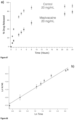
- the hydrogel controls (for example, retards or extends) the delivery of the bioactive agent, thereby potentially enhancing its therapeutic window. This is notably the case with local anaesthetic mepivacaine (see the Example below).
- hydrogels as described above can also be used to produce a paste of the invention for cleaning dental implants.
- the paste of the invention is specifically designed for cleaning dental implants, which have cleaning requirements that differ significantly from natural teeth.
- the inventor's knowledge there is currently no product on the market specially designed and optimized for implant surface decontamination.
- the paste of the invention allows removing biofilm contamination from titanium implant surfaces, while minimizing topographical changes to these surfaces (i.e. without affecting surface integrity).
- regular commercial toothpastes which are organic-based, are less effective in that context and may even contaminate the titanium implant surfaces - see the Examples below.
- the paste of the invention could allow dentists and patients to remove biofilm from implants, control the peri-implant infections and/or favor re-osseointegration in case of bone loss. It could also be used for surgical decontamination of implant surfaces or professional cleaning of implants during maintenance visits. It could also be used for daily personal care to clean titanium abutments in case of overdenture or even to clean exposed implant surfaces. Indeed, when wearing dental implants, titanium surfaces just below the crown, i.e. the "neck" of the implant, are commonly exposed.
- a paste for cleaning dental implants comprising the above hydrogel mixed with an abrasive agent.
- the hydrogel acts as a thickener and as carrier for the abrasive agent.
- the pH of the gel is between about 9 and about 10, especially is the implants to be cleans are made of titanium.
- One such gel is a gel in which, M I is Na + , M II is Mg 2+ , X is 0.56, Y is 0.13, and Z is 0.31.
- the paste is completely inorganic, i.e. it is free of organic compounds.
- the abrasive agent should preferably be avoided as they can induce surfaces scratches or rounded edges on the implant, thus potentially increasing plaque accumulation.
- the abrasive agent should have a relatively small average particle size, for example up to about 500 nm, preferably up to about 400 nm, and more preferably from about 200 to about 300 nm.
- Suitable abrasive agents include particles of silica, including magnesium phosphates silica, nano-silicates (that show osteoconductive properties that help inducing and accelerating bone regeneration) and/or calcium carbonate. More preferably, the abrasive agent is hydrated silica nanoparticles, especially those with average particles size of about 200 to about 300 nm.
- the abrasive agent can be present in the paste at a concentration ranging from about 5% to about 60%, preferably from about 20% to about 40%, and more preferably about 30% by weight based on the total weight of the paste.
- the paste for cleaning dental implants can comprise further additives, in particular such additives that are known as useful in dental cleaning pastes.
- additives include taste enhancers, coloring agents, sparkles as well as other functional ingredients.
- such additives should be carefully selected to avoid inducing contamination of the implants with organic compounds.
- to implant means to insert something into a person's body, for example (but not limited to) by surgery.
- An “implant” is a material that is intended/designed to be implanted into a person's body.
- to inject means to introduce something into a person's body using a needle.
- injectable is a material that is intended/designed to be injected into a person's body.
- the term "about” has its ordinary meaning. In embodiments, it may mean plus or minus 10% or plus or minus 5% of the numerical value qualified.
- Example 1 Magnesium Phosphate Gels That Up-Regulate Bone Formation and Bone Regeneration
- the title ternary system was investigated by varying the mole fraction of NaOH, Mg(OH) 2 , and H 3 PO 4 in different solutions. A fixed volume of 7 mL was used in all chemical reactions and the maximum reagents concentration was 10.5 mmol, in order to avoid any possible concentration effect.
- the ternary diagram was built using 141 different points obtained by mixing the three components at different mole fractions ( Figure 2 ). Precipitates obtained during the determination of the ternary diagram were prepared using the following procedure. 85 mg of Mg(OH) 2 were dissolved in 2.2 mL of H 3 PO 4 1.5 M and after complete dissolution 3.8 mL of NaOH 1.5 M were added under vigorous stirring.
- the diffraction patterns were processed with EVA software (Bruker AXS GmbH, Düsseldorf, Germany) and phase composition was determined by comparing the acquired spectra with the phases identified in the International Centre for Diffraction Data (ICDD) database PDF-4.
- the elemental composition of stable NMP suspensions was determined using Inductively Coupled Plasma Optical Emission Spectroscopy (ICP-OES) with a Thermo Scientific iCAP 6000 Series ICP-OES (Thermo Fisher Scientific Inc, East Grinstead, UK).
- ICP-OES Inductively Coupled Plasma Optical Emission Spectroscopy
- iCAP 6000 Series ICP-OES Thermo Fisher Scientific Inc, East Grinstead, UK.
- 6 mg of dried NMP powder was digested for 2 hours at 95 °C in 5 mL of HNO 3 67% trace metals basis and all samples were prepared in triplicate. After digestion, the samples were let to cool down at room temperature and then diluted with deionized water up to 50 mL. From this solution 1 mL was taken and diluted with deionized water to 10 mL and measured by ICP-OES.
- the calibration curves were prepared using freshly prepared standards solution of Mg 2+ , Na + , and PO 4 3- with a concentration of 10, 5, 2, 1, and 0.1 ppm in HNO 3 4%.
- the standard solutions were prepared by dilution from a certified standard solution of 1000 ppm in HNO 3 4% (SCP Science Inc, Baie D'Urfé, Canada).
- the analysis pump rate was set to 50 rpm
- the plasma radio frequency power was 1150 W
- the auxiliary and nebulizer gas flow were set to 0.5 L min -1 . All measurements were performed in axial/radius mode.
- FT-IR Fourier Transform Infrared Spectroscopy
- TGA Thermogravimetric analysis
- Colloidal suspensions of NMP were subjected to rheological measurements to determine the shear stress required for the gel-liquid transition, the liquid-gel transition time (t L-G ) upon removal of the shear stress, and the viscosity.
- the equilibration time before to run the measurements was set to 10 minutes and all the measurements were performed at room temperature.
- the force required to inject the NMP nanocrystals through an insulin needle of 160 ⁇ m of internal diameter was measured using the instrument Mach-1 V500cs and Mach-1 Motion software version 4.3.1 (Biomomentum Inc., Laval, Canada). The force was measured with a multiple-axis load cell of 70 N (resolution of 0.007 N) and acquisition rate of 100 Hz. The gel was loaded into the syringe avoiding the presence of bubble and then the plunger was inserted into the load cell. The force value was measured applying a constant vertical stage velocity of 1 mm s -1 (resolution of 0.1 ⁇ m).
- the syringe loaded with deionized water required a force of 0.14 ⁇ 0.01 N, while for the NMP gels of formulation A-D were comprised in a range of 0.22 ⁇ 0.03 N and 0.77 ⁇ 0.04 N.
- the required force to inject the NMP samples was considerably higher reaching a maximum of 18 ⁇ 1 N and a minimum of 9 ⁇ 1.2 N.
- Freeze-fracture replica was used to investigate the organization and morphology of the nanocrystals forming the thixotropic material.
- the stable NMP colloidal suspension was quickly frozen in liquid nitrogen (cooling rate >10 5 K s -1 ) immobilizing the nanocrystals instantaneously (Acharya et al., Journal of International Oral Health 2014, 6, 36 1).
- the resulting frozen suspension was fractured, the ice was removed by vacuum freeze etching, and a thin layer of carbon was sputtered onto the surface to produce a carbon replica.
- the sample surface was shadowed with platinum vapor and the carbon-metal replica was put on a Formvar/Carbon coated copper mesh-200 grid (2SPI, Structure Probe Inc, West Chester, USA) and examined by Transmission Electron Microscopy (TEM) using a Tecnai T12 working at 120 kV (FEI Inc, Hillsboro, Oregon, USA). Selected Area Electron Diffraction and TEM imaging of the water dispersion of the NMP nanocrystals were performed on a TEM grid Formvar/Carbon coated copper mesh-200 grid (2SPI, Structure Probe Inc, West Chester, USA).
- TEM Transmission Electron Microscopy
- the grid was prepared by deposition of a 5 ⁇ L drop of a 1% v/v water dispersion of the NMP gel, and the drop was blotted with filter paper after 90 seconds.
- Scanning Electron Microscopy (SEM) was used to characterize the nanocrystals at different time points during the reaction to obtain the NMP and the adhesion of osteoblast onto the surface of the nanocrystals.
- SEM Scanning Electron Microscopy
- an aliquot of 2 mL was withdrawn from the reaction and poured into a Buchner filter, washed with water first (20 mL), ethanol (20 mL), and then dried in a vacuum oven at 25 °C. This process was performed at 0.5, 5, 10, and 30 minutes from the beginning of the reaction.
- XPS measurements were carried out with a Thermo K-alpha spectrometer (Thermo Fisher Scientific Inc, East Grinstead, UK) equipped with monochromatic Al K ⁇ X-rays source operating at 1486.6 eV. Due to the non-conductive nature of the powder composing the gel, the charging effect was minimized using a low-energy flood-gun to provide efficient charge neutralization. To preclude charging effects in the resulting spectra the binding energy (BE) scale was calibrated from the hydrocarbon contamination using the C1s peak at 285.0 eV. During the measurement, the residual pressure inside the analysis chamber was 1 ⁇ 10 -8 mbar.
- the survey spectra were recorded with an X-ray beam diameter size of 400 ⁇ m and a passing energy of 200 eV, dwell time of 50 ms, and energy step size of 1 eV.
- High resolution spectra were recorded using a passing energy of 50 eV, dwell time of 50 ms, and energy step size of 0.1 eV.
- the surface depth profile experiment was realized using an Ar ions gun working at low current and ion energy of 500 eV that would produce an etching rate of 0.05 nm s -1 on a surface of TaO 2 .
- the etching time was 5 seconds and the process repeated for 5 times.
- Avantage software (5.932v) was used to fit photoelectron spectra using a least-squares algorithm.
- HF human fibroblast
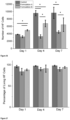
- HFs were kindly provided by Dr. C.Doillon (liable Laval). HFs were derived from foreskins after written informed consent which was approved by the Centre Hospitalier Universitaire de Quebec (CHUQ) Ethics Committee. HFs were seeded on circular coverslips in 24 well-plate (0.8x10⁵ well), cultured in DMEM cell culture media (10% FBS, 1% penicillin-streptomycin) at 37 °C in a humidified atmosphere of 5% CO 2 and grown overnight. A solution of 10% Alamar-Blue reagent was prepared in cell culture medium DMEM and used for the assay.
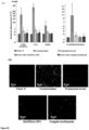
- HF cells without materials were used as positive control, NMP without HF cells was used as a blank, and colloidal suspension of formulation A and B were put in direct contact with HF cells for the cell viability test.
- the cell culture medium was removed and 500 ⁇ L of a 10% solution of Alamar-Blue reagent was added to each well and incubated at 37 °C in a humidified atmosphere of 5% CO 2 for 3 hours. From each sample 100 ⁇ L of culture medium was collected and deposited in a 96 well-plate. The fluorescence intensity was measured at 585 nm using a ⁇ exc of 550 nm using a Microplate Reader SpectraMax M2 (Molecular Devices, L.L.C. Sunnyvale, California, USA).
- HFs were seeded on circular coverslips in 24 well-plate (0.4x10⁵ well), cultured in DMEM cell culture media (10% FBS, 1% penicillin-streptomycin) at 37 °C in a humidified atmosphere of 5% CO 2 and grown overnight.
- the staining solution was prepared mixing calcein (2 ⁇ mol L -1 ), Etd-1 (4 ⁇ mol L -1 ), and Hoechst 33258 (4 ⁇ g mL -1 ).
- HF cells without materials were used as positive control, colloidal suspension formulation A and B were put in direct contact with HF cells for the Live-Dead assay.
- Negative control HF cells were treated with 70% of methanol for 30 minutes at room temperature. The culture medium was removed, and the cells were washed three times with 500 ⁇ L of phosphate buffer solution (PBS). 400 ⁇ L of the staining solution was added to each coverslip and incubated for 40 minutes at room temperature protected from light. Cells were washed again with PBS (500 ⁇ L) and the coverslips mounted on glass slides. Zeiss Axio Imager M2 (Carl Zeiss Microscopy GmbH, Goettingen, Germany) was used to take the photographs of the Live/Dead assay using three different sets of filters; green for calcein, red for Etd-1, and blue for Hoechst 33258. The same procedure was used for end point day 4 and 7. All assays were performed in triplicate.
- PBS phosphate buffer solution
- Mouse-derived bone marrow cells were kindly provided by Dr. S. Komarova (McGill University). Animal experiments were performed in accordance with the McGill University guidelines established by the Canadian Council on Animal Care.
- Mouse-derived bone marrow cells (mBCMs) were collected from mouse tibia and femora using a procedure previously described (C57BL6/J, male, 6 weeks old, purchased from Charles River) - see Hussein et al., Bone 2011, 48, 202 .
- mBMCs were cultured using a procedure previously described - see Tamimi et al., Acta Biomater. 2011, 7, 2678 .
- mBMCs were cultured in 75 cm 2 tissue culture flasks (2.5 ⁇ 10 6 cells cm -2 ) in MEM (Wisent Inc., Canada) with 10% serum (Fisher Scientific, Canada), 1% penicillin/streptomycin antibiotics (Wisent Inc., Canada), 1% sodium pyruvate (Wisent Inc.) and 50 ⁇ g mL -1 L-ascorbic acid (Sigma-Aldrich Co., USA). After 7-10 days, cells were detached with trypsin/EDTA (Wisent Inc.) and plated at a density of 10 4 cells cm -2 directly onto the surface of the materials or the tissue culture treated polystyrene (Corning Life Sciences, Lowell, MA, USA).
- mBMCs were cultured for 3, 5, 7, 14 and 21 days using MEM 10% serum, 1% antibiotics, 1% sodium pyruvate, 50 ⁇ g mL -1 ascorbic acid, 10 mM glycerol 2-phosphate disodium salt hydrate, and dexamethasone 1 ⁇ 10 -9 M. Cell cultures were supplemented with fresh medium every second day. Total RNA was isolated from mBMC primary cultures using TRI-zol ® reagent (Invitrogen TM , USA) following the manufacturer's protocol and quantified in a spectrophotometer by absorbance readings at 260 nm.
- RNA from each sample was reverse transcribed using a high-capacity cDNA reverse transcription kit (Applied Biosystems, USA) in accordance with the manufacturer's instructions.
- the resulting cDNAs were used for real-time PCR using Power SYBR Green PCR Master Mix (Applied Biosystems). Reactions were carried out in a 7500 Real-time PCR System (Applied Biosystems) for 40 cycles (95 °C for 15 s, 60 °C for 30 s and 72 °C for 45 s) after the initial 10-minute incubation at 95 °C.
- a cycle threshold value for each reaction was calculated using Applied Biosystems sequence detections software and the relative ratio of expression was determined using a previously described algorithm - see M.
- RunX2 run-related transcription factor 2: sense, 50-GGCTTGGGTTTCAGGTTAG-30; antisense, 50-CGGTTTCTTAGGGTCTTGGA-30
- TNALP tissue nonspecific alkaline phosphatase: sense, 50-GGGGACATGCAGTATGAGTT-30; antisense, 50-GGCCTGGTAGTTGTTGTGAG-30
- COL1A1 collagen type I, alpha 1: sense, 50-GAGGCATAAAGGGTCATCGTGG-30; antisense, 50-CATTAGGCGCAGGAAGGTCAGC-30
- OCN osteocalcin: sense, 50-TGAACAGACTCCGGCG-30; antisense, 50-GATACCGTAGATGCGTTTG-30
- OPN osteopontin: sense, 50-CTGCTAGTACACAAGCAGACA-30; antisense, 50-
- mice Twenty-four animals were used to assess the effect of NMP gel on bone healing and implant osseointegration. These animals were divided into two groups; first control (12 rats) and; second NMP gel (12 rats). To provide sufficient analgesia during surgical procedure, rats were injected with Carprofen (5-10 mg/kg, subcutaneous, Pfizer Animal Health, Quebec, QC) thirty minutes prior to surgical intervention. The rats were anesthetized with isoflurane (4% during the induction and 2.5% during the surgical procedure); the legs were shaved, disinfected with chlorhexidine gluconate solution (Omega Laboratories, Montreal, Canada) and covered with a sterile drape. A full thickness incision was made to expose the proximal third of the tibia.
- a uni-cortical defect (2.5 mm ⁇ ) was created in the right tibia using straight hand-piece under constant saline irrigation. The same procedure was performed in the contralateral side but custom made titanium (Ti) implant (1.5 mm ⁇ ⁇ 2.0 mm in depth) was placed in the defect.
- Ti titanium
- the Ti implants were coated with NMP gel before insertion in the defect and the contralateral defects were filled with the NMP gel (20 ⁇ L). The implant and the defects were not treated in the control group. Incisions were sutured using 5-0 monocryl sutures. In order to provide sufficient analgesia to the rats following surgery, they were administered with Carprofen every 24 hours for the first 3 days.
- Rats were allowed to heal for two weeks and then were euthanized using CO 2 asphyxiation, and the tibiae were extracted and preserved in 10% neutral buffered formalin (Richard Allan Scientific, Kalamazoo, MI). Samples were code labeled and codes were blinded to the person who did the analyses.
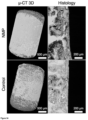
- the right tibiae (bone defect samples) were scanned using a micro-CT (SkyScan1172; SkyScan; Kontich, Belgium) set at 12.7 ⁇ m resolution, 50 kV voltage, 200 ⁇ A current, 0.5 degree rotation step and 0.5 mm aluminum filter.
- the original bone defect (2.5 mm ⁇ , full thickness of cortex) was identified as a region of interest (ROI).
- ROI was analyzed and the volume of the defect was calculated by subtracting the bone volume from the total volume of the ROI.
- the left tibiae samples with Ti implants were also scanned using a micro-CT (SkyScan1172; SkyScan; Kontich, Belgium) but set at 4.5 ⁇ m resolution, 100 kV voltage, 100 ⁇ A current, 0.4 degree rotation step and aluminum/copper filter.
- the reconstructed images were segmented by different thresholding to obtain two ROls.
- the first ROI included the titanium implant only by thresholding the intensity of the white color at low 80 and high 255.
- the second ROI included the implant/bone in the peri-implant area by an exact dilation of the the first ROI and followed by thresholding the intensity of the white color at low 10 and high 255.
- the final ROI (the peri-implant bone) was determined by subtracting the first ROI from the second ROI and the bone peri-implant area was analyzed.
- Primers used to amplify specific targets are as follows: RunX2 (runt-related transcription factor 2: sense, 50-GGCTTGGGTTTCAGGTTAG-30; antisense, 50-CGGTTTCTTAGGGTCTTGGA-30), TNALP (tissue nonspecific alkaline phosphatase: sense, 50-GGGGACATGCAGTATGAGTT-30; antisense, 50-GGCCTGGTAGTTGTTGTGAG-30), COL1A1 (collagen type I, alpha 1: sense, 50-GAGGCATAAAGGGTCATCGTGG-30; antisense, 50-CATTAGGCGCAGGAAGGTCAGC-30), OCN (osteocalcin: sense, 50-TGAACAGACTCCGGCG-30; antisense, 50-GATACCGTAGATGCGTTTG-30), and OPN (osteopontin: sense, 50-CTGCTAGTACACAAGCAGACA-30; antisense, 50-CATGAAATTCGGAATTTCAG-30).
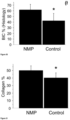
- osteoblasts data were presented as osteoblasts number per square millimeter of mineralized tissue (OB/mm 2 ).
- the percentage of mineralized tissue in the defect was analyzed using Image J (Wayne Rasband; National Institute of Health, Bethesda, Maryland) and data were presented as mineralized tissue present (MT %). All data are presented as mean ⁇ standard deviation.
- Left tibiae samples with Ti implants were dehydrated in ascending concentrations of ethanol (70% - 100%) and infiltrated with poly(methyl-methacrylate) histological resin (Technovit 9100, Heraeus Kulzer, Wehrheim, Germany).
- samples were sectioned into 30 ⁇ m thick histological slides using a diamond saw (SP1600, Leica Microsystems GmbH, Wetzlar, Germany) and stained using basic fuchsine and methylene blue. Histological sections were imaged using an optical micro-scope (Carl Zeiss Microscopy, Germany) and analyzed using ImageJ software (Wayne Rasband; National Institute of Health, Bethesda, Maryland). The bone-implant contact was calculated by dividing the bone-covered implant perimeter by the total implant perimeter.
- sample size for the in vivo part of the study was calculated to give a power of 80 % at 95 % confidence level to reject the null hypothesis that there is no difference between NMP gel-coated and non-coated implants.
- a 10 % difference was considered to be clinically relevant, 12 % potential standard deviation was assumed based on previous study, and a 10 % potential drop-out was expected (based on our previous experiment using similar model and intervention).
- Shapiro-Wilk test was used to assess data normality and Student's t test was used to compare between groups. Origin 9 software (Origin Lab Co., Northampton, MA) was used for data analyses. Statistical significance was set at p ⁇ 0.05.
- NMP Nanocrystalline Magnesium Phosphate
- NMP gels were obtained in a small region of the ternary diagram ( Figure 3 , area labelled "Stable colloidal suspension").
- the remaining crystals phases identified ranged from di- and tribasic magnesium phosphate such as Newberyite MgHPO 4 ⁇ 3H 2 O, Farringtonite Mg 3 (PO 4 ) 2 , Bobierrite Mg 3 (PO 4 ) 2 ⁇ 8H 2 O, and Brucite Mg(OH) 2 .
- di- and tribasic magnesium phosphate such as Newberyite MgHPO 4 ⁇ 3H 2 O, Farringtonite Mg 3 (PO 4 ) 2 , Bobierrite Mg 3 (PO 4 ) 2 ⁇ 8H 2 O, and Brucite Mg(OH) 2 .
- a new unidentified crystal phase (X-ray diffraction pattern shown in Figure 4 ) and unstable gel-like colloidal suspension were obtained ( Figure 3 , area labelled "New crystal phase and mixed Mg/PO 4 phases" surrounding the area labelled "Stable colloidal suspension”).
- the composition of the NMP was comprised of a range of [Mg + ], [Na + ], [PO 4 3 ], and [HPO 4 2- ] and the formula can be assumed to be Mg X Na Y (HPO 4 2- ) Z ⁇ (PO 4 3- ) T ⁇ nH 2 O with [5 ⁇ X ⁇ 7], [1 ⁇ Y ⁇ 2], [3 ⁇ Z ⁇ 5], and [1 ⁇ T ⁇ 3] where [7 ⁇ X+Y ⁇ 8] and [5 ⁇ Z+T ⁇ 6].
- the crystallization water was determined by thermogravimetric analysis and was comprised between [3 ⁇ n ⁇ 4] (Table 2 and Figure 5 ).
- the atomic ratio Mg/P was between 1 and 1.15, suggesting that the NMP gel might present similarity with the crystalline precipitate Newberyite MgHPO 4 ⁇ 3H 2 O.
- Figure 6 shows the pH of the colloidal suspension as a function of the reaction time.
- the mole fraction of H 3 PO 4 /NaOH/Mg(OH) 2 was 0.37/0.45/0.18 and the total volume was 7 mL.
- Figure 7 is a picture of the suspension after 30 seconds from the beginning of the reaction.
- the colloidal suspension was white and liquid, visible by tilting the tube.
- Figure 8 shows that after 10 minutes, the NMP colloidal suspension transformed from liquid to solid changing its color from white to gray.
- Figure 9 A and B show the NMP nanocrystals evolution during the reaction.
- the SEM micrographs show the evolution of the morphology nanocrystals after 30 s (d) and 30 minutes (e).
- the scale bar represents 500 nm.
- the zeta potential of the NMP nanocrystals showed an overall negative charge comprised between -10.1 ⁇ 4.3 and -18.1 ⁇ 6.7 mV.
- the NMP gel could be injected easily through an insulin needle and after manual injection the colloidal suspension would regain solid-like behavior ( Figure 15 ).
- the force required to inject the NMP material through the insulin needle was only 0.08-0.63 N more than the force required to inject water (0.14 ⁇ 0.03 N Experimental Section, above). After flipping the glass slide, the suspension behaved like a solid material.
- the real 3D structure of the colloidal suspension was revealed with TEM freeze-etching fracture technique due to the inherent sample technique preparation.
- the nanocrystals are kept in place by the underlying ice matrix, and after being exposed for replication, the carbon-metal replica shows the network formed by the nanocrystals.
- Low magnification TEM micrograph of the carbon-metal replica of a freeze-fractured suspension shows bundled NMP nanocrystals forming the 3D network, the water composing the medium of the hydrogel being in the empty space between the bundled nanocrystals ( Figure 16 ).
- the surface of the replica showed a stepped pattern revealing the thickness of the nanocrystals that was estimated to range between 4 and 7 nm ( Figure 17 ).
- TEM micrographs illustrated the high aspect ratio of the nanocrystals, and selected area electron diffraction showed the nanocrystalline nature of the biomaterial ( Figures 18 to 22 ).
- the synthesis of the thixotropic NMP biomaterial was achieved using different alkaline bases (NaOH, KOH, and LiOH) and the material showed very long term stability (> 1500 days, 20 °C).
- the stability was dramatically influenced by the type of alkali ion used. Smaller ions such as Li + and Na + (ionic radius 76 and 102 pm, respectively) produced stable hydrogels, whereas larger K + ions (ionic radius 138 pm) resulted in unstable hydrogels that eventually converted to Newberyite MgHPO 4 ⁇ 3H 2 O ( Figure 28 ).
- NMP contains both HPO 4 2- and PO 4 3- . This was demonstrated by performing onto the washed NMP powder FT-IR, solid state magic angle spinning (MAS) NMR, and XPS.
- MAS solid state magic angle spinning
- the FT-IR spectrum of formulation A ( Figure 29 ) showed a broad absorptions band from 3600 to 2600 cm -1 , indicating a combination of O-H stretching of intermolecular and weakly bonded crystallization water and stretching of (P)O-H.
- the OH stretching of the HPO 4 2- anion give rise to at least two broad bands around 2855 and 2385 cm -1 .
- the band around 2855 cm -1 was covered by the broad absorption of the water molecules, while the band at 2355 cm -1 was visible.
- the broad absorption at 1647 cm -1 was also attributed to a combination of HOH bending and rotation of residual crystallization and free water.
- Figure 30 shows the FT-IR spectrum of the same powder after calcination at 700 °C for 8 hours. As can be observed, the vibrations of the P-O-H moiety disappeared and new additional vibrational modes appeared in the frequency range of 1250-450 cm -1 that were assigned to the presence of P 2 O 7 4- , demonstrating the transformation of HPO 4 2- to P 2 O 7 4- .
- the 31 P spectra ( Figure 31 top) revealed two peaks. The left one without visible sidebands was assigned as PO 4 3- , whereas the right one with sidebands presented a stronger signal at the CP experiment and it was assigned as HPO 4 2- .
- MAS NMR showed that, HPO 4 2- cross polarizes intensely due to the small distance between hydrogen and phosphate ions in its chemical molecular structure. Therefore, HPO 4 2- yields a stronger signal in the CP MAS spectra, as compared with PO 4 3- .
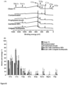
- XPS depth profile ( Figure 33 (c) ) also revealed that the concentration of sodium slightly decreased from 9.1 to 7.6 at. % after mild etching using Ar ions at very mild condition.
- the superficial excess of sodium on the outer surface of the nanocrystals could be due to the presence of negative charges on the faces of the nanocrystals, and those ions might be responsible of the stabilization of the metastable nanocrystalline phase.
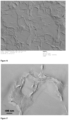
- Figure 34 shows the deposition of NMP on a negatively charged glass surface.
- the nanocrystals can adopt a parallel or perpendicular direction to the surface.
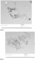
- Figure 35 show NMP powder deposited on a positively charged glass surface.
- the nanocrystals adopt only a parallel configuration to the glass surface.
- the amount of nanocrystals deposited onto the substrate surface with negative charge was considerably higher than the positive surface. Note the contrast in Figures 34 and 35 was enhanced for visibility in black and white.
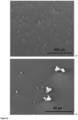
- SEM micrograph shows the morphology, adhesion, and colonization of differentiated osteoblasts from mouse bone marrow cells (mBMCs) cultured onto NMP biomaterial. After eight days of culture, the image shows cell-cell and cell-substrate interactions that enabled the formation of a macro-scale tissue construct ( Figure 42 ). Moreover, comparing the structure of the NMP nanocrystals before and after cells culture, it can be observed an increase of the nanocrystals porosity probably due to the dissolution of the NMP nanocrystals.
- Runt related transcription factor 2 (RunX2) is considered a key factor of osteogenesis due to the stimulation of osteoblast-related genes such as alkaline phosphatase (ALP), osteocalcin (OCN), osteopontin (OPN), and collagen, type I, alpha1 (COL1A1).
- ALP alkaline phosphatase
- OCN osteocalcin
- OPN osteopontin
- collagen, type I, alpha1 COL1A1
- the regulation activity of ALP is a key event that occurs during the early time of osteogenesis.
- Our result shows an early increase and up-regulation of ALP during the first five days respect MgHPO 4 ⁇ 3H 2 O and Mg 3 (PO 4 ) 2 ⁇ 22H 2 O indicating that NMP had a positive effect on ALP expression and promoted osteogenic differentiation ( Figure 43 ).
- OCN osteoblast differentiation and mineralization.
- OCN is secreted solely by osteoblasts and regulates body metabolism and bone building process, being the most specific marker for osteoblast differentiation and mineralization.
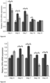
- Real Time-PCR of OCN showed that during the first 14 days, mBMCs cultured on NMP expressed significant higher levels of the gene than cells cultured on MgHPO 4 ⁇ 3H 2 O and Mg 3 (PO 4 ) 2 ⁇ 22H 2 O ( Figure 44 ).
- OPN or bone sialoprotein is a structural protein that accounts ⁇ 8% of all non-collagenous proteins found in bone, and it is mainly synthesized by pre-osteoblasts, osteoblasts and osteocytes. Our results shown that NMP up-regulated the expression of OPN up to 21 days ( Figure 45 ).
- COL1A1 a gene responsible to encode the production of pro-alpha1(l) chain of type I collagen that is a constituent of the ECM in connective tissue such as bone, skin, tendon, ligament and dentine.
- mBMCs cultured on NMP expressed higher levels of RunX2 than mBMCs cultured on Cattiite (Mg 3 (PO 4 ) 2 ⁇ 22H 2 O) and Newberyite after five days of incubation ( Figure 47 ).
- the novel NMP material has osteogenic properties and can trigger a series of events such as osteoblast cell proliferation, collagen synthesis, ECM maturation, and mineralization which follow the temporal pattern of osteogenic differentiation.
- the 2D material up-regulates the mRNA expression of RunX2/ALP and also the genes responsible of the formation of the extracellular matrix with bone-related protein OCN, OPN, and type I collagen.
- NPM accelerates bone healing and implant osseointegration
- NMP effects on bone formation were also investigated in vivo using rats' tibiae model.
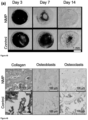
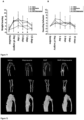
- Computerized micro-tomography ( ⁇ -CT) scans were performed on bone samples that were retrieved at different time points 3, 7 and 14 days after surgery for NMP treated and non-treated defects. At day 3, there is no visible difference on ⁇ -CT scan between the control and NMP group. On the other hand, at day 7 the ⁇ -CT scan clearly indicates that the tibial defect treated with NMP shown more bone formation in the defect compared to the control ones ( Figure 48 ). After 14 days, the tibial defect treated with NMP is almost fully filled with new bone while the control is still partially healed.
- FIB-SEM Focus Ion Beam SEM
- NMP effects on genes expression were further assessed in vivo by treating rats tibial defect with NMP. Bone samples were retrieved and assessed by qRT-PCR at different time points; 3, 7 and 14 days and showed that expression of COL1A1 and RunX2 were significantly up-regulated already at day 3 following surgery ( Figures 57 and 58 ).
- Osteoblast differentiation was up-regulated ( Figure 52 ) due to the NMP effects on osteoblastic gene markers ALP, OPN and RunX2 ( Figures 43 , 45 , 47 , and 57 ).
- the presence of magnesium and calcium ions increase the expression of osteoblast phenotype genes.
- Magnesium increases the expression of ALP, OPN and RunX2.
- calcium increases the expression of Col1.
- NMP which is rich of magnesium and calcium, enhanced osteoclasts proliferation and mineralization of the healing defects.
- Collagen synthesis was also up-regulated in NMP treated defects ( Figures 49 and 51 ) because NMP enhanced expression of COL1A1 ( Figures 46 and 58 ).
- the physical properties of this novel material can be tailored by varying the mole fraction of NaOH-Mg(OH) 2 -H 3 PO 4 .
- the 2D nanocrystals were characterized using a variety of analytical methods.
- the synthesized nanocrystals formed colloidal suspensions that behaved as physical hydrogels presenting long term stability, thixotropy, injectability, and high surface area.
- the easiness of synthesis and the interesting physicochemical properties of these materials open promising application in catalysis, layer by layer deposition, and electrodes for batteries.
- FeCl 2 ⁇ 4H 2 O and Mg(OH) 2 were purchased from Sigma-Aldrich (Milwaukee, WI, USA).
- Stable colloidal suspension of 2D nanocrystals was done using the following conditions. 85 mg of Mg(OH) 2 (1.45 mmol, mole fraction 0.14) were dissolved in 2.2 mL of H 3 PO 4 1.5 M (3.30 mmol, mole fraction 0.31). After complete dissolution of Mg(OH) 2 , 3.8 mL of NaOH 1.5 M (5.7 mmol, mole fraction 0.55) were added under vigorous stirring. The addition of NaOH provoked the instantaneous formation of a white colloidal suspension that after 6 minutes changed to a solid state with a grey color. The final pH of the colloidal suspension was 8.46. After 2 hours, the colloidal suspension was centrifuged at 4000 rpm for 5 minutes and the supernatant was discarded. The solid precipitate was washed with ethanol to remove the excess of water, vacuum dried at room temperature, and stored for characterization.
- the colloidal dispersion with Mg was also obtained by using magnesium chloride (MgCl 2 ⁇ 6H 2 O) instead of magnesium hydroxide (Mg(OH) 2 ).
- the reaction was carried out by dissolving 667.5 mg of MgCl 2 ⁇ 6H 2 O (3.275 mmol) in 2.5 mL of deionized water and 935 mg of Na 2 HPO 4 (6.525 mmol) in 10 mL of deionized water. After dissolving the solids, the Na 2 HPO 4 was poured into the solution of MgCl 2 ⁇ 6H 2 O under stirring.
- the resulting colloidal dispersion had a white color and after 20 minutes from the beginning of the reaction a grey thixotropic gel was obtained.
- NaMg(HPO 4 ) 2- (PO 4 ) 3- was also obtained by replacing Mg(OH) 2 with magnesium oxide (MgO). Briefly, 160 mg of MgO were dissolved in 11.2 mL of H 3 PO 4 1.5 M and 16.8 mL of NaOH 1.5 M were added under vigorous stirring. The colloidal suspension had a white color and after 10 minutes a grey thixotropic gel was obtained.
- MgO magnesium oxide
- the diffraction patterns were processed with EVA software (Bruker AXS GmbH, Düsseldorf, Germany) and phase composition was determined by comparing the acquired spectra with the phases identified in the International Centre for Diffraction Data (ICDD) database PDF-4.
- ICDD International Centre for Diffraction Data
- FT-IR Fourier Transform Infrared Spectroscopy
- TGA Thermogravimetric analysis
- Zeta potential measurements were carried out to assess the superficial charge of the nanocrystals using a Malvern Nano ZS equipped with disposable folded capillary cells (Malvern Instruments Ltd, Malvern, UK).
- the nanocrystals concentration used for the measurements was set to 20 mg mL -1 and the temperature was kept constant at 25 °C.
- the morphology of the different nanocrystals obtained was revealed by Scanning Electron Microscopy (SEM) using a FEI Inspect F-50 FE-SEM (FEI Inc, Hillsboro, Oregon, USA) operated at 10 kV. Prior analysis the samples were sputtered achieving a homogenous coating layer of 2 nm Pt (Leica EM ACE600, Leica Microsystems Inc, Concord, Ontario, Canada). The elemental composition of the colloidal suspension was determined using energy dispersive X-ray analysis spectroscopy (EDS) performed with an EDAX Octane Super Silicon Drift Detector and analyzed using TEAM TM software version 4.0.2 (AMETEK, Inc. Berwyn, PA, USA).
- EDS energy dispersive X-ray analysis spectroscopy
- the device was installed on a F-50 FE-SEM (FEI Inc, Hillsboro, Oregon, USA) operated in secondary electron mode at 10 kV.
- ZAF atomic number (Z), absorption (A), and fluorescence (F)
- TEAM TM software version 4.0.2 eZAF Smart Quant Results Acquisition
- the specific surface area of the products was measured by the Brunauer-Emmett-Teller (BET) method using nitrogen adsorption and desorption isotherms on an automated gas adsorption analyzer Tristar 3000 (Micromeritics Instrument Corporation Norcross, Georgia, USA).
- BET Brunauer-Emmett-Teller
- the force required to inject the thixotropic colloidal suspensions through an insulin needle of 160 ⁇ m of internal diameter was measured using a Mach-1 V500cs and Mach-1 Motion software version 4.3.1 (Biomomentum Inc., Laval, Canada). The force was measured with a multiple-axis load cell of 70 N (resolution of 0.007 N) and acquisition rate of 100 Hz. The gel was loaded into the syringe avoiding the presence of bubbles and then the plunger was inserted into the load cell. The force value was measured applying a constant vertical stage velocity of 1 mm s -1 (resolution of 0.1 ⁇ m).
- the colloidal suspensions formed physical gels.
- the colloidal dispersions showed a thixotropic behavior forming a physical gel.
- the nanocrystals synthesized presented a two-dimensional morphology that to form a physical hydrogel with a thixotropic clay-like behavior.
- Clays are plate-like poly-ions with a heterogeneous charge distribution that forms a physical gel in water at concentrations higher than 40 mg/mL due to the simultaneous presence of positive and negative charges that give rise to electrostatic and Van der Waals interactions. This allows the gel to behave as a thixotropic material due to the formation of a 3D network of particles known as the "house of cards" structure.
- Thixotropic materials can be liquified by applying mechanical energy allowing the physical gel to behave as a liquid; then when the mechanical stress is removed Brownian motions drive the particles into contact to reform the 3D network and the liquefied dispersion becomes gel-like again.
- the simultaneous presence of positive and negative charges allowed the novel synthesized nanocrystals dispersion to form a physical hydrogel with a "clay-like" behavior.
- the NMP gel could be injected easily through an insulin needle and after manual injection the colloidal suspension would regain solid-like behavior. After injection the material rapidly recovers its solid state and behaves again as solid, this is an important feature for coatings applications procedure.
- the FT-IR data, X-ray diffraction pattern of the dried and washed powders demonstrated the nanocrystalline nature of the 2D nanocrystals due to the presence of broad diffraction peaks.
- the implant-paste was developed by combining an inorganic thickening agent made of a nanocrystalline colloidal suspension (Nanocrystalline Magnesium Phosphate) and polishing nanoparticles of hydrated colloidal silica.
- the implant-paste formulation was optimized to decontaminate titanium surfaces coated with oral biofilm and compared to a commercial toothpaste (Colgate Total; Colgate-Palmoliven, USA). Surface morphology, bacterial load and attachment and chemical properties of titanium surfaces were analyzed and comparisons between different products were done using one-way ANOVA and independent samples t tests.
- An inorganic prophylaxis paste made of nanocrystalline magnesium phosphate gel (10% w/w) and (30% w/w) hydrated silica was superior to brushing alone and Colgate toothpaste in removing titanium surfaces contaminants and it did not cause surface alteration.
- the thixotropic and inorganic nature of the nanocrystalline magnesium phosphate implant-paste is ideal for cleaning implant surfaces because, unlike the Colgate and other commercial toothpastes, it does not contain organic-based thickeners that can adhere tightly on titanium surfaces and thus change their surface chemistry and moreover, does not abrade titanium.
- Nanocrystalline magnesium phosphate (NMP) gel is a novel inorganic colloidal suspension. It is stable biocompatible and thixotropic. NMP gel is silicate-free unlike other thixotropic inorganic materials such as silicate clays that could be more abrasive on implant surfaces. This novel gel is also rich in Na + cations that have toxic effect on bacteria and can disturb the biofilm structure by displacing the divalent cations (Ca ++ ).
- Conventional toothpastes comprise fluoride that can corrode Ti, organic compounds that can alter its surface chemistry and abrasives that can damage its surface microtexture. Accordingly, we hypothesized that prophylaxis pastes free of fluoride and organic compounds would be more efficient for cleaning dental implants. Thus, this study aimed at developing and optimizing a new "implant-paste" specifically designed for decontamination of dental implant.
- the implant-paste was developed by combining a thickening agent made of an inorganic nanocrystalline magnesium phosphate (NMP) gel with different concentration of an abrasive agent of hydrated silica nanoparticles.
- NMP nanocrystalline magnesium phosphate
- the liquid suspension changed its color from white to grey and possessed a solid and thixotropic behavior with the final suspension composed of 2D nanocrystals with an undulate structure.
- the solid content of the paste was then modified by adding 20, 30, 50, or 60% of hydrated silica nanoparticles with average aggregate particles size of 0.2-0.3 ⁇ m.
- the addition of hydrated silica nanoparticles increased the viscosity of the gel depending on the concentration used, however, the thixotropic behavior and pH of the initial gel were not affected (see Figure 59A ).
- Machined and polished titanium discs (grade 2, ⁇ 5.0 and 1.0 mm thick; McMaster-Carr, Cleveland, OH, United states) were used in this study. The discs were sequentially ultrasonicated in deionized water, acetone and ethanol for 15 minutes each, before drying over-night in a vacuum oven (Isotemp, Fisher Scientific, USA).
- Alginate impressions were taken to produce study models for each participant's upper jaw.
- a 1-mm-thick thermoplastic copolyester splints covering all maxillary teeth were produced.
- the splints were used to fix Ti discs at the buccal aspect of the premolar and molar areas, each splint housed 12 Ti discs.
- the participants were asked to wear the splints for 24 hours in order to allow for soft biofilm to accumulate on Ti surfaces.
- the participants were instructed to remove and store the splints during drinking or eating in phosphate buffered saline. After 24 hours, the splints were collected, the discs were washed with sterile saline solution (9%) and stored for further analysis.
- a rotary brush was used to clean biofilm-contaminated samples with water-intensive cooling at a speed of ⁇ 2500 rpm.
- the brush was held perpendicularly in gentle contact with the surfaces of the contaminated samples while moving in a circular motion.
- XPS X-ray Photoelectron Spectroscopy
- XPS is the most widely used surface analysis technique that measures the elemental composition, chemical state and electronic state of the elements within a material.
- the chemical composition of Ti surfaces was analyzed using X-ray Photoelectron Spectrometer (Thermo Fischer Scientific Inc, East Grinstead, UK). The in was equipped with a monochromatic Al K ⁇ X-Ray radiation source (1486.6 eV, ( ⁇ ) 0.834 nm) and an ultrahigh vacuum chamber (10 -9 torr). For all discs, survey scans were acquired over the range of 0-1350 eV with a pass energy of 200 eV and a resolution of 1.0 eV. A flood gun was used to neutralize the surface charging in all samples. Binding energies, peak areas and atom concentration ratios were obtained using the curve fitting function of Avantage (5.932v) analysis software (Thermo Fisher Scientific, Waltham, MA USA).
- Live/dead staining kit BocLight Bacterial Viability Kit L7012, Molecular Probes, Carlsbad, USA
- a LEXT 3D Confocal Microscope (Olympus America Inc., PA) was used to evaluate the surface roughness of polished Ti discs before and after decontamination.
- the surface roughness was characterized with roughness profile parameters [average roughness (Ra) and root mean square roughness (Rq)]; a method extensively used for assessing the surface roughness of implants. All values were determined at a cut-off length of 0.08 mm in 50 sections (evaluation length of 4 mm), and evaluated using the LEXT OLS4000 software (Olympus, America Inc., PA). Four discs were used for each group, and measurements were taken at five random areas from each disc.
- the primary outcome variables were surface chemical composition, surface roughness, and bacterial attachment and viability.
- data of the primary variables was statistically analyzed based on paired design for comparison of the measurements from before and after contamination and decontamination. The outcomes of different decontamination methods were also analyzed and compared.
- the XPS survey spectra of clean surfaces showed the presence of the following major peaks: O1s (Oxygen), C1s (Carbon), Ti2p (Titanium) and N1s (Nitrogen) (see Figure 60A and B) .
- C1s and N1s signals indicate the surface contamination while Ti2p signals demonstrate the presence of the TiO 2 oxide layer.
- composition of the NMP gel was optimized as mentioned above to obtain an alkaline pH of 9.6. to 10% w/w.
- the optimized implant-paste formulation was the one containing 10% NMP gel and 30% hydrated silica.
- the commercial toothpaste did not show a change in Ti or C levels.
- the optimized implant-paste is also able to remove the carbon-containing compounds that are adsorbed from atmosphere and usually detected on clean Ti surfaces without inducing a significant increase in their roughness as shown with confocal microscopy (see Figure 64 ).
- * significantly different from clean Ti surfaces
- a significantly different from Ti surfaces cleaned with the prophylaxis brush
- b significantly different from Ti surfaces brushed with optimized implant-paste (p ⁇ 0.05).
- the optimized implant-paste significantly reduced the atomic concentration of surfaces' contaminants (C and N) and increased the O and Ti levels. However, it did not induce any significant change in the Ti surface roughness (see Figure 64 ). Both results contrast with those obtained for surfaces cleaned with the brush alone or the brush with Colgate toothpaste ( Figure 65A and B ). The toothpaste significantly increased the C levels and surface roughness of Ti.
- Prophylaxis instruments such as brushes and rubber cup are used to remove biofilms attached to implant surfaces with or without using prophylaxis pastes.
- rotary brushes for cleaning Ti surfaces because they are inexpensive and accessible compared to titanium brushes and their plastic bristles should be stricte on Ti.
- Rotating cups were found to leave remnants of rubber particles on the implant surfaces after cleaning 5, 36.
- some cup materials are too abrasive and can cause Ti surface damage 37.
- a dentifrice is usually combined with brushes to adjunct the physical removal of plaque and stains through their chemical and physical additives, or to apply therapeutic and preventive agents to tooth surfaces.
- the dentifrice needs two main ingredients to achieve the mechanical cleaning; an abrasive agent and thickener to hold the abrasives in suspension during brushing.
- prophylaxis paste to decontaminate implant surfaces "implant-paste" and enhance the cleaning efficiency of the brush.
- the sole thickener was composed of an inorganic, silicate free Nanocrystalline Magnesium Phosphate (NMP) gel.
- the gel composition was optimized to obtain an alkaline pH of 9.6 because the corrosion resistance of Ti is high at this pH.
- the implant-paste can be in contact with intraoral structures and teeth for several hours when used for daily cleaning of Ti implants. Consequently, ideally, this optimized implant-paste to have a relatively alkaline pH to minimize potential tooth or implant damage.
- the optimized NMP gel has similar biocompatibility and thixotropic properties of Laponite (silicate clays); the most used inorganic thickener in toothpastes.
- Laponite silicate clays
- the optimized NMP gel has a stable consistency without the need for additional organic thickeners. This is an advantage of our novel gel over the clays-based toothpastes that require organic thickeners (i.e. xanthan gum) to provide optimal consistency.
- organic thickeners i.e. xanthan gum
- the other key component of the implant-paste that contributes to the physical removal of biofilm is the abrasive agent.
- hydrated silica nanoparticles were chosen as abrasives. It is a relatively safe, nontoxic ingredient and mostly compatible with other ingredients, such as glycerine and fluoride.
- low concentration of silicates shows osteoconductive properties that help to induce and accelerate bone regeneration.
- the cleaning effectiveness of the optimized prophylaxis paste was further confirmed by its ability to remove carbon-containing compound from clean Ti surfaces (controls), using the optimized brushing time, without causing alteration in their roughness ( Figure 64 ) .
- the significant drop in carbon level ( Figure 63 ) after cleaning confirms the effectiveness of the implant-paste in removing the carbon-containing compound that are normally adsorbed on Ti surface from atmosphere.
- the optimized inorganic implant-paste shows superior efficiency in decontaminating implants than organic-based Colgate toothpaste without damaging their surfaces integrity.
- the new inorganic implant-paste developed in this study can remove biofilm from contaminated Ti implants without affecting their surface integrity. Cleaning dental implants with current organic-based toothpastes contaminates the implants surfaces, changing their surface charge, roughness and chemistry, which could have negative impact on re-osseointegration.
- sodium magnesium phosphate nanocrystals are osteoinductive, thixotropic colloidal suspension and can be injected through high gauge needles and therefore can be used for minimal invasive interventions.
- this gel can control the release of local anesthetic (mepivacaine) in-vitro and in-vivo.
- mepivacaine local anesthetic
- the gel (NMP) for the in vitro release was prepared by dissolving 54 mg of Mg(OH) 2 (0.93 mmol, mole fraction 0.13) in 1.65 mL of H 3 PO 4 1.5 M (2.47 mmol, mole fraction 0.34), and subsequently 2.32 mL of NaOH 1.5 M (3.78 mmol, mole fraction 0.53) was added under vigorous stirring.
- mepivacaine hydrochloride was dissolved in 1 mL of gel previously prepared to reach different mepivacaine concentration.
- the pH of the resulting thixotropic colloidal suspension before the addition of mepivacaine was 7.95, while after the addition of the drug changed to 7.1.
- the gel + mepivacaine was placed in a Pur-L-Lyzer TM dialysis tube with a molecular weight cut-off of 12000 Da, while the control group is represented by the mepivacaine dissolved in PBS (2% w/v) and placed in a dialysis tubes with the same molecular weight cut-off.
- the tubes were incubated in 30 mL of phosphate buffer saline solution (PBS) at 37 °C, and the solution was changed at different time points comprised between 0 and 168 hours.
- PBS phosphate buffer saline solution
- the extracted solutions were analyzed with UV-Vis spectroscopy following the absorption of mepivacaine at 263 nm. All experiments were done in triplicates.
- the gel for the in vivo experiment was synthesized by dissolving 69 mg of Mg(OH) 2 (1.18 mmol, mole fraction 0.16) in 1.65 mL of H 3 PO 4 1.5 M (2.47 mmol, mole fraction 0.31), and subsequently 2.9 mL of NaOH 1.5 M (4.35 mmol, mole fraction 0.53) was added under vigorous stirring.
- 80 mg of mepivacaine hydrochloride were dissolved in 1 mL of gel previously prepared (8% w/v).
- the pH of the resulting thixotropic colloidal suspension before the addition of mepivacaine was 9.6, while after its addition the pH decreased to 7.6, being more compatible with physiological pH.
- the sensitivity to thermal stimuli was tested using radiant heat at different time points comprised between 0 and 120 minutes.
- mice tibial fracture model St-Arnaud, The Journal of steroid biochemistry and molecular biology 121, 254-256 (2010 )
- Carprofen (20 mg/kg; SC).
- the animal was then anesthetized with isoflurane (3-5% at the induction time and 2-2.5% during the maintenance period). After the animal shows signs of being fully anesthetized, the right leg was shaved and disinfected using chlorohexidine, then, the animal was covered with a sterile drape. A longitudinal skin incision was made in order to expose the right patellar tendon. The tendon was dissected and elevated in order to expose the proximal tibial tuberosity. A 27-gauge spinal needle was introduced into the intramedullary canal of the tibia.
- a tibial fracture was performed in a standardized manner using a twister ( Hiltunen et al., Journal of orthopaedic research 11, 305-312 (1993 )). Following creation of fracture, a single post-operative injection of the assigned treatment (20 ⁇ L) into the fracture site and the surgical site was closed using 5-0 Vicryl.
- Post-operative Animals' pain perception and locomotion were evaluated, at different time point; hours, 24 hours, 3 days, one week and two weeks post fracture, by assessing mice guarding ( Yasuda et al., Journal of pain research 6, 161 (2013 )) and weight bearing per each leg, respectively. Animals were allowed to habituate to the testing room for at least one hour prior to handling. Guarding behavior was assessed as following; each mouse was placed individually in a clear plastic box on an elevated stainless steel mesh. Both paws (of the fractured and non-fractured legs) were observed closely for 1-minute periods every five minutes for 60 minutes. A score of 0, 1, or 2 was given according to the postural position of each paw in most of the 1-minute scoring periods.
- Weight bearing test was performed as following; an incapacitance meter (IITC.inc, CA, US) was used for determination of hind paw weight distribution.
- Mouse was placed in an angled plexiglass chamber positioned so that each hind paw rest on a separate weighting plate. The weight exerted by each hind limb was measured and averaged over a period of 5 seconds. The change in hind paw weight distribution was calculated by determining the difference in the amount of weight (g) between the left and right limbs. Change in hind paw weight distribution between the left (fractured leg) and right (control) limbs was used as an index of pain in the fractured leg ( Bove et al., Osteoarthritis and cartilage 11, 821-830 (2003 )). Two weeks following fracture, mice were euthanized and tibial explants were assessed for bone healing and fracture resistance using micro-CT and three point pending test, respectively.
- the in vitro release experiment of mepivacaine demonstrates that, compared to the control formulation, the gel controlled the release of mepivacaine.
- the Korsmeyer-Peppa's model was used for the fitting of the cumulative drug release as shown in Figure 68 .
- the stability of mepivacaine in contact with the gel was checked after 24 hours from the beginning of the drug release experiment.
- the UV-Vis spectra show the molecular integrity of the drug indicating that mepivacaine is compatible with the gel ( Figure 69 ) .
- NMP combined with mepivacaine improved weight bearing on fractured leg and reduced post-operative pain ( Figure 71 ) .
- NMP and NMP + mepivacaine accelerated fracture healing. Indeed, Micro-CT sagittal, coronal sections and 3 D reconstructions showed that bone formation at fracture site was higher in NMP alone and in NMP combined with mepivacaine ( Figure 72 ).
- the biomaterial described here was injected through high gauge needle into hindpaw of mice and provided effective analgesia to the mice treated with gel + mepivacaine.
- NMP was able to control the release of mepivacaine for up to 24 hours.
- Higuchi and Korsmeyer-Peppas models indicated that mepivacaine was released by diffusion.
- the mepivacaine released by NMP presented no change in its molecular structure as shown by UV-Vis spectra ( Figure 69 ), indicating that the drug was compatible with NMP.
- Radiant heat test showed that NMP loaded with mepivacaine provides analgesia and the analgesic action of mepivacaine was prolonged by NMP ( Figure 70 ).
- Guarding and weight bearing tests revealed that NMP loaded with mepivacaine shorten the mobilization time and reduce postoperative pain ( Figure 71 ).
- Micro-CT data showed that NMP loaded with mepivacaine, accelerated bone healing ( Figure 72 ).
- Three point pending test indicated that NMP loaded with mepivacaine enhanced fracture resistance ( Figure 73 ).
Landscapes
- Health & Medical Sciences (AREA)
- Chemical & Material Sciences (AREA)
- Life Sciences & Earth Sciences (AREA)
- Medicinal Chemistry (AREA)
- Animal Behavior & Ethology (AREA)
- General Health & Medical Sciences (AREA)
- Public Health (AREA)
- Veterinary Medicine (AREA)
- Epidemiology (AREA)
- Dermatology (AREA)
- Organic Chemistry (AREA)
- Oral & Maxillofacial Surgery (AREA)
- Inorganic Chemistry (AREA)
- Pharmacology & Pharmacy (AREA)
- Transplantation (AREA)
- Engineering & Computer Science (AREA)
- Biomedical Technology (AREA)
- Orthopedic Medicine & Surgery (AREA)
- General Chemical & Material Sciences (AREA)
- Nuclear Medicine, Radiotherapy & Molecular Imaging (AREA)
- Chemical Kinetics & Catalysis (AREA)
- Molecular Biology (AREA)
- Dispersion Chemistry (AREA)
- Neurosurgery (AREA)
- Dentistry (AREA)
- Bioinformatics & Cheminformatics (AREA)
- Physical Education & Sports Medicine (AREA)
- Anesthesiology (AREA)
- Rheumatology (AREA)
- Medicinal Preparation (AREA)
- Pharmaceuticals Containing Other Organic And Inorganic Compounds (AREA)
- Materials For Medical Uses (AREA)
- Colloid Chemistry (AREA)
Claims (17)
- Pharmazeutische Zusammensetzung, die ein oder mehrere bioaktive Mittel, ausgewählt aus der Gruppe bestehend aus Anästhetika, Antibiotika, Hormonen, Wachstumsfaktoren, Betablockern und Proteinen, und ein Hydrogel als Träger für das bioaktive Mittel enthält, wobei das Hydrogel mindestens 1 % v/v einer kolloidalen Suspension von MI XMII YPZ zweidimensionalen Nanokristallen in Wasser umfasst, wobei:MI Na+ und/oder Li+ ist,MII Mg2+ oder eine Mischung von Mg2+ mit einem oder mehreren von Ni2+, Zn2+, Cu2+, Fe2+ und/oder Mn2+ ist,P eine Mischung von dibasischen Phosphat-Ionen (HPO4 2-) und tribasischen Phosphat-Ionen (PO4 3-) ist,X im Bereich von 0,43 bis 0,60 ist,Y im Bereich von 0,10 bis 0,18 ist, undZ im Bereich von 0,29 bis 0,46 ist,X, Y, Z Molfraktionen sind.
- Pharmazeutische Zusammensetzung nach Anspruch 1, die ein Implantat ist.
- Pharmazeutische Zusammensetzung nach Anspruch 1, die eine Injektionslösung ist.
- Pharmazeutische Zusammensetzung nach einem der vorhergehenden Ansprüche, wobei der bioaktive Wirkstoff aus der Gruppe ausgewählt ist, die aus Lokalanästhetika, Antibiotika und Betablockern besteht.
- Pharmazeutische Zusammensetzung nach einem der Ansprüche 1 bis 4 zur Verwendung für die gezielte Abgabe eines bioaktiven Wirkstoffs an einen Ort des Bedarfs eines Patienten durch Verabreichung der Zusammensetzung an den Ort des Bedarfs.
- Pharmazeutische Zusammensetzung nach Anspruch 5, wobei die Stelle des Bedarfs ein Knochendefekt oder eine Knochenverletzung ist.
- Pharmazeutische Zusammensetzung nach Anspruch 5 oder 6, wobei der Verabreichungsschritt das Implantieren des Hydrogels umfasst.
- Pharmazeutische Zusammensetzung nach Anspruch 5 oder 6, wobei der Verabreichungsschritt das Injizieren des Hydrogels umfasst.
- Paste zur Reinigung eines Zahnimplantats, wobei die Paste ein mit einem Schleifmittel vermischtes Hydrogel enthält, wobei das Hydrogel mindestens 1 % v/v einer kolloidalen Suspension von MI XMII YPZ zweidimensionalen Nanokristallen in Wasser umfasst, wobei:MI Na+ und/oder Li+ ist,MII Mg2+ oder eine Mischung von Mg2+ mit einem oder mehreren von Ni2+, Zn2+, Cu2+, Fe2+ und/oder Mn2+ ist,P eine Mischung von dibasischen Phosphat-Ionen (HPO4 2-) und tribasischen Phosphat-Ionen (PO4 3-) ist,X im Bereich von 0,43 bis 0,60 ist,Y im Bereich von 0,10 bis 0,18 ist, undZ im Bereich von 0,29 bis 0,46 ist,X, Y, Z Molfraktionen sind.
- Paste nach Anspruch 9, wobei das Hydrogel einen pH-Wert zwischen 9 und 10 aufweist.
- Paste nach Anspruch 9 oder 10, wobei MI Na+ ist, MII Mg2+ ist, X 0.50 ist, Y 0.13 ist, und Z 0.37 ist.
- Paste nach einem der Ansprüche 9 bis 11, wobei das Schleifmittel eine Kieselsäure ist.
- Paste nach einem der Ansprüche 9 bis 11, wobei es sich bei dem Schleifmittel um hydratisierte Siliziumdioxid-Nanopartikel handelt.
- Paste nach einem der Ansprüche 9 bis 11, wobei das Schleifmittel aus der Gruppe ausgewählt ist, die aus Magnesiumphosphat, Siliciumdioxid, einem Nanosilicat und Calciumcarbonat besteht.
- Paste nach einem der Ansprüche 9 bis 14, wobei die Schleifmittelteilchen eine durchschnittliche Teilchengröße von bis zu 500 nm, vorzugsweise bis zu 400 nm, aufweisen.
- Paste nach einem der Ansprüche 9 bis 15, die außerdem ein oder mehrere Additive enthält.
- Paste nach einem der Ansprüche 9 bis 16, die 5 bis 60 Gew.-%, vorzugsweise 20 bis 40 Gew.-%, des Schleifmittels, bezogen auf das Gesamtgewicht der Paste, enthält.
Applications Claiming Priority (3)
| Application Number | Priority Date | Filing Date | Title |
|---|---|---|---|
| US201562265570P | 2015-12-10 | 2015-12-10 | |
| EP16871858.3A EP3386547B1 (de) | 2015-12-10 | 2016-12-02 | Magnesiumphosphat-hydrogele |
| PCT/CA2016/051415 WO2017096469A1 (en) | 2015-12-10 | 2016-12-02 | Magnesium phosphate hydrogels |
Related Parent Applications (1)
| Application Number | Title | Priority Date | Filing Date |
|---|---|---|---|
| EP16871858.3A Division EP3386547B1 (de) | 2015-12-10 | 2016-12-02 | Magnesiumphosphat-hydrogele |
Publications (2)
| Publication Number | Publication Date |
|---|---|
| EP4104864A1 EP4104864A1 (de) | 2022-12-21 |
| EP4104864B1 true EP4104864B1 (de) | 2025-02-05 |
Family
ID=59012427
Family Applications (2)
| Application Number | Title | Priority Date | Filing Date |
|---|---|---|---|
| EP22186994.4A Active EP4104864B1 (de) | 2015-12-10 | 2016-12-02 | Magnesiumphosphat-hydrogele |
| EP16871858.3A Active EP3386547B1 (de) | 2015-12-10 | 2016-12-02 | Magnesiumphosphat-hydrogele |
Family Applications After (1)
| Application Number | Title | Priority Date | Filing Date |
|---|---|---|---|
| EP16871858.3A Active EP3386547B1 (de) | 2015-12-10 | 2016-12-02 | Magnesiumphosphat-hydrogele |
Country Status (5)
| Country | Link |
|---|---|
| US (2) | US10875772B2 (de) |
| EP (2) | EP4104864B1 (de) |
| CA (1) | CA3007064C (de) |
| ES (1) | ES2929118T3 (de) |
| WO (1) | WO2017096469A1 (de) |
Families Citing this family (6)
| Publication number | Priority date | Publication date | Assignee | Title |
|---|---|---|---|---|
| WO2018191615A1 (en) * | 2017-04-14 | 2018-10-18 | Northwestern University | Metal-substituted metal oxide materials for lithium ion batteries |
| WO2019071087A1 (en) * | 2017-10-06 | 2019-04-11 | DigiM Solution LLC | SYSTEM AND METHOD FOR CALCULATING PERFORMANCE OF CONTROLLED RELEASE OF MEDICINE USING IMAGES |
| CN110596066A (zh) * | 2019-09-28 | 2019-12-20 | 佛山克情日用品有限公司 | 一种餐具洁净程度检测设备 |
| CA3219195A1 (en) * | 2021-06-03 | 2022-12-08 | Magle Chemoswed Ab | A pharmaceutically acceptable aqueous gel composition for mrna delivery |
| CN113599574A (zh) * | 2021-07-28 | 2021-11-05 | 苏州大学 | 一种用于肌肉修复的再生材料及其制备方法 |
| WO2024082070A1 (en) * | 2022-10-21 | 2024-04-25 | Invicare Inc. | Bone implant with a magnesium phosphate xerogel coating |
Family Cites Families (6)
| Publication number | Priority date | Publication date | Assignee | Title |
|---|---|---|---|---|
| DE4237500A1 (de) * | 1992-11-06 | 1994-05-11 | Henkel Kgaa | Remineralisierendes Zahnpflegemittel |
| US5939051A (en) | 1998-02-27 | 1999-08-17 | Colgate-Palmolive Company | Dental abrasive |
| US6669928B1 (en) | 1999-05-13 | 2003-12-30 | Tamer International, Ltd. | Periodontal composition |
| US6451361B1 (en) * | 2001-05-29 | 2002-09-17 | Agri-Nutrients Technology Group, Inc. | Alkali metal magnesium phosphate hydrate buffering feed mineral |
| US20150150973A1 (en) | 2012-03-30 | 2015-06-04 | The Royal Institution For The Advancement Of Learning/Mcgill University | Magnesium phosphate gels |
| US20170056559A1 (en) * | 2014-03-14 | 2017-03-02 | Ecole Polytechnique Federale De Lausanne (Epfl) | Active Agent-Particle Combination Supporting Bone Regeneration |
-
2016
- 2016-12-02 EP EP22186994.4A patent/EP4104864B1/de active Active
- 2016-12-02 CA CA3007064A patent/CA3007064C/en active Active
- 2016-12-02 EP EP16871858.3A patent/EP3386547B1/de active Active
- 2016-12-02 US US16/060,462 patent/US10875772B2/en active Active
- 2016-12-02 WO PCT/CA2016/051415 patent/WO2017096469A1/en not_active Ceased
- 2016-12-02 ES ES16871858T patent/ES2929118T3/es active Active
-
2020
- 2020-12-04 US US17/111,940 patent/US11629055B2/en active Active
Also Published As
| Publication number | Publication date |
|---|---|
| CA3007064C (en) | 2023-01-24 |
| US20190002282A1 (en) | 2019-01-03 |
| WO2017096469A1 (en) | 2017-06-15 |
| EP3386547A1 (de) | 2018-10-17 |
| EP3386547A4 (de) | 2019-07-31 |
| ES2929118T3 (es) | 2022-11-24 |
| US11629055B2 (en) | 2023-04-18 |
| US20210163290A1 (en) | 2021-06-03 |
| US10875772B2 (en) | 2020-12-29 |
| EP3386547B1 (de) | 2022-07-27 |
| EP4104864A1 (de) | 2022-12-21 |
| CA3007064A1 (en) | 2017-06-15 |
Similar Documents
| Publication | Publication Date | Title |
|---|---|---|
| US11629055B2 (en) | Magnesium phosphate hydrogels | |
| Tovani et al. | Strontium calcium phosphate nanotubes as bioinspired building blocks for bone regeneration | |
| Chen et al. | Enhancement of critical-sized bone defect regeneration by magnesium oxide-reinforced 3D scaffold with improved osteogenic and angiogenic properties | |
| Li et al. | Porous lithium-doped hydroxyapatite scaffold seeded with hypoxia-preconditioned bone-marrow mesenchymal stem cells for bone-tissue regeneration | |
| Fu et al. | Fluorescence microscopic analysis of bone osseointegration of strontium-substituted hydroxyapatite implants | |
| Sobczak-Kupiec et al. | Effect of calcination parameters on behavior of bone hydroxyapatite in artificial saliva and its biosafety | |
| Pan et al. | Iron-doped brushite bone cement scaffold with enhanced osteoconductivity and antimicrobial properties for jaw regeneration | |
| Li et al. | Size effect of nano-hydroxyapatite on proliferation of odontoblast-like MDPC-23 cells | |
| Yao et al. | Enhanced osteogenesis of titanium with nano-Mg (OH) 2 film and a mechanism study via whole genome expression analysis | |
| Hesaraki et al. | Evaluation of a bioceramic-based nanocomposite material for controlled delivery of a non-steroidal anti-inflammatory drug | |
| Xiong et al. | Enhancing osseointegration and angiogenesis of Titanium implants through KMnO4-Modified Montmorillonite nano-clay coating | |
| Honma et al. | In vitro osteoconductivity of PMMA-Y2O3 composite resins | |
| Sugiura et al. | Inorganic silica hybrid octacalcium phosphate bone substitute: Harmonics to acceleration in biological metabolism and its curing process | |
| He et al. | Osteogenesis-inducing calcium phosphate nanoparticle precursors applied to titanium surfaces | |
| Tabatabaee et al. | Advances in bone repair: a bioengineered PMMA-based bone cement with enhanced otseoconductivity utilizing human-derived bone powder | |
| Ziąbka et al. | Biological evaluation of ZrO2 composites modified with different ceramics additives | |
| Bulajić et al. | Biocompatibility of mesoporous SBA‐16/hydroxyapatite nanocomposite and dentin demineralized particles on human dental pulp stem cells | |
| Wang et al. | Enhanced healing of rat calvarial critical size defect with selenium-doped lamellar biocomposites | |
| Tabari et al. | The Effect of Silicone Oil and Nano-hydroxyapatite/Chitosan Powder on Microhardness and Surface Structure of Primary Teeth Enamel After Iron Drop Consumption. | |
| Latifi et al. | Fabrication and characterization of chitosan based injectable thermosensitive hydrogels containing silica/calcium phosphate nanocomposite particles | |
| Lim et al. | Hydroxyapatite coating on damaged tooth surfaces by immersion | |
| US7767226B2 (en) | Calcium sulfate based nanoparticles | |
| Dang et al. | Sonochemical-synthesized biphasic tricalcium phosphate: Influence of synthesis parameters on the physicochemical properties and in vitro remineralization in artificial saliva | |
| Jairam et al. | Hydroxyapatite (HAp) nanoparticles in dentistry | |
| US9238047B1 (en) | Fluorapatite nano-crystalline coated non-ceramic hydrophilic hydroxylapatite bone grafting compositions and methods for promoting bone regeneration |
Legal Events
| Date | Code | Title | Description |
|---|---|---|---|
| PUAI | Public reference made under article 153(3) epc to a published international application that has entered the european phase |
Free format text: ORIGINAL CODE: 0009012 |
|
| STAA | Information on the status of an ep patent application or granted ep patent |
Free format text: STATUS: THE APPLICATION HAS BEEN PUBLISHED |
|
| AC | Divisional application: reference to earlier application |
Ref document number: 3386547 Country of ref document: EP Kind code of ref document: P |
|
| AK | Designated contracting states |
Kind code of ref document: A1 Designated state(s): AL AT BE BG CH CY CZ DE DK EE ES FI FR GB GR HR HU IE IS IT LI LT LU LV MC MK MT NL NO PL PT RO RS SE SI SK SM TR |
|
| STAA | Information on the status of an ep patent application or granted ep patent |
Free format text: STATUS: REQUEST FOR EXAMINATION WAS MADE |
|
| STAA | Information on the status of an ep patent application or granted ep patent |
Free format text: STATUS: EXAMINATION IS IN PROGRESS |
|
| 17P | Request for examination filed |
Effective date: 20230621 |
|
| RBV | Designated contracting states (corrected) |
Designated state(s): AL AT BE BG CH CY CZ DE DK EE ES FI FR GB GR HR HU IE IS IT LI LT LU LV MC MK MT NL NO PL PT RO RS SE SI SK SM TR |
|
| 17Q | First examination report despatched |
Effective date: 20230718 |
|
| GRAP | Despatch of communication of intention to grant a patent |
Free format text: ORIGINAL CODE: EPIDOSNIGR1 |
|
| STAA | Information on the status of an ep patent application or granted ep patent |
Free format text: STATUS: GRANT OF PATENT IS INTENDED |
|
| INTG | Intention to grant announced |
Effective date: 20240830 |
|
| GRAS | Grant fee paid |
Free format text: ORIGINAL CODE: EPIDOSNIGR3 |
|
| GRAA | (expected) grant |
Free format text: ORIGINAL CODE: 0009210 |
|
| STAA | Information on the status of an ep patent application or granted ep patent |
Free format text: STATUS: THE PATENT HAS BEEN GRANTED |
|
| AC | Divisional application: reference to earlier application |
Ref document number: 3386547 Country of ref document: EP Kind code of ref document: P |
|
| AK | Designated contracting states |
Kind code of ref document: B1 Designated state(s): AL AT BE BG CH CY CZ DE DK EE ES FI FR GB GR HR HU IE IS IT LI LT LU LV MC MK MT NL NO PL PT RO RS SE SI SK SM TR |
|
| REG | Reference to a national code |
Ref country code: GB Ref legal event code: FG4D |
|
| REG | Reference to a national code |
Ref country code: CH Ref legal event code: EP |
|
| REG | Reference to a national code |
Ref country code: IE Ref legal event code: FG4D |
|
| REG | Reference to a national code |
Ref country code: DE Ref legal event code: R096 Ref document number: 602016091163 Country of ref document: DE |
|
| REG | Reference to a national code |
Ref country code: NL Ref legal event code: MP Effective date: 20250205 |
|
| PG25 | Lapsed in a contracting state [announced via postgrant information from national office to epo] |
Ref country code: RS Free format text: LAPSE BECAUSE OF FAILURE TO SUBMIT A TRANSLATION OF THE DESCRIPTION OR TO PAY THE FEE WITHIN THE PRESCRIBED TIME-LIMIT Effective date: 20250505 |
|
| PG25 | Lapsed in a contracting state [announced via postgrant information from national office to epo] |
Ref country code: FI Free format text: LAPSE BECAUSE OF FAILURE TO SUBMIT A TRANSLATION OF THE DESCRIPTION OR TO PAY THE FEE WITHIN THE PRESCRIBED TIME-LIMIT Effective date: 20250205 |
|
| PG25 | Lapsed in a contracting state [announced via postgrant information from national office to epo] |
Ref country code: PL Free format text: LAPSE BECAUSE OF FAILURE TO SUBMIT A TRANSLATION OF THE DESCRIPTION OR TO PAY THE FEE WITHIN THE PRESCRIBED TIME-LIMIT Effective date: 20250205 |
|
| PG25 | Lapsed in a contracting state [announced via postgrant information from national office to epo] |
Ref country code: ES Free format text: LAPSE BECAUSE OF FAILURE TO SUBMIT A TRANSLATION OF THE DESCRIPTION OR TO PAY THE FEE WITHIN THE PRESCRIBED TIME-LIMIT Effective date: 20250205 |
|
| REG | Reference to a national code |
Ref country code: LT Ref legal event code: MG9D |
|
| PG25 | Lapsed in a contracting state [announced via postgrant information from national office to epo] |
Ref country code: NO Free format text: LAPSE BECAUSE OF FAILURE TO SUBMIT A TRANSLATION OF THE DESCRIPTION OR TO PAY THE FEE WITHIN THE PRESCRIBED TIME-LIMIT Effective date: 20250505 Ref country code: IS Free format text: LAPSE BECAUSE OF FAILURE TO SUBMIT A TRANSLATION OF THE DESCRIPTION OR TO PAY THE FEE WITHIN THE PRESCRIBED TIME-LIMIT Effective date: 20250605 |
|
| PG25 | Lapsed in a contracting state [announced via postgrant information from national office to epo] |
Ref country code: NL Free format text: LAPSE BECAUSE OF FAILURE TO SUBMIT A TRANSLATION OF THE DESCRIPTION OR TO PAY THE FEE WITHIN THE PRESCRIBED TIME-LIMIT Effective date: 20250205 |
|
| PG25 | Lapsed in a contracting state [announced via postgrant information from national office to epo] |
Ref country code: HR Free format text: LAPSE BECAUSE OF FAILURE TO SUBMIT A TRANSLATION OF THE DESCRIPTION OR TO PAY THE FEE WITHIN THE PRESCRIBED TIME-LIMIT Effective date: 20250205 |
|
| PG25 | Lapsed in a contracting state [announced via postgrant information from national office to epo] |
Ref country code: LV Free format text: LAPSE BECAUSE OF FAILURE TO SUBMIT A TRANSLATION OF THE DESCRIPTION OR TO PAY THE FEE WITHIN THE PRESCRIBED TIME-LIMIT Effective date: 20250205 Ref country code: PT Free format text: LAPSE BECAUSE OF FAILURE TO SUBMIT A TRANSLATION OF THE DESCRIPTION OR TO PAY THE FEE WITHIN THE PRESCRIBED TIME-LIMIT Effective date: 20250605 |
|
| PG25 | Lapsed in a contracting state [announced via postgrant information from national office to epo] |
Ref country code: GR Free format text: LAPSE BECAUSE OF FAILURE TO SUBMIT A TRANSLATION OF THE DESCRIPTION OR TO PAY THE FEE WITHIN THE PRESCRIBED TIME-LIMIT Effective date: 20250506 Ref country code: BG Free format text: LAPSE BECAUSE OF FAILURE TO SUBMIT A TRANSLATION OF THE DESCRIPTION OR TO PAY THE FEE WITHIN THE PRESCRIBED TIME-LIMIT Effective date: 20250205 |
|
| REG | Reference to a national code |
Ref country code: AT Ref legal event code: MK05 Ref document number: 1764780 Country of ref document: AT Kind code of ref document: T Effective date: 20250205 |
|
| PG25 | Lapsed in a contracting state [announced via postgrant information from national office to epo] |
Ref country code: SE Free format text: LAPSE BECAUSE OF FAILURE TO SUBMIT A TRANSLATION OF THE DESCRIPTION OR TO PAY THE FEE WITHIN THE PRESCRIBED TIME-LIMIT Effective date: 20250205 |
|
| PG25 | Lapsed in a contracting state [announced via postgrant information from national office to epo] |
Ref country code: SM Free format text: LAPSE BECAUSE OF FAILURE TO SUBMIT A TRANSLATION OF THE DESCRIPTION OR TO PAY THE FEE WITHIN THE PRESCRIBED TIME-LIMIT Effective date: 20250205 |
|
| PG25 | Lapsed in a contracting state [announced via postgrant information from national office to epo] |
Ref country code: DK Free format text: LAPSE BECAUSE OF FAILURE TO SUBMIT A TRANSLATION OF THE DESCRIPTION OR TO PAY THE FEE WITHIN THE PRESCRIBED TIME-LIMIT Effective date: 20250205 |
|
| PG25 | Lapsed in a contracting state [announced via postgrant information from national office to epo] |
Ref country code: IT Free format text: LAPSE BECAUSE OF FAILURE TO SUBMIT A TRANSLATION OF THE DESCRIPTION OR TO PAY THE FEE WITHIN THE PRESCRIBED TIME-LIMIT Effective date: 20250205 |
|
| PG25 | Lapsed in a contracting state [announced via postgrant information from national office to epo] |
Ref country code: AT Free format text: LAPSE BECAUSE OF FAILURE TO SUBMIT A TRANSLATION OF THE DESCRIPTION OR TO PAY THE FEE WITHIN THE PRESCRIBED TIME-LIMIT Effective date: 20250205 |
|
| PG25 | Lapsed in a contracting state [announced via postgrant information from national office to epo] |
Ref country code: EE Free format text: LAPSE BECAUSE OF FAILURE TO SUBMIT A TRANSLATION OF THE DESCRIPTION OR TO PAY THE FEE WITHIN THE PRESCRIBED TIME-LIMIT Effective date: 20250205 Ref country code: CZ Free format text: LAPSE BECAUSE OF FAILURE TO SUBMIT A TRANSLATION OF THE DESCRIPTION OR TO PAY THE FEE WITHIN THE PRESCRIBED TIME-LIMIT Effective date: 20250205 |
|
| PG25 | Lapsed in a contracting state [announced via postgrant information from national office to epo] |
Ref country code: RO Free format text: LAPSE BECAUSE OF FAILURE TO SUBMIT A TRANSLATION OF THE DESCRIPTION OR TO PAY THE FEE WITHIN THE PRESCRIBED TIME-LIMIT Effective date: 20250205 |
|
| PG25 | Lapsed in a contracting state [announced via postgrant information from national office to epo] |
Ref country code: SK Free format text: LAPSE BECAUSE OF FAILURE TO SUBMIT A TRANSLATION OF THE DESCRIPTION OR TO PAY THE FEE WITHIN THE PRESCRIBED TIME-LIMIT Effective date: 20250205 |
|
| REG | Reference to a national code |
Ref country code: DE Ref legal event code: R097 Ref document number: 602016091163 Country of ref document: DE |
|
| PLBE | No opposition filed within time limit |
Free format text: ORIGINAL CODE: 0009261 |
|
| STAA | Information on the status of an ep patent application or granted ep patent |
Free format text: STATUS: NO OPPOSITION FILED WITHIN TIME LIMIT |
|
| PGFP | Annual fee paid to national office [announced via postgrant information from national office to epo] |
Ref country code: GB Payment date: 20251229 Year of fee payment: 10 |
|
| 26N | No opposition filed |
Effective date: 20251106 |
|
| PGFP | Annual fee paid to national office [announced via postgrant information from national office to epo] |
Ref country code: FR Payment date: 20251226 Year of fee payment: 10 |
|
| PGFP | Annual fee paid to national office [announced via postgrant information from national office to epo] |
Ref country code: DE Payment date: 20251229 Year of fee payment: 10 |