EP4108779A1 - Procédé d'évaluation et de prédiction de maladies dérivées du placenta et kit associé - Google Patents
Procédé d'évaluation et de prédiction de maladies dérivées du placenta et kit associé Download PDFInfo
- Publication number
- EP4108779A1 EP4108779A1 EP20918272.4A EP20918272A EP4108779A1 EP 4108779 A1 EP4108779 A1 EP 4108779A1 EP 20918272 A EP20918272 A EP 20918272A EP 4108779 A1 EP4108779 A1 EP 4108779A1
- Authority
- EP
- European Patent Office
- Prior art keywords
- placenta
- derived
- methylation
- dna
- sample
- Prior art date
- Legal status (The legal status is an assumption and is not a legal conclusion. Google has not performed a legal analysis and makes no representation as to the accuracy of the status listed.)
- Withdrawn
Links
Images





Classifications
-
- C—CHEMISTRY; METALLURGY
- C12—BIOCHEMISTRY; BEER; SPIRITS; WINE; VINEGAR; MICROBIOLOGY; ENZYMOLOGY; MUTATION OR GENETIC ENGINEERING
- C12Q—MEASURING OR TESTING PROCESSES INVOLVING ENZYMES, NUCLEIC ACIDS OR MICROORGANISMS; COMPOSITIONS OR TEST PAPERS THEREFOR; PROCESSES OF PREPARING SUCH COMPOSITIONS; CONDITION-RESPONSIVE CONTROL IN MICROBIOLOGICAL OR ENZYMOLOGICAL PROCESSES
- C12Q1/00—Measuring or testing processes involving enzymes, nucleic acids or microorganisms; Compositions therefor; Processes of preparing such compositions
- C12Q1/68—Measuring or testing processes involving enzymes, nucleic acids or microorganisms; Compositions therefor; Processes of preparing such compositions involving nucleic acids
- C12Q1/6876—Nucleic acid products used in the analysis of nucleic acids, e.g. primers or probes
- C12Q1/6883—Nucleic acid products used in the analysis of nucleic acids, e.g. primers or probes for diseases caused by alterations of genetic material
-
- G—PHYSICS
- G16—INFORMATION AND COMMUNICATION TECHNOLOGY [ICT] SPECIALLY ADAPTED FOR SPECIFIC APPLICATION FIELDS
- G16B—BIOINFORMATICS, i.e. INFORMATION AND COMMUNICATION TECHNOLOGY [ICT] SPECIALLY ADAPTED FOR GENETIC OR PROTEIN-RELATED DATA PROCESSING IN COMPUTATIONAL MOLECULAR BIOLOGY
- G16B20/00—ICT specially adapted for functional genomics or proteomics, e.g. genotype-phenotype associations
- G16B20/20—Allele or variant detection, e.g. single nucleotide polymorphism [SNP] detection
-
- G—PHYSICS
- G16—INFORMATION AND COMMUNICATION TECHNOLOGY [ICT] SPECIALLY ADAPTED FOR SPECIFIC APPLICATION FIELDS
- G16H—HEALTHCARE INFORMATICS, i.e. INFORMATION AND COMMUNICATION TECHNOLOGY [ICT] SPECIALLY ADAPTED FOR THE HANDLING OR PROCESSING OF MEDICAL OR HEALTHCARE DATA
- G16H50/00—ICT specially adapted for medical diagnosis, medical simulation or medical data mining; ICT specially adapted for detecting, monitoring or modelling epidemics or pandemics
- G16H50/20—ICT specially adapted for medical diagnosis, medical simulation or medical data mining; ICT specially adapted for detecting, monitoring or modelling epidemics or pandemics for computer-aided diagnosis, e.g. based on medical expert systems
-
- G—PHYSICS
- G16—INFORMATION AND COMMUNICATION TECHNOLOGY [ICT] SPECIALLY ADAPTED FOR SPECIFIC APPLICATION FIELDS
- G16H—HEALTHCARE INFORMATICS, i.e. INFORMATION AND COMMUNICATION TECHNOLOGY [ICT] SPECIALLY ADAPTED FOR THE HANDLING OR PROCESSING OF MEDICAL OR HEALTHCARE DATA
- G16H50/00—ICT specially adapted for medical diagnosis, medical simulation or medical data mining; ICT specially adapted for detecting, monitoring or modelling epidemics or pandemics
- G16H50/30—ICT specially adapted for medical diagnosis, medical simulation or medical data mining; ICT specially adapted for detecting, monitoring or modelling epidemics or pandemics for calculating health indices; for individual health risk assessment
-
- C—CHEMISTRY; METALLURGY
- C12—BIOCHEMISTRY; BEER; SPIRITS; WINE; VINEGAR; MICROBIOLOGY; ENZYMOLOGY; MUTATION OR GENETIC ENGINEERING
- C12Q—MEASURING OR TESTING PROCESSES INVOLVING ENZYMES, NUCLEIC ACIDS OR MICROORGANISMS; COMPOSITIONS OR TEST PAPERS THEREFOR; PROCESSES OF PREPARING SUCH COMPOSITIONS; CONDITION-RESPONSIVE CONTROL IN MICROBIOLOGICAL OR ENZYMOLOGICAL PROCESSES
- C12Q2600/00—Oligonucleotides characterized by their use
- C12Q2600/154—Methylation markers
Definitions
- the present disclosure relates to the field of biological detection. Specifically, the present disclosure relates to a method for predicting and evaluating the risk of pregnancy complications associated with placental dysplasia, and a kit. More specifically, the present disclosure relates to a method for evaluating and predicting, at early pregnancy (12 to 16 weeks of gestation), the risk of pregnant women being afflicted with gestational hypertension and other types of placenta-derived diseases in the perinatal period, and a kit. More specifically, the present disclosure relates to a method for evaluating and predicting the risk of preeclampsia, and a kit.
- placenta Formation of a placenta is a vital guarantee for normal mammalian pregnancy and healthy fetal growth.
- the placenta is a maternal-fetal interface and plays important roles in, e.g., providing necessary nutrition support for a fetus and regulating maternal immune tolerance.
- the placenta originates from the extraembryonic ectoderm (ExE). Its formation course starts with implantation of the blastocyst, i.e., 6 to 7 days after fertilization, and its development course continues throughout the gestation.
- the blastocyst ready for implantation has differentiated into the inner cell mass (ICM) with the potential to develop into an embryo and the trophoectoderm (TE) with the potential to differentiate into a placental tissue.
- Placental growth and development involves such biological processes as degradation of extracellular matrix, trophoblastic invasion, cell differentiation of placental lineage, promotion of angiogenesis, cell migration, and regulation of tolerance of the maternal immune system.
- the placental development is also accompanied by dynamic epigenetic processes, such as a remodeling process of whole-genome methylation and regulation of expression levels by genomic imprinting, which plays important roles in determining the fate of the cell lineage and regulating the expression of placental development-associated genes and the embryonic development.
- DNA methylation modification is to transfer a methyl group to the fifth carbon atom of cytosine under the action of methyltransferases to form 5-methylcytosine. Most of DNA methylation modifications occur at CpG loci, with very few occurring at CHG or CHH loci (H represents C, T, or A). DNA methylation modifications are roughly similar in cells from the same developmental lineage. When cells undergo differentiation, transdifferentiation, canceration, etc., the DNA methylation modifications will be altered. Under different physiological and pathological conditions, the DNA methylation modification to the same cell may also be altered. Therefore, the pattern of the DNA methylation modification may be used to deduce the origin of its tissue and to evaluate the pathological and physiological status of the tissue cell where it is found.
- the DNA methylation modification is a highly dynamic process. In a few hours after zygote formation, an erasure of DNA methylation occurs progressively within a whole-genome range to thereby reduce the mean whole-genome methylation level from approximately 41% after fertilization to 9% at the 2-cell stage.
- the genome of blastocyst is almost completely hypo-methylated prior to implantation.
- the unerased methylation modification is known as methylation imprinting. After implantation of blastocyst, the de novo methylation of DNA will occur, which is closely related to the regulation of the expression of embryonic development-associated genes.
- intra- and extra-embryonic tissues show different patterns, with 80% of methylated loci being differentially modified at the whole-genome level in the intra- and extra-embryonic tissues; for an individual methylated locus, the DNA methylation modification in the intra-embryonic tissue is in an "all-or-none" pattern, i.e., a state that a particular methylated locus is completely methylated or completely hypo-methylated, whereas a majority of CpG loci in the extra-embryonic tissues are hemi-methylated.
- the methylation modifications to imprinted genes are regulated by an imprinting control region (ICR).
- ICR imprinting control region
- the ICR may remain methylated during the whole-genome erasure of methylations in the early development of a zygote, and the methylated ICR may affect surrounding genes such that they are methylation modified as well.
- Most of the ICRs in the oocyte are methylated, and they are mostly distributed in the CpG island (CGI) containing dense methylated loci.
- CGI CpG island
- genes are transcribed with an alternative promoter.
- the alternative promoters are upstream of the classic promoter on the CGI, and are mostly retrotransposons with a long terminal repeat (LTR), also referred to as endoretroviruses (ERVs).
- LTR long terminal repeat
- ERVs make up approximately 8% of the human genome. Human-derived ERVs lose their retrotransposon activity but retain their function as alternative promoters. For example, the transcription of the CYP19A1 gene in the placenta is driven by a tissue-specific promoter -- MER21 LTR, which belongs to the ERV family and enables placental cells to express tissue-specific transcripts through an alternative splicing mechanism.
- tissue-specific promoter -- MER21 LTR which belongs to the ERV family and enables placental cells to express tissue-specific transcripts through an alternative splicing mechanism.
- Preeclampsia is a common pregnancy complication that may seriously threaten lives of mother and fetus in the perinatal period.
- the morbidity of preeclampsia is approximately 2% to 6%
- preeclampsia is the second leading cause of maternal death.
- preeclampsia is typically characterized by first onset of hypertension and proteinuria after 20 weeks of gestation, often accompanied by edema and hyperuricemia. Onset of preeclampsia will cause fatal damages to the liver function, kidney function, and coagulation function of pregnant women.
- Pregnant women with severe preeclampsia may develop microangiopathic hemolytic anemia, elevated lactate dehydrogenase and liver enzymes, headache, or other neurological symptoms.
- preeclampsia is mainly caused by inadequate invasion of trophoblast into the uterine decidua and inadequate remodeling of distal uterine spiral arteries.
- preeclampsia there is still no clinical means of treating preeclampsia and the only cure is induction of labor.
- Several clinical trials have demonstrated that taking low-dose aspirin before 16 weeks of gestation may reduce the morbidity of preeclampsia.
- preeclampsia strengthen the early prevention of preeclampsia, and reduce its morbidity, there is a need to strengthen early screening of preeclampsia.
- Congo red dye could bind to the ⁇ -amyloid protein in urine of pregnant women with preeclampsia, and turns red.
- the PlGF level is decreased, while the sFlt1 level is increased. Therefore, the Congo red staining result and the PlGF/sFlt1 ratio may be used as indicators for clinical diagnosis and prediction of preeclampsia.
- the above biochemical assay is only applicable to people at 25 weeks of gestational age or more but not to people at less than 16 weeks of gestational age.
- cfDNA Cell-free DNA
- cfDNA is a single- or double-stranded DNA fragment present in human circulating blood, urine, and other body fluids. Most of cfDNAs are 200 bp or less in length, and they are present in plasma at very low concentrations (about 1 to about 100 ng/mL). cfDNA results mainly from apoptosis because its fragmentation pattern is very similar to DNA fragments generated by apoptosis. 90% or more of cfDNAs are derived from cells of the hematopoietic lineage.
- the cfDNAs in maternal plasma is a mixture of maternal and placental cfDNAs, of which fetal/placental cfDNAs are in a smaller proportion (proportion of placental cfDNAs: 3% to 15%).
- Existing methods for prenatal diagnosis have realized early screening for fetal genetic diseases, e.g., chromosomal polyploidization diseases such as Down's syndrome (T21), by deep sequencing of cfDNAs in maternal plasma.
- the present disclosure provides use of a reagent for detecting a methylation modification level of at least one DNA methylation modification difference locus or at least one DNA methylation haplotype in a sample to be tested in the preparation of a reagent or kit for detecting, monitoring, and/or predicting whether a subject has a placenta-derived gestational disorder.
- the present disclosure provides a kit for detecting a methylation modification level of at least one DNA methylation modification difference locus or at least one DNA methylation haplotype in a sample.
- the present disclosure provides a polynucleotide usable for detecting presence of a DNA methylation modification.
- the present disclosure provides a method for detecting a methylation modification level of at least one DNA methylation modification difference locus or at least one DNA methylation haplotype in a sample to be tested.
- the present disclosure provides a method for detecting, monitoring, and/or predicting whether a subject has a placenta-derived gestational disorder.
- the present disclosure provides a device for detecting, monitoring, and/or predicting whether a subject has a placenta-derived gestational disorder, and a non-volatile computer readable storage medium.
- the present disclosure provides a system for detecting a methylation modification level of at least one DNA methylation modification difference locus or at least one DNA methylation haplotype in a sample to be tested.
- the present disclosure provides a system for detecting, monitoring, and/or predicting whether a subject has a placenta-derived gestational disorder.
- the invention of the present disclosure provides a method for detecting a methylation modification level of at least one DNA methylation modification difference locus or at least one DNA methylation haplotype in a sample to be tested, a kit, and a corresponding detection system, which can be used for accurately screening, in vitro, whether a subject has a placenta-derived gestational disorder.
- the term "about” means that a value includes the error or standard deviation caused by the device or method used to measure the value.
- gestational age is a measure of the age of gestation, starting from a woman's last menstrual period (LMP) or a corresponding age estimated by an additional method.
- LMP menstrual period
- the gestational age refers to the embryonic or fetal age + two weeks.
- labor occurs within 37 to 42 weeks of gestational age, but it usually occurs at about 40 weeks of gestational age.
- infant pregnancy refers to the gestational age at or before 16 weeks.
- normal pregnancy refers to the pregnancy during which no significant pregnancy complications occur, the pregnancy complications including, but not limited to, gestational hypertension, gestational diabetes, twin-to-twin transfusion syndrome, fetal intrauterine growth restriction, HELLP syndrome, preeclampsia, severe preeclampsia, secondary preeclampsia, atypical preeclampsia, and eclampsia.
- control pregnancy refers to the pregnancy without preeclampsia, severe preeclampsia, secondary preeclampsia, atypical preeclampsia, or eclampsia.
- control pregnancy used herein may include "normal pregnancy”.
- placenta-derived gestational disorder refers to a disorder developed during pregnancy due to a developmental defect or loss of function of the placental tissue.
- the "placenta-derived gestational disorder” used herein is selected form gestational diabetes, twin-to-twin transfusion syndrome, fetal intrauterine growth restriction, gestational hypertension, eclampsia, preeclampsia, severe preeclampsia, secondary preeclampsia, atypical preeclampsia, or HELLP syndrome.
- gestational diabetes means that a woman, who does not have diabetes before pregnancy, has symptoms of hyperglycemia during pregnancy.
- twin-to-twin transfusion syndrome is the commonest complication occurring in monochorionic diamniotic twins. Because the monochorionic diamniotic twins share one placenta, in which the networks of a large number of blood vessels are anastomosed with one another, the blood of the two fetuses communicate directly, with one fetus being a donor twin with growth retardation, anemia, oliguria, oligohydramnios, or hypotension, while the other fetus being a recipient twin with plethora, polyuria, polyhydramnios, cardiomegaly, hypertension, or edema.
- the donor twin may die in the womb due to intrauterine growth distress, and the recipient twin may also die from heart failure.
- fetal growth restriction means that the fetal weight is less than the tenth percentile or less than two standard deviations of the average weight for the same gestational age.
- gestational hypertension refers to onset of diastolic blood pressure ⁇ 90 mm Hg and (or) systolic blood pressure ⁇ 140 mm Hg (measured at intervals between 6 hours and 1 week) after 20 weeks of gestational age without proteinuria, neurological symptoms, symptoms of liver or kidney damage, or symptoms of HELLP syndrome.
- preeclampsia refers to new onset of systolic blood pressure ⁇ 140 mm Hg and (or) diastolic blood pressure ⁇ 90 mm Hg after 20 weeks of gestational age, proteinuria ⁇ 300 mg/24 h, or qualitative test (+).
- preeclampsia may include severe preeclampsia, secondary preeclampsia, and atypical preeclampsia.
- “severe preeclampsia” refers to onset of systolic blood pressure ⁇ 160 mm Hg and (or) diastolic blood pressure ⁇ 110 mm Hg after 20 weeks of gestational age, 24-hour proteinuria level of greater than or equal to 2.0 g and (or) qualitative test (++) or more, together with at least one of: serum creatinine >106 ⁇ mol/L or elevated than before, blood platelets ⁇ 100,000/mm 3 , or developing microangiopathic hemolytic anemia, elevated lactate dehydrogenase or liver enzymes, with headache or other neurological or visual symptoms, persistent epigastric discomfort, or pulmonary edema.
- second preeclampsia refers to onset of systolic blood pressure ⁇ 140 mm Hg and (or) diastolic blood pressure ⁇ 90 mm Hg before pregnancy or before 20 weeks of gestational age, and onset of preeclampsia or eclampsia during pregnancy.
- "atypical preeclampsia” refers to occurrence of symptoms of gestational hypertension with one of: neurological symptoms, hemolysis, elevated liver enzymes, and low counts of platelets, but without proteinuria (abbreviated from preeclampsia without symptoms of renal impairment), or with proteinuria (more than two rapid strip tests being positive) along with one of: neurological symptoms, hemolysis, elevated liver enzymes, low counts of platelets, but without hypertension (abbreviated from preeclampsia without symptoms of hypertension).
- HELLP syndrome refers to a syndrome characterized by hemolysis, elevated liver enzymes, and low counts of platelets during pregnancy, which is a serious complication of gestational hypertension.
- transposon also known as a mobile gene or a jumping gene, is a DNA sequence that may be inserted and cleaved in a genome and can change its own location.
- Sequence identity and “percent identity” used herein refer to the percentage of nucleotides that are the same (i.e., identical) between two or more polynucleotides.
- sequence identity between two or more polynucleotides can be determined by aligning the nucleotide sequences of polynucleotides and scoring the number of positions at which nucleotides are identical in the aligned polynucleotides, and comparing the number of these positions with the number of positions at which nucleotides are different in the aligned polynucleotides.
- Polynucleotides may differ at one position by, e.g., containing a different nucleotide (i.e., substitution or mutation) or deleting a nucleotide (i.e., a nucleotide insertion or a nucleotide deletion in one or two polynucleotides).
- the sequence identity may be calculated by dividing the number of positions at which nucleotides are identical by the total number of polynucleotides. For example, the percent identity may be calculated by dividing the number of positions at which nucleotides are identical by the total number of nucleotides in the polynucleotides, and multiplying the result by 100.
- the present disclosure relates to the stringency of hybridization conditions used to define the degree of complementarity between two polynucleotides.
- the aforesaid polynucleotides may be selected from DNA.
- “Stringency” used herein refers to the temperature and ionic strength conditions during hybridization and the presence or absence of some organic solvents. The higher the stringency, the higher the degree of complementarity between the target nucleotide sequence and the labeled polynucleotide sequence.
- Stringent conditions refers to the temperature and ion conditions under which only nucleotide sequences with high-frequency complementary bases hybridize.
- hybridization under high-stringency or very high-stringency conditions describes the conditions for hybridization and washing.
- Guidance for hybridization reactions is described in Current Protocols in Molecular Biology, John Wiley & Sons, N.Y. (1989), 6.3.1-6.3.6 .
- high-stringency hybridization conditions means that following the standard DNA blotting procedures, a probe of at least 100 nucleotides in length pre-hybridizes or hybridizes for 12 to 24 hours at 42°C in 5X SSPE (saline sodium phosphate EDTA), 0.3% SDS, 200 ⁇ g/ml of sheared and denatured salmon sperm DNA, and 50% formamide. Finally, the vector material is washed three times at 65°C with 2X SSC and 0.2% SDS, each for 15 min.
- 5X SSPE saline sodium phosphate EDTA
- very high-stringency hybridization conditions means that following the standard DNA blotting procedures, a probe of at least 100 nucleotides in length pre-hybridizes or hybridizes for 12 to 24 hours at 42°C in 5X SSPE (saline sodium phosphate EDTA), 0.3% SDS, 200 ⁇ g/ml of sheared and denatured salmon sperm DNA, and 50% formamide. Finally, the vector material is washed three times at 70°C with 2X SSC and 0.2% SDS, each for 15 min.
- 5X SSPE saline sodium phosphate EDTA
- whole-genome bisulfite sequencing is also known as WGBS.
- the sequencing is based on the principle of treating a sample to be tested with bisulfite to convert the unmethylated C bases in the genome into U and then into T by PCR amplification, so that they are distinguished from the C bases originally with methylation modifications, and then aligned with the reference sequence by the high-throughput sequencing technology, thereby determining whether methylations occur at the CpG/CHG/CHH loci. This is particularly suitable for drawing whole-genome DNA methylation profiles at single-base resolution.
- cell-free DNA also known as cfDNA
- cfDNA is the partially degraded endogenous DNA that is free from extracellular parts in human circulating blood, urine, and other body fluids.
- cfDNAs are mostly 200 bp or less in length and present in plasma at very low concentrations (about 1 to about 100 ng/mL).
- genomic DNA also known as “gDNA” refers to the total DNA content of an organism in the haploid state.
- locus corresponds to a single locus, which may be a single base position or a group of related base positions, such as a CpG locus.
- DNA methylation refers to a process of transferring a methyl group from S-adenosyl methionine (SAM) as a methyl donor to a specific base under the catalysis of DNA methyltransferases in an organism.
- SAM S-adenosyl methionine
- methylation mainly takes place at the 5'C-terminal of nucleotide cytosine residues.
- cytosines in CpG dinucleotides where C is cytosine, G is guanine, and p is a phosphate group.
- DNA methylation may also take place on cytosine in nucleotide sequences such as CHG and CHH, where H is adenine, cytosine, or thymine. DNA methylation may also take place at, e.g., N6-methyladenine, other than cytosines. In addition, DNA methylations may further be in the form of 5-hydroxymethylcytosine, etc. In most cases, DNA methylations are induced by methylation modifications on oppolocus DNA strands or other CpG loci near DNA bases.
- DNA methylation may be used as a noun, referring to a methylated DNA fragment.
- demethylation refers to biochemical erasure of the methylation modification to the C base of the CpG locus on the original DNA fragment.
- the erasure process is realized mainly by specifically catalyzing the oxidization of 5-methylcytosine by a TET family oxidase to form 5-hydroxymethylcytosine, 5-formylcytosine, and 5-carbonylpyrimidine in this order, then forming uracil through the catalytic deamination reaction of APOBEC family enzymes, and finally erasing the uracil at this locus through replication and correction by DNA polymerases, and replacing it with the original cytosine.
- de novo methylation refers to a process of DNA methylation occurring on a DNA fragment that does not originally have any methylation modification. In particular, this process is not the same as the DNA methylation induced by the methylation modification to the oppolocus DNA strand caused by semi-conservative replication.
- hypo-methylation means that more than 50%, preferably more than 90%, more preferably more than 95% of the C bases at CpG loci on a DNA fragment are modified with methylation.
- “hyper-methylation” and “high methylation” have the same meaning and are interchangeable.
- hypo-methylation means that less than 50%, preferably less than 10%, more preferably less than 5% of the C bases at CpG loci on a DNA fragment are modified with methylation.
- DNA methylated locus refers to a single or multiple base position(s) at which DNA methylation modifications may occur, for example, a CpG locus, a CHG locus, or a CHH locus. In some instances, DNA methylated loci are equivalent to CpG loci.
- DNA differentially methylated locus also known as a “DNA methylation modification difference locus” refers to a single base position where the degrees of DNA methylation modifications are different in two groups of biological samples, for example, a CpG locus, a CHG locus, or a CHH locus.
- DNA differentially methylated region also known as a “DNA methylation difference region” or “DNA methylation modification difference region” refers to a genomic region consisting of a plurality of adjacent base positions where the degrees of DNA methylation modifications are different in two groups of biological samples. Of these, the distance between any two adjacent base positions does not exceed 20,000 nucleotides (bases).
- DNA methylation modification may be expressed as a single base methylation modification, or may be expressed as a mean of methylation modifications of multiple bases on a continuous region, or may be expressed as a methylation haplotype, or may also be expressed as a methylation monosomy.
- DNA methylation modification level may be described by overall quantitative parameter ⁇ or by the abundance of the methylation haplotype.
- the above-mentioned formula is applied to either a single base or a genomic region consisting of a plurality of adjacent bases.
- methylation haplotype also known as “methylation monosomy” refers to a combination of methylation modifications at two or more consecutive C-base loci on a single DNA fragment. Due to the relevance of the DNA methylation modifications on the same chromosome (DNA strand), the methylation modifications to a plurality of adjacent C-base loci may be correlated with one another, forming a certain fixed form. Therefore, the DNA methylation haplotype is an interlocking pattern of DNA methylation modifications on the same chromosome.
- methylated regions with a placenta-derived gestational disorder and “differentially methylated regions (DMRs) specific to a placenta with a placenta-derived gestational disorder” share the same meaning, and can be interchangeable.
- Pearson correlation coefficient can be used to measure the degree of correlation between two random variables by dividing the standard deviation of the two random variables by the covariance. Pearson correlation coefficient is a value between -1 and 1. If the linear relationship between two variables is strengthened, the correlation coefficient tends to be 1 or -1. If one variable increases and the other also increases, this indicates that they are positively correlated and the correlation coefficient is greater than 0. If one variable increases but the other decreases, this indicates that they are negatively correlated and the correlation coefficient is less than 0. If the correlation coefficient is equal to 0, this indicates that they do not have a linear relationship.
- similarity can be used to measure the quantitative similarity of two samples or two types of samples.
- it can be expressed quantitatively as the probability that a sample is surely from a certain type of specific samples, as calculated by a statistical principle. In other words, it corresponds to the probability that a sample randomly selected from a group of samples of a specific type is equivalent to a given target sample.
- “higher similarity” or “significantly higher” means that when the probability that a sample is from the specific samples of type A rather than from the specific samples of another type (e.g., rather than from the specific samples of type B) exceeds 90%, preferably exceeds 95%, more preferably exceeds 99%, the statistic value of similarity is significantly higher than expected; “lower similarity” or “significantly lower” means that when the probability that a sample is from the specific samples of type A rather than from the specific samples of another type (e.g., rather than from the specific samples of type B) is less than 10%, preferably less than 5%, more preferably less than 1%, the statistic value of similarity is significantly lower than expected.
- the method for measuring the similarity between two samples may be selected from Bayesian analysis, correlation, binomial distribution, beta distribution, negative binomial distribution, hypergeometric distribution, Normal distribution, Poisson distribution, Komolgorov-Smirnov test (KS test), and Shapiro-Wilk test.
- the method for measuring the proportion of sample similarity i.e., the probability that a sample is more likely from the specific samples of type A rather than the specific samples of type B
- KS test Komolgorov-Smirnov test
- Shapiro-Wilk test Shapiro-Wilk test.
- the method for measuring the proportion of sample similarity i.e., the probability that a sample is more likely from the specific samples of type A rather than the specific samples of type B
- likelihood ratio test chi-square test
- EP-007 probe or probe set is composed of a series of end-labeled DNA fragments of about 50 bp to about 70 bp in length, which is capable of capturing and enriching the genomic regions listed in Attached List 1 herein.
- the EP-007 probe or probe set includes a combination of whole-genome methylation or demethylation modifications to all CpG loci over this specific region.
- the EP-007 probe or probe set includes not only complementary sequences when the locus is an unmethylated cytosine, but also complementary sequences when the locus is a methylated cytosine.
- the probes in the "EP-007 probe or probe set" used herein are 50, 51, 52, 53, 54, 55, 56, 57, 58, 59, 60, 61, 62, 63, 64, 65, 66, 67, 68, 69, or 70 bp in length.
- the DNA methylation status may be analyzed by other techniques, such as microarray, real-time PCR, digital real-time PCR, and mass spectrometry ( Plongthongkum et al., Nature Reviews Genetics, 2014, 15 (10):647-61 ; Consortium et al., Nature Biotechnology, 2016, 34 (7):726 ).
- a person skilled in the art should be easily aware that, based on the specific DNA methylated markers provided herein, any method or reagent that can be used to detect the DNA methylation status may be used to detect, monitor, and/or predict whether a subject has a placenta-derived gestational disorder.
- the bisulfite conversion-based methylation detection method has become a conventional technique for detecting the DNA methylation status, and is well known to a person skilled in the art.
- the EZ DNA Methylation-Goldbisulfite conversion kit from the Zymo Research Corporation is used as a reagent for detecting the DNA methylation status, to enable the bisulfite conversion of the sample DNA.
- the inventors have discovered that it is possible to compare the methylation regions in the placental sample to be tested with those in the placenta in control pregnancy, determine a mean methylation level or an abundance of methylation haplotype in a differentially methylated region specific to the placenta with a particular placenta-derived gestational disorder in the sample to be tested by comparing the placental sample to be tested with the placental sample from a pregnancy with or without the particular placenta-derived gestational disorder, and then analyze the similarity between the placental sample to be tested and the placenta with or without the particular placenta-derived gestational disorder based on the mean methylation level or the abundance of methylation haplotype, thereby determining whether the source of the placental sample to be tested is related to the particular placenta-derived gestational disorder.
- the inventors have discovered that compared with the placenta in the control pregnancy, the placenta with the placenta-derived gestational disorder has a series of differentially methylated regions (DMRs) specific to the placenta with the placenta-derived gestational disorder, namely, the methylation regions with the placenta-derived gestational disorder.
- DMRs differentially methylated regions
- the placenta in normal pregnancy can be distinguished from the placenta from a pregnant woman with the placenta-derived gestational disorder selected from: gestational diabetes (GDM), twin-to-twin transfusion syndrome (TTTS), fetal intrauterine growth restriction, gestational hypertension (GHT), eclampsia, preeclampsia (PE), severe preeclampsia, secondary preeclampsia, atypical preeclampsia, or HELLP syndrome, thereby determining whether the subject has the placenta-derived gestational disorder.
- GDM gestational diabetes
- TTTS twin-to-twin transfusion syndrome
- GGT gestational hypertension
- PE preeclampsia
- PE severe preeclampsia
- secondary preeclampsia atypical preeclampsia
- HELLP syndrome thereby determining whether the subject has the placenta-derived gestational disorder.
- the placenta-derived gestational disorder is preeclampsia.
- the differentially methylated regions (PE-DMRs) specific to the preeclamptic placenta may be classified into preeclampsia-specific hyper-methylated regions (PE-hyper-DMRs, i.e., the genomic regions with an elevated methylation modification level in the preeclamptic placenta) and preeclampsia-specific hypo-methylated regions (PE-hypo-DMRs, i.e., the genomic regions with a decreased methylation modification level in the preeclamptic placenta).
- PE-hyper-DMRs preeclampsia-specific hyper-methylated regions
- PE-hypo-DMRs preeclampsia-specific hypo-methylated regions
- the inventors have discovered that by comparing the mean methylation levels in PE-DMRs, it is possible to distinguish germ cells or embryonic tissues at different developmental stages, including, but not limited to, ovum, sperm, 8-cell cleavage stage, morula, blastocyst-stage inner cell mass, and fetus at 6 weeks of gestation.
- the inventors have further studied PE-DMRs and discovered that PE-hyper-DMRs are mostly distributed in intergenic regions and introns. Moreover, the inventors have discovered a higher proportion of PE-hyper-DMRs on a series of human endoretroviruses (ERVs) in intergenic regions or introns.
- ERPs human endoretroviruses
- ERV enriching preeclampsia-specific hyper-methylated regions includes LTR12C, LTR12E, LTR13A, HERVE-int, LTR12D, and LTR13.
- the ERV significantly enriching preeclampsia-specific PE-hyper-DMRs is LTR12C.
- PE-hyper-DMRs that are in a hypo-methylation status in Homo sapiens spermatoblasts, but are in a hyper-methylation status in the homologous regions in Pan troglodytes. That is, the PE-hyper-DMRs are enriched in the hypo-methylated regions specific to Homo sapiens gametes. Furthermore, the chromatin accessibility to the PE-hyper-DMRs are specifically increased at the 8-cell stage of Homo sapiens embryonic development, that is, the PE-hyper-DMRs are enriched in the functional regions of the genome that play important roles in the specific stage of embryonic development.
- the inventors have discovered that in the preeclamptic placenta, a change in the methylation status of LTR12C is strongly correlated with the change in the chromatin accessibility status of its surrounding genes.
- PE-hypo-DMRs are enriched in introns of functional genes, introns of non-coding RNAs, 5'-untranslated regions, 3'-untranslated regions, and downstream regions of genes.
- PE-hypo-DMRs are significantly enriched in the de novo methylation region of the placental lineage, suggesting that the preeclamptic placenta is significantly defective in the course of de novo methylation.
- the chromatin accessibility to the PE-hypo-DMRs is specifically increased at the morula stage and blastocyst stage and in the placenta during the Homo sapiens embryonic development, and the expression of their surrounding genes is strongly increased at these stages of embryonic development, particularly in the differentiation process of extravillous trophoblasts (EVTs). That is, PE-hypo-DMRs are enriched in functional regions of the genome that play important roles in cell differentiation and placental development.
- the inventors have discovered that in the placental tissue, there are strongly correlated CpG loci in the preeclampsia-specific methylation regions.
- the methylation modifications carried on DNA fragments, from which such type of genomic regions are derived, can form a characteristic combination, i.e., a preeclampsia-specific methylation haplotype.
- the inventors have further discovered that the methylation patterns of the preeclampsia-specific methylation haplotypes differ markedly and consistently between the preeclamptic placenta and the placenta in control pregnancy. Meanwhile, such kind of methylation haplotypes is not present in the plasma of normal non-pregnant women.
- the inventors have discovered that the preeclampsia-specific methylation haplotype may be detected in and only in cfDNA fragments in the plasma of pregnant women afflicted with preeclampsia.
- the inventors have discovered that the difference in the abundance of such kind of methylation haplotypes in the plasma of pregnant women is in fact an indicator for the difference in the developmental and health conditions of placentas. To wit, it is possible to determine whether a subject pregnant woman has a placenta-derived disease by comparing the abundance of methylation haplotype in the plasma of the subject pregnant woman with that in the placenta with the placenta-derived gestational disorder and in the placenta in control pregnancy.
- the placenta when the abundance of the preeclampsia-specific methylation haplotype in plasma is significantly lower than the abundance in plasma of pregnant women with preeclampsia in a statistics sense and the abundance of the methylation haplotype specific to the control pregnancy is significantly higher than the abundance in plasma of pregnant women with preeclampsia in a statistics sense, the placenta may be considered to be in good developmental and health conditions. However, when the abundance of the preeclampsia-specific methylation haplotype is relatively increased, the placenta may be considered to be dysplastic, and placental-derived diseases such as preeclampsia are more likely to occur. Thus, the preeclampsia-specific methylation haplotype discovered by the inventors may be used to predict the risk of preeclampsia.
- the inventors have discovered that by comparing the sample to be tested with the placental samples with or without the placenta-derived gestational disorder to determine the mean methylation level of the sample to be tested in the differentially methylated regions specific to the placenta with the placenta-derived gestational disorder; and then by comparing the similarity between the detection results of the sample to be tested and the placental sample without the placenta-derived gestational disorder, it is possible to determine whether the subject has the placenta-derived gestational disorder.
- the subject has the placenta-derived gestational disorder if the similarity between the detection results is lower (i.e., the statistic value of similarity is significantly lower than expected), or the similarity between the detection results of the sample to be tested and the placental sample with the placenta-derived gestational disorder is higher (i.e., the statistic value of similarity is significantly higher than expected), the subject has the placenta-derived gestational disorder.
- the subject does not have the placenta-derived gestational disorder.
- the higher similarity (or significantly higher) or the lower similarity (or significantly lower) is determined by the following criterion: if the probability that this sample is from a certain group of the samples is less than 10%, preferably less than 5%, more preferably less than 1%, the similarity between the detection results is considered to be lower; if the probability that this sample is from a certain group of the samples is more than 90%, preferably more than 95%, more preferably more than 99%, the similarity between the detection results is considered to be higher.
- the inventors have discovered that by comparing the detection result of the DNA methylation haplotype in the sample to be tested or of the abundance of the DNA methylation haplotype with the detection result of a DNA methylation haplotype in a placental sample with or without the placenta-derived gestational disorder or of the abundance of the DNA methylation haplotype, it is possible to determine whether the subject has the placenta-derived gestational disorder.
- the subject has the placenta-derived gestational disorder if the similarity between the detection results of the sample to be tested and the placental sample without the placenta-derived gestational disorder is lower (i.e., the statistic value of similarity is significantly lower than expected), or the similarity between the detection results of the sample to be tested and the placental sample with the placenta-derived gestational disorder is higher (i.e., the statistic value of similarity is significantly higher than expected), the subject has the placenta-derived gestational disorder.
- the subject does not have the placenta-derived gestational disorder.
- the higher similarity (or significantly higher) or the lower similarity (or significantly lower) is determined by the following criterion: if the probability that this sample is from a certain group of the samples is less than 10%, preferably less than 5%, more preferably less than 1%, the similarity between the detection results is considered to be lower; if the probability that this sample is from a certain group of the samples is more than 90%, preferably more than 95%, more preferably more than 99%, the similarity between the detection results is considered to be higher.
- the inventors have developed a prediction model for the risk of preeclampsia using the plasma from 22 pregnant women at 11 to 18 gestational weeks. Specifically, cfDNAs are extracted from the plasma of pregnant women; the cfDNA fragments treated with sulfite are captured by a specific probe; and the DNA fragments containing the preeclampsia-specific methylation regions are enriched and subjected to high-throughput sequencing. The similarity between the cfDNA sample and the preeclamptic placenta and the placenta in control pregnancy is calculated to predict the risk of preeclampsia.
- the risk of preeclampsia can be predicted by calculating the Pearson correlation coefficient between the cfDNA sample and any type of placental samples.
- the Pearson correlation coefficient can be used to describe the degree of correlation between the preeclampsia-specific methylation haplotype in cfDNA from a subject pregnant woman and the placenta in control pregnancy and the preeclamptic placenta, i.e., r(CTRL) and r(PE). Further, r(CTRL) is compared with r(PE) to evaluate the risk of the subject pregnant woman being afflicted with preeclampsia during the pregnancy.
- r(CTRL) > r(PE)
- the detection and statistical method has achieved a sensitivity of 97% and specificity of 89% for the detection of pregnant women with preeclampsia among 159 peripheral blood samples from pregnant women.
- the correlation coefficient may also be used to predict the risk of pregnant women being afflicted with gestational hypertension, preeclampsia, severe preeclampsia, and secondary preeclampsia during the pregnancy.
- the correlation coefficient shows a good linear correlation with the highest systolic blood pressure during the first stage of labor for pregnant women, and this correlation coefficient may be used to predict the highest systolic blood pressure during labor.
- the "EP-007 probe or probe set” is composed of a series of 50- to 70-bp DNA fragments labeled with biotin at the 3'-end. It has the following four types of sequences (the design principle is as shown in FIG. 17a of the present disclosure):
- the labeling may be applied at the 3'-end.
- the labeling may include, but is not limited to, the labeling methods such as biotin labeling, avidin labeling, and fluorescent labeling, as long as the labeling enables the "EP-007 probe or probe set" to be detected by a detecting instrument.
- the detecting instrument is selected from fluorescence-detectable devices.
- the aspects involved herein may be implemented by utilizing any one of appropriate biological materials, and the examples described herein are for illustrative purposes only. It would not be difficult for professionals in this field to realize the detection described herein using other biological materials or a combination of several of the biological materials including, but not limited to, the materials such as blood, urine, feces, saliva, oral swabs, cervical secretions, cervical smears, amniocentesis, fetal villi samples, and fetal circulating cells.
- the materials such as blood, urine, feces, saliva, oral swabs, cervical secretions, cervical smears, amniocentesis, fetal villi samples, and fetal circulating cells.
- the experiments involved herein may be carried out by any one of appropriate DNA methylation modification assays, and the examples described herein are for illustrative purposes only. It would not be difficult for professionals in this field to realize the detection described herein by other methods or a combination of several of the methods including, but not limited to, the methods such as sulfite conversion, methylation modification by enzymatic conversion, Sanger sequencing, qPCR, digital PCR, time-of-flight mass spectrometry, methylation-specific PCR, methylation-specific restriction enzyme digestion, reduced representation bisulfite sequencing, whole-genome bisulfite sequencing, hybrid capture sequencing, amplicon sequencing, inverse PCR, dot blot, biochip hybridization, single-molecule sequencing, nanopore sequencing, loop-mediated isothermal amplification (LAMP), CRISPR enzyme-linked amplification reaction, and antibody cross-linking enzyme-linked amplification reaction.
- LAMP loop-mediated isothermal amplification
- the post-detection analysis involved herein may be implemented by any one of appropriate mathematical methods, and the examples described herein are for illustrative purposes only. It would not be difficult for professionals in this field to realize the analysis described herein by other methods or a combination of several of the methods including, but not limited to, t test, Z test, hypergeometric test, Fourier analysis, Wavelet analysis, Principal Component Analysis (PCA), t-Distributed Stochastic Neighbor Embedding (tSNE), Non-Negative Matrix Factorization (NMF), Support Vector Machine (SVM), k-Nearest Neighbor (KNN), k-means, Linear Model (LM), Generalized Linear Model (GLM), Gaussian Mixed Model (GMM), Neural Networks (NN), Random Forest (RF), Autoencoder, Deep Neural Networks (DNN) and variants thereof.
- PCA Principal Component Analysis
- tSNE t-Distributed Stochastic Neighbor Embedding
- Example 1 Extraction of gDNA from maternal placental tissues using Qiagen kit
- Digestion of placental tissue About 25 mg of placental tissues was weighed, and digestive juices (180 ⁇ L of buffer ALT + 20 ⁇ L of protease K) were formulated. They were mixed and then incubated at 65°C for 4 h until the placental tissues were completely digested.
- gDNA binding All of the incubated samples were transferred to an adsorption column and centrifuged, and the liquid waste was discarded.
- washing and elution The adsorption membrane was washed with buffer AW1 and buffer AW2 in sequence, and then added with an eluent to extract gDNA.
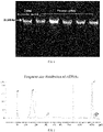
- the first lane from the left in FIG. 1 is the DNA molecular marker, i.e., the ⁇ DNA product (Takara) enzyme digested by Hind III.
- the size of the top band of the DNA molecular marker is 23,130 bp.
- the second to sixth lanes from the left are gDNAs extracted from different placental tissues. No obvious diffusion was observed from distribution of electrophoretic bands as shown in FIG. 1 , indicating that its integrity was good.
- Peripheral blood stored in blood collection tubes such as Hä MiniMax TM cfDNA non-invasive blood collection tubes (Apostle, Inc. Silicon Valley, California.) and Cell-Free DNA BCT CE blood collection tubes (Streck, Inc., La Vista, NE) (collected from Guangdong Maternal and Child Health Hospital, Clinical Research Approval No.: YL No. [201701044]);
- Sample preparation The cfDNA blood collection tubes containing peripheral blood were centrifuged in a centrifuge to stratify the peripheral blood and separate the plasma layer.
- Plasma lysis A lysis reaction solution (containing 0.4 mg/ml of protease K, plasma, and 1% SDS) was prepared, vortexed, centrifuged, and incubated at 60°C for 20 min. After incubation, the lysis reaction solution was cooled at room temperature.
- Elution of cfDNA After dried, the beads were eluted with an EB buffer and transferred into a new centrifuge tube for storage.
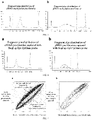
- FIG. 2 shows the fragment size distribution of cfDNAs as determined by an Agilent 2100 Bioanalyzer.
- the peaks at 35 bp and 10,380 bp are DNA molecular markers.
- the main peak of cfDNA is at approximate 161 bp.
- the samples from which cfDNAs were extracted were free of gDNA contamination, and the main peak of their fragment size distribution is at 161 bp, which is basically consistent with the theoretical value.
- Tequila V3 DNA Methylation Library Kit was used to prepare an end-repair reaction mix 1 in accordance with Table 1.
- the corresponding PCR program was as follows: 37°C 30 min, 95°C 10 min, heat dome 105°C, volume 20 ⁇ L.
- Table 1 Addition Volumes of Reagents in Reaction Mix for Repair Reaction System Volume of Single Sample ⁇ L cfDNA or gDNA 17 TQRB1 buffer 2 TQRE1 enzyme 1 Total volume 20
- Ligation reaction 1 Tequila V3 DNA Methylation Library Kit was used to prepare a reaction mix 2 on ice in accordance with Table 2. The corresponding PCR program was as follows: 37°C 30 min, 95°C 5 min, heat insulation at 10°C, heat dome 105°C, volume 30 ⁇ L. Table 2 Addition Volumes of Reagents for Ligation Reaction 1 Reaction System Volume of Single Sample ⁇ L Previous reaction 20 NF-H 2 O 1.5 TQLB1 buffer 4 TQLR1 reagent 2 TQLR2 reagent 1.5 TQLE1 enzyme 1 Total volume 30
- Bead-based purification 1 Tequila V3 DNA Methylation Library Kit, 138 ⁇ L of TQSB reagent, and 27 ⁇ L of XP beads were mixed well, then added with a PCR product obtained by linear amplification, incubated at room temperature, and placed on a magnetic stand for adsorption until the liquid was clear. Afterwards, the supernatant was removed, and a mixture of 180 ⁇ L of TQSB reagent and 100 ⁇ L of DEPC water to re-suspend the beads. After incubation at room temperature, absorption was performed over the magnetic stand until the liquid was clear. Afterwards, the supernatant was removed, and the beads were washed with 80% ethanol, followed by adding an eluent.
- DNA denaturation The PCR program was as follows: 95°C 10 min, heat dome 105°C, volume 10 ⁇ L. After the program operated for 5 min, the 0.2-mL PCR tube containing the sample was removed immediately from the PCR amplifier, inserted immediately on ice, and left for 2 min.
- Ligation reaction 2 Tequila V3 DNA Methylation Library Kit was used to prepare a reaction mix 4 on ice in accordance with Table 4. The PCR program was as follows: 25°C 15 min, heat dome 35°C, volume 20 ⁇ L. Table 4 Addition Volumes of Reagents for Ligation Reaction 2 Reaction System Volume of Single Sample ⁇ L Product of previous step 6.6 TQTHB 1 buffer 10 TQTHR1 reagent 2.4 TQHE1 enzyme 1 Total volume 20
- Bead-based purification 2 To the reaction product of the previous step, 17 ⁇ L of XP beads was added, mixed thoroughly with vortex, left to stand still at room temperature, and incubated for 5 min. After incubation, the mixture was adsorbed over a magnetic stand until the liquid was clear, and the supernatant was discarded. After washed twice with 80% ethanol, the beads were dried at room temperature and eluted with DEPC water.
- PCR amplification The PCR amplification system was as listed in Table 5. The corresponding reaction program was as follows: 98°C 45 s, 12X[98°C 15 s, 60°C 30 s, 72°C 30 s], 72°C 5 min, thermal insulation at 10°C, heat dome 105°C, volume 100 ⁇ L. Table 5 PCR Amplification System Reaction System Volume of Single Sample ⁇ L Eluent in previous reaction 73 TQAB1 reagent 20 TQAR1 reagent 2 TQIE2 enzyme 1 Index T5XX 2 (added separately) Index Q7XX 2 (added separately) Total volume 100
- Bead-based purification 3 To the PCR product of the previous step, 17 ⁇ L of XP beads was added, mixed thoroughly with vortex, left to stand still at room temperature, and incubated for 5 min, and then placed on a magnetic stand for adsorption until the liquid was clear. After the liquid waste was aspirated and discarded, the beads were washed twice with 80% ethanol. After the liquid waste was aspirated and discarded, the beads were dried at room temperature and then eluted with DPEC water to obtain a pre-library.
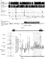
- the experimental results were as shown in FIG. 3 , showing the fragment size distribution of cfDNAs as determined by the Agilent 2100 Bioanalyzer.
- the peaks at 35 bp and 10,380 bp are DNA molecular markers.
- Example 4 Enrichment of whole-genome gDNA/cfDNA methylated loci using SeqCap EpiGiant probe
- Evaporation In accordance with Table 6, an evaporation system was prepared in a 1.5-mL EP tube using a hybrid elution kit of non-invasive preeclampsia early screening kit, and the EP tube was placed in a vacuum concentrator and evaporated at 60°C with the cap open.
- the library samples were constructed by the method described in Example 3.
- Table 6 Evaporation System Reaction System Volume of Single Component ⁇ L Library sample 500 ng TQBLKC reagent 2 TQBLKD reagent 4 TQBLKCHE reagent 2.5
- Hybridization reaction In accordance with Table 7, a hybridization system was prepared in a 200- ⁇ L PCR tube using a hybrid elution kit of non-invasive preeclampsia early screening kit, left to stand for 10 min after mixing well with vortex, and thereafter denatured in a PCR amplifier at 95°C for 10 min. At 8 min of denaturation, the PCR tube containing 4.5 ⁇ L of probe was placed in the PCR amplifier at 95°C to preheat the probe. After denaturation, the probe was charged into the hybridization system mixture at 47°C. After mixing with vortex, the PCR tube was placed in the PCR amplifier at 47°C (heat dome temperature: 57°C) and hybridized for 72 h. Table 7 Hybridization System Reaction System Volume of Single Sample ⁇ L Evaporated sample from previous step / TQHS1 7 TQHS2 3.5
- the liquid waste was aspirated and discarded and 180 ⁇ L of 1X TQWS3 was added and mixed well by vibration at room temperature for 1 min before absorbing over a magnetic stand until the liquid was clear.
- the liquid waste was aspirated and discarded and 23 ⁇ L of TQNF reagent was added and mixed well by vibration.
- Amplification of capture product In accordance with Table 8, an amplification system of the capture product was prepared using a hybrid elution kit of non-invasive preeclampsia early screening kit. The corresponding PCR program was as follows: 98°C 45s, 8X[98°C 15 s; 60°C 30 s; 72°C 30 s] 72°C 2 min, thermal insulation at 16°C, heat dome 105°C, volume 50 ⁇ L. Table 8 Amplification System of Capture product Reaction System Volume of Single Sample ⁇ L TQAE2 enzyme 25 TQPR3 reagent 2 Capture product (containing beads) 23 Total volume 50
- the hybrid elution kit of non-invasive preeclampsia early screening kit was used, and the above reaction product was placed on a magnetic stand for absorption until the liquid was clear and afterwards, the supernatant was was drawn and mixed with 90 ⁇ L of XP beads by vortex. After left to stand at room temperature for 5 min, the mixture was absorbed over a magnetic stand until the liquid was clear. The liquid waste was aspirated and discarded. The beads were washed twice with 80% ethanol, and air dried at room temperature with the cap open. After drying, the beads were added with 25 ⁇ L of TQEB reagent, and incubated at room temperature for 5 min before absorbing over a magnetic stand until the liquid was clear. The clear liquid was drawn into a new EP tube, thereby obtaining a purified capture product.
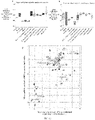
- FIG. 4 shows the fragment size of the libraries captured by the SeqCap EpiGiant probe, as determined by Agilent 2100 Bioanalyzer.
- the size of the above fragment is about 300 bp.
- FIG. 4a shows the library constructed based on placenta gDNAs, which has a fragment size of about 300 bp.
- FIG. 4b shows the library constructed based on cfDNAs in the plasma, which has a fragment size of about 300 bp.
- Example 5 Quality test on data produced by Illumina second-generation high-throughput sequencing platform for methylation libraries
- the KS test on their fragment length distribution and that of the standard cfDNAs shows that P>0.5
- the KS test on their fragment end base distribution and that of the standard cfDNAs show that P>0.5
- CHG>THG and CHH>THH were sampled and found that their conversion rates were ⁇ 99%.
- Example 1 The procedures for extraction of placenta gDNA, capture by SeqCap EpiGiant probes, and high-throughput sequencing were the same as those described in Example 1, Example 4, and Example 5.
- the coverage and methylation level at each CpG locus were extracted using PileOMeth (version 0.1.13). Conditions for quality control: The coverage at a single CpG locus was required to be ⁇ 200X. Each of the methylated loci was analyzed using the R language (3.5.2) to screen out the preeclampsia-specific methylation modification loci.
- Each of the CpG methylation modification loci was subjected to statistical detection by the t test, and required to meet the following conditions: the distribution of its methylation modifications in preeclamptic placental samples was significantly different from that of methylation modifications in control placental samples, i.e., p ⁇ 0.001; at the same time, the distribution of its methylation modifications in preeclamptic placental samples was significantly different from that of methylation modifications in plasma samples from healthy non-pregnant women, i.e., p ⁇ 0.001.
- the methylation levels at the CpG loci, obtained from the above analysis, were significantly and consistently different among different groups of samples, i.e., groups of preeclamptic placentas, control gestational placentas, and plasma from non-pregnant women.
- the CpG loci obtained from the above analysis were methylation modification loci specific to preeclamptic placentas (for the details, see Attached List 1).
- Tissue homogenization 20 mg of frozen tissues were weighed and put in a pre-cooled glass homogenizer, to which 1 ml of pre-cooled 1x HB buffer (as listed in Table 10, in which the 1x HB buffer was further prepared based on the 1.022xHB buffer listed in Table 9) was added, followed by grinding the tissues.
- the ground tissue homogenates were filtered through a 70- ⁇ m cell strainer and transferred into a low-adsorption tube, and centrifuged at 4°C for 5 min. The supernatant was discarded. The tissue homogenates were re-suspended by adding a fresh HB buffer.
- Iodixanol gradient centrifugation An equal volume of 50% iodixanol was added to the re-suspension to prepare 30% and 40% iodixanol, and construct a density gradient centrifugation system for nuclei enriched on 25%/30%/40% iodixanol, which was centrifuged at 3000 rcf at 4°C for 20 min. After centrifugation, the nuclear layer at the interface of 30% and 40% iodixanol was aspirated. The RSB buffer (Table 11) was added to dilute the nuclei.
- Tn5 transposase digestion-based library construction and purification After centrifugation at 500 rcf at 4°C for 10 min, the Tn5 transposase digestion was carried out according to the instructions of the VazymeTD501-02 TruePrepDNALibraryPrepKit.
- the enzyme-digested product was purified according to the Zymo D4061 Oligo Clean & Concentrator kit.
- the purified product was subjected to Index PCR according to TruePrep Index Kit V2 for Illumina.
- the PCR product was subjected to library purification with XP beads.
- the coverage was extracted with deeptools (version 3.3.0).
- R Software version 3.5.2 was used to analyze the coverage values of transcription factors and TSS region.
- the peak of the accessible chromatin enrichment region was analyzed using macs2 Software (version 2.2.4) and Genrich Software (version 0.6).
- diffBind and NarrowPeaks were used to analyze their differential regions and perform enrichment analysis (statistically analyzing the data using the R language).
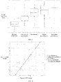
- the x-axis represents the mean methylation level of the placenta-specific differentially methylated regions in a placenta in normal pregnancy
- the y-axis represents the mean methylation levels of the same region in preeclamptic placentas.
- the distribution of black dots deviated obviously from the straight line y x, indicating that there were differentially methylated regions with significantly statistical differences in the preeclamptic placentas and in the placentas in normal pregnancy.
- FIG. 5b shows the density distribution curves of the non-differential regions (light gray) and the methylated regions with significantly statistical differences (dark gray) as shown in FIG. 5a .
- PE-DMRs differentially methylated regions
- PE-DMRs when aligned to the whole genome, were found to be concentrated in the gene regulatory regions, such as non-coding transcription regions (5'UTR/3'UTR), intergenic regions, introns, and some of exon regions.
- a further statistical analysis of enrichment showed that PE-hypo-DMRs were significantly enriched in introns, 5'UTR/3'UTR, and intron regions of non-coding RNAs; while PE-hyper-DMRs were mostly distributed in intergenic regions and intron regions, their distribution patterns were random.
- the alignment of the PE-hyper-DMRs with the transposable elements (TEs) in human genome revealed that the PE-hyper-DMRs were significantly enriched in LTR12 family transposons.
- about 30% of LTR12C transposons contained PE-hyper-DMRs.
- FIG. 8a shows the DNA methylation patterns in Pan troglodytes and Homo sapiens spermatoblasts.
- FIG. 8b shows the comparison of chromatin accessibility to the placenta in control pregnancy and the preeclamptic placenta.
- FIG. 8c shows the methylation patterns of the region in the placenta in control pregnancy and the preeclamptic placenta.
- hypo-methylated LTR12C specific to Homo sapiens spermatoblasts was in a hyper-methylation status in preeclamptic placentas ( FIG. 8c ).
- the DNA methylation status of the hypo-methylated LTR12C specific to Homo sapiens spermatoblasts was altered in preeclamptic placentas, accompanied by the change in chromatin accessibility ( FIG. 8b ).
- Example 7 Screening of de novo methylation regions specific to placental lineage
- methylation levels at CpG loci The coverage and methylation level were extracted from each of CpG loci using PileOMeth (version 0.1.13). Conditions for quality control: the coverage at a single CpG locus was required to be ⁇ 200X. Each of the methylated loci was analyzed using the R language (3.5.2) to screen out the preeclampsia-specific hyper-methylation modification locus, which was in a hypomethylation status in leukocyte of healthy non-pregnant women.
- Each of the CpG methylation modification loci was subjected to statistical detection by the t test, and required to meet the following conditions: the distribution of its methylation modifications in the placental sample in control pregnancy was significantly different from that of methylation modifications in the sample from leukocyte of healthy non-pregnant women, i.e., p ⁇ 0.001.
- the regions where differentially modified methylation loci screened were located were combined, and the methylation levels were averaged across the whole region, thereby screening out a HUVEC-specific hyper-methylated region.
- the intersection of the placenta-specific hyper-methylated region and HUVEC-specific hyper-methylated region was the de novo methylation region specific to placental lineage.
- Example 8 Enrichment of preeclampsia-specific hypo-methylated regions in de novo methylation region of placental lineage
- Placental de novo methylation regions and PE-DMRs were intersected to sort out the preeclampsia-specific differentially methylated regions that executed the placental de novo methylation, i.e., ExE-DN PE-DMRs, and the preeclampsia-specific differentially methylated regions that did not execute the placental de novo methylation, i.e., Not-ExE-DN PE-DMRs.
- FIG. 9 shows the mean regional methylation levels of the preeclampsia-specific differentially methylated regions that executed the placental de novo methylation, ExE-DN PE-DMRs (dark gray) and the preeclampsia-specific differentially methylated regions that did not execute the placental de novo methylation, Not-ExE-DN PE-DMRs (light gray).
- RNA expression data were extracted using Monocle (version 0.2.0). The data were subjected to dimensionality reduction by the principal component analysis method (reduce_dimension function, Monocle) and the UMAP method. For each cell, the mean expression levels of its homologous genes (genes within the range of 50 kbp base pairs near the PE-hypo-DMR or PE-hyper-DMR position) were calculated as the expression levels of PE-hypo-DMR genes and PE-hyper- DMR genes.
- VCTs placental villous cytotrophoblasts
- SCTs syncytiotrophoblasts
- EVTs extravillous trophoblasts
- Example 9 Distinguishing germ cells or embryonic tissues at different developmental stages by the mean methylation level of preeclampsia-specific methylated regions
- PE-hyper-DMRs and PE-hypo-DMRs could be obtained through the experiments and analyses described in the above-mentioned examples.
- Statistical analysis of the mean methylation levels of the above-mentioned regions in the methylation databases of human ovum, sperm, 8-cell cleavage stage, morula, inner cell mass at the blastocyst stage, and fetuses at 6 gestational weeks showed that the early embryonic development process following the gametogenesis involved a process of from extensive whole-genome demethylation (represented by 8-cell cleavage stage, morula, blastocyst) to de novo methylation (represented by normal placenta and embryo).
- epreeclamptic placentas could be well distinguished from early embryos/ fetuses based on the mean methylation levels of the preeclampsia-specific hyper-methylated regions and hypo-methylated regions.
- FIGS. 15a and 15b show the mean methylation levels of the preeclampsia-specific hyper-methylated and hypo-methylated regions in embryos (or tissues) at different developmental stages and placentas with different pregnancy complications, respectively.
- FIG. 15c shows a scatter plot drawn for germ cells or embryonic tissues at different developmental stages with the mean methylation level of the preeclampsia-specific hyper-methylated regions as the x-axis and the mean methylation level of the preeclampsia-specific hypo-methylated regions as y-axis.
- the preeclamptic placentas, placentas without the placenta-derived gestational disorder, sperms, ova, 8-cell stage, morula, and blastocyst could be well distinguished.
- PE-hyper-DMRs and PE-hypo-DMRs were specific to preeclamptic placentas.
- Example 10 Distinguishing placentas with different pregnancy complications by the mean methylation level of preeclampsia-specific methylated regions
- PE-hyper-DMRs and PE-hypo-DMRs could be obtained through the experiments and analyses described in the above-mentioned examples.
- Statistical analysis of the mean methylation levels of the above regions in the placentas with gestational hypertension, gestational diabetes, and twin-to-twin transfusion syndrome, placentas without the placenta-derived gestational disorder, and preeclamptic gestational placentas showed that the mean methylation levels of the preeclampsia-specific hyper-methylated regions and hypo-methylated regions could be used to distinguish different pregnancy complications ( FIG. 15c ). Therefore, there were specific methylation modification differences in the PE-hyper-DMRs and PE-hypo-DMRs in preeclamptic placentas.
- Example 11 Differences in methylation haplotypes in preeclampsia group and control pregnancy group
- Methylation haplotypes were extracted within the preeclampsia-specific differentially methylated regions obtained from the above analysis. Each of the haplotypes was analyzed using the R language (3.5.2) to screen out the preeclampsia-specific methylation haplotype, which was required to satisfy the condition that the frequency of this haplotype occurring in the preeclamptic placenta was at least 20 times that in the control placenta, or that the frequency of this haplotype occurring in the control placenta was at least 20 times that in the preeclamptic placenta.
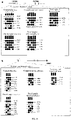
- FIGS. 16a and 16b show the methylation haplotypes in the preeclampsia-specific hyper-methylated region and hypo-methylated region, respectively.
- Example 12 Design and verification of EP-007 probe or probe set
- the EP-007 probe or probe set was designed based on the following basic principle:
- the probe was composed of several sequences that were 59 bp in length and complementary to the sequences of the methylation haplotype-specific loci after sulfite sequencing.
- the two probes covering adjacent loci contained 40-bp overlapping sequences.
- the probes had a biotin label at the 3'-end.
- Probe design was conducted on the genomic region where the preeclampsia-specific methylation haplotypes (Attached List 3) obtained from the above analysis were located to synthesize a DNA probe complementary thereto (Twist Inc., U.S.A.). As shown in FIG. 17a , a 59-bp DNA complementary strand was basically designed.
- the sequence of the target region desired to be detected was: >13 :29140769-29140827 (SEQ ID NO: 1).
- the synthesized DNA probes were subjected to double-stranded library construction and sequencing according to the operation instructions of the KAPA double-stranded DNA library construction kit. As listed in Table 12, the proportion of data less than 1X coverage was almost 0 and the normalized mean coverage of 0.2 or less was almost 0, so the probe could cover the selected region well.
- FIG. 17a shows the sequencing results after double-stranded library construction for the EP-007 probe or probe set.
- the x-axis represents the ratios of the coverage of different probe fragments to the mean coverage, and the y-axis represents the distribution density.
- the coverage distribution is in line with the Poisson distribution. Therefore, the synthetic quality of the EP-007 probe or probe set is unbiased and reliable.
- Example 13 Optimization of hybridization time and hybridization and eluting temperatures of EP-007 probe or probe set
- FIGS. 18a and 18b show the comparisons of the on target rates and the mean coverage in the sequencing results obtained at different combinations of the hybridization temperatures (47°C, 56°C, and 65°C) and the eluting temperatures (47°C, 56°C, and 65°C), respectively.
- the circle represents hybridization at 47°C
- the square represents hybridization at 56°C
- the triangle represents hybridization at 65°C.
- the capture effect was optimal in case of hybridization at 56°C and elution at 65°C. That is, the on target rate was greater than 30% (as shown in FIG. 18a ) and the mean coverage was the highest in case of the same data size (as shown in FIG. 18b ).
- FIG. 19 shows the on target rates of the library products when hybridized at the eluting temperature of 56°C for different hybridization time (4 h, 8 h, 12 h, 24 h, 36 h, 48 h, and 60 h). It was found that the on target rate reached a sequencing result of greater than 30% when the hybridization time was 12 h.
- Example 14 Capture of preeclamptic methylation modification difference loci in gDNA or cfDNA using EP-007 probe or probe set
- FIG. 20 shows the fragment size distribution of the library after probe capture as determined by Agilent 2100 Bioanalyzer. The peaks at 35 bp and 10,380 bp are DNA molecular markers.
- FIG. 20a shows the fragment size of the library constructed based on placenta gDNA
- FIG. 15b shows the fragment size of the library constructed based on cfDNA in plasma.
- the plasma from 22 pregnant women was subjected to experiments in molecular biology and analysis of the DNA methylation level.
- the experimental methods were the same as those described in Examples 2, 3, 5, 9, and 12. There were 6 pregnant women and 16 preeclamptic pregnant women.
- the methylation levels of each of the preeclampsia-specific methylation haplotype regions in Attached List 3 were averaged within the groups to obtain matrices M (CTRL) and M (PE), and calculate the Pearson correlation coefficients r (CTRL) and r (PE) of the control pregnancy group and the preeclampsia group by the leave-one-out method.
- a linear regression analysis between r(CTRL) and r(PE) and the clinical diagnosis of 22 pregnant women was made to obtain linear scores, which could be used to evaluate the degree of preeclampsia-associated placental dysplasia, that is, the degree of placental dysplasia in pregnant women afflicted with preeclampsia was high, and its linear score was high accordingly; however, the placental development of the control pregnancy was relatively good, so its linear score was low.
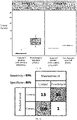
- FIG. 16 shows linear scores statistically calculated by correlation and linear regression after analyais of the methylation levels of PE-DMRs.
- Example 16 Verification of preeclampsia risk prediction model by retrospective clinical samples
- the experimental materials were the same as those used in Example 13, in which the peripheral blood samples drawn from a total of 159 pregnant women with preeclampsia or control pregnancy at 10 to 38 gestational weeks (Guangdong Maternal and Child Health Hospital, YL No. [201701044]).
- the mean methylation level of the subject in each region was calculated to obtain the matrix M (test), and the Pearson correlation coefficients between M (test) and M (CTRL) and M (PE), i.e., r (CTRL) and r (PE), were calculated, respectively. Further, r (CTRL) was compared with r (PE) to predict the risk of preeclampsia. If r (CTRL) > r (PE), the risk of preeclampsia was high; if r (CTRL) ⁇ r (PE), the risk of preeclampsia was low.
- this method may differentiate pregnant women with preeclampsia from pregnant women of control pregnancy, with the sensitivity of 97% (36/37) and the specificity of 89% (109/122).
- Example 17 Distinguishing different severities of subtypes of preeclampsia by the preeclampsia risk prediction model
- the experimental materials were the same as those used in Example 13, in which the peripheral blood samples drawn from a total of 159 pregnant women with preeclampsia or control pregnancy at 10 to 38 gestational weeks (Guangdong Maternal and Child Health Hospital, YL No. [201701044]).
- Example 18 Prediction of the highest pressure in the first stage of labor of pregnant women by the preeclampsia risk prediction model
- Example 13 The experimental materials were the same as those used in Example 13, in which the peripheral blood samples drawn from a total of 181 pregnant women with preeclampsia or control pregnancy at 10 to 38 gestational weeks (collected from Guangdong Maternal and Child Health Hospital, Clinical Research Approval No.: YL No. [201701044]).
- the regression statistical analysis was performed on r (CTRL) and r (PE) of 22 pregnant women and the highest pressure in the first stage of labor in the corresponding clinical outcomes, to establish a model between the similarity of the subject placenta to the control pregnancy and preeclamptic placenta and the highest systolic blood pressure in the first stage of labor of pregnant women. Then, the model was used to predict the highest systolic blood pressure in the first stage of labor of 159 pregnant women. As shown in FIG. 24 , the highest systolic blood pressure in the first stage of labor predicted by the model was in a good linear relationship with the highest systolic blood pressure measured in clinical.
Landscapes
- Health & Medical Sciences (AREA)
- Chemical & Material Sciences (AREA)
- Engineering & Computer Science (AREA)
- Life Sciences & Earth Sciences (AREA)
- Proteomics, Peptides & Aminoacids (AREA)
- Organic Chemistry (AREA)
- Genetics & Genomics (AREA)
- Medical Informatics (AREA)
- Analytical Chemistry (AREA)
- General Health & Medical Sciences (AREA)
- Zoology (AREA)
- Public Health (AREA)
- Pathology (AREA)
- Wood Science & Technology (AREA)
- Biomedical Technology (AREA)
- Bioinformatics & Cheminformatics (AREA)
- Physics & Mathematics (AREA)
- Biophysics (AREA)
- Molecular Biology (AREA)
- Biotechnology (AREA)
- Epidemiology (AREA)
- General Engineering & Computer Science (AREA)
- Data Mining & Analysis (AREA)
- Databases & Information Systems (AREA)
- Biochemistry (AREA)
- Microbiology (AREA)
- Immunology (AREA)
- Primary Health Care (AREA)
- Bioinformatics & Computational Biology (AREA)
- Evolutionary Biology (AREA)
- Spectroscopy & Molecular Physics (AREA)
- Theoretical Computer Science (AREA)
- Measuring Or Testing Involving Enzymes Or Micro-Organisms (AREA)
Applications Claiming Priority (2)
| Application Number | Priority Date | Filing Date | Title |
|---|---|---|---|
| CN202010084924.XA CN113249458B (zh) | 2020-02-10 | 2020-02-10 | 一种预测胎盘源性疾病的评估和预测的方法和试剂盒 |
| PCT/CN2020/120654 WO2021159722A1 (fr) | 2020-02-10 | 2020-10-13 | Procédé d'évaluation et de prédiction de maladies dérivées du placenta et kit associé |
Publications (2)
| Publication Number | Publication Date |
|---|---|
| EP4108779A1 true EP4108779A1 (fr) | 2022-12-28 |
| EP4108779A4 EP4108779A4 (fr) | 2024-02-07 |
Family
ID=77219837
Family Applications (1)
| Application Number | Title | Priority Date | Filing Date |
|---|---|---|---|
| EP20918272.4A Withdrawn EP4108779A4 (fr) | 2020-02-10 | 2020-10-13 | Procédé d'évaluation et de prédiction de maladies dérivées du placenta et kit associé |
Country Status (4)
| Country | Link |
|---|---|
| US (1) | US20230160011A1 (fr) |
| EP (1) | EP4108779A4 (fr) |
| CN (1) | CN113249458B (fr) |
| WO (1) | WO2021159722A1 (fr) |
Families Citing this family (6)
| Publication number | Priority date | Publication date | Assignee | Title |
|---|---|---|---|---|
| CA2938451C (fr) * | 2014-01-30 | 2023-10-17 | The Regents Of The University Of California | Haplotypage de methylation pour le diagnostic non invasif (monod) |
| CN114822682B (zh) * | 2022-04-12 | 2023-07-21 | 苏州市立医院 | 与早发型重度子痫前期发生相关的基因组合及其应用 |
| CN114774544A (zh) * | 2022-04-14 | 2022-07-22 | 武汉大学中南医院 | 膀胱癌检测的dna甲基化多重pcr试剂盒及其应用 |
| WO2025024536A1 (fr) * | 2023-07-24 | 2025-01-30 | University Of South Florida | Signatures de méthylation d'adn de leucocytes de sang maternel et de placenta prédisant un retard de croissance intra-utérin (fgr) |
| CN117604073A (zh) * | 2023-10-30 | 2024-02-27 | 上海微荷医学检验实验室有限公司 | 一种甲基化信号的探针及其应用 |
| CN117187381B (zh) * | 2023-11-03 | 2024-02-13 | 南京医科大学 | 一种用于妊娠期糖尿病早期辅助诊断的甲基化区域标志物组合及其应用 |
Family Cites Families (5)
| Publication number | Priority date | Publication date | Assignee | Title |
|---|---|---|---|---|
| WO2015136509A2 (fr) * | 2014-03-14 | 2015-09-17 | Genesis Theranostix Korlatolt Felelossegu Tarsasag | Cibles diagnostiques et thérapeutiques de la prééclampsie et d'autres complications étroitement associées à la grossesse |
| US10801067B2 (en) * | 2014-05-09 | 2020-10-13 | Eurofins Lifecodexx Gmbh | Detection of DNA that originates from a specific cell-type and related methods |
| HUE050491T2 (hu) * | 2015-11-10 | 2020-12-28 | Eurofins Lifecodexx Gmbh | Magzati kromoszomális aneuploidiák kimutatása olyan DNS régiókat alkalmazva, amelyek különbözõképpen vannak metilezve a magzat és a terhes nõstény között |
| CN106755492B (zh) * | 2017-01-24 | 2020-04-21 | 深圳金蕊科技有限公司 | 用于预测子痫前期的成套snp及其应用 |
| CN110004223A (zh) * | 2019-05-09 | 2019-07-12 | 苏州大学附属第一医院 | 一种用于预测早发型子痫前期发病的检测试剂盒 |
-
2020
- 2020-02-10 CN CN202010084924.XA patent/CN113249458B/zh active Active
- 2020-10-13 EP EP20918272.4A patent/EP4108779A4/fr not_active Withdrawn
- 2020-10-13 WO PCT/CN2020/120654 patent/WO2021159722A1/fr not_active Ceased
- 2020-10-13 US US17/798,427 patent/US20230160011A1/en not_active Abandoned
Also Published As
| Publication number | Publication date |
|---|---|
| CN113249458A (zh) | 2021-08-13 |
| US20230160011A1 (en) | 2023-05-25 |
| WO2021159722A1 (fr) | 2021-08-19 |
| EP4108779A4 (fr) | 2024-02-07 |
| CN113249458B (zh) | 2023-07-07 |
Similar Documents
| Publication | Publication Date | Title |
|---|---|---|
| US20200208212A1 (en) | Processes and Compositions for Methylation-Based Enrichment of Fetal Nucleic Acid From a Maternal Sample Useful for Non-Invasive Prenatal Diagnoses | |
| US20200362414A1 (en) | Processes and Compositions for Methylation-Based Enrichment of Fetal Nucleic Acid From a Maternal Sample Useful for Non-Invasive Prenatal Diagnoses | |
| US20230160011A1 (en) | Method for evaluating and predicting placenta-derived diseases and kit | |
| EP3103871B1 (fr) | Procédés pour l'enrichissiment en acide nucleic fétal d'un échantillon maternel, basé sur la méthylation et utilisation pour la quantification de l'acide nucleique fétal | |
| EP3099818B1 (fr) | Évaluation préimplantatoire d'embryons par détection de l'adn embryonnaire libre | |
| JP2018533953A (ja) | 胎児と妊娠女性との間において差次的にメチル化されるdna領域を用いる胎児染色体異数性の検出 | |
| AU2017251674B2 (en) | Processes and compositions for methylation-based enrichment of fetal nucleic acid from a maternal sample useful for non invasive prenatal diagnoses | |
| HK40043946A (en) | Processes and compositions for methylation-based enrichment of fetal nucleic acid from a maternal sample useful for non-invasive prenatal diagnoses | |
| HK40036431A (en) | Processes and compositions for methylation-based enrichment of fetal nucleic acid from a maternal sample useful for non-invasive prenatal diagnoses | |
| Liu et al. | Comparison of Genome-Wide DNA Methylation Profiles of Fetal Tissues Conceived by In Vitro Fertilization and Natural Conception | |
| HK1254596B (en) | Processes for methylation-based enrichment of fetal nucleic acid from a maternal sample useful for non-invasive prenatal diagnoses | |
| HK1229846A1 (en) | Processes for methylation-based enrichment of fetal nucleic acid from a maternal sample useful for fetal nucleic acid quantification | |
| HK1229846B (en) | Processes for methylation-based enrichment of fetal nucleic acid from a maternal sample useful for fetal nucleic acid quantification | |
| HK1158256B (en) | Processes and compositions for methylation-based enrichment of fetal nucleic acid from a maternal sample useful for non invasive prenatal diagnoses |
Legal Events
| Date | Code | Title | Description |
|---|---|---|---|
| STAA | Information on the status of an ep patent application or granted ep patent |
Free format text: STATUS: THE INTERNATIONAL PUBLICATION HAS BEEN MADE |
|
| PUAI | Public reference made under article 153(3) epc to a published international application that has entered the european phase |
Free format text: ORIGINAL CODE: 0009012 |
|
| STAA | Information on the status of an ep patent application or granted ep patent |
Free format text: STATUS: REQUEST FOR EXAMINATION WAS MADE |
|
| 17P | Request for examination filed |
Effective date: 20220912 |
|
| AK | Designated contracting states |
Kind code of ref document: A1 Designated state(s): AL AT BE BG CH CY CZ DE DK EE ES FI FR GB GR HR HU IE IS IT LI LT LU LV MC MK MT NL NO PL PT RO RS SE SI SK SM TR |
|
| DAV | Request for validation of the european patent (deleted) | ||
| DAX | Request for extension of the european patent (deleted) | ||
| RIC1 | Information provided on ipc code assigned before grant |
Ipc: C12Q 1/68 20180101AFI20230928BHEP |
|
| A4 | Supplementary search report drawn up and despatched |
Effective date: 20240110 |
|
| RIC1 | Information provided on ipc code assigned before grant |
Ipc: C12Q 1/68 20180101AFI20240104BHEP |
|
| STAA | Information on the status of an ep patent application or granted ep patent |
Free format text: STATUS: THE APPLICATION IS DEEMED TO BE WITHDRAWN |
|
| 18D | Application deemed to be withdrawn |
Effective date: 20240731 |