EP4111194B1 - Verfahren zur extraktion von zielzellen aus 3d-gewebe durch optische identifizierung - Google Patents
Verfahren zur extraktion von zielzellen aus 3d-gewebe durch optische identifizierung Download PDFInfo
- Publication number
- EP4111194B1 EP4111194B1 EP21708609.9A EP21708609A EP4111194B1 EP 4111194 B1 EP4111194 B1 EP 4111194B1 EP 21708609 A EP21708609 A EP 21708609A EP 4111194 B1 EP4111194 B1 EP 4111194B1
- Authority
- EP
- European Patent Office
- Prior art keywords
- target cells
- tissue
- cells
- imaging
- extraction
- Prior art date
- Legal status (The legal status is an assumption and is not a legal conclusion. Google has not performed a legal analysis and makes no representation as to the accuracy of the status listed.)
- Active
Links
Images






Classifications
-
- G—PHYSICS
- G01—MEASURING; TESTING
- G01N—INVESTIGATING OR ANALYSING MATERIALS BY DETERMINING THEIR CHEMICAL OR PHYSICAL PROPERTIES
- G01N33/00—Investigating or analysing materials by specific methods not covered by groups G01N1/00 - G01N31/00
- G01N33/48—Biological material, e.g. blood, urine; Haemocytometers
- G01N33/50—Chemical analysis of biological material, e.g. blood, urine; Testing involving biospecific ligand binding methods; Immunological testing
- G01N33/5005—Chemical analysis of biological material, e.g. blood, urine; Testing involving biospecific ligand binding methods; Immunological testing involving human or animal cells
-
- G—PHYSICS
- G01—MEASURING; TESTING
- G01N—INVESTIGATING OR ANALYSING MATERIALS BY DETERMINING THEIR CHEMICAL OR PHYSICAL PROPERTIES
- G01N1/00—Sampling; Preparing specimens for investigation
- G01N1/02—Devices for withdrawing samples
- G01N1/04—Devices for withdrawing samples in the solid state, e.g. by cutting
- G01N1/08—Devices for withdrawing samples in the solid state, e.g. by cutting involving an extracting tool, e.g. core bit
-
- C—CHEMISTRY; METALLURGY
- C12—BIOCHEMISTRY; BEER; SPIRITS; WINE; VINEGAR; MICROBIOLOGY; ENZYMOLOGY; MUTATION OR GENETIC ENGINEERING
- C12Q—MEASURING OR TESTING PROCESSES INVOLVING ENZYMES, NUCLEIC ACIDS OR MICROORGANISMS; COMPOSITIONS OR TEST PAPERS THEREFOR; PROCESSES OF PREPARING SUCH COMPOSITIONS; CONDITION-RESPONSIVE CONTROL IN MICROBIOLOGICAL OR ENZYMOLOGICAL PROCESSES
- C12Q1/00—Measuring or testing processes involving enzymes, nucleic acids or microorganisms; Compositions therefor; Processes of preparing such compositions
- C12Q1/68—Measuring or testing processes involving enzymes, nucleic acids or microorganisms; Compositions therefor; Processes of preparing such compositions involving nucleic acids
- C12Q1/6876—Nucleic acid products used in the analysis of nucleic acids, e.g. primers or probes
- C12Q1/6883—Nucleic acid products used in the analysis of nucleic acids, e.g. primers or probes for diseases caused by alterations of genetic material
- C12Q1/6886—Nucleic acid products used in the analysis of nucleic acids, e.g. primers or probes for diseases caused by alterations of genetic material for cancer
-
- G—PHYSICS
- G01—MEASURING; TESTING
- G01N—INVESTIGATING OR ANALYSING MATERIALS BY DETERMINING THEIR CHEMICAL OR PHYSICAL PROPERTIES
- G01N1/00—Sampling; Preparing specimens for investigation
- G01N1/28—Preparing specimens for investigation including physical details of (bio-)chemical methods covered elsewhere, e.g. G01N33/50, C12Q
- G01N1/30—Staining; Impregnating ; Fixation; Dehydration; Multistep processes for preparing samples of tissue, cell or nucleic acid material and the like for analysis
-
- G—PHYSICS
- G01—MEASURING; TESTING
- G01N—INVESTIGATING OR ANALYSING MATERIALS BY DETERMINING THEIR CHEMICAL OR PHYSICAL PROPERTIES
- G01N15/00—Investigating characteristics of particles; Investigating permeability, pore-volume or surface-area of porous materials
- G01N15/10—Investigating individual particles
- G01N15/14—Optical investigation techniques, e.g. flow cytometry
- G01N15/1429—Signal processing
- G01N15/1433—Signal processing using image recognition
-
- C—CHEMISTRY; METALLURGY
- C12—BIOCHEMISTRY; BEER; SPIRITS; WINE; VINEGAR; MICROBIOLOGY; ENZYMOLOGY; MUTATION OR GENETIC ENGINEERING
- C12Q—MEASURING OR TESTING PROCESSES INVOLVING ENZYMES, NUCLEIC ACIDS OR MICROORGANISMS; COMPOSITIONS OR TEST PAPERS THEREFOR; PROCESSES OF PREPARING SUCH COMPOSITIONS; CONDITION-RESPONSIVE CONTROL IN MICROBIOLOGICAL OR ENZYMOLOGICAL PROCESSES
- C12Q1/00—Measuring or testing processes involving enzymes, nucleic acids or microorganisms; Compositions therefor; Processes of preparing such compositions
- C12Q1/68—Measuring or testing processes involving enzymes, nucleic acids or microorganisms; Compositions therefor; Processes of preparing such compositions involving nucleic acids
- C12Q1/6813—Hybridisation assays
- C12Q1/6841—In situ hybridisation
-
- C—CHEMISTRY; METALLURGY
- C12—BIOCHEMISTRY; BEER; SPIRITS; WINE; VINEGAR; MICROBIOLOGY; ENZYMOLOGY; MUTATION OR GENETIC ENGINEERING
- C12Q—MEASURING OR TESTING PROCESSES INVOLVING ENZYMES, NUCLEIC ACIDS OR MICROORGANISMS; COMPOSITIONS OR TEST PAPERS THEREFOR; PROCESSES OF PREPARING SUCH COMPOSITIONS; CONDITION-RESPONSIVE CONTROL IN MICROBIOLOGICAL OR ENZYMOLOGICAL PROCESSES
- C12Q2600/00—Oligonucleotides characterized by their use
- C12Q2600/156—Polymorphic or mutational markers
-
- G—PHYSICS
- G01—MEASURING; TESTING
- G01N—INVESTIGATING OR ANALYSING MATERIALS BY DETERMINING THEIR CHEMICAL OR PHYSICAL PROPERTIES
- G01N1/00—Sampling; Preparing specimens for investigation
- G01N1/02—Devices for withdrawing samples
- G01N1/04—Devices for withdrawing samples in the solid state, e.g. by cutting
- G01N1/06—Devices for withdrawing samples in the solid state, e.g. by cutting providing a thin slice, e.g. microtome
-
- G—PHYSICS
- G01—MEASURING; TESTING
- G01N—INVESTIGATING OR ANALYSING MATERIALS BY DETERMINING THEIR CHEMICAL OR PHYSICAL PROPERTIES
- G01N15/00—Investigating characteristics of particles; Investigating permeability, pore-volume or surface-area of porous materials
- G01N15/10—Investigating individual particles
- G01N15/14—Optical investigation techniques, e.g. flow cytometry
- G01N2015/1497—Particle shape
Definitions
- the present invention relates to a method for selecting cells or parts of cells or groups of cells with or without their extracellular matrix from a three dimensional specimen by optical identification of target cells and subsequent isolation of target cells.
- immune cell therapy Today one of the most promising approaches to cure cancer is immune cell therapy. The success of immune cell therapy is first and foremost visible in the field of circulating tumors like leukemia. The goal is to extend the current immune therapy approach to solid tumors.
- Biological tissue is a heterogenous assembly of cells. Some cells, parts of cells or groups of cells can most likely only be distinguished from other cells by their localization within a tissue (e.g. vessels lying next to a tumor, versus vessels next to unaffected tissue), combined properties within a group of cells exerted only as a group but not individually.
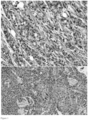
- cancerous tissue such as ductal adenocarcinoma displays a disorganized tissue architecture while unaffected tissue shows an organized architecture (see Figure 1 ) or glioblastoma show a very high density of cells expressing a combination of markers while unaffected neural tissue does not.
- Cancer is a broad group of diseases involving unregulated cell growth.
- cells divide and grow uncontrollably, forming malignant tumors, and invading nearby parts of the body. Cancer may also spread to more distant parts of the body through the lymphatic system or bloodstream.
- TME tumor microenvironment cells
- ECM extracellular matrix
- tissue in 2D using slices according standard procedures might not reveal the nature of the tissue and for instance a pathological assembly of cells.
- a three dimensional reconstruction of larger tissue areas, whole organs or organism with cellular resolution is at least very cumbersome and time consuming if not all impossible.
- tissue or organs can be dissociated so that the individual cells are available in suspension. Further analysis of the target cells can be performed by flow cytometry, flow sorting followed by single cell sequencing, microscopic approaches of even imaging flow cytometry. In this approach the morphological information is completely lost. Thus dissociation of tissues and subsequent analysis of single cells without prior spatial tagging can hardly be used to reconstruct three dimensional information.
- Cells can be extracted by submerging the tissue in a solution containing electrolytes, proteolytic enzymes, collagenases, proteases to digest the extracellular matrix.
- Other approaches use micro preparation like using micro-punches, microtome and micro scalpel or even laser micro dissection (https://eurekamag.com/research/066/022/066022108.php).
- Object of the invention was therefore to provide a method to select cells, parts, or group of cells by optical inspection, optionally tagging of selected areas and subsequent extraction of parts of interest.
- first object of the invention is a process for extracting target cells from a three dimensional biological specimen by the steps
- the method of the invention can be applied to any kind of tissue like an organism or a part of an organism such as an organ or a tumor.
- the subsequent analysis of the target cells may be performed by all state of the art.
- the selection of cells i.e. identifying the target cells may be performed to create an artificially generated assembly of cells or may be directed to specific organs or tissue of cells like a solid tumor i.e. to a naturally occurring assembly.
- Another object of the invention is an apparatus for extracting target cells from a three dimensional biological specimen
- an apparatus for extracting target cells from a three dimensional biological specimen comprising an a light sheet microscope as imaging unit for imaging the three dimensional specimen and identifying target cells; a data storage registering the spatial parameters (x,y,z coordinates) of the target cells; a crosslinking laser and a means for extraction the thus identified target cells.
- This patent application is directed to select single cell compartments or sub entities from a large entity of cells, e.g. tissue, tissue engineering products or cell cultures and to isolate these sub entities, i.e. to separate them from the tissue network, so that a further analysis of the target cells without loss of information on their location and/or intercellular environment (i.e. location within the surrounding tissue) becomes possible.
- a large entity of cells e.g. tissue, tissue engineering products or cell cultures
- This publication also aims to show different processes in which the technology of separation from a large entity can be used for applications in medical research, diagnostics and therapy.
- imaging the three-dimensional specimen refers to any optical imaging method such as a light sheet microscope or any kind of confocal microscope, resulting in identifying target cells either by labelling (tagging) or by optical parameters like shape, appearance, fluorescence or refractive properties (so called intrinsic optical properties).
- IMAGING modalities may include one or more of the following steps Contrasts are taken with or without STAINING
- FURTHER ANALYSIS may include one or more of the following steps
- target cells is used synonymous with “parts of interest”, “cell compartments”, “sub entities”, “sub volumes” or “ compartments of interests” and may include one or more of the following
- STAINING or "TAGGING” may include one or more of the following methods
- ENCAPUSLATION is directed to methods making the addressed volume resistant against optional tissue dissociation steps. The goal is to keep the morphology between the cells within the volume intact so that further detailed analysis of the target cells without loss of information on their location and/or intercellular environment (i.e. location within the surrounding tissue) becomes is possible.
- the encapsulation agent can optionally be dissolved to isolate intact single cells.
- "ENCAPUSLATION” therefore may include one or more of the following steps:
- Dissociation may include mechanical or enzymatic dissociation of specimens, filtering of encapsulated material, magnetic separation manual dissection and separation.
- Figure 1 shows the histology of a ductal adenocarcinoma of the pancreas (Wikipedia and http://www.genesdev.org/cgi/doi/10.1101/gad.323303.118)
- the specimen to be imaged is not labelled prior imaging (step b) and only intrinsic optical properties are used to distinguish different parts and cells of the specimen. Spatial information of the parts of interest in relation to the whole specimen is used to guide sectioning of the specimen for extraction of the parts of interest or for the preparation of slices for further analysis.
- the specimen is labelled prior, during or after imaging (step b) to specifically label single cells, groups of cells or parts of a cell to register spatial information of such parts of interest and thus guide sectioning of the specimen for extraction of the parts of interest or for the preparation of slices for further analysis.
- an optically inducible or crosslinking reagent is transferred to the specimen prior, during or after imaging and parts of interest are actively tagged by focusing light on the respective parts of interest.
- the method of the invention can be applied to research applications, e.g. in immune cell therapy, where the dissolved components are then examined by further microscopic methods or, in the case of interest in genetics, in sequencing applications.
- the background is that very large tissue parts are available, e.g. in the context of surgical removal of tumor tissue, but that tumors of different genetic origin are present, which react differently to therapeutic approaches.
- the invention is further directed to an apparatus specified by claim 4, which is configured to perform the different embodiments of the method of the invention.
- Exemplary workflows and protocols are provided in the section "
- the method of the invention can be applied in a therapeutic workflow where a cancer patient (100) is undergoes surgical removal of a tumor (101).
- the explanted tissue (102) is than dissociated (103) and representative entities of cells are injected (105) into an (humanized) animal model (104).
- the animals may undergo one or more different therapies (200 a-c).
- the most suitable therapy could then be applied to the patient.
- the organs (106 a-c) containing the tumor (101) are than explanted and prepared for microscopy. In this example light sheet microscopy (300) is used.
- Remaining tumor cells which are identified either due to labelling (staining) with tumor markers or by morphological recognition are extracted out of the organs (106 a-c) and further analysis can be applied to the micro-metastasis. This may include further immunostaining, flow cytometry or microscopy, sequencing. This results into a second level individualized selected therapy (201) based on the assumption that the survival of the cells in the animal model under the first therapy is similar to the survival in the human.
- Figure 3 shows a workflow for research.
- a treatment (200) is applied to either a cell culture (107), tissue culture or tissue engineering product (108) or to an animal model (104). After incubation the sample is taken or the organ (106) is extracted and analyzed by microscope (303). Illumination is done in case of light sheet microscopy with light sheets (302) and light sources (301). The 3D-image is analyzed and after identification of the compartments of interests (cells, sub-cellular compartments or parts of the tissue) they are extracted and further analyzed (400).
- Figure 4 shows an embodiment where the extraction of the compartments of interest is done within a preparational light sheet microscope.
- the tissue is moved between an imaging position under the objective (303) and a preparation position where the sample is sliced down to the level where one or more compartments of interested are located and can be reached for extraction.
- This embodiment comprises an appropriate sample holder (304) for holding the sample. A relative motion between the sample holder and the imaging and preparation position is possible. Subsequent imaging and slicing allows for a precise extraction of all selected sub-volumes.
- Figure 5 shows an embodiment where the extraction of the compartments of interest is done within a preparational microscope (401) like a light sheet microscope or any kind of confocal microscope.
- the embodiment contains an imaging position under the objective (303) and a preparation position (402) to slice the sample down to the level where one or more compartments should be extracted.
- the embodiment includes a sample holder (304) for holding the sample. A relative motion between the sample holder (304) and the imaging and preparation position is possible. Subsequent imaging and slicing allows for a precise extraction of all selected sub-volumes.
- the embodiment further provides the possibility for a more detailed optical analysis (403).
- the sample is pressed to an waveguide or high refraction index glass which can be illuminated by means of a light source, preferentially a laser (301).
- Evanescent waves are used to obtain high of super-resolved images from the sample.
- Figure 6 shows another embodiment of the preparation and isolation of the compartments of interest.
- precise extraction of selected sub-volumes (102) is done by means of laser micro dissection (307).
- the cutting beam can be directed to the sample within arbitrary shape.
- the dissection laser is coupled to the lens (303) at the margin of the aperture and scanned in a way to directly cutting a cone out of the sliced surface of the sample.
- an arbitrary line is cut down to a certain depth (10 to 200 ⁇ m). Further slicing (305) or direct extraction (306) releases and collects the sub volume or cell.
- Figure 7 shows another embodiment where a tagging light source (308) is directed to the selected sub compartments of interest.
- Optical tagging opens the possibility to tag different sub compartments (a,b,c) (e.g. metastasis of cells) individually. Identification or enumeration of target cells can be achieved by different intensities or colors or shape of the tagged objects.
- an activation level could be predefined as threshold set and the tagging could be done by generating different ratios between the dyes.
- An in-situ check of the tagging intensities and ratios is possible since the marking is done within the imaging instrument directly.
- the tagging could be done either by nonlinear activation of the switchable molecules by means of multi-photon activation or by one photon laser scanning activation.
- the focusing of the light can be done by special light modulators sequentially or simultaneously or in a similar way by direct projection with a DLP-device.
- the further processing of the sample could be done by dissection (305) under optical control and extraction of the tagged sub compartments (306)
- the sample (106) is prepared in a way that optical crosslinking (309) of tissue fragments is used to maintain the morphological structure of the target cells.
- This embodiment includes the extraction of the encapsulated compartments (310) to make them assessable to further microscopic approaches optionally involving rehydration and multiple label imaging.
- the encapsulation or crosslinking process could be done either by nonlinear activation of the crosslinking by means of multi-photon activation or by one photon laser scanning activation.
- the focusing of the light can be done by special light modulators sequentially or simultaneously or in a similar way by direct projection with a DLP-device.
- the crosslinking process can be performed directly within a light sheet microscope where the detection lens is used to direct the crosslinking laser (309) to the sample.
- Encapsulation or crosslinking methods are known to the person skilled in the art and are for example disclosed in " Advances in crosslinking strategies of biomedical hydrogels", Wu et al., Biomater Scie, 2019 (The Royal Society of chemistry )
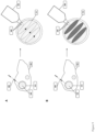
- Figure 9 shows two different embodiments of the invention where a light sheet microscope 303 (for example a tilted light sheet microscope) is used to analyze and subsequently mark or treat target cells in combination with laser scanning.
- the laser is directed through the detection objective. Since the treatment of the plane is also assessable by the light sheet at the same time, optical control of the process is possible.
- A) shows an encapsulation process where a margin of the sub volume is addressed layer by layer with the encapsulation laser (309).
- (B) shows a protocol where different layers addressed after imaging as a whole volume layer by layer. This in toto crosslinking of a sub-volume could be used to immobilize the morphological structure.
- FIG 10 shows the extraction of interaction zones between tumor (101) and immune cells (103) such as tumor microenvironment cells (TME). Further analysis can be used to understand the mechanisms of the attack and defense of e.g. CAR-T cells. And might be used to optimize CAR-T cell therapy especially by weakening the defence mechanisms by supplementary medication. This could be also used to support personalized therapy.
- TEE tumor microenvironment cells
- Figure 11 shows an example where crosslinking could be used in the analysis of cell compartments comprising an ensemble of immunological synapses.
- the crosslinking of the two cells e.g. tumor cell (101) and immune cell (118) allows an extraction of a fixed but morphologically intact immunological synapse. It could be also possible to isolate the volume containing the synapse. Further analysis could than include super-resolution microscopy to quantify the molecule exposed within the immunological synapsis.
- Figure 12 A shows the variant of the invention where crosslinking of DNA/RNA strands conjugated to an antibody to the cell offers the possibility to mark cells with a code or make the cell visible for DNA-PAINT Microscopy.
- Figure 12 B shows the crosslinking of magnetic beads to a cell surface or compartment followed by a washing step to remove all beads which are not cross-linked. The dissociated sample can than separated by magnetic cell separation.
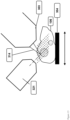
- FIG. 13 Another embodiment is depicted in Figure 13 , where the sample (106) is mounted on a sample holder (304).
- the sample holder is movable relative to the optics.
- the embodiment forms a tilted light-sheet (321).
- the image is detected through the objective (303) perpendicular to the light sheet.
- the system is combined with a photo treatment laser (314) which is applied through the imaging optic. In this configuration, subsequent imaging, treatment and stepping of the relative position between the sample holder and the optics allows to address large volumes as long as the sample is sufficiently thin.
- Figure 14 shows by way of example the embodiment of the invention where a sliced surface of a cleared sample is imaged with multi photon microscopy. Part A of Figure 14 shows in the middle a cell being located about 5 ⁇ m under the cutting plane. In Figure 14 , a top view of the slightly tilted cutting plane is depicted. Clearing was performed using Dibenzyl-ether. The cutting was performed with a conventional microtome.
- Figure 15 shows an laser scanning treatment protocol where a series (316) of acquisitions (315) is interrupted and either multiple regions of interest (318), combinations of lines (319) or a series of points (320) are treated (317).
- the treatment allows for not only structured photoactivation or crosslinking but also a marking, where the individual structures can be identified through the geometry of treatment.
- the subsequent analysis of the subcompartments is again a microscopic method. Data generated in this analysis step could be correlated to the initial imaging process, so that the detailed analysis could be viewed in the context of the whole sample which can be also the complete organism.
- subsequent extraction of compartments can be done.
- Super-resolution can be obtained with PAINT, dSTORM and DNA-PAINT. Staining procedures can be applied sequentially after sectioning or up front during the preparation the sample. By this means it is possible to combine the slicing with further analysis and super-resolution microscopy approaches, where the penetration of the light and also diffusion of reagents is or should be limited to the surface of the sample.
- all disclosed embodiments of the method of the invention may involve chemical dissociation of the tissue and subsequent collection of the optically tagged subcompartments.
- a back tracing of individual tagged compartments to its location within the sample is possible even in a situation where the sample is completely destroyed (disassembled) and dissolved for example with a gentleMACS instrument.
- the disassembly process can be speeded up by dissecting the tissue to a reasonably large number of slices. It is possible to design the slicing in a way that the tagged compartments are not destroyed.
- all disclosed embodiments of the method of the invention may involve further analyzing the encapsulated entity by means of sequencing. It is also possible to further dissociate the encapsulated compartment and to analyze the individual cells contained in the compartment. Thus differentiation between different interaction zones between tumor in its microenvironment is possible.
- the extraction of the encapsulated volumes can be done by means of slicing and preparation, where the preparation is supported by the crosslinking of the capsule.
- the extraction of the capsules could be also done by tissue dissociation where the crosslinking is designed in a way that is withstands the dissociation process.
Landscapes
- Health & Medical Sciences (AREA)
- Life Sciences & Earth Sciences (AREA)
- Chemical & Material Sciences (AREA)
- Immunology (AREA)
- Engineering & Computer Science (AREA)
- Analytical Chemistry (AREA)
- Pathology (AREA)
- General Health & Medical Sciences (AREA)
- Biochemistry (AREA)
- Physics & Mathematics (AREA)
- General Physics & Mathematics (AREA)
- Molecular Biology (AREA)
- Proteomics, Peptides & Aminoacids (AREA)
- Organic Chemistry (AREA)
- Biomedical Technology (AREA)
- Wood Science & Technology (AREA)
- Genetics & Genomics (AREA)
- Zoology (AREA)
- Microbiology (AREA)
- Biotechnology (AREA)
- Urology & Nephrology (AREA)
- Hematology (AREA)
- Hospice & Palliative Care (AREA)
- Bioinformatics & Cheminformatics (AREA)
- Oncology (AREA)
- Biophysics (AREA)
- General Engineering & Computer Science (AREA)
- Dispersion Chemistry (AREA)
- Signal Processing (AREA)
- Food Science & Technology (AREA)
- Tropical Medicine & Parasitology (AREA)
- Medicinal Chemistry (AREA)
- Cell Biology (AREA)
- Investigating Or Analysing Biological Materials (AREA)
- Sampling And Sample Adjustment (AREA)
- Computer Vision & Pattern Recognition (AREA)
Claims (4)
- Verfahren zur Extraktion von Zielzellen aus einer dreidimensionalen biologischen Probe durch die folgenden Schritte:a) Abbilden der dreidimensionalen Probe mit einem Lichtblattmikroskop,b) Identifizieren der Zielzellen,c) Registrieren der räumlichen Parameter (x, y, z-Koordinaten) der Zielzellen,d) Extrahieren der Zielzellen entsprechend ihren räumlichen Parametern,dadurch gekennzeichnet, dass die dreidimensionale Probe in mindestens einem der Schritte a), b), c) oder d) mit einem optisch induzierbaren Vernetzungsreagenz bereitgestellt und durch eine Ein-Photonen-Laser-Scan-Aktivierung vernetzt wird, wobei die Detektionslinse des Lichtblattmikroskops verwendet wird, um den Vernetzungslaser auf die Probe zu richten.
- Verfahren nach Anspruch 1, dadurch gekennzeichnet, dass die Zielzellen vor oder nach Schritt b) markiert werden.
- Verfahren nach Anspruch 1, dadurch gekennzeichnet, dass die Zielzellen in Schritt b) durch ihre intrinsischen optischen Eigenschaften identifiziert werden.
- Vorrichtung zum Extrahieren von Zielzellen aus einer dreidimensionalen biologischen Probe, umfassend ein Lichtblattmikroskop als Abbildungseinheit zum Abbilden der dreidimensionalen Probe und zum Identifizieren von Zielzellen; einen Datenspeicher, der die räumlichen Parameter (x,y,z-Koordinaten) der Zielzellen registriert; einen Vernetzungslaser und ein Mittel zum Extrahieren der so identifizierten Zielzellen.
Applications Claiming Priority (2)
| Application Number | Priority Date | Filing Date | Title |
|---|---|---|---|
| DE102020105311 | 2020-02-28 | ||
| PCT/EP2021/054663 WO2021170710A1 (en) | 2020-02-28 | 2021-02-25 | Method for extraction of target cells from 3d tissue by optical identification |
Publications (2)
| Publication Number | Publication Date |
|---|---|
| EP4111194A1 EP4111194A1 (de) | 2023-01-04 |
| EP4111194B1 true EP4111194B1 (de) | 2025-05-14 |
Family
ID=74797925
Family Applications (1)
| Application Number | Title | Priority Date | Filing Date |
|---|---|---|---|
| EP21708609.9A Active EP4111194B1 (de) | 2020-02-28 | 2021-02-25 | Verfahren zur extraktion von zielzellen aus 3d-gewebe durch optische identifizierung |
Country Status (3)
| Country | Link |
|---|---|
| US (1) | US12584824B2 (de) |
| EP (1) | EP4111194B1 (de) |
| WO (1) | WO2021170710A1 (de) |
Families Citing this family (1)
| Publication number | Priority date | Publication date | Assignee | Title |
|---|---|---|---|---|
| WO2025014758A1 (en) * | 2023-07-07 | 2025-01-16 | 10X Genomics, Inc. | Systems and methods for tissue bounds detection |
Family Cites Families (5)
| Publication number | Priority date | Publication date | Assignee | Title |
|---|---|---|---|---|
| KR102148747B1 (ko) * | 2012-08-09 | 2020-08-27 | 더 보드 오브 트러스티스 오브 더 리랜드 스탠포드 쥬니어 유니버시티 | 현미경적 분석을 위한 생물학적 표본을 준비하기 위한 방법과 조성물 |
| EP3083979B1 (de) * | 2013-12-19 | 2019-02-20 | Axon DX, LLC | Verfahren und vorrichtungen zur zelldetektion, -erfassung und -isolierung |
| WO2016115537A2 (en) * | 2015-01-15 | 2016-07-21 | Massachusetts Institute Of Technology | Systems, methods, and apparatus for in vitro single-cell identification and recovery |
| US10796781B2 (en) | 2015-12-07 | 2020-10-06 | Clarapath, Inc. | Spatial genomics with co-registered histology |
| JP6796379B2 (ja) | 2016-01-20 | 2020-12-09 | 地方独立行政法人東京都立産業技術研究センター | レーザーマイクロダイセクター及びレーザーマイクロダイセクション方法 |
-
2021
- 2021-02-25 EP EP21708609.9A patent/EP4111194B1/de active Active
- 2021-02-25 WO PCT/EP2021/054663 patent/WO2021170710A1/en not_active Ceased
- 2021-02-25 US US17/801,148 patent/US12584824B2/en active Active
Non-Patent Citations (1)
| Title |
|---|
| GALLION LUKE A ET AL: "Preserving single cells in space and time for analytical assays", TRAC TRENDS IN ANALYTICAL CHEMISTRY, ELSEVIER, AMSTERDAM, NL, vol. 122, 7 November 2019 (2019-11-07), XP086023658, ISSN: 0165-9936, [retrieved on 20191107], DOI: 10.1016/J.TRAC.2019.115723 * |
Also Published As
| Publication number | Publication date |
|---|---|
| US12584824B2 (en) | 2026-03-24 |
| US20230069991A1 (en) | 2023-03-09 |
| WO2021170710A1 (en) | 2021-09-02 |
| EP4111194A1 (de) | 2023-01-04 |
Similar Documents
| Publication | Publication Date | Title |
|---|---|---|
| Weiss et al. | Tutorial: practical considerations for tissue clearing and imaging | |
| US20230213415A1 (en) | Method and System for Imaging and Analysis of a Biological Specimen | |
| Friedenberger et al. | Fluorescence detection of protein clusters in individual cells and tissue sections by using toponome imaging system: sample preparation and measuring procedures | |
| Riesterer et al. | A workflow for visualizing human cancer biopsies using large-format electron microscopy | |
| Pirone et al. | Beyond fluorescence: advances in computational label-free full specificity in 3D quantitative phase microscopy | |
| Liu et al. | Nondestructive 3D pathology with light-sheet fluorescence microscopy for translational research and clinical assays | |
| Zhao et al. | Current status of tissue clearing and the path forward in neuroscience | |
| US20110124530A1 (en) | Pseudo-tissues and uses thereof | |
| Collinson et al. | Correlating 3D light to 3D electron microscopy for systems biology | |
| Leischner et al. | Formalin-induced fluorescence reveals cell shape and morphology in biological tissue samples | |
| US12613190B2 (en) | Quantitative liquid biopsy diagnostic system and methods | |
| EP4111194B1 (de) | Verfahren zur extraktion von zielzellen aus 3d-gewebe durch optische identifizierung | |
| Furia et al. | Automated multimodal fluorescence microscopy for hyperplex spatial-proteomics: Coupling microfluidic-based immunofluorescence to high resolution, high sensitivity, three-dimensional analysis of histological slides | |
| US20240375114A1 (en) | Biological sample holder and handler | |
| Velicky et al. | Saturated reconstruction of living brain tissue | |
| Ferri | Expansion Microscopy: A New Approach to Microscopic Evaluation | |
| Kochhan et al. | Zebrafish as a model to study chemokine function | |
| Ragan et al. | Two-photon tissue cytometry | |
| Fei et al. | Sub-voxel light-sheet microscopy for high-resolution, high-throughput volumetric imaging of large biomedical specimens | |
| Heymann et al. | Next-generation imaging: new insights from multicolor microscopy in liver biology and disease | |
| Schueth et al. | cleared tissue dual Selective Plane Illumination Microscopy (ct-dSPIM) for fast mesoscopic imaging of large-scale human tissues | |
| EP4672249A1 (de) | Verfahren und system zur trennung biochemischer moleküle | |
| Lin | Advancing Stimulated Raman Scattering Microscopy Through Deep Learning and Gel-Based Tissue Engineering | |
| Valet et al. | Cytomics: From cell States to predictive medicine | |
| Aureille et al. | Cell fluorescence photoactivation as a method to select and study cellular subpopulations grown in mechanically heterogeneous environments |
Legal Events
| Date | Code | Title | Description |
|---|---|---|---|
| STAA | Information on the status of an ep patent application or granted ep patent |
Free format text: STATUS: UNKNOWN |
|
| STAA | Information on the status of an ep patent application or granted ep patent |
Free format text: STATUS: THE INTERNATIONAL PUBLICATION HAS BEEN MADE |
|
| PUAI | Public reference made under article 153(3) epc to a published international application that has entered the european phase |
Free format text: ORIGINAL CODE: 0009012 |
|
| STAA | Information on the status of an ep patent application or granted ep patent |
Free format text: STATUS: REQUEST FOR EXAMINATION WAS MADE |
|
| 17P | Request for examination filed |
Effective date: 20220921 |
|
| AK | Designated contracting states |
Kind code of ref document: A1 Designated state(s): AL AT BE BG CH CY CZ DE DK EE ES FI FR GB GR HR HU IE IS IT LI LT LU LV MC MK MT NL NO PL PT RO RS SE SI SK SM TR |
|
| DAV | Request for validation of the european patent (deleted) | ||
| DAX | Request for extension of the european patent (deleted) | ||
| STAA | Information on the status of an ep patent application or granted ep patent |
Free format text: STATUS: EXAMINATION IS IN PROGRESS |
|
| 17Q | First examination report despatched |
Effective date: 20240606 |
|
| GRAP | Despatch of communication of intention to grant a patent |
Free format text: ORIGINAL CODE: EPIDOSNIGR1 |
|
| STAA | Information on the status of an ep patent application or granted ep patent |
Free format text: STATUS: GRANT OF PATENT IS INTENDED |
|
| INTG | Intention to grant announced |
Effective date: 20250103 |
|
| GRAS | Grant fee paid |
Free format text: ORIGINAL CODE: EPIDOSNIGR3 |
|
| GRAA | (expected) grant |
Free format text: ORIGINAL CODE: 0009210 |
|
| STAA | Information on the status of an ep patent application or granted ep patent |
Free format text: STATUS: THE PATENT HAS BEEN GRANTED |
|
| AK | Designated contracting states |
Kind code of ref document: B1 Designated state(s): AL AT BE BG CH CY CZ DE DK EE ES FI FR GB GR HR HU IE IS IT LI LT LU LV MC MK MT NL NO PL PT RO RS SE SI SK SM TR |
|
| REG | Reference to a national code |
Ref country code: GB Ref legal event code: FG4D |
|
| REG | Reference to a national code |
Ref country code: CH Ref legal event code: EP |
|
| REG | Reference to a national code |
Ref country code: DE Ref legal event code: R096 Ref document number: 602021030749 Country of ref document: DE |
|
| REG | Reference to a national code |
Ref country code: IE Ref legal event code: FG4D |
|
| REG | Reference to a national code |
Ref country code: NL Ref legal event code: MP Effective date: 20250514 |
|
| PG25 | Lapsed in a contracting state [announced via postgrant information from national office to epo] |
Ref country code: PT Free format text: LAPSE BECAUSE OF FAILURE TO SUBMIT A TRANSLATION OF THE DESCRIPTION OR TO PAY THE FEE WITHIN THE PRESCRIBED TIME-LIMIT Effective date: 20250915 Ref country code: FI Free format text: LAPSE BECAUSE OF FAILURE TO SUBMIT A TRANSLATION OF THE DESCRIPTION OR TO PAY THE FEE WITHIN THE PRESCRIBED TIME-LIMIT Effective date: 20250514 Ref country code: ES Free format text: LAPSE BECAUSE OF FAILURE TO SUBMIT A TRANSLATION OF THE DESCRIPTION OR TO PAY THE FEE WITHIN THE PRESCRIBED TIME-LIMIT Effective date: 20250514 |
|
| REG | Reference to a national code |
Ref country code: LT Ref legal event code: MG9D |
|
| PG25 | Lapsed in a contracting state [announced via postgrant information from national office to epo] |
Ref country code: NO Free format text: LAPSE BECAUSE OF FAILURE TO SUBMIT A TRANSLATION OF THE DESCRIPTION OR TO PAY THE FEE WITHIN THE PRESCRIBED TIME-LIMIT Effective date: 20250814 Ref country code: GR Free format text: LAPSE BECAUSE OF FAILURE TO SUBMIT A TRANSLATION OF THE DESCRIPTION OR TO PAY THE FEE WITHIN THE PRESCRIBED TIME-LIMIT Effective date: 20250815 |
|
| PG25 | Lapsed in a contracting state [announced via postgrant information from national office to epo] |
Ref country code: PL Free format text: LAPSE BECAUSE OF FAILURE TO SUBMIT A TRANSLATION OF THE DESCRIPTION OR TO PAY THE FEE WITHIN THE PRESCRIBED TIME-LIMIT Effective date: 20250514 Ref country code: NL Free format text: LAPSE BECAUSE OF FAILURE TO SUBMIT A TRANSLATION OF THE DESCRIPTION OR TO PAY THE FEE WITHIN THE PRESCRIBED TIME-LIMIT Effective date: 20250514 |
|
| REG | Reference to a national code |
Ref country code: AT Ref legal event code: MK05 Ref document number: 1795177 Country of ref document: AT Kind code of ref document: T Effective date: 20250514 |
|
| PG25 | Lapsed in a contracting state [announced via postgrant information from national office to epo] |
Ref country code: BG Free format text: LAPSE BECAUSE OF FAILURE TO SUBMIT A TRANSLATION OF THE DESCRIPTION OR TO PAY THE FEE WITHIN THE PRESCRIBED TIME-LIMIT Effective date: 20250514 |
|
| PG25 | Lapsed in a contracting state [announced via postgrant information from national office to epo] |
Ref country code: HR Free format text: LAPSE BECAUSE OF FAILURE TO SUBMIT A TRANSLATION OF THE DESCRIPTION OR TO PAY THE FEE WITHIN THE PRESCRIBED TIME-LIMIT Effective date: 20250514 |
|
| PG25 | Lapsed in a contracting state [announced via postgrant information from national office to epo] |
Ref country code: AT Free format text: LAPSE BECAUSE OF FAILURE TO SUBMIT A TRANSLATION OF THE DESCRIPTION OR TO PAY THE FEE WITHIN THE PRESCRIBED TIME-LIMIT Effective date: 20250514 |
|
| PG25 | Lapsed in a contracting state [announced via postgrant information from national office to epo] |
Ref country code: RS Free format text: LAPSE BECAUSE OF FAILURE TO SUBMIT A TRANSLATION OF THE DESCRIPTION OR TO PAY THE FEE WITHIN THE PRESCRIBED TIME-LIMIT Effective date: 20250814 |
|
| PG25 | Lapsed in a contracting state [announced via postgrant information from national office to epo] |
Ref country code: IS Free format text: LAPSE BECAUSE OF FAILURE TO SUBMIT A TRANSLATION OF THE DESCRIPTION OR TO PAY THE FEE WITHIN THE PRESCRIBED TIME-LIMIT Effective date: 20250914 |
|
| PG25 | Lapsed in a contracting state [announced via postgrant information from national office to epo] |
Ref country code: LV Free format text: LAPSE BECAUSE OF FAILURE TO SUBMIT A TRANSLATION OF THE DESCRIPTION OR TO PAY THE FEE WITHIN THE PRESCRIBED TIME-LIMIT Effective date: 20250514 |
|
| PG25 | Lapsed in a contracting state [announced via postgrant information from national office to epo] |
Ref country code: DK Free format text: LAPSE BECAUSE OF FAILURE TO SUBMIT A TRANSLATION OF THE DESCRIPTION OR TO PAY THE FEE WITHIN THE PRESCRIBED TIME-LIMIT Effective date: 20250514 Ref country code: SM Free format text: LAPSE BECAUSE OF FAILURE TO SUBMIT A TRANSLATION OF THE DESCRIPTION OR TO PAY THE FEE WITHIN THE PRESCRIBED TIME-LIMIT Effective date: 20250514 |
|
| PG25 | Lapsed in a contracting state [announced via postgrant information from national office to epo] |
Ref country code: CZ Free format text: LAPSE BECAUSE OF FAILURE TO SUBMIT A TRANSLATION OF THE DESCRIPTION OR TO PAY THE FEE WITHIN THE PRESCRIBED TIME-LIMIT Effective date: 20250514 |
|
| PG25 | Lapsed in a contracting state [announced via postgrant information from national office to epo] |
Ref country code: EE Free format text: LAPSE BECAUSE OF FAILURE TO SUBMIT A TRANSLATION OF THE DESCRIPTION OR TO PAY THE FEE WITHIN THE PRESCRIBED TIME-LIMIT Effective date: 20250514 |
|
| PG25 | Lapsed in a contracting state [announced via postgrant information from national office to epo] |
Ref country code: SK Free format text: LAPSE BECAUSE OF FAILURE TO SUBMIT A TRANSLATION OF THE DESCRIPTION OR TO PAY THE FEE WITHIN THE PRESCRIBED TIME-LIMIT Effective date: 20250514 |
|
| PG25 | Lapsed in a contracting state [announced via postgrant information from national office to epo] |
Ref country code: IT Free format text: LAPSE BECAUSE OF FAILURE TO SUBMIT A TRANSLATION OF THE DESCRIPTION OR TO PAY THE FEE WITHIN THE PRESCRIBED TIME-LIMIT Effective date: 20250514 |
|
| REG | Reference to a national code |
Ref country code: DE Ref legal event code: R097 Ref document number: 602021030749 Country of ref document: DE |
|
| PLBE | No opposition filed within time limit |
Free format text: ORIGINAL CODE: 0009261 |
|
| STAA | Information on the status of an ep patent application or granted ep patent |
Free format text: STATUS: NO OPPOSITION FILED WITHIN TIME LIMIT |
|
| REG | Reference to a national code |
Ref country code: CH Ref legal event code: L10 Free format text: ST27 STATUS EVENT CODE: U-0-0-L10-L00 (AS PROVIDED BY THE NATIONAL OFFICE) Effective date: 20260325 |
|
| PGFP | Annual fee paid to national office [announced via postgrant information from national office to epo] |
Ref country code: GB Payment date: 20260220 Year of fee payment: 6 |
|
| PGFP | Annual fee paid to national office [announced via postgrant information from national office to epo] |
Ref country code: DE Payment date: 20260218 Year of fee payment: 6 |
|
| PGFP | Annual fee paid to national office [announced via postgrant information from national office to epo] |
Ref country code: FR Payment date: 20260219 Year of fee payment: 6 |
|
| 26N | No opposition filed |
Effective date: 20260217 |