EP4111199B1 - Système de détection colorimétrique permettant la détection rapide de maladies pulmonaires infectieuses et masque facial doté dudit système de détection colorimétrique - Google Patents
Système de détection colorimétrique permettant la détection rapide de maladies pulmonaires infectieuses et masque facial doté dudit système de détection colorimétrique Download PDFInfo
- Publication number
- EP4111199B1 EP4111199B1 EP21714665.3A EP21714665A EP4111199B1 EP 4111199 B1 EP4111199 B1 EP 4111199B1 EP 21714665 A EP21714665 A EP 21714665A EP 4111199 B1 EP4111199 B1 EP 4111199B1
- Authority
- EP
- European Patent Office
- Prior art keywords
- antibodies
- substrate
- detection system
- aptamers
- colorimetric
- Prior art date
- Legal status (The legal status is an assumption and is not a legal conclusion. Google has not performed a legal analysis and makes no representation as to the accuracy of the status listed.)
- Active
Links
Images

Classifications
-
- G—PHYSICS
- G01—MEASURING; TESTING
- G01N—INVESTIGATING OR ANALYSING MATERIALS BY DETERMINING THEIR CHEMICAL OR PHYSICAL PROPERTIES
- G01N33/00—Investigating or analysing materials by specific methods not covered by groups G01N1/00 - G01N31/00
- G01N33/48—Biological material, e.g. blood, urine; Haemocytometers
- G01N33/50—Chemical analysis of biological material, e.g. blood, urine; Testing involving biospecific ligand binding methods; Immunological testing
- G01N33/58—Chemical analysis of biological material, e.g. blood, urine; Testing involving biospecific ligand binding methods; Immunological testing involving labelled substances
- G01N33/585—Chemical analysis of biological material, e.g. blood, urine; Testing involving biospecific ligand binding methods; Immunological testing involving labelled substances with a particulate label, e.g. coloured latex
-
- G—PHYSICS
- G01—MEASURING; TESTING
- G01N—INVESTIGATING OR ANALYSING MATERIALS BY DETERMINING THEIR CHEMICAL OR PHYSICAL PROPERTIES
- G01N33/00—Investigating or analysing materials by specific methods not covered by groups G01N1/00 - G01N31/00
- G01N33/48—Biological material, e.g. blood, urine; Haemocytometers
- G01N33/50—Chemical analysis of biological material, e.g. blood, urine; Testing involving biospecific ligand binding methods; Immunological testing
- G01N33/53—Immunoassay; Biospecific binding assay; Materials therefor
- G01N33/543—Immunoassay; Biospecific binding assay; Materials therefor with an insoluble carrier for immobilising immunochemicals
- G01N33/54366—Apparatus specially adapted for solid-phase testing
- G01N33/54386—Analytical elements
-
- A—HUMAN NECESSITIES
- A41—WEARING APPAREL
- A41D—OUTERWEAR; PROTECTIVE GARMENTS; ACCESSORIES
- A41D13/00—Professional, industrial or sporting protective garments, e.g. surgeons' gowns or garments protecting against blows or punches
- A41D13/05—Professional, industrial or sporting protective garments, e.g. surgeons' gowns or garments protecting against blows or punches protecting only a particular body part
- A41D13/11—Protective face masks, e.g. for surgical use, or for use in foul atmospheres
-
- C—CHEMISTRY; METALLURGY
- C12—BIOCHEMISTRY; BEER; SPIRITS; WINE; VINEGAR; MICROBIOLOGY; ENZYMOLOGY; MUTATION OR GENETIC ENGINEERING
- C12Q—MEASURING OR TESTING PROCESSES INVOLVING ENZYMES, NUCLEIC ACIDS OR MICROORGANISMS; COMPOSITIONS OR TEST PAPERS THEREFOR; PROCESSES OF PREPARING SUCH COMPOSITIONS; CONDITION-RESPONSIVE CONTROL IN MICROBIOLOGICAL OR ENZYMOLOGICAL PROCESSES
- C12Q1/00—Measuring or testing processes involving enzymes, nucleic acids or microorganisms; Compositions therefor; Processes of preparing such compositions
- C12Q1/34—Measuring or testing processes involving enzymes, nucleic acids or microorganisms; Compositions therefor; Processes of preparing such compositions involving hydrolase
- C12Q1/37—Measuring or testing processes involving enzymes, nucleic acids or microorganisms; Compositions therefor; Processes of preparing such compositions involving hydrolase involving peptidase or proteinase
-
- G—PHYSICS
- G01—MEASURING; TESTING
- G01N—INVESTIGATING OR ANALYSING MATERIALS BY DETERMINING THEIR CHEMICAL OR PHYSICAL PROPERTIES
- G01N21/00—Investigating or analysing materials by the use of optical means, i.e. using sub-millimetre waves, infrared, visible or ultraviolet light
- G01N21/75—Systems in which material is subjected to a chemical reaction, the progress or the result of the reaction being investigated
- G01N21/77—Systems in which material is subjected to a chemical reaction, the progress or the result of the reaction being investigated by observing the effect on a chemical indicator
- G01N21/78—Systems in which material is subjected to a chemical reaction, the progress or the result of the reaction being investigated by observing the effect on a chemical indicator producing a change of colour
-
- G—PHYSICS
- G01—MEASURING; TESTING
- G01N—INVESTIGATING OR ANALYSING MATERIALS BY DETERMINING THEIR CHEMICAL OR PHYSICAL PROPERTIES
- G01N33/00—Investigating or analysing materials by specific methods not covered by groups G01N1/00 - G01N31/00
- G01N33/48—Biological material, e.g. blood, urine; Haemocytometers
- G01N33/50—Chemical analysis of biological material, e.g. blood, urine; Testing involving biospecific ligand binding methods; Immunological testing
- G01N33/53—Immunoassay; Biospecific binding assay; Materials therefor
- G01N33/543—Immunoassay; Biospecific binding assay; Materials therefor with an insoluble carrier for immobilising immunochemicals
- G01N33/54393—Improving reaction conditions or stability, e.g. by coating or irradiation of surface, by reduction of non-specific binding, by promotion of specific binding
-
- G—PHYSICS
- G01—MEASURING; TESTING
- G01N—INVESTIGATING OR ANALYSING MATERIALS BY DETERMINING THEIR CHEMICAL OR PHYSICAL PROPERTIES
- G01N33/00—Investigating or analysing materials by specific methods not covered by groups G01N1/00 - G01N31/00
- G01N33/48—Biological material, e.g. blood, urine; Haemocytometers
- G01N33/50—Chemical analysis of biological material, e.g. blood, urine; Testing involving biospecific ligand binding methods; Immunological testing
- G01N33/53—Immunoassay; Biospecific binding assay; Materials therefor
- G01N33/569—Immunoassay; Biospecific binding assay; Materials therefor for microorganisms, e.g. protozoa, bacteria, viruses
- G01N33/56983—Viruses
-
- G—PHYSICS
- G01—MEASURING; TESTING
- G01N—INVESTIGATING OR ANALYSING MATERIALS BY DETERMINING THEIR CHEMICAL OR PHYSICAL PROPERTIES
- G01N33/00—Investigating or analysing materials by specific methods not covered by groups G01N1/00 - G01N31/00
- G01N33/48—Biological material, e.g. blood, urine; Haemocytometers
- G01N33/50—Chemical analysis of biological material, e.g. blood, urine; Testing involving biospecific ligand binding methods; Immunological testing
- G01N33/68—Chemical analysis of biological material, e.g. blood, urine; Testing involving biospecific ligand binding methods; Immunological testing involving proteins, peptides or amino acids
- G01N33/6884—Chemical analysis of biological material, e.g. blood, urine; Testing involving biospecific ligand binding methods; Immunological testing involving proteins, peptides or amino acids from lung
-
- G—PHYSICS
- G01—MEASURING; TESTING
- G01N—INVESTIGATING OR ANALYSING MATERIALS BY DETERMINING THEIR CHEMICAL OR PHYSICAL PROPERTIES
- G01N2333/00—Assays involving biological materials from specific organisms or of a specific nature
- G01N2333/90—Enzymes; Proenzymes
- G01N2333/914—Hydrolases (3)
- G01N2333/948—Hydrolases (3) acting on peptide bonds (3.4)
- G01N2333/95—Proteinases, i.e. endopeptidases (3.4.21-3.4.99)
- G01N2333/964—Proteinases, i.e. endopeptidases (3.4.21-3.4.99) derived from animal tissue
- G01N2333/96425—Proteinases, i.e. endopeptidases (3.4.21-3.4.99) derived from animal tissue from mammals
- G01N2333/96427—Proteinases, i.e. endopeptidases (3.4.21-3.4.99) derived from animal tissue from mammals in general
- G01N2333/9643—Proteinases, i.e. endopeptidases (3.4.21-3.4.99) derived from animal tissue from mammals in general with EC number
- G01N2333/96486—Metalloendopeptidases (3.4.24)
- G01N2333/96491—Metalloendopeptidases (3.4.24) with definite EC number
- G01N2333/96494—Matrix metalloproteases, e. g. 3.4.24.7
Definitions
- the present invention relates to a colorimetric detection system for rapid detection of lung diseases, a colorimetric mask comprising said detection system, a method for producing a colorimetric detection system, a method for producing a mask comprising said detections system and the use of dyed microsphere for a colorimetric detection system.
- Pulmonary infections are global health problems that can be spread anywhere. Spreading is in particular a problem in regions where many people live in cramped conditions. Likewise, seasonal holidays, such as carnival, or religious events, such as living as livingages, pose a challenge in preventing the spread of diseases. Viral and bacterial microbes play a major role in lung infections and different attempts have been undertaken to prevent spreading of these microbes. One way to prevent spreading would be a rapid detection method and isolation of infected persons. Another relatively efficient way of preventing spreading is the use of facial mask that cover mouth and nose. If a patient with a lung disease is coughing or sneezing into the mask, viruses and microbes such as bacteria are caught in the layers of the mask preventing their uptake by another person standing close to the patient.
- Indicator systems are known that are based on color changes.
- Schaude et al (Sensors 2017, 17, p. 1365 ) describes indicator cotton swabs for a faster and less expensive way to obtain information about a wound status.
- the method is based on cotton swabs covalently functionalized with an indicator dye that changes color upon pH change visible to the naked eye.
- the pH can be used to determine the efficiency of wound healing. In case the pH is not optimal for wound healing, a distinct treatment to change the wound pH can be applied.
- US 2019/0309357 A1 describes a detection system comprising a CRISPR system comprising an effector protein and one or more guide RNAs designed to bind to corresponding target molecules; an RNA masking construct and one or more detection aptamers coupled with a detectable ligand.
- Enzymes generate a detectable signal by releasing the detectable ligand form the molecule which can be detected by e.g. fluorescence.
- the detection system can be prepared in freeze-dried format for convenient distribution and point-of-care. A comparable system has been applied to face mask (according to www.businessinsider.com). However, the system still needs hours to obtain a diagnostic signal. Further, the detection system is based on PCR techniques, which is rather expensive. Fluorescence measurements are susceptible to interference leading to noise signals thus complicating the analysis.
- US 2010/092944 describes a colorimetric system for the detection of lung disease comprising antibodies coupled to a substrate for capturing a lung disease specific antigen characterized in that the colorimetric system further comprises dyed microspheres modified with the antibodies for visually detecting lung disease specific antigen wherein the antibodies are specific to an antigen of influenza virus and the solid support substrate is a hydrophilic material or cellulose.
- Wo 2009/126336 describes a similar system with the same components, an antibody coupled to a substrate for capturing a lung disease antigen and further comprising a dyed microsphere modified with antibodies.
- a first aspect of the invention relates to a colorimetric detection system for rapid detection of lung diseases.
- the detection system comprises antibodies and/or aptamers coupled to a substrate for capturing a lung disease specific antigen.
- the colorimetric system further comprises dyed microspheres modified with the antibodies and/or aptamers for visually detecting the lung disease specific antigen.
- the colorimetric detection system is further characterized in that the substrate comprises a protease, preferably a matrix-metalloproteinase.
- Antibodies are large Y-shaped proteins used by the immune system to identify pathogenic bacteria or viruses.
- the antibody recognizes a unique molecule of the pathogen, called the antigen.
- Aptamers are oligonucleotide or peptide molecules that bind to a specific target molecule. They can replace antibodies or used along antibodies for the same target.
- the antibodies and/or aptamers coupled to the substrate are preferably coupled by covalent linkage and can be considered as capture antibodies and/or aptamers.
- the modified dyed microspheres are preferably attached to the substrate by physical adsorption, thus being less strongly bound to the substrate than the capture antibodies/aptamers. However, they may also be bound by covalent linkage to the substrate.
- the antibodies and/or aptamers attached to the dyed microsphere are preferably of the same kind as the capture antibodies/aptamer but show at least the same antigen specificity as the capture antibodies/aptamers.
- the antibodies attached to the dyed microsphere may be referred to as detection antibodies and/or detection aptamers.
- theyed can refer to attached color-causing ligands; color pigments, compounds or dyes enclosed in the microsphere or with dye coated microspheres. Suitable dyes are for example Polybead ® dyed microsphere obtainable by Polysciences Inc. The Polybeads may have different colors such as blue, green, yellow, red, pink, orange.
- the Polybead ® microsphere can be Polybead ® Polystyrene Microsphere, in the description also referred to as polystyrene microsphere or microsphere.
- the dyed microspheres can be provided as a particle suspension with a concentration of 2.5 % solid per 100 mL.
- the suspension may comprise particle diameters in the range of 0.05 ⁇ m to 90 ⁇ m. Particles with a diameter in the range of 0.5 to 90 ⁇ m account for 10 % or less of the solids in the suspension.
- the microspheres can be functionalized with carboxyl-, hydroxyl-, or amine groups for coupling of the antibodies and aptamers.
- the dyed microspheres used for coupling with the antibodies and/or aptamers have a mean particle diameter of less than 6 ⁇ m, preferably even less than 3 ⁇ m, even more preferably less than 1 ⁇ m and most preferably less than 500 nm.
- the individual microspheres in the colorimetric detection system can have different nominal particle diameter, but preferably do not exceed 6 ⁇ m.
- the sphericity of the dyed microspheres is preferably > 0.9, more preferably > 0.93, most preferably > 0.96. Individual microspheres with a diameter of less than 6 ⁇ m are not visible to the naked human eye and only allow detection upon agglutination thus providing a simple and effective detections system.
- the substrate further comprises a protease, preferably a matrix-metalloproteinase.
- a protease preferably a matrix-metalloproteinase.
- the protease is coupled to the substrate by functional groups such as carboxyl-, hydroxyl- or amine groups present on the substrate.
- Other proteases are also possible, for example PR3 or Elastase.
- the protease has the advantage that a pathogen specific antigen coupled to the respective antibodies or aptamer are cleaved easier into smaller fragments (peptides and amino acids) thus allowing more detection antibodies and/or aptamers to agglutinate to a larger network and thus allowing a faster detection.
- the substrate can comprise discrete sections, each section being modified with antibodies and/or aptamers different from one of the other section for capturing a lung disease specific antigen different from the one captured in one of the other section.
- a detection system is provided that allows the detection of different pathogens simultaneously. Preferred are two to four discrete sections, even more three sections. However, the detection system can comprise even more discrete sections, being only limited by the size of the substrate to allow visual detection.
- one discrete section may comprise antibodies and/or aptamers specific for a pneumonia inducing bacteria while the other discrete section comprises antibodies and/or aptamers specific for a flue inducing antigen.
- the dyed microsphere may have different colors in order to distinguish the different diseases.
- the antibodies and/or aptamers can be influenza virus antibodies and/or aptamers; or a pneumonia or pneumonia-inducing antibodies and/or aptamers.
- antigens that may be detectable by the colorimetric detection system are for example related to: viruses causing COVID-19, MERS, SARS, Flue A, Para Flue A Flue B, Para Flu B; bacteria such as Streptococcus, pneumonia, legionella, tuberculosis; or fungus such as actinomyces, aspergillus, candida spp.
- bacteria such as Streptococcus, pneumonia, legionella, tuberculosis
- fungus such as actinomyces, aspergillus, candida spp.
- the list is not to be understood as exhaustive as basically every disease can be detected of which specific antibodies/aptamers are available.
- the substrate is a solid support.
- the substrate can be a hydrophilic material and/or oxidized cellulose.
- the substrate can be a cellulose based, paper-based or synthetic material.
- tissue paper, cotton or functionalized plastic material can serve as substrate.
- the substrate can basically have any shape. Due to its easy handling the strip format is preferred.
- the substrate may have functional groups to allow coupling of the capture antibodies and/or aptamers, for example carboxyl-, hydroxyl- or amine groups, preferably by conventional carbodiimde coupling techniques.
- the carbodiimide technique is in particular used to couple proteins and peptides via their n-terminus to carboxylic groups to form stable conjugates.
- the hydrophilicity allows the uptake of body liquids in the substrate. Basically, any hydrophilic substrate that provides the above-mentioned conditions can be used. Further examples are silica or metal hydroxide such as aluminium hydroxide.
- the working principle of the colorimetric system can be explained as follows: If a sample of salvia, sputum or nasal muscus of an infected person is applied to the substrate the substrate is humidified.
- the pathogen specific antigen is caught by the capture antibodies or aptamers covalently attached to the substrate.
- the protease allows cleavage of the antigen into smaller fragments for an efficient agglutination of detection antibodies.
- the humidified substrate allows movement of the detection antibodies.
- the detection antibodies or aptamers preferably attached to the substrate by physical adsorption, will attach to the pathogen/antivirus and may form agglutinated network.
- the agglutination of the detection antibodies/aptamers causes an agglutination of the dyed microsphere resulting in the color appearance of the respective dyed microspheres to the naked eye and thus allowing visual detection of the disease.
- Single dyed microsphere may be undetectable by the naked eye as described further above.
- the colorimetric detection system provides a simple, efficient and fast way of detecting lung infections.
- the system can be easily prepared with low costs.
- the color appears within the first 30 min after applying the sample to the substrate, preferably between 1 and 10 min after application and even more preferably between 1 to 5 min after application and most preferably between 1 and 3 min after application.
- the detection system does response faster than other known method such as fluorescent measurements and is less prone to errors. Further, no additional equipment is used for detection allowing its application everywhere.
- a second aspect of the invention relates to a colorimetric face mask for rapid detection of lung diseases.
- the face mask is preferably designed to cover the mouth and the nose of a person.
- the face mask comprises a first layer comprising the colorimetric detection system as previously described.
- the first layer can be made from the same material as the substrate.
- the substrate itself can serve as the first layer.
- the first layer can also be made of material different from the substrate.
- the first layer can comprise a hydrophobic material that prevents the adsorption of aqueous body liquids such as salvia.
- the first layer can be polypropylene or a hydrophobised material.
- the first layer can be impregnated material.
- the substrate can be attached to the first layer, for example by sewing, ticking or gluing.
- the face mask can comprise a second outer layer for protection against external pathogenic factors, preferably comprising a synthetic polymer such as polypropylene.
- the outer layer can be obtained by polymeric fibres for exampling by spinning or weaving.
- the outer layer is made of a hydrophobic material to avoid the uptake of pathogen-containing fluids such as aerosols, salvia or mucus of persons standing nearby.
- the outer layer can comprise impregnated material. Most preferably, the outer layer is impermeable to microparticle but allows easy breathing of the user.
- the impregnated material can be any textile or paper material, of natural or synthetic origin, that has been treated with resins or other substances known in the art, or even has been guanidine-functionalized, in order to avoid water uptake and/or to obtain an antimicrobial effect.
- the outer layer protects the user from being infected with a lung disease by others. At the same time is also prevents a false detection due to pathogens present in the environment instead of being present in the patient's body liquid. Further, it also avoids the visual detection by others which may lead to panic or the like.
- pathogenic factors all disease-causing pathogens, microbes and small particles are meant, in particular viruses, bacteria, fungus, dust and smog.
- the working principle of the face mask can be explained similar as done with regard to the colorimetric detection system with the difference that the pathogen specific antigen containing sample is provided by sneezing or coughing into the mask.
- the coughing or snoozing brings the salvia, sputum or nasal muscus to the mask and humidifies the substrate.
- the humidified substrate allows movement of detection antibodies to the antigen and the agglutination to the network allowing.
- the agglutination of the detection antibodies/aptamers causes an agglutination of the dyed microsphere resulting in the color appearance of the respective dyed microspheres to the naked eye and thus allowing visual detection of the disease.
- Single dyed microsphere may be undetectable by the naked eye as described further above.
- the colorimetric mask offers a cheap, fast and reliable method of detection a patient's or any other infected person's lung disease and at the same time prevents the spreading of the pathogens, thus lessen the risk of disease spreading.
- the color can appear within the first 30 min after release of the pathogen by e.g. sneezing or coughing, preferably between 1 and 10 min after release and even more preferably between 1 to 5 min after release and most preferably between 1 and 3 min.
- the first layer comprises the colorimetric detection system with two or more discrete sections for the detection of different pathogens as previously described.
- one discrete section may comprise antibodies and/or aptamers specific for a pneumonia inducing bacteria while the other discrete section comprises antibodies and/or aptamers specific for a flue inducing antigen.
- the dyed microsphere may have different colors in order to distinguish the different diseases.
- Such a mask allows the detection of several lung diseases and thus no separate mask for each kind of infection has to be provided.
- the colorimetric detection system can be located in the centre of the first layer, preferably in a circular arrangement. With “centre” is meant the middle of a plane forming cloth. Thus, the colorimetric detection system is closest to the mouth and nose, where the highest concentration on pathogen can be expected and thus leading to a fast detection. If more than one discrete section is used, the detection system is preferably arranged in a circle and divided into pie-shaped sections. However, other shapes may also be possible as long as a contact between pathogen and detection system can be ensured.
- the colorimetric mask can comprise a third inner layer for hiding the result of the visually detected lung disease.
- the third inner layer is preferably permeable to fluids and preferably comprises a cotton based material. Permeability can thus either be achieved by the material itself or by providing fluid passage, such as holes, in the third layer to allow contact of the pathogen containing body fluid with the detection system in the first layer. In principle, all material mentioned with regard to the first and second layer can be used for the third layer.
- the third layer avoids the visual detection of the disease by the patient himself.
- the visual appearance of the colors can only be interpreted by medical staff and thus avoiding revelation to the patient or surrounding people which may cause panic or fear.
- the mask may comprise fixation loops for fixing the mask to the head.
- the loops can be handcrafted rubber strings with two metal copper rings inside, rubber bands or cords for closing at the back of the head. This way, the mask is holed in place and wearing is comfortable. The mask can thus be worn for a couple of hours.
- a third aspect of the invention refers to a method for producing a colorimetric system as previously described.
- the method comprises the steps of
- the substrate may be a hydrophilic material already containing the reactive sites or the substrate may have been modified in a previous step.
- the substrate can originate from cellulose which has been oxidized.
- the substrate can be oxidized cellulose.
- the substrate can be any other hydrophilic material that provides reactive sites towards antibodies and/or aptamers.
- the antibodies and/or aptamers may be coupled to the substrate via the reactive groups.
- the reactive groups can be carboxyl-, hydroxyl-, amine- or sulfuric groups. Carboxyl groups are preferred.
- Coupling of the antibodies and aptamers can be conducted by carbodiimide crosslinking chemistry known in the art. In general, any coupling method that allows the N- or C-terminus of the antibody/aptamer to be coupled to the substrate is possible.
- Blocking of non-reactive sites is preferably obtained by using bovine serum albumin (BSA).
- BSA bovine serum albumin
- Attachment of the dyed microspheres modified with the antibodies and/or aptamers to the substrate is preferably obtained by physical adsorption.
- the substrate can be immersed into a solution comprising the modified dyed microspheres. Details to the microsphere have been previously provided and apply here analogously. After immersing the substrate into the solution, the substrate is stained in the color of the dyed microsphere. Washing the substrate with buffer solution will remove excess dyed microsphere resulting in an essentially colorless substrate. Even though not visible to the naked eye, microspheres will still be attached the substrate by preferably physical adsorption.
- the solution with the dyed microsphere modified with the antibodies and/or aptamers can be applied locally to the substrate, e.g. by drip on, spray on or the like. This is in particular advantageous if more than one antibody/aptamer system is applied to different discrete sections in the substrate.
- the dyed microsphere modified with antibodies and/or aptamers are preferably obtained by a separate step.
- Conventional dyed polystyrene microspheres can be obtained from Polyscience Inc. (USA).
- Non-modified dyed microspheres can have a mean diameter of less than 6 ⁇ m, preferably less than 3 ⁇ m, even more preferably less than 1 ⁇ m and most preferably less than 500 nm.
- the sphericity of the non-modified dyed microspheres is preferably > 0.9, more preferably > 0.93, most preferably > 0.96.
- the microspheres can be functionalized with carboxyl groups, but also hydroxyl- or amine groups are possible. Attachment of antibodies and aptamers can be conducted by using carbodiimide coupling techniques known in the art and previously described. After attaching the modified dyed microspheres to the substrate, the substrate can be dried before use.
- the capture antibody is coupled to the substrate together with a protease, preferably a matrix metalloprotease. But also other proteases fulfilling the respective function are suitable.
- the coupling can be obtained simultaneously with the antibodies/aptamers, preferably by using horseradish peroxidase (HRP), or in sequence.
- HRP horseradish peroxidase
- the method can provide the step of coupling two or more different antibodies and/or aptamers each specific for another lung disease causing antigen to the substrate.
- Antibodies and/or aptamers for one specific antigen are preferably coupled in a discrete section of the substrate. Coupling of capture antibodies and/or aptamers will preferably occur through chemical reaction such as carbodiimide crosslinking as previously described.
- a fourth aspect of the invention relates to a method for producing a colorimetric mask as previously described.
- the method comprises the step of providing a first layer with a colorimetric detection system obtainable by a method as previously described.
- the first layer can be made from the same material as the substrate.
- the substrate can even be the first layer itself.
- the first layer can also be made of material different from the substrate.
- the first layer can comprise a hydrophobic material that prevents the adsorption of aqueous body liquids such as salvia, sputum or nasal muscus.
- the first layer can be polypropylene, hydrophobised material or any previously described material.
- the substrate can be attached to the first layer, for example by sewing, ticking or gluing.
- the colorimetric detection system can be arranged in the centres of the first layer, meaning in the centre of the cloth forming the plane. Discrete sections of the colorimetric detections system can be arranged cicular and in pie-shape. But also other locations and shapes are possible as long as a contact between capture antibody/aptamer and antigen is provided.
- Another aspect of the invention refers to the use of dyed microsphere modified with antibody and/or aptamer in a colorimetric detection system as previously described.
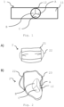
- FIG. 1 shows a colorimetric detection system 1 according to the invention.
- the colorimetric detection system 1 comprises a substrate 11, here in form of a strip.
- the detection area 12 in form of a circle with discrete detection sections A, B, C.
- Each detection section A, B and C are able to detect another pathogen specific antigen.
- section A can comprise influenza virus antibodies/aptamers coupled to blued-dyed microsphere.
- Section B can comprise pneumonia antibodies/aptamer coupled to green dyed microsphere.
- Section C can comprise Covid-19 antibodies/aptamer coupled to red dyed microsphere.
- the detection section A will change from colorless to blue while the other sections B and C remain colorless.
- the detection section B will change from colorless to green while the other sections A and C remain colorless.
- the detection section C will change from colorless to red while the other sections A and B remain colorless.
- Figure 2A shows the face mask 2 according to the invention from the front with the outer layer 21 and the fixation loops 22 on each side for fixation on the ears.
- Figure 2B shows the mask 2 from the backside, meaning the side facing mouth and nose of the user.
- the figure shows the first layer 23 comprising the detection area 12.
- the first layer also forms the substrate of the colorimetric detection system.
- the detection area 12 is provided as a circle in the centre of the mask 2, being the region closest to mouth and nose when in use.
- the detection area 12 is divided into three different pie-shaped sections A, B, C. Each section will be coupled with another antibodies/aptamer system specific for one particular antigen thus providing the possibility of detecting three different pathogen specific antigen with one mask.
- FIG. 3 shows the modification of the substrate.
- a cellulose substrate 31 is provided which is oxidized with sodium periodate (step a).
- capture antibodies or aptamers or both are added to the oxidized cellulose and coupled by known carbodiimide coupling techniques to the cellulose (step b).
- Step b also comprises the step of coupling protease to the cellulose using horseradish peroxidase (not shown).
- Previously prepared dyed microsphere modified with the antibodies/aptamers (detection antibodies) 32 are attached to the substrate by immersing the cellulose substrate into a solution containing the dyed microspheres (step c).
- the dyed microsphere are preferably obtained by reaction of colored polystyrene microspheres with free carboxyl groups with the respective antibodies and/or aptamers by the previously mentioned carbodiimide coupling techniques known in the art.
- the substrate 31 is dried, attached to a first layer of a mask or being the first layer itself and mounted together with an outer layer 21 to the colorimetric mask 2 of figure 2 .
- Figure 4 shows the working principle of the colorimetric detection system.
- the pathogen specific antigen 41 will be captured by the capture antibody/aptamer 42 (step d).
- the protease cleaves the antigen 41 into smaller fragments (step e), thus allowing more detection antibodies 32 to be agglutinated around the antigen 41 leading to a agglutinated network 43.
- the network 43 comprises sufficient detection antibodies 32 to become visible to the naked eye. Movement of the detection antibodies 32 to the antigen's 41 reactive sites is preferably achieved through capillary effects upon humidification of the substrate.
- MMP2 Matrix metalloproteinase-2
- Sodium phosphate and monopotassium phosphate Tris-Base, Sodium acetate; bovine serum albumin (BSA), sodium chloride, phosphate buffered saline (PBS), ethyl-3-(3-dimethylaminopropyl)-carbodiimide (EDC) , N-hydroxysuccinimide (NHS), ethylenediaminetetraacetic acid (EDTA) phosphate buffered were all purchased from Sigma-Aldrich (Darmstadt, Germany). Carboxylic acid functionalized microspheres of different colors (orange and blue) were obtained from Polysciences Inc. (Polysciences Inc., Warrington, PA).
- a pure cotton cellulose cloth was activated by immersing them in sodium periodate (NaIO 4 ) and of sulfuric acid (H 2 SO 4 ) overnight at room temperature to obtained oxidized cellulose.
- the oxidized cotton cellulose was washed with PBS buffer, subsequently, with cold distilled water, to remove excess of oxidizing agent.
- Periodate oxidation of cellulose cotton primarily activates hydroxyl groups into aldehyde or carboxyl groups, which will later be coupled with free amine groups of the antibodies or aptamers.
- hRSV Human respiratory syncytial virus
- Ab Human respiratory syncytial virus
- MMP2 protease were coated on the cotton cellulose to later support cleavage of the pathogen.
- the activated cotton cellulose was incubated into a well-mixed solution of hRSV Ab and PBS and incubated overnight at 4 °C. Accordingly, MMP2 containing coupling buffer was used to couple MMP2 onto the cellulose similar as to the method described for the antibody. Horseredish peroxidase was used for facilitate coupling.
- the cotton cellulose was washed to remove any unbounded antibody and MMP2 and thereafter 1% BSA was added to block the active sites of the cotton cellulose.
- a control cotton cellulose cloth was incubated with 1% BSA solution in PBS buffer for 30 min at room temperature.
- hRSV aptamer was washed with distilled water several times before being reacted with a coupling mixture of EDC/NHS overnight. Subsequently, the microspheres were washed with distilled water to remove excess coupling reagent. hRSV aptamer was thus linked to activated blue-dyed microspheres. Blocking of any unbound sites was achieved using 1% BSA solution in PBS for 30 min.
- the modified cotton cellulose with the attached antibodies from step 3 were washed with PBS followed by immersion the cotton cellulose into a solution of the modified microspheres of step 4, conjugated hRSV aptamer. Reaction was allowed to occur for a a couple of minutes. Subsequently, the cotton was washed with cold PBS to remove any unreacted microsphere. The cotton cellulose was dried and store at room temperature for 24 hrs.
- the cellulose cotton with the associated microspheres and capture antibodies were exposed to a sample of a patient's salvia, infected with respiratory syncytial virus. A blue color appeared within 3 min, visually displaying the infection.
Landscapes
- Health & Medical Sciences (AREA)
- Life Sciences & Earth Sciences (AREA)
- Engineering & Computer Science (AREA)
- Immunology (AREA)
- Chemical & Material Sciences (AREA)
- Molecular Biology (AREA)
- Biomedical Technology (AREA)
- Hematology (AREA)
- Urology & Nephrology (AREA)
- Physics & Mathematics (AREA)
- General Health & Medical Sciences (AREA)
- Biochemistry (AREA)
- Analytical Chemistry (AREA)
- General Physics & Mathematics (AREA)
- Pathology (AREA)
- Biotechnology (AREA)
- Microbiology (AREA)
- Cell Biology (AREA)
- Food Science & Technology (AREA)
- Medicinal Chemistry (AREA)
- Proteomics, Peptides & Aminoacids (AREA)
- Organic Chemistry (AREA)
- Virology (AREA)
- Chemical Kinetics & Catalysis (AREA)
- Wood Science & Technology (AREA)
- Zoology (AREA)
- Plasma & Fusion (AREA)
- Tropical Medicine & Parasitology (AREA)
- Biophysics (AREA)
- Bioinformatics & Cheminformatics (AREA)
- General Engineering & Computer Science (AREA)
- Genetics & Genomics (AREA)
- Physical Education & Sports Medicine (AREA)
- Textile Engineering (AREA)
- Measuring Or Testing Involving Enzymes Or Micro-Organisms (AREA)
Claims (14)
- Système de détection colorimétrique pour la détection rapide des maladies pulmonaires comprenant des anticorps et/ou des aptamères couplés à un substrat pour capturer un antigène spécifique d'une maladie pulmonaire, dans lequel le système colorimétrique comprend en outre des microsphères colorées modifiées avec les anticorps et/ou les aptamères pour détecter visuellement l'antigène spécifique d'une maladie pulmonaire, caractérisé en ce que le substrat comprend une protéase, de préférence une matrice-métallo-protéinase.
- Système de détection colorimétrique selon la revendication 1, dans lequel les microsphères colorées ont un diamètre moyen inférieur à 6 µm, de préférence inférieur à 3 µm, de préférence encore inférieur à 1 µm et de préférence inférieur à 500 nm.
- Système de détection colorimétrique selon l'une des revendications précédentes, dans lequel le substrat comprend des sections distinctes, chaque section étant modifiée par des anticorps et/ou des aptamères différents de l'une des autres sections pour capturer un antigène spécifique d'une maladie pulmonaire différent de celui capturé dans l'une des autres sections.
- Système de détection colorimétrique selon l'une des revendications précédentes, dans lequel les anticorps et/ou les aptamères sont des anticorps et/ou des aptamères du virus de la grippe, ou des anticorps et/ou des aptamères de la pneumonie ou induisant une pneumonie.
- Système de détection colorimétrique selon l'une des revendications précédentes, dans lequel le substrat est un matériau hydrophile et/ou de la cellulose oxydée.
- Un masque facial colorimétrique comprenant une première couche avec un système de détection colorimétrique selon les revendications 1 à 5.
- Le masque facial colorimétrique selon la revendication 6, comprenant une deuxième couche extérieure, de préférence en polymère synthétique, de préférence en polypropylène.
- Le masque colorimétrique selon l'une des revendications 6 à 7 comprend une troisième couche interne pour cacher le résultat de la maladie pulmonaire détectée visuellement.
- Le masque colorimétrique selon la revendication 8, dans lequel la troisième couche interne est perméable aux fluides et comprend de préférence un matériau à base de coton.
- Un procédé de fabrication d'un système de détection colorimétrique selon l'une des revendications 1 à 5, le procédé comprenant les étapes suivantes- Fournir un substrat avec des sites réactifs pour les anticorps et/ou les aptamères,- couplage d'anticorps et/ou d'aptamères spécifiques d'un antigène responsable d'une maladie pulmonaire aux sites réactifs,- blocage des sites actifs n'ayant pas réagi,- fixer des microsphères colorées modifiées par les anticorps et/ou les aptamères sur le substrat par adsorption physique, de préférence en immergeant le substrat dans une solution comprenant les microsphères colorées modifiées,caractérisé en ce qu'une protéase, de préférence une métalloprotéinase matricielle, est couplée au substrat.
- Le procédé selon la revendication 10, dans laquelle deux ou plus anticorps et/ou aptamères différents, chacun spécifique d'un autre antigène responsable d'une maladie pulmonaire, sont couplés au substrat.
- Un procédé de fabrication d'un masque selon l'une des revendications 6 à 9 comprenant les étapes suivantes :- Fournir une première couche avec un système de détection colorimétrique obtenu par un procédé selon l'une des revendications 10 à 11.
- Le procédé selon la revendication 12, dans lequel une deuxième couche extérieure est fournie, de préférence une couche extérieure comprenant un polymère synthétique.
- Utilisation d'une microsphère colorée modifiée par un anticorps et/ou un aptamère dans un système de détection colorimétrique selon l'une des revendications 1 à 5.
Applications Claiming Priority (2)
| Application Number | Priority Date | Filing Date | Title |
|---|---|---|---|
| US202062983620P | 2020-02-29 | 2020-02-29 | |
| PCT/IB2021/051658 WO2021171268A1 (fr) | 2020-02-29 | 2021-02-27 | Système de détection colorimétrique permettant la détection rapide de maladies pulmonaires infectieuses et masque facial doté dudit système de détection colorimétrique |
Publications (3)
| Publication Number | Publication Date |
|---|---|
| EP4111199A1 EP4111199A1 (fr) | 2023-01-04 |
| EP4111199C0 EP4111199C0 (fr) | 2025-06-25 |
| EP4111199B1 true EP4111199B1 (fr) | 2025-06-25 |
Family
ID=75252586
Family Applications (1)
| Application Number | Title | Priority Date | Filing Date |
|---|---|---|---|
| EP21714665.3A Active EP4111199B1 (fr) | 2020-02-29 | 2021-02-27 | Système de détection colorimétrique permettant la détection rapide de maladies pulmonaires infectieuses et masque facial doté dudit système de détection colorimétrique |
Country Status (4)
| Country | Link |
|---|---|
| US (1) | US20230184757A1 (fr) |
| EP (1) | EP4111199B1 (fr) |
| CN (1) | CN115210570A (fr) |
| WO (1) | WO2021171268A1 (fr) |
Families Citing this family (5)
| Publication number | Priority date | Publication date | Assignee | Title |
|---|---|---|---|---|
| US11345970B2 (en) * | 2020-03-12 | 2022-05-31 | New England Biolabs, Inc. | Rapid diagnostic test for LAMP |
| US11732315B2 (en) | 2020-03-12 | 2023-08-22 | New England Biolabs, Inc. | Rapid diagnostic test for lamp |
| KR102704892B1 (ko) * | 2021-11-23 | 2024-09-09 | 동의과학대학교 산학협력단 | 코로나바이러스 감별 마스크 |
| US20230140613A1 (en) * | 2022-03-30 | 2023-05-04 | Hasan Bagheri | Colorimetric system for detection of covid-19 using exhaled breath metabolites |
| WO2025005304A1 (fr) * | 2023-06-30 | 2025-01-02 | 株式会社デジタルストリーム | Élément de détection pour objet à détecter et procédé de détection d'objet à détecter à l'aide de celui-ci |
Family Cites Families (7)
| Publication number | Priority date | Publication date | Assignee | Title |
|---|---|---|---|---|
| WO1998029727A2 (fr) * | 1996-12-31 | 1998-07-09 | Stephon, Robert, L. | Dosage immunologique a microspheres ameliorees pour la detection des maladies veterinaires |
| US7595152B2 (en) * | 2005-07-01 | 2009-09-29 | Arbor Vita Corporation | Detection of influenza virus |
| EP2104451A4 (fr) * | 2006-01-25 | 2010-01-06 | Pipex Inc | Dispositifs de recueil de gouttelettes et méthodes de détection et de contrôle de maladies transmissibles par l'air, utilisant un rfid |
| WO2009126336A1 (fr) * | 2008-04-11 | 2009-10-15 | Becton, Dickinson And Company | Procédés de contrôle de la sensibilité et de la gamme dynamique d'un dosage homogène |
| KR20170063000A (ko) * | 2015-11-30 | 2017-06-08 | 고려대학교 산학협력단 | 바이러스 특이적 핵산앱타머-나노입자 복합체를 이용한 바이러스 검출방법 |
| PT3551753T (pt) | 2016-12-09 | 2022-09-02 | Harvard College | Diagnósticos baseados num sistema efetor de crispr |
| CN110540600B (zh) * | 2018-12-20 | 2021-01-29 | 湖北工业大学 | 基于肺炎克雷伯菌表面蛋白的乳胶微球免疫层析试纸的制备方法 |
-
2021
- 2021-02-27 EP EP21714665.3A patent/EP4111199B1/fr active Active
- 2021-02-27 US US17/802,408 patent/US20230184757A1/en active Pending
- 2021-02-27 WO PCT/IB2021/051658 patent/WO2021171268A1/fr not_active Ceased
- 2021-02-27 CN CN202180017082.9A patent/CN115210570A/zh active Pending
Non-Patent Citations (2)
| Title |
|---|
| GUERREIRO ANTÓNIO ET AL: "Influence of Surface-Imprinted Nanoparticles on Trypsin Activity", ADVANCED HEALTHCARE MATERIALS, vol. 3, no. 9, 20 March 2014 (2014-03-20), DE, pages 1426 - 1429, XP055799200, ISSN: 2192-2640, DOI: 10.1002/adhm.201300634 * |
| HIDEKAZU NISHINO ET AL: "Selective Protein Capture by Epitope Imprinting", ANGEWANDTE CHEMIE INTERNATIONAL EDITION, vol. 45, no. 15, 3 April 2006 (2006-04-03), pages 2392 - 2396, XP055009776, ISSN: 1433-7851, DOI: 10.1002/anie.200503760 * |
Also Published As
| Publication number | Publication date |
|---|---|
| EP4111199C0 (fr) | 2025-06-25 |
| WO2021171268A1 (fr) | 2021-09-02 |
| US20230184757A1 (en) | 2023-06-15 |
| CN115210570A (zh) | 2022-10-18 |
| EP4111199A1 (fr) | 2023-01-04 |
Similar Documents
| Publication | Publication Date | Title |
|---|---|---|
| EP4111199B1 (fr) | Système de détection colorimétrique permettant la détection rapide de maladies pulmonaires infectieuses et masque facial doté dudit système de détection colorimétrique | |
| EP1327885B1 (fr) | Kit de diagnostic simultane d'une pluralite de maladies infectieuses et technique de preparation de celui-ci | |
| KR101761426B1 (ko) | 간이 멤브레인 어세이 장치 | |
| US20170014450A1 (en) | Brain specific exosome based diagnostics and extracorporeal therapies | |
| CN107478837B (zh) | 基于磁微粒的微流控化学发光检测系统及其应用 | |
| WO2021216276A1 (fr) | Détection rapide au point d'intervention d'anticorps neutralisants contre un virus | |
| CA2513579A1 (fr) | Dosage immunologique et procede d'utilisation | |
| US20100322823A1 (en) | Rapid Detection of Post-Vaccination Antibody Response | |
| EP1826566B1 (fr) | Dispositif de test immunochromatographique | |
| KR102723283B1 (ko) | 백그라운드 노이즈를 저감한 면역크로마토그래피 장치 및 그 저감 방법 | |
| JP6656052B2 (ja) | 免疫測定方法、免疫測定用キット及びラテラルフロー型クロマトテストストリップ | |
| US9212386B2 (en) | Enzymatic cleavage based lateral flow assays | |
| CN202141725U (zh) | 一种定性/定量检测乙型肝炎病毒表面抗原的磁珠免疫层析试剂盒 | |
| CN101614740B (zh) | 甲型肝炎病毒抗原唾液快速检测试纸条 | |
| CN109212203A (zh) | 一种快速检测布鲁氏菌抗体的量子点免疫层析试纸条 | |
| CN117368469A (zh) | 一种呼吸道病毒抗原联合检测试纸卡、试剂盒 | |
| CN101614743A (zh) | 乙型肝炎病毒表面抗原唾液快速检测试纸条 | |
| CN101377494A (zh) | 一种检测结核抗体的化学发光免疫分析试剂盒 | |
| CN103235131A (zh) | 检测黄热病毒的侧向流免疫层析测定产品及其制备方法 | |
| CN117434260A (zh) | 一种同时检测五项呼吸道病毒的免疫层析试纸及其应用 | |
| CN212301577U (zh) | 长余辉-胶体金联用检测新冠病毒的免疫层析试纸条 | |
| KR20220156465A (ko) | 사스-코로나바이러스-2 항체를 검출하는 신규한 진단 장치 | |
| CN102445536A (zh) | 检测马脑炎病毒抗体的金标免疫试纸条及其制备方法和应用 | |
| CN209400545U (zh) | 狂犬病毒胶体金检测试剂及试剂盒 | |
| US20240418719A1 (en) | Device and method for pathogen detection |
Legal Events
| Date | Code | Title | Description |
|---|---|---|---|
| STAA | Information on the status of an ep patent application or granted ep patent |
Free format text: STATUS: UNKNOWN |
|
| STAA | Information on the status of an ep patent application or granted ep patent |
Free format text: STATUS: THE INTERNATIONAL PUBLICATION HAS BEEN MADE |
|
| PUAI | Public reference made under article 153(3) epc to a published international application that has entered the european phase |
Free format text: ORIGINAL CODE: 0009012 |
|
| STAA | Information on the status of an ep patent application or granted ep patent |
Free format text: STATUS: REQUEST FOR EXAMINATION WAS MADE |
|
| 17P | Request for examination filed |
Effective date: 20220816 |
|
| AK | Designated contracting states |
Kind code of ref document: A1 Designated state(s): AL AT BE BG CH CY CZ DE DK EE ES FI FR GB GR HR HU IE IS IT LI LT LU LV MC MK MT NL NO PL PT RO RS SE SI SK SM TR |
|
| DAV | Request for validation of the european patent (deleted) | ||
| DAX | Request for extension of the european patent (deleted) | ||
| GRAP | Despatch of communication of intention to grant a patent |
Free format text: ORIGINAL CODE: EPIDOSNIGR1 |
|
| STAA | Information on the status of an ep patent application or granted ep patent |
Free format text: STATUS: GRANT OF PATENT IS INTENDED |
|
| INTG | Intention to grant announced |
Effective date: 20250307 |
|
| RAP3 | Party data changed (applicant data changed or rights of an application transferred) |
Owner name: ALEKHMIMI, NUHA KHALID |
|
| RIN1 | Information on inventor provided before grant (corrected) |
Inventor name: ALEKHMIMI, NUHA KHALID |
|
| GRAS | Grant fee paid |
Free format text: ORIGINAL CODE: EPIDOSNIGR3 |
|
| GRAA | (expected) grant |
Free format text: ORIGINAL CODE: 0009210 |
|
| STAA | Information on the status of an ep patent application or granted ep patent |
Free format text: STATUS: THE PATENT HAS BEEN GRANTED |
|
| AK | Designated contracting states |
Kind code of ref document: B1 Designated state(s): AL AT BE BG CH CY CZ DE DK EE ES FI FR GB GR HR HU IE IS IT LI LT LU LV MC MK MT NL NO PL PT RO RS SE SI SK SM TR |
|
| REG | Reference to a national code |
Ref country code: GB Ref legal event code: FG4D |
|
| REG | Reference to a national code |
Ref country code: CH Ref legal event code: EP |
|
| REG | Reference to a national code |
Ref country code: CH Ref legal event code: EP |
|
| REG | Reference to a national code |
Ref country code: IE Ref legal event code: FG4D |
|
| REG | Reference to a national code |
Ref country code: DE Ref legal event code: R096 Ref document number: 602021032818 Country of ref document: DE |
|
| U01 | Request for unitary effect filed |
Effective date: 20250724 |
|
| U07 | Unitary effect registered |
Designated state(s): AT BE BG DE DK EE FI FR IT LT LU LV MT NL PT RO SE SI Effective date: 20250813 |
|
| PG25 | Lapsed in a contracting state [announced via postgrant information from national office to epo] |
Ref country code: NO Free format text: LAPSE BECAUSE OF FAILURE TO SUBMIT A TRANSLATION OF THE DESCRIPTION OR TO PAY THE FEE WITHIN THE PRESCRIBED TIME-LIMIT Effective date: 20250925 Ref country code: GR Free format text: LAPSE BECAUSE OF FAILURE TO SUBMIT A TRANSLATION OF THE DESCRIPTION OR TO PAY THE FEE WITHIN THE PRESCRIBED TIME-LIMIT Effective date: 20250926 |
|
| PG25 | Lapsed in a contracting state [announced via postgrant information from national office to epo] |
Ref country code: HR Free format text: LAPSE BECAUSE OF FAILURE TO SUBMIT A TRANSLATION OF THE DESCRIPTION OR TO PAY THE FEE WITHIN THE PRESCRIBED TIME-LIMIT Effective date: 20250625 |
|
| PG25 | Lapsed in a contracting state [announced via postgrant information from national office to epo] |
Ref country code: RS Free format text: LAPSE BECAUSE OF FAILURE TO SUBMIT A TRANSLATION OF THE DESCRIPTION OR TO PAY THE FEE WITHIN THE PRESCRIBED TIME-LIMIT Effective date: 20250925 |
|
| PG25 | Lapsed in a contracting state [announced via postgrant information from national office to epo] |
Ref country code: IS Free format text: LAPSE BECAUSE OF FAILURE TO SUBMIT A TRANSLATION OF THE DESCRIPTION OR TO PAY THE FEE WITHIN THE PRESCRIBED TIME-LIMIT Effective date: 20251025 |
|
| PG25 | Lapsed in a contracting state [announced via postgrant information from national office to epo] |
Ref country code: SM Free format text: LAPSE BECAUSE OF FAILURE TO SUBMIT A TRANSLATION OF THE DESCRIPTION OR TO PAY THE FEE WITHIN THE PRESCRIBED TIME-LIMIT Effective date: 20250625 |
|
| PG25 | Lapsed in a contracting state [announced via postgrant information from national office to epo] |
Ref country code: CZ Free format text: LAPSE BECAUSE OF FAILURE TO SUBMIT A TRANSLATION OF THE DESCRIPTION OR TO PAY THE FEE WITHIN THE PRESCRIBED TIME-LIMIT Effective date: 20250625 |
|
| PG25 | Lapsed in a contracting state [announced via postgrant information from national office to epo] |
Ref country code: PL Free format text: LAPSE BECAUSE OF FAILURE TO SUBMIT A TRANSLATION OF THE DESCRIPTION OR TO PAY THE FEE WITHIN THE PRESCRIBED TIME-LIMIT Effective date: 20250625 |
|
| PG25 | Lapsed in a contracting state [announced via postgrant information from national office to epo] |
Ref country code: SK Free format text: LAPSE BECAUSE OF FAILURE TO SUBMIT A TRANSLATION OF THE DESCRIPTION OR TO PAY THE FEE WITHIN THE PRESCRIBED TIME-LIMIT Effective date: 20250625 |
|
| PG25 | Lapsed in a contracting state [announced via postgrant information from national office to epo] |
Ref country code: ES Free format text: LAPSE BECAUSE OF FAILURE TO SUBMIT A TRANSLATION OF THE DESCRIPTION OR TO PAY THE FEE WITHIN THE PRESCRIBED TIME-LIMIT Effective date: 20250625 |