EP4113537A1 - Verfahren zum trainieren eines vorhersagemodells oder zur verarbeitung von mindestens einem vorkontrastbild, das unter verwendung des vorhersagemodells ein körperteil vor einer injektion von kontrastmittel darstellt - Google Patents
Verfahren zum trainieren eines vorhersagemodells oder zur verarbeitung von mindestens einem vorkontrastbild, das unter verwendung des vorhersagemodells ein körperteil vor einer injektion von kontrastmittel darstellt Download PDFInfo
- Publication number
- EP4113537A1 EP4113537A1 EP21315120.2A EP21315120A EP4113537A1 EP 4113537 A1 EP4113537 A1 EP 4113537A1 EP 21315120 A EP21315120 A EP 21315120A EP 4113537 A1 EP4113537 A1 EP 4113537A1
- Authority
- EP
- European Patent Office
- Prior art keywords
- contrast
- contrast image
- injection
- image
- parameter
- Prior art date
- Legal status (The legal status is an assumption and is not a legal conclusion. Google has not performed a legal analysis and makes no representation as to the accuracy of the status listed.)
- Withdrawn
Links
Images

Classifications
-
- G—PHYSICS
- G16—INFORMATION AND COMMUNICATION TECHNOLOGY [ICT] SPECIALLY ADAPTED FOR SPECIFIC APPLICATION FIELDS
- G16H—HEALTHCARE INFORMATICS, i.e. INFORMATION AND COMMUNICATION TECHNOLOGY [ICT] SPECIALLY ADAPTED FOR THE HANDLING OR PROCESSING OF MEDICAL OR HEALTHCARE DATA
- G16H40/00—ICT specially adapted for the management or administration of healthcare resources or facilities; ICT specially adapted for the management or operation of medical equipment or devices
- G16H40/60—ICT specially adapted for the management or administration of healthcare resources or facilities; ICT specially adapted for the management or operation of medical equipment or devices for the operation of medical equipment or devices
- G16H40/67—ICT specially adapted for the management or administration of healthcare resources or facilities; ICT specially adapted for the management or operation of medical equipment or devices for the operation of medical equipment or devices for remote operation
-
- A—HUMAN NECESSITIES
- A61—MEDICAL OR VETERINARY SCIENCE; HYGIENE
- A61B—DIAGNOSIS; SURGERY; IDENTIFICATION
- A61B6/00—Apparatus or devices for radiation diagnosis; Apparatus or devices for radiation diagnosis combined with radiation therapy equipment
- A61B6/48—Diagnostic techniques
- A61B6/481—Diagnostic techniques involving the use of contrast agents
-
- A—HUMAN NECESSITIES
- A61—MEDICAL OR VETERINARY SCIENCE; HYGIENE
- A61B—DIAGNOSIS; SURGERY; IDENTIFICATION
- A61B6/00—Apparatus or devices for radiation diagnosis; Apparatus or devices for radiation diagnosis combined with radiation therapy equipment
- A61B6/50—Apparatus or devices for radiation diagnosis; Apparatus or devices for radiation diagnosis combined with radiation therapy equipment specially adapted for specific body parts; specially adapted for specific clinical applications
- A61B6/502—Apparatus or devices for radiation diagnosis; Apparatus or devices for radiation diagnosis combined with radiation therapy equipment specially adapted for specific body parts; specially adapted for specific clinical applications for diagnosis of breast, i.e. mammography
-
- G—PHYSICS
- G06—COMPUTING OR CALCULATING; COUNTING
- G06T—IMAGE DATA PROCESSING OR GENERATION, IN GENERAL
- G06T7/00—Image analysis
- G06T7/0002—Inspection of images, e.g. flaw detection
- G06T7/0012—Biomedical image inspection
-
- G—PHYSICS
- G16—INFORMATION AND COMMUNICATION TECHNOLOGY [ICT] SPECIALLY ADAPTED FOR SPECIFIC APPLICATION FIELDS
- G16H—HEALTHCARE INFORMATICS, i.e. INFORMATION AND COMMUNICATION TECHNOLOGY [ICT] SPECIALLY ADAPTED FOR THE HANDLING OR PROCESSING OF MEDICAL OR HEALTHCARE DATA
- G16H30/00—ICT specially adapted for the handling or processing of medical images
- G16H30/40—ICT specially adapted for the handling or processing of medical images for processing medical images, e.g. editing
-
- G—PHYSICS
- G16—INFORMATION AND COMMUNICATION TECHNOLOGY [ICT] SPECIALLY ADAPTED FOR SPECIFIC APPLICATION FIELDS
- G16H—HEALTHCARE INFORMATICS, i.e. INFORMATION AND COMMUNICATION TECHNOLOGY [ICT] SPECIALLY ADAPTED FOR THE HANDLING OR PROCESSING OF MEDICAL OR HEALTHCARE DATA
- G16H50/00—ICT specially adapted for medical diagnosis, medical simulation or medical data mining; ICT specially adapted for detecting, monitoring or modelling epidemics or pandemics
- G16H50/20—ICT specially adapted for medical diagnosis, medical simulation or medical data mining; ICT specially adapted for detecting, monitoring or modelling epidemics or pandemics for computer-aided diagnosis, e.g. based on medical expert systems
-
- G—PHYSICS
- G16—INFORMATION AND COMMUNICATION TECHNOLOGY [ICT] SPECIALLY ADAPTED FOR SPECIFIC APPLICATION FIELDS
- G16H—HEALTHCARE INFORMATICS, i.e. INFORMATION AND COMMUNICATION TECHNOLOGY [ICT] SPECIALLY ADAPTED FOR THE HANDLING OR PROCESSING OF MEDICAL OR HEALTHCARE DATA
- G16H50/00—ICT specially adapted for medical diagnosis, medical simulation or medical data mining; ICT specially adapted for detecting, monitoring or modelling epidemics or pandemics
- G16H50/70—ICT specially adapted for medical diagnosis, medical simulation or medical data mining; ICT specially adapted for detecting, monitoring or modelling epidemics or pandemics for mining of medical data, e.g. analysing previous cases of other patients
-
- G—PHYSICS
- G06—COMPUTING OR CALCULATING; COUNTING
- G06T—IMAGE DATA PROCESSING OR GENERATION, IN GENERAL
- G06T2207/00—Indexing scheme for image analysis or image enhancement
- G06T2207/10—Image acquisition modality
- G06T2207/10116—X-ray image
-
- G—PHYSICS
- G06—COMPUTING OR CALCULATING; COUNTING
- G06T—IMAGE DATA PROCESSING OR GENERATION, IN GENERAL
- G06T2207/00—Indexing scheme for image analysis or image enhancement
- G06T2207/20—Special algorithmic details
- G06T2207/20084—Artificial neural networks [ANN]
Definitions
- the field of this invention is that of machine learning.
- the invention relates to methods for training an artificial intelligence (machine or deep learning) model (e.g. convolutional neural network) for processing at least a pre-contrast image, and for using such artificial intelligence model, in particular for the optimization of injection protocol parameters to lower radiation doses.
- an artificial intelligence machine or deep learning
- convolutional neural network e.g. convolutional neural network
- Contrast agent are substances used to increase the contrast of structures or fluids within the body in medical imaging.
- contrast agents enhance the radiodensity in a target tissue or structure.
- X-ray mammography is a non-contrast enhanced modality of first line investigation for breast cancer in women. Some studies highlighted that up to 30% of lesions are missed in non-contrast enhanced mammographies. Contrast-enhanced mammography (CEM) has shown an increase in sensitivity and, to a lesser extent, to specificity with respect to standard plain mammography, and could be conducted following a standard plain mammography that yielded abnormal or inconclusive findings. CEM shows promise as a valuable tool to identify and diagnose breast cancer, and it offers a fast and non-expensive alternative to magnetic resonance imaging (MRI).
- MRI magnetic resonance imaging
- CEM an iodinated contrast agent is injected intravenously to the patient and rapid repeated imaging is performed in order to obtain a temporal sequence of breast images with very high sensitivity.
- CEM is based on a dynamic x-ray-based imaging acquisition, involving radiation exposure (higher than a plain mammography), in contrast to MRI. Recent clinical guidelines suggest to lower exposure, as a precautionary consideration.. This is in alignment with ALARA principle, i.e. radiation dose As Low As Reasonably Achievable.
- Each image of a CEM may involve a lower radiation dose than a plain mammography, but CEM requires a plurality of images to be acquired. It is possible to decrease the radiation dose of each image by tweaking image acquisition parameters (settings of the medical imaging device, such as kVp (kilovoltage peak), spatial resolution, etc.) but it decreases the image quality. Note that it might be possible to « restore » low quality images at the diagnostic quality for instance by Al based algorithms, but it is not yet reliable.
- the total radiation dose (instead of considering the radiation dose of each image) while preserving the diagnostic value of the acquired CEM series, but is very difficult because of the interplay among said acquisition parameters, injection parameters (in particular the amount of contrast agent, contrast injection speed, and time delay to acquisition) and physiological parameters (for example cardiac output, patient specific hemodynamics parameters).
- injection parameters in particular the amount of contrast agent, contrast injection speed, and time delay to acquisition
- physiological parameters for example cardiac output, patient specific hemodynamics parameters.
- the third group cannot be changed but represents a given, fix set of parameters depending on individual patient physiology.
- the present invention provides according to a first aspect a method a method for processing at least a pre-contrast image depicting a body part prior to an injection of contrast agent, the method being characterized in that it comprises the implementation, by a data processor of a second server, of steps of:
- Step (a) also comprises obtaining value(s) of at least one context parameter of said pre-contrast image; said prediction model using said at least one context parameter as input at step (b) such that said contrast image has the same value(s) of context parameter(s) as the pre-contrast image:
- Said context parameter(s) is (are) physiological parameter(s) and/or acquisition parameter(s).
- Said injection parameter(s) comprises an injection duration prior to the . acquisition of the contrast image.
- Said pre-contrast image is acquired by an x-ray medical imaging device connected to the second server, said radiation dose being inflicted by the x-ray medical imaging device to said body part.
- Said pre-contrast and contrast images are mammographies.
- the method comprises a step (c) of providing said determined candidate value(s) of said injection parameter(s) to the x-ray medical device, and obtaining in response the contrast image depicting said body part during injection of contrast agent in accordance with the determined candidate value(s) of said injection parameter(s), acquired by said x-ray medical imaging device.
- Said contrast image, candidate value(s) are respectively a i-th contrast image and i-th candidate value(s), with i>0, the method comprising a step (d) of determining i+1-th candidate value(s) of the injection parameter by application of the prediction model to at least the i-th contrast image, such that the sequence of said pre-contrast image, each j-th contrast image, 0 ⁇ j ⁇ i, and the i+1-th contrast image depicting said body part during injection of contrast agent in accordance with the determined i+1-th candidate value(s) of said injection parameter(s) minimizes the radiation dose.
- Step (d) comprises combining the i-th contrast image with the pre-contrast image and/or at least one j-th contrast image, 0 ⁇ j ⁇ i, into a combined image, the prediction model being applied to the combined image.
- the method comprises a step (e) of providing said i+1-th candidate value(s) of said injection parameter(s) to the x-ray medical device, and obtaining in response a i+1-th contrast image depicting said body part during injection of contrast agent in accordance with the i+1-th candidate value(s) of said injection parameter(s), acquired by said medical imaging device.
- the method comprises recursively iterating steps (d) and (e) so as to obtain the sequence of successive contrast images.
- Said prediction model comprises a Convolutional Neural Network, CNN.
- the invention provides a method for training a prediction model, the method being characterized in that it comprises the implementation, by a data processor of a first server, for each of a plurality of training pre-contrast images from a base of training pre-contrast or contrast images respectively depicting a body part prior to and during an injection of contrast agent, each image being at least associated to reference value(s) of at least one injection parameter of said injection of contrast agent, of a step of determining candidate value(s) of said injection parameter(s) by application of the prediction model to said training pre-contrast image, such that the sequence of said training pre-contrast image and at least a contrast image depicting said body part during injection of contrast agent in accordance with the determined candidate value(s) of said injection parameter(s) minimizes a radiation dose; and verifying if the sequence of said theoretical contrast image and the contrast image minimize the radiation dose.
- the training pre-contrast or contrast images are organized in the base into sequences corresponding to the same injection, verifying for a training pre-contrast images if the sequence of said theoretical contrast image and the contrast image minimize the radiation dose comprises comparing the determined candidate value(s) of said injection parameter(s) with the reference value(s) of the injection parameter(s) associated to the training contrast image following said pre-contrast image in the same sequence.
- the invention provides a computer program product comprising code instructions to execute a method according to the first aspect for training at least a prediction model, or according to the second aspect for processing at least a pre-contrast image depicting a body part prior to an injection of contrast agent; a computer-readable medium, on which is stored a computer program product comprising code instructions for executing a method according to the first aspect for training at least a prediction model, or according to the second aspect for processing at least a pre-contrast image depicting a body part prior to an injection of contrast agent.
- pre-contrast image or "plain” image, it is meant an image depicting a given body part (to be monitored) prior to an injection of contrast agent, for a person or an animal.
- contrast image it is meant an image depicting said body part during or after the injection of contrast agent.
- the first one is the pre-contrast image
- each of the following is a contrast image
- the body part is a breast
- the (pre-contrast or contrast) images depict inner structures of breasts, in particular to measure breast density and morphologic features.
- the contrast images further highlight increased blood flow related to angiogenesis.
- the (pre-contrast or contrast) images are either directly acquired, or derived from images directly acquired, by a medical imaging device using ionizing radiations, in particular an X-ray medical imaging device such as a mammograph as explained, but also for instance an X-ray rotational scanner.
- a medical imaging device using ionizing radiations in particular an X-ray medical imaging device such as a mammograph as explained, but also for instance an X-ray rotational scanner.
- the acquisition of a said image may involve the injection of a contrast agent such as iodinated contrast agent.
- the prediction model is an artificial intelligence (Al) algorithm, e.g. a neural network (NN - and in particular a convolutional neural network, CNN) but possibly a Support Vector Machine (SVM), Random Forests (RF), etc., trained using machine learning (ML) or deep learning (DL) algorithms.
- Al artificial intelligence
- a CNN referred to as prediction CNN
- Al algorithms of different natures could be used.
- the above-mentioned methods are implemented within an architecture such as illustrated in Figure 1 , by means of a first and/or second server 1a, 1b.
- the first server 1a is the training server (implementing the training method) and the second server 1b is a processing server (implementing the processing method). It is fully possible that these two servers may be merged.
- Each of these servers 1a, 1b is typically remote computer equipment connected to an extended network 2 such as the Internet for data exchange.
- Each one comprises data processing means 11a, 11b of processor type (in particular the data processor 11a of the first server 1a have strong computing power, since learning is long and complex compared with ordinary use of the trained models), and optionally storage means 12a, 12b such as a computer memory e.g. a hard disk.
- the second server 1b may be connected to one or more medical imaging devices 10 as client equipment, to provide images to be processed, and receive back parameters.
- the imaging device 10 comprises an injector to perform the injection of contrast agent, said injector applying the injection parameters.
- the memory 12a of the first server 1a stores a training database i.e. a set of images referred to as training images (as opposed to so-called inputted images that precisely are sought to be processed).
- Each image of the database could be pre-contrast or contrast.
- images corresponding the same injection i.e. forming a sequence
- Each image/set of images is also associated to the corresponding parameters (at least one injection parameter, and preferably at least one context parameter chosen among a physiological parameter and/or acquisition parameter), and possibly to a quality level.
- Said quality level is in particular selected among a predefined plurality of possible quality levels.
- quality levels there are only two quality levels: "good image quality”, i.e. an image with diagnostic image quality; and “poor image quality", i.e. an image with non-diagnostic image quality. Note that there may be more quality levels.
- the quality levels of training images are typically obtained by consensus of a given number of expert radiologists.
- the method for processing at least a pre-contrast image starts with a step (a) of obtaining said pre-contrast image to be processed, preferably from a medical imaging device 10 connected to the second server 1a which has acquired said pre-contrast image.
- step (a) may also comprise obtaining value(s) of at least one context parameter of said pre-contrast image, typically physiological parameter(s) and/or acquisition parameter(s).
- physiological parameters are parameters related to the individual patient whose body part is depicted by the images (e.g. cardiac output, patient specific hemodynamics parameters, etc.)
- acquisition parameters are related to the medical imaging device 10 (i.e. settings of the medical imaging device 10, such as kVp, mA, exposure time, etc.).
- the present invention proposes to consider acquisition parameters as also not variable (like the physiological parameters) and focus only on injection protocol parameters (amount of contrast agent, contrast injection speed, time delay to acquisition, etc.) which are to be optimized.
- the injection protocol parameter to be optimized is the time delay to acquisition, i.e. the delay for the next image acquisition. For instance, by assuming that the injection immediately starts after the pre-contrast image is acquired, there is generally approximately two minutes before acquiring the first contrast image. Any image acquired before the right moment could be a useless radiation dose.
- the radiation dose delivered to the patient is astutely decreased by acquiring the minimal number of images at given time points.
- this step (a) may be implemented by the data processor 11b of the second server 1b and/or by the medical imaging device 10 if for instance it associates the parameters to the pre-contrast image.
- step (b) aims at determining candidate value(s) of the at least one injection parameter, i.e. the output of said prediction model is the value(s) of the injection parameter(s).
- candidate values are potentially optimized values, such that the sequence of said pre-contrast image and at least a contrast image depicting said body part during injection of contrast agent in accordance with the determined candidate value(s) of said injection parameter(s) minimize a radiation dose
- the prediction model predicts the values of the injections parameters which should lead to the realization of a contrast image minimizing the radiation dose.
- the prediction model advantageously uses the at least one context parameter as input at step (b), such that said theoretical contrast image further has the same value(s) of context parameter(s) as the pre-contrast image.
- the prediction model uses as input, the pre-contrast image and the value(s) of said context parameter(s) of said pre-contrast image, and outputs the candidate value(s) of the injection parameter(s).
- each context parameters acquisition parameter(s) and/or physiological parameter(s)
- the pre-contrast images and the subsequent contrast images are supposed to have the same values of the context parameters.
- predicting candidate value(s) of the injection parameter(s) minimizing the dose may be done according to several paradigms.
- said prediction model could be a direct estimation model if the training database is suitably constructed, i.e. if the sequences of training images are supposed to minimize the radiation dose.
- the value(s) of injection parameter(s) associated to the next contrast image of the same sequence can be considered as "expected" value(s) of injection parameter(s) minimizing the radiation dose (i.e. ground truth) for this pre-contrast image.
- the prediction model takes as input the inputted pre-contrast and estimates the associated value(s) of injection parameter(s).
- Suitable CNN are well known to the skilled person, see for example VGG-16 or AlexNet.
- the prediction model uses a property of the radiation dose: image quality and radiation dose are linked by specific equations in x-ray imaging.
- image quality increases almost linearly within some radiation dose ranges and the overall relationship can be modelled by a logistic function, see for instance the document General equations for optimal selection of diagnostic image acquisition parameters in clinical X-ray imaging, by Xiaoming Zheng.
- the database could be labelled with a quality level and let the model learn all the correspondences by means of a data-driven empirical approach.
- the prediction model is provided by said equations linking image quality and radiation dose, and it actually determines the candidate value(s) of the injection parameter(s), such that the theoretical contrast image depicting said body part during injection of contrast agent in accordance with the determined candidate value(s) of said injection parameter(s) is expected to present a target quality level corresponding to a . minimal radiation dose.
- the prediction model predicts the values of the injections parameters which should lead to the realization of a contrast image with the target quality level that will actually minimize the radiation dose.
- said "target” quality is defined as a function of the radiation dose.
- a contrast image presenting a target quality level can be construed as an inverse problem. Indeed, it is actually easier to train a "test" model outputting an estimated quality of contrast image from the pre-contrast image and the candidate value(s) of the injection parameter(s) than a direct prediction model. The idea is thus to predict the candidate value(s) of the injection parameter(s) by trial-and-error, i.e. to iteratively test several possible values until reaching the target quality level.
- the tested values can be randomly selected, or according to a pattern.
- the "test” model may be a two-step model (i.e. two sub-models) which (1) simulates the theoretical contrast image (or even generates it) from the pre-contrast image and the candidate value(s) of the injection parameter(s) (and the set context parameter(s)), and (2) estimates the quality of the simulated theoretical contrast image:
- the first sub-model may be a generator model for example based on a GAN (Generative adversarial network) trained for generating synthetic contrast images (a discriminator module of the GAN tries to distinguish original contrast images from a database from synthetic contrast images).
- the second sub-model is a classification model, that could be any Al algorithm, in particular a CNN, taking as input the contrast image and determining its quality level. CNN for classification of images are well known to the skilled person, see for example VGG-16 or AlexNet.
- a classification model is very efficient, as the quality level can be chosen among a predefined plurality of possible quality levels as alternate classes.
- the method preferably comprises a step (c) of providing said determined candidate value(s) of said injection parameter(s) to the medical device 10.
- a contrast image depicting said body part during injection of contrast agent in accordance with the determined candidate value(s) of said injection parameter(s) can be acquired by said medical imaging device 10. Because of the use of the candidate value(s) of said injection parameter(s), the sequence of the pre-contrast image and the contrast image is expected to minimize the radiation dose inflicted to the patient.
- Step (c) advantageously further comprises obtaining in response (at data processor 11b of a second server 1b, from the imaging device 10) the acquired contrast image, in particular for iterating the process.
- the present method can be applied to static contrast acquisition (e.g. two images, the pre-contrast image and one contrast image, for example portal or delayed phase), but also to dynamic contrast acquisition (as it is generally the case for CEM) involving a sequence of contrast images, i.e. a plurality of contrast images depicting said body part during injection of contrast agent.
- static contrast acquisition e.g. two images, the pre-contrast image and one contrast image, for example portal or delayed phase
- dynamic contrast acquisition as it is generally the case for CEM
- a sequence of contrast images i.e. a plurality of contrast images depicting said body part during injection of contrast agent.
- the present method could be performed recursively for ensuring that the complete sequence of the pre-contrast image and each contrast image minimizes the radiation dose.
- each contrast image and candidate value(s) is respectively referred to as i-th contrast image and i-th candidate value(s), with i>0.
- the pre-contrast image is acquired, then the first contrast image, the second contrast image, etc.
- the present method preferably comprises a step (d) of determining the i+1-th candidate value(s) of said injection parameter(s): the sequence of the pre-contrast image and each subsequent contrast image (i.e. including each j-th contrast image, 0 ⁇ j ⁇ i, and the i+1-th contrast image depicting said body part during injection of contrast agent in accordance with the determined i+1-th candidate value(s) of said injection parameter(s)) still minimizes the radiation dose.
- Each j-th contrast image, 0 ⁇ j ⁇ i is already available, only the i+1-th contrast image is yet to be acquired.
- step (d) knowing the pre-contrast image and each j-th contrast image up to the i-th one, the candidate value(s) of said injection parameter(s) for the i+1-th contrast image are optimized to continue minimizing the radiation dose.
- the injection protocol parameter to be optimized is preferably the time delay to acquisition, i.e. the delay between the acquisition of the i-th contrast image and the acquisition of the i+1-th contrast image.
- step (d) can be seen as a generic version of step (b), with step (b) as the "0-th” iteration of the step (d), and then the step (d) repeated as many times as there are further contrast images after the first one.
- the method preferably comprises a step (e) of providing said i+1-th candidate value(s) of said injection parameter(s) to the medical device 10, which is similar to step (c).
- a i+1-th contrast image depicting said body part during injection of contrast agent in accordance with the i+1-th candidate value(s) of said injection parameter(s) can acquired by said medical imaging device 10.
- Step (f) advantageously further comprises obtaining in response (at data processor 11b of a second server 1b, from the imaging device 10) the acquired i+1-th contrast image, in particular again for recursively minimizing the radiation dose.
- step (d) a new occurrence of step (d) may be performed, i.e. determining i+2-th candidate value(s) of the injection parameter(s), etc.
- the method advantageously comprises recursively iterating steps (d) to (e) so as to obtain a sequence of successive contrast images.
- said sequence may be interrupted (i.e. no more image is acquired) for instance if a fixed time observational window (e.g. 2 minutes) is over, or if a specific metric (e.g. difference, mutual information, etc.) between two consecutive images is below/above a given threshold.
- a fixed time observational window e.g. 2 minutes
- a specific metric e.g. difference, mutual information, etc.
- the prediction model may be only applied to the i-th contrast image, but preferably, step (e) comprises combining the i-th contrast image with the pre-contrast image and/or at least one j-th contrast image, 0 ⁇ j ⁇ i, into a combined image (even preferably combining the i-th contrast image with the pre-contrast image and each j-th contrast image, 0 ⁇ j ⁇ i, i.e. all the i+1 previously acquired images), the prediction model being applied to the combined image.
- the information from previously acquired image may be taken into account when determining the i+1-th candidate value(s) so as to refine this determination and improve the chances to have a really minimal radiation zone over the whole sequence.
- a training method implemented by the data processor 11a of the first server 1a. Said method trains the prediction model, for processing at least a pre-contrast image depicting a body part prior to an injection of contrast agent.
- training it is meant the determination of the optimal values of parameters and weights for this Al model.
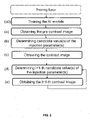
- the model used in the processing method is preferably trained according to the present training method, hence referred to at step (a0) in the figure 2 .
- the model may be directly taken “off the shelf” with preset values of parameters and weights.
- Said training method is similar to the previously described processing method, but is iteratively performed on training images of the training database, i.e. a base of training pre-contrast or contrast images respectively depicting a body part prior to and during an injection of contrast agent, each image being associated to reference value(s) of at least one injection parameter of said injection of contrast agent and organized into sequences corresponding to the same injection.
- Training images are preferably further associated to a reference quality level.
- the training method comprises, for each of a plurality of training pre-contrast images from the training base, a step of determining candidate value(s) of said injection parameter(s) by application of the prediction model to said training pre-contrast image, such that the sequence of said training pre-contrast image and at least a contrast image depicting said body part during injection of contrast agent in accordance with the determined candidate value(s) of said injection parameter(s) minimizes a radiation dose; and verifying if the sequence of said theoretical contrast image and the contrast image minimize the radiation dose.
- the training may be direct.
- minimizing the radiation dose for a given training pre-contrast image is equivalent to produce a "real" contrast image as close as possible to the training contrast image following said pre-contrast image in the same sequence, i.e. equivalent to predict candidate value(s) of said injection parameter(s) as close as possible to the reference value(s) of the injection parameter(s) associated to said training contrast image.
- said prediction of model can be verified by comparing the determined candidate value(s) and reference value(s) of the injection parameter(s).
- the prediction model may be trained so as to predict candidate value(s) of the injection parameter(s), such that the theoretical contrast image depicting said body part during injection of contrast agent in accordance with the determined candidate value(s) of said injection parameter(s) is expected to present a target quality level corresponding to a minimal radiation dose.
- the training may again be direct (if there is an identified contrast image presenting said target quality level corresponding to the minimal radiation dose belonging to the same sequence as said training pre-contrast image, said "theoretical" contrast image can be verified by comparing the determined candidate value(s) and reference value(s) of the injection parameter(s) of said identified training image), or there may be two sub-models that are independently trained on the training base:
- the invention provides a computer program product comprising code instructions to execute a method (particularly on the data processor 11a, 11b of the first or second server 1a, 1b) according to the second aspect of the invention for training at least a prediction model, or a method according to the first aspect of the invention for processing at least a pre-contrast image depicting a body part prior to an injection of contrast agent, and storage means readable by computer equipment (memory of the first or second server 1a, 1b) provided with this computer program product.
Landscapes
- Health & Medical Sciences (AREA)
- Engineering & Computer Science (AREA)
- Medical Informatics (AREA)
- Public Health (AREA)
- General Health & Medical Sciences (AREA)
- Life Sciences & Earth Sciences (AREA)
- Biomedical Technology (AREA)
- Nuclear Medicine, Radiotherapy & Molecular Imaging (AREA)
- Radiology & Medical Imaging (AREA)
- Primary Health Care (AREA)
- Epidemiology (AREA)
- Pathology (AREA)
- Physics & Mathematics (AREA)
- Data Mining & Analysis (AREA)
- Optics & Photonics (AREA)
- High Energy & Nuclear Physics (AREA)
- Biophysics (AREA)
- Veterinary Medicine (AREA)
- Heart & Thoracic Surgery (AREA)
- Molecular Biology (AREA)
- Surgery (AREA)
- Animal Behavior & Ethology (AREA)
- Databases & Information Systems (AREA)
- Theoretical Computer Science (AREA)
- Dentistry (AREA)
- General Physics & Mathematics (AREA)
- Oral & Maxillofacial Surgery (AREA)
- Computer Vision & Pattern Recognition (AREA)
- Quality & Reliability (AREA)
- Business, Economics & Management (AREA)
- General Business, Economics & Management (AREA)
- Apparatus For Radiation Diagnosis (AREA)
Priority Applications (4)
| Application Number | Priority Date | Filing Date | Title |
|---|---|---|---|
| EP21315120.2A EP4113537A1 (de) | 2021-06-30 | 2021-06-30 | Verfahren zum trainieren eines vorhersagemodells oder zur verarbeitung von mindestens einem vorkontrastbild, das unter verwendung des vorhersagemodells ein körperteil vor einer injektion von kontrastmittel darstellt |
| EP22740855.6A EP4364165A1 (de) | 2021-06-30 | 2022-06-30 | Verfahren zum trainieren eines vorhersagemodells oder zum verarbeiten mindestens eines einen körperteil darstellenden vorkontrastbildes vor einer injektion eines kontrastmittels |
| US18/570,245 US20240274273A1 (en) | 2021-06-30 | 2022-06-30 | Methods for training a prediction model, or for processing at least a pre-contrast image depicting a body part prior to an injection of contrast agent using said prediction model |
| PCT/EP2022/068061 WO2023275246A1 (en) | 2021-06-30 | 2022-06-30 | Methods for training a prediction model, or for processing at least a pre-contrast image depicting a body part prior to an injection of contrast agent using said prediction model |
Applications Claiming Priority (1)
| Application Number | Priority Date | Filing Date | Title |
|---|---|---|---|
| EP21315120.2A EP4113537A1 (de) | 2021-06-30 | 2021-06-30 | Verfahren zum trainieren eines vorhersagemodells oder zur verarbeitung von mindestens einem vorkontrastbild, das unter verwendung des vorhersagemodells ein körperteil vor einer injektion von kontrastmittel darstellt |
Publications (1)
| Publication Number | Publication Date |
|---|---|
| EP4113537A1 true EP4113537A1 (de) | 2023-01-04 |
Family
ID=77179958
Family Applications (2)
| Application Number | Title | Priority Date | Filing Date |
|---|---|---|---|
| EP21315120.2A Withdrawn EP4113537A1 (de) | 2021-06-30 | 2021-06-30 | Verfahren zum trainieren eines vorhersagemodells oder zur verarbeitung von mindestens einem vorkontrastbild, das unter verwendung des vorhersagemodells ein körperteil vor einer injektion von kontrastmittel darstellt |
| EP22740855.6A Pending EP4364165A1 (de) | 2021-06-30 | 2022-06-30 | Verfahren zum trainieren eines vorhersagemodells oder zum verarbeiten mindestens eines einen körperteil darstellenden vorkontrastbildes vor einer injektion eines kontrastmittels |
Family Applications After (1)
| Application Number | Title | Priority Date | Filing Date |
|---|---|---|---|
| EP22740855.6A Pending EP4364165A1 (de) | 2021-06-30 | 2022-06-30 | Verfahren zum trainieren eines vorhersagemodells oder zum verarbeiten mindestens eines einen körperteil darstellenden vorkontrastbildes vor einer injektion eines kontrastmittels |
Country Status (3)
| Country | Link |
|---|---|
| US (1) | US20240274273A1 (de) |
| EP (2) | EP4113537A1 (de) |
| WO (1) | WO2023275246A1 (de) |
Citations (4)
| Publication number | Priority date | Publication date | Assignee | Title |
|---|---|---|---|---|
| WO2017035097A1 (en) * | 2015-08-21 | 2017-03-02 | Duke University | Systems and methods for determining image quality and models based on contrast injection protocols |
| US20190108634A1 (en) * | 2017-10-09 | 2019-04-11 | The Board Of Trustees Of The Leland Stanford Junior University | Contrast Dose Reduction for Medical Imaging Using Deep Learning |
| EP3552550A1 (de) * | 2018-04-11 | 2019-10-16 | Siemens Healthcare GmbH | Auf maschinenlernen basierende kontrastmittelverabreichung |
| US20210133960A1 (en) * | 2019-11-01 | 2021-05-06 | GE Precision Healthcare LLC | Methods and systems for an adaptive five-zone perfusion scan |
Family Cites Families (6)
| Publication number | Priority date | Publication date | Assignee | Title |
|---|---|---|---|---|
| WO2002032292A2 (en) * | 2000-10-18 | 2002-04-25 | The Board Of Trustess Of The Leland Stanford Junior University | Methods for development and use of diagnostic and therapeutic agents |
| US20100331673A1 (en) * | 2009-06-26 | 2010-12-30 | Michael Maschke | System and method for reducing patient risk of allergic reaction to contrast agents or medical material |
| US20110282194A1 (en) * | 2010-05-06 | 2011-11-17 | Bruce Reiner | Method and apparatus of quantitative analysis and data mining of medical imaging agent administration |
| US20180071452A1 (en) * | 2016-09-13 | 2018-03-15 | Siemens Healthcare Gmbh | System and Method for Optimizing Contrast Imaging of a Patient |
| EP4165397B1 (de) * | 2020-06-15 | 2026-02-25 | EosDx Inc. | Auf diffraktometer basierendes globales in-situ-diagnosesystem |
| EP4016106A1 (de) * | 2020-12-18 | 2022-06-22 | Guerbet | Verfahren zum trainieren von mindestens einem vorhersagemodell für medizinische bildgebung oder zur verarbeitung von mindestens einem vorkontrastbild, das unter verwendung des vorhersagemodells ein körperteil vor einer injektion von kontrastmittel darstellt |
-
2021
- 2021-06-30 EP EP21315120.2A patent/EP4113537A1/de not_active Withdrawn
-
2022
- 2022-06-30 US US18/570,245 patent/US20240274273A1/en active Pending
- 2022-06-30 WO PCT/EP2022/068061 patent/WO2023275246A1/en not_active Ceased
- 2022-06-30 EP EP22740855.6A patent/EP4364165A1/de active Pending
Patent Citations (4)
| Publication number | Priority date | Publication date | Assignee | Title |
|---|---|---|---|---|
| WO2017035097A1 (en) * | 2015-08-21 | 2017-03-02 | Duke University | Systems and methods for determining image quality and models based on contrast injection protocols |
| US20190108634A1 (en) * | 2017-10-09 | 2019-04-11 | The Board Of Trustees Of The Leland Stanford Junior University | Contrast Dose Reduction for Medical Imaging Using Deep Learning |
| EP3552550A1 (de) * | 2018-04-11 | 2019-10-16 | Siemens Healthcare GmbH | Auf maschinenlernen basierende kontrastmittelverabreichung |
| US20210133960A1 (en) * | 2019-11-01 | 2021-05-06 | GE Precision Healthcare LLC | Methods and systems for an adaptive five-zone perfusion scan |
Also Published As
| Publication number | Publication date |
|---|---|
| US20240274273A1 (en) | 2024-08-15 |
| EP4364165A1 (de) | 2024-05-08 |
| WO2023275246A1 (en) | 2023-01-05 |
Similar Documents
| Publication | Publication Date | Title |
|---|---|---|
| US12223649B2 (en) | Systems and methods for medical acquisition processing and machine learning for anatomical assessment | |
| CN112292086B (zh) | 超声病变评估及相关联的设备、系统和方法 | |
| CN111739614B (zh) | 医学图像增强 | |
| US11080895B2 (en) | Generating simulated body parts for images | |
| EP3649955A1 (de) | Deep learning basierte spektrale bolusverfolgung | |
| US20240054648A1 (en) | Methods for training at least a prediction model, or for processing at least a pre-contrast image depicting a body part prior to an injection of contrast agent using said prediction model | |
| US11301961B2 (en) | Medical signal processing apparatus and model learning apparatus | |
| JP7106307B2 (ja) | 医用画像診断装置、医用信号復元方法、医用信号復元プログラム、モデル学習方法、モデル学習プログラム、および磁気共鳴イメージング装置 | |
| Mohebbian et al. | Classifying MRI motion severity using a stacked ensemble approach | |
| CN110910342B (zh) | 通过使用深度学习来分析骨骼创伤 | |
| KR20230030810A (ko) | 데이터 생성 방법 및 이를 이용한 학습방법 및 장치 | |
| WO2020209826A1 (en) | Deep reinforcement learning for computer assisted reading and analysis | |
| Niu et al. | Noise entangled GAN for low-dose CT simulation | |
| Nasimova et al. | Generating and Evaluating Synthetic Medical Images | |
| US20240274273A1 (en) | Methods for training a prediction model, or for processing at least a pre-contrast image depicting a body part prior to an injection of contrast agent using said prediction model | |
| Lei et al. | Generative adversarial network for image synthesis | |
| Singh et al. | MDA-GAN: Multiscale and dual attention generative adversarial network for bone suppression in chest x-rays | |
| US20240265537A1 (en) | Methods for training a tomosynthesis reconstruction model, or for generating at least one contrast tomogram depicting a target body part during an injection of contrast agent | |
| JP7841930B2 (ja) | 医用画像処理装置、医用画像処理装置の動作方法及びプログラム | |
| Wu et al. | Cardiac Phase Estimation Using Deep Learning Analysis of Pulsed-Mode Projections: Toward Autonomous Cardiac CT Imaging | |
| EP4657379A1 (de) | Hybride intelligenz für wissensgesteuerte synthese von realistischen läsionen | |
| da Silva Corado | Echocardiography Automatic Image Quality Enhancement Using Generative Adversarial Networks | |
| Corado | Echocardiography Automatic Image Quality Enhancement Using Generative Adversarial Networks | |
| Reasat et al. | MSTT-199: MRI Dataset for Musculoskeletal Soft Tissue Tumor Segmentation | |
| MohammadiNasab et al. | No-reference based automatic parameter optimization for iterative reconstruction using a novel search space aware crow search algorithm |
Legal Events
| Date | Code | Title | Description |
|---|---|---|---|
| PUAI | Public reference made under article 153(3) epc to a published international application that has entered the european phase |
Free format text: ORIGINAL CODE: 0009012 |
|
| STAA | Information on the status of an ep patent application or granted ep patent |
Free format text: STATUS: THE APPLICATION HAS BEEN PUBLISHED |
|
| AK | Designated contracting states |
Kind code of ref document: A1 Designated state(s): AL AT BE BG CH CY CZ DE DK EE ES FI FR GB GR HR HU IE IS IT LI LT LU LV MC MK MT NL NO PL PT RO RS SE SI SK SM TR |
|
| STAA | Information on the status of an ep patent application or granted ep patent |
Free format text: STATUS: THE APPLICATION IS DEEMED TO BE WITHDRAWN |
|
| 18D | Application deemed to be withdrawn |
Effective date: 20230705 |