EP4120186A1 - Systemes et procedes mis en oeuvre par ordinateur pour la detection et la caracterisation d'objets - Google Patents
Systemes et procedes mis en oeuvre par ordinateur pour la detection et la caracterisation d'objets Download PDFInfo
- Publication number
- EP4120186A1 EP4120186A1 EP21185179.5A EP21185179A EP4120186A1 EP 4120186 A1 EP4120186 A1 EP 4120186A1 EP 21185179 A EP21185179 A EP 21185179A EP 4120186 A1 EP4120186 A1 EP 4120186A1
- Authority
- EP
- European Patent Office
- Prior art keywords
- interest
- classification
- location
- size
- network
- Prior art date
- Legal status (The legal status is an assumption and is not a legal conclusion. Google has not performed a legal analysis and makes no representation as to the accuracy of the status listed.)
- Withdrawn
Links
Images






Classifications
-
- G—PHYSICS
- G06—COMPUTING OR CALCULATING; COUNTING
- G06T—IMAGE DATA PROCESSING OR GENERATION, IN GENERAL
- G06T7/00—Image analysis
- G06T7/0002—Inspection of images, e.g. flaw detection
- G06T7/0012—Biomedical image inspection
-
- A—HUMAN NECESSITIES
- A61—MEDICAL OR VETERINARY SCIENCE; HYGIENE
- A61B—DIAGNOSIS; SURGERY; IDENTIFICATION
- A61B1/00—Instruments for performing medical examinations of the interior of cavities or tubes of the body by visual or photographical inspection, e.g. endoscopes; Illuminating arrangements therefor
- A61B1/00002—Operational features of endoscopes
- A61B1/00004—Operational features of endoscopes characterised by electronic signal processing
- A61B1/00009—Operational features of endoscopes characterised by electronic signal processing of image signals during a use of endoscope
- A61B1/000096—Operational features of endoscopes characterised by electronic signal processing of image signals during a use of endoscope using artificial intelligence
-
- G—PHYSICS
- G06—COMPUTING OR CALCULATING; COUNTING
- G06T—IMAGE DATA PROCESSING OR GENERATION, IN GENERAL
- G06T7/00—Image analysis
- G06T7/60—Analysis of geometric attributes
- G06T7/62—Analysis of geometric attributes of area, perimeter, diameter or volume
-
- G—PHYSICS
- G06—COMPUTING OR CALCULATING; COUNTING
- G06T—IMAGE DATA PROCESSING OR GENERATION, IN GENERAL
- G06T7/00—Image analysis
- G06T7/70—Determining position or orientation of objects or cameras
- G06T7/73—Determining position or orientation of objects or cameras using feature-based methods
-
- G—PHYSICS
- G06—COMPUTING OR CALCULATING; COUNTING
- G06V—IMAGE OR VIDEO RECOGNITION OR UNDERSTANDING
- G06V10/00—Arrangements for image or video recognition or understanding
- G06V10/20—Image preprocessing
- G06V10/255—Detecting or recognising potential candidate objects based on visual cues, e.g. shapes
-
- G—PHYSICS
- G06—COMPUTING OR CALCULATING; COUNTING
- G06V—IMAGE OR VIDEO RECOGNITION OR UNDERSTANDING
- G06V10/00—Arrangements for image or video recognition or understanding
- G06V10/70—Arrangements for image or video recognition or understanding using pattern recognition or machine learning
- G06V10/764—Arrangements for image or video recognition or understanding using pattern recognition or machine learning using classification, e.g. of video objects
-
- G—PHYSICS
- G06—COMPUTING OR CALCULATING; COUNTING
- G06T—IMAGE DATA PROCESSING OR GENERATION, IN GENERAL
- G06T2200/00—Indexing scheme for image data processing or generation, in general
- G06T2200/24—Indexing scheme for image data processing or generation, in general involving graphical user interfaces [GUIs]
-
- G—PHYSICS
- G06—COMPUTING OR CALCULATING; COUNTING
- G06T—IMAGE DATA PROCESSING OR GENERATION, IN GENERAL
- G06T2207/00—Indexing scheme for image analysis or image enhancement
- G06T2207/10—Image acquisition modality
- G06T2207/10016—Video; Image sequence
-
- G—PHYSICS
- G06—COMPUTING OR CALCULATING; COUNTING
- G06T—IMAGE DATA PROCESSING OR GENERATION, IN GENERAL
- G06T2207/00—Indexing scheme for image analysis or image enhancement
- G06T2207/10—Image acquisition modality
- G06T2207/10068—Endoscopic image
-
- G—PHYSICS
- G06—COMPUTING OR CALCULATING; COUNTING
- G06T—IMAGE DATA PROCESSING OR GENERATION, IN GENERAL
- G06T2207/00—Indexing scheme for image analysis or image enhancement
- G06T2207/20—Special algorithmic details
- G06T2207/20081—Training; Learning
-
- G—PHYSICS
- G06—COMPUTING OR CALCULATING; COUNTING
- G06T—IMAGE DATA PROCESSING OR GENERATION, IN GENERAL
- G06T2207/00—Indexing scheme for image analysis or image enhancement
- G06T2207/20—Special algorithmic details
- G06T2207/20084—Artificial neural networks [ANN]
-
- G—PHYSICS
- G06—COMPUTING OR CALCULATING; COUNTING
- G06T—IMAGE DATA PROCESSING OR GENERATION, IN GENERAL
- G06T2207/00—Indexing scheme for image analysis or image enhancement
- G06T2207/30—Subject of image; Context of image processing
- G06T2207/30004—Biomedical image processing
- G06T2207/30028—Colon; Small intestine
-
- G—PHYSICS
- G06—COMPUTING OR CALCULATING; COUNTING
- G06T—IMAGE DATA PROCESSING OR GENERATION, IN GENERAL
- G06T2207/00—Indexing scheme for image analysis or image enhancement
- G06T2207/30—Subject of image; Context of image processing
- G06T2207/30004—Biomedical image processing
- G06T2207/30028—Colon; Small intestine
- G06T2207/30032—Colon polyp
-
- G—PHYSICS
- G06—COMPUTING OR CALCULATING; COUNTING
- G06T—IMAGE DATA PROCESSING OR GENERATION, IN GENERAL
- G06T2207/00—Indexing scheme for image analysis or image enhancement
- G06T2207/30—Subject of image; Context of image processing
- G06T2207/30004—Biomedical image processing
- G06T2207/30096—Tumor; Lesion
-
- G—PHYSICS
- G06—COMPUTING OR CALCULATING; COUNTING
- G06T—IMAGE DATA PROCESSING OR GENERATION, IN GENERAL
- G06T2207/00—Indexing scheme for image analysis or image enhancement
- G06T2207/30—Subject of image; Context of image processing
- G06T2207/30168—Image quality inspection
Definitions
- the present disclosure relates generally to the field of imaging systems and computer-implemented systems and methods for processing real-time video. More specifically, and without limitation, this disclosure relates to systems, methods, and computer-readable media for processing frames of real-time video and performing object detection and characterization.
- the systems and methods disclosed herein may be used in various applications, such as medical image analysis for polyp detection and characterization, including determining the classification, size, and location of polyps.
- the systems and methods disclosed herein may also be implemented to provide real-time image processing capabilities, such as identifying, based on one or more of object characteristics, a medical guideline and presenting, in real-time on a display device, information for the medical guideline.
- Modern vision and image analysis systems require the ability to detect and characterize objects of interest in a scene.
- An object of interest may be a person, place, feature, or thing.
- the accuracy of object detection and characterization is important to ensure a proper diagnosis and/or treatment.
- Example objects of interest in medical applications include lesions, polyps and/or other abnormalities on or of human tissue.
- extant systems may lack the capability to detect variations in object types and/or produce false positives. Some also suffer from limited response time or the inability to process real-time video signals. Still further, extant systems may not provide object characterization capabilities or only limited object information.
- systems, methods, and computer-readable media are provided for processing real-time video, including for processing frames of real-time video and performing object detection and characterization.
- Embodiments of the present disclosure also relate to systems and methods for object detection and characterization using real-time video from a medical image device.
- object detection and characterization may include polyp detection and characterization, as well as other abnormality detections and characterizations.
- a system of one or more computers can be configured to perform particular operations or actions by virtue of having software, firmware, hardware, or a combination of them installed for the system that in operation causes or cause the system to perform the actions.
- One or more computer programs can be configured to perform operations or actions by virtue of including instructions that, when executed by data processing apparatus (such as one or more processors), cause the apparatus to perform such operations or actions.
- the computer-implemented system may include at least one processor configured to receive a real-time video captured from a medical image device during a medical procedure, where the real-time video includes a plurality of frames.
- the at least one processor may be further configured to detect an object of interest in the plurality of frames and apply one or more neural networks that implement: a trained classification network configured to determine a classification of the object of interest, a trained location network configured to determine a location associated with the object of interest, and a trained size network configured to determine a size associated with the object of interest.
- the at least one processor may be configured to identify, based on one or more of the classification, the location, and the size of the object of interest, a medical guideline.
- the at least one processor may be further configured to present, in real-time on a display device during the medical procedure, information for the identified medical guideline.
- Other embodiments include corresponding computer methods, apparatus, and computer programs recorded on one or more computer storage devices, each configured to perform the above operations or features.
- the medical procedure may include at least one of an endoscopy, a gastroscopy, a colonoscopy, or an enteroscopy.
- the object of interest may include at least one of a formation on or of human tissue, a change in human tissue from one type of cell to another type of cell, an absence of human tissue from a location where the human tissue is expected, or a lesion.
- the object of interest may be a polyp.
- the information for the identified medical guideline may include an instruction to leave or resect the object of interest.
- the information for the identified medical guideline may also include a type of resection.
- the at least one processor may be further configured to generate a confidence value associated with the identified medical guideline.
- the determined classification may be based on at least one of a histological classification, a morphological classification, a structural classification, or a malignancy classification.
- the determined location associated with the object of interest may be a location in a human body.
- the location in the human body may be one of a location in a rectum, sigmoid colon, descending colon, transverse colon, ascending colon, or cecum.
- the determined size associated with the object of interest may be a numeric value or a size classification.
- the at least one processor may be further configured to apply one or more neural networks that implement a trained quality network to: determine a frame quality associated with at least one of the plurality of frames, and generate a confidence value associated with the determined frame quality.
- the at least one processor may be further configured to: aggregate data associated with the determined classification, location, and size when at least one of the determined frame quality or the confidence value is above a predetermined threshold and present, on the display device, at least a portion of the aggregated data.
- the at least one processor may be configured to detect a plurality of objects of interest in the plurality of frames and determine a plurality of classifications and sizes associated with the plurality of objects of interest, where a classification and a size in the plurality of classifications and sizes are associated with a detected object of interest in the detected plurality of objects of interest.
- the computer-implemented system may include at least one processor configured to receive a real-time video, where the real-time video includes a plurality of frames collected during a medical procedure.
- the at least one processor may be further configured to detect an object of interest in the plurality of frames and apply one or more neural networks that implement a trained characterization network configured to: determine a plurality of features associated with the object of interest, and determine confidence values associated with the plurality of features.
- the at least one processor may be further configured to identify, based on one or more of the plurality of features and the confidence values, a medical guideline and present, in real-time on a display device during the medical procedure, information for the identified medical guideline.
- Other embodiments include corresponding computer methods, apparatus, and computer programs recorded on one or more computer storage devices, each configured to perform the above operations or features.
- the medical procedure may include at least one of an endoscopy, a gastroscopy, a colonoscopy, or an enteroscopy.
- the object of interest may include at least one of a formation on or of human tissue, a change in human tissue from one type of cell to another type of cell, an absence of human tissue from a location where the human tissue is expected, or a lesion.
- the object of interest may be a polyp.
- the information for the medical guideline may include an instruction to leave or resect the object of interest.
- the information for the identified medical guideline may also include a type of resection.
- the at least one processor may be further configured to generate a confidence value associated with the identified medical guideline.
- the trained characterization network may include: a trained classification network configured to determine a classification associated with the object of interest and to generate a classification confidence value associated with the determined classification, a trained location network configured to determine a location associated with the object of interest and to generate a location confidence value associated with the determined location, and a trained size network configured to determine a size associated with the object of interest and to generate a size confidence value associated with the determined size.
- the at least one processor may be further configured to present, on the display device, information associated with at least one of the classification, the location, or the size.
- the at least one processor may be further configured to: apply one or more neural networks that implement a trained quality network configured to determine a frame quality associated with at least one of the plurality of frames; and generate a confidence value associated with the determined frame quality.
- the at least one processor may be further configured to aggregate data associated with the plurality of features when at least one of the determined frame quality or the confidence value is above a predetermined threshold and present, on the display device, at least a portion of the aggregated data.
- the at least one processor may be further configured to detect a plurality of objects of interest in the plurality of frames, determine a plurality of sets of features associated with the plurality of objects of interest, where a set of features in the plurality of sets of features includes characterization and size information associated with a detected object of interest in the plurality of objects of interest, and present, on the display device, information associated with one or more sets of features in the plurality of sets of features. Implementations of these and the other above-described operations and techniques may include hardware, a method or process, or computer software on a computer-accessible medium.
- the computer-implemented system may include at least one processor configured to detect an object of interest in a plurality of frames received from a medical image device and characterize the object of interest, the characterization including determining a plurality of features associated with the object of interest.
- the plurality of features may include a location and a size of the object of interest.
- the at least one processor may be further configured to aggregate, when the object of interest persists over more than one of the plurality of frames, information associated with the determined location and size of the object of interest.
- the at least one processor may present, on a display device, when the determined location is in a first body region and the determined size is within a first range, the aggregated information for the object of interest and present, on the display device, when the determined location is in a second body region and the determined size is within a second range, information indicating a status of the characterization of the object of interest.
- Other embodiments include corresponding computer methods, apparatus, and computer programs recorded on one or more computer storage devices, each configured to perform the above operations or features.
- Implementations may include one or more of the following features.
- the at least one processor may be further configured to identify, based on the determined location and size of the object of interest, a medical guideline and present, on the display device, information associated with the identified medical guideline.
- the plurality of features further may include a classification of the object of interest, the classification being based on at least one of a histological classification, a morphological classification, a structural classification, or a malignancy classification.
- the determined location associated with the object of interest may be a location in at least one of a rectum, sigmoid colon, descending colon, transverse colon, ascending colon, or cecum.
- the determined size associated with the object of interest may be a numeric value or a size classification.
- the at least one processor may be further configured to detect a plurality of objects of interest in the plurality of frames, characterize the plurality of objects of interest, the characterization including determining a plurality of sets of features associated with the plurality of objects of interest, where a set of features in the plurality of sets of features includes characterization and size information associated with a detected object of interest in the plurality of objects of interest and present, on the display device, information associated with one or more sets of features in the plurality of sets of features. Implementations of these and other above-described operations and techniques may include hardware, a method or process, or computer software on a computer-accessible medium.
- the computer-implemented method may include receiving a real-time video captured from a medical image device during a medical procedure, the real-time video may include a plurality of frames.
- the method further includes detecting an object of interest in the plurality of frames, applying one or more neural networks that implement: a trained classification network configured to determine a classification of the object of interest, a trained location network configured to determine a location associated with the object of interest, and a trained size network configured to determine a size associated with the object of interest.
- the method also includes identifying, based on one or more of the classification, the location, and the size, a medical guideline and presenting, in real-time on a display device during the medical procedure, information for the identified medical guideline.
- Implementations of the present disclosure may include programs or instructions that are machine constructed and/or programmed specifically for performing functions associated with the disclosed operations or actions. Still further, non-transitory computer-readable storage media may be used that store program instructions, which are executable by at least one processor to perform the steps and/or methods described herein.
- Embodiments described herein may refer to a non-transitory computer readable medium containing instructions that when executed by at least one processor, cause the at least one processor to perform a method or set of operations.
- Non-transitory computer readable mediums may be any medium capable of storing data in any memory in a way that may be read by any computing device with a processor to carry out methods or any other instructions stored in the memory.
- the non-transitory computer readable medium may be implemented as software, firmware, hardware, or any combination thereof.
- Software may preferably be implemented as an application program tangibly embodied on a program storage unit or computer readable medium consisting of parts, or of certain devices and/or a combination of devices.
- the application program may be uploaded to, and executed by, a machine comprising any suitable architecture.
- the machine may be implemented on a computer platform having hardware such as one or more central processing units (“CPUs"), a memory, and input/output interfaces.
- the computer platform may also include an operating system and microinstruction code.
- the various processes and functions described in this disclosure may be either part of the microinstruction code or part of the application program, or any combination thereof, which may be executed by a CPU, whether or not such a computer or processor is explicitly shown.
- various other peripheral units may be connected to the computer platform such as an additional data storage unit and a printing unit.
- a non-transitory computer readable medium may be any computer readable medium except for a transitory propagating signal.
- the memory may include any mechanism for storing electronic data or instructions, including Random Access Memory (RAM), a Read-Only Memory (ROM), a hard disk, an optical disk, a magnetic medium, a flash memory, other permanent, fixed, volatile or non-volatile memory.
- RAM Random Access Memory
- ROM Read-Only Memory
- the memory may include one or more separate storage devices collocated or disbursed, capable of storing data structures, instructions, or any other data.
- the memory may further include a memory portion containing instructions for the processor to execute.
- the memory may also be used as a working memory device for the processors or as a temporary storage.
- a processor may be any physical device or group of devices having electric circuitry that performs a logic operation on input or inputs.
- the at least one processor may include one or more integrated circuits (IC), including application-specific integrated circuit (ASIC), microchips, microcontrollers, microprocessors, all or part of a central processing unit (CPU), graphics processing unit (GPU), digital signal processor (DSP), field-programmable gate array (FPGA), server, virtual server, or other circuits suitable for executing instructions or performing logic operations.
- the instructions executed by at least one processor may, for example, be pre-loaded into a memory integrated with or embedded into the controller or may be stored in a separate memory.
- the at least one processor may include more than one processor.
- Each processor may have a similar construction, or the processors may be of differing constructions that are electrically connected or disconnected from each other.
- the processors may be separate circuits or integrated in a single circuit.
- the processors may be configured to operate independently or collaboratively.
- the processors may be coupled electrically, magnetically, optically, acoustically, mechanically or by other means that permit them to interact.
- a network may constitute any type of physical or wireless computer networking arrangement used to exchange data.
- a network may be the Internet, a private data network, a virtual private network using a public network, a Wi-Fi network, a LAN or WAN network, and/or other suitable connections that may enable information exchange among various components of the system.
- a network may include one or more physical links used to exchange data, such as Ethernet, coaxial cables, twisted pair cables, fiber optics, or any other suitable physical medium for exchanging data.
- a network may also include a public switched telephone network ("PSTN”) and/or a wireless cellular network.
- PSTN public switched telephone network
- a network may be a secured network or unsecured network.
- one or more components of the system may communicate directly through a dedicated communication network.
- Direct communications may use any suitable technologies, including, for example, BLUETOOTH TM , BLUETOOTH LE TM (BLE), Wi-Fi, near field communications (NFC), or other suitable communication methods that provide a medium for exchanging data and/or information between separate entities.
- machine learning networks or algorithms may be trained using training examples, for example in the cases described below.
- Some non-limiting examples of such machine learning algorithms may include classification algorithms, data regressions algorithms, image segmentation algorithms, visual detection algorithms (such as object detectors, face detectors, person detectors, motion detectors, edge detectors, etc.), visual recognition algorithms (such as face recognition, person recognition, object recognition, etc.), speech recognition algorithms, mathematical embedding algorithms, natural language processing algorithms, support vector machines, random forests, nearest neighbors algorithms, deep learning algorithms, artificial neural network algorithms, convolutional neural network algorithms, recursive neural network algorithms, linear machine learning models, non-linear machine learning models, ensemble algorithms, and so forth.
- a trained machine learning network or algorithm may comprise an inference model, such as a predictive model, a classification model, a regression model, a clustering model, a segmentation model, an artificial neural network (such as a deep neural network, a convolutional neural network, a recursive neural network, etc.), a random forest, a support vector machine, and so forth.
- the training examples may include example inputs together with the desired outputs corresponding to the example inputs.
- training machine learning algorithms using the training examples may generate a trained machine learning algorithm, and the trained machine learning algorithm may be used to estimate outputs for inputs not included in the training examples.
- the training may be supervised or non-supervised, or a combination thereof.
- validation examples and/or test examples may include example inputs together with the desired outputs corresponding to the example inputs, a trained machine learning algorithm and/or an intermediately trained machine learning algorithm may be used to estimate outputs for the example inputs of the validation examples and/or test examples, the estimated outputs may be compared to the corresponding desired outputs, and the trained machine learning algorithm and/or the intermediately trained machine learning algorithm may be evaluated based on a result of the comparison.
- a machine learning algorithm may have parameters and hyper parameters, where the hyper parameters are set manually by a person or automatically by a process external to the machine learning algorithm (such as a hyper parameter search algorithm), and the parameters of the machine learning algorithm are set by the machine learning algorithm according to the training examples.
- the hyper-parameters are set according to the training examples and the validation examples, and the parameters are set according to the training examples and the selected hyper-parameters.
- the machine learning networks or algorithms may be further retrained based on any output.
- Certain embodiments disclosed herein may include computer-implemented systems for performing operations or methods comprising a series of steps.
- the computer-implemented systems and methods may be implemented by one or more computing devices, which may include one or more processors as described herein, configured to process real-time video.
- the computing device may be one or more computers or any other devices capable of processing data.
- Such computing devices may include a display such as an LED display, augmented reality (AR), or virtual reality (VR) display.
- the computing device may also be implemented in a computing system that includes a back end component (e.g., as a data server), or that includes a middleware component (e.g., an application server), or that includes a front end component (e.g., a user device having a graphical user interface or a Web browser through which a user can interact with an implementation of the systems and techniques described here), or any combination of such back end, middleware, or front end components.
- the components of the system and/or the computing device can be interconnected by any form or medium of digital data communication (e.g., a communication network). Examples of communication networks include a local area network ("LAN”), a wide area network (“WAN”), and the Internet.
- the computing device can include clients and servers. A client and server are generally remote from each other and typically interact through a communication network. The relationship of client and server arises by virtue of computer programs running on the respective computers and having a client-server relationship to each other.
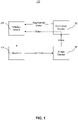
- FIG. 1 illustrates an example computer-implemented system 100 for processing real-time video, according to embodiments of the present disclosure.
- system 100 includes an image device 140 and an operator 120 who operates and controls image device 140 through control signals sent from operator 120 to image device 140.
- operator 120 may be a physician or other health care professional.
- Image device 140 may comprise a medical imaging device, such as an endoscopy imaging device, an X-ray machine, a computed tomography (CT) machine, a magnetic resonance imaging (MRI) machine, or other medical imaging device that produces videos or one or more images of a human body or a portion thereof.
- CT computed tomography
- MRI magnetic resonance imaging
- Operator 120 may control image device 140 by controlling, among other things, a capture rate of image device 140 and/or a movement or navigation of image device 140, e.g., through or relative to the human body of a patient or individual.
- image device 140 may comprise a swallowable capsule device or other form of capsule endoscopy device as opposed to a conventional endoscopy imaging device inserted through a cavity of the human body.
- image device 140 may transmit the captured video as a plurality of image frames to a computing device 160.
- Computing device 160 may comprise one or more processors to process the video, as described herein (see, e.g., FIG. 2 ).
- the one or more of the processors may be implemented as separate component(s) (not shown) that are not part of computing device 160 but in network communication therewith.
- the one or more processors of computing device 160 may implement one or more networks, such as trained neural networks. Examples of neural networks include an object detection network, a classification detection network, a location detection network, a size detection network, or a frame quality detection network, as further described herein.
- Computing device 160 may receive and process the plurality of image frames from image device 140.
- control or information signals may be exchanged between computing device 160 and operator 120 for purposes for controlling or instructing the creation one or more augmented videos. These control or information signals may be communicated as data through image device 140 or directly from operator 120 to computing device 160. Examples of control and information signals include signals for controlling components of computing device 160, such as an object detection network, a classification detection network, a location detection network, a size detection network, or a frame quality detection network, as described herein.
- computing device 160 may process and augment the video received from image device 140 and then transmit the augmented video to a display device 180.
- the video augmentation or modification may comprise providing one or more overlays, alphanumeric characters, shapes, diagrams, images, animated images, or any other suitable graphical representation in or with the video frames.
- the video augmentation may provide information related to an object of interest, such as classification, size and/or location information. Additionally or alternatively, the video augmentation may provide information related to a medical guideline.
- computing device 160 may also be configured to relay the original, non-augmented video from image device 140 directly to display device 180.
- computing device 160 may perform a direct relay under predetermined conditions, such as when there is no overlay or other augmentation to be generated.
- computing device 160 may perform a direct relay if operator 120 transmits a command as part of a control signal to computing device 160 to do so.
- the commands from operator 120 may be generated by operation of button(s) and/or key(s) included on an operator device and/or an input device (not shown), such as a mouse click, a cursor hover, a mouseover, a button press, a keyboard input, a voice command, an interaction performed in virtual or augmented reality, or any other input.
- computing device 160 may process the video from image device 140 and create a modified video stream to send to display device 180.
- the modified video may comprise the original image frames with the augmenting information to be displayed to the operator via display device 180.
- Display device 180 may comprise any suitable display or similar hardware for displaying the video or modified video, such as an LCD, LED, or OLED display, an augmented reality display, or a virtual reality display.
- FIG. 2 is a block diagram of an example computing device 200 for processing real-time video, consistent with embodiments of the present disclosure.
- Computing device 200 may be used in connection with the implementation of the example system of FIG. 1 (including, e.g., computing device 160). It is to be understood that in some embodiments the computing device may include multiple sub-systems, such as cloud computing systems, servers, or any other suitable components for receiving and processing real-time video.
- computing device 200 may include one or more processor(s) 230, which may include, for example, one or more integrated circuits (IC), including application-specific integrated circuit (ASIC), microchips, microcontrollers, microprocessors, all or part of a central processing unit (CPU), graphics processing unit (GPU), digital signal processor (DSP), field-programmable gate array (FPGA), server, virtual server, or other circuits suitable for executing instructions or performing logic operations, as noted above.
- processor(s) 230 may include, or may be a component of, a larger processing unit implemented with one or more processors.
- the one or more processors 230 may be implemented with any combination of general-purpose microprocessors, microcontrollers, digital signal processors (DSPs), field programmable gate array (FPGAs), programmable logic devices (PLDs), controllers, state machines, gated logic, discrete hardware components, dedicated hardware finite state machines, or any other suitable entities that can perform calculations or other manipulations of information.
- DSPs digital signal processors
- FPGAs field programmable gate array
- PLDs programmable logic devices
- controllers state machines, gated logic, discrete hardware components, dedicated hardware finite state machines, or any other suitable entities that can perform calculations or other manipulations of information.
- processor(s) 230 may be communicatively connected via a bus or network 250 to a memory 240.
- Bus or network 250 may be adapted to communicate data and other forms of information.
- Memory 240 may include a memory portion 245 that contains instructions that when executed by the processor(s) 230, perform the operations and methods described in more detail herein.
- Memory 240 may also be used as a working memory for processor(s) 230, a temporary storage, and other memory or storage roles, as the case may be.
- memory 240 may be a volatile memory such as, but not limited to, random access memory (RAM), or non-volatile memory (NVM), such as, but not limited to, flash memory.
- RAM random access memory
- NVM non-volatile memory
- Processor(s) 230 may also be communicatively connected via bus or network 250 to one or more I/O device 210.
- I/O device 210 may include any type of input and/or output device or periphery device.
- I/O device 210 may including one or more network interface cards, APIs, data ports, and/or other components for supporting connectivity with processor(s) 230 via network 250.
- processor(s) 230 and the other components (210, 240) of computing device 200 may be communicatively connected to a database or storage device 220.
- Storage device 220 may electronically store data in an organized format, structure, or set of files.
- Storage device 220 may include a database management system to facilitate data storage and retrieval. While illustrated in FIG. 2 as a single device, it is to be understood that storage device 220 may include multiple devices either collocated or distributed. In some embodiments, storage device 220 may be implemented on a remote network, such as a cloud storage.
- Processor(s) 230 and/or memory 240 may also include machine-readable media for storing software or sets of instructions.
- "Software” as used herein refers broadly to any type of instructions, whether referred to as software, firmware, middleware, microcode, hardware description language, or otherwise. Instructions may include code (e.g., in source code format, binary code format, executable code format, or any other suitable format of code). The instructions, when executed by one or more processors 230, may cause the processor(s) to perform the various operations and functions described in further detail herein.
- Implementations of computing device 200 are not limited to the example embodiment shown in FIG. 2 .
- the number and arrangement of components (210, 220, 230, 240) may be modified and rearranged.
- computing device 200 may be in electronic communication with other network(s), including the Internet, a local area network, a wide area network, a metro area network, and other networks capable of enabling communication between the elements of the computing architecture.
- computing device 200 may retrieve data or other information described herein from any source, including storage device 220 as well as from network(s) or other database(s).
- computing device 200 may include one or more machine-learning models used to implement the neural networks described herein, and may retrieve or receive weights or parameters of machine-learning models, training information or training feedback, medical guidelines and/or guideline rules, and/or any other data and information described herein.
- Systems, methods, and computer-readable media are provided for processing real-time video.
- the systems and methods described herein may be implemented with the aid of at least one processor or non-transitory computer readable medium, such as a CPU, FPGA, ASIC, or any other processing structure(s) or storage medium of the computing device.
- "Real-time video,” as used herein, may refer to video received by the at least one processor, computing device, and/or system without perceptible delay from the video's source (e.g., an image device).
- the at least one processor may be configured to receive real-time video captured from a medical image device during a medical procedure, consistent with disclosed embodiments.
- a medical image device may be any device capable of producing videos or one or more images of a human body or a portion thereof, such as an endoscopy device, an X-ray machine, a CT machine, or an MRI machine, as described above.
- a medical procedure may be any action performed with the intention of determining, detecting, measuring, or diagnosing a patient condition, such as an endoscopy, a gastroscopy, a colonoscopy, or an enteroscopy.
- the medical procedure may be used to identify objects of interest (e.g., lesions or polyps) in a location in the human body. Locations in the human body may be the rectum, sigmoid colon, descending colon, transverse colon, ascending colon, or cecum. It is to be understood, however, that the disclosed systems and methods may be employed in other contexts and applications.
- the real-time video may comprise a plurality of frames, consistent with disclosed embodiments.
- a "frame,” as used herein, may refer to any digital representation such as a collection of pixels representing of a scene or field of view in the real-time video.
- a pixel may represent a discrete element characterized by a value or intensity in a color space (e.g., based on the RGB, RYB, CMY, CMYK, or YUV color models).
- a frame may be encoded in any appropriate format, such as Joint Photographic Experts Group (JPEG) format, Graphics Interchange Format (GIF), bitmap format, Scalable Vector Graphics (SVG) format, Encapsulated PostScript (EPS) format, or the like.
- JPEG Joint Photographic Experts Group
- GIF Graphics Interchange Format
- SVG Scalable Vector Graphics
- EPS Encapsulated PostScript
- video may refer to any digital representation of a scene or area of interest comprised of a plurality of frames in sequence.
- a video may be encoded in any appropriate format, such as a Moving Picture Experts Group (MPEG) format, a flash video format, an Audio Video Interleave (AVI) format, or any other format.
- MPEG Moving Picture Experts Group
- AVI Audio Video Interleave
- a video need not be encoded, and may more generally include a plurality of frames.
- the frames may be in any order, including a random order.
- a video or plurality of frames may be paired with audio.
- the plurality of frames may include representations of an object of interest.
- An "object of interest,” as used herein, may refer to any visual item or feature in the plurality of frames the detection or characterization of which may be desired.
- an object of interest may be a person, place, entity, feature, area, or any other distinguishable visual item or thing.
- an object of interest may include at least one of a formation on or of human tissue, a change in human tissue from one type of cell to another type of cell, an absence of human tissue from a location where the human tissue is expected, or a lesion.
- Examples of objects of interest in a video captured by an image device may include a polyp (a growth protruding from a gastro-intestinal mucosa), a tumor (a swelling of a part of the body), a bruise (a change from healthy cells to discolored cells), a depression (an absence of human tissue), or an ulcer or abscess (tissue that has suffered damage, i.e., a lesion).
- a polyp a growth protruding from a gastro-intestinal mucosa
- a tumor a swelling of a part of the body
- a bruise a change from healthy cells to discolored cells
- a depression an absence of human tissue
- an ulcer or abscess tissue that has suffered damage, i.e., a lesion
- polyp may refer to growths or lesions of the gastro-intestinal mucosa, and may more generally be used herein to refer to a candidate tissue the detection and characterization of which may be of interest. Polyps may be characterized based on classification, such as based on a histological classification, a morphological classification, a structural classification, or a malignancy classification.
- polyps may be histologically classified using the Narrow-Band Imaging International Colorectal Endoscopic (NICE) or the Vienna classification.
- NICE Narrow-Band Imaging International Colorectal Endoscopic
- a polyp may be one of three (3) types, as follows: (Type 1) sessile serrated polyp or hyperplastic polyp; (Type 2) conventional adenoma; and (Type 3) cancer with deep submucosal invasion.
- a polyp may be one of five (5) types, as follows: (Category 1) negative for neoplasia/dysplasia; (Category 2) indefinite for neoplasia/dysplasia; (Category 3) non-invasive low grade neoplasia (low grade adenoma/dysplasia); (Category 4) mucosal high grade neoplasia, such as high grade adenoma/dysplasia, non-invasive carcinoma (carcinoma in-situ), or suspicion of invasive carcinoma; and (Category 5) invasive neoplasia, intramucosal carcinoma, submucosal carcinoma, or the like.
- a polyp may be morphologically classified using the Paris classification.
- a polyp may be one of three (3) general types, as follows: (Type I) elevated or polypoid forms, such as pedunculated, sessile, and broad-based; (Type II) flat or superficial forms, such as flat and elevated, completely flat, and superficially depressed; and (Type III) excavated forms, including excavated and ulcerated.
- Type I are often referred to as polypoid forms while Types II and III are often referred to as non-polypoid forms.
- a polyp may also be structurally classified based on its shape or appearance.
- a polyp may be classified as benign if its surface is smooth or round in appearance, or as non-benign or malignant if its surface includes abnormal growths or is irregular in appearance.
- a polyp may also be classified based on malignancy.
- a malignancy may be based on a degree of invasion of a disease, such as cancer invasiveness.
- a polyp may, for example, be classified as benign when there is a small or no invasion of cancer in or around the polyp, and the polyp may be classified as malignant or cancerous when there is invasion of cancer in or around the polyp.
- Other classifications will be apparent based on this disclosure and may be selected according to the particular application. Accordingly, the present disclosure is not limited to any particular classification or type of object of interest.
- a polyp may also be characterized based on its size.
- a polyp size may be expressed as a numeric value or a size classification.
- the size of a polyp may be, for example, expressed using any suitable metric value such as millimeters (mm) although any other metric may be used (e.g., inches).
- a polyp may thus have a size of 1 mm, 5 mm, 10 mm, and so forth.
- a polyp size may also be expressed as a classification based on one or more suitable size categories, such as (1) “diminutive” or “small” for polyps having a size less than or equal to 5 mm, (2) “non-diminutive” or “large” for polyps having a size between 6 mm and 9 mm, and (3) “very large” for polyps having a size equal to or greater than 10 mm.
- suitable size categories such as (1) “diminutive” or “small” for polyps having a size less than or equal to 5 mm, (2) “non-diminutive” or “large” for polyps having a size between 6 mm and 9 mm, and (3) “very large” for polyps having a size equal to or greater than 10 mm.
- suitable size categories such as (1) “diminutive” or “small” for polyps having a size less than or equal to 5 mm, (2) “non-diminutive” or “large” for polyps having a size between 6
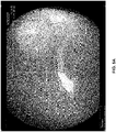
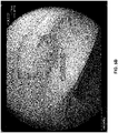
- FIGS. 3A and 3B illustrate frame images of example polyps, consistent with embodiments of the present disclosure.
- the illustrated polyps are characterized based on a classification and a size, consistent with the description above.
- the illustrated polyp is classified as non-adenoma (i.e., not adenomatous) with a diminutive size (i.e., having a size less than or equal to 5 mm).
- the illustrated polyp is classified as adenoma (i.e., pre-cancerous) with a non-diminutive size (i.e., having a size between 6 mm and 9 mm).
- Other characterizations, classifications, and size categories may be used, as explained herein.
- the at least one processor of computing device 160 may be configured to detect an object of interest in the plurality of frames.
- An object of interest may be detected based on, for example, a determination of the presence or absence of an object of interest in the plurality of frames of the video.
- Object detection may be implemented using, for example, machine learning detection networks or algorithms, conventional detection algorithms, or a combination of both.
- the plurality of frames may be fed to one or more neural networks (e.g., a deep neural network, a convolutional neural network, a recursive neural network, etc.), a random forest, a support vector machine, or any other suitable model, as described above, trained to detect the object of interest.
- neural networks e.g., a deep neural network, a convolutional neural network, a recursive neural network, etc.
- a random forest e.g., a support vector machine, or any other suitable model, as described above, trained to detect the object of interest.
- Machine learning detection algorithms, models, or weights may be stored in the computing device and/or system, or they may be fetched from a network or database prior to detection.
- a machine learning detection network or algorithm may be re-trained based on one or more of its outputs, such as true/false positive detections or true/false negatives detections.
- the feedback for re-training may be generated automatically by the system or the computing device, or it may be manually inputted by the operator or another user (e.g., through a mouse or keyboard or other input device). Weights or other parameters of the machine learning detection network or algorithm may be adjusted based on the feedback.
- conventional non-machine learning detection algorithms may be used, either alone or in combination with the machine learning detection networks or algorithms.
- the presence of the polyps may be detected using one or more machine learning detection networks or algorithms, conventional detection algorithms, or a combination of both, consistent with the disclosed embodiments.
- the detection of the polyps may include a determined location of the polyp in the frame and be indicated using any suitable graphical representation, which may be overlaid over a frame in which it is detected.
- each illustrated polyp is surrounded by a rectangular box indicating the detection of the illustrated polyps.
- the detection may be represented using any other graphical representation (e.g., another shape, a color, a pattern, an image, a video, or an alphanumeric character) or the detection may not be represented at all.
- the at least one processor of computing device 160 may be configured to apply one or more neural networks that implement a trained characterization network configured to determine a plurality of features associated with the object of interest, consistent with the disclosed embodiments.
- the trained characterization network may comprise one or more suitable machine learning networks or algorithms for determining a plurality of features associated with the object of interest, including one or more neural networks (e.g., a deep neural network, a convolutional neural network, a recursive neural network, etc.), a random forest, a support vector machine, or any other suitable model as described above trained to determine features of the object of interest in a plurality of objects of interest.
- the characterization network may be trained using a plurality of training frames or portions thereof labeled based on the desired features (e.g., classification, size, or location). For example, a first set of training frames (or portions of frames) containing or not containing an object of interest may be labeled as having a feature, and a second set of training frames (or portions of frames) containing or not containing an object of interest may be labeled as not having the feature. Weights or other parameters of the characterization network may be adjusted based on its output with respect to a third, non-labeled set of training frames (or portions of frames) until a convergence or other metric is achieved, and the process may be repeated with additional training frames (or portions thereof) or with live data, as described herein.
- desired features e.g., classification, size, or location
- the trained characterization network may be configured to determine confidence values associated with the plurality of features.
- a confidence value for an identified feature may refer to an indication of the level of certainty associated with the identified feature. For example, a confidence value of 0.9 or 90% may indicate that there is a ninety percent certainty that the identified feature is present in an object of interest, while a confidence value of 0.4 or 40% may indicate that there is a forty percent certainty that the identified feature is present, and so forth.
- the at least one processor of computing device 160 may be configured to apply one or more specific machine learning networks or algorithms trained to detect one or more specific features.
- the at least one processor may be configured to apply one or more neural networks that implement a trained classification network configured to determine a classification of the object of interest.
- the trained classification network may also be configured to generate a classification confidence value associated with the determined classification.
- the classification may be the same or similar as those described above (e.g., based on at least one of a histological classification, a morphological classification, a structural classification, or a malignancy classification).
- the classification network may be trained using a plurality of training frames or portions thereof labeled based on one of more classifications.
- a first set of training frames (or portions of frames) containing or not containing an object of interest may be labeled as "adenoma”
- a second set of training frames (or portions of frames) containing or not containing an object of interest may be labeled as "non-adenoma” or another classification (e.g., "serrated”).
- Other labeling conventions could be used both in binary (e.g. “hyperplastic” vs “non-hyperplastic") and in multiple classes (e.g. "adenoma” vs “sessile serrated” vs "hyperplastic”).
- Weights or other parameters of the classification network may be adjusted based on its output with respect to a third, non-labeled set of training frames (or portions of frames) until a convergence or other metric is achieved, and the process may be repeated with additional training frames (or portions thereof) or with live data as described herein.
- the at least one processor of computing device 160 may be configured to apply one or more neural networks that implement a trained location network configured to determine a location associated with the object of interest.
- the trained location network may also be configured to generate a location confidence value associated with the determined location.
- the location may be the same or similar as those described above (e.g., a location in a human body, such as the rectum, sigmoid colon, descending colon, transverse colon, ascending colon, or cecum).
- the location network may be trained using a plurality of training frames or portions thereof labeled based on one or more locations (e.g., body locations).
- a first set of training frames (or portions of frames) containing or not containing an object of interest may be labeled as "sigma rectum”
- a second set of training frames (or portions of frames) containing or not containing an object of interest may be labeled as "not sigma rectum” or another body location (e.g., "ascending colon”).
- Weights or other parameters of the location network may be adjusted based on its output with respect to a third, non-labeled set of training frames (or portions of frames) until a convergence or other metric is achieved, and the process may be repeated with additional training frames (or portions thereof) or with live data as described herein.
- the at least one processor of computing device 160 may be configured to apply one or more neural networks that implement a trained size network configured to determine a size associated with the object of interest, and in some embodiments the trained classification network may also be configured to generate a size confidence value associated with the determined size.
- the size may be the same or similar as those described above (e.g., a numeric value or a size classification).
- the location network may be trained using a plurality of training frames or portions thereof labeled based on size.
- a first set of training frames (or portions of frames) containing or not containing an object of interest may be labeled as “diminutive” or "small”
- a second set of training frames (or portions of frames) containing or not containing an object of interest may be labeled as “non-diminutive” or “not small” or another size value (e.g., "large” or “10 mm”).
- Weights or other parameters of the location network may be adjusted based on its output with respect to a third, non-labeled set of training frames (or portions of frames) until a convergence or other metric is achieved, and the process may be repeated with additional training frames (or portions thereof) or with live data as described herein.
- the trained classification, location, and/or size networks may be stored in the computing device and/or system, or they may be fetched from a network or database prior to characterization.
- the trained classification, location, and/or size networks may be re-trained based on one or more outputs, such as true or false feature detections.
- the feedback for re-training may be generated automatically by the system or the computing device, or it may be manually inputted by the operator or another user (e.g., through a mouse or keyboard or other input device). Weights or other parameters of the trained classification, location, and/or size networks may be adjusted based on the feedback.
- conventional non-machine learning detection algorithms may be used, either alone or in combination with the trained classification, location, and/or size networks.
- the at least one processor of computing device 160 may be configured to identify, based on one or more of the plurality of features and/or the confidence values, a medical guideline.
- a “medical guideline,” as used herein, may refer to any information provided with the aim of aiding in the determination, diagnosis, or treatment of a patient condition.
- the identified medical guideline may include an instruction to leave or resect the object of interest.
- the medical guideline may also include an identification or description of a specific type of resection.
- the medical guideline may include an instruction to perform Endoscopic Mucosal Resection (EMR) to resect small polyps or precancerous growths, or to perform Endoscopic Submucosal Dissection (ESD) to resect large polyps or likely-cancerous growths, or any other type of resection.
- EMR Endoscopic Mucosal Resection
- ESD Endoscopic Submucosal Dissection
- Other medical guidelines may be identified based on the specific application or context, however, such as examining the object of interest further, performing other medical examinations, performing an operation, prescribing a drug or medicine, or administrating other treatments.
- the at least one processor may also be configured to present, in real-time on a display device during capture (e.g., during a medical procedure), information for the identified medical guideline, as described above.
- the displayed information for the identified medical guideline may be in any suitable representation, such as one or more alphanumeric characters (e.g., the words “leave” or “resect", or indicating more specifically the type of resection (e.g., "EMR” or “EDS”), shapes (e.g., a check mark or a cross sign), colors (e.g., green or red), images (e.g., an image of a hand or a medical instrument), videos (e.g., a video of a suggested procedure), or a combination thereof.
- alphanumeric characters e.g., the words “leave” or “resect", or indicating more specifically the type of resection (e.g., "EMR” or “EDS”)
- shapes e.g., a check mark or a cross sign
- colors e.g., green or red
- images e.g., an image of a hand or a medical instrument
- videos e.g.,
- the at least one processor may be configured to generate a confidence value associated with the identified medical guideline.
- a confidence value for an identified medical guideline may refer to an indication of the level of certainty associated with the identified medical guideline. For example, a confidence value of 0.9 or 90% may indicate that there is a ninety percent certainty that the identified medical guideline is correct, while a confidence value of 0.4 or 40% may indicate that there is a forty percent certainty that the identified medical guideline is correct, and so forth. Other values, metrics, or representations may be used to represent a confidence value, however.
- a confidence value may be categorized as "high confidence” when the confidence value is above a predetermined threshold (e.g., above 66% confidence), "low confidence” when the confidence value is below a predetermined threshold (e.g., below 33% confidence), or "undetermined” when between two predetermined thresholds (e.g., 33% to 66% confidence).
- the at least one processor may also present a confidence score associated with the medical guideline.
- a confidence score may be represented as alphanumeric characters along with the medical guideline, such as “leave - high confidence” or "resect - 30% confidence," although any other suitable representation may be used (e.g., a shape, a color, an image, a video, or a combination thereof).
- the at least one processor may be configured to identify, based on one or more of the classification, the location, and the size, the medical guideline.
- the medical guideline may be to leave the polyp when it is classified as hyperplastic and to resect the polyp when it is classified as dysplastic or neoplastic.
- the medical guideline may be to leave the polyp when it is determined to be equal to or less than 5 mm or "diminutive" in size and to resect the polyp when it is determined to be greater than 5 mm or "small” or “large” in size.
- the medical guideline may be to leave the polyp when it is determined to be in a non-harmful body location and to resect the polyp when it is determined to be in a harmful body location.
- the medical guideline may be determined based on a combination of the classification, the location, and the size.
- the medical guideline may be to leave the polyp when it is determined to be a hyperplastic polyp located in the sigma rectum having a size that is equal to or less than 5 mm in size.
- the medical guideline may be to leave the polyp when it is determined to be a hyperplastic polyp located in the rectum having a "diminutive" size.
- the medical guideline may be to resect the polyp when it is determined to be an adenoma located in the caecum having a "diminutive" size.
- the medical guideline may be to resect the polyp when it is determined to be an adenoma located in the ascending colon having a "non-diminutive" size.
- a confidence value associated with the relevant characterization may be used to determine the medical guideline.
- the medical guideline may be to resect the polyp only when there is a ninety percent confidence value that the polyp is neoplastic, 10 mm or "large” in size, and/or in a harmful body location, and to leave the polyp otherwise.
- Confidence values could be expressed also as “high confidence” or “low confidence”. Other confidence values and characterizations may be used, however, as described above.
- the medical guideline determinations disclosed herein are provided for illustrative purposes only and are not intended to be exhaustive, and other medical guidelines will be apparent to those having ordinary skill in the art.
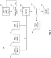
- FIG. 4 illustrates an exemplary system of processing real-time video, consistent with embodiments of the present disclosure.
- a real-time processing system 400 may comprise an image device 410, an object detector 420, a characterization network 430, and a display device 470.
- Image device 410 may be the same or similar to image device 140 described above in connection with FIG. 1 (e.g., an endoscopy machine, an X-ray machine, a CT machine, an MRI machine, or any other medical imaging device), and display device 470 may be the same or similar as the display device 180 also described above in connection with FIG. 1 (e.g., an LCD, LED, or OLED display, an augmented reality display, a virtual reality display, or the like).
- Image device 410 may be configured to capture real-time video, which in some embodiments may be captured during a medical procedure (e.g., an endoscopic procedure), as described above.
- Image device 410 may be configured to feed the captured real-time video to object detector 420.
- Object detector 420 may comprise one or more machine learning detection networks or algorithms, conventional detection algorithms, or a combination of both, as described above.
- Object detector 420 may be configured to detect an object of interest, such as a polyp, in frames of the real-time video captured by image device 410.
- Object detector 420 may also be configured to output a plurality of detections when two or more objects of interest are present in frames of the real-time video.
- Object detector 420 may be configured to output any detection(s) to characterization network 430.
- Characterization network 430 may include one or more trained machine learning algorithms (e.g., one or more neural networks) configured to determine a plurality of features in the objects of interest(s) detected by object detector 420, as described above. As shown in FIG. 4 , characterization network 430 may include trained machine learning networks or algorithms configured to detect specific features, such as classification network 440 configured to determine a classification of the detected object of interest(s), location network 450 configured to determine a body location of the detected object of interest(s), and size network 460 configured to determine a size of the detected object of interest(s). It is to be understood, however, that characterization network 430 may be modified to include all, some, more, or none of the machine learning networks depicted in FIG. 4 . For example, in some embodiments characterization network 430 may be a single network configured to determine all the features of interest, or characterization network 430 may include machine learning networks to determine features other than classification, location, or size, depending on the specific application or context.
- trained machine learning algorithms e.g., one
- characterization network 430 may be configured to identify and output information associated with the determined features to display device 470.
- characterization network 430 may also be configured to identify and output a medical guideline based on the determined features and/or confidence values associated with the determined features, as described above.
- Display device 470 may subsequently present, in real-time during capture (e.g., during a medical procedure), information associated with the plurality of features and/or the medical guideline.
- characterization network 430 may determine and display device 470 may display classification information (e.g., "adenoma” or “non-adenoma”, or “hyperplastic” or “non-hyperplastic”), location information (e.g., “rectum” or “caecum”), size information (e.g., “diminutive” or “non-diminutive” or “small” or “large”), and/or a medical guideline (e.g., “leave” or “resect” or “biopsy”) for the detected object of interest.
- classification information e.g., "adenoma” or “non-adenoma”, or “hyperplastic” or “non-hyperplastic”
- location information e.g., “rectum” or “caecum”
- size information e.g., “diminutive” or “non-diminutive” or “small” or “large”
- a medical guideline e.g., "lea
- characterization network 430 may be configured to generate and output characterization and/or medical guideline information for each individual object of interest (e.g., as a feature vector or another suitable data model) and display 470 may consequently display characterization and/or medical guideline information for each individual object of interest (e.g., on or near each object of interest).
- one or more computing devices may be used to implement object detector 420 and characterization network 430.
- Such computing device(s) may include one or more processors and configured to modify the video from the image device with augmenting information, including the above-described information determined by object detector 420 and characterization network 430.
- augmented video may be fed to display 470 for viewing by the operator of image device 410 and other users.
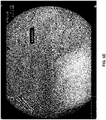
- FIGS. 5A-5E illustrate example augmented frames containing characterization and medical guideline information, consistent with embodiments of the present disclosure.
- the augmented frames illustrated in FIGS. 5A-5E may be displayed in real-time during capture of the video (e.g., during a medical examination), as discussed above. While polyps are illustrated in the example frames of FIGS. 5A-5E , it is to be understood that the disclosed embodiments may be used with any other object of interest.
- the frames may include characterization information associated with one or more features, such as classification, size, and/or location of a polyp. For example, as shown in FIG.
- a polyp may be classified as a non-adenoma, and a corresponding representation such as "non-ade” may be displayed; or, as shown in FIGS. 5B and 5C , the polyp may be classified as an adenoma, and a corresponding representation such as "ade” may be displayed.
- a polyp may be determined to have a diminutive size, and a corresponding representation such as "dim” may be displayed; or, as shown in FIG. 5B , the polyp may be determined to have a non-diminutive size, and a corresponding representation such as "non-dim” may be displayed.
- FIGS. 5A a polyp may be classified as a non-adenoma, and a corresponding representation such as "non-ade” may be displayed; or, as shown in FIGS. 5B and 5C , the polyp may be determined to have a non-diminutive size, and a corresponding representation such as "non-dim” may
- the body location of a polyp may be determined and a corresponding representation may be displayed, such as "Rectum” ( FIG. 5A ), “Ascending” ( FIG. 5B ), “Caecum” ( FIG. 5C ), and “Sigma - rectum” ( FIG. 5D ). If the location cannot be determined, then no location information may be displayed with the augmented frame.
- Other representations may be used, consistent with the present disclosure, such as one or more images, icons, videos, shapes, or numbers.
- a medical guideline may also be displayed.
- the medical guideline based on the classification, size, and/or location may be to not remove the polyp, and a corresponding representation such as "leave” may be displayed.
- the medical guide may be to remove the polyp (or to perform any other procedure or treatment), and a corresponding representation such as "resect” may be displayed.
- characterization and medical guideline information may be displayed (and determined) independently for each polyp in a plurality of polyps in the frames. As shown in FIG.
- separate classification and size information may be displayed adjacent to each detected polyp, which may be different depending on the context.
- other information other than characterization and medical guideline information may be displayed.
- a state of the characterization network and/or the computing device may be displayed, such as whether the characterization network and/or the computing device is currently "analyzing" the frames ( FIG. 5D ), there is “no prediction” ( FIG. 5E ), or there is no characterization yet (shown as a blank field for the object detected in the middle of the frame in FIG. 5E ).
- Examples of other information that may be displayed include the characterization network and/or the computing device partially characterizing the polyp, a confidence value being too low, an error having occurred, the system restarting, a user action or input, or any other information associated with the processing of the real-time video.
- the at least one processor may be configured to apply one or more neural networks that implement a trained quality network configured to determine a frame quality associated with at least one of the plurality of frames.
- a "frame quality,” as used herein, may refer to a degree of visual clarity of one or more frames for purposes of performing the operations described herein.
- a frame quality may be based on any visual characteristic, such as blurriness, sharpness, brightness, lighting, exposure, contrast, movement, visibility, or any other feature of one or more frames.
- the trained frame quality network may be trained to generate a numeric value and/or a quality classification associated with a frame quality.
- the trained frame quality network may be configured to output a number (e.g., 0.7) associated with the frame quality, or it may be configured to assign a quality class to a frame (e.g., "sufficient quality” or "not sufficient quality”).
- the trained frame quality network may comprise one or more suitable machine learning networks or algorithms for determining a quality value associated with one or more frames in the real-time video, including one or more neural networks (e.g., a deep neural network, a convolutional neural network, a recursive neural network, etc.), a random forest, a support vector machine, or any other suitable model as described above trained to determine a frame quality.
- the frame quality network may be trained using a plurality of training frames or portions thereof labeled based on one or more quality values or classifications.
- a first set of training frames may be labeled as "sufficient quality”
- a second set of training frames may be labeled as "not sufficient quality”
- Weights or other parameters of the frame quality network may be adjusted based on its output with respect to a third, non-labeled set of training frames (or portions of frames) until a convergence or other metric is achieved, and the process may be repeated with additional training frames (or portions thereof) or with live data as described herein.
- the trained frame quality network may be stored in the computing device and/or system, or it may be fetched from a network or database prior to determining the frame quality.
- the trained frame quality network may be re-trained based on one or more of its outputs, such as accurate or inaccurate frame quality detections.
- the feedback for re-training may be generated automatically by the system or the computing device, or it may be manually inputted by the operator or another user (e.g., through a mouse or keyboard or other input device). Weights or other parameters of the trained frame quality network may be adjusted based on the feedback.
- conventional non-machine learning frame quality detection networks or algorithms may be used, either alone or in combination with the trained frame quality network.
- the trained frame quality network may be configured to generate a confidence value associated with the determined frame quality.
- a confidence value for a determined frame quality may refer to an indication of the level of certainty associated with the determined frame quality. For example, a confidence value of 0.9 or 90% may indicate that there is a ninety percent certainty that the determined frame quality is correct, while a confidence value of 0.4 or 40% may indicate that there is a forty percent certainty that the determined frame quality is correct, and so forth. Other values, metrics, or representations may be used to represent a confidence value, however.
- a confidence value may be categorized as "high confidence” when the confidence value is above a predetermined threshold (e.g., above 66% confidence), "low confidence” when the confidence value is below a predetermined threshold (e.g., below 33% confidence), or "undetermined” when between two predetermined thresholds (e.g., 33% to 66% confidence).
- the at least one processor may be configured to present, in real-time on the display device, information for the determined frame quality in any suitable format (e.g., the frame quality value and/or classification, a thumbs up or thumbs down, a check mark or cross sign, or a color).
- the at least one processor of the computing device or system may be configured to aggregate data associated with a determined characterization (e.g., classification, location, and/or size) when at least one of the determined frame quality or the confidence value is above a predetermined threshold, consistent with the embodiments of the present disclosure. Aggregation in this context may refer to any operation for combining, collecting, or receiving multiple data. For example, in embodiments where the classification, location, and size of an object of interest in a frame are determined, the at least one processor may be configured to collect the classification, location, and size determination from the characterization network only when the determined frame quality from the frame is above the predetermined threshold (e.g., having a frame quality greater than 0.4 or being classified as "sufficient quality").
- a determined characterization e.g., classification, location, and/or size
- the at least one processor may be configured to present, on the display device, at least a portion of the aggregated data.
- the aggregated data may be displayed in the same or similar manner as described above (e.g., using an LED display, virtual reality display, or augmented reality display).
- the at least one processor may be configured to aggregate, when the object of interest persists over more than one of the plurality of frames, information associated with the determined features (e.g., location and size) of the object of interest.
- a "persistence,” or variations thereof as used herein, may refer to an object of interest's continued presence in a location of one or more frames.
- a persistence may be determined using any process for comparing the presence of an object of interest in one or more frames.
- an Intersection over Union (IoU) value for the location of an object of interest in two or more image frames may be calculated, and the IoU value may be compared with a threshold to determine whether the object of interest persists over more than one of the frames.
- Area of Overlap is the area where the object of interest is present in two or more frames
- Area of Union is the total area where the object of interest is present in the two or more frames.
- an IoU value above 0.5 e.g., approximately 0.6 or 0.7 or higher, such as 0.8 or 0.9
- an IoU value below 0.5 e.g., approximately 0.4 or lower
- Other methods of determining a persistence may be used, however, depending on the application or context.
- information associated with the determined features (e.g., location and size) of the object of interest may be aggregated, in the same or similar manner as discussed above.
- the at least one processor may be configured to present, on a display device, the aggregated data or a portion thereof. In this manner, information may be displayed only for objects of interest that are sufficiently present in two or more frames, so as to avoid displaying useless or distracting information during capture (e.g., during a medical procedure).
- aggregated information may be displayed based on one or more criteria.
- the at least one processor may be configured to present, on a display device, when the determined location is in a first body region and the determined size is within a first range, the aggregated information (e.g., location and size) for the object of interest. Further, the at least one processor may be configured to present, on the display device, when the determined location is in a second body region and the determined size is within a second range, information indicating a status of the characterization of the object of interest (i.e., non-aggregated information).
- classification information i.e., non-aggregated information
- location and size i.e., the aggregated information
- only relevant information may be displayed to an operator based on predetermined aggregation rules and/or display criteria so as to provide only critical information during capture (e.g., during a medical procedure).
- FIG. 6 illustrates another exemplary system 600 for processing real-time video, consistent with disclosed embodiments.
- a real-time processing system 600 may comprise an object detector 610, a frame quality network 620, a characterization network 630, a tracker 640, an aggregator 650, and a display device 660.
- the components (610, 620, 630, 640, 650) of FIG. 6 may be implemented with computing device(s) or one or more processors.
- Object detector 610 may be the same or similar as object detector 420 described above in connection with FIG. 4 (e.g., a machine learning detection network of algorithm or a conventional detection algorithm), characterization network 630 may be the same or similar to characterization network 430 discussed above in connection with FIG.
- display device 470 may be the same or similar as the display device 180 described above in connection with FIG. 1 (e.g., an LCD, LED, or OLED display, augmented reality display, or virtual reality display).
- FIG. 4 it can be seen that additional operations may be performed before, after, or concurrently with characterization network 630.
- frame quality network 620, tracker 640, and aggregator 650 may perform operations at any time with respect to the operations of the characterization network 630.
- arrows are used in FIG. 6 and other figures to denote a general flow of information in some embodiments, it is to be understood that operations may happen in a different order or concurrently with one or more other operations, and other steps may be added or skipped altogether.
- object detector 610 may detect one or more objects of interest in the plurality of frames and may send its output to frame quality network 620 and/or characterization network 630.
- frame quality network 620 may be part of characterization network 630.
- Frame quality network 620 may be configured to determine a frame quality and/or a confidence value associated with the frame quality for frames containing a detected object of interest, as described above, and send its output to characterization network 630 and/or tracker 640.
- Characterization network 630 may identify one or more features and/or confidence values associated with the one or more features of the detected object of interest, as discussed above.
- characterization network 630 may be configured to detect (or provide an output for) features of the object of interest only in frames where the frame quality and/or the confidence value associated with the frame quality are above a predetermined threshold (e.g., greater than 0.4 or classified as "sufficient quality").
- Tracker 640 may be configured to determine a persistence of a detected object of interest, as described above.
- tracker 640 may receive feature detections from characterization network 630 and may be configured to only provide them as output when it determines that the detected object of interest persists over more than one of the frames or a predetermined number of frames.
- tracker 640 may be configured to receive a frame quality and/or a confidence value associated with the frame quality, which it may utilize with its persistence determination to determine whether to provide an output (e.g., it may provide an output only when the detected object of interest persists in more than one of the frames or a predetermined number of frames and the frame quality and/or confidence value are above a predetermined threshold).
- Aggregator 650 may receive outputs from any of the previously mentioned components, including object detector 610, frame quality network 620, characterization network 630, and/or tracker 640, and it may aggregate or combine any of the received information based on one or more criteria for presentation on display 660, as discussed above. For example, aggregator 650 may receive information associated with features detected by characterization network 630 to determine what features to aggregate depending on predefined criteria. For example, aggregator 650 may output, to display device 660, when the determined location is in a first body region and the determined size is within a first range, the aggregated location and size information for the object of interest.
- aggregator 650 may output, to display device 660, when the determined location is in a second body region and the determined size is within a second range, information indicating a status of the characterization of the object of interest instead.
- Aggregator 650 may use other rules and criteria to output information for presentation on display device 660, as discussed above.
- FIG. 7 illustrates a block diagram of an example method 700 for aggregating information for presentation on a display, consistent with embodiments of the present disclosure.
- the example method 700 may be implemented with one or more processors.
- method 700 is implemented with a computing device or system, such as system 600 of FIG. 6 . It will be appreciated that this is a non-limiting example.
- a frame quality network may be applied to a frame containing or not containing an object of interest to determine a frame quality and a confidence value associated with the frame quality, as discussed above.
- the frame quality and the confidence value may be compared to a threshold to determine whether the frame has a sufficient frame quality. If the frame has a sufficient frame quality, then the method proceeds to aggregate data. If the frame does not have sufficient frame quality, then the method returns to step 710 so that another frame can be examined.
- a classification network, a location network, and a size network may be applied to determine a classification, location, and size of the detected object of interest, respectively, as discussed above.
- the frame has a sufficient frame quality, at least a portion or set of the classification, location, and size information is aggregated.
- one or more criteria is applied to determine what portion of the aggregated data to present for display (e.g., whether the determined location is in a first body region and the determined size is within a first range, or whether determined location is in a second body region and the determined size is within a second range), as discussed above.
- a first portion of the aggregated information may be displayed. If a second set of criteria are met, then at step 790 a second portion of the aggregated information may be displayed.
- FIG. 8 illustrates a block diagram of an example method 800 for processing real-time video, consistent with embodiments of the present disclosure.
- the example method 800 may be implemented with computing device(s) or one or more processors.
- method 800 is implemented with a computing device or system, such as system 100 of FIG. 1 or system 400 or FIG. 4 . It will be appreciated that these are non-limiting examples.
- FIG. 8 includes steps 801 to 813.
- at least one processor may receive a real-time video captured from a medical image device during a medical procedure, the real-time video comprising a plurality of frames, as discussed above.
- the at least one processor may detect an object of interest in the plurality of frames, as discussed above.
- the at least one processor may apply one or more neural networks that implement a trained classification network configured to determine a classification of the object of interest, as discussed above.
- the at least one processor may apply one or more neural networks that implement a trained location network configured to determine a location associated with the object of interest, as discussed above.
- the at least one processor may apply one or more neural networks that implement a trained size network configured to determine a size associated with the object of interest, as discussed above.
- the at least one processor may identify, based on one or more of the classification, the location, and the size, a medical guideline, as discussed above.
- the at least one processor may present, in real-time on a display device during the medical procedure, information for the identified medical guideline, as discussed above.
- each block in a flowchart or diagram may represent a module, segment, or portion of code, which includes one or more executable instructions for implementing the specified logical functions.
- functions indicated in a block may occur out of order noted in the figures.
- two blocks shown in succession may be executed or implemented substantially concurrently, or two blocks may sometimes be executed in reverse order, depending upon the functionality involved.
- some blocks may also be omitted.
Landscapes
- Engineering & Computer Science (AREA)
- Physics & Mathematics (AREA)
- Health & Medical Sciences (AREA)
- Theoretical Computer Science (AREA)
- General Physics & Mathematics (AREA)
- Medical Informatics (AREA)
- Computer Vision & Pattern Recognition (AREA)
- General Health & Medical Sciences (AREA)
- Life Sciences & Earth Sciences (AREA)
- Nuclear Medicine, Radiotherapy & Molecular Imaging (AREA)
- Radiology & Medical Imaging (AREA)
- Evolutionary Computation (AREA)
- Artificial Intelligence (AREA)
- Surgery (AREA)
- Multimedia (AREA)
- Quality & Reliability (AREA)
- Pathology (AREA)
- Heart & Thoracic Surgery (AREA)
- Signal Processing (AREA)
- Veterinary Medicine (AREA)
- Biophysics (AREA)
- Optics & Photonics (AREA)
- Databases & Information Systems (AREA)
- Computing Systems (AREA)
- Biomedical Technology (AREA)
- Software Systems (AREA)
- Molecular Biology (AREA)
- Animal Behavior & Ethology (AREA)
- Public Health (AREA)
- Geometry (AREA)
- Measuring And Recording Apparatus For Diagnosis (AREA)
- Image Analysis (AREA)
- Apparatus For Radiation Diagnosis (AREA)
Priority Applications (13)
| Application Number | Priority Date | Filing Date | Title |
|---|---|---|---|
| EP21185179.5A EP4120186A1 (fr) | 2021-07-12 | 2021-07-12 | Systemes et procedes mis en oeuvre par ordinateur pour la detection et la caracterisation d'objets |
| US17/862,353 US20230007982A1 (en) | 2021-07-12 | 2022-07-11 | Computer-implemented systems and methods for object detection and characterization |
| TW111125956A TW202309926A (zh) | 2021-07-12 | 2022-07-11 | 用於目標偵測及特徵化之電腦執行系統及方法 |
| KR1020247004829A KR20240033023A (ko) | 2021-07-12 | 2022-07-12 | 대상 검출 및 특성화를 위한 컴퓨터-구현 시스템들 및 방법들 |
| MX2024000658A MX2024000658A (es) | 2021-07-12 | 2022-07-12 | Sistemas y metodos implementados por computadora para la deteccion y caracterizacion de objetos. |
| EP22750805.8A EP4371061A1 (fr) | 2021-07-12 | 2022-07-12 | Systèmes et procédés mis en oeuvre par ordinateur pour la détection et la caractérisation d'objets |
| AU2022312668A AU2022312668A1 (en) | 2021-07-12 | 2022-07-12 | Computer-implemented systems and methods for object detection and characterization |
| CA3222272A CA3222272A1 (fr) | 2021-07-12 | 2022-07-12 | Systemes et procedes mis en ?uvre par ordinateur pour la detection et la caracterisation d'objets |
| ARP220101826A AR126437A1 (es) | 2020-07-12 | 2022-07-12 | Sistemas y métodos implementados por computadora para detección y caracterización de objetos |
| IL310111A IL310111A (en) | 2021-07-12 | 2022-07-12 | Computer applied systems and methods for object identification and characterization |
| US18/578,337 US20240296560A1 (en) | 2021-07-12 | 2022-07-12 | Computer-implemented systems and methods for object detection and characterization |
| PCT/EP2022/069364 WO2023285407A1 (fr) | 2021-07-12 | 2022-07-12 | Systèmes et procédés mis en œuvre par ordinateur pour la détection et la caractérisation d'objets |
| JP2024501862A JP2024526751A (ja) | 2021-07-12 | 2022-07-12 | 物体検出のためのコンピュータ実行システム及び方法 |
Applications Claiming Priority (1)
| Application Number | Priority Date | Filing Date | Title |
|---|---|---|---|
| EP21185179.5A EP4120186A1 (fr) | 2021-07-12 | 2021-07-12 | Systemes et procedes mis en oeuvre par ordinateur pour la detection et la caracterisation d'objets |
Publications (1)
| Publication Number | Publication Date |
|---|---|
| EP4120186A1 true EP4120186A1 (fr) | 2023-01-18 |
Family
ID=76920503
Family Applications (1)
| Application Number | Title | Priority Date | Filing Date |
|---|---|---|---|
| EP21185179.5A Withdrawn EP4120186A1 (fr) | 2020-07-12 | 2021-07-12 | Systemes et procedes mis en oeuvre par ordinateur pour la detection et la caracterisation d'objets |
Country Status (3)
| Country | Link |
|---|---|
| US (1) | US20230007982A1 (fr) |
| EP (1) | EP4120186A1 (fr) |
| AR (1) | AR126437A1 (fr) |
Families Citing this family (8)
| Publication number | Priority date | Publication date | Assignee | Title |
|---|---|---|---|---|
| WO2021168408A1 (fr) | 2020-02-20 | 2021-08-26 | Smith & Nephew, Inc. | Procédés d'analyse de vidéo arthroscopique et dispositifs associés |
| WO2021203082A1 (fr) | 2020-04-03 | 2021-10-07 | Smith & Nephew, Inc. | Interface utilisateur pour marqueurs numériques en arthroscopie |
| WO2021203077A1 (fr) * | 2020-04-03 | 2021-10-07 | Smith & Nephew, Inc. | Procédés de segmentation de vidéo de chirurgie arthroscopique et dispositifs associés |
| US12026874B2 (en) * | 2021-08-04 | 2024-07-02 | Magentiq Eye Ltd | Systems and methods for detection and analysis of polyps in colon images |
| AR127744A1 (es) * | 2021-10-08 | 2024-02-28 | Cosmo Artificial Intelligence Ai Ltd | Sistemas y métodos implementados por computadora para analizar la calidad del examen de un procedimiento endoscópico |
| US12087454B2 (en) * | 2022-11-17 | 2024-09-10 | Aignostics GmbH | Systems and methods for the detection and classification of biological structures |
| WO2024163104A1 (fr) * | 2023-01-31 | 2024-08-08 | Verily Life Sciences Llc | Systèmes endoscopiques et procédés pour ré-identifier des polypes à l'aide d'images d'entrée multivues |
| CN121169909B (zh) * | 2025-10-29 | 2026-03-24 | 常熟市第一人民医院 | 基于图像处理的小肠胶囊内镜辅助方法及系统 |
Family Cites Families (7)
| Publication number | Priority date | Publication date | Assignee | Title |
|---|---|---|---|---|
| WO2017042812A2 (fr) * | 2015-09-10 | 2017-03-16 | Magentiq Eye Ltd. | Système et procédé de détection de régions tissulaires suspectes dans un protocole endoscopique |
| EP3392832A1 (fr) * | 2017-04-21 | 2018-10-24 | General Electric Company | Procédés et systèmes d'apprentissage machine par segmentation de risque d'organisme automatisée |
| US10108850B1 (en) * | 2017-04-24 | 2018-10-23 | Intel Corporation | Recognition, reidentification and security enhancements using autonomous machines |
| US10810460B2 (en) * | 2018-06-13 | 2020-10-20 | Cosmo Artificial Intelligence—AI Limited | Systems and methods for training generative adversarial networks and use of trained generative adversarial networks |
| US11100633B2 (en) * | 2018-06-13 | 2021-08-24 | Cosmo Artificial Intelligence—Al Limited | Systems and methods for processing real-time video from a medical image device and detecting objects in the video |
| US10929669B2 (en) * | 2019-06-04 | 2021-02-23 | Magentiq Eye Ltd | Systems and methods for processing colon images and videos |
| WO2022049577A1 (fr) * | 2020-09-02 | 2022-03-10 | Given Imaging Ltd. | Systèmes et procédés de comparaison d'images d'indicateurs d'événements |
-
2021
- 2021-07-12 EP EP21185179.5A patent/EP4120186A1/fr not_active Withdrawn
-
2022
- 2022-07-11 US US17/862,353 patent/US20230007982A1/en active Pending
- 2022-07-12 AR ARP220101826A patent/AR126437A1/es not_active Application Discontinuation
Non-Patent Citations (2)
| Title |
|---|
| MICHAEL F BYRNE ET AL: "Real-time differentiation of adenomatous and hyperplastic diminutive colorectal polyps during analysis of unaltered videos of standard colonoscopy using a deep learning model", GUT MICROBIOTA, vol. 68, no. 1, 1 January 2019 (2019-01-01), UK, pages 94 - 100, XP055736721, ISSN: 0017-5749, DOI: 10.1136/gutjnl-2017-314547 * |
| YU TIAN ET AL: "Detecting, Localising and Classifying Polyps from Colonoscopy Videos using Deep Learning", ARXIV.ORG, CORNELL UNIVERSITY LIBRARY, 201 OLIN LIBRARY CORNELL UNIVERSITY ITHACA, NY 14853, 9 January 2021 (2021-01-09), XP081856074 * |
Also Published As
| Publication number | Publication date |
|---|---|
| US20230007982A1 (en) | 2023-01-12 |
| AR126437A1 (es) | 2023-10-11 |
Similar Documents
| Publication | Publication Date | Title |
|---|---|---|
| EP4120186A1 (fr) | Systemes et procedes mis en oeuvre par ordinateur pour la detection et la caracterisation d'objets | |
| EP4412554B1 (fr) | Systèmes et procédés mis en oeuvre par ordinateur pour analyser une qualité d'examen pour une intervention endoscopique | |
| KR102791421B1 (ko) | 의료영상 장치로부터의 실시간 비디오 처리 및 그 비디오로부터 오브젝트를 검출하기 위한 시스템 및 방법 | |
| CN113496489B (zh) | 内窥镜图像分类模型的训练方法、图像分类方法和装置 | |
| US10957043B2 (en) | AI systems for detecting and sizing lesions | |
| KR102826170B1 (ko) | 적대적 생성 네트워크를 훈련시키기 위한 시스템 및 방법, 그리고 훈련된 적대적 생성 네트워크의 사용 | |
| US20240296560A1 (en) | Computer-implemented systems and methods for object detection and characterization | |
| Zhou et al. | LPCANet: Classification of laryngeal cancer histopathological images using a CNN with position attention and channel attention mechanisms | |
| US20250349116A1 (en) | Computer-implemented systems and methods for intelligent image analysis using spatio-temporal information | |
| US20240013509A1 (en) | Computer-implemented systems and methods for intelligent image analysis using spatio-temporal information | |
| WO2022160070A1 (fr) | Système apte à l'apprentissage automatique pour interventions sur anomalies de peau | |
| CN117651970A (zh) | 用于对象检测和表征的计算机实现的系统和方法 | |
| Hill | Segmentation of oral optical coherence tomography with deep learning | |
| HK40129517A (en) | Computer-implemented systems and methods for analyzing examination quality for an endoscopic procedure |
Legal Events
| Date | Code | Title | Description |
|---|---|---|---|
| PUAI | Public reference made under article 153(3) epc to a published international application that has entered the european phase |
Free format text: ORIGINAL CODE: 0009012 |
|
| STAA | Information on the status of an ep patent application or granted ep patent |
Free format text: STATUS: THE APPLICATION HAS BEEN PUBLISHED |
|
| AK | Designated contracting states |
Kind code of ref document: A1 Designated state(s): AL AT BE BG CH CY CZ DE DK EE ES FI FR GB GR HR HU IE IS IT LI LT LU LV MC MK MT NL NO PL PT RO RS SE SI SK SM TR |
|
| P01 | Opt-out of the competence of the unified patent court (upc) registered |
Effective date: 20230525 |
|
| STAA | Information on the status of an ep patent application or granted ep patent |
Free format text: STATUS: REQUEST FOR EXAMINATION WAS MADE |
|
| 17P | Request for examination filed |
Effective date: 20230717 |
|
| RBV | Designated contracting states (corrected) |
Designated state(s): AL AT BE BG CH CY CZ DE DK EE ES FI FR GB GR HR HU IE IS IT LI LT LU LV MC MK MT NL NO PL PT RO RS SE SI SK SM TR |
|
| STAA | Information on the status of an ep patent application or granted ep patent |
Free format text: STATUS: EXAMINATION IS IN PROGRESS |
|
| 17Q | First examination report despatched |
Effective date: 20231005 |
|
| STAA | Information on the status of an ep patent application or granted ep patent |
Free format text: STATUS: THE APPLICATION HAS BEEN WITHDRAWN |
|
| 18W | Application withdrawn |
Effective date: 20240724 |