EP4132365B1 - Ensemble pour naviguer dans une intervention endovasculaire périphérique par ultrasons à réalité augmentée - Google Patents
Ensemble pour naviguer dans une intervention endovasculaire périphérique par ultrasons à réalité augmentée Download PDFInfo
- Publication number
- EP4132365B1 EP4132365B1 EP21714815.4A EP21714815A EP4132365B1 EP 4132365 B1 EP4132365 B1 EP 4132365B1 EP 21714815 A EP21714815 A EP 21714815A EP 4132365 B1 EP4132365 B1 EP 4132365B1
- Authority
- EP
- European Patent Office
- Prior art keywords
- ultrasound
- augmented reality
- intervention
- patient
- reality device
- Prior art date
- Legal status (The legal status is an assumption and is not a legal conclusion. Google has not performed a legal analysis and makes no representation as to the accuracy of the status listed.)
- Active
Links
Images


Classifications
-
- A—HUMAN NECESSITIES
- A61—MEDICAL OR VETERINARY SCIENCE; HYGIENE
- A61B—DIAGNOSIS; SURGERY; IDENTIFICATION
- A61B8/00—Diagnosis using ultrasonic, sonic or infrasonic waves
- A61B8/08—Clinical applications
- A61B8/0833—Clinical applications involving detecting or locating foreign bodies or organic structures
- A61B8/0841—Clinical applications involving detecting or locating foreign bodies or organic structures for locating instruments
-
- A—HUMAN NECESSITIES
- A61—MEDICAL OR VETERINARY SCIENCE; HYGIENE
- A61B—DIAGNOSIS; SURGERY; IDENTIFICATION
- A61B8/00—Diagnosis using ultrasonic, sonic or infrasonic waves
- A61B8/08—Clinical applications
- A61B8/0833—Clinical applications involving detecting or locating foreign bodies or organic structures
- A61B8/085—Clinical applications involving detecting or locating foreign bodies or organic structures for locating body or organic structures, e.g. tumours, calculi, blood vessels, nodules
-
- A—HUMAN NECESSITIES
- A61—MEDICAL OR VETERINARY SCIENCE; HYGIENE
- A61B—DIAGNOSIS; SURGERY; IDENTIFICATION
- A61B8/00—Diagnosis using ultrasonic, sonic or infrasonic waves
- A61B8/42—Details of probe positioning or probe attachment to the patient
- A61B8/4209—Details of probe positioning or probe attachment to the patient by using holders, e.g. positioning frames
- A61B8/4218—Details of probe positioning or probe attachment to the patient by using holders, e.g. positioning frames characterised by articulated arms
-
- A—HUMAN NECESSITIES
- A61—MEDICAL OR VETERINARY SCIENCE; HYGIENE
- A61B—DIAGNOSIS; SURGERY; IDENTIFICATION
- A61B8/00—Diagnosis using ultrasonic, sonic or infrasonic waves
- A61B8/46—Ultrasonic, sonic or infrasonic diagnostic devices with special arrangements for interfacing with the operator or the patient
- A61B8/461—Displaying means of special interest
- A61B8/462—Displaying means of special interest characterised by constructional features of the display
-
- A—HUMAN NECESSITIES
- A61—MEDICAL OR VETERINARY SCIENCE; HYGIENE
- A61B—DIAGNOSIS; SURGERY; IDENTIFICATION
- A61B8/00—Diagnosis using ultrasonic, sonic or infrasonic waves
- A61B8/46—Ultrasonic, sonic or infrasonic diagnostic devices with special arrangements for interfacing with the operator or the patient
- A61B8/467—Ultrasonic, sonic or infrasonic diagnostic devices with special arrangements for interfacing with the operator or the patient characterised by special input means
-
- G—PHYSICS
- G06—COMPUTING OR CALCULATING; COUNTING
- G06T—IMAGE DATA PROCESSING OR GENERATION, IN GENERAL
- G06T19/00—Manipulating three-dimensional [3D] models or images for computer graphics
- G06T19/006—Mixed reality
Definitions
- the invention relates to an ultrasound-augmented reality peripheral endovascular intervention navigation arrangement.
- endovascular techniques have fundamentally changed the world of conventional open vascular surgery and ushered in a new era in the treatment of vascular surgery patients. Thanks to endovascular techniques, patients can now be treated minimally invasively without major access trauma. This also allows the treatment of patients who are no longer amenable to conventional open surgical treatment for health reasons. Furthermore, patients treated endovascularly have fewer postoperative complications and therefore a shorter hospital stay.
- Endovascular interventions are now part of the standard repertoire of every vascular surgeon and are increasingly replacing conventional open surgical procedures. Endovascular intervention is now recommended for the majority of peripheral vascular pathologies. It is estimated that by 2026, 75 to 95% of vascular surgical procedures will be performed endovascularly.
- Another disadvantage of using X-rays in endovascular interventions is the risk of kidney damage caused by the application of contrast media. Contrast media-induced hypoxic kidney damage is assumed to be the cause. The incidence of acute kidney damage in peripheral interventions is up to 10%.
- the guideline of the German Society of Nephrology therefore recommends a risk assessment, including in particular screening for pre-existing impairment of renal function, in all patients before examinations with intravascular administration of iodine-containing contrast media.
- Protective measures include intravenous Volume expansion with isotonic saline or sodium bicarbonate solution and the administration of oral N-acetylcysteine together with intravenous isotonic crystalloid solution are recommended.
- the best prevention is to administer the lowest possible dose of contrast medium or to consider alternative imaging methods without contrast medium.
- fluoroscopy only allows two-dimensional navigation in a three-dimensional anatomical structure. This makes navigation difficult, especially for complex procedures, and leads to longer fluoroscopy times and thus to an increased administration of contrast medium.
- Intravascular sonography Compared to conventional angiography, this provides a detailed image of the vessel wall. Furthermore, a three-dimensional reconstruction of vascular pathologies is possible. Intravascular sonography has therefore been shown to be superior to conventional angiography in differentiating between stenoses and re-stenoses.
- a similar technique is optical coherence tomography.
- the vessel is not scanned with ultrasound but with light.
- the result is an image similar to that of intravascular sonography, but with significantly better resolution.
- Both the application of intravascular sonography and optical coherence tomography are useful in peripheral interventions, for example in Secco G., Garatoni C., Parisi R. et. al. (2015), "Optical Coherence Tomography Guidance during Peripheral Vascular Intervention", Cardiovascular and Interventional Radiology 38: 768-772, doi: 10.1007/s00270-014-0868-3 or in print Karnabatidis D, Katsanos K, Paraskevopoulos I, et. al. (2011), "Frequency-domain intravascular optical coherence tomography of the femoropopliteal artery", Cardiovascular and Interventional Radiology 34: 1172-1181, doi: 10.1007/s00270-010-0092-8 described.
- both techniques are currently only an additive application within the framework of a conventional intervention. X-rays are still required for navigation of the endovascular intervention. Nevertheless, both techniques provide differentiated information about the internal structure of the vessels.
- Deterministic damage is usually the result of massive killing of cells in an organ or tissue system through apoptosis or necrosis. If too many cells are killed in a tissue or organ, the affected organ will lose its function. Deterministic radiation damage is caused by high doses of ionizing radiation and occurs when a threshold value is exceeded.
- Stochastic radiation damage occurs when, as a result of radiation exposure, the DNA is inadequately or incorrectly repaired and the genetic information of a cell is changed. During the natural process of cell division, the altered/mutated cells then multiply. This process can lead to the development of cancer in body cells years after radiation exposure. If the genetic information in the germ cells produced in the testes or ovaries is altered, this can lead to genetic damage in subsequent generations. The occurrence of radiation-induced genetic damage has not yet been observed in humans, but has been observed in animal studies. Stochastic radiation damage can occur in body cells and germ cells even with low doses of ionizing radiation. This means that stochastic damage can occur with both low and high doses, but does not necessarily. The probability of this type of radiation damage occurring increases with increasing radiation exposure. A long time can pass between exposure to radiation and the disease.
- the publication WO 2018/211235 A1 discloses a visualization system for needling.
- the visualization system allows the user, who may be a surgeon, to recognize the position of the needle and the position of the organs within the volume where the procedure is performed.
- an example system may include a processor configured to determine a placement of a 2D medical image in a 3D virtual space, determine a placement of a virtual medical device in the 3D space, determine an intersection point based on the placement of the 2D medical image and the placement of the virtual medical device, and/or determine a dynamic projection point for the virtual medical device based at least in part on the determined intersection point.
- the processor may cause a display to display a rendering of the 2D medical image and a projection of the virtual medical device onto the 2D medical image from a perspective of the dynamic projection point.
- the display may include a display communicatively coupled to an imaging medical device. The field of view of the may be parallel to a 2D region associated with the 2D medical image.
- the publication DE 10 2014 226 240 A1 a system and a method for robot-assisted medical treatment of a patient is known, wherein the system comprises a manipulator, a medical visualization device which is attached to the manipulator in order to be moved by the manipulator, and a medical instrument which is connected to at least one marker in order to be able to detect the position of the medical instrument, and wherein the manipulator is intended to move the visualization device in such a way that it is oriented depending on the position of the medical instrument.
- a medical workstation which has a medical instrument which is intended to be at least partially introduced into the interior of a living being for the treatment thereof, an imaging device which is designed to create image data sets of the interior of the living being during the treatment, and a robot, wherein the robot comprises a robot arm having a plurality of links arranged one behind the other, on which the imaging device or the medical instrument can be arranged, and a control device provided for moving the robot arm, which is designed to move the robot arm in such a way that the imaging device attached to the robot arm follows a movement of the medical instrument or the medical instrument attached to the robot arm follows a movement of the imaging device.
- the present invention is based on the object of providing an X-ray-free and contrast-free navigation arrangement for carrying out a peripheral endovascular intervention.
- the ultrasound augmented reality peripheral endovascular intervention navigation assembly comprises an augmented reality device according to claim 1.
- the computer can also have access to the material logistics system, whereby material suggestions for the intervention material are issued and used material is marked via the system database of the material logistics system based on the ultrasound results.
- the robot arm with ultrasound head can be placed on the patient either manually or automatically. Before the robot arm with ultrasound head is placed on the patient, the robot arm is manually placed on the opposite side of the patient's organ system to be treated. A system check is then carried out to check that the robot system is functioning correctly. If the robot arm is functioning correctly, the ultrasound head is connected to the robot arm. If the final placement of the robot arm with ultrasound head on the patient is carried out manually, the first waypoint, i.e. the starting position, is set manually or virtually using voice control. Alternatively, the robot arm with ultrasound head can be placed automatically using an optical patient recognition system. The target organ is located via the robot arm and automatically recognized as such.
- the ultrasound area on the patient can be defined manually or automatically. If the ultrasound area is defined manually, the start and end points are defined by manually guiding the robot arm to the appropriate positions. The ultrasound area is defined automatically based on the patient data previously read into the system from the patient management system.
- Defining an ultrasound region on a patient involves defining intervals within that ultrasound region to measure endovascular flow velocity.
- the endovascular flow velocity is used to calculate virtual ultrasound angiography and to generate a virtual three-dimensional vessel model.
- Virtual ultrasound angiography is used to evaluate vascular pathology, for example to find hemodynamically relevant stenoses, and as a comparison basis for the final angiography.
- visualizing virtual ultrasound angiography using the augmented reality device it is possible to display the flow kinetics in a dynamic sequence, whereby the display duration can be selected, similar to a conventional angiography.
- the flow velocities are displayed in color-coded form, as in duplex sonography.
- the surgeon can display individual local flow velocities by virtually pointing at the virtual ultrasound angiography.
- the local flow velocities can be linked to a waypoint so that they can be called up again later virtually manually or via voice control.
- the virtual three-dimensional vessel model is displayed as a navigation aid during the manual intervention via the augmented reality device.
- the surgeon is shown the catheter position/the position of the ultrasound head in real time during the intervention via the augmented reality device.
- the robot arm with ultrasound head automatically returns to the first waypoint/starting point.
- the intervention then begins with a sonographically controlled manual puncture of the vessel. A sheath is inserted manually and the intervention wire is advanced.
- the intervention wire is recognized by the system and the tip of the wire is automatically tracked as it is advanced distally.
- the augmented reality device can output several different data sets in parallel, it is possible to display both the ultrasound image, i.e. the wire tracking, and a navigation overview, i.e. the position of the ultrasound head relative to the virtual three-dimensional vessel model, at the same time. This makes it easier for the surgeon to carry out the intervention.
- preinterventional and/or interventional waypoints can be set in virtual ultrasound angiography and/or in the virtual three-dimensional vascular model, it is possible to compare preinterventional and postinterventional ultrasound results.
- waypoints can be set at any point, just like before the intervention.
- the waypoints are visually displayed and marked on the virtual three-dimensional representation of the vessel. If necessary, the robot arm with ultrasound head can return to any set waypoint automatically under voice control or virtually manually by touching the waypoint.
- This function can also be used to measure, for example, the correct stent size by measuring the length between two waypoints and calculating the average vessel diameter between these same waypoints. The result is displayed via the augmented reality device.
- Real-time data can be called up or displayed throughout the intervention.
- the manometer readings can be displayed virtually using the augmented reality device.
- the surgeon then has the real-time image and balloon pressure together and does not have to constantly change the direction of view.
- the material logistics system can access it even during an intervention. If, for example, the surgeon has determined a required stent size during an intervention, the material logistics system can access the system database. For example, all stent sizes in stock are stored in the system database. The system can make a material suggestion at this point. If the material is then used, it is automatically marked as used in the system. This function can be used equivalently for a wide variety of intervention materials, such as balloon sizes for angioplasties. Access to the material logistics system can be done either via voice control or virtually manually.
- FIG. 1 is a schematic representation of the detection of the superficial femoral artery 1.
- the branch of the deep femoral aorta 2 from the superficial femoral aorta 1 is visible.
- a robot arm 5 with an ultrasound head 6 touches the patient's surface and moves along it from a starting point.
- the ultrasound head 5 has a defined ultrasound sector 7 and performs an ultrasound through the patient's tissue 3 to diagnose/evaluate a section of the superficial femoral artery 1.
- a waypoint 8 was set by the surgeon.
- a stenosis 4 is shown in an area of the severe femoral aorta 1. This stenosis 4 is evaluated by the robot arm 5 with ultrasound head 6 during the automated ultrasound scan.
- the surgeon can set any number of waypoints 8.
- the start point and the end point for the automated ultrasound scan were either determined based on the existing patient data or manually by guiding the robot arm to the corresponding Positions defined.
- the automated ultrasound scan is performed from proximal to distal in two planes.
- flow velocity measurements are taken at predetermined intervals for later calculations.
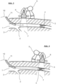
- Fig. 2 shows a schematic representation of the automatic ultrasound scan from Figure 1 in axial view.
- the femur 9 and the superficial femoral aorta 1 are shown in the patient's tissue 3. Both the femur 9 and the superficial femoral aorta 1 are located in the ultrasound sector 7 of the robot arm 5 with ultrasound head 6.
- Fig. 3 is a schematic representation of the detection of the intervention wire 10 in the vascular system during insertion of the intervention wire 10.
- the intervention wire 10 is introduced here through a sheath 11 into the superficial femoral aorta 1.
- the tip of the intervention wire 10 is detected by the system and automatically tracked by the robot arm 5 with ultrasound head 6.
- the tip of the intervention wire 10 is thus continuously located in the ultrasound sector 7 of the robot arm 5 with ultrasound head 6.
- Fig. 4 shows a schematic representation of the automatic tracking of the robot arm 5 from the previous figures to the target lesion, i.e. the stenosis 4.
- the surgeon is shown both the ultrasound image for wire tracking and a navigation overview for a representation of the ultrasound head 6 relative to the virtual three-dimensional vessel model in the augmented reality device.
- FIG. 5 A schematic representation of the automatic control of the ultrasound head 6 to the intervention focus is shown.
- a stent 12 has been inserted in the area of the stenosis 4 from the previous figures.
- the ultrasound sector 7 of the robot arm 5 with ultrasound head 6 is located in this very area of interest.
- the stent 12 was inserted using the intervention wire 10.
- Fig. 6 shows a schematic representation of the duplex sonographic control of the area of the superficial femoral aorta 1 into which the stent 12 was inserted, i.e. after the intervention.
- the ultrasound sector 7 of the robot arm 5 with ultrasound head 6 encompasses the newly inserted stent 4.
- the intervention wire 10 is guided back out of the patient via the sheath 11.
- a final virtual ultrasound angiography is created and compared with the initial angiography. This allows the success of the intervention to be quantified.
- the device according to the invention allows the performance of peripheral, endovascular interventions that are completely free of X-rays and contrast agents. As previously described, this leads to a significant minimization of health risks for the surgeon and the patient when performing these procedures.
Landscapes
- Health & Medical Sciences (AREA)
- Life Sciences & Earth Sciences (AREA)
- Engineering & Computer Science (AREA)
- Physics & Mathematics (AREA)
- Surgery (AREA)
- Heart & Thoracic Surgery (AREA)
- Veterinary Medicine (AREA)
- Public Health (AREA)
- General Health & Medical Sciences (AREA)
- Animal Behavior & Ethology (AREA)
- Biophysics (AREA)
- Nuclear Medicine, Radiotherapy & Molecular Imaging (AREA)
- Pathology (AREA)
- Radiology & Medical Imaging (AREA)
- Biomedical Technology (AREA)
- Molecular Biology (AREA)
- Medical Informatics (AREA)
- Software Systems (AREA)
- Computer Graphics (AREA)
- Computer Hardware Design (AREA)
- General Engineering & Computer Science (AREA)
- Theoretical Computer Science (AREA)
- General Physics & Mathematics (AREA)
- Vascular Medicine (AREA)
- Apparatus For Radiation Diagnosis (AREA)
- Ultra Sonic Daignosis Equipment (AREA)
Claims (2)
- Dispositif de navigation d'intervention endovasculaire périphérique en réalité augmentée par ultrasons, destiné à être utilisé sur un patient sur lequel une intervention endovasculaire périphérique doit être effectuée, comprenant:- un dispositif de réalité augmentée;- un système robotique comprenant un bras robotique (5) avec une tête à ultrasons (6);- un ordinateur ayant accès à un système de gestion de patients dans lequel le patient est enregistré;
dans lequel- le dispositif de réalité augmentée est conçu comme des lunettes de réalité augmentée tridimensionnelles,- le patient est un être vivant ou un modèle à des fins de simulation;- l'ordinateur, le dispositif de réalité augmentée et le système robotique sont reliés et communiquent entre eux par le biais d'un logiciel;- le bras robotisé (5) et le dispositif de réalité augmentée sont à commande vocale et/ou à commande manuelle virtuelle;
dans lequel- le bras robotisé (5) est conçu avec une tête à ultrasons (6) pour effectuer une échographie automatisée sur un patient,- le dispositif de réalité augmentée est conçu pour une représentation visuelle des résultats de l'ultrason,- lors de l'exécution de l'intervention dans le dispositif de réalité augmentéedans lequel le système de navigation est configuré pour exécuter un procédé de navigation qui comprend les opérations suivantes:- différents résultats d'ultrasons peuvent être représentés et/ou appelés visuellement simultanément en parallèle et/ou de manière combinée,- les résultats actuels des ultrasons sont transmis par le bras robotisé (5) avec la tête à ultrasons (6) et peuvent être visualisés, et- des données en temps réel peuvent être appelées et / ou affichées,a. Saisie des données du patient dans l'ordinateur avec le système de gestion des patients;b. Configuration du dispositif de réalité augmentée connecté à l'ordinateur;c. Préparer le système robotique connecté à l'ordinateur et au dispositif de réalité augmentée et le placer sur le patient placé;d. Définition d'une zone d'ultrasons pour une intervention endovasculaire périphérique sur le patient, ce qui implique la définition d'intervalles dans cette zone d'ultrasons pour mesurer le débit endovasculaire;e. Exécution automatique d'une échographie par le bras robotisé (5) dans la zone d'échographief. Analyser les résultats des ultrasons pour créer une angiographie virtuelle par ultrasons et un modèle vasculaire tridimensionnel virtuel et les visualiser par l'intermédiaire du dispositif de réalité augmentée, le modèle vasculaire tridimensionnel virtuel servant d'aide à la navigation pendant une intervention manuelle;g. Retour automatique du bras robotisé (5) au début de la zone d'échographie;h. Réalisation de l'intervention endovasculaire périphérique comprenant:- la reconnaissance par le bras robotique du matériel d'intervention qui est introduit manuellement dans le patient à l'aide des données du dispositif de réalité augmentée, et- Réalisation automatique d'une échographie accompagnant l'insertion, la position de la tête à ultrasons(6) étant fournie par le dispositif de réalité augmentée pendant l'intervention manuelle;i. Exécution automatique d'une échographie par le bras robotisé (5) dans la zone d'échographie après la fin de l'intervention ; etj. traitement post-interventionnel des données ultrasonores obtenues lors de l'intervention. - Système de navigation selon la revendication 1,
caractérisé en ce que
l'ordinateur présente un accès au système logistique de matériel, des propositions de matériel pour le matériel d'intervention étant émises par l'intermédiaire de la base de données du système logistique de matériel à l'aide des résultats des ultrasons et le matériel usagé étant marqué.
Applications Claiming Priority (2)
| Application Number | Priority Date | Filing Date | Title |
|---|---|---|---|
| DE102020109593.7A DE102020109593B3 (de) | 2020-04-06 | 2020-04-06 | Ultraschall-Erweiterte Realität-Peripher Endovaskulär Intervention-Navigationsverfahren sowie zugehörige Ultraschall-Erweiterte Realität-Peripher Endovaskulär Intervention-Navigationsanordnung |
| PCT/DE2021/100275 WO2021204325A1 (fr) | 2020-04-06 | 2021-03-18 | Méthode de navigation dans une intervention endovasculaire périphérique par ultrasons à réalité augmentée et ensemble associé pour naviguer dans une intervention endovasculaire périphérique par ultrasons à réalité augmentée |
Publications (3)
| Publication Number | Publication Date |
|---|---|
| EP4132365A1 EP4132365A1 (fr) | 2023-02-15 |
| EP4132365B1 true EP4132365B1 (fr) | 2025-01-08 |
| EP4132365C0 EP4132365C0 (fr) | 2025-01-08 |
Family
ID=75267444
Family Applications (1)
| Application Number | Title | Priority Date | Filing Date |
|---|---|---|---|
| EP21714815.4A Active EP4132365B1 (fr) | 2020-04-06 | 2021-03-18 | Ensemble pour naviguer dans une intervention endovasculaire périphérique par ultrasons à réalité augmentée |
Country Status (3)
| Country | Link |
|---|---|
| EP (1) | EP4132365B1 (fr) |
| DE (1) | DE102020109593B3 (fr) |
| WO (1) | WO2021204325A1 (fr) |
Families Citing this family (3)
| Publication number | Priority date | Publication date | Assignee | Title |
|---|---|---|---|---|
| JP7695177B2 (ja) * | 2021-11-19 | 2025-06-18 | 朝日インテック株式会社 | 医療装置 |
| CN116741387B (zh) * | 2023-06-26 | 2025-05-02 | 强联智创(北京)科技有限公司 | 由计算机实施的介入耗材的模拟方法及相关产品 |
| WO2025128801A1 (fr) * | 2023-12-15 | 2025-06-19 | Intuitive Surgical Operations, Inc. | Dissection assistée par ordinateur |
Family Cites Families (5)
| Publication number | Priority date | Publication date | Assignee | Title |
|---|---|---|---|---|
| US20060176242A1 (en) * | 2005-02-08 | 2006-08-10 | Blue Belt Technologies, Inc. | Augmented reality device and method |
| DE102011005917A1 (de) | 2011-03-22 | 2012-09-27 | Kuka Laboratories Gmbh | Medizinischer Arbeitsplatz |
| DE102014226240A1 (de) | 2014-12-17 | 2016-06-23 | Kuka Roboter Gmbh | System zur roboterunterstützten medizinischen Behandlung |
| GB2562502A (en) * | 2017-05-16 | 2018-11-21 | Medaphor Ltd | Visualisation system for needling |
| US11484365B2 (en) | 2018-01-23 | 2022-11-01 | Inneroptic Technology, Inc. | Medical image guidance |
-
2020
- 2020-04-06 DE DE102020109593.7A patent/DE102020109593B3/de active Active
-
2021
- 2021-03-18 EP EP21714815.4A patent/EP4132365B1/fr active Active
- 2021-03-18 WO PCT/DE2021/100275 patent/WO2021204325A1/fr not_active Ceased
Also Published As
| Publication number | Publication date |
|---|---|
| EP4132365A1 (fr) | 2023-02-15 |
| EP4132365C0 (fr) | 2025-01-08 |
| DE102020109593B3 (de) | 2021-09-23 |
| WO2021204325A1 (fr) | 2021-10-14 |
Similar Documents
| Publication | Publication Date | Title |
|---|---|---|
| EP0652726B1 (fr) | Procede de visualisation de l'interieur de corps | |
| DE102009009158B4 (de) | Lokalisierung eines medizinischen Instruments in einem präinvasiv aufgenommenen Tomographie-Bilddatensatz | |
| EP1127545B1 (fr) | Procédé pour individuer des objets en radiothérapie | |
| DE10322739B4 (de) | Verfahren zur markerlosen Navigation in präoperativen 3D-Bildern unter Verwendung eines intraoperativ gewonnenen 3D-C-Bogen-Bildes | |
| EP4132365B1 (fr) | Ensemble pour naviguer dans une intervention endovasculaire périphérique par ultrasons à réalité augmentée | |
| EP2632382B2 (fr) | Accessoire de navigation pour appareils optiques en médecine et procédé associé | |
| DE102020121807A1 (de) | Ansichtsänderung für die ultraschalldiagnostik während der untersuchung | |
| DE102006001884A1 (de) | Verfahren zur bildlichen Unterstützung einer Führung eines in einen Hohlraum eines Untersuchungsobjekts mindestens teilweise eingeführten medizinischen Instruments | |
| DE10325003A1 (de) | Visualisierung von 2D/ 3D-fusionierten Bilddaten für die Katheterangiografie | |
| DE60038730T2 (de) | Vorrichtung und verfahren zur messung anatomischer objekte mittels koordinierter fluoroskopie | |
| DE102009025077A1 (de) | System zur Orientierungsunterstützung und Darstellung eines Instruments im Inneren eines Untersuchungsobjektes insbesondere im menschlichen Körper | |
| EP3323347B1 (fr) | Soutien basé sur l'image lors d'une procédure chirurgicale mini-invasive | |
| WO2021151843A1 (fr) | Ensemble comprenant un dispositif oct pour déterminer une reconstruction 3d d'un volume de région d'objet, programme d'ordinateur et son procédé mis en œuvre par ordinateur | |
| DE102021102158A1 (de) | Ultraschallbilderfassungsoptimierung gemäss verschiedenen atmungsmodi | |
| DE102012214589B4 (de) | Verfahren und Vorrichtung zur Darstellung eines biologischen Hohlorgans | |
| DE102020201928A1 (de) | Verfahren zur Deformationskorrektur | |
| Tradigo et al. | On the reliability of measurements for a stent positioning simulation system | |
| EP1959391A1 (fr) | Détermination du tracé tridimensionnel du bord d'une structure anatomique | |
| WO2011120795A1 (fr) | Procédé pour la détermination de données volumiques tridimensionnelles et équipement d'imagerie | |
| DE102019215001B4 (de) | Verfahren zur Bildunterstützung bei der Navigation und System | |
| DE102013208897B4 (de) | Verfahren und Vorrichtung zur Darstellung eines Zielbereiches eines Hohlorgans eines Untersuchungsobjektes | |
| EP4172977B1 (fr) | Modèle d'entraînement médical avec au moins un modèle de vaisseau sanguin | |
| DE102005022901B4 (de) | Verfahren und Vorrichtung zur Orientierungsbestimmung eines sich in einem Objekt befindlichen Instruments | |
| DE10359317A1 (de) | Verfahren zur gezielten Navigation eines medizinischen Instruments, insbesondere eines Katheders | |
| DE102024209278B3 (de) | Überlagern eines Entfaltungsbilds mit einer aktuellen Projektion |
Legal Events
| Date | Code | Title | Description |
|---|---|---|---|
| STAA | Information on the status of an ep patent application or granted ep patent |
Free format text: STATUS: UNKNOWN |
|
| STAA | Information on the status of an ep patent application or granted ep patent |
Free format text: STATUS: THE INTERNATIONAL PUBLICATION HAS BEEN MADE |
|
| PUAI | Public reference made under article 153(3) epc to a published international application that has entered the european phase |
Free format text: ORIGINAL CODE: 0009012 |
|
| STAA | Information on the status of an ep patent application or granted ep patent |
Free format text: STATUS: REQUEST FOR EXAMINATION WAS MADE |
|
| 17P | Request for examination filed |
Effective date: 20221104 |
|
| AK | Designated contracting states |
Kind code of ref document: A1 Designated state(s): AL AT BE BG CH CY CZ DE DK EE ES FI FR GB GR HR HU IE IS IT LI LT LU LV MC MK MT NL NO PL PT RO RS SE SI SK SM TR |
|
| DAV | Request for validation of the european patent (deleted) | ||
| DAX | Request for extension of the european patent (deleted) | ||
| GRAP | Despatch of communication of intention to grant a patent |
Free format text: ORIGINAL CODE: EPIDOSNIGR1 |
|
| STAA | Information on the status of an ep patent application or granted ep patent |
Free format text: STATUS: GRANT OF PATENT IS INTENDED |
|
| INTG | Intention to grant announced |
Effective date: 20240801 |
|
| RAP3 | Party data changed (applicant data changed or rights of an application transferred) |
Owner name: UNIVERSITAET ZU LUEBECK |
|
| GRAS | Grant fee paid |
Free format text: ORIGINAL CODE: EPIDOSNIGR3 |
|
| GRAA | (expected) grant |
Free format text: ORIGINAL CODE: 0009210 |
|
| STAA | Information on the status of an ep patent application or granted ep patent |
Free format text: STATUS: THE PATENT HAS BEEN GRANTED |
|
| AK | Designated contracting states |
Kind code of ref document: B1 Designated state(s): AL AT BE BG CH CY CZ DE DK EE ES FI FR GB GR HR HU IE IS IT LI LT LU LV MC MK MT NL NO PL PT RO RS SE SI SK SM TR |
|
| REG | Reference to a national code |
Ref country code: GB Ref legal event code: FG4D Free format text: NOT ENGLISH |
|
| REG | Reference to a national code |
Ref country code: CH Ref legal event code: EP |
|
| REG | Reference to a national code |
Ref country code: DE Ref legal event code: R096 Ref document number: 502021006366 Country of ref document: DE |
|
| REG | Reference to a national code |
Ref country code: IE Ref legal event code: FG4D Free format text: LANGUAGE OF EP DOCUMENT: GERMAN |
|
| U01 | Request for unitary effect filed |
Effective date: 20250206 |
|
| U07 | Unitary effect registered |
Designated state(s): AT BE BG DE DK EE FI FR IT LT LU LV MT NL PT RO SE SI Effective date: 20250212 |
|
| U20 | Renewal fee for the european patent with unitary effect paid |
Year of fee payment: 5 Effective date: 20250327 |
|
| PG25 | Lapsed in a contracting state [announced via postgrant information from national office to epo] |
Ref country code: RS Free format text: LAPSE BECAUSE OF FAILURE TO SUBMIT A TRANSLATION OF THE DESCRIPTION OR TO PAY THE FEE WITHIN THE PRESCRIBED TIME-LIMIT Effective date: 20250408 |
|
| PG25 | Lapsed in a contracting state [announced via postgrant information from national office to epo] |
Ref country code: PL Free format text: LAPSE BECAUSE OF FAILURE TO SUBMIT A TRANSLATION OF THE DESCRIPTION OR TO PAY THE FEE WITHIN THE PRESCRIBED TIME-LIMIT Effective date: 20250108 |
|
| PG25 | Lapsed in a contracting state [announced via postgrant information from national office to epo] |
Ref country code: ES Free format text: LAPSE BECAUSE OF FAILURE TO SUBMIT A TRANSLATION OF THE DESCRIPTION OR TO PAY THE FEE WITHIN THE PRESCRIBED TIME-LIMIT Effective date: 20250108 |
|
| PG25 | Lapsed in a contracting state [announced via postgrant information from national office to epo] |
Ref country code: IS Free format text: LAPSE BECAUSE OF FAILURE TO SUBMIT A TRANSLATION OF THE DESCRIPTION OR TO PAY THE FEE WITHIN THE PRESCRIBED TIME-LIMIT Effective date: 20250508 Ref country code: NO Free format text: LAPSE BECAUSE OF FAILURE TO SUBMIT A TRANSLATION OF THE DESCRIPTION OR TO PAY THE FEE WITHIN THE PRESCRIBED TIME-LIMIT Effective date: 20250408 |
|
| PG25 | Lapsed in a contracting state [announced via postgrant information from national office to epo] |
Ref country code: HR Free format text: LAPSE BECAUSE OF FAILURE TO SUBMIT A TRANSLATION OF THE DESCRIPTION OR TO PAY THE FEE WITHIN THE PRESCRIBED TIME-LIMIT Effective date: 20250108 |
|
| PG25 | Lapsed in a contracting state [announced via postgrant information from national office to epo] |
Ref country code: GR Free format text: LAPSE BECAUSE OF FAILURE TO SUBMIT A TRANSLATION OF THE DESCRIPTION OR TO PAY THE FEE WITHIN THE PRESCRIBED TIME-LIMIT Effective date: 20250409 |
|
| PG25 | Lapsed in a contracting state [announced via postgrant information from national office to epo] |
Ref country code: SM Free format text: LAPSE BECAUSE OF FAILURE TO SUBMIT A TRANSLATION OF THE DESCRIPTION OR TO PAY THE FEE WITHIN THE PRESCRIBED TIME-LIMIT Effective date: 20250108 |
|
| PG25 | Lapsed in a contracting state [announced via postgrant information from national office to epo] |
Ref country code: MC Free format text: LAPSE BECAUSE OF FAILURE TO SUBMIT A TRANSLATION OF THE DESCRIPTION OR TO PAY THE FEE WITHIN THE PRESCRIBED TIME-LIMIT Effective date: 20250108 |
|
| PG25 | Lapsed in a contracting state [announced via postgrant information from national office to epo] |
Ref country code: CZ Free format text: LAPSE BECAUSE OF FAILURE TO SUBMIT A TRANSLATION OF THE DESCRIPTION OR TO PAY THE FEE WITHIN THE PRESCRIBED TIME-LIMIT Effective date: 20250108 |
|
| REG | Reference to a national code |
Ref country code: CH Ref legal event code: H13 Free format text: ST27 STATUS EVENT CODE: U-0-0-H10-H13 (AS PROVIDED BY THE NATIONAL OFFICE) Effective date: 20251023 |
|
| PG25 | Lapsed in a contracting state [announced via postgrant information from national office to epo] |
Ref country code: SK Free format text: LAPSE BECAUSE OF FAILURE TO SUBMIT A TRANSLATION OF THE DESCRIPTION OR TO PAY THE FEE WITHIN THE PRESCRIBED TIME-LIMIT Effective date: 20250108 |
|
| PLBE | No opposition filed within time limit |
Free format text: ORIGINAL CODE: 0009261 |
|
| STAA | Information on the status of an ep patent application or granted ep patent |
Free format text: STATUS: NO OPPOSITION FILED WITHIN TIME LIMIT |
|
| 26N | No opposition filed |
Effective date: 20251009 |
|
| PG25 | Lapsed in a contracting state [announced via postgrant information from national office to epo] |
Ref country code: CH Free format text: LAPSE BECAUSE OF NON-PAYMENT OF DUE FEES Effective date: 20250331 |
|
| PG25 | Lapsed in a contracting state [announced via postgrant information from national office to epo] |
Ref country code: IE Free format text: LAPSE BECAUSE OF NON-PAYMENT OF DUE FEES Effective date: 20250318 |
|
| U20 | Renewal fee for the european patent with unitary effect paid |
Year of fee payment: 6 Effective date: 20260218 |
|
| PGFP | Annual fee paid to national office [announced via postgrant information from national office to epo] |
Ref country code: GB Payment date: 20260217 Year of fee payment: 6 |