EP4137510A1 - Pd-1-mutantes polypeptid und herstellung davon und verwendung davon - Google Patents
Pd-1-mutantes polypeptid und herstellung davon und verwendung davon Download PDFInfo
- Publication number
- EP4137510A1 EP4137510A1 EP21789230.6A EP21789230A EP4137510A1 EP 4137510 A1 EP4137510 A1 EP 4137510A1 EP 21789230 A EP21789230 A EP 21789230A EP 4137510 A1 EP4137510 A1 EP 4137510A1
- Authority
- EP
- European Patent Office
- Prior art keywords
- seq
- amino acid
- acid sequence
- antibody
- fusion protein
- Prior art date
- Legal status (The legal status is an assumption and is not a legal conclusion. Google has not performed a legal analysis and makes no representation as to the accuracy of the status listed.)
- Pending
Links
Images






Classifications
-
- A—HUMAN NECESSITIES
- A61—MEDICAL OR VETERINARY SCIENCE; HYGIENE
- A61P—SPECIFIC THERAPEUTIC ACTIVITY OF CHEMICAL COMPOUNDS OR MEDICINAL PREPARATIONS
- A61P37/00—Drugs for immunological or allergic disorders
-
- A—HUMAN NECESSITIES
- A61—MEDICAL OR VETERINARY SCIENCE; HYGIENE
- A61P—SPECIFIC THERAPEUTIC ACTIVITY OF CHEMICAL COMPOUNDS OR MEDICINAL PREPARATIONS
- A61P35/00—Antineoplastic agents
-
- A—HUMAN NECESSITIES
- A61—MEDICAL OR VETERINARY SCIENCE; HYGIENE
- A61P—SPECIFIC THERAPEUTIC ACTIVITY OF CHEMICAL COMPOUNDS OR MEDICINAL PREPARATIONS
- A61P37/00—Drugs for immunological or allergic disorders
- A61P37/02—Immunomodulators
-
- C—CHEMISTRY; METALLURGY
- C07—ORGANIC CHEMISTRY
- C07K—PEPTIDES
- C07K14/00—Peptides having more than 20 amino acids; Gastrins; Somatostatins; Melanotropins; Derivatives thereof
- C07K14/435—Peptides having more than 20 amino acids; Gastrins; Somatostatins; Melanotropins; Derivatives thereof from animals; from humans
- C07K14/705—Receptors; Cell surface antigens; Cell surface determinants
- C07K14/70503—Immunoglobulin superfamily
- C07K14/70521—CD28, CD152
-
- C—CHEMISTRY; METALLURGY
- C07—ORGANIC CHEMISTRY
- C07K—PEPTIDES
- C07K16/00—Immunoglobulins [IG], e.g. monoclonal or polyclonal antibodies
- C07K16/18—Immunoglobulins [IG], e.g. monoclonal or polyclonal antibodies against material from animals or humans
-
- C—CHEMISTRY; METALLURGY
- C07—ORGANIC CHEMISTRY
- C07K—PEPTIDES
- C07K16/00—Immunoglobulins [IG], e.g. monoclonal or polyclonal antibodies
- C07K16/18—Immunoglobulins [IG], e.g. monoclonal or polyclonal antibodies against material from animals or humans
- C07K16/28—Immunoglobulins [IG], e.g. monoclonal or polyclonal antibodies against material from animals or humans against receptors, cell surface antigens or cell surface determinants
-
- C—CHEMISTRY; METALLURGY
- C07—ORGANIC CHEMISTRY
- C07K—PEPTIDES
- C07K16/00—Immunoglobulins [IG], e.g. monoclonal or polyclonal antibodies
- C07K16/18—Immunoglobulins [IG], e.g. monoclonal or polyclonal antibodies against material from animals or humans
- C07K16/28—Immunoglobulins [IG], e.g. monoclonal or polyclonal antibodies against material from animals or humans against receptors, cell surface antigens or cell surface determinants
- C07K16/2803—Immunoglobulins [IG], e.g. monoclonal or polyclonal antibodies against material from animals or humans against receptors, cell surface antigens or cell surface determinants against the immunoglobulin superfamily
-
- C—CHEMISTRY; METALLURGY
- C07—ORGANIC CHEMISTRY
- C07K—PEPTIDES
- C07K16/00—Immunoglobulins [IG], e.g. monoclonal or polyclonal antibodies
- C07K16/18—Immunoglobulins [IG], e.g. monoclonal or polyclonal antibodies against material from animals or humans
- C07K16/28—Immunoglobulins [IG], e.g. monoclonal or polyclonal antibodies against material from animals or humans against receptors, cell surface antigens or cell surface determinants
- C07K16/2803—Immunoglobulins [IG], e.g. monoclonal or polyclonal antibodies against material from animals or humans against receptors, cell surface antigens or cell surface determinants against the immunoglobulin superfamily
- C07K16/2818—Immunoglobulins [IG], e.g. monoclonal or polyclonal antibodies against material from animals or humans against receptors, cell surface antigens or cell surface determinants against the immunoglobulin superfamily against CD28 or CD152
-
- C—CHEMISTRY; METALLURGY
- C07—ORGANIC CHEMISTRY
- C07K—PEPTIDES
- C07K16/00—Immunoglobulins [IG], e.g. monoclonal or polyclonal antibodies
- C07K16/18—Immunoglobulins [IG], e.g. monoclonal or polyclonal antibodies against material from animals or humans
- C07K16/28—Immunoglobulins [IG], e.g. monoclonal or polyclonal antibodies against material from animals or humans against receptors, cell surface antigens or cell surface determinants
- C07K16/2839—Immunoglobulins [IG], e.g. monoclonal or polyclonal antibodies against material from animals or humans against receptors, cell surface antigens or cell surface determinants against the integrin superfamily
- C07K16/2848—Immunoglobulins [IG], e.g. monoclonal or polyclonal antibodies against material from animals or humans against receptors, cell surface antigens or cell surface determinants against the integrin superfamily against integrin beta3-subunit-containing molecules, e.g. CD41, CD51, CD61
-
- C—CHEMISTRY; METALLURGY
- C07—ORGANIC CHEMISTRY
- C07K—PEPTIDES
- C07K16/00—Immunoglobulins [IG], e.g. monoclonal or polyclonal antibodies
- C07K16/18—Immunoglobulins [IG], e.g. monoclonal or polyclonal antibodies against material from animals or humans
- C07K16/28—Immunoglobulins [IG], e.g. monoclonal or polyclonal antibodies against material from animals or humans against receptors, cell surface antigens or cell surface determinants
- C07K16/2863—Immunoglobulins [IG], e.g. monoclonal or polyclonal antibodies against material from animals or humans against receptors, cell surface antigens or cell surface determinants against receptors for growth factors, growth regulators
-
- C—CHEMISTRY; METALLURGY
- C07—ORGANIC CHEMISTRY
- C07K—PEPTIDES
- C07K16/00—Immunoglobulins [IG], e.g. monoclonal or polyclonal antibodies
- C07K16/18—Immunoglobulins [IG], e.g. monoclonal or polyclonal antibodies against material from animals or humans
- C07K16/28—Immunoglobulins [IG], e.g. monoclonal or polyclonal antibodies against material from animals or humans against receptors, cell surface antigens or cell surface determinants
- C07K16/2878—Immunoglobulins [IG], e.g. monoclonal or polyclonal antibodies against material from animals or humans against receptors, cell surface antigens or cell surface determinants against the NGF-receptor/TNF-receptor superfamily, e.g. CD27, CD30, CD40, CD95
-
- C—CHEMISTRY; METALLURGY
- C07—ORGANIC CHEMISTRY
- C07K—PEPTIDES
- C07K16/00—Immunoglobulins [IG], e.g. monoclonal or polyclonal antibodies
- C07K16/18—Immunoglobulins [IG], e.g. monoclonal or polyclonal antibodies against material from animals or humans
- C07K16/28—Immunoglobulins [IG], e.g. monoclonal or polyclonal antibodies against material from animals or humans against receptors, cell surface antigens or cell surface determinants
- C07K16/2896—Immunoglobulins [IG], e.g. monoclonal or polyclonal antibodies against material from animals or humans against receptors, cell surface antigens or cell surface determinants against molecules with a "CD"-designation, not provided for elsewhere
-
- C—CHEMISTRY; METALLURGY
- C07—ORGANIC CHEMISTRY
- C07K—PEPTIDES
- C07K16/00—Immunoglobulins [IG], e.g. monoclonal or polyclonal antibodies
- C07K16/18—Immunoglobulins [IG], e.g. monoclonal or polyclonal antibodies against material from animals or humans
- C07K16/28—Immunoglobulins [IG], e.g. monoclonal or polyclonal antibodies against material from animals or humans against receptors, cell surface antigens or cell surface determinants
- C07K16/30—Immunoglobulins [IG], e.g. monoclonal or polyclonal antibodies against material from animals or humans against receptors, cell surface antigens or cell surface determinants from tumour cells
- C07K16/303—Liver or Pancreas
-
- C—CHEMISTRY; METALLURGY
- C07—ORGANIC CHEMISTRY
- C07K—PEPTIDES
- C07K16/00—Immunoglobulins [IG], e.g. monoclonal or polyclonal antibodies
- C07K16/18—Immunoglobulins [IG], e.g. monoclonal or polyclonal antibodies against material from animals or humans
- C07K16/28—Immunoglobulins [IG], e.g. monoclonal or polyclonal antibodies against material from animals or humans against receptors, cell surface antigens or cell surface determinants
- C07K16/30—Immunoglobulins [IG], e.g. monoclonal or polyclonal antibodies against material from animals or humans against receptors, cell surface antigens or cell surface determinants from tumour cells
- C07K16/3069—Reproductive system, e.g. ovaria, uterus, testes, prostate
-
- C—CHEMISTRY; METALLURGY
- C07—ORGANIC CHEMISTRY
- C07K—PEPTIDES
- C07K16/00—Immunoglobulins [IG], e.g. monoclonal or polyclonal antibodies
- C07K16/18—Immunoglobulins [IG], e.g. monoclonal or polyclonal antibodies against material from animals or humans
- C07K16/28—Immunoglobulins [IG], e.g. monoclonal or polyclonal antibodies against material from animals or humans against receptors, cell surface antigens or cell surface determinants
- C07K16/30—Immunoglobulins [IG], e.g. monoclonal or polyclonal antibodies against material from animals or humans against receptors, cell surface antigens or cell surface determinants from tumour cells
- C07K16/3076—Immunoglobulins [IG], e.g. monoclonal or polyclonal antibodies against material from animals or humans against receptors, cell surface antigens or cell surface determinants from tumour cells against structure-related tumour-associated moieties
- C07K16/3092—Immunoglobulins [IG], e.g. monoclonal or polyclonal antibodies against material from animals or humans against receptors, cell surface antigens or cell surface determinants from tumour cells against structure-related tumour-associated moieties against tumour-associated mucins
-
- C—CHEMISTRY; METALLURGY
- C07—ORGANIC CHEMISTRY
- C07K—PEPTIDES
- C07K16/00—Immunoglobulins [IG], e.g. monoclonal or polyclonal antibodies
- C07K16/18—Immunoglobulins [IG], e.g. monoclonal or polyclonal antibodies against material from animals or humans
- C07K16/32—Immunoglobulins [IG], e.g. monoclonal or polyclonal antibodies against material from animals or humans against translation products of oncogenes
-
- A—HUMAN NECESSITIES
- A61—MEDICAL OR VETERINARY SCIENCE; HYGIENE
- A61K—PREPARATIONS FOR MEDICAL, DENTAL OR TOILETRY PURPOSES
- A61K39/00—Medicinal preparations containing antigens or antibodies
- A61K2039/505—Medicinal preparations containing antigens or antibodies comprising antibodies
-
- A—HUMAN NECESSITIES
- A61—MEDICAL OR VETERINARY SCIENCE; HYGIENE
- A61K—PREPARATIONS FOR MEDICAL, DENTAL OR TOILETRY PURPOSES
- A61K38/00—Medicinal preparations containing peptides
-
- C—CHEMISTRY; METALLURGY
- C07—ORGANIC CHEMISTRY
- C07K—PEPTIDES
- C07K19/00—Hybrid peptides, i.e. peptides covalently bound to nucleic acids, or non-covalently bound protein-protein complexes
-
- C—CHEMISTRY; METALLURGY
- C07—ORGANIC CHEMISTRY
- C07K—PEPTIDES
- C07K2317/00—Immunoglobulins specific features
- C07K2317/50—Immunoglobulins specific features characterized by immunoglobulin fragments
- C07K2317/52—Constant or Fc region; Isotype
- C07K2317/524—CH2 domain
-
- C—CHEMISTRY; METALLURGY
- C07—ORGANIC CHEMISTRY
- C07K—PEPTIDES
- C07K2317/00—Immunoglobulins specific features
- C07K2317/50—Immunoglobulins specific features characterized by immunoglobulin fragments
- C07K2317/52—Constant or Fc region; Isotype
- C07K2317/526—CH3 domain
-
- C—CHEMISTRY; METALLURGY
- C07—ORGANIC CHEMISTRY
- C07K—PEPTIDES
- C07K2317/00—Immunoglobulins specific features
- C07K2317/50—Immunoglobulins specific features characterized by immunoglobulin fragments
- C07K2317/56—Immunoglobulins specific features characterized by immunoglobulin fragments variable (Fv) region, i.e. VH and/or VL
- C07K2317/565—Complementarity determining region [CDR]
-
- C—CHEMISTRY; METALLURGY
- C07—ORGANIC CHEMISTRY
- C07K—PEPTIDES
- C07K2317/00—Immunoglobulins specific features
- C07K2317/90—Immunoglobulins specific features characterized by (pharmaco)kinetic aspects or by stability of the immunoglobulin
- C07K2317/92—Affinity (KD), association rate (Ka), dissociation rate (Kd) or EC50 value
-
- C—CHEMISTRY; METALLURGY
- C07—ORGANIC CHEMISTRY
- C07K—PEPTIDES
- C07K2319/00—Fusion polypeptide
- C07K2319/30—Non-immunoglobulin-derived peptide or protein having an immunoglobulin constant or Fc region, or a fragment thereof, attached thereto
-
- C—CHEMISTRY; METALLURGY
- C07—ORGANIC CHEMISTRY
- C07K—PEPTIDES
- C07K2319/00—Fusion polypeptide
- C07K2319/33—Fusion polypeptide fusions for targeting to specific cell types, e.g. tissue specific targeting, targeting of a bacterial subspecies
Definitions
- the present invention relates to the field of biopharmaceuticals, and in particular to a high-affinity PD-1 mutant polypeptide, and an antibody fusion protein containing the PD-1 mutant polypeptide and preparation and application thereof.
- PD-1 is an immunosuppressive molecule that belongs to the CD28 family. Its structure includes: an extracellular immunoglobulin variable region (IgV)-like domain, a hydrophobic transmembrane region, and an intracellular region. PD-1 is expressed on CD4-CD8-thymocytes, and inducibly expressed on activated T cells, B cells, bone marrow cells, dendritic cells, natural killer cells, monocytes etc. The continuous expression of PD-1 on T cells may induce T cell exhaustion.
- IgV immunoglobulin variable region
- the expression of PD-1 by tumor-infiltrating lymphocytes may affect T cell function, weaken cytokine secretion, and weaken the tumor-killing effect of T cells, and is closely related to the poor prognosis and high tumor recurrence rate of patients with renal cell carcinoma and non-small cell lung cancer ( Pan Jiajia, et al.; Journal of China Pharmaceutical University 2016, 47(1):9-18 ).
- PD-1 has two ligands, namely PD-L1 and PD-L2, both of which belong to the B7 family.
- PD-L1 is widely expressed in activated B cells, T cells, macrophages, dendritic cells (DCs), NK cells etc.
- PD-L1 is also expressed on the surface of many tumor cells, such as lung cancer, breast cancer, malignant melanoma, esophageal cancer, gastric cancer, and pancreatic cancer.
- the high expression of PD-L1 or PD-L2 on surface of tumor cells binds to PD-1 on T cells to transduce negative regulatory signals, leading to the immune apoptosis and immune incompetence of tumor antigen-specificity T cells, and making tumor cells escape from the immune surveillance and killing ( Pan Jiajia et al.; Journal of China Pharmaceutical University 2016, 47(1):9-18 ). Therefore, with PD-1/PD-Ls signaling pathway as the target, the development of blockers against PD-1 or PD-Ls can enhance the killing of tumor cells by T cells.
- the PD-1 mutant polypeptide (with 2 amino acid mutations) of the present invention not only retains the physicochemical properties and the same epitope binding similar to that of wild-type PD-1, but also greatly improves its affinity to PD-L1 (pM), and the possibility of off-target is low; in addition, the PD-1 mutant polypeptide can cross-react with PD-L1 from human, monkey and mouse, providing great convenience for studies on pharmacodynamics and toxicology in animal models.
- the PD-1 mutant polypeptide Compared with PD-L1 antibodies, the PD-1 mutant polypeptide has a single domain, and is easy to construct bispecific antibodies.
- the antibody fusion protein formed by fusing the PD-1 mutant polypeptide with the antibody targeted to a tumor antigen on the one hand, can specifically confine the PD-1 mutant polypeptide on or near tumor cells and in the tumor microenvironment through high-affinity ((higher than the affinity of the PD-1 mutant polypeptide with PD-L1)tumor antigen-specificity antibody, greatly reducing the extensive immune activation caused by conventional immune checkpoint inhibitors in vivo; and on the other hand, the fast binding and fast dissociation characteristics of the PD-1 mutant polypeptide enable it to block the PD-1/PD-L1 signaling pathway, avoiding the adverse reactions resulting from continuous activation of immune cells by the fast binding and slow dissociation properties of the conventional immune checkpoint inhibitors.
- the faster in vivo metabolism of the PD-1 mutant than that of conventional antibody fragments helps
- a PD-1 mutant polypeptide in an aspect of the present invention, has a nucleic acid sequence as shown in SEQ ID NO: 1, or has an amino acid sequence as shown in SEQ ID NO: 2.
- an antibody fusion protein containing the PD-1 mutant polypeptide is provided.
- the antibody fusion protein includes the PD-1 mutant polypeptide and an antibody targeted to a tumor cell surface antigen.
- the tumor cell surface antigen is selected from EGFR, HER2, HER3, GPC3, PSMA, CD27, CD33, CD37, CD38, CD51, CD56, mesothelin, HGFR, Nectin-4, Mucin 5AC, folate receptor ⁇ , carbonic anhydrase IX (CAIX), DLL1, Notch2, Notch3 and endoglin etc.
- the tumor antigen is selected from EGFR.
- the antibody targeted to the tumor cell surface antigen EGFR has sequences HCDR1, HCDR2 and HCDR3 as shown in SEQ ID NO: 13, SEQ ID NO: 14, and SEQ ID NO: 15, and sequences LCDR1, LCDR2 and LCDR3 as shown in SEQ ID NO: 16, SEQ ID NO: 17, and SEQ ID NO: 18.
- the antibody targeted to the tumor antigen EGFR has a heavy chain variable region (VH) with an amino acid sequence as shown in SEQ ID NO: 34, and a light chain variable region (VL) with an amino acid sequence as shown in SEQ ID NO: 36.
- the antibody targeted to the tumor cell surface antigen EGFR has a VH with an amino acid sequence as shown in SEQ ID NO: 65, and a VL with an amino acid sequence as shown in SEQ ID NO: 66. In some embodiments, the antibody targeted to the tumor antigen EGFR has a heavy chain (HC) with an amino acid sequence as shown in SEQ ID NO: 4, and a light chain (LC) with an amino acid sequence as shown in SEQ ID NO: 6 .
- HC heavy chain
- LC light chain
- the antibody fusion protein containing the PD-1 mutant polypeptide and the antibody targeted to the tumor cell surface antigen EGFR has a HC with an amino acid sequence as shown in SEQ ID NO: 4 and a LC with an amino acid sequence as shown in SEQ ID NO: 28.
- the tumor antigen is selected from HER2.
- the antibody targeted to the tumor cell surface antigen HER2 has sequences HCDR1, HCDR2 and HCDR3 as shown in SEQ ID NO: 19, SEQ ID NO: 20, and SEQ ID NO: 21, and sequences LCDR1, LCDR2 and LCDR3 as shown in SEQ ID NO: 22, SEQ ID NO: 23, and SEQ ID NO: 24.
- the antibody targeted to the tumor cell surface antigen HER2 has a VH with an amino acid sequence as shown in SEQ ID NO: 38, and a VL with an amino acid sequence as shown in SEQ ID NO: 40.
- the antibody targeted to the tumor cell surface antigen HER2 has a VH with an amino acid sequence as shown in SEQ ID NO: 67, and a VL with an amino acid sequence as shown in SEQ ID NO: 68.
- the antibody targeted to the tumor antigen HER2 has an HC with an amino acid sequence as shown in SEQ ID NO: 8 and an LC with an amino acid sequence as shown in SEQ ID NO: 8.
- the antibody fusion protein containing the PD-1 mutant polypeptide and the antibody targeted to the tumor antigen HER2 has a HC with an amino acid sequence as shown in SEQ ID NO: 10 and a LC with an amino acid sequence as shown in SEQ ID NO: 32.
- the tumor cell surface antigen is selected from GPC-3.
- the antibody targeted to the tumor cell surface antigen GPC3 has sequences HCDR1, HCDR2, and HCDR3 as shown in SEQ ID NO: 45, SEQ ID NO: 46, and SEQ ID NO: 47, and sequences LCDR1, LCDR2, and LCDR3 as shown in SEQ ID NO: 48, SEQ ID NO: 49, and SEQ ID NO: 50.
- the antibody targeted to the tumor cell surface antigen GPC-3 has a VH with an amino acid sequence as shown in SEQ ID NO: 42, and a VL with an amino acid sequence as shown in SEQ ID NO: 44.
- the antibody targeted to the tumor antigen GPC-3 has an HC with an amino acid sequence as shown in SEQ ID NO: 58 and an LC with an amino acid sequence as shown in SEQ ID NO: 60.
- the antibody fusion protein containing the PD-1 mutant polypeptide and the antibody targeted to the tumor antigen GPC-3 has a HC with an amino acid sequence as shown in SEQ ID NO: 52 and a LC with an amino acid sequence as shown in SEQ ID NO: 60.
- the antibody fusion protein containing the PD-1 mutant polypeptide and the antibody targeted to the tumor antigen GPC-3 has a HC with an amino acid sequence as shown in SEQ ID NO: 58 and a LC with an amino acid sequence as shown in SEQ ID NO: 54. In some embodiments, the antibody fusion protein containing the PD-1 mutant polypeptide and the antibody targeted to the tumor antigen GPC-3 has a HC with an amino acid sequence as shown in SEQ ID NO: 58 and a LC with an amino acid sequence as shown in SEQ ID NO: 56.
- the tumor cell surface antigen is selected from PSMA.
- the antibody targeted to the tumor cell surface antigen PSMA has a VH with an amino acid sequence as shown in SEQ ID NO: 62, and a VL with an amino acid sequence as shown in SEQ ID NO: 64.
- the tumor cell surface antigen is selected from CD27.
- the antibody targeted to the tumor cell surface antigen CD27 has a VH with an amino acid sequence as shown in SEQ ID NO: 69, and a VL with an amino acid sequence as shown in SEQ ID NO: 70.
- the tumor cell surface antigen is selected from CD33.
- the antibody targeted to the tumor cell surface antigen CD33 has a VH with an amino acid sequence as shown in SEQ ID NO: 71, and a VL with an amino acid sequence as shown in SEQ ID NO: 72.
- the tumor cell surface antigen is selected from CD37.
- the antibody targeted to the tumor cell surface antigen CD37 has a VH with an amino acid sequence as shown in SEQ ID NO: 73, and a VL with an amino acid sequence as shown in SEQ ID NO: 74.
- the tumor cell surface antigen is selected from CD38.
- the antibody targeted to the tumor cell surface antigen CD38 has a VH with an amino acid sequence as shown in SEQ ID NO: 75, and a VL with an amino acid sequence as shown in SEQ ID NO: 76.
- the tumor cell surface antigen is selected from CD51.
- the antibody targeted to the tumor cell surface antigen CD51 has a VH with an amino acid sequence as shown in SEQ ID NO: 77, and a VL with an amino acid sequence as shown in SEQ ID NO: 78.
- the tumor cell surface antigen is selected from CD56.
- the antibody targeted to the tumor cell surface antigen CD56 has a VH with an amino acid sequence as shown in SEQ ID NO: 79, and a VL with an amino acid sequence as shown in SEQ ID NO: 80.
- the tumor cell surface antigen is selected from mesothelin.
- the antibody targeted to the tumor cell surface antigen mesothelin has a VH with an amino acid sequence as shown in SEQ ID NO: 81, and a VL with an amino acid sequence as shown in SEQ ID NO: 82.
- the tumor cell surface antigen is selected from HGFR.
- the antibody targeted to the tumor cell surface antigen HGFR has a VH with an amino acid sequence as shown in SEQ ID NO: 83, and a VL with an amino acid sequence as shown in SEQ ID NO: 84.
- the tumor cell surface antigen is selected from Nectin-4.
- the antibody targeted to the tumor cell surface antigen Nectin-4 has a VH with an amino acid sequence as shown in SEQ ID NO: 85, and a VL with an amino acid sequence as shown in SEQ ID NO: 86.
- the tumor cell surface antigen is selected from Mucin5AC.
- the antibody targeted to the tumor cell surface antigen Mucin5AC has a VH with an amino acid sequence as shown in SEQ ID NO: 87, and a VL with an amino acid sequence as shown in SEQ ID NO: 88.
- the tumor cell surface antigen is selected from folate receptor ⁇ .
- the antibody targeted to the tumor cell surface antigen folate receptor ⁇ has a VH with an amino acid sequence as shown in SEQ ID NO: 89, and a VL with an amino acid sequence as shown in SEQ ID NO: 90.
- the tumor cell surface antigen is carbonic anhydrase IX (CAIX).
- the antibody targeted to the tumor cell surface antigen carbonic anhydrase IX (CAIX) has a VH with an amino acid sequence as shown in SEQ ID NO: 91, and a VL with an amino acid sequence as shown in SEQ ID NO: 92.
- the tumor cell surface antigen is selected from DLL1.
- the antibody targeted to the tumor cell surface antigen DLL1 has a VH with an amino acid sequence as shown in SEQ ID NO: 93, and a VL with an amino acid sequence as shown in SEQ ID NO: 94.
- the tumor cell surface antigen is selected from Notch 2/3.
- the antibody targeted to the tumor cell surface antigen Notch 2/3 has a VH with an amino acid sequence as shown in SEQ ID NO: 95, and a VL with an amino acid sequence as shown in SEQ ID NO: 96.
- the tumor cell surface antigen is selected from endoglin.
- the antibody targeted to the tumor cell surface antigen endoglin has a VH with an amino acid sequence as shown in SEQ ID NO: 97, and a VL with an amino acid sequence as shown in SEQ ID NO: 98.
- the tumor cell surface antigen is selected from HER3.
- the antibody targeted to the tumor cell surface antigen HER3 has a VH with an amino acid sequence as shown in SEQ ID NO: 99, and a VL with an amino acid sequence as shown in SEQ ID NO: 100.
- a polynucleotide is provided, and it encodes the above PD-1 mutant polypeptide or the antibody fusion protein containing the PD-1 mutant polypeptide.
- the polynucleotide is selected from nucleotide sequences in the following group consisting of: SEQ ID NO: 1, 3, 5, 7, 9, 11, 27, 31, 51, 53, 55, 57, and 59.
- a vector containing the above PD-1 mutant polypeptide or the antibody fusion protein sequence containing the PD-1 mutant polypeptide is provided.
- a host cell in an aspect of the present invention, includes the above vector.
- a pharmaceutical composition in an aspect of the present invention, includes a pharmaceutically acceptable carrier and the above PD-1 mutant polypeptide or an antibody fusion protein containing the PD-1 mutant polypeptide.
- an application of the PD-1 mutant polypeptide or the antibody fusion protein containing the PD-1 mutant polypeptide as inhibitor of PD-1-PD-L1 interaction is provided.
- an application of the PD-1 mutant polypeptide or the antibody fusion protein containing the PD-1 mutant polypeptide as an immunomodulator is provided.
- an application of the PD-1 mutant polypeptide or the antibody fusion protein containing the PD-1 mutant polypeptide for treatment of tumors or cancersis includes: B cell lymphoma, breast cancer, ovarian cancer, NSCLC, low-grade glioma, Hodgkin lymphoma, chronic lymphocytic leukemia (CLL), multiple myeloma, metastatic colorectal cancer, Merkel cell tumor, non-small cell lung cancer (NSCLC), squamous NSCLC, pancreatic cancer, neuromesothelioma, breast cancer, HER2-positive metastatic breast cancer, metastatic gastric cancer, liver cancer, urothelial cell carcinoma, bladder cancer, pancreatic cancer, ovarian cancer, renal cell carcinoma, multiple myeloma, soft tissue sarcoma, primary peritoneal cancer etc.
- the affinity (pM) of the high-affinity PD-1 mutant polypeptide of the present invention with PD-L1 is greatly improved.
- the affinity of the antibody fusion protein containing the PD-1 mutant polypeptide with the recombinant PD-L1 is 39-109 times higher than that of the antibody fusion protein containing the wild-type PD-1 with the recombinant PD-L1.
- the affinity of the antibody fusion protein containing the PD-1 mutant polypeptide with the cell surface PD-L1 is greatly enhanced relative to the affinity (almost no binding) of the antibody fusion protein containing the wild-type PD-1 with the cell surface PD-L1. Therefore, the PD-1 mutant of the present invention may be used as acompetitive inhibitor of PD-1-PD-L1 interaction.
- the PD-1 mutant of the present invention may also be used as animmunomodulator for, for example, the immunotherapy of tumors.
- CDR refers to the complementarity determining region in an immunoglobulin variable region sequence.
- CDR1, CDR2, and CDR3 CDR1, CDR2, and CDR3.
- CDR group refers to a group of three CDRs that appear in a single variable region capable of binding an antigen. The exact boundaries of these CDRs are already defined according to different systems. A system described by Kabat (Kabat et al. (1987) and (1991)) not only provides a clear residue numbering system applicable to any variable regions of an antibody or binding protein, but also provides a precise definition of residue boundaries of the three CDRs in each heavy chain or light chain sequence.
- L1 and H1 refer to light chain and heavy chain regions, respectively. These regions may be referred to as Chothia CDR, with a boundary overlapped with the Kabat CDR.
- tumor cell surface antigen refers to an antigen that is expressed on the surface of a tumor cell and may be targeted for disease treatment.
- the targeted antigen is selected from the antigen that is preferentially expressed on the surface of tumor cells (such as solid tumors or hematological tumor cells): non-limiting examples of specific tumor-associated antigen include, for example, EGFR, HER2, HER3, HER4, GPC3, PSMA, CD27, CD33, CD37, CD38, CD51, CD56, mesothelin, HGFR, Nectin-4, Mucin 5AC, folate receptor ⁇ , carbonic anhydrase IX (CAIX), DLL1, Notch2, Notch3, endoglin, MUC1, MUC2, MUC3A, MUC3B, MUC4, MUC5B, MUC6, MUC7, MUC8, MUC12, MUC13, MUC15, MUC16, MUC17, MUC19, MUC20, V
- aEGFR VH anti-human EGFR antibody heavy chain variable region
- aEGFR VL EGFR antibody light chain variable region
- aHER2 VH anti-human HER2 antibody heavy chain variable region
- aHER2 VL anti-human HER2 antibody light chain variable region
- the amplified aEGFR VH and aHER2 VH genes were cloned into the pFuse-hIgG1-Fc2 vector (InvivoGen) (herein 9 mutations contained in hIgG1-Fc on the vector: E233P, L234V, L235A, ⁇ G236, A327G, A330S, P331S, E356D, and M358L are all completed by our laboratory), and aEGFR VL and aHER2 VL were cloned into the pFuse2-CLIg-Hk vector (InvivoGen) by enzyme digestion and ligation.
- InvivoGen pFuse-hIgG1-Fc2 vector
- the amplified PD-1m gene was cloned into the N-terminal of a EGFR antibody light chain or the N-terminal of aHER2 antibody light chain constructed as described above by enzyme digestion and ligation. All constructed vectors were verified by sequencing.
- Table 1 Sequence listing Construct Nucleotide sequence Amino acid sequence Seq ID No: Seq ID No: PD-1m 1 2 aEGFR HC 3 4 aEGFR LC 5 6 aHER2 LC 7 8 aHER2 HC 9 10 PD-1 11 12 aEGFR CDR1-H / 13 aEGFR CDR2-H / 14 aEGFR CDR3-H / 15 aEGFR CDR1-L / 16 aEGFR CDR2-L / 17 aEGFR CDR3-L / 18 aHER2 CDR1-H / 19 aHER2 CDR2-H / 20 aHER2 CDR3-H / 21 aHER2 CDR1-L / 22 aHER2 CDR2-L / 23 aHER2 CDR3-L / 24 PD-1-aEGFR-LC 25 26 PD-1m-aEGFR-LC 27 28 PD-1-aHER2-LC 29 30 PD-1m-aHER2-LC 31
- the heavy chain and light chain of the fusion protein expression vector constructed in Embodiment 1 were transiently transfected into FreeStyle HEK293 cells (ThermoFisher), and the amount of the plasmid for the heavy chain and the plasmid for the light chain during transfection is a molar ratio of 1:1.
- 28 ml of FreeStyle HEK 293 (3 ⁇ 10 7 cells/ml) were seeded in a 125 ml cell culture flask, plasmids were diluted with 1 ml of Opti-MEM (Invitrogen), and then added to 1 ml of Opti-MEM containing 60 ⁇ l 293Fectin (Invitrogen).
- plasmid-293Fectin mixture was added to the cell culture medium, and incubated at 125rpm, 37°C and 5% CO 2 .
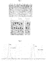
- Cell culture supernatant was collected at 96 h after transfection, and purified by Protein A Resin (Genscript). The purified proteins were applied to SDS-PAGE. Results are shown in Fig. 1 , indicating that the antibody fusion protein with expected molecular weight was successfully expressed.
- the antibody fusion protein purified by Protein A resin was analyzed by size exclusion chromatography by GE's AKTA .
- the column used was: Superdex 200 Increase 10/300 GL size exclusion chromatography column, and the solution used was PBS buffer (0.010 M phosphate buffer, 0.0027 M KCl, 0.14 M NaCl, pH 7.4). It is seen from the chromatogram in Fig. 2 that the expression of the antibody fusion protein of PD-1 and aEGFR or aHER2 has considerable purity.
- hEGFR-hIGg1Fc (SinoBiological) (100 ng/well) was coated in a 96-well plate, and incubated overnight at 4°C. After blocked with PBST (0.5% Tween-20 in PBS) containing 2% skimmed milk powder for 1 hour at room temperature, the gradiently diluted antibody (aEGFR) fusion proteins (10pM-1.2nM) were added and incubated at room temperature for 2 h.
- aEGFR gradiently diluted antibody
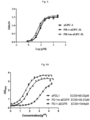
- the antibody fusion proteins PD-1m-aEGFR and PD-1-aEGFR have similar affinities to EGFR.
- hHER2-His (Aero) (100 ng/well) was coated in a 96-well plate, and incubated overnight at 4°C. After blocked with PBST (0.5% Tween-20 in PBS)containing 2% skimmed milk powder for 1 hour at room temperature, the gradiently diluted antibody (aHER2) fusion proteins (10pM-1.2nM) were added and incubated at room temperature for 2 h. After being washed with PBST containing 2% skimmed milk powder for 4-5 times, anti-human kappa light chain-HRP (Sigma A7146, 1:3000) secondary antibody was added and incubated at room temperature for 1 h, and then washed with PBST containing 2% skimmed milk powder for 4-5 times.
- aHER2 gradiently diluted antibody
- Color development was performed by QuantaBlu fluorescent peroxidase substrate (Life technologies, Cat.15169) and read at 325 nm and 420 nm, or by TMB color reagent (BioLegend , Cat. 421101) and read at 650 nm (unterminated) or 450 nm (terminated). Data was analyzed by nonlinear regression using specific binding model with Prizm Graphpad software.
- PD-1-aHER2 and PD-1m-aHER2 have similar affinities to HER2. Compared with the positive control aHER2, the fusion of PD-1 or PD-1m has little effect on the binding of the scaffold antibody to HER2.
- hGPC-3 (BioLegend) (100 ng/well) was coated in a 96-well plate, and incubated overnight at 4°C. After blocked with PBST(0.5% Tween-20 in PBS) containing 2% skimmed milk powder for 1 h at room temperature, the antibody (aGPC-3) fusion proteins in gradient dilutions (25pM-3nM) were added and incubated at room temperature for 2 h.
- the affinities of PD-1-aGPC-3 and PD-1m-aGPC-3 to GPC3 are similar to the affinity of scaffold antibody aGPC-3, indicating that the fusion of PD-1 or PD-1m has little effect on the binding of scaffold antibody to GPC-3.
- hPD-L1-hIGg1Fc (SinoBiological) or mouse PD-L1-Fc (SinoBiological) (100ng/well) was coated in 96-well plates, and incubated overnight at 4°C. After blocked with PBST(0.5% Tween-20 in PBS) containing 2% skimmed milk powder at room temperature for 1 h, the antibody fusion protein in gradient dilutions (25pM-3nM) was added and incubated at room temperature for 2 h.
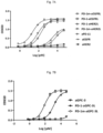
- the affinity of PD-1m antibody fusion protein (PD-1m-aEGFRL, PD-1m-aHER2L or PD-1m-aGPC-3L) to human PD-L1 is similar to that of the positive control aPD-L1 to human PD-L1, and is much higher than that of PD-1 antibody fusion protein to human PD-L1 (25-35 times).
- the affinity of PD-1m antibody fusion protein (PD-1m-aEGFRL, PD-1m-aHER2L or PD-1m-aGPC-3L) to mouse PD-L1 is similar to that of the positive control aPD-L1 to mouse PD-L1, and is much higher than that of PD-1 antibody fusion protein to mouse PD-L1.
- A431, MC38-hEGFR, CHO-PD-L1, HepG2, and Hepa1-6 cells were cultured. After trypsinization, 2*10 5 cells were taken and washed with pre-cooled PBS for 3 times. After blocked with PBS containing 2% FBS at 4°C, supernatant was discarded by centrifugation, 100 pL of the antibody fusion proteins with different concentrations diluted by the PBS solution containing 2% FBS were added and incubated at 4°C for 1 h.
- the unbound antibody fusion protein was washed off with PBS containing 2% FBS, and secondary antibody Mouse Anti-Human IgG Fc-APC (Southern biotech) was added and incubated at 4°C for 1 h. After being washed with PBS containing 2% FBS for three times, the fluorescence intensity was detected by flow cytometry.
- Fig. 8 Results of the binding of PD-1m-aEGFRL to cells are shown in Fig. 8 .
- the binding of PD-1m-aEGFRL and PD-1-aEGFRL to HepG2 cell surface EGFR is very similar with a slight reduction( Fig. 8A ), which is consistent with ELISA results.
- PD-1m-aEGFRL can bind to hPD-L1 on the surface of CHO-hPD-L1 cells, and the binding activity is similar to that of aPD-L1 ( Fig. 8B ), which is consistent with ELISA results.
- PD-1m-aEGFRL or PD-1-aEGFRL has slightly better cell binding activity to MC38-hEGFR ( Fig. 8C ), indicating a synergistic effect.
- Fig. 9 Results of the binding of PD-1m-aGPC-3L to cells are shown in Fig. 9 .
- the binding of PD-1m-a GPC-3L and PD-1-aGPC-3L to HepG2 cell surface GPC-3 is very similar with a slight reduction( Fig. 9A ).
- PD-1m-aGPC-3L can bind to hPD-L1 (9B) on the surface of A431 cells and mPDL1 (9C) on the surface of Hepa1-6 cells.
- MC38 cells expressing human EGFR (MC38-hEGFR, constructed by our laboratory) (DMEM medium containing 10% FBS, 1% double antibody) were cultured. After trypsinization, 2*10 5 cells were taken and washed with pre-cooled PBS for 3 times. After being blocked with PBS containing 2% FBS at 4°C, supernatant was discarded by centrifugation, 100 pL of the antibody fusion proteins with different concentrations diluted by the PBS solution containing 2% FBS were added and incubated at 4°C for 1 h.
- the unbound antibody fusion protein was washed off with PBS containing 2% FBS, and secondary antibody Mouse Anti-Human IgG Fc-APC (Southern biotech) was added and incubated at 4°C for 1 h. After being washed with PBS containing 2% FBS for three times, the fluorescence intensity was detected by flow cytometry.
- the unbound antibody fusion protein was washed off with PBS containing 2% FBS, a secondary antibody Mouse Anti-Human IgG Fc-APC (southern biotech) is added and incubated at 4°C for 1 h. After being washed with PBS containing 2% FBS for three times, the fluorescence intensity was detected by flow cytometry.
- Results are shown in Fig. 10 .
- the binding of PD-1m-aEGFRL to MC38-hEGFR cells can be weakened by free hEGFR-his in the solution, but the magnitude of is less than that of the binding ability of the positive control aEGFR or PD-1-aEGFR to the MC38-hEGFR cells.
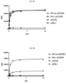
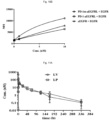
- the antibody fusion protein was intraperitoneal-injected (I.P.) or tail-vein-injected (I.V.) into SD male rats (3 rats/group). Heparin anticoagulant blood was collected from the tail vein at 15 min, 30 min, 1h, 4h, 8h, 1d, 2d, 3d, 5d, 7d and 14d after injection. After centrifugation, plasma was collected, and stored at -80°C for further use.
- the antibody fusion protein and scaffold antibody in the plasma was performed with reference to Embodiment 4.1.
- the scaffold of the antibody fusion protein has a longer half-life than that of the antibody fusion protein.
- the antibody fusion protein can effectively inhibit tumor growth without significant side effects such as weight loss.
- the control bispecific antibody(PD1m-aGPC3) without EGFR targeting cannot inhibit tumor growth at all, proving that the tumor-targeted PD1m-aEGFR has good selectivity and safety.
Landscapes
- Health & Medical Sciences (AREA)
- Chemical & Material Sciences (AREA)
- Organic Chemistry (AREA)
- Immunology (AREA)
- Life Sciences & Earth Sciences (AREA)
- Medicinal Chemistry (AREA)
- General Health & Medical Sciences (AREA)
- Biophysics (AREA)
- Biochemistry (AREA)
- Molecular Biology (AREA)
- Genetics & Genomics (AREA)
- Proteomics, Peptides & Aminoacids (AREA)
- Cell Biology (AREA)
- Chemical Kinetics & Catalysis (AREA)
- Public Health (AREA)
- Veterinary Medicine (AREA)
- Nuclear Medicine, Radiotherapy & Molecular Imaging (AREA)
- Animal Behavior & Ethology (AREA)
- Pharmacology & Pharmacy (AREA)
- Gastroenterology & Hepatology (AREA)
- General Chemical & Material Sciences (AREA)
- Bioinformatics & Cheminformatics (AREA)
- Engineering & Computer Science (AREA)
- Zoology (AREA)
- Toxicology (AREA)
- Oncology (AREA)
- Gynecology & Obstetrics (AREA)
- Pregnancy & Childbirth (AREA)
- Reproductive Health (AREA)
- Peptides Or Proteins (AREA)
- Medicines Containing Antibodies Or Antigens For Use As Internal Diagnostic Agents (AREA)
Applications Claiming Priority (2)
| Application Number | Priority Date | Filing Date | Title |
|---|---|---|---|
| CN202010299304 | 2020-04-16 | ||
| PCT/CN2021/087864 WO2021209049A1 (zh) | 2020-04-16 | 2021-04-16 | 一种pd-1突变体多肽及其制备和用途 |
Publications (2)
| Publication Number | Publication Date |
|---|---|
| EP4137510A1 true EP4137510A1 (de) | 2023-02-22 |
| EP4137510A4 EP4137510A4 (de) | 2023-11-01 |
Family
ID=78083924
Family Applications (1)
| Application Number | Title | Priority Date | Filing Date |
|---|---|---|---|
| EP21789230.6A Pending EP4137510A4 (de) | 2020-04-16 | 2021-04-16 | Pd-1-mutantes polypeptid und herstellung davon und verwendung davon |
Country Status (4)
| Country | Link |
|---|---|
| US (1) | US20230287081A1 (de) |
| EP (1) | EP4137510A4 (de) |
| CN (1) | CN115708411A (de) |
| WO (1) | WO2021209049A1 (de) |
Families Citing this family (1)
| Publication number | Priority date | Publication date | Assignee | Title |
|---|---|---|---|---|
| CN116004541B (zh) * | 2022-08-04 | 2025-01-28 | 四川大学华西医院 | 具有高亲和力结合肿瘤细胞pd-l1的细胞膜材料及其制备方法和应用 |
Family Cites Families (13)
| Publication number | Priority date | Publication date | Assignee | Title |
|---|---|---|---|---|
| CA3253628A1 (en) * | 2010-03-05 | 2025-11-29 | The Johns Hopkins University | Compositions and methods for targeted immunomodulatory antibodies and fusion proteins |
| US8628773B2 (en) * | 2011-04-07 | 2014-01-14 | Amgen Inc. | Antigen binding proteins |
| CN103172741B (zh) * | 2011-12-20 | 2018-04-27 | 智翔(上海)医药科技有限公司 | 全人源抗egfr抗体 |
| TWI681969B (zh) * | 2014-01-23 | 2020-01-11 | 美商再生元醫藥公司 | 針對pd-1的人類抗體 |
| CN106967173A (zh) * | 2016-01-13 | 2017-07-21 | 上海嘉和生物科技有限公司 | 一种重组嵌合膜蛋白及其制备和应用 |
| CA3055200A1 (en) * | 2017-03-03 | 2018-09-07 | Obsidian Therapeutics, Inc. | Compositions and methods for immunotherapy |
| US20200157226A1 (en) * | 2017-05-23 | 2020-05-21 | Dragonfly Therapeutics, Inc. | Proteins binding nkg2d, cd16 and a tumor-associated antigen |
| EP3668893A4 (de) * | 2017-08-16 | 2021-08-04 | Dragonfly Therapeutics, Inc. | Nkg2d, cd16 und egfr, hla-e ccr4 oder pd-l1 bindende proteine |
| WO2019151771A1 (ko) * | 2018-02-02 | 2019-08-08 | 주식회사 뉴라클제네틱스 | Pd-l1 결합력이 증대된 pd-1 변이체 |
| KR102171766B1 (ko) * | 2018-02-02 | 2020-10-29 | 주식회사 뉴라클제네틱스 | Pd-l1 결합력이 증대된 pd-1 변이체 |
| WO2019241758A1 (en) * | 2018-06-15 | 2019-12-19 | Alpine Immune Sciences, Inc. | Pd-1 variant immunomodulatory proteins and uses thereof |
| EP3820887A4 (de) * | 2018-07-11 | 2022-04-20 | KAHR Medical Ltd. | Pd1-4-1bbl-fusionsproteinvariante und ihre verwendunge |
| CN108997492B (zh) * | 2018-07-19 | 2022-04-12 | 海珂分子(北京)科技有限责任公司 | 高亲和力的pd1胞外区突变体多肽及相关融合蛋白 |
-
2021
- 2021-04-16 US US17/918,919 patent/US20230287081A1/en active Pending
- 2021-04-16 WO PCT/CN2021/087864 patent/WO2021209049A1/zh not_active Ceased
- 2021-04-16 CN CN202180028808.9A patent/CN115708411A/zh active Pending
- 2021-04-16 EP EP21789230.6A patent/EP4137510A4/de active Pending
Also Published As
| Publication number | Publication date |
|---|---|
| EP4137510A4 (de) | 2023-11-01 |
| CN115708411A (zh) | 2023-02-21 |
| US20230287081A1 (en) | 2023-09-14 |
| WO2021209049A1 (zh) | 2021-10-21 |
Similar Documents
| Publication | Publication Date | Title |
|---|---|---|
| CN112638950B (zh) | 一种双特异性抗体 | |
| JP7713886B2 (ja) | Cd86バリアント免疫調節タンパク質およびその使用 | |
| US12522638B2 (en) | IL12 receptor agonists and methods of use thereof | |
| WO2020177627A1 (zh) | 一种双特异抗体 | |
| US20250000946A1 (en) | Il-2 variants and fusion proteins thereof | |
| CN117836328A (zh) | 一种多特异性抗原结合蛋白及其应用 | |
| EP4573111A1 (de) | Interferonrezeptoragonisten und verwendungen davon | |
| EP4137510A1 (de) | Pd-1-mutantes polypeptid und herstellung davon und verwendung davon | |
| JP2025529805A (ja) | インターフェロンプロタンパク質及びその使用 | |
| JP2025520175A (ja) | インターロイキン-2プロタンパク質及びその使用 | |
| WO2024199458A1 (en) | Il-2 variants with improved stability and compositions thereof | |
| WO2025199243A1 (en) | Trivalent multispecific binding molecules and methods of use thereof | |
| WO2025245494A1 (en) | Tumor-targeted split il12 receptor agonists | |
| JP2026504072A (ja) | Il12受容体アゴニストおよびその使用方法 | |
| WO2026039791A1 (en) | Tumor-targeted split il2 receptor agonists | |
| EP4673470A2 (de) | Multispezifische moleküle mit einem peptid-mhc-komplex mit einer mhc-domäne und einem antigenpeptid und einem immunzellenantigen-targeting-teil | |
| JP2025519158A (ja) | インターロイキン-2プロタンパク質及びその使用 | |
| WO2026076013A1 (en) | Tumor-targeted il2 receptor agonists and multispecific t-cell engagers |
Legal Events
| Date | Code | Title | Description |
|---|---|---|---|
| STAA | Information on the status of an ep patent application or granted ep patent |
Free format text: STATUS: THE INTERNATIONAL PUBLICATION HAS BEEN MADE |
|
| PUAI | Public reference made under article 153(3) epc to a published international application that has entered the european phase |
Free format text: ORIGINAL CODE: 0009012 |
|
| STAA | Information on the status of an ep patent application or granted ep patent |
Free format text: STATUS: REQUEST FOR EXAMINATION WAS MADE |
|
| 17P | Request for examination filed |
Effective date: 20221116 |
|
| AK | Designated contracting states |
Kind code of ref document: A1 Designated state(s): AL AT BE BG CH CY CZ DE DK EE ES FI FR GB GR HR HU IE IS IT LI LT LU LV MC MK MT NL NO PL PT RO RS SE SI SK SM TR |
|
| DAV | Request for validation of the european patent (deleted) | ||
| DAX | Request for extension of the european patent (deleted) | ||
| RAP1 | Party data changed (applicant data changed or rights of an application transferred) |
Owner name: YICHEN THERAPEUTICS LIMITED |
|
| A4 | Supplementary search report drawn up and despatched |
Effective date: 20231002 |
|
| RIC1 | Information provided on ipc code assigned before grant |
Ipc: A61P 37/00 20060101ALI20230926BHEP Ipc: A61P 35/00 20060101ALI20230926BHEP Ipc: A61K 38/17 20060101ALI20230926BHEP Ipc: C12N 15/09 20060101ALI20230926BHEP Ipc: C07K 19/00 20060101ALI20230926BHEP Ipc: C07K 14/705 20060101AFI20230926BHEP |