EP4140475A1 - Lipoparticles pseudo-virales synthétiques - Google Patents
Lipoparticles pseudo-virales synthétiques Download PDFInfo
- Publication number
- EP4140475A1 EP4140475A1 EP21193839.4A EP21193839A EP4140475A1 EP 4140475 A1 EP4140475 A1 EP 4140475A1 EP 21193839 A EP21193839 A EP 21193839A EP 4140475 A1 EP4140475 A1 EP 4140475A1
- Authority
- EP
- European Patent Office
- Prior art keywords
- lipoparticle
- sample
- spike protein
- lipid
- ectodomain
- Prior art date
- Legal status (The legal status is an assumption and is not a legal conclusion. Google has not performed a legal analysis and makes no representation as to the accuracy of the status listed.)
- Ceased
Links
Images


Classifications
-
- A—HUMAN NECESSITIES
- A61—MEDICAL OR VETERINARY SCIENCE; HYGIENE
- A61K—PREPARATIONS FOR MEDICAL, DENTAL OR TOILETRY PURPOSES
- A61K9/00—Medicinal preparations characterised by special physical form
- A61K9/0012—Galenical forms characterised by the site of application
- A61K9/0019—Injectable compositions; Intramuscular, intravenous, arterial, subcutaneous administration; Compositions to be administered through the skin in an invasive manner
-
- A—HUMAN NECESSITIES
- A61—MEDICAL OR VETERINARY SCIENCE; HYGIENE
- A61K—PREPARATIONS FOR MEDICAL, DENTAL OR TOILETRY PURPOSES
- A61K39/00—Medicinal preparations containing antigens or antibodies
- A61K39/12—Viral antigens
-
- A—HUMAN NECESSITIES
- A61—MEDICAL OR VETERINARY SCIENCE; HYGIENE
- A61K—PREPARATIONS FOR MEDICAL, DENTAL OR TOILETRY PURPOSES
- A61K9/00—Medicinal preparations characterised by special physical form
- A61K9/08—Solutions
-
- A—HUMAN NECESSITIES
- A61—MEDICAL OR VETERINARY SCIENCE; HYGIENE
- A61K—PREPARATIONS FOR MEDICAL, DENTAL OR TOILETRY PURPOSES
- A61K9/00—Medicinal preparations characterised by special physical form
- A61K9/10—Dispersions; Emulsions
- A61K9/127—Synthetic bilayered vehicles, e.g. liposomes or liposomes with cholesterol as the only non-phosphatidyl surfactant
-
- A—HUMAN NECESSITIES
- A61—MEDICAL OR VETERINARY SCIENCE; HYGIENE
- A61K—PREPARATIONS FOR MEDICAL, DENTAL OR TOILETRY PURPOSES
- A61K9/00—Medicinal preparations characterised by special physical form
- A61K9/10—Dispersions; Emulsions
- A61K9/127—Synthetic bilayered vehicles, e.g. liposomes or liposomes with cholesterol as the only non-phosphatidyl surfactant
- A61K9/1277—Preparation processes; Proliposomes
-
- A—HUMAN NECESSITIES
- A61—MEDICAL OR VETERINARY SCIENCE; HYGIENE
- A61K—PREPARATIONS FOR MEDICAL, DENTAL OR TOILETRY PURPOSES
- A61K9/00—Medicinal preparations characterised by special physical form
- A61K9/48—Preparations in capsules, e.g. of gelatin, of chocolate
- A61K9/50—Microcapsules having a gas, liquid or semi-solid filling; Solid microparticles or pellets surrounded by a distinct coating layer, e.g. coated microspheres, coated drug crystals
- A61K9/51—Nanocapsules; Nanoparticles
- A61K9/5107—Excipients; Inactive ingredients
- A61K9/5123—Organic compounds, e.g. fats, sugars
-
- A—HUMAN NECESSITIES
- A61—MEDICAL OR VETERINARY SCIENCE; HYGIENE
- A61P—SPECIFIC THERAPEUTIC ACTIVITY OF CHEMICAL COMPOUNDS OR MEDICINAL PREPARATIONS
- A61P31/00—Antiinfectives, i.e. antibiotics, antiseptics, chemotherapeutics
- A61P31/12—Antivirals
- A61P31/14—Antivirals for RNA viruses
-
- A—HUMAN NECESSITIES
- A61—MEDICAL OR VETERINARY SCIENCE; HYGIENE
- A61P—SPECIFIC THERAPEUTIC ACTIVITY OF CHEMICAL COMPOUNDS OR MEDICINAL PREPARATIONS
- A61P31/00—Antiinfectives, i.e. antibiotics, antiseptics, chemotherapeutics
- A61P31/12—Antivirals
- A61P31/14—Antivirals for RNA viruses
- A61P31/16—Antivirals for RNA viruses for influenza or rhinoviruses
-
- A—HUMAN NECESSITIES
- A61—MEDICAL OR VETERINARY SCIENCE; HYGIENE
- A61P—SPECIFIC THERAPEUTIC ACTIVITY OF CHEMICAL COMPOUNDS OR MEDICINAL PREPARATIONS
- A61P31/00—Antiinfectives, i.e. antibiotics, antiseptics, chemotherapeutics
- A61P31/12—Antivirals
- A61P31/20—Antivirals for DNA viruses
-
- A—HUMAN NECESSITIES
- A61—MEDICAL OR VETERINARY SCIENCE; HYGIENE
- A61K—PREPARATIONS FOR MEDICAL, DENTAL OR TOILETRY PURPOSES
- A61K39/00—Medicinal preparations containing antigens or antibodies
- A61K2039/555—Medicinal preparations containing antigens or antibodies characterised by a specific combination antigen/adjuvant
- A61K2039/55511—Organic adjuvants
- A61K2039/55555—Liposomes; Vesicles, e.g. nanoparticles; Spheres, e.g. nanospheres; Polymers
-
- C—CHEMISTRY; METALLURGY
- C12—BIOCHEMISTRY; BEER; SPIRITS; WINE; VINEGAR; MICROBIOLOGY; ENZYMOLOGY; MUTATION OR GENETIC ENGINEERING
- C12N—MICROORGANISMS OR ENZYMES; COMPOSITIONS THEREOF; PROPAGATING, PRESERVING, OR MAINTAINING MICROORGANISMS; MUTATION OR GENETIC ENGINEERING; CULTURE MEDIA
- C12N2730/00—Reverse transcribing DNA viruses
- C12N2730/00011—Details
- C12N2730/10011—Hepadnaviridae
- C12N2730/10111—Orthohepadnavirus, e.g. hepatitis B virus
- C12N2730/10134—Use of virus or viral component as vaccine, e.g. live-attenuated or inactivated virus, VLP, viral protein
-
- C—CHEMISTRY; METALLURGY
- C12—BIOCHEMISTRY; BEER; SPIRITS; WINE; VINEGAR; MICROBIOLOGY; ENZYMOLOGY; MUTATION OR GENETIC ENGINEERING
- C12N—MICROORGANISMS OR ENZYMES; COMPOSITIONS THEREOF; PROPAGATING, PRESERVING, OR MAINTAINING MICROORGANISMS; MUTATION OR GENETIC ENGINEERING; CULTURE MEDIA
- C12N2770/00—MICROORGANISMS OR ENZYMES; COMPOSITIONS THEREOF; PROPAGATING, PRESERVING, OR MAINTAINING MICROORGANISMS; MUTATION OR GENETIC ENGINEERING; CULTURE MEDIA ssRNA viruses positive-sense
- C12N2770/00011—Details
- C12N2770/20011—Coronaviridae
- C12N2770/20034—Use of virus or viral component as vaccine, e.g. live-attenuated or inactivated virus, VLP, viral protein
-
- C—CHEMISTRY; METALLURGY
- C12—BIOCHEMISTRY; BEER; SPIRITS; WINE; VINEGAR; MICROBIOLOGY; ENZYMOLOGY; MUTATION OR GENETIC ENGINEERING
- C12N—MICROORGANISMS OR ENZYMES; COMPOSITIONS THEREOF; PROPAGATING, PRESERVING, OR MAINTAINING MICROORGANISMS; MUTATION OR GENETIC ENGINEERING; CULTURE MEDIA
- C12N2770/00—MICROORGANISMS OR ENZYMES; COMPOSITIONS THEREOF; PROPAGATING, PRESERVING, OR MAINTAINING MICROORGANISMS; MUTATION OR GENETIC ENGINEERING; CULTURE MEDIA ssRNA viruses positive-sense
- C12N2770/00011—Details
- C12N2770/24011—Flaviviridae
- C12N2770/24111—Flavivirus, e.g. yellow fever virus, dengue, JEV
- C12N2770/24134—Use of virus or viral component as vaccine, e.g. live-attenuated or inactivated virus, VLP, viral protein
Definitions
- the present invention is directed to non-infectious synthetic lipoparticles.
- Said lipoparticles comprise a cell surface receptor binding Spike protein ectodomain on their surface, which renders the lipoparticles particularly useful for studying the binding and/or retention of said lipoparticles to cells.
- SARS-CoV-2 infection is initiated by binding of the virus to the target receptor angiotensin converting enzyme 2 (ACE2), mediated by the trimeric Spike (S) glycoprotein.
- ACE2 angiotensin converting enzyme 2
- S trimeric Spike glycoprotein.
- HIV uses a fusion co-receptor, typically either the chemokine receptors CXCR4 or CCR5. Preventing receptor binding is a common strategy for treating or preventing pathogen infection by inhibition of viral entry.
- Membrane vesicles prepared by mechanically disrupting cells (Top-down approach), are a common source of membrane protein for many drug screens currently conducted.
- membrane vesicles are heterogeneous, impure, and not particularly stable.
- the receptors within them may be misoriented, a minor component of total protein, and derived from intracellular organelles.
- pseudovirus approaches as well as in in vitro protein binding assays often do not reproduce the biophysical features of the natural pathogen (virion, cell), which are essential for assessing particulate and multivalent binding effects.
- pathogen receptor interactions are generally slow and laborious. In many cases, such techniques require transfection of individual receptors, the growth of cells, and the use of the pathogen in its infectious form, thereby requiring at least a safety level 2 laboratory environment. Also, protein interactions are detected using immunoprecipitation, Western blotting, or radiolabeled proteins.
- SARS-CoV-2 enveloped severe acute respiratory syndrome-coronavirus 2
- SARS-CoV-2 infection is initiated by binding of the virus to the target receptor angiotensin converting enzyme 2 (ACE2), mediated by the trimeric Spike (S) glycoprotein.
- ACE2 angiotensin converting enzyme 2
- S2 trimeric Spike glycoprotein.
- RBD target receptor angiotensin converting enzyme 2
- S2 induces fusion of the viral envelope with the target cell membrane.
- S is the major focus of COVID-19 vaccine development, the primary target for neutralizing antibodies and the main diagnostic antigen for SARS-CoV-2 infections.
- Structural analyses of S revealed substantial conformational rearrangements of RBD during the infection process.
- the RBDs lie flat on the S surface and are therefore less accessible for ACE2 binding.
- at least one RBD erects from S to expose the receptor binding motif, enabling ACE2 binding.
- Each monomer within the S trimer can undergo a closed-to-open transition and consecutive priming by ACE2.
- ACE2 ACE2 binding motif
- Molecular mechanisms that modulate S open-to-closed equilibrium and their implications for SARS-CoV-2 infection and immune evasion remain poorly understood. Exogenous control of this equilibrium, however, could greatly benefit COVID-19 therapy development and promote prospective pandemic prevention.
- the objective of the present invention lies in the provision of synthetic particles that mimic pathogens, particularly viruses, that can be prepared under low-biosafety constraints and that are particularly useful for studying the binding and/or retention to living cells.
- synthetic lipoparticles comprising a lipid bilayer decorated with cell receptor binding Spike protein ectodomain are particularly beneficial for assessing receptor binding.
- the so formed synthetic lipoparticles are molecularly defined and suitable for systematic analysis of cell attachment effects.
- the synthetic lipoparticles can be produced in vitro in a bottom-up approach from small unilamellar vesicles. Due to the bottom-up approach the constitution of the lipid bilayer in regard of the lipid components, the Spike protein and particle size, can be adjusted precisely to mimic the biophysical features of various natural pathogens, such as viruses or virions, thereby allowing for integrated assessment of particulate and multivalent binding effects between the pathogen and living cells.
- the synthetic lipoparticles according to the invention lack genetically encoded information, so that no higher biosafety standards have to be met, which facilitates the production and use of the synthetic lipoparticles.
- the present invention is directed to a non-infectious synthetic lipoparticle comprising: a lipid bilayer comprising
- the synthetic lipoparticles of the present invention do not contain infectious nucleic acids, such as DNA or RNA, wherein infectious refers to nucleic acids which, upon being taken up by a permissive host cell, are capable of producing multiple, e.g., one or more copies of the same nucleic acid (replicating) and the nucleic acid represent a complete infectious virus particle.
- infectious nucleic acids such as DNA or RNA
- the present invention is directed to a synthetic lipoparticle comprising: a lipid bilayer comprising
- the synthetic particles of the present invention mainly consist of a lipid bilayer and are therefore considered as lipoparticles.
- the lipid bilayer forms a shell or a sphere in the nanometer range similar to the outer layer of viruses or virions, the viral envelope, so that the synthetic lipoparticles of the present invention exhibit biophysical properties similar or identical to natural viruses or virions.
- the lipoparticles are virus-like or viral particles or minimal virions.
- the present invention is directed to a non-infectious synthetic virus-like particle comprising: a lipid bilayer comprising
- the present invention is directed to a non-infectious synthetic viral particle comprising: a lipid bilayer comprising
- the present invention is directed to a non-infectious synthetic minimal virion comprising: a lipid bilayer comprising
- the present invention is directed to a non-infectious synthetic liposome comprising: a lipid bilayer comprising
- the present invention is directed to a non-infectious synthetic lipoparticle comprising: a small unilamellar vesicle comprising
- the synthetic lipoparticles of the present invention comprise a Spike protein ectodomain that is bound to a lipid.
- the lipid is aggregated in the lipid bilayer such that the Spike protein ectodomain is arranged at the outside of the lipid bilayer.
- the Spike protein ectodomain is located at the outer surface of the bilayer so that the Spike protein can interact with cell surface receptors.
- the present invention is directed to a non-infectious synthetic lipoparticle comprising: a vesicle-forming lipid bilayer comprising
- the present invention is directed to a non-infectious synthetic lipoparticle comprising: a vesicle-forming lipid bilayer comprising
- the present invention is directed to synthetic lipoparticles comprising a lipid bilayer decorated with cell receptor binding proteins, wherein the lipid bilayer is molecularly defined to precisely mimic the biophysical features of various natural pathogens, such as viruses or virions.
- the present invention is directed to a non-infectious synthetic lipoparticle comprising: a lipid bilayer comprising
- Suitable phosphatidylcholines for the lipid bilayer are exemplarily, but not restricted to, 1,2-dimyristoyl-sn-glycero-3-phosphocholine (DMPC), 1,2-dihexanoyl-sn-glycero-3-phosphocholine (DHPC), 1,2-dipalmitoleoyl-sn-glycero-3-phosphocholine, 1,2-dipalmitoyl-sn-glycero-3-phosphocholine (DPPC), 1-palmitoyl-2-oleoyl-glycero-3-phosphocholine (POPC), 1,2-distearoyl-sn-glycero-3-phosphocholine (DSPC), 1,2-dilauroyl-sn-glycero-3-phosphocholine (DLPC), and 1,2-dioleoyl-sn-glycero-3-phosphocholine (DOPC).
- DMPC 1,2-dimyristoyl-sn-glycero-3-phospho
- Suitable anionic lipids for the lipid bilayer are for example, but not limited to, phosphatidylserines, such as 1,2-dioleoyl-sn-glycero-3-phospho-l-serine (DOPS) and phosphatidylglycerols, such as 1,2-dioleoyl-sn-glycero-3-phospho-(1'-rac-glycerol) (DOPG), and phosphatidylinositols, such as 1,2-dioleoyl-sn-glycero-3-phospho-(1'-myo-inositol) (DOPI).
- DOPS 1,2-dioleoyl-sn-glycero-3-phospho-l-serine
- DOPG 1,2-dioleoyl-sn-glycero-3-phospho-(1'-rac-glycerol)
- DOPI 1,2-dioleoyl-sn-gly
- the non-infectious synthetic lipoparticle comprises a lipid bilayer comprising
- the non-infectious synthetic lipoparticle comprises a lipid bilayer comprising
- the non-infectious synthetic lipoparticle comprises a lipid bilayer comprising
- the non-infectious synthetic lipoparticle comprises a lipid bilayer comprising
- the non-infectious synthetic lipoparticle comprises a lipid bilayer comprising
- the non-infectious synthetic lipoparticle comprises a lipid bilayer comprising
- the lipid bilayer of the inventive lipoparticles is composed of at least one neutral amphiphile, at least one cationic lipid and at least one anionic lipid in order to optimally reproduce the biophysical properties of viral pathogens.
- the non-infectious synthetic lipoparticle comprises: a lipid bilayer comprising
- Suitable cationic lipids for the lipid bilayer are for example, but not limited to, cholesterols, such as 18:1 cholesterol, 3 ⁇ -[N-(N',N'-Dimethylaminoethane)-carbamoyl]cholesterol (DAC-Chol) and 3 ⁇ -[N-(N',N'-dimethylaminoethane)-carbamoyl]cholesterol (DC-Chol), and sphingosylphosphorylcholine and N-acyl derivatives, such as N-palmitoyl-D-erythro-sphingosylphosphorylcholine, N-heptadecanoyl-D-erythro-sphingosylphosphorylcholine, N-oleoyl-D-erythro-sphingosylphosphorylcholine N -stearoyl-D-erythro-sphingosylphosphorylcholine, N-lignoceroyl-D-erythro-sphingosylphospho
- the non-infectious synthetic lipoparticle comprises a lipid bilayer comprising
- the non-infectious synthetic lipoparticle comprises a lipid bilayer comprising
- the non-infectious synthetic lipoparticle comprises a lipid bilayer comprising
- the non-infectious synthetic lipoparticle comprises a lipid bilayer comprising
- the non-infectious synthetic lipoparticle comprises: a lipid bilayer comprising - 40-50 mol% 1,2-dioleoyl-sn-glycero-3-phosphocholine, - 15-25 mol% 1,2-dioleoyl-sn-glycero-3-phosphoethanolamine, - 2-4 mol% 1,2-dioleoyl-sn-glycero-3-phospho-l-serine, - 12 mol% 1,2-dioleoyl-sn-glycero-3-phospho-(1'-myo-inositol), - 14 mol% cholesterol, - 3 mol% N -stearoyl-D-erythro-sphingosylphosphorylcholine, and - 1 mol% of a lipid-bound Spike protein ectodomain, wherein the Spike protein ectodomain binds to a cell surface receptor, wherein the lipoparticle has a diameter between 20
- the non-infectious synthetic lipoparticle comprises: a lipid bilayer comprising - 80-85 mol% 1,2-dioleoyl-sn-glycero-3-phosphocholine, - 5-10 mol% 1,2-dioleoyl-sn-glycero-3-phosphoethanolamine, - 5 mol% 1,2-dioleoyl-sn-glycero-3-phospho-(1'-myo-inositol), - 3 mol% N -stearoyl-D-erythro-sphingosylphosphorylcholine, and - 1 mol% of a lipid-bound Spike protein ectodomain, wherein the Spike protein ectodomain binds to a cell surface receptor, wherein the lipoparticle has a diameter between 20 nm and 300 nm as measured by dynamic light scattering.
- the lipid bilayer of the inventive lipoparticles may further comprise a neutral lipid, preferably a ceramide, such as N-stearoyl-D-erythro-sphingosine.
- the non-infectious synthetic lipoparticle comprises a lipid bilayer comprising
- the non-infectious synthetic lipoparticle comprises a lipid bilayer comprising
- the non-infectious synthetic lipoparticle comprises a lipid bilayer comprising
- the non-infectious synthetic lipoparticle comprises a lipid bilayer comprising
- the non-infectious synthetic lipoparticle comprises: a lipid bilayer comprising - 40 mol% 1,2-dioleoyl-sn-glycero-3-phosphocholine, - 30 mol% 1,2-dioleoyl-sn-glycero-3-phosphoethanolamine, - 22 mol% N -stearoyl-D-erythro-sphingosylphosphorylcholine, - 5 mol% N-stearoyl-D-erythro-sphingosine, and - 1 mol% of a lipid-bound Spike protein ectodomain, wherein the Spike protein ectodomain binds to a cell surface receptor, wherein the lipoparticle has a diameter between 20 nm and 300 nm as measured by dynamic light scattering.
- the Spike protein ectodomain can be bound to the lipid by any conventional binding method such as covalent binding (ester bond formation or amide bond formation) or affinity binding (e.g. Ni 2+ /His-tag).
- the lipid is equipped with suitable reactive groups.
- the lipid is a reactive lipid in such a manner that it can bind or immobilise the Spike protein ectodomain.
- the Spike protein ectodomain is also equipped with a reactive group or a tag for binding or immobilising to the lipid.
- the Spike protein ectodomain can be bound to the lipid prior vesicle formation or the Spike protein ectodomain can be bound to the already formed lipid bilayer vesicle.
- the latter approach has the advantage that all Spike protein ectodomain is located at the outer surface of the lipid bilayer vesicle and none of it in the inner cavity of the vesicle, where it cannot interact with a cell surface receptor.
- the present invention is directed to a non-infectious synthetic lipoparticle comprising: a vesicle-forming lipid bilayer comprising
- the Spike protein ectodomain can be any protein which interacts or binds to a cell surface receptor, preferably a receptor of a human cell.
- the present invention is also directed to a non-infectious synthetic lipoparticle comprising: a lipid bilayer comprising
- the Spike protein ectodomain is bound via a His-tag to a Ni-NTA functionalized lipid.
- the present invention is directed to a non-infectious synthetic lipoparticle comprising: a vesicle-forming lipid bilayer comprising
- the Spike protein ectodomain may be derived from any surface protein which is known for binding to cell surface receptors and/or which is known for being involved in viral entry mechanism of viruses.
- the present invention is also directed to a non-infectious synthetic lipoparticle comprising: a lipid bilayer comprising
- the Spike protein ectodomain is selected from SARS-CoV-2 Spike protein ectodomain or a mutant thereof, SARS-CoV Spike protein ectodomain or a mutant thereof, MERS-CoV Spike protein ectodomain or a mutant thereof, HIV-1 gp 120 protein, Hepatitis B Surface Antigen adw subtype 2 (HBVsAG), Hepatitis C E1 protein, Ebolavirus glycoprotein, Marburg virus glycoprotein (AMARV GP), Influenza virus A hemagglutinin H1 protein, and Dengue virus 2 E-glycoprotein.
- HBVsAG Hepatitis B Surface Antigen adw subtype 2
- AMARV GP Marburg virus glycoprotein
- Influenza virus A hemagglutinin H1 protein and Dengue virus 2 E-glycoprotein.
- the present invention is also directed to a non-infectious synthetic lipoparticle comprising: a lipid bilayer comprising
- the Spike protein ectodomain is a SARS-CoV-2 Spike protein ectodomain mutant homotrimer.
- the present invention is also directed to a non-infectious synthetic lipoparticle comprising: a lipid bilayer comprising
- the Spike protein ectodomain is a SARS-CoV-2 Spike protein ectodomain mutant homotrimer, wherein each monomer of the SARS-CoV-2 Spike protein ectodomain comprises an amino acid sequence identical to the sequence as set forth in SEQ ID NO 5 and wherein each monomer comprises further a single point mutation at one of the following sites: L4, T6, P12, D66, D124, R176, D201, R232, K403, L411, E470, N487, A556, D600, H641, Q663, P667, A684, T699, S965, T1010, and D1101.
- the present invention is also directed to a non-infectious synthetic lipoparticle comprising: a lipid bilayer comprising
- the Spike protein ectodomain is a SARS-CoV-2 Spike protein ectodomain mutant homotrimer, wherein each monomer comprises an amino acid sequence identical to one of the sequences as set forth in SEQ ID NOs 6 to 29.
- the present invention is also directed to a non-infectious synthetic lipoparticle comprising: a lipid bilayer comprising
- Spike protein ectodomains usually contain free fatty acids when they are produced by recombinant techniques.
- the inventors have found that these free fatty acids (e.g. palmitic acid, oleic acid, linoleic acid and arachidonic acid) significantly influence the receptor binding of the Spike protein ectodomains.
- free fatty acids e.g. palmitic acid, oleic acid, linoleic acid and arachidonic acid
- Removal of free fatty acids can be achieved by treating the proteins with Lipidex-1000 (a hydroxypropyl beaded dextran substituted with approximately 10% by weight long chain alkyl ethers averaging 15 carbons in length), thereby leading to the corresponding apo Spike protein ectodomains.
- the present invention is also directed to a non-infectious synthetic lipoparticle comprising: a lipid bilayer comprising
- the inventive lipoparticle can have a DLS diameter between 20 nm and 300 nm to optimally reproduce the biophysical properties of viral pathogens. Suitable DLS diameters are between 20 nm and 300 nm, and more preferably between 80 nm and 140 nm. Preferably, the diameter of the inventive lipoparticle is between 100 nm and 120 nm.
- the non-infectious synthetic lipoparticle comprises: a lipid bilayer comprising
- the non-infectious synthetic lipoparticle comprises: a lipid bilayer comprising
- the non-infectious synthetic lipoparticle comprises: a lipid bilayer comprising
- the lipoparticle may further comprise a fluorophore in the lipid bilayer.
- the fluorophore may be immobilized to the surface of the lipid bilayer by a bond to a lipid which is aggregated in the lipid bilayer.
- the non-infectious synthetic lipoparticle comprises: a lipid bilayer comprising
- the non-infectious synthetic lipoparticle comprises: a lipid bilayer comprising
- the fluorophore is bound or conjugated to a neutral amphiphile as lipid, such as a phosphatidylethanolamine.
- a neutral amphiphile such as a phosphatidylethanolamine.
- the fluorophore is bound or conjugated to 1,2-dioleoyl-sn-glycero-3-phosphoethanolamine (DOPE).
- DOPE 1,2-dioleoyl-sn-glycero-3-phosphoethanolamine
- the non-infectious synthetic lipoparticle comprises: a lipid bilayer comprising
- the fluorophore may be any suitable molecular probe, which has an absorption wavelength above 500 nm, such as Lissamine Rhodamine B, fluorescein, and Atto488 ( CAS number 923585-42-6 ).
- the lipid-bound fluorophore may be present in the lipid bilayer of the inventive lipoparticles in the amount of 0.1 mol% to 5 mol%, more preferably 0.2 mol% to 4 mol%, more preferably 0.3 mol% to 3.5 mol%, more preferably 0.4 mol% to 3 mol%, more preferably 0.5 mol% to 2.5 mol%, more preferably 0.6 mol% to 2 mol%, more preferably 0.7 mol% to 1.5 mol%, and most preferably 0.8 mol% to 1.2 mol%.
- the lipid-bound fluorophore is present in the lipid bilayer of the inventive lipoparticles in the amount of 1 mol%.
- the non-infectious synthetic lipoparticle comprises: a lipid bilayer comprising
- the non-infectious synthetic lipoparticle comprises: a lipid bilayer comprising
- the non-infectious synthetic lipoparticle comprises: a lipid bilayer comprising
- the non-infectious synthetic lipoparticle comprises a lipid bilayer comprising
- the non-infectious synthetic lipoparticle comprises a lipid bilayer comprising
- the non-infectious synthetic lipoparticle comprises a lipid bilayer comprising
- the non-infectious synthetic lipoparticle comprises a lipid bilayer comprising
- the non-infectious synthetic lipoparticle comprises a lipid bilayer comprising
- the non-infectious synthetic lipoparticle comprises a lipid bilayer comprising
- the non-infectious synthetic lipoparticle comprises: a lipid bilayer comprising
- the non-infectious synthetic lipoparticle comprises a lipid bilayer comprising
- the non-infectious synthetic lipoparticle comprises a lipid bilayer comprising
- the non-infectious synthetic lipoparticle comprises a lipid bilayer comprising
- the non-infectious synthetic lipoparticle comprises a lipid bilayer comprising
- the non-infectious synthetic lipoparticle comprises: a lipid bilayer comprising - 40-50 mol% 1,2-dioleoyl-sn-glycero-3-phosphocholine, - 15-25 mol% 1,2-dioleoyl-sn-glycero-3-phosphoethanolamine, - 2-4 mol% 1,2-dioleoyl-sn-glycero-3-phospho-l-serine, - 12 mol% 1,2-dioleoyl-sn-glycero-3-phospho-(1'-myo-inositol), - 14 mol% cholesterol, - 3 mol% N -stearoyl-D-erythro-sphingosylphosphorylcholine, - 1 mol% 1,2-dioleoyl-sn-glycero-3-phosphoethanolamine- N -(lissamine rhodamine B Sulfonyl), and - 1 mol%
- the Spike protein ectodomain is bound via a His-Tag to Ni-NTA functionalized lipid, preferably the nickel salt of 1,2-dioleoyl-sn-glycero-3-[(N-(5-amino-1-carboxypentyl)iminodiacetic acid)succinyl].
- the non-infectious synthetic lipoparticle comprises: a lipid bilayer comprising - 80-85 mol% 1,2-dioleoyl-sn-glycero-3-phosphocholine, - 5-10 mol% 1,2-dioleoyl-sn-glycero-3-phosphoethanolamine, - 5 mol% 1,2-dioleoyl-sn-glycero-3-phospho-(1 '-myo-inositol), - 3 mol% N -stearoyl-D-erythro-sphingosylphosphorylcholine, - 1 mol% 1,2-dioleoyl-sn-glycero-3-phosphoethanolamine- N -(lissamine rhodamine B Sulfonyl), and - 1 mol% of a lipid-bound Spike protein ectodomain, wherein the Spike protein ectodomain binds to a cell surface receptor, wherein the
- the Spike protein ectodomain is bound via a His-Tag to Ni-NTA functionalized lipid, preferably the nickel salt of 1,2-dioleoyl-sn-glycero-3-[(N-(5-amino-1-carboxypentyl)iminodiacetic acid)succinyl].
- the lipid bilayer of the inventive lipoparticles may further comprise a neutral lipid, preferably a ceramide, such as N-stearoyl-D-erythro-sphingosine.
- the non-infectious synthetic lipoparticle comprises a lipid bilayer comprising
- the non-infectious synthetic lipoparticle comprises a lipid bilayer comprising
- the non-infectious synthetic lipoparticle comprises a lipid bilayer comprising
- the non-infectious synthetic lipoparticle comprises: a lipid bilayer comprising - 40 mol% 1,2-dioleoyl-sn-glycero-3-phosphocholine, - 30 mol% 1,2-dioleoyl-sn-glycero-3-phosphoethanolamine, - 22 mol% N -stearoyl-D-erythro-sphingosylphosphorylcholine, - 5 mol% N-stearoyl-D-erythro-sphingosine, - 1 mol% 1,2-dioleoyl-sn-glycero-3-phosphoethanolamine- N -(lissamine rhodamine B Sulfonyl), and - 1 mol% of a lipid-bound Spike protein ectodomain, wherein the Spike protein ectodomain binds to a cell surface receptor, wherein the lipoparticle has a diameter between 20 nm and 300 nm
- the Spike protein ectodomain is bound via a His-Tag to Ni-NTA functionalized lipid, preferably the nickel salt of 1,2-dioleoyl-sn-glycero-3-[(N-(5-amino-1-carboxypentyl)iminodiacetic acid)succinyl].
- the non-infectious synthetic lipoparticle comprises: a lipid bilayer comprising
- the non-infectious synthetic lipoparticle comprises: a lipid bilayer comprising
- the non-infectious synthetic lipoparticle comprises: a lipid bilayer comprising
- the non-infectious synthetic lipoparticle comprises: a lipid bilayer comprising
- a further aspect of the present invention is directed to a method for quantification of Spike mediated binding of a lipoparticle to a cell in a sample comprising the steps:
- the total number of lipoparticles in the sample is detected or determined prior to washing the sample.
- inventive method for quantification of Spike mediated binding of the lipoparticle to a cell in a sample comprises the steps:
- the inventive method for quantification of Spike mediated binding of the lipoparticle to a cell in a sample comprises the steps:
- the inventive method for quantification of Spike mediated binding of the lipoparticle to a cell in a sample comprises the steps:
- the inventive method for quantification of Spike mediated binding of the lipoparticle to a cell in a sample comprises the steps:
- the inventive method for quantification of Spike mediated binding of the lipoparticle to a cell in a sample comprises the steps:
- the inventive method for quantification of Spike mediated binding of the lipoparticle to a cell in a sample comprises the steps:
- the inventive method for quantification of Spike mediated binding of the lipoparticle to a cell in a sample comprises the steps:
- the inventive method for quantification of Spike mediated binding of the lipoparticle to a cell in a sample comprises the steps:
- the inventive method for quantification of Spike mediated binding of the lipoparticle to a cell in a sample comprises the steps: (a) providing a lipoparticle comprising: a lipid bilayer comprising - 40-50 mol% 1,2-dioleoyl-sn-glycero-3-phosphocholine, - 15-25 mol% 1,2-dioleoyl-sn-glycero-3-phosphoethanolamine, - 2-4 mol% 1,2-dioleoyl-sn-glycero-3-phospho-l-serine, - 12 mol% 1,2-dioleoyl-sn-glycero-3-phospho-(1'-myo-inositol), - 14 mol% cholesterol, - 3 mol% N -stearoyl-D-erythro-sphingosylphosphorylcholine, - 1 mol% 1,2-dioleoyl-sn-glycero-3-phosphoethanolamine
- the inventive method for quantification of Spike mediated binding of the lipoparticle to a cell in a sample comprises the steps: (a) providing a lipoparticle comprising: a lipid bilayer comprising - 80-85 mol% 1,2-dioleoyl-sn-glycero-3-phosphocholine, - 5-10 mol% 1,2-dioleoyl-sn-glycero-3-phosphoethanolamine, - 5 mol% 1,2-dioleoyl-sn-glycero-3-phospho-(1'-myo-inositol), - 3 mol% N -stearoyl-D-erythro-sphingosylphosphorylcholine, - 1 mol% 1,2-dioleoyl-sn-glycero-3-phosphoethanolamine- N- (lissamine rhodamine B Sulfonyl), and - 1 mol% of a lipid-bound Spike protein e
- the inventive method for quantification of Spike mediated binding of the lipoparticle to a cell in a sample comprises the steps: (a) providing a lipoparticle comprising: a lipid bilayer comprising - 40 mol% 1,2-dioleoyl-sn-glycero-3-phosphocholine, - 30 mol% 1,2-dioleoyl-sn-glycero-3-phosphoethanolamine, - 22 mol% N -stearoyl-D-erythro-sphingosylphosphorylcholine, - 5 mol% N-stearoyl-D-erythro-sphingosine, - 1 mol% 1,2-dioleoyl-sn-glycero-3-phosphoethanolamine- N- (lissamine rhodamine B Sulfonyl), and - 1 mol% of a lipid-bound Spike protein ectodomain, wherein the Spike protein ectodomain binds to
- the lipoparticles have preferably a diameter between 80 nm and 140 nm as measured by dynamic light scattering.
- the lipid-bound fluorophore is present in the lipid bilayer of the inventive lipoparticles in the amount of 1 mol%.
- the Spike protein ectodomain is bound via a His-Tag to Ni-NTA functionalized lipid, preferably the nickel salt of 1,2-dioleoyl-sn-glycero-3-[(N-(5-amino-1-carboxypentyl)iminodiacetic acid)succinyl].
- the inventive method for quantification of Spike mediated binding of the lipoparticle to a cell in a sample comprises the steps:
- the inventive method for quantification of Spike mediated binding of the lipoparticle to a cell in a sample comprises the steps:
- the inventive method for quantification of Spike mediated binding of the lipoparticle to a cell in a sample comprises the steps:
- the inventive method for quantification of Spike mediated binding of the lipoparticle to a cell in a sample comprises the steps:
- a further aspect of the present invention is directed to a method for quantification of binding of a lipoparticle to an antibody that specifically binds to the Spike protein in a sample comprising the steps:
- the inventive method for quantification of binding of a lipoparticle to an antibody in a sample comprises the steps:
- the inventive method for quantification of binding of a lipoparticle to an antibody in a sample comprises the steps:
- the inventive method for quantification of binding of a lipoparticle to an antibody in a sample comprises the steps:
- the inventive method for quantification of binding of a lipoparticle to an antibody in a sample comprises the steps:
- the inventive method for quantification of binding of a lipoparticle to an antibody in a sample comprises the steps:
- the inventive method for quantification of binding of a lipoparticle to an antibody in a sample comprises the steps:
- the inventive method for quantification of binding of a lipoparticle to an antibody in a sample comprises the steps:
- the inventive method for quantification of binding of a lipoparticle to an antibody in a sample comprises the steps: (a) providing a lipoparticle comprising: a lipid bilayer comprising - 40-50 mol% 1,2-dioleoyl-sn-glycero-3-phosphocholine, - 15-25 mol% 1,2-dioleoyl-sn-glycero-3-phosphoethanolamine, - 2-4 mol% 1,2-dioleoyl-sn-glycero-3-phospho-l-serine, - 12 mol% 1,2-dioleoyl-sn-glycero-3-phospho-(1'-myo-inositol), - 14 mol% cholesterol, - 3 mol% N -stearoyl-D-erythro-sphingosylphosphorylcholine, - 1 mol% 1,2-dioleoyl-sn-glycero-3-phosphoethanolamine- N- (
- the inventive method for quantification of binding of a lipoparticle to an antibody in a sample comprises the steps: (a) providing a lipoparticle comprising: a lipid bilayer comprising - 80-85 mol% 1,2-dioleoyl-sn-glycero-3-phosphocholine, - 5-10 mol% 1,2-dioleoyl-sn-glycero-3-phosphoethanolamine, - 5 mol% 1,2-dioleoyl-sn-glycero-3-phospho-(1'-myo-inositol), - 3 mol% N -stearoyl-D-erythro-sphingosylphosphorylcholine, - 1 mol% 1,2-dioleoyl-sn-glycero-3-phosphoethanolamine- N- (lissamine rhodamine B Sulfonyl), and - 1 mol% of a lipid-bound Spike protein ectodomain,
- the inventive method for quantification of binding of a lipoparticle to an antibody in a sample comprises the steps: (a) providing a lipoparticle comprising: a lipid bilayer comprising - 40 mol% 1,2-dioleoyl-sn-glycero-3-phosphocholine, - 30 mol% 1,2-dioleoyl-sn-glycero-3-phosphoethanolamine, - 22 mol% N -stearoyl-D-erythro-sphingosylphosphorylcholine, - 5 mol% N-stearoyl-D-erythro-sphingosine, - 1 mol% 1,2-dioleoyl-sn-glycero-3-phosphoethanolamine- N- (lissamine rhodamine B Sulfonyl), and - 1 mol% of a lipid-bound Spike protein ectodomain, wherein the Spike protein ectodomain binds to a cell surface
- the lipoparticles have preferably a diameter between 80 nm and 140 nm as measured by dynamic light scattering.
- the lipid-bound fluorophore is present in the lipid bilayer of the inventive lipoparticles in the amount of 1 mol%.
- the Spike protein ectodomain is bound via a His-Tag to Ni-NTA functionalized lipid, preferably the nickel salt of 1,2-dioleoyl-sn-glycero-3-[(N-(5-amino-1-carboxypentyl)iminodiacetic acid)succinyl].
- the inventive method for quantification of binding of a lipoparticle to an antibody in a sample comprises the steps:
- the inventive method for quantification of binding of a lipoparticle to an antibody in a sample comprises the steps:
- the inventive method for quantification of binding of a lipoparticle to an antibody in a sample comprises the steps:
- the inventive method for quantification of binding of a lipoparticle to an antibody in a sample comprises the steps:
- a further aspect of the present invention is directed to a method for producing a lipoparticle according to the present invention.
- the method comprises the steps:
- a further aspect of the present invention is directed to a method for screening for and/or identifying an agent or molecule that can block or inhibit Spike mediated binding of a synthetic lipoparticle to a cell in a sample comprising the steps:
- the inventive method for screening for and/or identifying an agent or molecule that can block or inhibit Spike mediated binding of a synthetic lipoparticle to a cell in a sample comprises the steps:
- the inventive method for screening for and/or identifying an agent or molecule that can block or inhibit Spike mediated binding of a synthetic lipoparticle to a cell in a sample comprises the steps:
- the inventive method for screening for and/or identifying an agent or molecule that can block or inhibit Spike mediated binding of a synthetic lipoparticle to a cell in a sample comprises the steps:
- the inventive method for screening for and/or identifying an agent or molecule that can block or inhibit Spike mediated binding of a synthetic lipoparticle to a cell in a sample comprises the steps:
- the inventive method for screening for and/or identifying an agent or molecule that can block or inhibit Spike mediated binding of a synthetic lipoparticle to a cell in a sample comprises the steps:
- the inventive method for screening for and/or identifying an agent or molecule that can block or inhibit Spike mediated binding of a synthetic lipoparticle to a cell in a sample comprises the steps:
- the inventive method for screening for and/or identifying an agent or molecule that can block or inhibit Spike mediated binding of a synthetic lipoparticle to a cell in a sample comprises the steps:
- the inventive method for screening for and/or identifying an agent or molecule that can block or inhibit Spike mediated binding of a synthetic lipoparticle to a cell in a sample comprises the steps: (a1) providing a lipoparticle comprising: a lipid bilayer comprising - 40-50 mol% 1,2-dioleoyl-sn-glycero-3-phosphocholine, - 15-25 mol% 1,2-dioleoyl-sn-glycero-3-phosphoethanolamine, - 2-4 mol% 1,2-dioleoyl-sn-glycero-3-phospho-l-serine, - 12 mol% 1,2-dioleoyl-sn-glycero-3-phospho-(1 '-myo-inositol), - 14 mol% cholesterol, - 3 mol% N -stearoyl-d-erythro-sphingosylphosphorylcholine, - 1 mol% 1,2-dio
- the inventive method for screening for and/or identifying an agent or molecule that can block or inhibit Spike mediated binding of a synthetic lipoparticle to a cell in a sample comprises the steps: (a1) providing a lipoparticle comprising: a lipid bilayer comprising - 80-85 mol% 1,2-dioleoyl-sn-glycero-3-phosphocholine, - 5-10 mol% 1,2-dioleoyl-sn-glycero-3-phosphoethanolamine, - 5 mol% 1,2-dioleoyl-sn-glycero-3-phospho-(1'-myo-inositol), - 3 mol% N -stearoyl-d-erythro-sphingosylphosphorylcholine, - 1 mol% 1,2-dioleoyl-sn-glycero-3-phosphoethanolamine- N- (lissamine rhodamine B Sulfonyl), and -
- the inventive method for screening for and/or identifying an agent or molecule that can block or inhibit Spike mediated binding of a synthetic lipoparticle to a cell in a sample comprises the steps: (a1) providing a lipoparticle comprising: a lipid bilayer comprising - 40 mol% 1,2-dioleoyl-sn-glycero-3-phosphocholine, - 30 mol% 1,2-dioleoyl-sn-glycero-3-phosphoethanolamine, - 22 mol% N -stearoyl-d-erythro-sphingosylphosphorylcholine, - 5 mol% N-stearoyl-d-erythro-sphingosine, - 1 mol% 1,2-dioleoyl-sn-glycero-3-phosphoethanolamine- N- (lissamine rhodamine B Sulfonyl), and - 1 mol% of a lipid-bound Spike protein ectodomain
- the lipoparticles have preferably a diameter between 80 nm and 140 nm as measured by dynamic light scattering.
- the lipid-bound fluorophore is present in the lipid bilayer of the inventive lipoparticles in the amount of 1 mol%.
- the Spike protein ectodomain is bound via a His-Tag to Ni-NTA functionalized lipid, preferably the nickel salt of 1,2-dioleoyl-sn-glycero-3-[(N-(5-amino-1-carboxypentyl)iminodiacetic acid)succinyl].
- the Spike protein ectodomain is selected from the SARS-CoV-2 Spike protein ectodomain or a mutant thereof, SARS-CoV Spike protein ectodomain or a mutant thereof, MERS-CoV Spike protein ectodomain or a mutant thereof, human immunodeficiency virus type 1 envelope Spike protein ectodomain or a mutant thereof, large Hepatitis B virus envelope Spike protein ectodomain or a mutant thereof, Hepatitis C virus or a mutant thereof, Herpes simplex virus 1 glycoprotein B, C, D, H, or L ectodomain or a mutant thereof, Herpes simplex virus 2 glycoprotein B, C, D, H, or L ectodomain or a mutant thereof, Ebolavirus glycoprotein ectodomain or a mutant thereof, Marburg virus glycoprotein ectodomain or a mutant thereof, Influenza virus A hemagglutinin ectodomain or mutant thereof, Influenza virus B hemagglut
- the Spike protein ectodomain is selected from SARS-CoV-2 Spike protein ectodomain or a mutant thereof, SARS-CoV Spike protein ectodomain or a mutant thereof, MERS-CoV Spike protein ectodomain or a mutant thereof, HIV-1 gp 120 protein, Hepatitis B Surface Antigen adw subtype 2 (HBVsAG), Hepatitis C E1 protein, Ebolavirus glycoprotein, Marburg virus glycoprotein (AMARV GP), Influenza virus A hemagglutinin H1 protein, and Dengue virus 2 E-glycoprotein.
- HBVsAG Hepatitis B Surface Antigen adw subtype 2
- AMARV GP Marburg virus glycoprotein
- Influenza virus A hemagglutinin H1 protein and Dengue virus 2 E-glycoprotein.
- Dexamethasone (BioReagent), Tretinoin (pharmaceutical standard), Acitretin (>98%), Vitamin K2, Carbenoxolone disodium salt (>98%), palmitic acid (>99%), oleic acid (>99%), linoleic acid (>99%) arachidonic acid (analytical standard), oxalyl chloride (>99%), 3-picolylamine (>99%) were purchased from Sigma Aldrich, Germany. LinRGD (linear Arg-Gly-Asp tripeptide) was custom synthesized by PSL GmbH, Germany. Transparent flat bottom 96-well plates were purchased from TTP, Switzerland. Human Insulin was purchased from Millipore Sigma.
- Lipidex-1000 resin was purchased from Perkin Elmer, Germany.
- DMSO for cell culture use was purchased from Omnilab, Germany.
- Sicastar Ni2+-NTA silica beads were purchased from Micromod GmbH, Germany.
- Human IgG COVID-19 convalescent plasma fractionated purified and lyophilized was purchased from innovative Research, USA.
- MCF-7 cells were obtained from ATCC, USA.
- QCM-D sensor crystals QS-QSX303 were obtained from Quantum Design GmbH, Germany.
- SUVs and lipoparticles were produced by manual extrusion through track-etched polycarbonate filter membranes.
- lipids dissolved in chloroform stock solutions were mixed at the desired lipid ratio in glass vials and subsequently dried under vacuum for at least 15 min to evaporate the chloroform completely.
- the obtained lipid film was rehydrated to a final lipid concentration of 6 mM in PBS for at least 5 min and afterwards shaken for at least 5 min at 1000 rpm on a horizontal shaker.
- This liposome solution was extruded 9 times through a 50 nm radius pore size filter.
- the SUV solution was diluted to a final concentration of 100 ⁇ M in PBS, corresponding to 10 ⁇ M final DGS-NTA(Ni 2+ ).
- 5 ⁇ M of recombinant histidine-tagged ectodomain was added and incubated for coupling for at least 15 min.
- FAs fatty acids
- Recombinant wild-type (Wuhan) SARS-CoV-2 S protein with a mutated furin site was produced as described previously ( Nature 581, 221-3 224 (2020 )).
- the construct comprises amino acids 1 to 1213, lacks the native transmembrane domain which is replaced with a C-terminal thrombin cleavage site followed by a T4-foldon trimerization domain and a hexa-histidine tag.
- the polybasic cleavage site has been removed (RRAR to A mutation).
- S protein was expressed using the MultiBac baculovirus expression system in Hi5 cells.
- protein was eluted using a step gradient of elution buffer (65 mM NaH 2 PO 4 , 300 mM NaCl, 235 mM imidazole, pH 7.5).
- elution buffer 65 mM NaH 2 PO 4 , 300 mM NaCl, 235 mM imidazole, pH 7.5.
- fractions containing SARS-CoV-2 S protein were pooled and concentrated using 50 kDa MWCO Amicon centrifugal filter units (EMD Millipore) and finally buffer-exchanged in phosphate-buffered saline (PBS) pH 7.5.
- the protein was then subjected to size exclusion chromatography (SEC) using a Superdex 200 increase 10/300 column (GE Healthcare) in PBS pH 7.5.
- SEC size exclusion chromatography
- Peak fractions from SEC were then analysed using reducing SDS-PAGE and then fractions containing SARS-CoV-2 S protein were pooled and concentrated 5 using 50 kDa MWCO Amicon centrifugal filter units (EMD Millipore) and finally aliquoted and flash frozen in liquid nitrogen for storage at -80°C until further use.
- EMD Millipore Amicon centrifugal filter units
- the S(R403A) mutant was prepared by modifying the wild-type (Wuhan) S expression construct with the point mutation using the QuickChange site-directed mutagenesis kit (Qiagen).
- SARS-CoV S encoding gene was synthesized (Genscript Inc, New Jersey USA). The construct comprises amino acids 14 to 1193, preceded by a GP64 secretion signal sequence (amino acids MVSAIVLYVLLAAAAHSAFA, SEQ ID No. 35) and contains a C-terminal thrombin cleavage site followed by a T4-foldon trimerization domain and a hexa-histidine affinity purification tag. The synthetic gene was inserted into pACEBac1 s. Protein was produced and purified as described above for SARS-CoV-2 S.
- MERS-CoV S encoding gene was synthesized (Genscript Inc, New Jersey USA) and cloned into pACEBac1. This construct comprises amino acids 18 to 1294, preceded by the GP64 secretion signal sequence and contains a C-terminal thrombin cleavage site followed by a T4-foldon trimerization domain and a hexa-histidine affinity purification tag. Protein was produced and purified as described above for SARS-CoV-2 S.
- Apo SARS-CoV-2 S protein lacking free fatty acid was produced from purified SARS-CoV-2 S by Lipidex treatment. Briefly, purified SARS-CoV-2 S protein was incubated with pre-equilibrated lipidex-1000 resin (Perkin Elmer; cat no. 6008301) in PBS pH 7.5 overnight at 4°C on a roller shaker. Following this, Lipidex-treated S protein was separated from the resin using a gravity flow column. The integrity of the protein was confirmed by size exclusion chromatography (SEC) using a S200 10/300 increase column (GE Healthcare) and SDS-PAGE.
- SEC size exclusion chromatography
- Neutralizing nanobody ADAH11 against the RBM of Spike ectodomains was selected from a synthetic library using in vitro selection by ribosome display. Following selection, the ADAH11 coding sequence was cloned into pHEN6 plasmid containing a PelB signal sequence at the N-terminus, and a hexa-histidine and 3X FLAG tag at the C-terminus. ADAH11 was expressed in E. coli TG1 cells in 2x YT medium overnight at 30°C induced with 1mM IPTG (Isopropyl ⁇ -D-1-thiogalactopyranoside). Cells were harvested by centrifugation at 3200g for 15 minutes at 4°C.
- IPTG Isopropyl ⁇ -D-1-thiogalactopyranoside
- ADAH11 wash buffer 1 50 mM Tris, 200 mM NaCl, 10 mM Imidazole pH 8.0.
- a gravity flow column Bio-Rad was used to separate the resin bound with ADAH11 from unbound lysate.
- This resin was then washed with 20 CV of ADAH11 wash buffer 1, followed by 30 CV wash with ADAH11 wash buffer 2 (50 mM Tris pH 8.0, 200 mM NaCl, 20 mM Imidazole pH 8.0).
- ADAH11 elution buffer 50 mM Tris, 200 mM NaCl, 500 mM Imidazole pH 8.0. Elution fractions were analyzed using reducing SDS-PAGE. Elution fractions containing ADAH11 protein were pooled and dialyzed against PBS pH 7.5. Dialyzed protein was concentrated using 10 kDa MWCO Amicon centrifugal filter units (EMD Millipore) and then injected on a Superdex 200 increase 10/300 size exclusion chromatography (SEC) column (GE Healthcare) in PBS pH 7.5.
- EMD Millipore Amicon centrifugal filter units
- SEC Superdex 200 increase 10/300 size exclusion chromatography
- Peak fractions from SEC were analyzed using reducing SDS-PAGE and fractions containing ADAH11 were pooled and concentrated using 10 kDa MWCO Amicon centrifugal filter units (EMD Millipore). Finally, the protein was aliquoted and flash frozen in liquid nitrogen for storage at -80°C until further use.
- MCF-7 cells were seeded at a density of 50.000 cells/well in flat-bottom 96-well plates in 100 ⁇ L culture medium. Cells were either incubated with 20 ⁇ M linRGD or with addition of 20 ⁇ L PBS (mock) for 24 hours. Subsequently, Hoechst33342 was added to a final concentration of 10 ⁇ M to the cell layers and incubated for 10 min. Cells were then washed twice with 200 ⁇ L PBS and fixed for 20 min with 4% paraformaldehyde.
- lipoparticles were diluted to a final particle concentration of 2*10 2 particles/mL.
- the samples were applied to glow-discharged C-Flat 1.2/1.3 4 C grids (Protochips) and plunge-frozen using a Vitrobot Mark IV (FEI, now Thermo Scientific) at the following settings: temperature of 22 °C, humidity of 100%, 0s wait time, 2-3s blot time, blot force -1, and 0s drain time.
- FEI Vitrobot Mark IV
- the data were acquired on a Titan Krios electron microscope operated at 300 kV equipped with a Falcon 3EC direct detector, a Volta phase plate, and a Cs Corrector (CEOS GmbH).
- micrographs were acquired with the software EPU (Thermo Scientific) in linear and counting mode at magnified pixel sizes of 1.39 ⁇ and 0.85 ⁇ , at doses of 40-60 e-/sq ⁇ , at defocus values ranging from -0.25 ⁇ m to -2.0 ⁇ m using the Volta phase plate or the 100 ⁇ m objective aperture.
- EPU Thermo Scientific
- Tilt series were acquired with the software TOMO (Thermo Scientific) at tilt angles ranging from -60° to 60° at 3° increments with defocus values ranging from -0.3 ⁇ m to -1.0 ⁇ m with individual micrograph movies recorded with 10 frames in counting mode at a magnified pixel size of 0.85 ⁇ , a total dose per micrograph of 3e-/sq ⁇ , using the Volta phase plate.
- Micrograph movies were aligned using MotionCor2 in the RELION3 software suite.
- Tilt series were binned by a factor of four for further processing in Etomo (IMOD). For each tilt series, the micrographs were aligned using patch tracking.
- the final tomograms were generated with a filtered back-projection of the aligned micrographs.
- Tomogram slices were FFT band-pass filtered between 3 and 40 pixel and subsequently a 2x2 pixel Gaussian blur filter was applied. Images were contrast corrected by visual inspection.
- SUVs supported lipid bilayers
- EggPG L- ⁇ -phosphatidylglycerol
- EggPC 77 mol% L- ⁇ -phosphatidylcholine
- 1,2-Dioleoyl-sn-glycero-3-phosphoethanolamin 2 mol% 1,2-dioleoyl-sn-glycero-3-[(N-(5-amino-1-carboxypentyl)iminodiacetic acid)succinyl] (18:1 DGS-NTA) and 1 mol% 1,2-Dioleoyl-sn-glycero- 3-phosphoethanolamin
- Atto488-conjugate were used for SLB formation at a lipid concentration of 1.2 mM and a final MgCl 2 concentration of 2mM.
- the crystal sensor S3 not being functionalized with the ACE2-receptor was treated in the same way with the washing buffer . After 11 min, the washing buffer was removed and 200 ⁇ L of a solution containing 1.5 ⁇ M MiniVs (total lipid concentration) was added to sensors S1 and S3. To the crystal of sensor S2, a 200 ⁇ L PBS solution containing 1.5 ⁇ M (total lipid concentration) naive SUVs was added. Sensor S4 was treated with 200 ⁇ L PBS containing soluble recombinant S. The solutions were incubated on the crystal sensors for 55 min and afterwards washed with PBS to remove unbound components.
- MCF-7 cells were cultured in Dulbecco's Modified Eagle Medium supplemented with 4.5 g/l glucose, 1 % L-glutamine, 1 % penicillin/streptomycin, 0.01 mg/mL recombinant human insulin and 10 % fetal bovine serum. Cells were routinely cultured at 37 °C and 5 % CO 2 atmosphere and passaged at ⁇ 80 % confluency by detachment with 0.05% trypsin/EDTA treatment.
- MCF-7 cells were cultured in Dulbecco's Modified Eagle Medium (DMEM) supplemented with 4.5 g/l glucose, 1 % L-glutamine, 1 % penicillin/streptomycin, 0.01 mg/mL recombinant human insulin and 10 % fetal bovine serum.
- DMEM Dulbecco's Modified Eagle Medium
- A549-ACE2 cells were kindly provided by M. Cortese and R. Bartenschlager (Heidelberg University) and cultivated in DMEM supplemented with 10% fetal calf serum (Capricorn), 100 U/mL penicillin, 100 ⁇ g/mL streptomycin and 1% non-essential amino acids (all from Gibco) and 0.5 mg/mL G418.
- Cells were routinely cultured at 37 °C and 5 % CO 2 atmosphere and passaged at ⁇ 80 % confluency based on 0.05 % trypsin/EDTA treatment.
- the SARS-CoV-2 isolate Bavpat1/2020 was obtained by the European Virology Archive (Ref-SKU: 026V-03883) at passage 2. Virus stocks were generated by passaging the virus two times in VeroE6 cells. Virus stocks were titrated by infection of VeroE6 cells.
- the subsequent LC-MSMS analysis was performed using a Shimadzu Nexera system hyphenated to a Sciex QTRAP 4500 system.
- the LC system was supplied with water (A) and acetonitrile (B) in LCMS grade from Biosolve. The solvents were supplemented with 0.1 % LCMS grade formic acid. 6 ⁇ L of the sample solutions were fractionated with a Supelco Titan C18 column 100 x 2.1 mm, 1.9 ⁇ , at 45°C. Whereas the following gradient was applied: 0 min -40% B; 1.5 min: 40% B; 6.5 min - 98% B; 8.0 min - 98% B; 8.1 min -40% B and 9.5 min - 98% B.
- the mass spectrometer was controlled using the Analyst 1.7 software and was operated in ESI positive mode.
- An optimal target compound ionization was achieved by setting the following source parameters: Curtain gas 35, temperature 550 °C, ionization voltage 5500 V, nebulizer gas 65, heater gas 80 and collision gas 9.
- the targeted derivatized fatty acids were detected in MRM mode. Fragmentation of the monitored PA derivatized FA yielded a characteristic MSMS fragment with m/z 92. For linoleic acid additional parent to fragment MSMS transitions were recorded.
- PA derivatized linoleic acid (LAP-92: 371.3 - 92.1 Da, Dwell 90 ms, declustering potential (DP) 80, entrance potential (EP) 10, collision energy (CE) 43 , cell exit potential (CXP) 11, LAP-109: 371.3 - 109.1 Da, Dwell 40 ms, DP 80, EP 10, CE 36, CXP 10, LAP-163: 371.3 - 163.1 Da, Dwell 15 ms, DP 80, EP 10, CE45, CXP 11); PA derivatized deuterated linoleic acid (LADP-92: 375.3 - 92.1 Da, Dwell 50 ms, DP 80, EP 10, CE 43, CXP 11); PA derivatized palmitic acid (PAP-92: 347.3 - 92.1 Da, Dwell 50 ms, DP 80, EP 10, CE 43, CXP 11); PA derivatized oleic acid (OAP-92:
- a set of positive and negative control samples was measured under the same conditions to ensure high quality results.
- a sample preparation related contamination of the samples was exclude using a negative control sample, that was generated by using a water aliquot instead of the protein solution for the sample work-up.
- cell medium was exchanged from the seeding medium (DMEM 40 supplemented with phenol red, 4.5 g/l glucose, 1 % L-glutamine, 1 % penicillin/streptomycin, 0.01 mg/mL recombinant human insulin and 10 % fetal bovine serum) to low-serum medium (DMEM supplemented without phenol red, 4.5 g/l glucose, 1 % L-glutamine, 1 % penicillin/streptomycin, 0.01 mg/mL recombinant human insulin and 0.5 % fetal bovine serum) to reduce the amount of serum-derived FAs far below physiologically relevant concentrations.
- DMEM 40 seeding medium
- low-serum medium DMEM supplemented without phenol red, 4.5 g/l glucose, 1 % L-glutamine, 1 % penicillin/streptomycin, 0.01 mg/mL recombinant human insulin and 0.5 % fetal bovine serum
- rhodamine fluorescence was measured at 9 different positions and 1300 ⁇ m distance to well wall in each well using an Infinite M200 TECAN plate reader controlled by TECAN iControl software with an in-built gain optimization and excitation/emission setting adjusted to 555/585 nm. Wells were then washed three times with PBS and subsequently fixed with 100 ⁇ L 4% paraformaldehyde. After 10 min fixation, rhodamine fluorescence was again measured in each well with the settings mentioned above.
- retention analysis binding could then be deduced from residual fluorescence in each well (for assessment of patient IgG neutralization) or retention values could be calculated by dividing the residuals fluorescence to the initial fluorescence intensity before washing. All measurements were performed in triplicates. As retention show significant variations depending on cell seeding density, cell viability and vesicle preparation, a condition of naive SUVS (i.e. without protein on the surface) was added to all experimental batches for normalization proposes. For time resolved measurements of retention, separate wells for each time point were prepared and evaluated 5 sequentially.
- multiplicity of infection (MOI) measurements were performed by confocal imaging.
- both lipoparticle types D614G and B1.1.7 were incubated with MCF-7 cell in glass bottom 8-well LabTek chambers for 24 hours. After incubations, cells were washed three times with PBS and subsequently imaging in at least 8 z-positions (1 ⁇ m stack distance) in both channels. Maximum intensity projects were made by ImageJ and lipoparticles of each type were manually counted for 5 individual cell groups.
- R is the retention value of the respective lipoparticle S configuration without linRGD
- R linRGD is the retention value of the corresponding lipoparticle S configuration under addition of 20 ⁇ M linRGD
- R nativeS is the retention value lipoparticles presenting native S.
- R corr is calculated by R SUVs R drugSUVs and R is the retention value of the respective lipoparticle Apo S configuration under drug incubation
- R apo is the retention value of lipoparticles with Apo S without drug incubation
- R SUVs is the retention value of SUVs without drug incubation
- R drugSUVs is the retention value of SUVs under drug incubation.
- Lipoparticle concentration was determined by NTA with a ZetaView Quatt Video Microscope PMX-420 (Particle Metrix, Inning am Ammersee, Germany). Alignment was performed using 100 nm polystyrene beads diluted 1:250000 (v:v) in MilliQ water. Lipoparticles were diluted in PBS to a final concentration of 50-150 particles/frame. The observation cell was equilibrated with PBS before 1 mL of the sample was injected. One acquisition cycle was performed at 11 positions of the observation cell, in scatter mode using the 488 nm laser and at a temperature of 24 °C. The following settings were used for acquisition: sensitivity 80, shutter 100, frame rate 30, medium quality. Postacquisition parameters were set as follows: minimal brightness 30, minimal area 10, maximal area 1000, trace length 15. All 11 positions were included in the analysis, using over 500 traced particles in total.
- ADAH11 neutralization was titrated in a concentration range of 7.4 nM and 1.5 ⁇ M and retention assays for ADAH11 neutralization were performed at 1 ⁇ M ADAH11. Neutralization of purified donor IgGs was measured at a final concentration of 3.3 ⁇ g/mL.
- immunoglobulin mediated reduction in retention was calculated by: R ⁇ R immunoglobulin R nativeS where R is the retention value of the respective lipoparticle Apo S configuration without immunoglobulin, R immunoglobulin is the retention value of the respective lipoparticle Apo S configuration under immunoglobulin incubation and R nativeS is the retention value of lipoparticles presenting native S without immunoglobulin incubation.
- R is the retention value of the respective lipoparticle Apo S configuration without immunoglobulin
- R immunoglobulin is the retention value of the respective lipoparticle Apo S configuration under immunoglobulin incubation
- R nativeS is the retention value of lipoparticles presenting native S without immunoglobulin incubation.
- 5x10 4 A549-ACE2 cells were seeded in 24-well plates on the day prior to infection. Cells were incubated with 300 ⁇ L of SUV or lipoparticle dilution for 2.5 h. 100 ⁇ L of virus suspension containing 5x10 4 infectious virus particles (multiplicity of infection of 1) were added to each well and incubated for 2 h. Medium was removed, and cells washed two times with sterile PBS before addition of 1 mL of fresh medium. Cells were harvested 18 h post infection and total RNA extracted using NucleoSpin RNA Plus kit (Macherey-Nagel), following manufacturer's instructions. cDNA was generated using the High-Capacity cDNA Reverse Transcription kit (Applied Biosystems) following manufacturer's instructions.
- GAPDH and SARS-CoV-2 Orf7a mRNA were determined by using the iTaq Universal SYBR Green 2x (Bio-Rad). Reactions were performed on an CFX96 (Bio-Rad) using the following program: 95 °C for 3 min and 45 cycles as follows: 95 °C for 10 s, 60 °C for 30 s.
- GAPDH mRNA level was used for normalization of input RNA. Relative abundance of each specific mRNA was determined by using the ⁇ CT method. The following primers were used:
- Example 1 Lipoparticles with recombinant Spike protein ectodomains
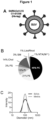
- Lipoparticles were produced as described above from a lipid formulation composed of 45 mol% DOPC, 21 mol% DOPE, 3 mol% DOPS, 12 mol% DOPI, 14 mol% cholesterol, and 3 mol% N-stearoyl-D-erythro-sphingosylphosphorylcholine (SM), and decorated with recombinant SARS-CoV-2 D614G S ectodomains (C1-G1236 with C-terminal trimerization domain and histidine-tag), or recombinant B1.1.7 variant S ectodomain, or recombinant SARS-CoV-2 R403A S ectodomains, or recombinant SARS-CoV S ectodomains, or recombinant MERS-CoV S ectodomains, or recombinant apo SARS-CoV-2 D614G S ectodomains, each via NTA(Ni 2+ ) functionalized 18:1 DGS.
- a negative zeta potential of -39.2 mV in H 2 O and -3.4 mV in isosmotic buffer were measured, which is consistent with natural SARS-CoV-2 virions.
- These immobilized, prefusion-stabilized S variants with deleted polybasic cleavage site were applied to specifically analyze RBD-mediated receptor binding independently from S2-induced membrane fusion.
- Hepatitis B virus lipoparticles were prepared as follows: small unilamellar vesicles with a lipid composition of 83 mol% DOPC, 7 mol% DOPE, 5 mol% DOPI, 3 mol% SM, 1 mol% DGS-NTA and 1 mol% LissRhodamine PE were produced by mechanical extrusion in the same way the minimal SARS-CoV-2 lipoparticles described above.
- Dengue virus lipoparticles were prepared as follows: small unilamellar vesicles with a lipid composition of 40 mol% DOPC, 30 mol% DOPE, 22 mol% SM, 5 mol% ceramides, 1 mol% DOPS, 1 mol% DGS-NTA and 1 mol% LissRhodamine PE were produced my mechanical extrusion in the same way the minimal SARS-CoV-2 lipoparticles described above. Retention of these lipoparticles to HepG2 cells was either measured as plain lipoparticles ( Figure 4, C ) or as lipoparticles that were conjugated with recombinant Dengue virus 2 E-glycoprotein (SEQ ID No. 30) via a histidine-tag on the C-terminus of the protein ( Figure 4, F ).
- Example 3 Influence of the fatty acid binding pocket (FABP) on regulation of SARS-CoV-2 spike protein cell binding
- Lipoparticles allowed for a systematic assessment of changes in SARS-CoV-2 spike protein (S)-mediated cell interactions as a function of FABP occupancy. Because recombinant native S binds an a priori undefined set of free fatty acids (FFAs) during expression and purification, first FFA-depleted S (apoS) was generated by treatment of the purified native S samples with alkoxylated lipophilic columns. Compared to the native S, apoS-decorated lipoparticles displayed increased binding to human epithelial cells ( Figure 5A ). This is consistent with a model where FFAs lock a closed RBD conformation, thereby reducing exposure of the receptor binding motif (RBM).
- RBM receptor binding motif
- SARS-CoV-2 Signal peptide S1 Subunit, Receptor Binding Site, S2 Subunit 2 native SARS-CoV-2 Spike ectodomain protein construct, furin cleavage site RRAR replaced by A, C-terminal thrombin cleavage site, T4 foldon trimerization domain, hexa-His tag 3 C-terminal thrombin cleavage site 4 T4 foldon trimerization domain 5 SARS-CoV-2 Spike protein native ectodomain 6 SARS-CoV-2 Spike protein native ectodomain L4F mutant 7 SARS-CoV-2 Spike protein native ectodomain T6N mutant 8 SARS-CoV-2 Spike protein native ectodomain P12S mutant 9 SARS-CoV-2 Spike protein native ectodomain D66A mutant 10 SARS-CoV-2 Spike protein native ectodomain D124Y mutant 11 SARS-CoV-2 Spike protein native ectodomain R176S
Landscapes
- Health & Medical Sciences (AREA)
- Life Sciences & Earth Sciences (AREA)
- Chemical & Material Sciences (AREA)
- Medicinal Chemistry (AREA)
- Pharmacology & Pharmacy (AREA)
- Animal Behavior & Ethology (AREA)
- General Health & Medical Sciences (AREA)
- Public Health (AREA)
- Veterinary Medicine (AREA)
- Virology (AREA)
- Epidemiology (AREA)
- General Chemical & Material Sciences (AREA)
- Engineering & Computer Science (AREA)
- Communicable Diseases (AREA)
- Oncology (AREA)
- Chemical Kinetics & Catalysis (AREA)
- Molecular Biology (AREA)
- Nuclear Medicine, Radiotherapy & Molecular Imaging (AREA)
- Organic Chemistry (AREA)
- Dispersion Chemistry (AREA)
- Bioinformatics & Cheminformatics (AREA)
- Dermatology (AREA)
- Biomedical Technology (AREA)
- Nanotechnology (AREA)
- Optics & Photonics (AREA)
- Biotechnology (AREA)
- Physics & Mathematics (AREA)
- Pulmonology (AREA)
- Immunology (AREA)
- Microbiology (AREA)
- Mycology (AREA)
- Peptides Or Proteins (AREA)
- Medicines Containing Material From Animals Or Micro-Organisms (AREA)
Priority Applications (2)
| Application Number | Priority Date | Filing Date | Title |
|---|---|---|---|
| EP21193839.4A EP4140475A1 (fr) | 2021-08-30 | 2021-08-30 | Lipoparticles pseudo-virales synthétiques |
| PCT/EP2022/073986 WO2023031137A2 (fr) | 2021-08-30 | 2022-08-29 | Lipoparticules de type viral synthétique |
Applications Claiming Priority (1)
| Application Number | Priority Date | Filing Date | Title |
|---|---|---|---|
| EP21193839.4A EP4140475A1 (fr) | 2021-08-30 | 2021-08-30 | Lipoparticles pseudo-virales synthétiques |
Publications (1)
| Publication Number | Publication Date |
|---|---|
| EP4140475A1 true EP4140475A1 (fr) | 2023-03-01 |
Family
ID=77543418
Family Applications (1)
| Application Number | Title | Priority Date | Filing Date |
|---|---|---|---|
| EP21193839.4A Ceased EP4140475A1 (fr) | 2021-08-30 | 2021-08-30 | Lipoparticles pseudo-virales synthétiques |
Country Status (2)
| Country | Link |
|---|---|
| EP (1) | EP4140475A1 (fr) |
| WO (1) | WO2023031137A2 (fr) |
Citations (3)
| Publication number | Priority date | Publication date | Assignee | Title |
|---|---|---|---|---|
| WO2012128628A1 (fr) * | 2011-03-22 | 2012-09-27 | Mucosis B.V. | Compositions immunogènes sous forme particulaire et leurs procédés de production |
| WO2019032463A1 (fr) * | 2017-08-07 | 2019-02-14 | Icahn School Of Medicine At Mount Sinai | Compositions immunogènes comprenant des polypeptides chimères d'hémagglutinine du virus de la grippe et as01 et leurs utilisations |
| WO2019143969A1 (fr) * | 2018-01-18 | 2019-07-25 | Claudio Maldonado | Traitement d'un dysfonctionnement microvasculaire |
-
2021
- 2021-08-30 EP EP21193839.4A patent/EP4140475A1/fr not_active Ceased
-
2022
- 2022-08-29 WO PCT/EP2022/073986 patent/WO2023031137A2/fr not_active Ceased
Patent Citations (3)
| Publication number | Priority date | Publication date | Assignee | Title |
|---|---|---|---|---|
| WO2012128628A1 (fr) * | 2011-03-22 | 2012-09-27 | Mucosis B.V. | Compositions immunogènes sous forme particulaire et leurs procédés de production |
| WO2019032463A1 (fr) * | 2017-08-07 | 2019-02-14 | Icahn School Of Medicine At Mount Sinai | Compositions immunogènes comprenant des polypeptides chimères d'hémagglutinine du virus de la grippe et as01 et leurs utilisations |
| WO2019143969A1 (fr) * | 2018-01-18 | 2019-07-25 | Claudio Maldonado | Traitement d'un dysfonctionnement microvasculaire |
Non-Patent Citations (2)
| Title |
|---|
| BRUNO SAINZ ET AL: "The Aromatic Domain of the Coronavirus Class I Viral Fusion Protein Induces Membrane Permeabilization: Putative Role during Viral Entry †", BIOCHEMISTRY, vol. 44, no. 3, 1 January 2005 (2005-01-01), pages 947 - 958, XP055388821, ISSN: 0006-2960, DOI: 10.1021/bi048515g * |
| NATURE, vol. 581, no. 221-3, 2020, pages 224 |
Also Published As
| Publication number | Publication date |
|---|---|
| WO2023031137A2 (fr) | 2023-03-09 |
| WO2023031137A3 (fr) | 2023-05-04 |
Similar Documents
| Publication | Publication Date | Title |
|---|---|---|
| Brock et al. | Efficient cell delivery mediated by lipid‐specific endosomal escape of supercharged branched peptides | |
| Erazo-Oliveras et al. | The late endosome and its lipid BMP act as gateways for efficient cytosolic access of the delivery agent dfTAT and its macromolecular cargos | |
| Johansson et al. | Cellular and biophysical barriers to lipid nanoparticle mediated delivery of RNA to the cytosol | |
| Gudlur et al. | Peptide nanovesicles formed by the self-assembly of branched amphiphilic peptides | |
| Staufer et al. | Synthetic virions reveal fatty acid-coupled adaptive immunogenicity of SARS-CoV-2 spike glycoprotein | |
| Bobone et al. | Phosphatidylserine lateral organization influences the interaction of influenza virus matrix protein 1 with lipid membranes | |
| Buehler et al. | Bioengineered vaults: Self-assembling protein shell–lipophilic core nanoparticles for drug delivery | |
| Sancini et al. | Functionalization with TAT-peptide enhances blood-brain barrier crossing in vitro of nanoliposomes carrying a curcumin-derivative to bind amyloid-β peptide | |
| US11504338B2 (en) | GM3 functionalized nanoparticles | |
| Brock et al. | Mechanism of cell penetration by permeabilization of late endosomes: interplay between a multivalent TAT peptide and bis (monoacylglycero) phosphate | |
| Cavaco et al. | DPepH3, an improved peptide shuttle for receptor-independent transport across the blood-brain barrier | |
| Somiya et al. | Intracellular trafficking of bio-nanocapsule–liposome complex: Identification of fusogenic activity in the pre-S1 region of hepatitis B virus surface antigen L protein | |
| US20210330809A1 (en) | Lipid-coated particles for treating viral infections | |
| Alter et al. | High efficiency preparation of monodisperse plasma membrane derived extracellular vesicles for therapeutic applications | |
| Szecskó et al. | PepH3-modified nanocarriers for delivery of therapeutics across the blood-brain barrier | |
| Milogrodzka et al. | Effect of cholesterol on biomimetic membrane curvature and coronavirus fusion peptide encapsulation | |
| Carradori et al. | The origin of neural stem cells impacts their interactions with targeted-lipid nanocapsules: Potential role of plasma membrane lipid composition and fluidity | |
| Dhankher et al. | Coiled coil exposure and histidine tags drive function of an intracellular protein drug carrier | |
| Takeda et al. | An in vitro lipid-mixing assay to investigate the fusion between small extracellular vesicles and endosome | |
| Akita et al. | Transcytosis‐Targeting Peptide: A Conductor of Liposomal Nanoparticles through the Endothelial Cell Barrier | |
| Amiar et al. | A fatty acid-ordered plasma membrane environment is critical for Ebola virus matrix protein assembly and budding | |
| EP4140475A1 (fr) | Lipoparticles pseudo-virales synthétiques | |
| WO2023027082A1 (fr) | Liposome-exosome hybride lié par un peptide, exosome lié par un peptide, composition les contenant et procédé pour leur formation | |
| Morris et al. | Enhanced pulmonary absorption of a macromolecule through coupling to a sequence-specific phage display-derived peptide | |
| Kim et al. | Membrane fusogenic high-density lipoprotein nanoparticles |
Legal Events
| Date | Code | Title | Description |
|---|---|---|---|
| PUAI | Public reference made under article 153(3) epc to a published international application that has entered the european phase |
Free format text: ORIGINAL CODE: 0009012 |
|
| STAA | Information on the status of an ep patent application or granted ep patent |
Free format text: STATUS: THE APPLICATION HAS BEEN PUBLISHED |
|
| AK | Designated contracting states |
Kind code of ref document: A1 Designated state(s): AL AT BE BG CH CY CZ DE DK EE ES FI FR GB GR HR HU IE IS IT LI LT LU LV MC MK MT NL NO PL PT RO RS SE SI SK SM TR |
|
| STAA | Information on the status of an ep patent application or granted ep patent |
Free format text: STATUS: THE APPLICATION HAS BEEN REFUSED |
|
| 18R | Application refused |
Effective date: 20230323 |