EP4159845A1 - Kulturmedium für laryngealkarzinomepithelzellen, kulturverfahren und anwendung davon - Google Patents
Kulturmedium für laryngealkarzinomepithelzellen, kulturverfahren und anwendung davon Download PDFInfo
- Publication number
- EP4159845A1 EP4159845A1 EP20937456.0A EP20937456A EP4159845A1 EP 4159845 A1 EP4159845 A1 EP 4159845A1 EP 20937456 A EP20937456 A EP 20937456A EP 4159845 A1 EP4159845 A1 EP 4159845A1
- Authority
- EP
- European Patent Office
- Prior art keywords
- cells
- laryngeal cancer
- alkyl
- culture medium
- primary
- Prior art date
- Legal status (The legal status is an assumption and is not a legal conclusion. Google has not performed a legal analysis and makes no representation as to the accuracy of the status listed.)
- Pending
Links
- 206010023841 laryngeal neoplasm Diseases 0.000 title claims abstract description 188
- 206010023825 Laryngeal cancer Diseases 0.000 title claims abstract description 187
- 210000002919 epithelial cell Anatomy 0.000 title claims abstract description 72
- 238000012136 culture method Methods 0.000 title abstract description 9
- 239000001963 growth medium Substances 0.000 title description 49
- 210000004027 cell Anatomy 0.000 claims abstract description 260
- 239000003814 drug Substances 0.000 claims abstract description 61
- 229940079593 drug Drugs 0.000 claims abstract description 59
- 239000006143 cell culture medium Substances 0.000 claims abstract description 27
- 238000012258 culturing Methods 0.000 claims abstract description 27
- 101001066435 Homo sapiens Hepatocyte growth factor-like protein Proteins 0.000 claims abstract description 24
- 101000880431 Homo sapiens Serine/threonine-protein kinase 4 Proteins 0.000 claims abstract description 24
- 229940043355 kinase inhibitor Drugs 0.000 claims abstract description 24
- 239000003757 phosphotransferase inhibitor Substances 0.000 claims abstract description 24
- 238000012216 screening Methods 0.000 claims abstract description 5
- 102100032813 Hepatocyte growth factor-like protein Human genes 0.000 claims abstract 6
- 238000000034 method Methods 0.000 claims description 40
- 230000005251 gamma ray Effects 0.000 claims description 20
- 125000004169 (C1-C6) alkyl group Chemical group 0.000 claims description 17
- 102000003972 Fibroblast growth factor 7 Human genes 0.000 claims description 16
- 108090000385 Fibroblast growth factor 7 Proteins 0.000 claims description 16
- 102000003745 Hepatocyte Growth Factor Human genes 0.000 claims description 16
- 108090000100 Hepatocyte Growth Factor Proteins 0.000 claims description 16
- 101000599951 Homo sapiens Insulin-like growth factor I Proteins 0.000 claims description 16
- 102100037852 Insulin-like growth factor I Human genes 0.000 claims description 16
- 150000001875 compounds Chemical class 0.000 claims description 16
- 229940098448 fibroblast growth factor 7 Drugs 0.000 claims description 16
- NOESYZHRGYRDHS-UHFFFAOYSA-N insulin Chemical compound N1C(=O)C(NC(=O)C(CCC(N)=O)NC(=O)C(CCC(O)=O)NC(=O)C(C(C)C)NC(=O)C(NC(=O)CN)C(C)CC)CSSCC(C(NC(CO)C(=O)NC(CC(C)C)C(=O)NC(CC=2C=CC(O)=CC=2)C(=O)NC(CCC(N)=O)C(=O)NC(CC(C)C)C(=O)NC(CCC(O)=O)C(=O)NC(CC(N)=O)C(=O)NC(CC=2C=CC(O)=CC=2)C(=O)NC(CSSCC(NC(=O)C(C(C)C)NC(=O)C(CC(C)C)NC(=O)C(CC=2C=CC(O)=CC=2)NC(=O)C(CC(C)C)NC(=O)C(C)NC(=O)C(CCC(O)=O)NC(=O)C(C(C)C)NC(=O)C(CC(C)C)NC(=O)C(CC=2NC=NC=2)NC(=O)C(CO)NC(=O)CNC2=O)C(=O)NCC(=O)NC(CCC(O)=O)C(=O)NC(CCCNC(N)=N)C(=O)NCC(=O)NC(CC=3C=CC=CC=3)C(=O)NC(CC=3C=CC=CC=3)C(=O)NC(CC=3C=CC(O)=CC=3)C(=O)NC(C(C)O)C(=O)N3C(CCC3)C(=O)NC(CCCCN)C(=O)NC(C)C(O)=O)C(=O)NC(CC(N)=O)C(O)=O)=O)NC(=O)C(C(C)CC)NC(=O)C(CO)NC(=O)C(C(C)O)NC(=O)C1CSSCC2NC(=O)C(CC(C)C)NC(=O)C(NC(=O)C(CCC(N)=O)NC(=O)C(CC(N)=O)NC(=O)C(NC(=O)C(N)CC=1C=CC=CC=1)C(C)C)CC1=CN=CN1 NOESYZHRGYRDHS-UHFFFAOYSA-N 0.000 claims description 16
- 239000003112 inhibitor Substances 0.000 claims description 13
- 229910052711 selenium Inorganic materials 0.000 claims description 13
- 239000011669 selenium Substances 0.000 claims description 13
- 229940091258 selenium supplement Drugs 0.000 claims description 13
- 238000007865 diluting Methods 0.000 claims description 10
- 125000000171 (C1-C6) haloalkyl group Chemical group 0.000 claims description 8
- 125000005913 (C3-C6) cycloalkyl group Chemical group 0.000 claims description 8
- 102000004877 Insulin Human genes 0.000 claims description 8
- 108090001061 Insulin Proteins 0.000 claims description 8
- 229940125396 insulin Drugs 0.000 claims description 8
- 210000000130 stem cell Anatomy 0.000 claims description 8
- 210000000251 trophoblastic cell Anatomy 0.000 claims description 8
- 102000004338 Transferrin Human genes 0.000 claims description 7
- 108090000901 Transferrin Proteins 0.000 claims description 7
- BVTBRVFYZUCAKH-UHFFFAOYSA-L disodium selenite Chemical compound [Na+].[Na+].[O-][Se]([O-])=O BVTBRVFYZUCAKH-UHFFFAOYSA-L 0.000 claims description 7
- 229910052739 hydrogen Inorganic materials 0.000 claims description 7
- 239000001257 hydrogen Substances 0.000 claims description 7
- 229960001471 sodium selenite Drugs 0.000 claims description 7
- 239000011781 sodium selenite Substances 0.000 claims description 7
- 235000015921 sodium selenite Nutrition 0.000 claims description 7
- 239000012581 transferrin Substances 0.000 claims description 7
- 230000003833 cell viability Effects 0.000 claims description 6
- 125000001316 cycloalkyl alkyl group Chemical group 0.000 claims description 6
- 125000001997 phenyl group Chemical group [H]C1=C([H])C([H])=C(*)C([H])=C1[H] 0.000 claims description 6
- 239000011435 rock Substances 0.000 claims description 6
- 150000003839 salts Chemical class 0.000 claims description 6
- 210000002966 serum Anatomy 0.000 claims description 6
- 241000283690 Bos taurus Species 0.000 claims description 5
- 102100033456 TGF-beta receptor type-1 Human genes 0.000 claims description 5
- 108010011702 Transforming Growth Factor-beta Type I Receptor Proteins 0.000 claims description 5
- 229910052736 halogen Inorganic materials 0.000 claims description 5
- 150000002431 hydrogen Chemical class 0.000 claims description 5
- -1 hydroxyl C1-C6 alkyl Chemical group 0.000 claims description 5
- 230000001817 pituitary effect Effects 0.000 claims description 5
- 239000012453 solvate Substances 0.000 claims description 5
- 102000041829 R-spondin family Human genes 0.000 claims description 4
- 108091078718 R-spondin family Proteins 0.000 claims description 4
- 102000013814 Wnt Human genes 0.000 claims description 4
- 108050003627 Wnt Proteins 0.000 claims description 4
- 125000001797 benzyl group Chemical group [H]C1=C([H])C([H])=C(C([H])=C1[H])C([H])([H])* 0.000 claims description 4
- 125000001153 fluoro group Chemical group F* 0.000 claims description 4
- 125000002496 methyl group Chemical group [H]C([H])([H])* 0.000 claims description 4
- 125000001544 thienyl group Chemical group 0.000 claims description 4
- 125000004191 (C1-C6) alkoxy group Chemical group 0.000 claims description 3
- 239000000556 agonist Substances 0.000 claims description 3
- 125000005843 halogen group Chemical group 0.000 claims description 3
- 125000002023 trifluoromethyl group Chemical group FC(F)(F)* 0.000 claims description 3
- 125000006273 (C1-C3) alkyl group Chemical group 0.000 claims description 2
- PWKSKIMOESPYIA-BYPYZUCNSA-N L-N-acetyl-Cysteine Chemical compound CC(=O)N[C@@H](CS)C(O)=O PWKSKIMOESPYIA-BYPYZUCNSA-N 0.000 claims description 2
- DFPAKSUCGFBDDF-ZQBYOMGUSA-N [14c]-nicotinamide Chemical compound N[14C](=O)C1=CC=CN=C1 DFPAKSUCGFBDDF-ZQBYOMGUSA-N 0.000 claims description 2
- 229960004308 acetylcysteine Drugs 0.000 claims description 2
- 125000003118 aryl group Chemical group 0.000 claims description 2
- 208000037265 diseases, disorders, signs and symptoms Diseases 0.000 claims description 2
- NGOGFTYYXHNFQH-UHFFFAOYSA-N fasudil Chemical compound C=1C=CC2=CN=CC=C2C=1S(=O)(=O)N1CCCNCC1 NGOGFTYYXHNFQH-UHFFFAOYSA-N 0.000 claims description 2
- 229960002435 fasudil Drugs 0.000 claims description 2
- 150000002367 halogens Chemical class 0.000 claims description 2
- 125000001072 heteroaryl group Chemical group 0.000 claims description 2
- 125000004435 hydrogen atom Chemical group [H]* 0.000 claims description 2
- 125000001624 naphthyl group Chemical group 0.000 claims description 2
- 238000004113 cell culture Methods 0.000 abstract description 9
- 238000011156 evaluation Methods 0.000 abstract description 4
- 210000002993 trophoblast Anatomy 0.000 abstract 1
- 210000001519 tissue Anatomy 0.000 description 96
- 239000002609 medium Substances 0.000 description 54
- 239000000203 mixture Substances 0.000 description 37
- 239000000523 sample Substances 0.000 description 36
- 238000005516 engineering process Methods 0.000 description 32
- 238000009472 formulation Methods 0.000 description 31
- 206010028980 Neoplasm Diseases 0.000 description 30
- 201000011510 cancer Diseases 0.000 description 30
- 230000029087 digestion Effects 0.000 description 22
- 210000002220 organoid Anatomy 0.000 description 21
- 239000000243 solution Substances 0.000 description 21
- 102100037629 Serine/threonine-protein kinase 4 Human genes 0.000 description 18
- 239000000654 additive Substances 0.000 description 17
- 238000000338 in vitro Methods 0.000 description 17
- 230000000996 additive effect Effects 0.000 description 16
- 230000000694 effects Effects 0.000 description 16
- 230000035755 proliferation Effects 0.000 description 16
- 230000035945 sensitivity Effects 0.000 description 16
- 239000006144 Dulbecco’s modified Eagle's medium Substances 0.000 description 15
- 239000006228 supernatant Substances 0.000 description 14
- 229930182555 Penicillin Natural products 0.000 description 13
- JGSARLDLIJGVTE-MBNYWOFBSA-N Penicillin G Chemical compound N([C@H]1[C@H]2SC([C@@H](N2C1=O)C(O)=O)(C)C)C(=O)CC1=CC=CC=C1 JGSARLDLIJGVTE-MBNYWOFBSA-N 0.000 description 13
- 102000004142 Trypsin Human genes 0.000 description 13
- 108090000631 Trypsin Proteins 0.000 description 13
- 239000012530 fluid Substances 0.000 description 13
- 229940049954 penicillin Drugs 0.000 description 13
- 239000012588 trypsin Substances 0.000 description 13
- UCSJYZPVAKXKNQ-HZYVHMACSA-N streptomycin Chemical compound CN[C@H]1[C@H](O)[C@@H](O)[C@H](CO)O[C@H]1O[C@@H]1[C@](C=O)(O)[C@H](C)O[C@H]1O[C@@H]1[C@@H](NC(N)=N)[C@H](O)[C@@H](NC(N)=N)[C@H](O)[C@H]1O UCSJYZPVAKXKNQ-HZYVHMACSA-N 0.000 description 12
- 238000012360 testing method Methods 0.000 description 12
- 108091003079 Bovine Serum Albumin Proteins 0.000 description 11
- 229940125904 compound 1 Drugs 0.000 description 11
- 238000003384 imaging method Methods 0.000 description 10
- 239000013028 medium composition Substances 0.000 description 10
- 238000002360 preparation method Methods 0.000 description 10
- XLYOFNOQVPJJNP-UHFFFAOYSA-N water Chemical compound O XLYOFNOQVPJJNP-UHFFFAOYSA-N 0.000 description 10
- RTZKZFJDLAIYFH-UHFFFAOYSA-N Diethyl ether Chemical compound CCOCC RTZKZFJDLAIYFH-UHFFFAOYSA-N 0.000 description 9
- IAZDPXIOMUYVGZ-UHFFFAOYSA-N Dimethylsulphoxide Chemical compound CS(C)=O IAZDPXIOMUYVGZ-UHFFFAOYSA-N 0.000 description 9
- 239000000306 component Substances 0.000 description 9
- 239000012091 fetal bovine serum Substances 0.000 description 9
- 230000001737 promoting effect Effects 0.000 description 9
- 210000004881 tumor cell Anatomy 0.000 description 9
- 230000015572 biosynthetic process Effects 0.000 description 8
- 230000004663 cell proliferation Effects 0.000 description 8
- 239000006285 cell suspension Substances 0.000 description 8
- 239000000499 gel Substances 0.000 description 8
- 230000012010 growth Effects 0.000 description 8
- 230000008672 reprogramming Effects 0.000 description 8
- 210000000601 blood cell Anatomy 0.000 description 7
- 230000001079 digestive effect Effects 0.000 description 7
- 238000011081 inoculation Methods 0.000 description 7
- CSCPPACGZOOCGX-UHFFFAOYSA-N Acetone Chemical compound CC(C)=O CSCPPACGZOOCGX-UHFFFAOYSA-N 0.000 description 6
- XEKOWRVHYACXOJ-UHFFFAOYSA-N Ethyl acetate Chemical compound CCOC(C)=O XEKOWRVHYACXOJ-UHFFFAOYSA-N 0.000 description 6
- OKKJLVBELUTLKV-UHFFFAOYSA-N Methanol Chemical compound OC OKKJLVBELUTLKV-UHFFFAOYSA-N 0.000 description 6
- 241000699670 Mus sp. Species 0.000 description 6
- 239000002246 antineoplastic agent Substances 0.000 description 6
- JYGXADMDTFJGBT-VWUMJDOOSA-N hydrocortisone Chemical compound O=C1CC[C@]2(C)[C@H]3[C@@H](O)C[C@](C)([C@@](CC4)(O)C(=O)CO)[C@@H]4[C@@H]3CCC2=C1 JYGXADMDTFJGBT-VWUMJDOOSA-N 0.000 description 6
- 239000002904 solvent Substances 0.000 description 6
- 229960005322 streptomycin Drugs 0.000 description 6
- 101000804764 Homo sapiens Lymphotactin Proteins 0.000 description 5
- 102100035304 Lymphotactin Human genes 0.000 description 5
- 101100503363 Schistosoma mansoni SCM-2 gene Proteins 0.000 description 5
- 210000004369 blood Anatomy 0.000 description 5
- 239000008280 blood Substances 0.000 description 5
- 210000004748 cultured cell Anatomy 0.000 description 5
- 210000000981 epithelium Anatomy 0.000 description 5
- 238000002474 experimental method Methods 0.000 description 5
- 239000011550 stock solution Substances 0.000 description 5
- 230000004083 survival effect Effects 0.000 description 5
- 102000029816 Collagenase Human genes 0.000 description 4
- 108060005980 Collagenase Proteins 0.000 description 4
- 102100027881 Tumor protein 63 Human genes 0.000 description 4
- 230000010261 cell growth Effects 0.000 description 4
- 238000012054 celltiter-glo Methods 0.000 description 4
- 229960002424 collagenase Drugs 0.000 description 4
- 230000000052 comparative effect Effects 0.000 description 4
- 239000012043 crude product Substances 0.000 description 4
- 238000010790 dilution Methods 0.000 description 4
- 239000012895 dilution Substances 0.000 description 4
- AUZONCFQVSMFAP-UHFFFAOYSA-N disulfiram Chemical compound CCN(CC)C(=S)SSC(=S)N(CC)CC AUZONCFQVSMFAP-UHFFFAOYSA-N 0.000 description 4
- 238000007877 drug screening Methods 0.000 description 4
- 239000012634 fragment Substances 0.000 description 4
- 238000004895 liquid chromatography mass spectrometry Methods 0.000 description 4
- 239000011159 matrix material Substances 0.000 description 4
- 238000002156 mixing Methods 0.000 description 4
- 239000012074 organic phase Substances 0.000 description 4
- 238000007747 plating Methods 0.000 description 4
- 230000008569 process Effects 0.000 description 4
- 238000002271 resection Methods 0.000 description 4
- 239000013049 sediment Substances 0.000 description 4
- VBEQCZHXXJYVRD-GACYYNSASA-N uroanthelone Chemical compound C([C@@H](C(=O)N[C@H](C(=O)N[C@@H](CS)C(=O)N[C@@H](CC(N)=O)C(=O)N[C@@H](CS)C(=O)N[C@H](C(=O)N[C@@H]([C@@H](C)CC)C(=O)NCC(=O)N[C@@H](CC=1C=CC(O)=CC=1)C(=O)N[C@@H](CO)C(=O)NCC(=O)N[C@@H](CC(O)=O)C(=O)N[C@@H](CCCNC(N)=N)C(=O)N[C@@H](CS)C(=O)N[C@@H](CCC(N)=O)C(=O)N[C@@H]([C@@H](C)O)C(=O)N[C@@H](CCCNC(N)=N)C(=O)N[C@@H](CC(O)=O)C(=O)N[C@@H](CC(C)C)C(=O)N[C@@H](CCCNC(N)=N)C(=O)N[C@@H](CC=1C2=CC=CC=C2NC=1)C(=O)N[C@@H](CC=1C2=CC=CC=C2NC=1)C(=O)N[C@@H](CCC(O)=O)C(=O)N[C@@H](CC(C)C)C(=O)N[C@@H](CCCNC(N)=N)C(O)=O)C(C)C)[C@@H](C)O)NC(=O)[C@H](CO)NC(=O)[C@H](CC(O)=O)NC(=O)[C@H](CC(C)C)NC(=O)[C@H](CO)NC(=O)[C@H](CCC(O)=O)NC(=O)[C@@H](NC(=O)[C@H](CC=1NC=NC=1)NC(=O)[C@H](CCSC)NC(=O)[C@H](CS)NC(=O)[C@@H](NC(=O)CNC(=O)CNC(=O)[C@H](CC(N)=O)NC(=O)[C@H](CC(C)C)NC(=O)[C@H](CS)NC(=O)[C@H](CC=1C=CC(O)=CC=1)NC(=O)CNC(=O)[C@H](CC(O)=O)NC(=O)[C@H](CC=1C=CC(O)=CC=1)NC(=O)[C@H](CO)NC(=O)[C@H](CO)NC(=O)[C@H]1N(CCC1)C(=O)[C@H](CS)NC(=O)CNC(=O)[C@H]1N(CCC1)C(=O)[C@H](CC=1C=CC(O)=CC=1)NC(=O)[C@H](CO)NC(=O)[C@@H](N)CC(N)=O)C(C)C)[C@@H](C)CC)C1=CC=C(O)C=C1 VBEQCZHXXJYVRD-GACYYNSASA-N 0.000 description 4
- JEEZAZSTUWUWQK-UHFFFAOYSA-N 2-chloro-7-(2,6-difluorophenyl)-5,8-dimethyl-7H-pteridin-6-one Chemical compound ClC1=NC=2N(C(C(N(C=2C=N1)C)=O)C1=C(C=CC=C1F)F)C JEEZAZSTUWUWQK-UHFFFAOYSA-N 0.000 description 3
- APZCECSEMLNKFK-UHFFFAOYSA-N 2-chloro-7-(2,6-difluorophenyl)-7,8-dihydro-5H-pteridin-6-one Chemical compound ClC1=NC=2NC(C(NC=2C=N1)=O)C1=C(C=CC=C1F)F APZCECSEMLNKFK-UHFFFAOYSA-N 0.000 description 3
- FWBHETKCLVMNFS-UHFFFAOYSA-N 4',6-Diamino-2-phenylindol Chemical compound C1=CC(C(=N)N)=CC=C1C1=CC2=CC=C(C(N)=N)C=C2N1 FWBHETKCLVMNFS-UHFFFAOYSA-N 0.000 description 3
- QTBSBXVTEAMEQO-UHFFFAOYSA-N Acetic acid Chemical compound CC(O)=O QTBSBXVTEAMEQO-UHFFFAOYSA-N 0.000 description 3
- 102400001368 Epidermal growth factor Human genes 0.000 description 3
- 101800003838 Epidermal growth factor Proteins 0.000 description 3
- 108010037362 Extracellular Matrix Proteins Proteins 0.000 description 3
- 102000010834 Extracellular Matrix Proteins Human genes 0.000 description 3
- 102000018233 Fibroblast Growth Factor Human genes 0.000 description 3
- 108050007372 Fibroblast Growth Factor Proteins 0.000 description 3
- 102000004864 Fibroblast growth factor 10 Human genes 0.000 description 3
- 108090001047 Fibroblast growth factor 10 Proteins 0.000 description 3
- WSFSSNUMVMOOMR-UHFFFAOYSA-N Formaldehyde Chemical compound O=C WSFSSNUMVMOOMR-UHFFFAOYSA-N 0.000 description 3
- 229920004890 Triton X-100 Polymers 0.000 description 3
- 239000013504 Triton X-100 Substances 0.000 description 3
- 229940041181 antineoplastic drug Drugs 0.000 description 3
- 238000001574 biopsy Methods 0.000 description 3
- 230000000903 blocking effect Effects 0.000 description 3
- 229940044683 chemotherapy drug Drugs 0.000 description 3
- 238000010586 diagram Methods 0.000 description 3
- 229940116977 epidermal growth factor Drugs 0.000 description 3
- 210000002744 extracellular matrix Anatomy 0.000 description 3
- 229940126864 fibroblast growth factor Drugs 0.000 description 3
- 229960000890 hydrocortisone Drugs 0.000 description 3
- 230000002055 immunohistochemical effect Effects 0.000 description 3
- IGMWMQOJLFFZRK-UHFFFAOYSA-N methyl 2-[(2-chloro-5-nitropyrimidin-4-yl)amino]-2-(2,6-difluorophenyl)acetate Chemical compound ClC1=NC=C(C(=N1)NC(C(=O)OC)C1=C(C=CC=C1F)F)[N+](=O)[O-] IGMWMQOJLFFZRK-UHFFFAOYSA-N 0.000 description 3
- GXMZVLXPZSATTL-UHFFFAOYSA-N methyl 2-amino-2-(2,6-difluorophenyl)acetate Chemical compound COC(=O)C(N)C1=C(F)C=CC=C1F GXMZVLXPZSATTL-UHFFFAOYSA-N 0.000 description 3
- 239000011541 reaction mixture Substances 0.000 description 3
- 238000010186 staining Methods 0.000 description 3
- 238000003860 storage Methods 0.000 description 3
- 102100037651 AP-2 complex subunit sigma Human genes 0.000 description 2
- UXVMQQNJUSDDNG-UHFFFAOYSA-L Calcium chloride Chemical compound [Cl-].[Cl-].[Ca+2] UXVMQQNJUSDDNG-UHFFFAOYSA-L 0.000 description 2
- 208000005623 Carcinogenesis Diseases 0.000 description 2
- 102000053602 DNA Human genes 0.000 description 2
- 108020004414 DNA Proteins 0.000 description 2
- 239000012981 Hank's balanced salt solution Substances 0.000 description 2
- 101000806914 Homo sapiens AP-2 complex subunit sigma Proteins 0.000 description 2
- 101001095308 Homo sapiens Periostin Proteins 0.000 description 2
- 101000880439 Homo sapiens Serine/threonine-protein kinase 3 Proteins 0.000 description 2
- 108010003272 Hyaluronate lyase Proteins 0.000 description 2
- 102000001974 Hyaluronidases Human genes 0.000 description 2
- 239000005411 L01XE02 - Gefitinib Substances 0.000 description 2
- 239000005551 L01XE03 - Erlotinib Substances 0.000 description 2
- 102400000058 Neuregulin-1 Human genes 0.000 description 2
- 108090000556 Neuregulin-1 Proteins 0.000 description 2
- 229930040373 Paraformaldehyde Natural products 0.000 description 2
- 239000012980 RPMI-1640 medium Substances 0.000 description 2
- 102100037628 Serine/threonine-protein kinase 3 Human genes 0.000 description 2
- PMZURENOXWZQFD-UHFFFAOYSA-L Sodium Sulfate Chemical compound [Na+].[Na+].[O-]S([O-])(=O)=O PMZURENOXWZQFD-UHFFFAOYSA-L 0.000 description 2
- 239000000090 biomarker Substances 0.000 description 2
- GXJABQQUPOEUTA-RDJZCZTQSA-N bortezomib Chemical compound C([C@@H](C(=O)N[C@@H](CC(C)C)B(O)O)NC(=O)C=1N=CC=NC=1)C1=CC=CC=C1 GXJABQQUPOEUTA-RDJZCZTQSA-N 0.000 description 2
- 229960001467 bortezomib Drugs 0.000 description 2
- 229940098773 bovine serum albumin Drugs 0.000 description 2
- BTANRVKWQNVYAZ-UHFFFAOYSA-N butan-2-ol Chemical compound CCC(C)O BTANRVKWQNVYAZ-UHFFFAOYSA-N 0.000 description 2
- 239000001110 calcium chloride Substances 0.000 description 2
- 229910001628 calcium chloride Inorganic materials 0.000 description 2
- 230000036952 cancer formation Effects 0.000 description 2
- 231100000504 carcinogenesis Toxicity 0.000 description 2
- 239000013592 cell lysate Substances 0.000 description 2
- 239000013065 commercial product Substances 0.000 description 2
- 230000009089 cytolysis Effects 0.000 description 2
- 238000001514 detection method Methods 0.000 description 2
- 238000011161 development Methods 0.000 description 2
- 230000018109 developmental process Effects 0.000 description 2
- 229960002563 disulfiram Drugs 0.000 description 2
- 229960001433 erlotinib Drugs 0.000 description 2
- AAKJLRGGTJKAMG-UHFFFAOYSA-N erlotinib Chemical compound C=12C=C(OCCOC)C(OCCOC)=CC2=NC=NC=1NC1=CC=CC(C#C)=C1 AAKJLRGGTJKAMG-UHFFFAOYSA-N 0.000 description 2
- 239000003797 essential amino acid Substances 0.000 description 2
- 235000020776 essential amino acid Nutrition 0.000 description 2
- 239000007850 fluorescent dye Substances 0.000 description 2
- 238000011990 functional testing Methods 0.000 description 2
- 229960002584 gefitinib Drugs 0.000 description 2
- XGALLCVXEZPNRQ-UHFFFAOYSA-N gefitinib Chemical compound C=12C=C(OCCCN3CCOCC3)C(OC)=CC2=NC=NC=1NC1=CC=C(F)C(Cl)=C1 XGALLCVXEZPNRQ-UHFFFAOYSA-N 0.000 description 2
- ZDXPYRJPNDTMRX-UHFFFAOYSA-N glutamine Natural products OC(=O)C(N)CCC(N)=O ZDXPYRJPNDTMRX-UHFFFAOYSA-N 0.000 description 2
- 239000003102 growth factor Substances 0.000 description 2
- 102000048431 human POSTN Human genes 0.000 description 2
- 229960002773 hyaluronidase Drugs 0.000 description 2
- 210000002510 keratinocyte Anatomy 0.000 description 2
- 239000007788 liquid Substances 0.000 description 2
- 108010082117 matrigel Proteins 0.000 description 2
- 238000001000 micrograph Methods 0.000 description 2
- 238000012986 modification Methods 0.000 description 2
- 230000004048 modification Effects 0.000 description 2
- 230000017074 necrotic cell death Effects 0.000 description 2
- 230000001338 necrotic effect Effects 0.000 description 2
- 239000002547 new drug Substances 0.000 description 2
- 102000045246 noggin Human genes 0.000 description 2
- 108700007229 noggin Proteins 0.000 description 2
- AICOOMRHRUFYCM-ZRRPKQBOSA-N oxazine, 1 Chemical compound C([C@@H]1[C@H](C(C[C@]2(C)[C@@H]([C@H](C)N(C)C)[C@H](O)C[C@]21C)=O)CC1=CC2)C[C@H]1[C@@]1(C)[C@H]2N=C(C(C)C)OC1 AICOOMRHRUFYCM-ZRRPKQBOSA-N 0.000 description 2
- 229920002866 paraformaldehyde Polymers 0.000 description 2
- 230000001575 pathological effect Effects 0.000 description 2
- 239000012071 phase Substances 0.000 description 2
- 229920003023 plastic Polymers 0.000 description 2
- BWHMMNNQKKPAPP-UHFFFAOYSA-L potassium carbonate Chemical compound [K+].[K+].[O-]C([O-])=O BWHMMNNQKKPAPP-UHFFFAOYSA-L 0.000 description 2
- 239000013630 prepared media Substances 0.000 description 2
- 230000001172 regenerating effect Effects 0.000 description 2
- 238000011160 research Methods 0.000 description 2
- 230000004044 response Effects 0.000 description 2
- 238000007789 sealing Methods 0.000 description 2
- 150000003384 small molecules Chemical class 0.000 description 2
- HPALAKNZSZLMCH-UHFFFAOYSA-M sodium;chloride;hydrate Chemical class O.[Na+].[Cl-] HPALAKNZSZLMCH-UHFFFAOYSA-M 0.000 description 2
- 206010041823 squamous cell carcinoma Diseases 0.000 description 2
- 238000004659 sterilization and disinfection Methods 0.000 description 2
- FYSNRJHAOHDILO-UHFFFAOYSA-N thionyl chloride Chemical compound ClS(Cl)=O FYSNRJHAOHDILO-UHFFFAOYSA-N 0.000 description 2
- JOXIMZWYDAKGHI-UHFFFAOYSA-N toluene-4-sulfonic acid Chemical compound CC1=CC=C(S(O)(=O)=O)C=C1 JOXIMZWYDAKGHI-UHFFFAOYSA-N 0.000 description 2
- INUSQTPGSHFGHM-UHFFFAOYSA-N 2,4-dichloro-5-nitropyrimidine Chemical compound [O-][N+](=O)C1=CN=C(Cl)N=C1Cl INUSQTPGSHFGHM-UHFFFAOYSA-N 0.000 description 1
- UOXJNGFFPMOZDM-UHFFFAOYSA-N 2-[di(propan-2-yl)amino]ethylsulfanyl-methylphosphinic acid Chemical compound CC(C)N(C(C)C)CCSP(C)(O)=O UOXJNGFFPMOZDM-UHFFFAOYSA-N 0.000 description 1
- UQFQFMHLBQOCLY-UHFFFAOYSA-N 2-azaniumyl-2-(2,6-difluorophenyl)acetate Chemical compound OC(=O)C(N)C1=C(F)C=CC=C1F UQFQFMHLBQOCLY-UHFFFAOYSA-N 0.000 description 1
- APKFDSVGJQXUKY-KKGHZKTASA-N Amphotericin-B Natural products O[C@H]1[C@@H](N)[C@H](O)[C@@H](C)O[C@H]1O[C@H]1C=CC=CC=CC=CC=CC=CC=C[C@H](C)[C@@H](O)[C@@H](C)[C@H](C)OC(=O)C[C@H](O)C[C@H](O)CC[C@@H](O)[C@H](O)C[C@H](O)C[C@](O)(C[C@H](O)[C@H]2C(O)=O)O[C@H]2C1 APKFDSVGJQXUKY-KKGHZKTASA-N 0.000 description 1
- 102000009016 Cholera Toxin Human genes 0.000 description 1
- 108010049048 Cholera Toxin Proteins 0.000 description 1
- 229930182566 Gentamicin Natural products 0.000 description 1
- CEAZRRDELHUEMR-URQXQFDESA-N Gentamicin Chemical compound O1[C@H](C(C)NC)CC[C@@H](N)[C@H]1O[C@H]1[C@H](O)[C@@H](O[C@@H]2[C@@H]([C@@H](NC)[C@@](C)(O)CO2)O)[C@H](N)C[C@@H]1N CEAZRRDELHUEMR-URQXQFDESA-N 0.000 description 1
- 239000007756 Ham's F12 Nutrient Mixture Substances 0.000 description 1
- 241000282412 Homo Species 0.000 description 1
- 101000825954 Homo sapiens R-spondin-1 Proteins 0.000 description 1
- XEEYBQQBJWHFJM-UHFFFAOYSA-N Iron Chemical compound [Fe] XEEYBQQBJWHFJM-UHFFFAOYSA-N 0.000 description 1
- 102000011782 Keratins Human genes 0.000 description 1
- 108010076876 Keratins Proteins 0.000 description 1
- 241000124008 Mammalia Species 0.000 description 1
- 241001529936 Murinae Species 0.000 description 1
- FXHOOIRPVKKKFG-UHFFFAOYSA-N N,N-Dimethylacetamide Chemical compound CN(C)C(C)=O FXHOOIRPVKKKFG-UHFFFAOYSA-N 0.000 description 1
- 208000002454 Nasopharyngeal Carcinoma Diseases 0.000 description 1
- 206010061306 Nasopharyngeal cancer Diseases 0.000 description 1
- 241000283973 Oryctolagus cuniculus Species 0.000 description 1
- 208000002193 Pain Diseases 0.000 description 1
- 108091000080 Phosphotransferase Proteins 0.000 description 1
- 102100022762 R-spondin-1 Human genes 0.000 description 1
- KEAYESYHFKHZAL-UHFFFAOYSA-N Sodium Chemical compound [Na] KEAYESYHFKHZAL-UHFFFAOYSA-N 0.000 description 1
- UIIMBOGNXHQVGW-UHFFFAOYSA-M Sodium bicarbonate Chemical class [Na+].OC([O-])=O UIIMBOGNXHQVGW-UHFFFAOYSA-M 0.000 description 1
- 208000036765 Squamous cell carcinoma of the esophagus Diseases 0.000 description 1
- QTENRWWVYAAPBI-YZTFXSNBSA-N Streptomycin sulfate Chemical compound OS(O)(=O)=O.OS(O)(=O)=O.OS(O)(=O)=O.CN[C@H]1[C@H](O)[C@@H](O)[C@H](CO)O[C@H]1O[C@@H]1[C@](C=O)(O)[C@H](C)O[C@H]1O[C@H]1[C@H](N=C(N)N)[C@@H](O)[C@H](N=C(N)N)[C@@H](O)[C@@H]1O.CN[C@H]1[C@H](O)[C@@H](O)[C@H](CO)O[C@H]1O[C@@H]1[C@](C=O)(O)[C@H](C)O[C@H]1O[C@H]1[C@H](N=C(N)N)[C@@H](O)[C@H](N=C(N)N)[C@@H](O)[C@@H]1O QTENRWWVYAAPBI-YZTFXSNBSA-N 0.000 description 1
- OKJPEAGHQZHRQV-UHFFFAOYSA-N Triiodomethane Natural products IC(I)I OKJPEAGHQZHRQV-UHFFFAOYSA-N 0.000 description 1
- 240000004922 Vigna radiata Species 0.000 description 1
- 235000010721 Vigna radiata var radiata Nutrition 0.000 description 1
- 235000011469 Vigna radiata var sublobata Nutrition 0.000 description 1
- 230000001464 adherent effect Effects 0.000 description 1
- 210000000577 adipose tissue Anatomy 0.000 description 1
- 210000004504 adult stem cell Anatomy 0.000 description 1
- 230000004931 aggregating effect Effects 0.000 description 1
- 238000003915 air pollution Methods 0.000 description 1
- APKFDSVGJQXUKY-INPOYWNPSA-N amphotericin B Chemical compound O[C@H]1[C@@H](N)[C@H](O)[C@@H](C)O[C@H]1O[C@H]1/C=C/C=C/C=C/C=C/C=C/C=C/C=C/[C@H](C)[C@@H](O)[C@@H](C)[C@H](C)OC(=O)C[C@H](O)C[C@H](O)CC[C@@H](O)[C@H](O)C[C@H](O)C[C@](O)(C[C@H](O)[C@H]2C(O)=O)O[C@H]2C1 APKFDSVGJQXUKY-INPOYWNPSA-N 0.000 description 1
- 229960003942 amphotericin b Drugs 0.000 description 1
- 229940125644 antibody drug Drugs 0.000 description 1
- 238000003149 assay kit Methods 0.000 description 1
- 210000001099 axilla Anatomy 0.000 description 1
- 239000007640 basal medium Substances 0.000 description 1
- 230000009286 beneficial effect Effects 0.000 description 1
- 239000007474 bm medium Substances 0.000 description 1
- 238000003570 cell viability assay Methods 0.000 description 1
- 230000001413 cellular effect Effects 0.000 description 1
- 239000003153 chemical reaction reagent Substances 0.000 description 1
- 125000001309 chloro group Chemical group Cl* 0.000 description 1
- 210000001072 colon Anatomy 0.000 description 1
- 238000004891 communication Methods 0.000 description 1
- 210000000795 conjunctiva Anatomy 0.000 description 1
- 210000004087 cornea Anatomy 0.000 description 1
- 238000005138 cryopreservation Methods 0.000 description 1
- 230000001186 cumulative effect Effects 0.000 description 1
- 230000007812 deficiency Effects 0.000 description 1
- 239000003085 diluting agent Substances 0.000 description 1
- 238000003113 dilution method Methods 0.000 description 1
- 231100000673 dose–response relationship Toxicity 0.000 description 1
- 238000001647 drug administration Methods 0.000 description 1
- 238000003255 drug test Methods 0.000 description 1
- 210000001198 duodenum Anatomy 0.000 description 1
- 239000000975 dye Substances 0.000 description 1
- 208000007276 esophageal squamous cell carcinoma Diseases 0.000 description 1
- 210000003238 esophagus Anatomy 0.000 description 1
- 230000005284 excitation Effects 0.000 description 1
- 210000002950 fibroblast Anatomy 0.000 description 1
- 239000000706 filtrate Substances 0.000 description 1
- 210000003194 forelimb Anatomy 0.000 description 1
- 229960002518 gentamicin Drugs 0.000 description 1
- 210000004209 hair Anatomy 0.000 description 1
- 125000000623 heterocyclic group Chemical group 0.000 description 1
- 238000013537 high throughput screening Methods 0.000 description 1
- 210000003405 ileum Anatomy 0.000 description 1
- 238000010166 immunofluorescence Methods 0.000 description 1
- 238000003125 immunofluorescent labeling Methods 0.000 description 1
- 238000013115 immunohistochemical detection Methods 0.000 description 1
- 238000003364 immunohistochemistry Methods 0.000 description 1
- 238000011534 incubation Methods 0.000 description 1
- 230000002401 inhibitory effect Effects 0.000 description 1
- 230000003993 interaction Effects 0.000 description 1
- INQOMBQAUSQDDS-UHFFFAOYSA-N iodomethane Chemical compound IC INQOMBQAUSQDDS-UHFFFAOYSA-N 0.000 description 1
- 238000002955 isolation Methods 0.000 description 1
- 210000001630 jejunum Anatomy 0.000 description 1
- 210000000738 kidney tubule Anatomy 0.000 description 1
- 210000004561 lacrimal apparatus Anatomy 0.000 description 1
- 210000002429 large intestine Anatomy 0.000 description 1
- 201000004959 laryngeal benign neoplasm Diseases 0.000 description 1
- 210000004185 liver Anatomy 0.000 description 1
- 230000007774 longterm Effects 0.000 description 1
- 238000004020 luminiscence type Methods 0.000 description 1
- 210000004072 lung Anatomy 0.000 description 1
- 210000005075 mammary gland Anatomy 0.000 description 1
- 238000001819 mass spectrum Methods 0.000 description 1
- 239000012533 medium component Substances 0.000 description 1
- 210000004379 membrane Anatomy 0.000 description 1
- 239000012528 membrane Substances 0.000 description 1
- 125000000956 methoxy group Chemical group [H]C([H])([H])O* 0.000 description 1
- 230000004784 molecular pathogenesis Effects 0.000 description 1
- 210000002200 mouth mucosa Anatomy 0.000 description 1
- 208000018795 nasal cavity and paranasal sinus carcinoma Diseases 0.000 description 1
- 201000011216 nasopharynx carcinoma Diseases 0.000 description 1
- 238000011017 operating method Methods 0.000 description 1
- 238000005457 optimization Methods 0.000 description 1
- 210000000056 organ Anatomy 0.000 description 1
- 210000004798 organs belonging to the digestive system Anatomy 0.000 description 1
- 230000036407 pain Effects 0.000 description 1
- 210000000496 pancreas Anatomy 0.000 description 1
- 239000012188 paraffin wax Substances 0.000 description 1
- 238000010827 pathological analysis Methods 0.000 description 1
- 239000013610 patient sample Substances 0.000 description 1
- 230000000149 penetrating effect Effects 0.000 description 1
- 102000020233 phosphotransferase Human genes 0.000 description 1
- 125000005936 piperidyl group Chemical group 0.000 description 1
- 230000036470 plasma concentration Effects 0.000 description 1
- 229910000027 potassium carbonate Inorganic materials 0.000 description 1
- 210000002307 prostate Anatomy 0.000 description 1
- 210000003079 salivary gland Anatomy 0.000 description 1
- 230000011664 signaling Effects 0.000 description 1
- 238000010898 silica gel chromatography Methods 0.000 description 1
- 210000003491 skin Anatomy 0.000 description 1
- 210000000813 small intestine Anatomy 0.000 description 1
- 239000012312 sodium hydride Substances 0.000 description 1
- 229910000104 sodium hydride Inorganic materials 0.000 description 1
- 239000007787 solid Substances 0.000 description 1
- 238000007711 solidification Methods 0.000 description 1
- 230000008023 solidification Effects 0.000 description 1
- 210000002784 stomach Anatomy 0.000 description 1
- FDDDEECHVMSUSB-UHFFFAOYSA-N sulfanilamide Chemical compound NC1=CC=C(S(N)(=O)=O)C=C1 FDDDEECHVMSUSB-UHFFFAOYSA-N 0.000 description 1
- 229940124530 sulfonamide Drugs 0.000 description 1
- 239000013589 supplement Substances 0.000 description 1
- 125000001412 tetrahydropyranyl group Chemical group 0.000 description 1
- 238000002560 therapeutic procedure Methods 0.000 description 1
- 231100000419 toxicity Toxicity 0.000 description 1
- 230000001988 toxicity Effects 0.000 description 1
- 210000003437 trachea Anatomy 0.000 description 1
- 230000005740 tumor formation Effects 0.000 description 1
- 210000003932 urinary bladder Anatomy 0.000 description 1
Images




Classifications
-
- G—PHYSICS
- G01—MEASURING; TESTING
- G01N—INVESTIGATING OR ANALYSING MATERIALS BY DETERMINING THEIR CHEMICAL OR PHYSICAL PROPERTIES
- G01N33/00—Investigating or analysing materials by specific methods not covered by groups G01N1/00 - G01N31/00
- G01N33/48—Biological material, e.g. blood, urine; Haemocytometers
- G01N33/50—Chemical analysis of biological material, e.g. blood, urine; Testing involving biospecific ligand binding methods; Immunological testing
- G01N33/5005—Chemical analysis of biological material, e.g. blood, urine; Testing involving biospecific ligand binding methods; Immunological testing involving human or animal cells
- G01N33/5008—Chemical analysis of biological material, e.g. blood, urine; Testing involving biospecific ligand binding methods; Immunological testing involving human or animal cells for testing or evaluating the effect of chemical or biological compounds, e.g. drugs, cosmetics
- G01N33/5011—Chemical analysis of biological material, e.g. blood, urine; Testing involving biospecific ligand binding methods; Immunological testing involving human or animal cells for testing or evaluating the effect of chemical or biological compounds, e.g. drugs, cosmetics for testing antineoplastic activity
-
- C—CHEMISTRY; METALLURGY
- C12—BIOCHEMISTRY; BEER; SPIRITS; WINE; VINEGAR; MICROBIOLOGY; ENZYMOLOGY; MUTATION OR GENETIC ENGINEERING
- C12N—MICROORGANISMS OR ENZYMES; COMPOSITIONS THEREOF; PROPAGATING, PRESERVING, OR MAINTAINING MICROORGANISMS; MUTATION OR GENETIC ENGINEERING; CULTURE MEDIA
- C12N5/00—Undifferentiated human, animal or plant cells, e.g. cell lines; Tissues; Cultivation or maintenance thereof; Culture media therefor
- C12N5/06—Animal cells or tissues; Human cells or tissues
- C12N5/0602—Vertebrate cells
- C12N5/0693—Tumour cells; Cancer cells
-
- C—CHEMISTRY; METALLURGY
- C07—ORGANIC CHEMISTRY
- C07D—HETEROCYCLIC COMPOUNDS
- C07D241/00—Heterocyclic compounds containing 1,4-diazine or hydrogenated 1,4-diazine rings
- C07D241/36—Heterocyclic compounds containing 1,4-diazine or hydrogenated 1,4-diazine rings condensed with carbocyclic rings or ring systems
- C07D241/50—Heterocyclic compounds containing 1,4-diazine or hydrogenated 1,4-diazine rings condensed with carbocyclic rings or ring systems with hetero atoms directly attached to ring nitrogen atoms
- C07D241/54—Nitrogen atoms
-
- C—CHEMISTRY; METALLURGY
- C12—BIOCHEMISTRY; BEER; SPIRITS; WINE; VINEGAR; MICROBIOLOGY; ENZYMOLOGY; MUTATION OR GENETIC ENGINEERING
- C12N—MICROORGANISMS OR ENZYMES; COMPOSITIONS THEREOF; PROPAGATING, PRESERVING, OR MAINTAINING MICROORGANISMS; MUTATION OR GENETIC ENGINEERING; CULTURE MEDIA
- C12N5/00—Undifferentiated human, animal or plant cells, e.g. cell lines; Tissues; Cultivation or maintenance thereof; Culture media therefor
- C12N5/06—Animal cells or tissues; Human cells or tissues
- C12N5/0602—Vertebrate cells
- C12N5/0625—Epidermal cells, skin cells; Cells of the oral mucosa
-
- C—CHEMISTRY; METALLURGY
- C12—BIOCHEMISTRY; BEER; SPIRITS; WINE; VINEGAR; MICROBIOLOGY; ENZYMOLOGY; MUTATION OR GENETIC ENGINEERING
- C12N—MICROORGANISMS OR ENZYMES; COMPOSITIONS THEREOF; PROPAGATING, PRESERVING, OR MAINTAINING MICROORGANISMS; MUTATION OR GENETIC ENGINEERING; CULTURE MEDIA
- C12N5/00—Undifferentiated human, animal or plant cells, e.g. cell lines; Tissues; Cultivation or maintenance thereof; Culture media therefor
- C12N5/06—Animal cells or tissues; Human cells or tissues
- C12N5/0602—Vertebrate cells
- C12N5/0625—Epidermal cells, skin cells; Cells of the oral mucosa
- C12N5/0632—Cells of the oral mucosa
-
- C—CHEMISTRY; METALLURGY
- C12—BIOCHEMISTRY; BEER; SPIRITS; WINE; VINEGAR; MICROBIOLOGY; ENZYMOLOGY; MUTATION OR GENETIC ENGINEERING
- C12N—MICROORGANISMS OR ENZYMES; COMPOSITIONS THEREOF; PROPAGATING, PRESERVING, OR MAINTAINING MICROORGANISMS; MUTATION OR GENETIC ENGINEERING; CULTURE MEDIA
- C12N2500/00—Specific components of cell culture medium
- C12N2500/05—Inorganic components
- C12N2500/10—Metals; Metal chelators
- C12N2500/20—Transition metals
- C12N2500/24—Iron; Fe chelators; Transferrin
- C12N2500/25—Insulin-transferrin; Insulin-transferrin-selenium
-
- C—CHEMISTRY; METALLURGY
- C12—BIOCHEMISTRY; BEER; SPIRITS; WINE; VINEGAR; MICROBIOLOGY; ENZYMOLOGY; MUTATION OR GENETIC ENGINEERING
- C12N—MICROORGANISMS OR ENZYMES; COMPOSITIONS THEREOF; PROPAGATING, PRESERVING, OR MAINTAINING MICROORGANISMS; MUTATION OR GENETIC ENGINEERING; CULTURE MEDIA
- C12N2500/00—Specific components of cell culture medium
- C12N2500/99—Serum-free medium
-
- C—CHEMISTRY; METALLURGY
- C12—BIOCHEMISTRY; BEER; SPIRITS; WINE; VINEGAR; MICROBIOLOGY; ENZYMOLOGY; MUTATION OR GENETIC ENGINEERING
- C12N—MICROORGANISMS OR ENZYMES; COMPOSITIONS THEREOF; PROPAGATING, PRESERVING, OR MAINTAINING MICROORGANISMS; MUTATION OR GENETIC ENGINEERING; CULTURE MEDIA
- C12N2501/00—Active agents used in cell culture processes, e.g. differentation
- C12N2501/10—Growth factors
-
- C—CHEMISTRY; METALLURGY
- C12—BIOCHEMISTRY; BEER; SPIRITS; WINE; VINEGAR; MICROBIOLOGY; ENZYMOLOGY; MUTATION OR GENETIC ENGINEERING
- C12N—MICROORGANISMS OR ENZYMES; COMPOSITIONS THEREOF; PROPAGATING, PRESERVING, OR MAINTAINING MICROORGANISMS; MUTATION OR GENETIC ENGINEERING; CULTURE MEDIA
- C12N2501/00—Active agents used in cell culture processes, e.g. differentation
- C12N2501/10—Growth factors
- C12N2501/105—Insulin-like growth factors [IGF]
-
- C—CHEMISTRY; METALLURGY
- C12—BIOCHEMISTRY; BEER; SPIRITS; WINE; VINEGAR; MICROBIOLOGY; ENZYMOLOGY; MUTATION OR GENETIC ENGINEERING
- C12N—MICROORGANISMS OR ENZYMES; COMPOSITIONS THEREOF; PROPAGATING, PRESERVING, OR MAINTAINING MICROORGANISMS; MUTATION OR GENETIC ENGINEERING; CULTURE MEDIA
- C12N2501/00—Active agents used in cell culture processes, e.g. differentation
- C12N2501/10—Growth factors
- C12N2501/11—Epidermal growth factor [EGF]
-
- C—CHEMISTRY; METALLURGY
- C12—BIOCHEMISTRY; BEER; SPIRITS; WINE; VINEGAR; MICROBIOLOGY; ENZYMOLOGY; MUTATION OR GENETIC ENGINEERING
- C12N—MICROORGANISMS OR ENZYMES; COMPOSITIONS THEREOF; PROPAGATING, PRESERVING, OR MAINTAINING MICROORGANISMS; MUTATION OR GENETIC ENGINEERING; CULTURE MEDIA
- C12N2501/00—Active agents used in cell culture processes, e.g. differentation
- C12N2501/10—Growth factors
- C12N2501/115—Basic fibroblast growth factor (bFGF, FGF-2)
-
- C—CHEMISTRY; METALLURGY
- C12—BIOCHEMISTRY; BEER; SPIRITS; WINE; VINEGAR; MICROBIOLOGY; ENZYMOLOGY; MUTATION OR GENETIC ENGINEERING
- C12N—MICROORGANISMS OR ENZYMES; COMPOSITIONS THEREOF; PROPAGATING, PRESERVING, OR MAINTAINING MICROORGANISMS; MUTATION OR GENETIC ENGINEERING; CULTURE MEDIA
- C12N2501/00—Active agents used in cell culture processes, e.g. differentation
- C12N2501/10—Growth factors
- C12N2501/12—Hepatocyte growth factor [HGF]
-
- C—CHEMISTRY; METALLURGY
- C12—BIOCHEMISTRY; BEER; SPIRITS; WINE; VINEGAR; MICROBIOLOGY; ENZYMOLOGY; MUTATION OR GENETIC ENGINEERING
- C12N—MICROORGANISMS OR ENZYMES; COMPOSITIONS THEREOF; PROPAGATING, PRESERVING, OR MAINTAINING MICROORGANISMS; MUTATION OR GENETIC ENGINEERING; CULTURE MEDIA
- C12N2501/00—Active agents used in cell culture processes, e.g. differentation
- C12N2501/10—Growth factors
- C12N2501/15—Transforming growth factor beta (TGF-β)
-
- C—CHEMISTRY; METALLURGY
- C12—BIOCHEMISTRY; BEER; SPIRITS; WINE; VINEGAR; MICROBIOLOGY; ENZYMOLOGY; MUTATION OR GENETIC ENGINEERING
- C12N—MICROORGANISMS OR ENZYMES; COMPOSITIONS THEREOF; PROPAGATING, PRESERVING, OR MAINTAINING MICROORGANISMS; MUTATION OR GENETIC ENGINEERING; CULTURE MEDIA
- C12N2501/00—Active agents used in cell culture processes, e.g. differentation
- C12N2501/70—Enzymes
- C12N2501/72—Transferases [EC 2.]
- C12N2501/727—Kinases (EC 2.7.)
-
- C—CHEMISTRY; METALLURGY
- C12—BIOCHEMISTRY; BEER; SPIRITS; WINE; VINEGAR; MICROBIOLOGY; ENZYMOLOGY; MUTATION OR GENETIC ENGINEERING
- C12N—MICROORGANISMS OR ENZYMES; COMPOSITIONS THEREOF; PROPAGATING, PRESERVING, OR MAINTAINING MICROORGANISMS; MUTATION OR GENETIC ENGINEERING; CULTURE MEDIA
- C12N2501/00—Active agents used in cell culture processes, e.g. differentation
- C12N2501/999—Small molecules not provided for elsewhere
-
- C—CHEMISTRY; METALLURGY
- C12—BIOCHEMISTRY; BEER; SPIRITS; WINE; VINEGAR; MICROBIOLOGY; ENZYMOLOGY; MUTATION OR GENETIC ENGINEERING
- C12N—MICROORGANISMS OR ENZYMES; COMPOSITIONS THEREOF; PROPAGATING, PRESERVING, OR MAINTAINING MICROORGANISMS; MUTATION OR GENETIC ENGINEERING; CULTURE MEDIA
- C12N2503/00—Use of cells in diagnostics
- C12N2503/02—Drug screening
-
- C—CHEMISTRY; METALLURGY
- C12—BIOCHEMISTRY; BEER; SPIRITS; WINE; VINEGAR; MICROBIOLOGY; ENZYMOLOGY; MUTATION OR GENETIC ENGINEERING
- C12N—MICROORGANISMS OR ENZYMES; COMPOSITIONS THEREOF; PROPAGATING, PRESERVING, OR MAINTAINING MICROORGANISMS; MUTATION OR GENETIC ENGINEERING; CULTURE MEDIA
- C12N2533/00—Supports or coatings for cell culture, characterised by material
- C12N2533/90—Substrates of biological origin, e.g. extracellular matrix, decellularised tissue
Definitions
- the invention relates to the field of medical technology, in particular to a culture medium and a culture method for culturing or expanding primary laryngeal cancer epithelial cells in vitro, and also to a method and use of the cultured cells in the efficacy evaluation and screening of drugs.
- the incidence rate of laryngeal cancer accounts for 5.7%-7.6% of malignant tumors in body. It ranks third only to nasopharyngeal carcinoma, and nasal cavity and paranasal sinus cancer in the field of otolaryngology.
- the prevalent age is 50 to 70.
- the incidence rate is the highest in northeast, and the majority of the patients are males.
- the etiology is not very clear, but the cities with heavy air pollution have higher incidence rate than those with light pollution. Because the molecular pathogenesis of laryngeal cancer is still unclear, and the treatment methods for patients with laryngeal cancer, especially those in the middle and late stages, are still limited, resulting in a low five-year survival rate in clinical practice and lack of personalized and accurate drug guidance.
- Functional testing refers to the method of detecting the sensitivity of antitumor drugs on cancer patient cells in vitro.
- the key to apply this method is to develop tumor cell models that have short growth cycle and can represent the biological characteristics of laryngeal cancer patients.
- the cell model should be easy to operate to quickly and efficiently predict the efficacy of clinical medication, so as to give precise medication guidance to cancer patients in a timely manner.
- the organoid technology is a technology that embeds the patient's autologous primary epithelial cells into the extracellular matrix for 3D in vitro culture; however, the culture medium of this technology needs to be added with a variety of specific growth factors (such as Wnt proteins and R-spondin family proteins), which is expensive and unsuitable for extensive use in clinical.
- the organoid needs to be embedded in the extracellular matrix gel during the entire culture process, and the plating steps of cell inoculation, passage, and drug sensitivity test are cumbersome and time-consuming as compared to 2D culture operations.
- the size of the organoid formed by this technology is difficult to control, and some organoids may grow too large and cause an internal necrosis.
- the organoid technology has poorer operability and applicability than the 2D culture technology. It requires professional technicians to operate, and thus, is not suitable for extensive and wide use in clinical for in vitro drug sensitivity testing ( Nick Barker, Nat Cell Biol, 18(3): 246-54, 2016 ).
- Cell reprogramming is a technique in which a patient's autologous primary epithelial cells are co-cultured with murine-derived feeder cells.
- murine-derived feeder cells it has not been reported in the literature to test the laryngeal cancer samples, so it is unknown whether the medium components reported in the literature can rapidly expand the primary laryngeal cancer cells.
- the cultured laryngeal cancer cells can represent the biological characteristics of the laryngeal cancer patients.
- the invention intends to provide a culture medium for culturing primary laryngeal cancer epithelial cells and a method for culturing primary laryngeal cancer epithelial cells using the culture medium.
- the culture medium and the culture method for primary laryngeal cancer epithelial cells of the invention can achieve the goal of short in vitro culture period, controllable cost, and convenient operation.
- the technology is applied to construct a primary tumor cell model of laryngeal cancer, the primary laryngeal cancer cells with the biological characteristics of the laryngeal cancer patients can be obtained, which can be applied in new drug screening and in vitro drug sensitivity test.
- One aspect of the invention is to provide a primary cell culture medium for culturing primary laryngeal cancer epithelial cells, comprising an MST1/2 kinase inhibitor, wherein the MST1/2 kinase inhibitor comprises a compound of Formula (I) or a pharmaceutically acceptable salt, or a solvate thereof.
- the MST1/2 kinase inhibitor comprises a compound of Formula (Ia) or a pharmaceutically acceptable salt, or a solvate thereof, wherein,
- the MST1/2 kinase inhibitor is at least one selected from the following compounds or a pharmaceutically acceptable salt, or a solvate thereof. 1 2 3 4 5 6 7 8 9 10 11 12 13 14 15 16 17 18 19 20 21 22 23 24 25 26 27 28 29 30 31 32 33 34 35 36 37 38 39 40 41 42 43 44 45 46 47 48 49 50 51 52 53 54 55 56 57 58 59
- the MST1/2 kinase inhibitor of the invention is Compound 1.
- the amount of MST1/2 kinase inhibitor in the culture medium is usually 1.25 ⁇ M-10 ⁇ M, preferably 2.5 ⁇ M-10 ⁇ M, more preferably 5 ⁇ M.
- the primary cell culture medium of the invention further contains one or more of the following factors: insulin-like growth factor 1 (IGF-1); fibroblast growth factor 7 (FGF7); insulin-transferrin-selenium complex (ITS); hepatocyte growth factor (HGF); a ROCK kinase inhibitor selected from at least one of Y27632, Fasudil, and H-1152; and a TGF ⁇ type I receptor inhibitor selected from at least one of A83-01, SB431542, Repsox, SB505124, SB525334, SD208, LY36494, and SJN2511.
- IGF-1 insulin-like growth factor 1
- FGF7 fibroblast growth factor 7
- ITS insulin-transferrin-selenium complex
- HGF hepatocyte growth factor
- ROCK kinase inhibitor selected from at least one of Y27632, Fasudil, and H-1152
- TGF ⁇ type I receptor inhibitor selected from at least one of A83-01, SB431542, Repsox,
- the amount of fibroblast growth factor 7 in the culture medium is preferably 5-80 ng/ml, more preferably 20-80 ng/ml;
- the respective amounts of insulin / transferrin / sodium selenite in the insulin-transferrin-selenium complex are 5-20 ⁇ g/ml, 2.5-10 ⁇ g/ml, 2.5-10 ng/ml, respectively, and preferably 10-20 ⁇ g/ml, 5-10 ⁇ g/ml, 5-10 ng/ml, respectively;
- the amount of insulin-like growth factor 1 is preferably 10-80 ng/ml, more preferably 10-40 ng/ml;
- the amount of hepatocyte growth factor is preferably 10-80 ng/ml, more preferably 10-40 ng/ml;
- the ROCK kinase inhibitor is preferably Y27632, and the amount of the ROCK kinase inhibitor is preferably 1.25-20 ⁇ M, more preferably 2.5-10 ⁇ M;
- the composition of this medium is supplemented with an MST1/2 kinase inhibitor, but does not contain uncertain components such as serum, bovine pituitary extract or the like, niche factors that are necessary for organoid culture such as Wnt agonists, R-spondin family proteins, BMP inhibitors or the like, nor does it contain nicotinamide, and N-acetylcysteine, thereby greatly reducing the cost of the medium, simplifying the operation process of preparing the medium, and realizing the in vitro culture of the primary laryngeal cancer epithelial cells with controllable cost and convenient operation.
- MST1/2 kinase inhibitor Compared to the cell conditional reprogramming medium and the organoid medium, the composition of this medium is supplemented with an MST1/2 kinase inhibitor, but does not contain uncertain components such as serum, bovine pituitary extract or the like, niche factors that are necessary for organoid culture such as Wnt agonists, R-spondin family proteins
- the primary laryngeal cancer epithelial cells can be selected from laryngeal cancer cells, normal laryngeal cancer epithelial cells, and laryngeal cancer epithelial stem cells.
- One aspect of the invention is to provide a method for culturing the primary laryngeal cancer epithelial cells, comprising the following steps:
- the primary laryngeal cancer epithelial cells can be derived, for example, from the laryngeal cancer tissue samples and paracancerous tissue samples.
- the laryngeal cancer tissue samples are derived from the cancer tissue of the informed and consenting laryngeal cancer patient by surgical resection, and the paracancerous tissue samples are collected from the laryngeal cancer tissue at least 5 cm away from the laryngeal tissue. Collection of the aforementioned tissue samples is performed within half an hour of the surgical excision or the biopsy.
- the tissue sample from non-necrotic sites is cut, with its volume of more than 0.5 cm 3 , and then the tissue sample is placed in pre-cooled 3-5 mL DMEM/F12 medium, which is contained in a plastic sterile centrifuge tube with a lid, and transported to the laboratory on ice.
- the DMEM/F12 medium contains 1-2 vol% of penicillin/streptomycin, and/or 0.2-0.4 vol% of Primocin (hereinafter referred to as the tissue transport fluid).
- the concentration range of streptomycin is 25-400 ⁇ g/mL, preferably 50-200 ⁇ g/mL, more preferably 200 ⁇ g/mL
- the concentration range of penicillin is 25-400 U/mL, preferably 50-200 U/mL, more preferably 200 U/mL
- the concentration range is 25-400 ⁇ g/mL, preferably 50-200 ⁇ g/mL, more preferably 100 ⁇ g/mL.
- tissue sample is transferred to a cell culture dish, which is then rinsed with the transport fluid, and the blood cells on the surface of the tissue sample are washed away.
- the rinsed tissue sample is transferred to another new culture dish, with the addition of 1-3 mL of the transport fluid, and the tissue sample is divided into tissue fragments less than 3 mm 3 in volume using a sterile scalpel blade and a forceps.
- the tissue sample fragments are transferred to a centrifuge tube, which is centrifuged at 1000-3000 rpm for 3-5 minutes in a tabletop centrifuge (Sigma, 3-18K); after discarding the supernatant, the tissue transport fluid and the tissue digestive solution are added in a ratio of 1:1 (the dosage is about 5 mL of tissue digestive solution per 10 mg of tissue; the preparation method of the tissue digestive solution comprises: dissolving 1-2 mg/mL collagenase II, 1-2 mg/mL collagenase IV, 50-100 U/mL deoxyribonucleic acid I, 0.5-1 mg/mL hyaluronidase, 0.1-0.5 mg/mL calcium chloride, 5-10 mg/mL bovine serum albumin in HBSS and RPMI-1640 with a volume ratio of 1:1); then the sample is numbered and sealed with sealing film, and is then digested in a constant-temperature shaker (Zhichu Instrument ZQLY-180N) at 37°C, 200-300 revolutions
- undigested tissue blocks are filtered through a cell filter screen (the cell screen mesh size is, for example 70 ⁇ m); the tissue blocks on the filter screen are rinsed with the tissue transport fluid; the residual cells are rinsed into a centrifuge tube and centrifuged with a tabletop centrifuge at 1000-3000 rpm for 3-5 minutes.
- a cell filter screen the cell screen mesh size is, for example 70 ⁇ m
- the remaining cell clusters are observed to determine whether blood cells are remained; if there are blood cells, 1-5 mL blood cell lysate (purchased from Sigma) is added, which is then mixed well, lysed at 4°C for 10-20 minutes, with shaking and mixing well once every 5 minutes; after lysis, the resultant is take out and centrifuged at 1000-3000 rpm for 3-5 minutes. After discarding the supernatant, the primary cell culture medium of the invention is added for resuspension. The total number of cells is obtained by counting with a flow imaging counter (JIMBIO FIL, Jiangsu Jimbio Technology Co., Ltd.).
- step (3) Inoculating the primary laryngeal cancer epithelial cells isolated in step (3) in the culture vessel that pre-inoculated with trophoblastic cells, and culturing by using the primary cell culture medium obtained in step (1).
- NIH-3T3 cells that are irradiated with ⁇ -ray with irradiation dose of 35 Gy, are pre-inocluated at a density of 2 ⁇ 10 4 to 4 ⁇ 10 4 cells/cm 2 (e.g., 2 ⁇ 10 4 cells/cm 2 ) in one well of a multi-well plate, and after being adhered to the wall, primary laryngeal cancer cells are inoculated at a density of 2 ⁇ 10 4 to 8 ⁇ 10 4 cells/cm 2 (e.g., 4 ⁇ 10 4 cells/cm 2 ); 0.5-2 mL of primary epithelial cell culture medium is added to each well, and then the plate is cultured in a cell incubator for 8-16 days, for example, under the conditions of 37°C, 5% CO 2 ; fresh primary cell culture medium is used for replacing every 4 days during the culture; digestion and passaging are performed when the primary laryngeal cancer epithelial cells grow to a cell density that accounts for about 80% to 90% of the bottom area
- this step does not need to mix the primary cells with the matrix gel uniformly on ice to form gel droplets and wait for the solidification of the gel droplets before adding the medium. In addition, it saves the amount of the expensive extracellular matrix gel and simplifies the operation steps.
- the cell clones formed in the culture container converge to cover 80% of the bottom area, the supernatant is discarded, and 1-2 mL of 0.25% trypsin (purchased from Thermo Fisher) is added for digestion for 1 minute; after removing 0.25% trypsin, 1-2 mL of 0.05% trypsin is added again for cell digestion, which is then incubated at room temperature for 5-20 min.
- trypsin purchased from Thermo Fisher
- the digested cells are resuspended in 2-4 mL of culture liquid containing, for example, 5% (v/v) fetal bovine serum, 100 U/mL penicillin, and 100 ⁇ g/mL streptomycin, and are centrifuged at 1000-3000 rpm for 3-5 minutes.
- the digested single cells are resuspended using the primary cell culture medium of the invention, and the obtained cell suspension is placed in a T25 cell culture flask pre-laid with trophoblastic cells for continuous culture.
- the pretreatment operation of T25 cell culture flask is the same as that in step (2).
- the expanded laryngeal cancer epithelial cells grow in 2D, avoiding the non-uniform size of organoids and internal necrosis in overgrown organoids that may occur in the expansion using organoid technology.
- the laryngeal cancer epithelial cells especially the laryngeal cancer cells, cultured by the culturing method for primary laryngeal cancer epithelial cells of the invention, can be used for drug efficacy evaluation and drug screening, which comprises the following steps:
- each well is added with, for example, 10 ⁇ L of Cell Titer-Glo reagent (purchased from Promega), and after uniformly shaking, the chemiluminescence intensity of each well is measured with a fluorescence microplate reader. Taking the drug concentration as the abscissa and the fluorescence intensity as the ordinate, GraphPad Prism software is used to draw the drug dose-effect curve based on the measured values, and the inhibitory intensity of the drugs on the proliferation of the tested cells are calculated.
- Cell Titer-Glo reagent purchased from Promega
- the interaction with the drug is quicker than the drug test time in the organoid technology (the average administration time in the organoid technology is 6 days).
- the laryngeal cancer epithelial cells derived from humans or other mammals including laryngeal cancer cells, normal laryngeal cancer epithelial cells, laryngeal cancer epithelial stem cells, or tissues comprising at least any of these cells, can be cultured.
- the culture medium of the invention can also be used to develop a kit for expansion and culture of primary laryngeal cancer cells in vitro.
- the cells obtained by the culture method of this embodiment can be used in regenerative medicine, basic medical research of laryngeal cancer epithelial cells, screening of drug responses, and development of new drugs for ESCC diseases, and the like.
- the epithelial cells include differentiated epithelial cells and epithelial stem cells obtained from epithelial tissues.
- Epithelial stem cells refer to the cells with long-term self-renewal ability that may differentiate into epithelial cells, and the stem cells originated from epithelial tissues.
- epithelial tissues include cornea, oral mucosa, skin, conjunctiva, bladder, renal tubule, kidney, digestive organs (esophagus, stomach, duodenum, small intestine (including jejunum and ileum), large intestine (including colon)), liver, pancreas, mammary gland, salivary gland, lacrimal gland, prostate, hair root, trachea, lung, etc.
- the cell culture medium of the embodiment is preferably the culture medium for laryngeal cancer originated epithelial cells.
- epithelial tumor cells refer to the cells obtained by tumorigenesis of cells originated from the aforementioned epithelial tissues.
- organs refers to a three-dimensional, organ-like cellular tissue formed by spontaneously organizing and aggregating cells within a controlled space in high density.
- MST1/2 kinase inhibitor refers to any inhibitor that directly or indirectly negatively regulates MST1/2 signaling.
- MST1/2 kinase inhibitors reduce the activity of MST1/2 kinase by, for example, binding to the same. Since MST1 and MST2 have similar structures, MST1/2 kinase inhibitors may be, for example, compounds that bind to MST1 or MST2 and reduce the activity thereof.
- Laryngeal cancer tissue samples were obtained from the cancer tissues of three informed and consenting laryngeal cancer patients by surgical resection, namely Sample Nos. 0S0020, 0S0021, and 0S0022. One of the samples (No. 0S0020) will be described below.
- tissue samples were collected within half an hour after the surgical excision or the biopsy. More specifically, in a sterile environment, tissue samples from non-necrotic sites were cut with a volume of more than 0.5 cm 3 and were placed in 4 mL of pre-cooled tissue transport fluid (specific formulation shown in Table 1). The transport fluid was placed in a 5 mL plastic sterile cryopreservation tube with a lid (purchased from Guangzhou Jet Bio-Filtration Co., Ltd.) and cold chain (0-10°C) transported to the laboratory. Tabel 1.
- tissue transport fluid Components of tissue transport fluid Supplier Final concentration DMEM/F12 Corning 97.8 vol.% Primocin Invivogen 0.2 vol.% (concentration of commercial product: 50 mg/ml) penicillin / streptomycin solution double antibody Corning 2 vol.% (concentration of commercial product: 10000 U/ml penicillin, 10 mg/ml streptomycin) Table 2.
- tissue digestive solution Supplier Final concentration HBSS Gibco 50 vol.% RPMI-1640 Corning 50 vol.% collagenase II Sigma 2 mg/mL collagenase IV Sigma 2 mg/mL deoxyribonucleic acid I Sigma 50 U/mL hyaluronidase Sigma 0.5 mg/mL calcium chloride Sangon Biotech 0.33 mg/mL bovine serum albumin Sangon Biotech 10 mg/mL
- the tissue sample (No. 0S0020) was transferred to a 100 mm cell culture dish (purchased from NEST). The tissue sample was rinsed with the tissue transport fluid. The residual blood on the surface of the tissue sample was washed away. Excess tissues such as fat on the surface of the tissue sample were removed. The rinsed tissue sample was transferred to another new 100 mm culture dish; 2 mL of transport fluid was added, and a sterile scalpel blade and a forceps were used to divide the tissue sample into tissue fragments less than 3 mm 3 in volume.
- tissue sample fragments were transferred to a 15 mL centrifuge tube, and centrifuged at 1500 rpm for 4 minutes in a tabletop centrifuge (Sigma, 3-18K); after discarding the supernatant, the tissue transport fluid and the tissue digestive solution were added in a ratio of 1:1 (the dosage is about 5 mL of tissue digestive solution per 10 mg of tissue; specific formulation was shown in Table 2); then the sample was numbered and sealed with sealing film, and was then digested in a constant-temperature shaker (Zhichu Instrument ZQLY-180N) at 37°C, 300 revolutions; whether the digestion was completed was determined via observation every 1 hour.
- a constant-temperature shaker Zhichu Instrument ZQLY-180N
- tissue blocks were filtered through a 70 ⁇ m filter screen; the tissue blocks on the filter screen were rinsed with the tissue transport fluid; the residual cells were rinsed into a centrifuge tube and centrifuged at 1500 rpm for 4 minutes.
- the remaining cell clusters were observed to determine whether blood cells were remained; if there were blood cells, 3 mL blood cell lysate (purchased from Sigma) was added, which was then mixed well, lysed at 4°C for 15 minutes, with shaking and mixing well once every 5 minutes; after lysis, the resultant was take out and centrifuged at 1500 rpm for 4 minutes. The supernatant was discarded to provide digested and isolated primary laryngeal cancer cells, which were added with basic medium (BM) for resuspension.
- BM basic medium
- the basic medium was prepared by adding 0.2 vol.% of Primocin (purchased from Invivogen, with a concentration of 50 mg/mL) to the commercial DMEM/F-12 medium to provide a final concentration of 100 ⁇ g/mL.
- the total number of cells was 2,080,000, which was obtained by counting with a flow imaging counter (JIMBIO FIL, Jiangsu Jimbio Technology Co., Ltd.).
- the cultured NIH-3T3 cells (purchased from ATCC, and cultured in DMEM culture solution containing 10% fetal bovine serum) were digested with 0.25% trypsin (purchased from Thermo Fisher), and the digestion was terminated by using DMEM culture solution (purchased from Corning) containing 5%(v/v) fetal bovine serum (purchased from ExCell Biotech Co., Ltd.), 100 U/mL penicillin and 100 ⁇ g/mL streptomycin; the resultant was collected into a 15 mL centrifuge tube and centrifuged at 1500 rpm for 4 minutes, and then the supernatant was discarded.
- the centrifuged cell sediments were resuspended in the above DMEM culture solution containing 10% fetal bovine serum and were counted with a flow imaging counter (JIMBIO FIL, Jiangsu Jimbio Technology Co., Ltd.).
- the cells were irradiated with ⁇ -ray of 35 Gy irradiation dose, and then were inoculated in a culture vessel at a density of 2 ⁇ 10 4 cells/cm 2 .
- the cells were incubated in a 37°C incubator until the cells adhere to the wall. Before the inoculation of primary cells, the culture medium was removed from the culture vessel.
- BM basic medium
- Primocin purchased from Invivogen, with a concentration of 50 mg/mL
- the culture mediums with different components were added at a volume of 500 ⁇ l/well to 48-well plates which were pre-laid with ⁇ -ray irradiated NIH-3T3 cells.
- Laryngeal cancer cells No. 0S0064 isolated from laryngeal cancer tissue according to the same method as described in Example 1 were inoculated at a cell count of 4 ⁇ 10 4 cells/well in the above-mentioned 48-well culture plates which were pre-laid with ⁇ -ray irradiated NIH-3T3 cells. After surface disinfection, the plates were placed in a 37°C, 5% CO 2 incubator (purchased from Thermo Fisher), such that equal numbers of freshly isolated laryngeal cancer cells (No.
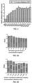
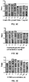
- 0S0064 were cultured under different medium formulations. The culture mediums were replaced and the trophoblastic cells were supplemented every 4 days. after the start of culture. After 10 days of culture, cell counts were performed. The basic medium (BM) without addition of any additive was used as the experimental control. The results were shown in Figs. 1A-1F .
- the ordinate in the figures shows the ratio of the number of cells obtained after culture in different mediums to the number of cells obtained after culture in basic medium BM.
- adding different concentrations of different factors according to Table 3 to BM would result in different effects on cell proliferation.
- B27 additive, N2 additive, insulin-transferrin-selenium complex, hepatocyte growth factor, insulin-like growth factor 1, fibroblast growth factor 7, Compound 1, Y27632 and A83-01 provided certain promoting effects on cell proliferation in specific concentration ranges.
- the culture mediums with different components were added at a volume of 500 ⁇ l/well to 48-well plates which were pre-laid with ⁇ -ray irradiated NIH-3T3 cells, and simultaneously, the BM medium was used as the experiment control.
- Laryngeal cancer cells No. 0S0065 isolated from laryngeal cancer tissue according to the method described in Example 1 were inoculated at a cell count of 4 ⁇ 10 4 cells/well in the above-mentioned 48-well culture plates which were pre-laid with ⁇ -ray irradiated NIH-3T3 cells.
- the plate was placed in a 37°C, 5% CO 2 incubator (purchased from Thermo Fisher), such that equal numbers of freshly isolated laryngeal cancer cells (No. 0S0065) were cultured under different medium formulations. After 7 days of culture, cell counts were performed. The results were shown in Fig. 2 .
- No. 7 is the most preferred culture medium in this patent for culturing and expanding primary laryngeal cancer cells (hereinafter abbreviated as SCM).
- SCM primary laryngeal cancer cells
- fibroblast growth factor 7 FGF-7
- IGF-1 insulin-like growth factor 1
- HGF hepatocyte growth factor
- ITS insulin-transferrin-selenium complex
- Laryngeal cancer epithelial cells originated from cancer tissue were isolated and obtained from the cancer tissue of a laryngeal cancer patient (Sample No. 0S0005) using the same method as in Example 1.
- laryngeal cancer cells originated from cancer tissue were counted with a flow imaging counter (JIMBIO FIL, Jiangsu Jimbio Technology Co., Ltd.) to obtain the total number of cells.
- the cells were inoculated in a 12-well plate which was pre-laid with ⁇ -ray irradiated NIH-3T3 cells at a density of 4 ⁇ 10 4 cells/cm 2 .
- the centrifuged cell sediments were resuspended in the basic medium BM and were counted with a flow imaging counter (JIMBIO FIL, Jiangsu Jimbio Technology Co., Ltd.) to obtain the total number of cells.
- the obtained cells were used in the following cultivation experiments.
- the digested cell suspension was diluted with the above Formulations 1-7 respectively, and were seeded into a 48-well plate which was pre-laid with ⁇ -ray irradiated NIH-3T3 cells with 10,000 cells and 250 ⁇ L volume per well.
- the medium of Formulation 1 When using the medium of Formulation 1, to a 48-well plate inoculated with primary cells was added the prepared fibroblast growth factor 7, with 250 ⁇ L per well, such that the final concentrations of fibroblast growth factor 7 were 80 ng/ml, 40 ng/ml, 20 ng/ml, 10 ng/ml, 5 ng/ml, 2.5 ng/ml, and 1.25 ng/ml, respectively; the medium of Formulation 1 was used as the control well (BC).
- the medium of Formulation 3 When using the medium of Formulation 3, to a 48-well plate inoculated with primary cells was added the prepared insulin-like growth factor 1, with 250 ⁇ L per well, such that the final concentrations of insulin-like growth factor 1 were 80 ng/ml, 40 ng/ml, 20 ng/ml, 10 ng/ml, 5 ng/ml, 2.5 ng/ml, and 1.25 ng/ml, respectively; the medium of Formulation 3 was used as the control well (BC).
- the medium of Formulation 4 When using the medium of Formulation 4, to a 48-well plate inoculated with primary cells was added the prepared hepatocyte growth factor, with 250 ⁇ L per well, such that the final concentrations of hepatocyte growth factor were 80 ng/ml, 40 ng/ml, 20 ng/ml, 10 ng/ml, 5 ng/ml, 2.5 ng/ml, and 1.25 ng/ml, respectively; the medium of Formulation 4 was used as the control well (BC).
- the medium of Formulation 5 When using the medium of Formulation 5, to a 48-well plate inoculated with primary cells was added the prepared Y27632, with 250 ⁇ L per well, such that the final concentrations of Y27632 were 40 ⁇ M, 20 ⁇ M, 10 ⁇ M, 5 ⁇ M, 2.5 ⁇ M, 1.25 ⁇ M, and 0.625 ⁇ M, respectively; the medium of Formulation 5 was used as the control well (BC).
- the medium of Formulation 6 When using the medium of Formulation 6, to a 48-well plate inoculated with primary cells was added the prepared Compound 1, with 250 ⁇ L per well, such that the final concentrations of Compound 1 were 40 ⁇ M, 20 ⁇ M, 10 ⁇ M, 5 ⁇ M, 2.5 ⁇ M, 1.25 ⁇ M, and 0.625 ⁇ M, respectively; the medium of Formulation 6 was used as the control well (BC).
- the medium of Formulation 7 When using the medium of Formulation 7, to a 48-well plate inoculated with primary cells was added the prepared A83-01, with 250 ⁇ L per well, such that the final concentrations of A83-01 were 4000 nM, 2000 nM, 1000 nM, 500 nM, 250 nM, 125 nM, and 62.5 nM, respectively; the medium of Formulation 7 was used as the control well (BC).
- the ratios were calculated by referring to the cell numbers in the control well (BC), and the results were shown in Figs. 3A-3G .
- the ratio represents a ratio of the number of cells of the first passage cultured by using each culture medium to the number of cells of the first passage cultured by the corresponding control well.
- the ratio is greater than 1, it indicates that the proliferation promoting effect of the prepared medium containing different concentrations of factors or small molecular compounds is preferable over that of the control well medium; if the ratio is less than 1, it indicates that the proliferation promoting effect of the prepared medium containing different concentrations of factors or small molecular compounds is poorer than that of the control well medium.
- the amount of fibroblast growth factor 7 in the culture medium is preferably 5 ng/ml-80 ng/ml, more preferably 20 ng/ml-80 ng/ml;
- the volume concentration of insulin-transferrin-selenium complex is preferably 1:25-1:100, more preferably 1:25-1:150 (corresponding to the final concentrations of insulin / transferrin / sodium selenite of 5-20 ⁇ g/ml, 2.5-10 ⁇ g/ml, 2.5-10 ng/ml, respectively, and more preferably 10-20 ⁇ g/ml, 5-10 ⁇ g/ml, 5-10 ng/ml, respectively);
- the amount of insulin-like growth factor 1 is preferably 10 ng/ml-80 ng/ml, more preferably 10 ng/ml-40 ng/ml;
- the amount of hepatocyte growth factor is preferably 10 ng/ml-80 ng/ml, more preferably 10 ng/
- Laryngeal cancer epithelial cells originated from cancer tissue were isolated and obtained from the cancer tissue of a laryngeal cancer patient (Sample No. 0S0003) using the same method as in Example 1.
- laryngeal cancer cells orginated from cancer tissue were counted with a flow imaging counter (JIMBIO FIL, Jiangsu Jimbio Technology Co., Ltd.) to obtain the total number of cells.
- the cells were inoculated in a 12-well plate which was pre-laid with ⁇ -ray irradiated NIH-3T3 cells at a density of 4 ⁇ 10 4 cells/cm 2 .
- 2 mL of the prepared culture medium for primary laryngeal cancer epithelial cells SCM was added to the 12-well plate, which was then placed in a 37 °C , 5% CO 2 incubator (purchased from Thermo Fisher) for culture.
- Fig. 4A is a microscopy image (photographed by a 100x inverted phase contrast microscope) of the cells which were cultured to day 4 after inoculation at a density of 4 ⁇ 10 4 cells/cm 2 into a 12-well plate which was pre-laid with ⁇ -ray irradiated NIH-3T3 cells according to this Example. Microscopic observation shows that the cultured primary laryngeal cancer cells derived from the cancer tissue had formed large clones.
- Fig. 4B is a microscopy image (photographed by a 100x inverted phase contrast microscope) of the cells which were cultured to day 12 after inoculation according to this Example, which shows that the field of vision was full of cells. It can be seen from Figs.
- Culture medium for primary laryngeal cancer epithelial cell SCM was prepared in the same method of Example 2, and basic medium BM was prepared as control. As another control, a culture medium FM used in the cell conditional reprogramming technology was prepared additionally.
- the formulation of the culture medium is shown in Table 5.
- culture mediums for primary laryngeal cancer cell SCM-1 and SCM-2 were prepared, and the formulations of the culture mediums were obtained by replacing insulin-transferrin-selenium complex in medium SCM wth 1:50 volume ratio of B27 additive and 1:100 volume ratio of N2 additive, respectively.
- KSFM medium a commercial medium Defined Keratinocyte SFM (hereinafter also referred to as "KSFM medium”) was purchased from Gibco, and the formulation of the culture medium is shown in Table 6. Table 5.
- Composition of culture medium FM used in the cell conditional reprogramming technology Medium composition Supplier Final concentration DMEM medium Corning 65 vol.% Fetal bovine serum Gibico 10 vol.% Ham's F12 Nutrient Mixture Gibico 25 vol.% Hydrocortisone Sigma-Aldrich 25 ng/ml Epidermal growth factor R&D 0.125 ng/ml Insulin Sigma-Aldrich 5 ⁇ g/ml Amphotericin B Sigma-Aldrich 250 ng/ml Gentamicin Gibico 10 ⁇ g/ml Cholera Toxin Sigma-Aldrich 0.1 nM Y27632 Enzo 10 ⁇ M Table 6.
- Composition of commercial medium Defined Keratinocyte SFM (KSFM) Medium composition Supplier Final concentration Defined Keratinocyte-S
- the primary laryngeal cancer cells No. 0S0006 derived from laryngeal cancer tissues were obtained.
- the cells were cultured at the same density (4 ⁇ 10 4 cells/cm 2 ) under the following 6 culture conditions:
- the cell clone formation and the cell proliferation status under the cultivation of each medium in the 24-well plate was observed, and the cell growth status was recorded by taking pictures with a microscope (EVOS M500, Invitrogen).
- the centrifuged cell sediments were resuspended in the culture medium of the invention and were counted with a flow imaging counter (JIMBIO FIL, Jiangsu Jimbio Technology Co., Ltd.) to obtain the total number of cells, which was 317,000.
- the cells cultured under the other five culture conditions were digested and counted in the same operation process as above.
- the total number of cells cultured by using the mediums FM, SCM-1, SCM-2, KSFM and BM were 166,700, 266,100, 277,200, 95,600 and 35,900, respectively.
- Fig. 5 is a plot of the total number of cells amplified from No. 0S0006 cells under different conditions.
- Fig. 6 is a comparative diagram of cell proliferation effects obtained by culturing the primary laryngeal cancer cells isolated from six laryngeal cancer patient samples (No. 0S0011, OS0012, OS0013, OS0014, OS0015, 0S0016) according to Example 1 for 7 days under the conditions of six different culture mediums, where ⁇ represents a moderate clone formation ability and proliferation promoting effect, ⁇ ⁇ represents a significant clone formation ability and proliferation promoting effect, ⁇ ⁇ ⁇ represents an extremely significant clone formation ability and proliferation promoting effect, and ⁇ represents no clone formation. It can be confirmed from the figure that the culture medium SCM has significant superiority over the other five culture conditions in terms of clone formation ability and cell proliferation promoting effect in cultivation of the laryngeal cancer tissue originated primary cell.
- the culture medium for primary laryngeal cancer epithelial cell SCM and the culture mediums FM, SCM-1, SCM-2, KSFM and BM as controls were obtained by using the same method as in (1) of this Example.
- the laryngeal cancer tissue originated primary laryngeal cancer cells (No. 0S0004) were cultured in six culture mediums, and then digested, passaged and counted by using the same method as in (1) of this Example.
- the cultured cells were digested, collected and counted according to the above operating method.
- the cells were inoculated at a density of 4 ⁇ 10 4 cells/well again and cultured continuously.
- Fig. 7 shows the growth curves of 0S0004 cells under six different culture conditions drawn by Graphpad Prism software.
- the abscissa represents the days of cell culture, and the ordinate represents the multiple of cumulative cell proliferation, i.e., the multiple of cell expansion in the culture period. The larger the value, the more multiples the cell expands in certain period, that is, the more cells are expanded.
- the slope represents the rate of cell expansion. It can be confirmed from the figure that the proliferation rates of laryngeal cancer epithelial cells cultured with the culture medium SCM of the invention and SCM-1, SCM-2 were superior to the other three culture conditions, and it can also be confirmed that the technology of the invention can continuously culture the primary laryngeal cancer epithelial cells.
- Laryngeal cancer epithelial cells originated from cancer tissue were isolated and obtained from the cancer tissue of a laryngeal cancer patient (Sample No. 0S0015) using the same method as in Example 1.
- laryngeal cancer cells originated from cancer tissue were counted with a flow imaging counter (JIMBIO FIL, Jiangsu Jimbio Technology Co., Ltd.) to obtain the total number of cells.
- the cells were inoculated in a 24-well plate which was pre-laid with ⁇ -ray irradiated NIH-3T3 cells at a density of 4 ⁇ 10 4 cells/cm 2 .
- PBS + 0.3% Triton X-100 was prepared in advance to dilute the antibody, wherein the squamous carcinoma specific antibody p63 (purchased from CST) was diluted in a ratio of 1:50. The blocking solution was discarded, and the prepared primary antibody diluent was added. The resultant was incubated in a refrigerator at 4°C overnight. The resultant was taken out at 4°C, balanced to room temperature, continued for incubation at 37°C for 1 hour, and then washed with PBS for 5 minutes ⁇ 3 times.
- PBS + 0.3% Triton X-100 was prepared in advance for secondary antibody dilution, wherein the rabbit fluorescent secondary antibody (purchased from Thermo Fisher) with excitation light of 488 nm was diluted in a ratio of 1:1000. The resultant was incubated at room temperature in the dark for 1 hour, and then was washed with PBS for 5 minutes ⁇ 3 times.
- Non-specific fluorescent dye DAPI (purchased from Sigma) was diluted with PBS in volume ratio of 1:1000, which was used for staining at room temperature in the dark for 5 minutes. The resultant was washed with PBS for 5 minutes ⁇ 3 times. Pictures were taken under microscope (EVOS M500, Invitrogen) for record.
- Figs. 8A and 8B show the fluorescent pictures taken in different fields under the 10X objective lens, respectively; in which, Fig. 8A is a picture of staining the nuclei with non-specific fluorescent dye DAPI, and Fig. 8B is a picture of staining with laryngeal cancer specific antibody p63 (located in the nuclei). As shown in the figures, the positions marked with nuclei in Fig. 8A were all stained green in Fig. 8B , indicating that the cultured cells are laryngeal squamous carcinoma cells, which are consistent with the clinical pathological diagnosis.
- Cancer tissue (Sample No. 0S0001) about the size of a mung bean was taken from a surgical resection sample of a laryngeal cancer patient, and immersed in 1 mL of 4% paraformaldehyde.
- Laryngeal cancer epithelial cells (Sample No. 0S0001) were obtained from the remaining cancer tissue in the same method of Example 1.
- Sample 0S0001 was continuously cultured to the sixth passage using the culture medium SCM of the invention according to the method of Example 3.
- Immunohistochemistry assay was used to detect the expression of important laryngeal cancer-related biomarkers in the original tissue of Sample 0S0001 and the primary cells obtained by continuous culture to the sixth passage.
- the tissue was fixed with 4% paraformaldehyde, embedded in paraffin, and cut into tissue sections of 4 ⁇ m thickness with a microtome. Routine immunohistochemical detection was then performed (for detailed steps, see Li et al., Nature Communication, (2016) 9: 2983 ).
- the primary antibodies used were Cytokeratin (pCK) (purchased from CST), p63 antibody (purchased from CST), and Ki67 antibody (purchased from R&D).
- Fig. 9 is a comparison of immunohistochemical results of the original tissue cells and the laryngeal cancer cells obtained by culturing the cells with the culture medium SCM of the invention.
- Fig. 9 confirms that the expression of laryngeal cancer-related biomarkers on the cells that were cultured from the laryngeal cancer cells (Sample No. 0S0001) by the technology of the invention to the 6th passage, was consistent with the expression of markers on the original tissue section from which the cells were derived. This suggests that the cells cultured by the technology of the invention maintain the original pathological characteristics of the cancer tissues of the laryngeal cancer patients.
- Laryngeal cancer cells (No. 0S0003) were isolated and obtained from the cancer tissues of one pathologically diagnosed laryngeal cancer patient by using the same method as in Example 1. 0S0003 was cultured using the culture medium SCM of the invention according to the method of Example 3, and when the number of laryngeal cancer cells reached 1 ⁇ 10 7 , the laryngeal cancer cells were digested and collected by using the same method of Example 4.
- the culture medium for laryngeal cancer cells of the invention SCM and Matrigel ® (purchased from BD Biosciences) were mixed at a ratio of 1:1, and 100 ⁇ L of the culture medium mixed with Matrigel was used to resuspend 5 ⁇ 10 6 laryngeal cancer cells, and the resultant was injected into the laryngeal cancer fat pad and the axilla of the right forelimb of 6-week-old female highly immunodeficient mice (NCG) (purchased from Nanjing Model Animal Research Center), respectively. The volume and growth rate of tumors in mice generated from the laryngeal cancer cells were observed and photographed every three days.
- Tumor formation can be observed in both of the two tumor cell inoculation sites of the mice on day 21 after tumor cell inoculation. From day 21 to day 40, the tumor proliferation in mice was obvious. This indicates that the cancer tissue-derived laryngeal cancer cells cultured by the culture method of the invention have tumorigenicity in mice.
- the laryngeal cancer cells cultured from the patient-derived laryngeal tumor samples can be used to test the sensitivity of the tumor cells of the patient to different drugs.
- Figs. 10A and 10B respectively represent the sensitivity of the laryngeal cancer cells cultured from surgically resected cancer tissue samples of two different laryngeal cancer patients (Sample No. 0S0020 and Sample No. 0S0022) to two chemotherapeutic drugs Bortezomib and Disulfiram, and to targeted drugs Gefitinib and Erlotinib.
- Fig. 10A shows the sensitivity results of laryngeal cancer cells cultured from Sample No. 0S0020 on four drugs
- Fig. 14B shows the sensitivity results of laryngeal cancer cells cultured from Sample No. No. 0S0022 on four drugs.
- the concentrations on the abscissa corresponding to the dash lines are the maximum blood concentrations C max of these four drugs in human body.
- the results show that the cells from the same patient have different sensitivities to different drugs at their respective maximum blood concentrations in human body, and the cells from different patients also have different sensitivities to the drugs at the maximum blood concentrations in human body. According to the results, the effectiveness of the drugs in the clinical use to the laryngeal cancer patients can be determined.
- the invention provides a culture medium and a culture method for culturing or expanding primary laryngeal cancer epithelial cells in vitro.
- the cell progeny and organoid cultured using the culture medium and culture method of the invention can be used for the efficacy evaluation and screening of drugs, toxicity determination, and regenerative medicine. Therefore, the invention is applicable in the industry.
Landscapes
- Health & Medical Sciences (AREA)
- Engineering & Computer Science (AREA)
- Life Sciences & Earth Sciences (AREA)
- Biomedical Technology (AREA)
- Chemical & Material Sciences (AREA)
- Organic Chemistry (AREA)
- Biotechnology (AREA)
- Bioinformatics & Cheminformatics (AREA)
- Genetics & Genomics (AREA)
- Wood Science & Technology (AREA)
- Zoology (AREA)
- Cell Biology (AREA)
- Biochemistry (AREA)
- General Health & Medical Sciences (AREA)
- Microbiology (AREA)
- Immunology (AREA)
- General Engineering & Computer Science (AREA)
- Hematology (AREA)
- Urology & Nephrology (AREA)
- Molecular Biology (AREA)
- Oncology (AREA)
- Physics & Mathematics (AREA)
- Tropical Medicine & Parasitology (AREA)
- Dermatology (AREA)
- Food Science & Technology (AREA)
- Medicinal Chemistry (AREA)
- Toxicology (AREA)
- Analytical Chemistry (AREA)
- General Physics & Mathematics (AREA)
- Pathology (AREA)
- Pharmaceuticals Containing Other Organic And Inorganic Compounds (AREA)
- Micro-Organisms Or Cultivation Processes Thereof (AREA)
- Measuring Or Testing Involving Enzymes Or Micro-Organisms (AREA)
- Peptides Or Proteins (AREA)
Applications Claiming Priority (2)
| Application Number | Priority Date | Filing Date | Title |
|---|---|---|---|
| CN202010454162.8A CN113717941B (zh) | 2020-05-26 | 2020-05-26 | 一种用于喉癌上皮细胞的培养基、培养方法及其应用 |
| PCT/CN2020/103428 WO2021237905A1 (zh) | 2020-05-26 | 2020-07-22 | 一种用于喉癌上皮细胞的培养基、培养方法及其应用 |
Publications (2)
| Publication Number | Publication Date |
|---|---|
| EP4159845A1 true EP4159845A1 (de) | 2023-04-05 |
| EP4159845A4 EP4159845A4 (de) | 2024-10-02 |
Family
ID=78671978
Family Applications (1)
| Application Number | Title | Priority Date | Filing Date |
|---|---|---|---|
| EP20937456.0A Pending EP4159845A4 (de) | 2020-05-26 | 2020-07-22 | Kulturmedium für laryngealkarzinomepithelzellen, kulturverfahren und anwendung davon |
Country Status (5)
| Country | Link |
|---|---|
| US (1) | US20230212523A1 (de) |
| EP (1) | EP4159845A4 (de) |
| JP (1) | JP7503662B2 (de) |
| CN (1) | CN113717941B (de) |
| WO (1) | WO2021237905A1 (de) |
Families Citing this family (1)
| Publication number | Priority date | Publication date | Assignee | Title |
|---|---|---|---|---|
| CN115595308B (zh) * | 2021-07-08 | 2025-10-10 | 合肥中科普瑞昇生物医药科技有限公司 | 一种用于肺癌上皮细胞的培养基、培养方法及其应用 |
Family Cites Families (9)
| Publication number | Priority date | Publication date | Assignee | Title |
|---|---|---|---|---|
| GB0621205D0 (en) * | 2006-10-25 | 2006-12-06 | Chroma Therapeutics Ltd | Inhibitors of PLK |
| CN102787092B (zh) * | 2011-05-17 | 2018-01-09 | 李晖 | 培养基、细胞培养用试剂盒及细胞培养方法 |
| CN103116006B (zh) * | 2013-01-25 | 2015-09-30 | 重庆医科大学附属第一医院 | 一种抗癌药物的新型筛选方法 |
| MX387025B (es) * | 2016-03-04 | 2025-03-19 | Anhui New Star Pharmaceutical Dev Co Ltd | Compuestos de anillo de siete miembros de pirimidina |
| CN105801582A (zh) * | 2016-04-12 | 2016-07-27 | 合肥工业大学 | 一类新型二氢蝶啶酮类衍生物及其制备方法和在医药上的用途 |
| GB201715930D0 (en) * | 2017-09-30 | 2017-11-15 | Upside Biotechnologies Ltd | Cell Culture Medium |
| CN111039944B (zh) | 2018-10-12 | 2021-11-23 | 中国科学院合肥物质科学研究院 | Mst1激酶抑制剂及其用途 |
| CN113528444B (zh) * | 2020-04-15 | 2023-05-23 | 合肥中科普瑞昇生物医药科技有限公司 | 一种用于食管鳞癌上皮细胞的培养基、培养方法及其应用 |
| CN113528425B (zh) * | 2020-04-15 | 2023-05-30 | 合肥中科普瑞昇生物医药科技有限公司 | 一种用于乳腺上皮肿瘤细胞的培养基和培养方法 |
-
2020
- 2020-05-26 CN CN202010454162.8A patent/CN113717941B/zh active Active
- 2020-07-22 EP EP20937456.0A patent/EP4159845A4/de active Pending
- 2020-07-22 JP JP2022573183A patent/JP7503662B2/ja active Active
- 2020-07-22 US US17/927,530 patent/US20230212523A1/en active Pending
- 2020-07-22 WO PCT/CN2020/103428 patent/WO2021237905A1/zh not_active Ceased
Also Published As
| Publication number | Publication date |
|---|---|
| JP2023534890A (ja) | 2023-08-15 |
| EP4159845A4 (de) | 2024-10-02 |
| CN113717941A (zh) | 2021-11-30 |
| US20230212523A1 (en) | 2023-07-06 |
| CN113717941B (zh) | 2023-11-14 |
| WO2021237905A1 (zh) | 2021-12-02 |
| JP7503662B2 (ja) | 2024-06-20 |
Similar Documents
| Publication | Publication Date | Title |
|---|---|---|
| EP4137565A1 (de) | Kulturmedium für epithelzellen von speiseröhrenplattenepithelkarzinom, kulturverfahren und anwendung davon | |
| EP4056685A1 (de) | Primäres brustepithelzellen-kulturmedium, kulturverfahren und dessen verwendung | |
| CN113969262B (zh) | 一种用于肺癌上皮细胞的培养基、培养方法及其应用 | |
| CN115960831B (zh) | 肠癌类器官的培养基及培养方法 | |
| EP4159845A1 (de) | Kulturmedium für laryngealkarzinomepithelzellen, kulturverfahren und anwendung davon | |
| EP4368706A1 (de) | Kulturmedium und kulturverfahren für lungenkrebsepithelzellen und anwendung davon | |
| CN115975935B (zh) | 一种用于宫颈癌原代细胞的培养基和培养方法 | |
| EP4403626A1 (de) | Kulturmedium und kulturverfahren für lungenkrebsepithelzellen und anwendung davon | |
| CN115960815B (zh) | 肺癌胸水来源类器官的培养基、培养方法及其应用 | |
| RU2814719C1 (ru) | Культуральная среда для эпителиальных клеток рака гортани, способ культивирования и их применение | |
| WO2023060764A1 (zh) | 胃癌原代细胞的培养基和培养方法 | |
| RU2838489C2 (ru) | Среда для культивирования эпителиальных клеток рака лёгкого, способ их культивирования и применение | |
| RU2842810C2 (ru) | Культуральная среда и способ культивирования эпителиальных клеток рака легкого и их применение | |
| CN115975936B (zh) | 宫颈癌原代细胞的培养基和培养方法 | |
| RU2816529C1 (ru) | Культуральная среда для эпителиальных клеток плоскоклеточной карциномы пищевода, способ культивирования и их применение | |
| CN115975933B (zh) | 卵巢癌原代细胞的培养基、培养方法及其应用 | |
| CN115975910B (zh) | 胃癌类器官的培养基及培养方法 |
Legal Events
| Date | Code | Title | Description |
|---|---|---|---|
| STAA | Information on the status of an ep patent application or granted ep patent |
Free format text: STATUS: THE INTERNATIONAL PUBLICATION HAS BEEN MADE |
|
| PUAI | Public reference made under article 153(3) epc to a published international application that has entered the european phase |
Free format text: ORIGINAL CODE: 0009012 |
|
| STAA | Information on the status of an ep patent application or granted ep patent |
Free format text: STATUS: REQUEST FOR EXAMINATION WAS MADE |
|
| 17P | Request for examination filed |
Effective date: 20221205 |
|
| AK | Designated contracting states |
Kind code of ref document: A1 Designated state(s): AL AT BE BG CH CY CZ DE DK EE ES FI FR GB GR HR HU IE IS IT LI LT LU LV MC MK MT NL NO PL PT RO RS SE SI SK SM TR |
|
| DAV | Request for validation of the european patent (deleted) | ||
| DAX | Request for extension of the european patent (deleted) | ||
| A4 | Supplementary search report drawn up and despatched |
Effective date: 20240830 |
|
| RIC1 | Information provided on ipc code assigned before grant |
Ipc: G01N 33/15 20060101ALI20240826BHEP Ipc: C12Q 1/02 20060101ALI20240826BHEP Ipc: C12N 5/071 20100101ALI20240826BHEP Ipc: C12N 5/09 20100101AFI20240826BHEP |