EP4163363A1 - Verfahren zur herstellung einer zusammensetzung zur kultivierung von lungenorganoiden, zusammensetzung dafür und organoidkulturverfahren damit - Google Patents
Verfahren zur herstellung einer zusammensetzung zur kultivierung von lungenorganoiden, zusammensetzung dafür und organoidkulturverfahren damit Download PDFInfo
- Publication number
- EP4163363A1 EP4163363A1 EP21832927.4A EP21832927A EP4163363A1 EP 4163363 A1 EP4163363 A1 EP 4163363A1 EP 21832927 A EP21832927 A EP 21832927A EP 4163363 A1 EP4163363 A1 EP 4163363A1
- Authority
- EP
- European Patent Office
- Prior art keywords
- lung
- decm
- tissue
- composition
- organoid
- Prior art date
- Legal status (The legal status is an assumption and is not a legal conclusion. Google has not performed a legal analysis and makes no representation as to the accuracy of the status listed.)
- Pending
Links
Images








Classifications
-
- C—CHEMISTRY; METALLURGY
- C12—BIOCHEMISTRY; BEER; SPIRITS; WINE; VINEGAR; MICROBIOLOGY; ENZYMOLOGY; MUTATION OR GENETIC ENGINEERING
- C12N—MICROORGANISMS OR ENZYMES; COMPOSITIONS THEREOF; PROPAGATING, PRESERVING, OR MAINTAINING MICROORGANISMS; MUTATION OR GENETIC ENGINEERING; CULTURE MEDIA
- C12N5/00—Undifferentiated human, animal or plant cells, e.g. cell lines; Tissues; Cultivation or maintenance thereof; Culture media therefor
- C12N5/0081—Purging biological preparations of unwanted cells
-
- C—CHEMISTRY; METALLURGY
- C12—BIOCHEMISTRY; BEER; SPIRITS; WINE; VINEGAR; MICROBIOLOGY; ENZYMOLOGY; MUTATION OR GENETIC ENGINEERING
- C12N—MICROORGANISMS OR ENZYMES; COMPOSITIONS THEREOF; PROPAGATING, PRESERVING, OR MAINTAINING MICROORGANISMS; MUTATION OR GENETIC ENGINEERING; CULTURE MEDIA
- C12N5/00—Undifferentiated human, animal or plant cells, e.g. cell lines; Tissues; Cultivation or maintenance thereof; Culture media therefor
- C12N5/06—Animal cells or tissues; Human cells or tissues
- C12N5/0602—Vertebrate cells
- C12N5/0688—Cells from the lungs or the respiratory tract
-
- A—HUMAN NECESSITIES
- A61—MEDICAL OR VETERINARY SCIENCE; HYGIENE
- A61L—METHODS OR APPARATUS FOR STERILISING MATERIALS OR OBJECTS IN GENERAL; DISINFECTION, STERILISATION OR DEODORISATION OF AIR; CHEMICAL ASPECTS OF BANDAGES, DRESSINGS, ABSORBENT PADS OR SURGICAL ARTICLES; MATERIALS FOR BANDAGES, DRESSINGS, ABSORBENT PADS OR SURGICAL ARTICLES
- A61L27/00—Materials for grafts or prostheses or for coating grafts or prostheses
- A61L27/36—Materials for grafts or prostheses or for coating grafts or prostheses containing ingredients of undetermined constitution or reaction products thereof, e.g. transplant tissue, natural bone, extracellular matrix
- A61L27/3604—Materials for grafts or prostheses or for coating grafts or prostheses containing ingredients of undetermined constitution or reaction products thereof, e.g. transplant tissue, natural bone, extracellular matrix characterised by the human or animal origin of the biological material, e.g. hair, fascia, fish scales, silk, shellac, pericardium, pleura, renal tissue, amniotic membrane, parenchymal tissue, fetal tissue, muscle tissue, fat tissue, enamel
- A61L27/3633—Extracellular matrix [ECM]
-
- A—HUMAN NECESSITIES
- A61—MEDICAL OR VETERINARY SCIENCE; HYGIENE
- A61L—METHODS OR APPARATUS FOR STERILISING MATERIALS OR OBJECTS IN GENERAL; DISINFECTION, STERILISATION OR DEODORISATION OF AIR; CHEMICAL ASPECTS OF BANDAGES, DRESSINGS, ABSORBENT PADS OR SURGICAL ARTICLES; MATERIALS FOR BANDAGES, DRESSINGS, ABSORBENT PADS OR SURGICAL ARTICLES
- A61L27/00—Materials for grafts or prostheses or for coating grafts or prostheses
- A61L27/36—Materials for grafts or prostheses or for coating grafts or prostheses containing ingredients of undetermined constitution or reaction products thereof, e.g. transplant tissue, natural bone, extracellular matrix
- A61L27/3683—Materials for grafts or prostheses or for coating grafts or prostheses containing ingredients of undetermined constitution or reaction products thereof, e.g. transplant tissue, natural bone, extracellular matrix subjected to a specific treatment prior to implantation, e.g. decellularising, demineralising, grinding, cellular disruption/non-collagenous protein removal, anti-calcification, crosslinking, supercritical fluid extraction, enzyme treatment
- A61L27/3687—Materials for grafts or prostheses or for coating grafts or prostheses containing ingredients of undetermined constitution or reaction products thereof, e.g. transplant tissue, natural bone, extracellular matrix subjected to a specific treatment prior to implantation, e.g. decellularising, demineralising, grinding, cellular disruption/non-collagenous protein removal, anti-calcification, crosslinking, supercritical fluid extraction, enzyme treatment characterised by the use of chemical agents in the treatment, e.g. specific enzymes, detergents, capping agents, crosslinkers, anticalcification agents
-
- A—HUMAN NECESSITIES
- A61—MEDICAL OR VETERINARY SCIENCE; HYGIENE
- A61L—METHODS OR APPARATUS FOR STERILISING MATERIALS OR OBJECTS IN GENERAL; DISINFECTION, STERILISATION OR DEODORISATION OF AIR; CHEMICAL ASPECTS OF BANDAGES, DRESSINGS, ABSORBENT PADS OR SURGICAL ARTICLES; MATERIALS FOR BANDAGES, DRESSINGS, ABSORBENT PADS OR SURGICAL ARTICLES
- A61L2430/00—Materials or treatment for tissue regeneration
- A61L2430/40—Preparation and treatment of biological tissue for implantation, e.g. decellularisation, cross-linking
-
- C—CHEMISTRY; METALLURGY
- C12—BIOCHEMISTRY; BEER; SPIRITS; WINE; VINEGAR; MICROBIOLOGY; ENZYMOLOGY; MUTATION OR GENETIC ENGINEERING
- C12N—MICROORGANISMS OR ENZYMES; COMPOSITIONS THEREOF; PROPAGATING, PRESERVING, OR MAINTAINING MICROORGANISMS; MUTATION OR GENETIC ENGINEERING; CULTURE MEDIA
- C12N2513/00—3D culture
-
- C—CHEMISTRY; METALLURGY
- C12—BIOCHEMISTRY; BEER; SPIRITS; WINE; VINEGAR; MICROBIOLOGY; ENZYMOLOGY; MUTATION OR GENETIC ENGINEERING
- C12N—MICROORGANISMS OR ENZYMES; COMPOSITIONS THEREOF; PROPAGATING, PRESERVING, OR MAINTAINING MICROORGANISMS; MUTATION OR GENETIC ENGINEERING; CULTURE MEDIA
- C12N2533/00—Supports or coatings for cell culture, characterised by material
- C12N2533/90—Substrates of biological origin, e.g. extracellular matrix, decellularised tissue
Definitions
- the present invention relates to a preparation method of a composition for culturing lung organoids, a composition thereby, and an organoid culture method using the same.
- Organoids are three-dimensional cell aggregates formed from stem cells through phylogeny and differentiation, and they allow for the observation of the development process of organs and the reproduction of actual tissues in terms of function and structure.
- a support or scaffold which plays a supporting role so that cells can be attached and grow, also plays a very important role in biological tissue engineering, and it also plays an important role in the growth of cells seeded in a porous structure and cells migrating from the surroundings of the tissue.
- Most of the cells in the human body are adherent cells that are attached to grow, and if there is no place to be attached to, the cells cannot grow and eventually die. Therefore, the scaffold should provide a compatible environment for cell adhesion, differentiation, growth and cell migration.
- Such a scaffold can be made of various materials, and research to develop a scaffold by using a natural material, a synthetic polymer, bioceramics, and a polymer-ceramic composite material is being actively conducted. Accordingly, organoids that mimic various organs have been developed so far, and these are mainly cultured in Matrigel, a three-dimensional culture environment.
- Matrigel is widely used for organoid culture, it is derived from mouse sarcoma, and there is no particular substitute. Therefore, despite the high cost, there is no choice but to rely on Matrigel. In addition, certain components are dominating in Matrigel and there is a limitation in reflecting the tissue-specific characteristics. To complement and replace this, decellularization of tissues and organs has been studied as a promising method for preparing functional scaffolds for cell culture and transplantation, and there is an emerging need for the method. However, the organoids produced by the current organoid culture technology have many differences from the actual human tissues in terms of the degree of differentiation and functions, and thus the development of a technology for culturing more mature organoids is required.
- Non-Patent Document 1 teaches that a decellularized lung tissue scaffold can be used for lung tissue regeneration. However, the decellularization process is not specifically disclosed therein.
- the whole lung was decellularized in a number of previous studies, but in the present invention, decellularization was performed by separating the bronchus and micro-bronchus and using the alveoli only (see Example 1 of the present invention, including the details about removing the bronchus and micro-bronchus during the lung arrangement). Even if a conventional decellularization process is employed, there is a difference in the content of residual DNA, GAGs, and collagen. In particular, in the present invention, decellularization conditions were established such that a large amount of collagen VI, laminin, and fibronectin components can remain (see Example 8) .
- Patent Document 1 discloses a preparation method of a brain organoid culture composition in which brain tissue is decellularized and lyophilized, the lyophilized tissue is dispersed and then solutionized in an acidic solution, and the pH is adjusted.
- this is for brain tissue and not for the lung.
- pepsin or trypsin can be used as an acidic solution, and just the general gelation temperature (25 to 38°C) after pH adjustment is described.
- Non-Patent Document 1 Balestrini, JL. et al" Biomaterials (2016) 102:220-230
- the purpose of the present invention is to solve the problem that the Matrigel-based culture system, which is an extract derived from animal cancer tissue, has a large difference between batches, is incapable of simulating the environment of the actual tissue, and has an insufficient efficiency of tissue differentiation and development.
- the composition of the present invention is to provide an optimal environment for organoid culture by creating an actual tissue-like environment and to improve organ-specific differentiation potency.
- another purpose of the present invention is to provide a composition for culturing an organoid, which has excellent physical properties of a decellularized tissue and which can also significantly increase the organoid culture efficiency, and a preparation method of the same.
- another purpose of the present invention is to provide a composition for culturing an organoid that is capable of providing excellent physical properties of a decellularized tissue, preventing gelation, and enabling uniform hydrogelation, and a preparation method of the same.
- the present invention provides a preparation method of a composition for culturing lung organoids, the composition thereby, and a organoid culture method using the same.
- the present invention provides a preparation method of a composition for culturing lung organoids, the method comprising: S1) decellularizing lung tissue; S2) lyophilizing the decellularized tissue; S3) pulverizing the lyophilized tissue; S4) lysing the pulverized tissue by adding a proteopeptic enzyme and an acid; and S5) adjusting the pH by adding a basic solution to the lysate.
- the decellularized tissue contains an actual tissue-specific extracellular matrix component, it is able to provide a physical, mechanical, and biochemical environment of the tissue, and is very efficient in promoting the differentiation into lung tissue cells and their tissue-specific functionality.
- the "organoid” refers to a micro-sized biological organ produced in the form of an artificial organ by culturing cells derived from tissues or pluripotent stem cells in a 3D form.
- the organoids are three-dimensional tissue analogues including organ-specific cells that are developed from stem cells and that self-organize in a manner that is similar to the in vivo state, and they can develop into specific tissues by patterning limited factors (for example, growth factors).
- the organoid has the intrinsic physiological properties of the cell and may have an anatomical structure that mimics the original state of a cell mixture (including not only limited cell types but also residual stem cells and proximal physiological niche). Through a three-dimensional culture method, the organoid may have a shape and a tissue-specific function, such as that of an organ in which cells and cellular functions are better arranged and which has functionality.
- the S1) decellularizing may comprise, twice or more, a process of adding at least one solution selected from sodium dodecyl sulfate (SDS), polyethylene glycol (PEG), Triton X-100, ethylenediaminetetraacetic acid (EDTA), peracetic acid, deoxycholic acid, 3-[(3-cholamidopropyl)dimethylammonio]-1-propanesulfonate (CHAPS), sodium deoxycholate, ammonium hydroxide, trypsin, benzonase, DNase, calcium chloride, magnesium sulfate, sodium azide (NaN 3 ) and sodium chloride.
- S1) may comprise a washing process in the middle of the two decellularization processes.
- the tissue may be stirred during the decellularization.
- Decellularized extracellular matrix can be obtained through the method described above, and the "dECM” means a natural scaffold for cell growth prepared through decellularization of tissues found in mammals and multicellular organisms.
- the extracellular matrix may be collagen, elastin, laminins, glycosaminoglycans, proteoglycans, antimicrobials, chemoattractants, cytokines, and a mixture of structural and non-structural biomolecules that are not limited by growth factors.
- the extracellular matrix contain about 90% collagen in various forms in mammals. Extracellular matrixes derived from various biological tissues may have different overall structures and compositions due to the unique role required for each tissue.
- the decellularizing may comprise: a1) washing a lung tissue; a2) treating with at least one selected from peracetic acid, triton-X, sodium dodecyl sulfate and sodium chloride; a3) washing; a4) treating with at least one selected from peracetic acid, triton-X, sodium dodecyl sulfate and sodium chloride; a5) washing; a6) treating with at least one selected from peracetic acid, triton-X, sodium dodecyl sulfate and sodium chloride; and a7) washing.
- the decellularizing may comprise: a1) washing a lung tissue; a2) treating with at least one selected from peracetic acid, triton-X, sodium dodecyl sulfate and sodium chloride; a3) washing; a4) treating with at least one selected from peracetic acid, triton-X, sodium dodecyl sulfate and sodium chloride; a5) washing; a6) treating with at least one selected from peracetic acid, triton-X, sodium dodecyl sulfate and sodium chloride; and a7) washing.
- the washing of a1), a3), a5), or a7) may be performed with at least one selected from distilled water, physiological saline, and phosphate buffered saline.
- a composition for culturing organoids having excellent differentiation into tissue cells and having excellent tissue specificity can be obtained.
- a composition capable of maintaining the mutational properties of lung cancer organoids may be obtained in the lung dECM scaffold using the same.
- a1) may be performed for 3 to 48 hours, a2) for 12 to 120 hours, a3) for 6 to 24 hours, a4) for 0.5 to 4 hours, a5) for 6 to 48 hours, a6) for 0.5 to 4 hours, and a7) for 1 to 6 hours.
- a1) may be performed for 3 to 24 hours, a2) for 24 to 48 hours, a3) for 9 to 18 hours, a4) for 1 to 3 hours, a5) for 12 to 36 hours, a6) for 1 to 3 hours, and a7) for 2 to 4 hours.
- the S2) lyophilizing is for lyophilizing a tissue decellularized in S1).
- the lyophilizing methods or specific conditions are not particularly limited, and various methods or conditions known in the art may be used.
- the decellularized tissue may be frozen at -100°C to -50°C, and then dried at -70°C to -30°C for 1 to 10 days.
- the S3) pulverizing is for pulverizing the tissue lyophilized in S2).
- the pulverizing methods or sizes, and specific conditions are not particularly limited, and various methods or sizes and conditions known in the art may be used.
- the lyophilized tissue may be put into a mortar and pulverized by using liquid nitrogen.
- the S4) lysing is for lysing the tissue pulverized in S3) above.
- the pulverized tissue can be lysed by adding a proteopeptic enzyme and an acid to the same, and stirring the resulting mixture.
- the present invention is characterized in that a proteopeptic enzyme and an acid are added to the pulverized tissue to lysate by stirring the resulting mixture at a speed of 330 rpm to 500 rpm for 72 to 96 hours.
- the resulting mixture may be lysed by stirring at a speed of 330 rpm for 72 to 96 hours.
- the resulting mixture may be lysed by stirring at a speed of 330 rpm to 500 rpm for 96 hours. More preferably, the resulting mixture may be lysed by stirring at a speed of 330 rpm for 96 hours.
- the modulus values representing the physical properties for each stirring time were found to be in the order of 24 h ⁇ 72 h ⁇ 96 h > 120 h. In other words, it was confirmed that the physical properties were best when the stirring time was 72-96 hours. In addition, although the physical properties continued to increase when the stirring time was 120 hours, the experiment was difficult because many bubbles were generated when the organoid is encapsulated. In particular, death of many organoids was found in the dECM stirred for 24 hours and 120 hours, indicating that the properties are important in the culture of an organoid. In addition, the culture tendency in the organoid culture was improved in the order of 24 h ⁇ 72 h ⁇ 96 h > 120 h (120 h ⁇ 72 h).
- the modulus values representing the physical properties for each stirring speed were found to be in the order of 80 ⁇ 150 ⁇ 330 ⁇ 500 > 800 > 1000 (80 ⁇ 1000 ⁇ 150 ⁇ 800 ⁇ 330 ⁇ 500) rpm, and the best physical properties were found at 330-500 rpm.
- the culture efficiency in the organoid culture was in the order of 80 ⁇ 250 « 330 ⁇ 500 » 1000 (80 ⁇ 250 ⁇ 1000 « 330 ⁇ 500) and that the dome formation was impossible at 80 rpm.
- death of many organoids was found with no living organoids observed in the dECM stirred at 80, 250, and 1000 rpm, indicating that the properties are important in the culture of an organoid, and compatible conditions could be set accordingly.
- the proteopeptic enzyme in S4) may be pepsin or trypsin. Preferably, it may be pepsin.
- the acid in S4) may be hydrochloric acid or acetic acid.
- it may be acetic acid.
- the S5) adjusting the pH is for adjusting the pH by adding a basic solution to the lysate in S4) above.
- the method of adding a basic solution or the type of the basic solution to be used is not particularly limited, and various basic solutions known in the art may be used.
- the final pH can be adjusted to be 7 to 7.5.
- the present invention is particularly characterized in that the S5) adjusting the pH is performed within a range of 1°C to 10°C in order to prevent gelation. Preferably, it may be performed within a range of 2°C to 7°C. In other words, the pH adjusting process may not be performed at room temperature, but may be performed in a lower refrigeration temperature range.
- the S5) adjusting the pH may be performed on ice in order to prevent gelation.
- the expression of performing "on ice” means that the lysate is present on the surface of the ice or provided in the ice.
- the pH may be adjusted by adding a base, after inserting the container containing the lysate into ice.
- the present inventors found that not in the process of decellularization but in the process of adjusting the pH by adding a basic solution to the decellularization lysate, the temperature (1°C to 10°C) and/or conditions (on ice) affect the hydrogelation of the composition and the subsequent encapsulation of organoids, and then completed the present invention.
- the process of adjusting pH by adding a basic solution to the decellularized lysate is performed "on ice (at 1°C to 10°C)," the gelation of the lysate may be prevented, and uniform hydrogelation may be possible.
- the lung dECM scaffold prepared in this way has excellent rheological properties (physical properties) and culture effects, compared to the case where the lung dECM scaffold was prepared differently.
- the decellularization composition of which pH was adjusted on ice according to the present invention was able to form a uniform gel during the gelation process, but the decellularization composition of which pH was adjusted at room temperature (RT) underwent partial gelation and aggregation and so the dome shape was not maintained but broken and the physical properties thereof (complex modulus (G*)) were also low.
- RT room temperature
- the present invention may be characterized in that the pH adjustment process is not performed at room temperature but in a lower refrigeration temperature range, and further, the basic solution is stored at a refrigeration temperature within a range of 1°C to 10°C in advance before use.
- a basic solution stored at a refrigeration temperature may preferably be used because the gelation prevention and uniform hydrogelation may be performed better.
- tissue cells 950 or more of tissue cells, specifically 970 or more of the tissue cells can be removed, compared to the tissue cells before the treatment.
- the glucosaminoglycan content may be 90% to 120%, specifically 95% to 110%, compared to the glucosaminoglycan content before the treatment.
- the collagen content may be 200% to 2000%, specifically 500% to 1700%, compared to the collagen content before the treatment.
- an excellent cell removal rate may be obtained, and the useful components can be retained so that a scaffold based on the retained useful components may be used to obtain an organoid culture composition in which the organoid culture is performed well (culture, expansion, proliferation) and which has excellent differentiation into tissue cells and excellent tissue specificity.
- a cytokine-rich composition that is excellent for organoid culture may be obtained through the method described above. Specifically, compared to Matrigel, at least one cytokine selected from angiopoietin-1, insulin growth factor, TGF-beta, and placental growth factor may have been increased. Through this, an organoid culture composition in which the organoid culture is performed well (culture, expansion, proliferation) and which has excellent differentiation into tissue cells and excellent tissue specificity may be obtained.
- the present invention also provides a composition for culturing lung organoids prepared by the method described above.
- the composition may contain 0.1% to 10% (w/v) of dried decellularization tissue based on the total volume of the composition, specifically, 0.1% to 50 (w/v).
- dECM decellularization scaffold
- the composition of the present invention may further include Matrigel.
- the "Matrigel” is a protein complex extracted from EHS (Engelbreth-Holm-Swarm) mouse sarcoma cells (name of product manufactured by BD Bioscience) and may comprise an extracellular matrix, such as laminin, collagen and heparan sulfate proteoglycan, and fibroblast growth factor (FGF), epidermal growth factor (EGF), insulin-like growth factor (IGF), TGF- ⁇ or platelet-derived growth factor (PDGF).
- EHS Engelbreth-Holm-Swarm mouse sarcoma cells
- FGF fibroblast growth factor
- EGF epidermal growth factor
- IGF insulin-like growth factor
- TGF- ⁇ platelet-derived growth factor
- the composition may contain 0.1% to 10% (w/v) of dried decellularization tissue based on the total volume of the composition, specifically 0.1 to 50 (w/v).
- dECM decellularization scaffold
- the present invention also provides a lung organoid culture method comprising: culturing a lung organoid in the composition for culturing lung organoids described above.
- the culture refers to a process of maintaining and growing cells under compatible conditions, and compatible conditions may mean, for example, the temperature at which the cells are maintained, nutrient availability, atmospheric CO 2 content, and cell density.
- compatible culture conditions for maintaining, proliferating, expanding and differentiating different types of cells are known and documented in the art.
- the compatible conditions for the formation of the organoid may be conditions that facilitate or allow cell differentiation and formation of multicellular structures.
- the present invention also provides a lung organoid culture method for culturing a lung organoid, the method comprising: culturing a lung organoid in the composition for culturing lung organoids described above.
- the present invention also provides an extracellular matrix scaffold for culturing organoids prepared from the composition for culturing lung organoids described above.
- the present invention also provides a lung organoid culture method comprising: mixing the extracellular matrix support for culturing organoids and the lung organoid.
- the present invention is capable of creating an environment that is more similar to an actual tissue than a conventional Matrigel-based culture system, and in particular, exhibits an effect of facilitating tissue differentiation in lung organoid culture and effectively developing into a form that is similar to an actual tissue.
- the present invention exhibits effectiveness by maintaining genetic mutations even in tumor-derived organoid culture.
- the decellularized tissue when stirred for a specific time and/or at a specific rpm in lysing the decellularized tissue with a proteopeptic enzyme and an acid according to the present invention, the physical properties of the decellularized tissue become excellent and the organoid culture rate is significantly increased.
- the process of adjusting the pH by adding a basic solution to the decellularization solution according to the present invention is performed under specific conditions, the physical properties of the decellularized tissue become excellent, gelation can be prevented, and uniform hydrogelation can be achieved.
- the trimmed lung tissue is frozen at - 80°C for 24 hours. (Store at -80°C until use) After slicing the frozen lung tissue to a thickness of 1-2 mm, and the sliced lung tissue is moved to a beaker. In order to prevent contamination of the tissue and evaporation of the solution, the entrance of the beaker must be covered with aluminum foil.
- the tissue is washed with distilled water (dH 2 O; secondary water) for 3 hours until almost no blood comes out.
- the tissue is cut into small pieces by using scissors to remove the remaining micro-bronchus. In this step, the amount of the tissue is reduced.
- 1% SDS is treated within 24 hours. Washing is performed with distilled water for 12 hours to remove the remaining SDS.
- the tissue is treated with 1 M sodium chloride (NaCl) for 2 hours. Washing is performed with distilled water for 24 hours to remove residual reagents.
- ethanol containing peracetic acid is treated for 2 hours.
- tertiary distilled water tertiary distilled water (ddH 2 O) is used as all base buffers.
- washing is performed with 1X PBS for 1 hour.
- the tissue is moved to a 50 mL conical tube by using forceps.
- the tissue is lyophilized at -50°C for 4 days.
- the washing process employs a stirrer at a speed of 100-150 rpm.
- the stirring speed is kept within 100 rpm to ensure that the tissue and the reagent can react sufficiently.
- forceps and scissors are used to loosen the tissue in order to prevent congelation of the tissue.
- the beaker and the magnetic bar are washed with distilled water in each step in order to remove the remaining reagent.
- the lyophilized lung dECM prepared in Example 1 is pulverized with liquid nitrogen by using a mortar and a pestle.
- the lung dECM is put into a 50 mL conical tube and lysed in 0.5 M acetic acid with pepsin for 96 hours.
- a cell strainer is used to filter the lung dECM powder that is not lysed.
- the steps hereinafter should be performed on ice.
- a pH indicator strip is used to observe the change of pH, and 10 N sodium hydroxide (NaOH) is added to adjust the final pH to 7-7.5.
- Cold 1X PBS is added to adjust to the final volume.
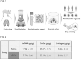
- the contents of dsDNA, GAGs and collagen in the decellularized tissue is confirmed, compared to the native tissue.
- GeneJET Genomic DNA Purification Kit was used to separate dsDNA from the native and decellularized tissues. The separation was carried out in the same manner as described in the manufacturer's protocol. The dsDNA content in the decellularized tissue compared to the native tissue was confirmed by using Quant-iT TM PicoGreen TM dsDNA Assay Kit. The assay was carried out in the same manner as described in the manufacturer's protocol.
- a papain solution is prepared for the digestion of dECM.
- the amount of the solution can be adjusted according to the number of samples. The specific process is described below. 1 0.1 M sodium phosphate (monobasic), 0.5 mM Na2-EDTA, and 5 mM cysteine-hydrochloric acid are dissolved in autoclaved distilled water. 2 The pH of the solution is adjusted to 6.5 by adding 10 N sodium hydroxide. (3) A papain stock prepared at 10 mg/mL is added to the solution above, and the resulting mixture is mixed well with a vortex mixer to be mixed homogeneously.
- a solution for the dimethyl-methylene blue (DMMB) assay is prepared.
- the specific process is described below. 1
- 1,9-dimethyl-methylene blue zinc chloride double salt, glycine, and sodium chloride are dissolved in autoclaved distilled water.
- the pH is adjusted to 3 by adding 0.5 M HCl solution, while measuring the change of pH by using a bench-top pH meter.
- an aliquot of the solution is taken in a conical tube, which is wrapped with aluminum foil and stored.
- a 10 mg/mL chondroitin sulfate A solution stock is prepared at 100 ⁇ g/mL by using 1X PBS.
- a solution for the hydroxyproline assay is prepared. The specific process is described below.
- the lyophilized tissue is put into a 1.7 mL microcentrifuge tube (e-tube) to which the papain solution is added, and the resulting mixture is mixed well with a vortex mixer.
- the e-tube containing the tissue is inserted into rubber racks, which are floated on water added into a beaker, and then the tissue is digested at 60°C for 16 hours. However, the tissue should be completely lysed without any remaining tissue. Centrifugation is performed at 12000 x g for 20 minutes, and the supernatant is taken and transferred to a new e-tube.
- GAGs Glycosaminoglycans
- a DMMB assay is performed to quantify the GAGs in the dECM.
- the specific process is described below. 1
- a standard in a specific range according to the purpose of the experiment is prepared by using chondroitin sulfate A solution stock prepared at 100 ug/mL and distilled water. 2
- the standard and samples are loaded in triplicate into a 96-well plate at 40 ⁇ L/well.
- the DMMB dye is added at 160 ⁇ L/well by using a multi-channel pipettor.
- the absorbance is measured at a 525 nm wavelength with a microplate reader.
- a hydroxyproline assay is performed to quantify the amount of collagen in the dECM.
- the specific process is described below. 1
- the tissue lysed in the papain solution and the same amount of hydrochloric acid are subject to a reaction in an oven at 120°C for 16 hours.
- the residue remaining on the glass wall is dried and cooled at room temperature for 3 hours, and then lysed again in 1X PBS.
- (3) The completely lysed sample is transferred to an e-tube and centrifuged at 5,000 x g for 10 minutes at 4°C, and only the supernatant is transferred to a new e-tube.
- the 100 ug/mL hydroxyproline solution stock is diluted with distilled water to prepare a standard in a specific range according to the purpose of the experiment.
- the standard and samples are loaded in triplicate into a 96-well plate at 50 ⁇ L/well.
- the chloramine T solution in a volume of 50 ⁇ L is added, and then the resulting mixture is subject to a reaction at room temperature for 20 minutes.
- the P-DAB solution in a volume of 50 ⁇ L is added and then the resulting mixture is subject to a reaction 60°C for 30 minutes. Since P-DAB is sensitive to light, the loading should be conducted with the light off, and then the plate should be wrapped with aluminum foil to perform incubation. 8 After cooling for 30 minutes at room temperature, the absorbance is measured at a 540 nm with a microplate reader.
- the DNA content was 2.05%, and more than 97% of the cells were removed.
- the content of GAGs, of which loss occurs mainly in the decellularization process was about 110% in the lung dECM, which was similar to that of the native group. Therefore, the decellularization process was considered as having been well established.
- the collagen content in the lung dECM was 573% compared to the native group, and so it was verified that cells were removed during the decellularization process and the main components of the extracellular matrix were well preserved.
- the lung dECM scaffold is prepared at pH 7 to evaluate the rheological properties.
- the 20 mm cone plate geometry (cone diameter of 20 mm at a 2° angle) is set in the rate-controlled mode of a rheometer.
- An experiment sequence in the installed software program (TRIOS) is crated to measure the viscosity, gelation kinetics, and dynamic modulus of the dECM scaffold.
- the dECM scaffold Before the measurement, the dECM scaffold is placed on the plate at 37°C for 30 minutes. The frequency-dependent storage modulus (G') and loss modulus (G") of the dECM scaffold are measured at 2% strain within a range of 0.1-100 rad/s.
- the amount of the dECM loading is set to be 250 ⁇ L, and the running GAP is set to be 500 um. Before running, the dECM that has escaped from the periphery of the cone plate is removed as much as possible with a thin scraper or the like.
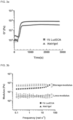
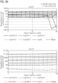
- FIGS. 3a and 3b The analytical results are shown in FIGS. 3a and 3b .
- Matrigel and the neutralized lung dECM hydrogels exhibit thermal response behavior, show a solution-like phase at a temperature below 15°C, and transform into a gel-like phase (crosslinked gel) when the temperature is increased to a physiological temperature (37°C).
- Matrigel and the lung dECM showed a drastic increase in the complex modulus in a similar pattern at 37°C.
- the neutralized lung dECM showed a fluid-to-gel transition for 50 minutes ( FIG. 3a ).
- the lung dECM scaffold is adjusted to pH 7.
- the lung dECM gelated at 37°C for 30 minutes is immersed in 4% paraformaldehyde to fix. Samples are washed 3 times with 1X PBS for 10 minutes each and 2 more times with running D.W. Ethanol at 30%, 50%, 700, 90% and 100% is prepared and treated according to the sequence for 10 minutes each.
- HMDS Hexamethyldisilazane
- the lung dECM scaffold is prepared by adjusting the pH to 7. After adding the RIPA buffer to the neutralized dECM, which is kept in ice and subject to a reaction for about 30 minutes. The mixture is mixed well by using a vortex mixer once every 5 minutes. The total protein in the dECM is quantified by using the BCA protein assay kit. The standard is prepared by diluting a 2 mg/mL stock of bovine serum albumin (BSA) with ddH 2 O. The lung dECM dissociated in the RIPA buffer is used as a stock solution, and samples are prepared by diluting by factors of 2/5/10/50/100/200/500/1000 times with ddH 2 O.
- BSA bovine serum albumin
- the standard and the samples are loaded in triplicate into a 96-well plate at 20 ⁇ L/well each.
- the blank is the RIPA buffer.
- the required amount of the BCA solution is calculated, and Solution A and Solution B are mixed at the ratio of 49:1. It should be ensured that the tube is wrapped with aluminum foil, and Solution A and Solution B solutions are not mixed in advance but are mixed after loading the samples.
- the mixed solution is poured into the reservoir and loaded at 160 ⁇ L/well each by using a multi-channel pipettor.
- the entire 96-well plate is wrapped with aluminum foil and subject to a reaction in an incubator at 37°C for 30 minutes.
- the absorbance is measured at a 540 nm wavelength with a microplate reader.
- a standard curve is drawn and the total protein in the samples is calculated.
- the dilution factor was determined within the recommended protein loading range (load 50 to 500 ⁇ g of total protein) provided in the cytokine array protocol.
- the diluted solution was prepared by diluting the lung dECM stock solution to a concentration of 50 ⁇ g/mL so that the total protein became 50 ⁇ g.
- the analysis method was carried out in the same manner as the manufacturer's protocol (RayBio ® C-Series Porcine cytokine array 1).
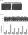
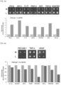
- FIGS. 4a to 4e show the results of analyzing the residual cytokines in the lung dECM.
- SCF stem cell growth factor
- AGN-1 angiopoietin 1
- the lung dECM contains a large amount of cytokines produced and secreted by immune cells, vascular cells, and other stromal cells in actual tissues, and thus can mimic the tissue microenvironment more similarly.
- the lung dECM contains cytokines related to angiogenesis and cell proliferation in a large amount, and thus is more effective in the organoid culture than Matrigel.
- Tissue staining (Hematoxylin & Eosin, Masson's trichrome stain) of the lung dECM prepared in Example 1 was performed.
- the histological (H&E, MT staining) analysis confirmed that the cells were removed from the tissue after the decellularization process, while the extracellular matrix component mimicking the biochemical microenvironment was maintained.
- the residual protein of the lung dECM prepared in Example 1 was analyzed by using LC-MS/MS (Liquid Chromatography-Mass spectrometry).
- glycoproteins have sugar chains in the middle of the sequence and the sugar chains may cause problems in the mass value later, the sugar portions are removed and the glycoprotein portions are changed into the forms of normal peptides.
- the database of the individuals is imported and analyzed by using the sequence library.
- the experimental results confirmed that the lung dECM contained a large amount of collagen VI, which is related to cell survival and functional improvement. It was also confirmed that laminin, which is an important factor to the organoid culture, was present in a large amount and in addition to laminin, fibronecitn, which is important for cell adhesion, growth and differentiation, was present in a large amount. Additionally, the results confirmed that tissue-specific extracellular matrix proteins remained even after the decellularization process so that the decellularization-based lung dECM scaffold developed in the present invention can help the organoid growth and improve the organoid differentiation and functions.
- hydrogel formulation conditions similar to the gelation tendency of a scaffold (Matrigel) that is often used for organoid culture were screened. The verification was performed by using the dECM alone or by using a mixture of Matrigel and the dECM.
- the lung dECM was tested based on the concentrations of 20, 1.5%, and 1%, and subsequent experiments were conducted only based on the concentrations of 2% and 1%.
- the overall details are similar to "Example 4: Rheological Characterization of Lung dECM".
- the lung dECM scaffold is prepared by adjusting the pH thereof to 7-7.5.
- the measuring plate PP25 is mounted on the rheometer to perform the evaluation.
- the amount of dECM loading is 300 ⁇ L or more, and the running GAP is set to 300 um for the lung.
- the dECM that has escaped from the periphery of the measuring plate is removed as much as possible with a scraper.
- the sol-gel transition, complex viscosity, and frequency sweep of the dECM scaffold are measured by creating an experiment under the following conditions in the installed software program (RheoCompass 1.19).
- the frequency-dependent storage modulus (G') and loss modulus (G") of the completely gelated dECM scaffold is measured by changing the frequency from 500 rad/s to 1 rad/s at 2% strain and at 37°C.
- FIG. 8a gelation kinetics
- FIG. 8b frequency sweep
- Matrigel and neutralized lung dECM hydrogels exhibit thermal reaction behavior and a solution-like phase at a temperature of lower than 5°C. As the temperature increases to 37°C, the same as in the body, it is transformed into a gel-like state.
- the sol-gel transition and frequency sweep were analyzed by a rheometer with Matrigel as a control, with the lung dECM alone, and with the mixed gels prepared by mixing the same with Matrigel at the ratios described in Example 9 (1), and the results observed from each organ are described below.
- FIGS. 8a and 8b show the results of analyzing the dECM alone and the mixtures prepared by mixing the same with Matrigel at the ratios described above, after preparing the lung dECM at 2% and 1% according to one embodiment.
- gelation was observed in the control (Matrigel) and the mixtures prepared mixing at the dECM-to-Matrigel ratios of 0.5:1, 1:1 and 2:1 among the ratios described in Example 9 (1).
- gelation was all observed in the control (Matrigel) and the experimental groups described above.
- the neutralized lung dECM and the mixture prepared at the selected ratio based on the results described above among the mixtures described in Example 8 were subject to gelation for 30 minutes ( FIG. 8a ), and it was confirmed that after the hydrogel was stabilized, they had physical properties that are similar to those of Matrigel under a dynamic external force in the range from a high shear frequency to a low shear frequency (500-1 rad/s) ( FIG. 8b ).
- gelation was performed at 37°C for 40 minutes or longer.
- organoid culture is performed based on the dECM.
- a medium is prepared by mixing Glutamax, HEPES, and Anti-anti.
- growth factors such as Rspondin-1, B27, nicotinamide, N-acetylcysteine, SB202190, A83-01, mNoggin, hFGF10, mFGF7, and Primocin are added.
- the organoid that is being cultured in Matrigel is recovered.
- the specific process is described below. 1
- the Matrigel in which the lung organoid is growing is physically destroyed through pipetting.
- the organoid is disassembled with a syringe and a needle.
- Pipetting is additionally performed by using a 10 ml tip.
- Centrifugation is performed at 4°C and 200xg for 5 minutes.
- the supernatant is removed to obtain an organoid pellet.
- the organoid pellet of (5) is washed by pipetting with a medium prepared based on Advanced DMEM/F12 by mixing Glutamax, HEPES, and Anti-anti.
- Centrifugation is performed at 4°C and 200xg for 5 minutes. 8
- the supernatant is removed to obtain an organoid pellet.
- the amount required for embedding is calculated and the organoid pellet is mixed with the dECM scaffold made in Example 3 (a mixture prepared according to the mixing ratio of Matrigel and dECM) .
- the dECM mixture of 40-50 ul is dropped into a 24-well plate (or culture plate).
- 11 Incubation is performed at 37°C for 30-40 minutes.
- 12 The organoid culture medium containing Y-27632 is added.
- the medium is replaced by an organoid culture medium that does not contain Y-27632 once every 2-3 days. 14 After 7-14 days following the subculture, depending on the state of the organoid, subculture is performed for long-term culture (more than 30 days).
- live cells are confirmed by a Live/Dead assay to confirm that the dECM scaffold is nontoxic to the organoid culture.
- the toxicity of the dECM scaffold was confirmed by staining and observing the live cells and dead cells of the organoid cultured in the dECM by using the Invitrogen Live/Dead ® Viability/Cytotoxicity Kit.
- the dECM scaffold in which organoid is being cultured is washed with pre-warmed DPBS (1X) 3 times for at 37°C for 5 minutes each. Afterwards, the method was performed in the same manner as the manufacturer's protocol.
- the proliferation potential of the organoid in the dECM is evaluated with the CCK-8 assay, which confirms whether the organoid cultured on the dECM scaffold proliferates based on the respiration rate of the cells.
- the proliferation of an organoid (and cell) being cultured in the dECM was confirmed by measuring the respiration volume of the organoid cultured in the dECM by using DOJINDO's CCK-8 assay kit.
- the dECM scaffold in which the organoid is being cultured is washed with pre-warmed DPBS (1X) 3 times at 37°C for 5 minutes each. Afterwards, the method was performed in the same manner as the manufacturer's protocol. In the analysis of the results, the occurrence of the proliferation was confirmed by comparing the absorbance values of the samples for each period.
- the tumor organoid cultured on the dECM scaffold is effective with the tumor organoid cultured on the scaffold which is often used in the organoid culture (Matrigel).
- the scaffold and Matrigel on which the human-derived lung tumor organoid is growing is obtained by physical destroying the same through pipetting. Centrifugation is performed by table-top centrifugation or by using a microcentrifuge at 13,000 rpm. The supernatant is removed, leaving only the human-derived lung tumor organoid pellet.
- the genomic DNA was extracted by using HiGene TM Genomic DNA prep Kit. The following procedures were carried out in the same manner as described in the animal tissue manual of the manufacturer's protocol.
- sequencing of the genomic DNA extracted in (1) is performed.
- the sequencing was performed by direct sequencing, and was commissioned by Macrogen.
- FIG. 11 shows the experimental results of confirming by the direct sequencing whether, among the markers for distinguishing tumors, the mutations and positions thereof are retained the same as those of human-derived tumors, in order to verify the effectiveness of the human-derived tumor organoid cultured in the lung dECM-based hydrogels.
- a tumor organoid having the Exon 19 deletion mutation which is a typical mutation of lung cancer
- tumor organoids having the KRAS mutation were cultured in the lung 1% dECM, wherein the tumor organoid having the Exon 19 deletion mutation was cultured for 8 days and the tumor organoids having the KRAS mutation were each cultured for 20 days and 28 days, and the results showed that each of the mutations and the position thereof were retained.
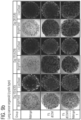
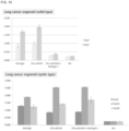
- FIG. 12a shows the results of the microscopic observation of the long-term culture of the solid type for 48 days among the human-derived lung tumor organoids based on the 1% lung dECM prepared according to an Example of the present invention under the conditions of using the dECM alone and mixing the same with Matrigel according to the ratios described above, wherein FIG. 12b shows the results of the long-term culture of the cystic type for 28 days and FIG. 12a shows the analytical results of the cytotoxicity of the organoid on Day 17, Day 30 and Day 44 during the culture of the solid type for 48 days. The results confirmed that the scaffold of the present invention maintains excellent properties in the long-term culture of the tumor organoids.
- This Example describes the experimental method in which the stirring time and speed were varied in the S4) lysing according to the present invention, and the results obtained therefrom.
- Example 2 The experimental method was carried out based on the methods described in Example 2 and Examples 10 to 12 of the present invention.
- a decellularization composition was prepared according to the method of Example 2 of the present invention.
- the pulverized dECM prepared in Example 2 was put into a 50 mL conical tube, and 0.5 M acetic acid was added together with pepsin and lysed at a stirring speed of 330 rpm for 24 hours, 72 hours, 96 hours and 120 hours, respectively.
- the mixture was lysed at a stirring speed of 80 rpm, 150 rpm, 330 rpm, 500 rpm, 800 rpm, and 1000 rpm for 96 hours.
- the lysed dECM was filtered by using a cell strainer to filter out the lung dECM powder that was not lysed.
- 10X PBS and 10 N sodium hydroxide (NaOH) were added, wherein the PBS and NaOH were stored at a refrigerated temperature before use and used in a cold state for the pH adjustment.
- the lung organoid was cultured according to the method of Example 10 of the present invention.
- the lung organoid was obtained with a 1000 ul tip and a pipette according to the method of Example 10 of the present invention, and collected in a conical tube. After that, the organoid was primarily disassembled by using a syringe and a needle, and then pipetting was additionally performed with a 10 ml tip and a pipette aid. The disassembled organoid was obtained by subjecting the same to centrifugation and removing the supernatant.
- the number of the organoids required for this experiment and the required amount of the dECM were calculated by calculating an appropriate sub-culture ratio depending on the state of the organoid (40 ul dECM required per dome).
- the calculated amount of the organoid pellet was mixed with each of the dECMs prepared by applying different stirring time (24 hours, 72 hours, 96 hours, 120 hours) and stirring speed (80 rpm, 150 rpm, 330 rpm, 500 rpm, 800 rpm, 1000 rpm), and the resulting mixtures were encapsulated.
- the dECM mixed with organoids was dropped in an amount of 40 ul onto a culture plate by using a pipette to form a dome for organoid culture, which was the subject to gelation at 37°C for 30-40 minutes. After that, an organoid culture medium was added, and the added medium was replaced with new one once every 2-3 days. Four days after the encapsulation of the organoid, the culture state of the organoid was observed by the method of Examples 11 and 12 according to the present invention.
- FIGS. 13 to 16 The physical properties of the lung dECM and the results of the organoid culture for each stirring time are shown in FIGS. 13 to 16 .
- FIG. 13 is a graph showing the modulus value of the lung dECM when the stirring time was changed in the S4) lysing according to an Example of the present invention
- FIG. 14 is a graph showing the gelation characteristic (gelation kinetic) value of the lung dECM for each stirring time
- FIG. 15 is a photograph showing the lung organoid culture state for each stirring time
- FIG. 16 is a graph showing the proliferation rate of the lung organoid culture for each stirring time.
- the modulus value representing the physical properties for each stirring time, was in the order of 24 h ⁇ 72 h ⁇ 96 h ⁇ 120 h. Therefore, it was confirmed the physical properties were best when the stirring time was 72-96 hours ( FIGS. 13 and 14 ).
- FIG. 17 is a graph showing the modulus value of the lung dECM when the stirring speed was changed in the S4) lysing according to an Example of the present invention
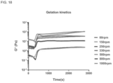
- FIG. 18 is a graph showing the gelation characteristic (gelation kinetic) value of the lung dECM for each stirring speed
- FIG. 19 is a photograph showing the lung organoid culture state for each stirring speed
- FIG. 20 is a graph showing the proliferation rate of the lung organoid culture for each stirring speed.
- the modulus value representing the physical properties for each stirring speed, was in the order of 80 ⁇ 150 ⁇ 330 ⁇ 500 > 800 > 1000 (80 ⁇ 1000 ⁇ 150 ⁇ 800 ⁇ 330 ⁇ 500) rpm, and it was confirmed the physical properties were best when the stirring speed was 330-500 ( FIGS. 17 and 18 ).
- the culture efficiency was in the order of 80 ⁇ 250 « 330 ⁇ 500 » 1000 (80 ⁇ 250 ⁇ 1000 « 330 ⁇ 500), and dome formation was impossible at 80 rpm.
- the dECM stirred at 80, 250, and 1000 rpm, a lot of organoid deaths occurred and no live organoid was observed, indicating that the physical properties of the organoid culture are important, and appropriate conditions could be set accordingly ( FIGS. 19 and 20 ) .
- the present inventors learned that the conditions compatible with the organoid culture are 330-500 rpm in terms of the stirring speed.
- This Example describes the experimental method in which the implementation conditions and temperature were varied in the S5) adjusting pH according to the present invention, and the results obtained therefrom.
- the experimental method was carried out based on the method described in Example 2 of the present invention.
- the pH adjustment process was performed while the tube containing the lysate was kept in ice.
- FIG. 21 is a photograph for explaining the process of performing the S5) adjusting pH on ice according to an Example of the present invention.
- the pH adjustment process was performed in a state in which the container containing the decellularization composition according to the present invention was completely submerged in ice.
- the pH adjustment process was performed while the container containing the decellularization composition was placed at room temperature (about 26°C).
- the physical properties of the lung dECM lysate for each condition of the pH adjustment process are as shown in FIGS. 22 to 25 .
- FIGS. 22 to 24 are photographs showing the gelation state of the composition for which the S5) adjusting pH was performed on ice according to an Example of the present invention
- FIG. 25 is a graph showing the modulus value of the composition for which the S5) adjusting pH was performed on ice according to an Example of the present invention.
- the decellularization composition was gelated at 37°C for 30 minutes in the same manner as the organoid culture condition, and then the hydrogels made under each pH adjustment condition were observed.
- the results also showed that the separation of the water layer and the gelated decellularization composition occurred more significantly in the hydrogel of which pH was adjusted at room temperature (RT), confirming that the hydrogel fails to maintain the gel stably and so is incompatible with the organoid culture (see FIG. 24 ).
- the decellularization composition of which pH was adjusted at room temperature was difficult to perform pipetting when encapsulating the organoid, and the dome was not maintained but was broken during the culturing of the encapsulated organoid or the culture itself was difficult because the organoid was not encapsulated homogeneously ( FIG. 22 , 23 and 24 ).
- the decellularization composition of which pH was adjusted on ice according to the present invention was able to form a homogeneous gel during the gelation process, but the decellularization composition of which pH was adjusted at room temperature (RT) was partially gelated and aggregated, and the dome was not maintained but was broken, exhibiting low physical properties (complex modulus (G*)) (see FIG. 25 ).
- FIG. 26 is a graph showing the gelation kinetic value of the composition for which the S5) adjusting pH was performed on ice according to an Example of the present invention.
- the decellularization composition of which pH was adjusted on ice according to the present invention showed a clear increase in the complex modulus (G*) value during temperature change (4-37°C), whereas the decellularization composition of which pH was adjusted at room temperature (RT) already showed a high initial G* value and the change of the value was insignificant.
- the initial state of the dECM should be a sol state (storage modulus ⁇ loss modulus) as the temperature changes.
- the initial state of the dECM since the initial state of the dECM was already a gel state (storage modulus > loss modulus), not only the pipetting in the encapsulation during the organoid culture but also the encapsulation for the organoid culture could not be carried out, indicating that the decellularization composition was not compatible with the organoid culture (see FIG. 27 ).
Landscapes
- Health & Medical Sciences (AREA)
- Life Sciences & Earth Sciences (AREA)
- Engineering & Computer Science (AREA)
- Biomedical Technology (AREA)
- Chemical & Material Sciences (AREA)
- Zoology (AREA)
- Wood Science & Technology (AREA)
- Organic Chemistry (AREA)
- Genetics & Genomics (AREA)
- Bioinformatics & Cheminformatics (AREA)
- Biotechnology (AREA)
- General Health & Medical Sciences (AREA)
- General Engineering & Computer Science (AREA)
- Biochemistry (AREA)
- Microbiology (AREA)
- Cell Biology (AREA)
- Pulmonology (AREA)
- Molecular Biology (AREA)
- Chemical Kinetics & Catalysis (AREA)
- Botany (AREA)
- Veterinary Medicine (AREA)
- Public Health (AREA)
- Animal Behavior & Ethology (AREA)
- Epidemiology (AREA)
- Transplantation (AREA)
- Oral & Maxillofacial Surgery (AREA)
- Medicinal Chemistry (AREA)
- Dermatology (AREA)
- Urology & Nephrology (AREA)
- Biophysics (AREA)
- General Chemical & Material Sciences (AREA)
- Micro-Organisms Or Cultivation Processes Thereof (AREA)
Applications Claiming Priority (3)
| Application Number | Priority Date | Filing Date | Title |
|---|---|---|---|
| KR20200080518 | 2020-06-30 | ||
| KR1020210056942A KR102282070B1 (ko) | 2020-06-30 | 2021-04-30 | 폐 오가노이드 배양용 조성물의 제조 방법, 그 조성물 및 이를 이용한 오가노이드 배양 방법 |
| PCT/KR2021/007930 WO2022005098A1 (ko) | 2020-06-30 | 2021-06-24 | 폐 오가노이드 배양용 조성물의 제조 방법, 그 조성물 및 이를 이용한 오가노이드 배양 방법 |
Publications (2)
| Publication Number | Publication Date |
|---|---|
| EP4163363A1 true EP4163363A1 (de) | 2023-04-12 |
| EP4163363A4 EP4163363A4 (de) | 2024-07-17 |
Family
ID=77125330
Family Applications (1)
| Application Number | Title | Priority Date | Filing Date |
|---|---|---|---|
| EP21832927.4A Pending EP4163363A4 (de) | 2020-06-30 | 2021-06-24 | Verfahren zur herstellung einer zusammensetzung zur kultivierung von lungenorganoiden, zusammensetzung dafür und organoidkulturverfahren damit |
Country Status (4)
| Country | Link |
|---|---|
| US (1) | US20230287354A1 (de) |
| EP (1) | EP4163363A4 (de) |
| KR (1) | KR102282070B1 (de) |
| WO (1) | WO2022005098A1 (de) |
Families Citing this family (4)
| Publication number | Priority date | Publication date | Assignee | Title |
|---|---|---|---|---|
| CN114457019A (zh) * | 2022-02-17 | 2022-05-10 | 安徽骆华生物科技有限公司 | 一种基于多孔微球构建肺癌类器官模型及其培养方法与用途 |
| CN114686421B (zh) * | 2022-04-18 | 2023-10-10 | 中山大学 | 一种肺组织脱细胞外基质微载体的制备方法及其应用 |
| KR102952410B1 (ko) * | 2023-01-20 | 2026-04-14 | 에스지바이오사이언스 주식회사 | 육종 튜머로이드 제조 방법 및 그의 용도 |
| KR102942588B1 (ko) | 2025-02-20 | 2026-03-20 | 재단법인대구경북과학기술원 | 후두 오가노이드의 제조방법 및 이의 용도 |
Family Cites Families (12)
| Publication number | Priority date | Publication date | Assignee | Title |
|---|---|---|---|---|
| AU2009298560B2 (en) * | 2008-09-30 | 2014-05-01 | The Regents Of The University Of California | Compositions and methods for tissue repair with extracellular matrices |
| CN102282252B (zh) * | 2008-11-19 | 2017-07-04 | 人类起源公司 | 羊膜来源的贴壁细胞 |
| KR101822662B1 (ko) * | 2009-02-04 | 2018-01-26 | 예일 유니버시티 | 폐 조직 엔지니어링 |
| GB201421092D0 (en) * | 2014-11-27 | 2015-01-14 | Koninklijke Nederlandse Akademie Van Wetenschappen | Culture medium |
| WO2017008035A1 (en) * | 2015-07-08 | 2017-01-12 | The Trustees Of The University Of Pennesylvania | Decellularized organ-derived tissue engineering scaffolds |
| KR101860798B1 (ko) | 2015-12-29 | 2018-05-25 | 포항공과대학교 산학협력단 | 뇌 조직 탈세포 방법 및 뇌 조직 배양 방법 |
| US20190201586A1 (en) * | 2016-06-24 | 2019-07-04 | Washington State University | Three-Dimensional Tissue Matrix Scaffold System |
| US11918702B2 (en) * | 2017-03-24 | 2024-03-05 | The Trustees Of Columbia University In The City Of New York | Generation of lung bud organoids with branching structures and uses thereof for lung disease modeling |
| KR102015368B1 (ko) | 2018-01-30 | 2019-08-28 | 재단법인대구경북과학기술원 | 오일을 이용한 조직재생용 탈세포 지지체의 제조방법 및 조직 탈세포 처리 조성물 |
| EP3587564B1 (de) | 2018-06-21 | 2022-10-26 | Industry-Academic Cooperation Foundation, Yonsei University | Zusammensetzung zur kultivierung von gehirnorganoiden auf basis von dezellularisierter hirnmatrix und verfahren zur herstellung davon |
| KR102103783B1 (ko) * | 2018-06-21 | 2020-04-23 | 한국원자력의학원 | 침샘 조직의 탈세포화 세포외기질-하이드로겔 및 침샘 오거나이드를 제조하는 방법 |
| KR102102153B1 (ko) * | 2018-08-30 | 2020-04-27 | 연세대학교 산학협력단 | 탈세포 조직 기반 세포 봉입 및 배양용 비드, 및 이의 용도 |
-
2021
- 2021-04-30 KR KR1020210056942A patent/KR102282070B1/ko active Active
- 2021-06-21 US US18/012,958 patent/US20230287354A1/en active Pending
- 2021-06-24 WO PCT/KR2021/007930 patent/WO2022005098A1/ko not_active Ceased
- 2021-06-24 EP EP21832927.4A patent/EP4163363A4/de active Pending
Also Published As
| Publication number | Publication date |
|---|---|
| EP4163363A4 (de) | 2024-07-17 |
| KR102282070B1 (ko) | 2021-07-27 |
| WO2022005098A1 (ko) | 2022-01-06 |
| US20230287354A1 (en) | 2023-09-14 |
Similar Documents
| Publication | Publication Date | Title |
|---|---|---|
| US20230287354A1 (en) | Method for preparing composition for culturing lung organoids, composition therefor, and organoid culture method using same | |
| Schwarz et al. | 3D printing and characterization of human nasoseptal chondrocytes laden dual crosslinked oxidized alginate-gelatin hydrogels for cartilage repair approaches | |
| EP4155384A1 (de) | Verfahren zur herstellung einer zusammensetzung zur kultivierung pankreatischer organoide, zusammensetzung dafür und verfahren zur kultivierung von organoiden unter verwendung davon | |
| Campo et al. | Tissue-specific decellularized endometrial substratum mimicking different physiological conditions influences in vitro embryo development in a rabbit model | |
| KR101954953B1 (ko) | 3차원 프린팅용 바이오 잉크 조성물 및 이의 제조방법 | |
| Sobreiro-Almeida et al. | Decellularized kidney extracellular matrix bioinks recapitulate renal 3D microenvironment in vitro | |
| US20190201586A1 (en) | Three-Dimensional Tissue Matrix Scaffold System | |
| US12472285B2 (en) | Eggshell particle containing hydrogels and prepolymer compositions for biomedical applications | |
| Asgari et al. | Optimization of decellularized human placental macroporous scaffolds for spermatogonial stem cells homing | |
| Liu et al. | Egg white improves the biological properties of an alginate-methylcellulose bioink for 3D bioprinting of volumetric bone constructs | |
| Song et al. | Development of novel apoptosis-assisted lung tissue decellularization methods | |
| JP6871274B2 (ja) | 汗腺の三次元細胞培養モデルを含有するインビトロ完全皮膚モデル | |
| Kara Özenler et al. | 3D bioprinting of mouse pre-osteoblasts and human MSCs using bioinks consisting of gelatin and decellularized bone particles | |
| US20220389375A1 (en) | Extracellular Matrix Gels, and Organoid Cultures Comprising the Same | |
| EP4261273A1 (de) | Gerüst aus dezellularisiertem fettgewebe zur kultivierung von organoiden und verfahren zur herstellung davon | |
| CN116867892A (zh) | 三维组织体的制造方法和脂肪来源干细胞的分化促进方法 | |
| EP4098740A1 (de) | Gerüst aus dezellularisierter extrazellulärer herzmatrix zur kultivierung und transplantation von herzorganoiden sowie verfahren zu seiner herstellung | |
| Uslu et al. | Pectin hydrogels crosslinked via peptide nanofibers for designing cell-instructive dynamic microenvironment | |
| EP4098739A1 (de) | Aus dezellularisiertem organgewebe stammendes gerüst für die organische organoidkultur und -transplantation sowie herstellungsverfahren dafür | |
| Özdinç et al. | Development and characterization of decellularized lung extracellular matrix hydrogels | |
| Nie et al. | Self-organizing gelatin–polycaprplactone materials with good fluid transmission can promote full-thickness skin regeneration | |
| CN110575567A (zh) | 一种支架复合材料及其制备方法、应用 | |
| Jiang et al. | Effect of in-situ synthesized nano-hydroxyapatite/collagen composite hydrogel on osteoblasts growth in vitro | |
| Silva et al. | Comparable decellularization of fetal and adult cardiac tissue explants as 3D-like platforms for in vitro studies. | |
| Palierse et al. | Tuning the mechanical properties and printability of viscoelastic skin-derived hydrogels for 3D cell culture |
Legal Events
| Date | Code | Title | Description |
|---|---|---|---|
| STAA | Information on the status of an ep patent application or granted ep patent |
Free format text: STATUS: THE INTERNATIONAL PUBLICATION HAS BEEN MADE |
|
| PUAI | Public reference made under article 153(3) epc to a published international application that has entered the european phase |
Free format text: ORIGINAL CODE: 0009012 |
|
| STAA | Information on the status of an ep patent application or granted ep patent |
Free format text: STATUS: REQUEST FOR EXAMINATION WAS MADE |
|
| 17P | Request for examination filed |
Effective date: 20221216 |
|
| AK | Designated contracting states |
Kind code of ref document: A1 Designated state(s): AL AT BE BG CH CY CZ DE DK EE ES FI FR GB GR HR HU IE IS IT LI LT LU LV MC MK MT NL NO PL PT RO RS SE SI SK SM TR |
|
| DAV | Request for validation of the european patent (deleted) | ||
| DAX | Request for extension of the european patent (deleted) | ||
| A4 | Supplementary search report drawn up and despatched |
Effective date: 20240614 |
|
| RIC1 | Information provided on ipc code assigned before grant |
Ipc: A61L 27/36 20060101ALI20240610BHEP Ipc: A61L 27/38 20060101ALI20240610BHEP Ipc: C12N 5/071 20100101ALI20240610BHEP Ipc: C12N 5/00 20060101AFI20240610BHEP |