EP4166084A1 - Système de détection et d'intervention basé sur le sommeil - Google Patents
Système de détection et d'intervention basé sur le sommeil Download PDFInfo
- Publication number
- EP4166084A1 EP4166084A1 EP21202960.7A EP21202960A EP4166084A1 EP 4166084 A1 EP4166084 A1 EP 4166084A1 EP 21202960 A EP21202960 A EP 21202960A EP 4166084 A1 EP4166084 A1 EP 4166084A1
- Authority
- EP
- European Patent Office
- Prior art keywords
- sleep
- template
- intervention system
- slow wave
- patient
- Prior art date
- Legal status (The legal status is an assumption and is not a legal conclusion. Google has not performed a legal analysis and makes no representation as to the accuracy of the status listed.)
- Withdrawn
Links
Images


Classifications
-
- A—HUMAN NECESSITIES
- A61—MEDICAL OR VETERINARY SCIENCE; HYGIENE
- A61B—DIAGNOSIS; SURGERY; IDENTIFICATION
- A61B5/00—Measuring for diagnostic purposes; Identification of persons
- A61B5/48—Other medical applications
- A61B5/4806—Sleep evaluation
- A61B5/4812—Detecting sleep stages or cycles
-
- A—HUMAN NECESSITIES
- A61—MEDICAL OR VETERINARY SCIENCE; HYGIENE
- A61M—DEVICES FOR INTRODUCING MEDIA INTO, OR ONTO, THE BODY; DEVICES FOR TRANSDUCING BODY MEDIA OR FOR TAKING MEDIA FROM THE BODY; DEVICES FOR PRODUCING OR ENDING SLEEP OR STUPOR
- A61M21/00—Other devices or methods to cause a change in the state of consciousness; Devices for producing or ending sleep by mechanical, optical, or acoustical means, e.g. for hypnosis
- A61M21/02—Other devices or methods to cause a change in the state of consciousness; Devices for producing or ending sleep by mechanical, optical, or acoustical means, e.g. for hypnosis for inducing sleep or relaxation, e.g. by direct nerve stimulation, hypnosis, analgesia
-
- A—HUMAN NECESSITIES
- A61—MEDICAL OR VETERINARY SCIENCE; HYGIENE
- A61B—DIAGNOSIS; SURGERY; IDENTIFICATION
- A61B5/00—Measuring for diagnostic purposes; Identification of persons
- A61B5/24—Detecting, measuring or recording bioelectric or biomagnetic signals of the body or parts thereof
- A61B5/316—Modalities, i.e. specific diagnostic methods
- A61B5/369—Electroencephalography [EEG]
- A61B5/372—Analysis of electroencephalograms
-
- A—HUMAN NECESSITIES
- A61—MEDICAL OR VETERINARY SCIENCE; HYGIENE
- A61B—DIAGNOSIS; SURGERY; IDENTIFICATION
- A61B5/00—Measuring for diagnostic purposes; Identification of persons
- A61B5/72—Signal processing specially adapted for physiological signals or for diagnostic purposes
- A61B5/7235—Details of waveform analysis
- A61B5/7246—Details of waveform analysis using correlation, e.g. template matching or determination of similarity
-
- A—HUMAN NECESSITIES
- A61—MEDICAL OR VETERINARY SCIENCE; HYGIENE
- A61B—DIAGNOSIS; SURGERY; IDENTIFICATION
- A61B5/00—Measuring for diagnostic purposes; Identification of persons
- A61B5/72—Signal processing specially adapted for physiological signals or for diagnostic purposes
- A61B5/7271—Specific aspects of physiological measurement analysis
- A61B5/7282—Event detection, e.g. detecting unique waveforms indicative of a medical condition
-
- G—PHYSICS
- G16—INFORMATION AND COMMUNICATION TECHNOLOGY [ICT] SPECIALLY ADAPTED FOR SPECIFIC APPLICATION FIELDS
- G16H—HEALTHCARE INFORMATICS, i.e. INFORMATION AND COMMUNICATION TECHNOLOGY [ICT] SPECIALLY ADAPTED FOR THE HANDLING OR PROCESSING OF MEDICAL OR HEALTHCARE DATA
- G16H20/00—ICT specially adapted for therapies or health-improving plans, e.g. for handling prescriptions, for steering therapy or for monitoring patient compliance
- G16H20/70—ICT specially adapted for therapies or health-improving plans, e.g. for handling prescriptions, for steering therapy or for monitoring patient compliance relating to mental therapies, e.g. psychological therapy or autogenous training
-
- A—HUMAN NECESSITIES
- A61—MEDICAL OR VETERINARY SCIENCE; HYGIENE
- A61B—DIAGNOSIS; SURGERY; IDENTIFICATION
- A61B5/00—Measuring for diagnostic purposes; Identification of persons
- A61B5/72—Signal processing specially adapted for physiological signals or for diagnostic purposes
- A61B5/7203—Signal processing specially adapted for physiological signals or for diagnostic purposes for noise prevention, reduction or removal
-
- A—HUMAN NECESSITIES
- A61—MEDICAL OR VETERINARY SCIENCE; HYGIENE
- A61M—DEVICES FOR INTRODUCING MEDIA INTO, OR ONTO, THE BODY; DEVICES FOR TRANSDUCING BODY MEDIA OR FOR TAKING MEDIA FROM THE BODY; DEVICES FOR PRODUCING OR ENDING SLEEP OR STUPOR
- A61M21/00—Other devices or methods to cause a change in the state of consciousness; Devices for producing or ending sleep by mechanical, optical, or acoustical means, e.g. for hypnosis
- A61M2021/0005—Other devices or methods to cause a change in the state of consciousness; Devices for producing or ending sleep by mechanical, optical, or acoustical means, e.g. for hypnosis by the use of a particular sense, or stimulus
- A61M2021/0027—Other devices or methods to cause a change in the state of consciousness; Devices for producing or ending sleep by mechanical, optical, or acoustical means, e.g. for hypnosis by the use of a particular sense, or stimulus by the hearing sense
-
- A—HUMAN NECESSITIES
- A61—MEDICAL OR VETERINARY SCIENCE; HYGIENE
- A61M—DEVICES FOR INTRODUCING MEDIA INTO, OR ONTO, THE BODY; DEVICES FOR TRANSDUCING BODY MEDIA OR FOR TAKING MEDIA FROM THE BODY; DEVICES FOR PRODUCING OR ENDING SLEEP OR STUPOR
- A61M2205/00—General characteristics of the apparatus
- A61M2205/33—Controlling, regulating or measuring
- A61M2205/3317—Electromagnetic, inductive or dielectric measuring means
-
- A—HUMAN NECESSITIES
- A61—MEDICAL OR VETERINARY SCIENCE; HYGIENE
- A61M—DEVICES FOR INTRODUCING MEDIA INTO, OR ONTO, THE BODY; DEVICES FOR TRANSDUCING BODY MEDIA OR FOR TAKING MEDIA FROM THE BODY; DEVICES FOR PRODUCING OR ENDING SLEEP OR STUPOR
- A61M2209/00—Ancillary equipment
- A61M2209/08—Supports for equipment
- A61M2209/088—Supports for equipment on the body
-
- A—HUMAN NECESSITIES
- A61—MEDICAL OR VETERINARY SCIENCE; HYGIENE
- A61M—DEVICES FOR INTRODUCING MEDIA INTO, OR ONTO, THE BODY; DEVICES FOR TRANSDUCING BODY MEDIA OR FOR TAKING MEDIA FROM THE BODY; DEVICES FOR PRODUCING OR ENDING SLEEP OR STUPOR
- A61M2230/00—Measuring parameters of the user
- A61M2230/08—Other bio-electrical signals
- A61M2230/10—Electroencephalographic signals
Definitions
- the present invention relates to a sleep-based detection and intervention system for the treatment of major depressive disorder (MDD).
- MDD major depressive disorder
- MDD is listed by the World Health Organization as the leading cause for illness-related reduction of quality of life worldwide.
- Therapeutic sleep deprivation has been known since the 1970s to exert a strong and rapid antidepressant effect on individuals with MDD.
- the effect is short-lived, with most patients relapsing following a period of subsequent sleep.
- the burden on the patient of staying awake for an entire night is a significant limitation of the treatment.
- the disclosure of WO 2018/104459 A1 pertains to determining the timing of stimulation provided to a subject during sleep.
- the system receives a raw signal carrying information related to slow wave activity; buffers a portion of the raw signal; determines a timing of slow wave events in the buffered portion of the raw signal; filters the raw signal; determines a timing of slow wave events in the filtered raw signal; compares the timing of the slow wave events in the buffered portion of the raw signal to the timing of the slow wave events in the filtered raw signal; determines a first correction factor associated with reducing slow wave activity in the subject and a second correction factor associated with enhancing slow wave activity in the subject; and adjusts a timing of the stimulation provided to the subject during the sleep session based on the first and/or second correction factors.
- delivering auditory and/or other stimulation during slow wave sleep influences the amplitude of subsequent sleep slow waves.
- the nature of the influence depends on the timing of the stimulation with respect to the sleep slow wave phase and/or characteristics.
- the subsequent slow waves have a higher amplitude (e.g., they are enhanced).
- the enhancement effect is believed to be beneficial for increasing the restorative value of sleep.
- Delivering the stimulation in the vicinity of a slow wave negative peak may have a reducing effect (e.g., a decrease in amplitude) on the amplitude of the subsequent positive peak. This effect may be useful in situations where deprivation of slow wave sleep is sought, as for example in the framework of depression relief.
- the system comprises an electroencephalography (EEG) cap with multiple electrodes configured for recording an EEG of a patient's brain, an earphone device configured for one ear or the ears of the patient (i.e. the earphone device may be placed in or onto one or both ears of the patient) and a processing unit being connected to the EEG cap and to the earphone device.
- the processing unit includes a template-based algorithm configured for detecting slow wave sleep of the patient based on the EEG recorded by the multiple electrodes of the EEG cap, and an acoustic intervention protocol configured for providing a noise stimulation to the patient via the earphone device when slow wave sleep is detected by the template-based algorithm.
- EEG is an electrophysiological monitoring method to record electrical activity on the scalp that has been shown to represent the macroscopic activity of the surface layer of the brain underneath (brain activity). It is typically non-invasive, with the electrodes placed along the scalp. It is noted that EEG may also be used to diagnose epilepsy, which causes abnormalities in EEG readings, but also to diagnose sleep disorders, depth of anesthesia, coma, encephalopathies, and brain death.
- electrodes includes all types of measuring units and/or sensors which are capable of measuring brain activities respectively the corresponding voltage values.
- the electrode placement sites on the head respectively scalp generally refer to the lobe or area of the brain they are reading from.
- the electronic placement sites are commonly designated frontal (F), temporal (T), parietal (P), occipital (O) and central (C).
- processing unit may include a personal computer or a mobile communication device such as a laptop computer a tablet or a mobile phone or the like.
- earphone device may include earphones, headsets, headphones, earbud headphones and the like.
- template-based algorithm generally includes the means used by the processing unit primarily for the purpose of detecting slow wave sleep (SWS) of a patient.
- SWS slow wave sleep
- SWS is defined as a 30-s epoch containing at least approximately 6 slow waves (SWs) of >75 mV amplitude at frontal channels.
- the template-based algorithm is based on a template, referring to averaged slow wave sleep information of multiple test persons.
- template refers to this averaged slow wave sleep information. More precisely, the (average) slow wave sleep information used for creating the template is in the form of multiple whole head topographies from each of the multiple test persons recorded at the time-point of a frontal slow wave peak in the electroencephalogram.
- the frontal slow wave peaks are measured by one or more electrodes of the EEG cap in the frontal region of the brain, in particular in the region of the frontal lobe. If a frontal slow wave peak occurs, the voltage values of all electrodes of the EEG cap are recorded to form the whole head topography.
- the whole head topography may therefore be regarded as a sort of "screen shot" of the patient's brain on the basis of the recorded voltage values.
- the template based algorithm is configured to determine a correlation of whole head topographies of the patient with the (average) template, i.e. in order to detect slow wave sleep.
- This approach has proven to provide a significantly improved accuracy for determining the occurrence of SWS.
- the template-based algorithm is configured to use the determined correlation of the whole head topographies of the patient with the template (i.e. as predictors) in a linear model including a first regressor and a second regressor.
- the linear model is preferably a linear regression model.
- the first, template-based, regressor comprises the slow oscillation (SO) power of the template correlation, the variance of the slow oscillation (SO) power of the template correlation and a relative percentage of the slow oscillation (SO) power of the template correlation.
- SO slow oscillation
- SO variance of the slow oscillation
- SO relative percentage of the slow oscillation
- SWs slow waves
- the second regressor includes the average global gamma power of the electroencephalogram.
- the average global gamma power represents the average gamma power measured by the electrodes of the EEG cap across the whole head (all channels) in a frequency range of about 30 Hz to about 40 Hz.
- the correlation of the whole head topographies of the patient with the template is calculated over a moving window, respectively, wherein the slow oscillation power of the template correlation, the variance of SO power of the template correlation and the percentage of power in SO frequency band are preferably calculated across a 10 s moving window and wherein the average global gamma power of the encephalogram is preferably calculated across a 4 s moving window.
- the slow oscillation power of the template correlation, the variance of SO power of the template correlation and the percentage of power in SO frequency band are preferably calculated across a 10 s moving window and wherein the average global gamma power of the encephalogram is preferably calculated across a 4 s moving window.
- the template-based algorithm is configured to determine the presence of artifacts/arousals based on an (additional) artifact/arousal detection process which runs parallel to the SWS detection.
- Artifacts/arousals may for instance include blink of an eye, eye movements, swallowing, muscle twitching, pulse and poorly fitting electrodes. In this manner, any corruption of the measuring results may be avoided.
- this negates a (positive) decision of the SWS detection process (i.e. which is carried out by the general linear model with the first regressor and the second regressor).
- the artifact/arousal detection process includes the voltage range of the electroencephalogram, the amplitude and the frontal delta power (frequency of delta brain waves measured by the frontal electrodes respectively calculated on the frontal channels of the electroencephalogram) as well as the average global gamma power (frequency of gamma brain waves measured by all electrodes respectively calculated on all channels of the electroencephalogram) of the electroencephalogram.
- the frontal delta power is preferably measured in a frequency range of about 0.1 Hz and about 4 Hz. This has proven to be the most effective approach for measuring delta brain waves.
- the frontal amplitude and the frontal delta power and the average global gamma power of the electroencephalogram are determined across a 4 s moving window and checked every 0.5 s.
- the accuracy of the determination scheme may be improved.
- delta brain waves represent the slowest frequency and highest amplitude brain waves, i.e. with a frequency between approximately 0.1 Hz and approximately 4 Hz, and an amplitude of approximately 100 ⁇ V to approximately 200 ⁇ V.
- Delta brain waves are usually associated with the deep sleep stage N3 of NREM (non-rapid eye movement) sleep, also known as slow wave sleep.
- NREM sleep generally includes three stages N1, N2 and N3.
- Stage N1 of NREM sleep marks the transition from wakefulness to sleep. This stage typically lasts less than 10 minutes and is marked by a slowing of your heartbeat, breathing, and eye movements, as well as the relaxation of your muscles.
- Stage N2 of NREM sleep is a period of light sleep before you enter deeper sleep and lasts roughly 20 minutes. Stage N2 is characterized by further slowing of both the heartbeat and breathing, and the brain begins to produce bursts of rapid, rhythmic brain wave activity known as sleep spindles.
- stage N3 of NREM sleep is the deepest period of sleep and lasts for about 20 to 40 minutes. The heartbeat and breathing slow down to their lowest levels, and the muscles are relaxed such that it may be hard to awaken a person in this stage of sleep.
- gamma brain waves also called gamma rhythm
- gamma brain waves have the highest frequency among all brain waves, generally between 25 Hz and 140 Hz (wherein the region between about 30 Hz and about 40 Hz being presently of particular interest).
- Gamma brain waves are generally associated with large scale brain network activity and cognitive phenomena such as working memory, attention, and perceptual grouping.
- the acoustic intervention protocol provides for randomized noise stimulation.
- pink noise is used for the noise stimulation.
- Pink noise is a signal or process with a frequency spectrum such that the power spectral density (power per frequency interval) is inversely proportional to the frequency of the signal.
- each octave interval halving or doubling in frequency
- white noise is however also possible herein and also the use other types of tones. It is noted in this context that white noise generally comprises a heterogeneous mixture of sound waves extending over a wide frequency range.
- the randomized noise stimulation has a randomized duration of about 50 ms to about 500 ms. This duration has proven to be particularly effective.
- the randomized noise stimulation has a randomized linear increase of volume from about 40 dB to about 106 dB in 60s until slow wave sleep is no longer detected by the template-based algorithm. This form of increase is less stressful for the patient.
- the linear increase of volume is combined with random walks between about +/- 2.5 dB.
- the random walks are configured according to an Ornstein-Uhlenbeck process. This process can be considered to be a modification of the random walk in continuous time, or Wiener process, in which the properties of the process have been changed so that there is a tendency of the walk to move back towards a central location, with a greater attraction when the process is further away from the center.
- the randomized noise stimulation has randomized interstimulus intervals of about 1 s to about 4 s (i.e. intervals without any tone).
- the acoustic intervention protocol is configured to provide that upon artifact/arousal detection noise stimulation is reset and suppressed for a period of time, preferably for about 35 s, which has proven to be a particularly effective intervention scheme.
- a further aspect of the present invention comprises a method of sleep-based detection and intervention for the treatment of major depression disorder.
- the method comprises the following steps: placing an electroencephalography cap on the head (respectively the scalp) of a patient; placing an earphone device in or onto one ear or both ears of the patient; providing a processing unit being connected to the electroencephalography cap and to the earphone device; detecting slow wave sleep based on the electroencephalogram by a template-based algorithm comprised in the processing unit; and providing a noise stimulation via the earphone device to the patient when slow wave sleep is detected.
- the noise stimulation is reset and suppressed for a period of time, preferably for about 35 s, which has proven to be a particularly effective intervention scheme.
- the improved slow wave sleep (SWS) detection approach in humans ensures a robust all night SWS detection across different age groups.
- the detection approach relies on spatial filtering that is based on the topographical distribution of potentials across the cortex during frontal slow waves.
- the degree of correlation with this template will fluctuate in a rhythm typical of SWS.
- These rhythmic, coherent changes in correlation across channels can thereby be used as a biologically relevant and highly robust marker for SWS. Its characteristics makes it less sensitive to the changes in slow wave amplitudes within individuals (e.g. throughout the night) as well as to differences between individuals (e.g. young versus elderly, and different patient populations).
- the aforementioned achievements are further supported by the new intervention protocol for disrupting SWS, involving the application of noise of increasing volume upon detection of SWS.
- the human brain is particularly sensitive to auditory input during sleep, potentially to detect distant threats in the dark. Stronger stimulation activates arousal-pathways disrupting sleep, which is capitalized on for SWS stage disruption.
- a full randomization of noise application including duration, interstimulus interval and volume increase (random walk) is applied with the present invention for the first time.
- system and method according to the present invention may also be applied for the treatment of other diseases, as for example the treatment of epileptic seizures and the like.
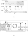
- Fig. 1 shows a schematic illustration of an inventive sleep-based detection and intervention system 1 for treatment of major depressive disorder.
- the system comprises an EEG cap 5 with multiple electrodes 6a, 6b, 6c, 6d, 6e, 6f, 6g, 6h configured for recording an electroencephalogram of a patient's brain wherein the electrodes detect electric potential differences at the scalp of the patient 7.
- electrodes 6a and 6b are for example placed in the frontal region, electrodes 6c and 6d in the central region, electrodes 6e and 6f in the parietal region and electrodes 6g and 6h in the occipital region.
- the electrodes at the lateral head portions i.e. left temporal and right temporal regions) are not illustrated herein.
- Earphone device 4 is configured to be placed in or onto one or both ears of the patient 7 and to provide for acoustic noise when activated.
- the earphone device 4 may be in the form of earbuds, earphones or a headset or the like.
- the system 1 further comprises a processing unit 2 which is connected to the EEG cap 5 and to the earphone device 4.
- the processing unit 2 includes a template-based algorithm 33 configured for detecting slow wave sleep of the patient 7 based on the EEG recorded by the multiple electrodes 6a, 6b, 6c, 6d, 6e, 6f, 6g, 6h of the EEG cap 5, and an acoustic intervention protocol 8 configured for providing a noise stimulation to the patient 7 via the earphone device 4 when slow wave sleep is detected by the template-based algorithm 33.
- FIG. 2 the creation of a template 3 in accordance with the present invention is schematically illustrated.
- Frontal signals in the form of frontal slow waves, in particular fronto-central slow waves, of multiple test persons are detected.
- whole head topographies 9 at the time-point of a frontal slow wave peak cf. red points in the graph, respectively
- a frontal slow wave peak cf. red points in the graph, respectively
- an average template is created across all individual test persons which is herein referred to as the template 3.
- slow waves show a typical fronto-central maximum in the power spectrum
- the detection of slow waves was limited to the fronto-central channels that during slow wave sleep express the largest power in the slow wave frequency range of 0.5 Hz - 2 Hz (the power in the slow wave frequency range is also referred to as SO power or slow oscillation power).
- An average signal was calculated for six frontal electrodes.
- the EEG was bandpass filtered between 0.15 Hz and 2 Hz.
- time points of positive to negative zero crossings were identified in the signal. The lowest and highest value between every 2 of these time points were detected (i.e. one negative and one positive peak between two succeeding positive to negative zero crossings).
- EEG potentials at the peak of the positive and the negative half-wave of all detected SWs were calculated per EEG channel.
- a prototypical voltage distribution also described as a topographic or spatial template
- Grand mean averages across all subjects were calculated per EEG channel to create a template across all individuals.
- the template was normalized by its maximum absolute value, i.e. by dividing each element of the vector by the vector's maximum absolute value.
- Fig. 3 the application of the template according to the present invention is schematically illustrated.
- whole head topographies 9' of the patient 7 are correlated with the template 3.
- Whole head potentials 9' correlate positively with the template upon frontal slow wave peaks, negatively upon troughs, while correlation decreases between peaks and troughs or for artifacts.
- the power of the, for slow waves distinctive ⁇ 1 Hz rhythmic fluctuation of the correlation over time, marks the presence of slow wave sleep.
- it is a correlation it is insensitive to the amplitude of recorded potentials. This is advantageous firstly as different individuals have different amplitudes of slow waves. In particular, the amplitude of slow waves decline with age. Second, it makes the detection insensitive to huge artifacts, in particular if detection is turned on during wakefulness when the subject respectively the patient is still moving around (cf. section "Artifacts" in the graph).
- the template correlation shows to what extent the brain state of the patient (measured as whole head topography 9') approaches, in a certain moment, the template (which reflects the frontal up-state).
- the template which reflects the frontal up-state.
- the left red dot frontal up-state
- the point-by-point correlation over time is shown in the graph at the bottom of Fig. 3 .
- a further spectral analysis occurs which results in the power of template correlation (0.5 - 2 Hz). It is important to note that only if over a longer period of time a high power is measured and other conditions are met (cf. below in connection with Fig. 4 ) the acoustic noise stimulation is triggered.
- the idea here is that the noise stimulation shall not start when (only) a single slow wave occurs (this is also possible in case of lighter sleep) but only if real deep sleep is present. In doing so, slow wave sleep detection becomes very robust.
- the sleep state of slow wave sleep is defined herein according to the presence of a certain amount of slow waves within an extended period. It was found that a 10 s moving window struck a good balance between reliably detecting a change into slow wave sleep, while also being reactive to changes (it is noted that a longer time-window would react slower to changes in the sleep state).
- Fig. 4 schematically depicts slow wave sleep detection and closed-loop suppression through acoustic stimulation.
- the template correlation is calculated over a moving window of EEG signal.
- the power, variance of power and percentage of power are used together with global gamma power to detect slow wave sleep.
- Frontal amplitude, voltage range and delta power together with global gamma power are used as artifact/arousal markers for quick suppression of stimulation.
- linear regression model with data from the 10 s moving window (moving in 0.5 s steps) - i.e. an extracted section of the EEG data - was used to detect slow wave sleep.
- Two regressors R1 and R2 are used herein for detecting slow wave sleep which are based on the correlation with the template (first, template-based, regressor R1) as well as on gamma power of the EEG data (second, EEG-based, regressor R2).
- the first (template-based) regressor R1 the following three characteristics of the template correlation across the 10 s moving window were selected to be used as a regressor to identify a period as slow wave sleep.
- the second regressor R2 includes the average gamma power across all channels (across a 4 s moving window).
- the arousal/artifact detection (A1) includes the voltage range (maximum threshold), amplitude (minimum and maximum) and delta power (minimum threshold), calculated on frontal channels as well as the average global gamma power (maximum threshold).
- A1 includes the voltage range (maximum threshold), amplitude (minimum and maximum) and delta power (minimum threshold), calculated on frontal channels as well as the average global gamma power (maximum threshold).
- frontal amplitude and delta power and the average gamma power were based on a 4 s data, and checked per 0.5 s.
- the voltage range was based on 200 ms data, checked continuously.
- stimulation was reset and suppressed for 35 s.
- Fig. 5 is schematical illustration of a pulsed noise stimulation protocol.
- bursts of pink noise are applied with a randomized duration and inter-onset interval.
- a random walk (Ornstein-Uhlenbeck process) is superimposed on a linear increase of volume, to add unpredictability in volume. In this manner slow waves may be suppressed in a particularly effective manner.
- Fig. 6 depicts one example of successful slow wave sleep suppression through pulsed noise of increasing volume in accordance with an inventive acoustic intervention protocol 8.
- the noise stimulation involves the application of bursts of pink noise with a randomized duration of 50 ms to 500 ms.
- the volume is increasing as a function of time from about 40 dB (40 dB is the lowest possible value) to about 70 dB (as highest value in this example) in about 40 s in this example, until slow wave sleep is no longer detected by the template-based algorithm.
- the linear increase of volume is combined with random walks between +-2.5 dB (Ornstein-Uhlenbeck process) to add unpredictability in volume.
- the inter-stimulus-intervals are randomized between about 1 s and about 4 s.
- SWS is defined as a 30-s epoch containing at least approximately 6 slow waves (SWs) of >75 mV amplitude at frontal channels.
- the disclosure also covers all further features shown in the figures individually although they may not have been described in the afore or following description. Also, single alternatives of the embodiments described in the figures and the description and single alternatives of features thereof can be disclaimed from the subject matter of the invention or from disclosed subject matter.
- the disclosure comprises subject matter consisting of the features defined in the claims or the exemplary embodiments as well as subject matter comprising said features.
Landscapes
- Health & Medical Sciences (AREA)
- Life Sciences & Earth Sciences (AREA)
- Engineering & Computer Science (AREA)
- Public Health (AREA)
- General Health & Medical Sciences (AREA)
- Animal Behavior & Ethology (AREA)
- Heart & Thoracic Surgery (AREA)
- Biomedical Technology (AREA)
- Physics & Mathematics (AREA)
- Veterinary Medicine (AREA)
- Medical Informatics (AREA)
- Psychology (AREA)
- Anesthesiology (AREA)
- Psychiatry (AREA)
- Biophysics (AREA)
- Pathology (AREA)
- Molecular Biology (AREA)
- Surgery (AREA)
- Pain & Pain Management (AREA)
- Acoustics & Sound (AREA)
- Hematology (AREA)
- Computer Vision & Pattern Recognition (AREA)
- Artificial Intelligence (AREA)
- Physiology (AREA)
- Signal Processing (AREA)
- Primary Health Care (AREA)
- Epidemiology (AREA)
- Social Psychology (AREA)
- Hospice & Palliative Care (AREA)
- Developmental Disabilities (AREA)
- Child & Adolescent Psychology (AREA)
- Measurement And Recording Of Electrical Phenomena And Electrical Characteristics Of The Living Body (AREA)
- Measurement Of The Respiration, Hearing Ability, Form, And Blood Characteristics Of Living Organisms (AREA)
Priority Applications (4)
| Application Number | Priority Date | Filing Date | Title |
|---|---|---|---|
| EP21202960.7A EP4166084A1 (fr) | 2021-10-15 | 2021-10-15 | Système de détection et d'intervention basé sur le sommeil |
| EP22802949.2A EP4415623A1 (fr) | 2021-10-15 | 2022-10-14 | Système de détection et d'intervention basé sur le sommeil |
| PCT/EP2022/078612 WO2023062179A1 (fr) | 2021-10-15 | 2022-10-14 | Système de détection et d'intervention basé sur le sommeil |
| US18/700,883 US20240408346A1 (en) | 2021-10-15 | 2022-10-14 | Sleep-based detection and intervention system |
Applications Claiming Priority (1)
| Application Number | Priority Date | Filing Date | Title |
|---|---|---|---|
| EP21202960.7A EP4166084A1 (fr) | 2021-10-15 | 2021-10-15 | Système de détection et d'intervention basé sur le sommeil |
Publications (1)
| Publication Number | Publication Date |
|---|---|
| EP4166084A1 true EP4166084A1 (fr) | 2023-04-19 |
Family
ID=78500351
Family Applications (2)
| Application Number | Title | Priority Date | Filing Date |
|---|---|---|---|
| EP21202960.7A Withdrawn EP4166084A1 (fr) | 2021-10-15 | 2021-10-15 | Système de détection et d'intervention basé sur le sommeil |
| EP22802949.2A Pending EP4415623A1 (fr) | 2021-10-15 | 2022-10-14 | Système de détection et d'intervention basé sur le sommeil |
Family Applications After (1)
| Application Number | Title | Priority Date | Filing Date |
|---|---|---|---|
| EP22802949.2A Pending EP4415623A1 (fr) | 2021-10-15 | 2022-10-14 | Système de détection et d'intervention basé sur le sommeil |
Country Status (3)
| Country | Link |
|---|---|
| US (1) | US20240408346A1 (fr) |
| EP (2) | EP4166084A1 (fr) |
| WO (1) | WO2023062179A1 (fr) |
Cited By (2)
| Publication number | Priority date | Publication date | Assignee | Title |
|---|---|---|---|---|
| CN116725553A (zh) * | 2023-05-19 | 2023-09-12 | 武汉大学 | 基于时频域关联特征的睡眠循环交替模式检测方法及装置 |
| WO2025184463A1 (fr) * | 2024-02-29 | 2025-09-04 | Massachusetts Institute Of Technology | Stimulation cérébrale non invasive en boucle fermée avec imagerie simultanée pour réguler l'écoulement de liquide céphalorachidien chez les êtres humains |
Families Citing this family (1)
| Publication number | Priority date | Publication date | Assignee | Title |
|---|---|---|---|---|
| CN118253009B (zh) * | 2024-04-11 | 2024-12-03 | 电子科技大学 | 基于闭环声刺激和光生物调控的睡眠剥夺系统及使用方法 |
Citations (1)
| Publication number | Priority date | Publication date | Assignee | Title |
|---|---|---|---|---|
| WO2018104459A1 (fr) | 2016-12-08 | 2018-06-14 | Koninklijke Philips N.V. | Détermination de synchronisation corrigée de stimulation appliquée à un sujet pendant le sommeil |
-
2021
- 2021-10-15 EP EP21202960.7A patent/EP4166084A1/fr not_active Withdrawn
-
2022
- 2022-10-14 WO PCT/EP2022/078612 patent/WO2023062179A1/fr not_active Ceased
- 2022-10-14 EP EP22802949.2A patent/EP4415623A1/fr active Pending
- 2022-10-14 US US18/700,883 patent/US20240408346A1/en active Pending
Patent Citations (1)
| Publication number | Priority date | Publication date | Assignee | Title |
|---|---|---|---|---|
| WO2018104459A1 (fr) | 2016-12-08 | 2018-06-14 | Koninklijke Philips N.V. | Détermination de synchronisation corrigée de stimulation appliquée à un sujet pendant le sommeil |
Non-Patent Citations (5)
| Title |
|---|
| DANILENKO K ET AL: "Effectiveness of auditory closed-loop stimulation during sleep on depression", EUROPEAN NEUROPSYCHOPHARMACOLOGY, vol. 29, 12 February 2019 (2019-02-12), XP085600075, ISSN: 0924-977X, DOI: 10.1016/J.EURONEURO.2018.11.1048 * |
| ERIC C LANDSNESS ET AL: "Antidepressant effects of selective slow wave sleep deprivation in major depression: A high-density EEG investigation", JOURNAL OF PSYCHIATRIC RESEARCH, ELSEVIER LTD, GB, vol. 45, no. 8, 3 February 2011 (2011-02-03), pages 1019 - 1026, XP028244824, ISSN: 0022-3956, [retrieved on 20110212], DOI: 10.1016/J.JPSYCHIRES.2011.02.003 * |
| FEHÉR K. D. ET AL: "Predicting slow wave sleep through a topographic template of slow oscillations", JOURNAL OF SLEEP RESEARCH, vol. 29, 15 September 2020 (2020-09-15), GB, pages 128 - 129, XP055906057, ISSN: 0962-1105, DOI: 10.1111/jsr.13181 * |
| PAPALAMBROS NELLY A. ET AL: "Acoustic enhancement of sleep slow oscillations in mild cognitive impairment", ANNALS OF CLINICAL AND TRANSLATIONAL NEUROLOGY, vol. 6, no. 7, 1 July 2019 (2019-07-01), GB, pages 1191 - 1201, XP055906591, ISSN: 2328-9503, DOI: 10.1002/acn3.796 * |
| PREHN-KRISTENSEN ALEXANDER ET AL: "Acoustic closed-loop stimulation during sleep improves consolidation of reward-related memory information in healthy children but not in children with attention-deficit hyperactivity disorder", SLEEP, vol. 43, no. 8, 8 February 2020 (2020-02-08), US, XP055906567, ISSN: 0161-8105, DOI: 10.1093/sleep/zsaa017 * |
Cited By (2)
| Publication number | Priority date | Publication date | Assignee | Title |
|---|---|---|---|---|
| CN116725553A (zh) * | 2023-05-19 | 2023-09-12 | 武汉大学 | 基于时频域关联特征的睡眠循环交替模式检测方法及装置 |
| WO2025184463A1 (fr) * | 2024-02-29 | 2025-09-04 | Massachusetts Institute Of Technology | Stimulation cérébrale non invasive en boucle fermée avec imagerie simultanée pour réguler l'écoulement de liquide céphalorachidien chez les êtres humains |
Also Published As
| Publication number | Publication date |
|---|---|
| WO2023062179A1 (fr) | 2023-04-20 |
| US20240408346A1 (en) | 2024-12-12 |
| EP4415623A1 (fr) | 2024-08-21 |
Similar Documents
| Publication | Publication Date | Title |
|---|---|---|
| US20240408346A1 (en) | Sleep-based detection and intervention system | |
| US12239423B2 (en) | Detection of patient conditions using signals sensed on or near the head | |
| Anson et al. | Reduced vestibular function is associated with longer, slower steps in healthy adults during normal speed walking | |
| JP7218367B2 (ja) | ユーザの認知領域を強化するためにユーザに感覚刺激を供給するためのシステム及び方法 | |
| JP2007515200A (ja) | 脳波を使用した神経障害の治療有効性の評価システムおよび評価方法 | |
| JP2007515200A5 (fr) | ||
| JP2016539758A (ja) | 多相性睡眠管理システム、その操作方法、睡眠分析機器、現状睡眠フェーズ分類方法、多相性睡眠管理システム、および多相性睡眠管理における睡眠分析機器の使用 | |
| CN107997751A (zh) | 一种基于生物反馈的智能耳机系统 | |
| CN119386346B (zh) | 生物健康系统、生理状态调控方法、穿戴设备及存储介质 | |
| JP2020515306A (ja) | 閉ループ両耳感覚刺激での両側インイヤ型eeg記録のためのシステム | |
| Yue et al. | Heart rate and heart rate variability as classification features for mental fatigue using short-term PPG signals via smartphones instead of ECG recordings | |
| US20240172949A1 (en) | Systems and methods for measuring intracranial pressure | |
| JP2023153785A (ja) | 治療装置 | |
| Moumane et al. | Signal quality evaluation of an in-ear EEG device in comparison to a conventional cap system | |
| Xu et al. | Earable multimodal sensing and stimulation: a prospective toward unobtrusive closed-loop biofeedback | |
| WO2024086209A2 (fr) | Dispositifs et procédés de neuromodulation | |
| CN120094067B (zh) | 基于超声刺激的睡眠-觉醒昼夜节律障碍调节方法及装置 | |
| CN119523497A (zh) | 利用脑电功率谱特征评估酒精使用障碍患者治疗效果的系统 | |
| CN1805767A (zh) | 评估脑电图对神经紊乱治疗效果的系统和方法 | |
| Webster et al. | The effect of stimulus probability on P3 in the respiratory-related evoked potential | |
| Zinn et al. | Parametrization of the dying brain: A case report from ICU bed-side EEG monitoring | |
| KR102909434B1 (ko) | 바이노럴 비트 기반의 뇌파 유도 장치 및 바이노럴 비트 기반의 뇌파 유도 방법 | |
| Avramidou et al. | From Ear-EEG to Ear-ExG: The Jaw Artifact is a Keeper | |
| WO2025212538A1 (fr) | Sensations auditives binaurales de battement guidées par rétroaction d'électroencéphalographie | |
| Datar et al. | Based on EEG Signal |
Legal Events
| Date | Code | Title | Description |
|---|---|---|---|
| PUAI | Public reference made under article 153(3) epc to a published international application that has entered the european phase |
Free format text: ORIGINAL CODE: 0009012 |
|
| STAA | Information on the status of an ep patent application or granted ep patent |
Free format text: STATUS: THE APPLICATION HAS BEEN PUBLISHED |
|
| AK | Designated contracting states |
Kind code of ref document: A1 Designated state(s): AL AT BE BG CH CY CZ DE DK EE ES FI FR GB GR HR HU IE IS IT LI LT LU LV MC MK MT NL NO PL PT RO RS SE SI SK SM TR |
|
| STAA | Information on the status of an ep patent application or granted ep patent |
Free format text: STATUS: THE APPLICATION IS DEEMED TO BE WITHDRAWN |
|
| 18D | Application deemed to be withdrawn |
Effective date: 20231020 |